Zurich Open Repository and Archive University of Zurich Main Library Strickhofstrasse 39 CH-8057 Zurich www.zora.uzh.ch Year: 2019 CAD-CAM milled versus rapidly prototyped (3D-printed) complete dentures: An in vitro evaluation of trueness Kalberer, Nicole ; Mehl, Albert ; Schimmel, Martin ; Müller, Frauke ; Srinivasan, Murali Abstract: STATEMENT OF PROBLEM Complete dentures fabricated by computer-aided design and computer-aided manufacturing (CAD-CAM) techniques have become popular. The 2 principal CAD- CAM techniques, milling and rapid prototyping (3D printing), used in the fabrication of complete dentures have been reported to yield clinically acceptable results. However, clinical trials or in vitro studies that evaluated the accuracy of the 2 manufacturing techniques are lacking. PURPOSE The purpose of this in vitro study was to compare the diferences in trueness between the CAD-CAM milled and 3D-printed complete dentures. MATERIAL AND METHODS Two groups of identical maxillary complete dentures were fabricated. A 3D-printed denture group (3DPD) (n=10) and a milled denture group (MDG) (n=10) from a reference maxillary edentulous model. The intaglio surfaces of the fabricated complete dentures were scanned at baseline using a laboratory scanner. The complete dentures were then immersed in an artifcial saliva solution for a period of 21 days, followed by a second scan (after immersion in saliva). A third scan (after the wet-dry cycle) was then made after 21 days, during which the complete dentures were maintained in the artifcial saliva solution during the day and stored dry at night. A purpose-built 3D comparison software program was used to analyze the diferences in the trueness of the complete dentures. The analyses were performed for the entire intaglio surface and specifc regions of interest: posterior crest, palatal vault, posterior palatal seal area, tuberosity, anterior ridge, vestibular fange, and mid-palatal raphae. Independent t tests, ANOVA, and post hoc tests were used for statistical analyses (=.05). RESULTS The trueness of the milled prostheses was signifcantly better than that of the rapid prototyping group with regard to the entire intaglio surface (P<.001), posterior crest (P<.001), palatal vault (P<.001), posterior palatal seal area (P<.001), tuberosity (P<.001), anterior ridge (baseline: P<.001; after immersion in saliva: P=.001; after the wet-dry cycle: P=.011), vestibular fange (P<.001), and mid-palatal raphae (P<.001). CONCLUSIONS The CAD-CAM, milled complete dentures, under the present manufacturing standards, were superior to the rapidly prototyped complete dentures in terms of trueness of the intaglio surfaces. However, further research is needed on the biomechanical, clinical, and patient-centered outcome measures to determine the true superiority of one technique over the other with regard to fabricating complete dentures by CAD-CAM techniques. DOI: https://doi.org/10.1016/j.prosdent.2018.09.001 Posted at the Zurich Open Repository and Archive, University of Zurich ZORA URL: https://doi.org/10.5167/uzh-180704 Journal Article Accepted Version Originally published at:

Welcome message from author
This document is posted to help you gain knowledge. Please leave a comment to let me know what you think about it! Share it to your friends and learn new things together.
Transcript
Zurich Open Repository andArchiveUniversity of ZurichMain LibraryStrickhofstrasse 39CH-8057 Zurichwww.zora.uzh.ch
Year: 2019
CAD-CAM milled versus rapidly prototyped (3D-printed) completedentures: An in vitro evaluation of trueness
Kalberer, Nicole ; Mehl, Albert ; Schimmel, Martin ; Müller, Frauke ; Srinivasan, Murali
Abstract: STATEMENT OF PROBLEM Complete dentures fabricated by computer-aided design andcomputer-aided manufacturing (CAD-CAM) techniques have become popular. The 2 principal CAD-CAM techniques, milling and rapid prototyping (3D printing), used in the fabrication of complete dentureshave been reported to yield clinically acceptable results. However, clinical trials or in vitro studies thatevaluated the accuracy of the 2 manufacturing techniques are lacking. PURPOSE The purpose of thisin vitro study was to compare the differences in trueness between the CAD-CAM milled and 3D-printedcomplete dentures. MATERIAL AND METHODS Two groups of identical maxillary complete dentureswere fabricated. A 3D-printed denture group (3DPD) (n=10) and a milled denture group (MDG) (n=10)from a reference maxillary edentulous model. The intaglio surfaces of the fabricated complete dentureswere scanned at baseline using a laboratory scanner. The complete dentures were then immersed in anartificial saliva solution for a period of 21 days, followed by a second scan (after immersion in saliva). Athird scan (after the wet-dry cycle) was then made after 21 days, during which the complete dentureswere maintained in the artificial saliva solution during the day and stored dry at night. A purpose-built3D comparison software program was used to analyze the differences in the trueness of the completedentures. The analyses were performed for the entire intaglio surface and specific regions of interest:posterior crest, palatal vault, posterior palatal seal area, tuberosity, anterior ridge, vestibular flange,and mid-palatal raphae. Independent t tests, ANOVA, and post hoc tests were used for statisticalanalyses (=.05). RESULTS The trueness of the milled prostheses was significantly better than that ofthe rapid prototyping group with regard to the entire intaglio surface (P<.001), posterior crest (P<.001),palatal vault (P<.001), posterior palatal seal area (P<.001), tuberosity (P<.001), anterior ridge (baseline:P<.001; after immersion in saliva: P=.001; after the wet-dry cycle: P=.011), vestibular flange (P<.001),and mid-palatal raphae (P<.001). CONCLUSIONS The CAD-CAM, milled complete dentures, underthe present manufacturing standards, were superior to the rapidly prototyped complete dentures in termsof trueness of the intaglio surfaces. However, further research is needed on the biomechanical, clinical,and patient-centered outcome measures to determine the true superiority of one technique over the otherwith regard to fabricating complete dentures by CAD-CAM techniques.
DOI: https://doi.org/10.1016/j.prosdent.2018.09.001
Posted at the Zurich Open Repository and Archive, University of ZurichZORA URL: https://doi.org/10.5167/uzh-180704Journal ArticleAccepted Version
Originally published at:

Kalberer, Nicole; Mehl, Albert; Schimmel, Martin; Müller, Frauke; Srinivasan, Murali (2019). CAD-CAMmilled versus rapidly prototyped (3D-printed) complete dentures: An in vitro evaluation of trueness.Journal of Prosthetic Dentistry, 121(4):637-643.DOI: https://doi.org/10.1016/j.prosdent.2018.09.001
2
1
Title: CAD/CAM milled versus rapidly-prototyped (3D-printed) complete dentures:
an in vitro evaluation of trueness.
Authors: Nicole Kalberer Med. dent.1
Albert Mehl Dr. med. dent. Dr. rer. biol. hum.2
Martin Schimmel, Dr. med. dent., MAS 1, 3
Frauke Müller Dr. med. dent. habil.1, 4
Murali Srinivasan Dr. med. dent, BDS, MDS, MBA, MAS1
Author affiliations:
1- Division of Gerodontology and Removable Prosthodontics, University
Clinics of Dental Medicine, University of Geneva, Geneva, Switzerland.
2- Clinic of Preventive Dentistry, Periodontology and Cariology, Center of
Dental Medicine, University of Zurich, Zurich, Switzerland.
3- Division of Gerodontology, School of Dental Medicine, Bern,
Switzerland.
4- Service of Geriatrics, Department of Internal Medicine, Rehabilitation and
Geriatrics, University Hospitals of Geneva, Thônex, Switzerland.
Corresponding Author:
Prof. Frauke Müller, Dr. med. dent. habil.,
Division of Gerodontology and Removable Prosthodontics,
University Clinics of Dental Medicine, University of Geneva,
Rue Barthélemy-Menn, CH-1205 Geneva, Switzerland.
Tel. No: +41 22 3794060, Fax: +41 22 3794052
Email: [email protected]
2
ABSTRACT (348/400)
Statement of problem Computer Aided Design/ Computer Aided Manufacturing
(CAD/CAM) manufactured Complete Removable Dental Prostheses (CRDPs) have evolved
exponentially in the last decade. The two principal CAD/CAM techniques, milling and rapid
prototyping (3D-printing), employed in the fabrication of CRDPs have been reported to yield
clinically acceptable results. However, the accuracy of the two manufacturing techniques has
never been compared, either in bench experiments or by clinical trials.
Purpose This in vitro bench experiment aimed to compare the differences in trueness
between the CAD/CAM milled CRDPs and rapidly prototyped (RP) CRDPs.
Material and Methods Two groups of identical maxillary CRDPs were manufactured
(Group#1: RP: n=10; Group#2: Milled: n=10) from a reference maxillary edentulous model.
The intaglio surfaces of the fabricated CRDPs were first scanned (BL) using a laboratory
scanner. The CRDPs were then immersed in an artificial saliva solution for a period of 21
days, following which a second scan (PIS) was done. A third scan (WDC) was then made
after 21 days, during which the CRDPs were maintained in the saliva solution during day and
stored dry at night. A purpose-built 3D comparison software was used to analyze the
differences in the trueness of the CRDPs. The analyses were performed for the entire intaglio
surface, and specific regions of interest (posterior crest, palatal-vault, posterior palatal seal
area (PPS), tuberosity, anterior-ridge, vestibular-flange and mid-palatal raphae). ANOVA and
post-hoc tests were applied for statistical analyses (𝛼=0.05).
Results The trueness of the milled prostheses was significantly better than that of the RP
group with regards to the entire intaglio surface (p<0.001), posterior crest (p<0.001), palatal-
vault (p<0.001), PPS (p<0.001), tuberosity (p<0.001), anterior-ridge (BL: p<0.001; PIS:
p=0.001; WDC: p=0.011), vestibular-flange (p<0.001), and mid- palatal raphae (p<0.001).
3
Conclusion The CAD/CAM milled CRDPs, under the present manufacturing standards, are
superior to the rapidly prototyped CRDPs in terms of trueness of the intaglio surfaces.
However, further research is needed on a larger number of biomechanical, clinical and patient
centered outcome measures, to evidence the true superiority of one technique over the other
with regards to manufacturing CRDPs with CAD/CAM techniques, taking the rapidly
evolving technical possibilities into account.
Clinical implications: This study provides evidence to help in the clinical decision making
for choosing the appropriate CAD/CAM manufacturing technique for fabricating CRDPs. The
study also provides sufficient information to encourage future research to clinically validate
the findings of this bench experiment.
4
Introduction
The fabrication of complete removable dental prostheses (CRDPs) by computer aided
designing and manufacturing (CAD/CAM) methods has witnessed a phenomenal rise, in both
clinical and laboratory practices, during the recent years.1 This gaining popularity may be
attributed to the considerable improvements in the CAD/CAM techniques, the growing
awareness amongst the dental practitioners and laboratory technicians along with an
increasing flexibility to combine parts of the digital workflow with conventional
clinical/laboratory protocols. To date, two established CAD/CAM techniques, either by a
computerized numeric control (CNC) subtractive milling process or by a system of rapid
prototyping (RP) commonly known as 3D printing, the latter being an additive manufacturing
process are available to fabricate CAD/CAM CRDPs. Most manufacturers currently employ
the milling technique for commercial production of CRDPs, while the RP method is mainly
used for fabricating provisional or try-in CRDPs and, on a smaller scale, definitive CRDPs.
Whereas the milling process implies the loss of large quantities of denture base material, the
more recent 3D prototyping promises a more sustainable additive approach by using less
denture resin.
CRDPs manufactured with either of the two CAD/CAM techniques have been
documented. When compared to the conventional CRDPs, CAD/CAM milled CRDPs show
similar or better fit of the intaglio surfaces, equal biocompatibility and improved mechanical
properties.2-7 High patient and clinician satisfaction have also been reported with CAD/CAM
milled CRDPs.8, 9 The clinical protocols considerably reduce the chairside time, while the
manufacturing process may reduce the laboratory fees in some countries.10 CRDPs
manufactured by RP technique have also elicited comparable patient satisfaction when
compared to conventional CRDPs;11, 12 RP has been further used in CRDP fabrication for
precise reproduction of denture bases and printed wax patterns.13, 14 Although both techniques
5
are successful in manufacturing clinically acceptable CRDPs, no study, till date, has actually
compared the precision of the intaglio surface between the CRDPs manufactured by RP (3D
printing) and a milled technique. This study aims to compare the trueness of the intaglio
surfaces CRDPs manufactured by the CAD/CAM milling technique with those fabricated
using the RP (3D printing) technology. Therefore, the null hypothesis set for this in vitro
study was that there is no difference in the trueness of the intaglio surfaces of CRDPs
manufactured either by CAD/CAM RP or milling techniques.
Materials and Methods
This in vitro study was conducted in the Division of Gerodontology and Removable
Prosthodontics, University Clinics of Dental Medicine, University of Geneva, Switzerland.
An ethical approval was not required for performing this study, because no patient records or
data were used for this bench experiment. The color mapping and analysis of the differences
were done at the Division of Computerized Restorative Dentistry, Clinic for Preventive
Dentistry, Periodontology and Cariology, Center for Dental Medicine, University of Zurich,
Switzerland.
Samples size
The sample size for the current study was calculated using the results from a previously
published study.3 The effect size (dz=1.5004) and the required sample size were calculated for
a=0.05 and a power of 0.95 (1-b err prob), assuming a normal distribution. For this study, a
sample size of 9 was obtained and subsequently increased to 10 per group, to remain
consistent with previous similar published studies and to avoid errors.2, 3 The power analysis
was performed using the freeware (G*Power for Mac OSX, Version 3.1.9.2, Düsseldorf,
Germany).15
6
Master reference model
A completely edentulous maxillary cobalt-chrome model served as the master reference
model for the current study. This model has been used in a previous experiment.3 All the
CRDP specimens used in this bench experiment were fabricated using the scan of this
reference model.
Master reference scan
A master scan of the reference model was performed using a laboratory scanner (IScan
D103i, Imetric 3D SA, Courgenay, Switzerland). The high-resolution scanner is calibrated to
a precision of 6µm,16 with a manufacturer specified nominal point spacing of 6–8µm with a
repeatability of 10µm at an accuracy of 20µm. The bundle scanner software is equipped with
an auto-align function that aligns multiple scan sets and the resultant information is stored in a
3D *.stl-format.
CRDPs CAD Design
The file of the master scan was exported in an electronic format (*.stl) to the CAD/CAM
CRDP manufacturer using a purpose-built software (AvaDentTM Connect software, version
3.52, AvaDentTM, Global Dental Science Europe BV, Tilburg, Netherlands). The anatomical
landmarks were identified and the peripheral limits were marked on a virtual model in the
AvaDentTM design software, which then served to design the final CRDP. A digital preview
was generated and sent for approval to the investigators before manufacturing. Both, milled
and 3D printed CRDPs used the same design.
7
Study groups
A total of 20 CRDPs were fabricated using the scan master reference model applying the two
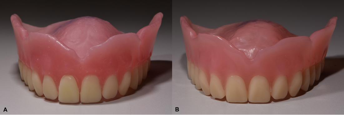
mentioned CAD/CAM manufacturing techniques (figure 1). Group 1 (n=10) comprised of
CRDPs manufactured using the RP technique (NextDent B.V., Soesterberg, Netherlands),
while group 2 consisted of 10 fully milled CRDPs (AvaDentTM, Global Dental Science Europe
BV, Tilburg, Netherlands).
Lubricant media
A liquid media was a custom-fabricated artificial saliva solution, manufactured solely for the
purpose of these bench experiments; its composition has been described in detail in
previously published studies.3, 17, 18
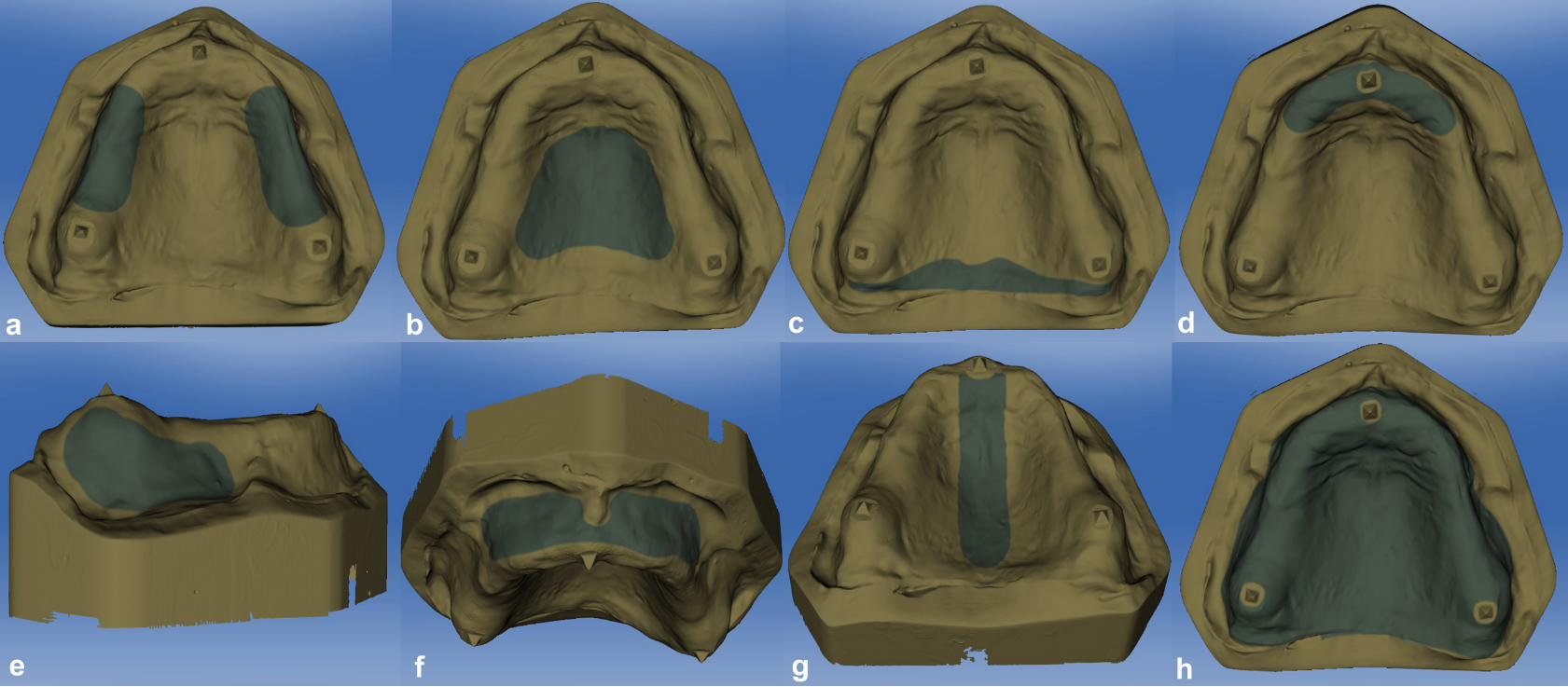
Entire intaglio surface and specific regions of interest (figure 2)
Based on clinical relevance for denture function, the entire intaglio surface and certain regions
of interests were selected for analysis:
a. Posterior crest,
b. Palatal vault,
c. Posterior palatal seal area (PPS),
d. Anterior-ridge,
e. Tuberosities,
f. Vestibular-flange, and
g. Mid-palatal raphae (MPR).
8
Protocol
At first, the master reference model was scanned to form the master scan data file, which was
used for the manufacturing of the CRDPs. This master scan was also used later for data
analysis and comparison. After manufacturing, the specimens were quality-checked for any
defects. At baseline (BL) the intaglio surfaces of the CRDPs specimens (n=20 specimens;
Group 1: n=10, Group 2: n=10) were scanned. Subsequently the samples were incubated in an
artificial saliva solution for a period of 21 days at room temperature. At the end of this period,
a second scan of the intaglio surface was performed (post-immersion-scan; PIS). The
following 21 days the specimens were immersed during the day in the artificial saliva solution
and were stored dry during the night. The intaglio surface was then scanned a third and last
time (wet/dry cycle; WDC).
Scan procedure and 3D comparison
All intaglio surfaces were scanned by a single investigator (NK), adhering to principles of
extra-oral laboratory scanning procedures as recommended by the manufacturer, using the
same aforesaid laboratory scanner. For comparative analyses a purpose-built 3D comparison
software was used (Oracheck version 2.10, Cyfex, Switzerland). The scan file of the master
reference model was inverted and, on which, the intaglio surface scans of all specimens were
superimposed.3, 19 The software calculated the 3D ? distances between the superimposed
matching points. Mean values and standard deviations were calculated for the entire intaglio
surface as well as the regions of interest.
Statistical analysis
Normal distribution was confirmed before ANOVA and post hoc tests were used to
demonstrate any significant differences between the groups with respect to the entire intaglio
9
surfaces and the specified regions of interests investigated. All statistical analyses were
performed using the SPSS® software package (version 24.0. IBM® Corporation, Armonk, NY,
USA).
Results
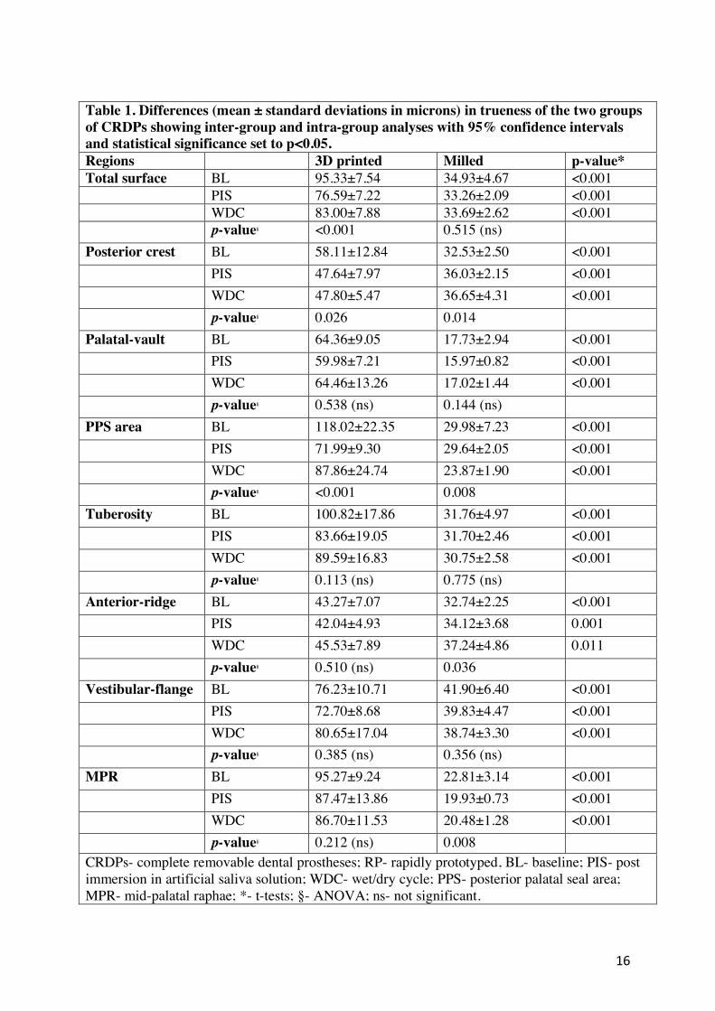
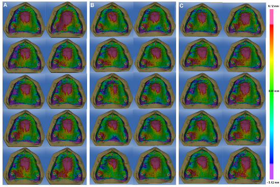
Trueness of the entire intaglio surface (Table 1, Figures 3 and 4)
Inter-group results (Group#1 versus Group#2)
At the given time-points, BL, PIS, and WDC, the trueness of the entire intaglio surface was
significantly better in the CRDPs of group #2 than those of group #1 (p<0.001).
Intra-group results (BL versus PIS; BL versus WDC; PIS versus WDC)
Within group analysis revealed that there was a significant difference in the trueness of the
entire intaglio surface in group#1 when compared between BL and PIS (p<0.001), BL and
WDC (p=0.003), but not between PIS and WDC (p=0.205). Group#2 did not show any
statistically significant differences in trueness between the 3 evaluated time points.
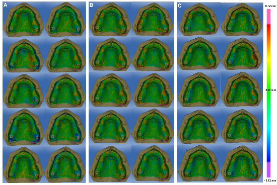
Trueness in the regions of interest (Table 1, Figures 3 and 4)
Inter-group results (Group#1 versus Group#2)
The trueness of the CRDPs in group #2 was significantly better at BL, PIS and after WDC,
with regards to the posterior crest (p<0.001), the palatal-vault (p<0.001), PPS (p<0.001), the
anterior-ridge (BL: p<0.001; PIS: p=0.001; WDC: p=0.011), the tuberosities (p<0.001), the
vestibular-flange (p<0.001), and the MPR (p<0.001).

10
Intra-group results (BL versus PIS; BL versus WDC; PIS versus WDC)
In group#1, there was a significant difference for the PPS area when compared between BL
and PIS (p<0.001) as well as between BL and WDC (p=0.007) but no difference between PIS
and WDC (p=0.261). The trueness in PPS area, improved after the incubation in saliva.
No significant differences were observed in the trueness of the other investigated regions of
interest for group#1.
Group#2 showed significant differences in the posterior crest (BL versus WDC: p=0.020),
PPS area (BL versus WDC: p=0.015; PIS versus WDC: p=0.023), anterior-ridge (BL versus
WDC: p=0.037), and in the MPR area (BL versus PIS: p=0.010; BL versus WDC: p=0.045).
Discussion
The fabrication of CRDPs by subtractive milling or by additive rapid prototyping are recent
developments in the field of complete denture prosthodontics. Although, both techniques
utilize a digital image file designed by a CAD software to manufacture the CRDPs, the two
modes of fabrication however are entirely different from one another. In the milling method,
the CRDP is fabricated by a milling station using a pre-polymerized polymethylmethacrylate
(PMMA) puck manufactured under high pressure. While the RP technique uses photo-
sensitive liquid resins, repetitively layered on a support structure and polymerized by an ultra-
violet (UV) or a visible light source. Distinct advantages and disadvantages for each of the
two techniques do exist. Manufacturing CRDPs from a pre-polymerized PMMA puck may be
advantageous in eliminating ill-effects such as shrinkage and porosities, caused by the
packing and polymerization process. Also, they possibly contain lower levels of residual
monomer, and seem to afford superior material properties. The residual monomer content of
the milled CRDPs was however, not markedly reduced when compared with conventional
heat-polymerized CRDPs, but was observed to be significantly lower when compared to
11
CRDPs manufactured with auto-polymerizing resins.6 These might be important factors to
consider while comparing them with rapidly prototyped CRDPs. The RP technique uses
uncured resins for manufacturing the CRDPs and once manufactured, it requires an additional
final light-polymerization step to complete the curing process. During the RP workflow,
polymerization shrinkage is theoretically possible as CRDPs are not completely polymerized
before the final light polymerization procedure. A deformation of the prostheses can always
occur when demounting the partially cured CRDP from the build platform, despite adequate
care being exercised. Furthermore, a residual layer of uncured resin invariably remains on the
finished prostheses, which has to be eliminated by thorough rinsing with a suitable solvent.
On the flip side, the claimed advantages of an additive manufacturing process include higher
accuracy, limiting material wastage, and low infrastructure costs, however, these have not yet
been scientifically proven with regards to CRDP fabrication. Theoretically, solely on the basis
of the different manufacturing processes, a logical difference in the accuracy of the fabricated
CRDPs should exist, but both techniques have been documented to be clinically acceptable if
not superior when compared to conventional methods.2-13 The superiority, if existing, of one
CAD/CAM technique over the other has not been investigated so far.
The results of this in vitro study demonstrate that the trueness of the CAD/CAM
milled CRDPs was statistically better than the rapidly prototyped CRDPs both for the entire
intaglio surface and the specific regions of interest. Therefore, the initially set null hypotheses
is rejected by virtue of the findings of this study. Whether this difference in the trueness is
clinically relevant remains debatable, as studies have demonstrated that the accuracy of
rapidly prototyped CRDPs have clinically acceptable levels of precision and have also
reported good patient and clinician satisfaction.11-14 A further important aspect to consider is
whether the rapidly prototyped CRDPs would be dimensionally stable over long-term given
the fact that they are being manufactured using light-polymerizing resins, and no studies exist
12
in the current literature that elucidate on this aspect. Despite the inferior trueness in the
present study, it seems worthwhile to invest in perfecting the RP techniques, as they present
some substantial advantages to the CAD/CAM milling techniques. Sustainability and
responsible use of our planets resources were stated a political priority by the United Nations.
With a projected estimate of 61 million dentures to be made in 2020 for the US alone,20 global
numbers are expected to be exponentially higher. Therefore a substantial limitation of
environmental pollution with plastic particles may be achieved, if judicious manufacturing
techniques are adopted and may well further justify the developments of the RP techniques in
a humanitarian aspect. Small 3D printers cost a fraction of a professional milling machine,
and could possibly be afforded in those economically poor and non-industrialized parts of this
world, where edentulism is most prevalent and skilled dental technicians are scarce. On-site
manufacturing would also avoid shipping costs. In a long-term perspective, access to CRDPs
may be extended to patient groups who are currently deprived of restorative oral health care.
Technical improvements in terms of trueness can be expected in the near future, as
CAD/CAM techniques are developing very rapidly. But before recommending RP CRDP
manufacturing as a standard manufacturing procedure, more research is needed. There are no
studies on the monomer-based ester compounds that are used in rapid prototyping with
regards to allergenic potentials, residual monomer levels, material and color stability, material
compatibility to conventional relines, mechanical properties, and biocompatibility. The
appearance of the two different denture types has to be studied as esthetics are of increasing
importance in our modern society. Last but not least, patient centered outcome measures have
to be considered.

13
Conclusions
The CADCAM milled CRDPs, under the present manufacturing standards, are superior to the
rapidly prototyped CRDPs in terms of trueness of the intaglio surfaces. However, further
research is needed on a larger number of biomechanical clinical and patient centered outcome
measures, to evidence the true superiority of one technique over the other with regards to
CRDPs.
14
References
1. Baba NZ, AlRumaih HS, Goodacre BJ, Goodacre CJ. Current techniques in CAD/CAM
denture fabrication. Gen Dent. 2016;64:23-8.
2. Goodacre BJ, Goodacre CJ, Baba NZ, Kattadiyil MT. Comparison of denture base
adaptation between CAD-CAM and conventional fabrication techniques. J Prosthet Dent.
2016;116:249-56.
3. Srinivasan M, Cantin Y, Mehl A, Gjengedal H, Müller F, Schimmel M. CAD/CAM milled
removable complete dentures: an in vitro evaluation of trueness. Clin Oral Investig.
2017;21:2007-19.
4. Srinivasan M, Gjengedal H, Cattani-Lorente M, Moussa M, Durual S, Schimmel M et al.
CAD/CAM milled complete removable dental prostheses: an in vitro evaluation of
biocompatibility, mechanical properties, and surface roughness. Dent Mater J. 2018:doi:
10.4012/dmj.2017-207. [Epub - ahead of print.]
5. Steinmassl O, Dumfahrt H, Grunert I, Steinmassl PA. CAD/CAM produces dentures with
improved fit. Clin Oral Investig. 2018:doi: 10.1007/s00784-018-2369-2. [Epub ahead of
print.]
6. Steinmassl PA, Wiedemair V, Huck C, Klaunzer F, Steinmassl O, Grunert I et al. Do
CAD/CAM dentures really release less monomer than conventional dentures? Clin Oral
Investig. 2017;21:1697-705.
7. Steinmassl O, Dumfahrt H, Grunert I, Steinmassl PA. Influence of CAD/CAM fabrication
on denture surface properties. J Oral Rehabil. 2018: doi: 10.1111/joor.12621. [Epub - ahead
of print.]
8. Kattadiyil MT, Jekki R, Goodacre CJ, Baba NZ. Comparison of treatment outcomes in
digital and conventional complete removable dental prosthesis fabrications in a predoctoral
setting. J Prosthet Dent. 2015;114:818-25.
9. Bidra AS, Farrell K, Burnham D, Dhingra A, Taylor TD, Kuo CL. Prospective cohort pilot
study of 2-visit CAD/CAM monolithic complete dentures and implant-retained overdentures:
Clinical and patient-centered outcomes. J Prosthet Dent. 2016;115:578-586.
10. Srinivasan M, Schimmel M, Naharro M, Müller F. CAD/CAM Milled Complete
Dentures: Time and Cost Analysis. J Dent Res. 2017;96(Spec Iss A):3660.
11. Pereyra NM, Marano J, Subramanian G, Quek S, Leff D. Comparison of Patient
Satisfaction in the Fabrication of Conventional Dentures vs. DENTCA (CAD/CAM)
Dentures: A Case Report. J N J Dent Assoc. 2015;86:26-33.

15
12. Ucar Y, Akova T, Aysan I. Mechanical properties of polyamide versus different PMMA
denture base materials. Journal of prosthodontics: official journal of the American College of
Prosthodontists. 2012;21:173-6.
13. Chen H, Wang H, Lv P, Wang Y, Sun Y. Quantitative Evaluation of Tissue Surface
Adaption of CAD-Designed and 3D Printed Wax Pattern of Maxillary Complete Denture.
Biomed Res Int. 2015;2015:453968.
14. Inokoshi M, Kanazawa M, Minakuchi S. Evaluation of a complete denture trial method
applying rapid prototyping. Dent Mater J. 2012;31:40-6.
15. Faul F, Erdfelder E, Buchner A, Lang AG. Statistical power analyses using G*Power 3.1:
tests for correlation and regression analyses. Behavior research methods. 2009;41:1149-60.
16. Papaspyridakos P, Gallucci GO, Chen CJ, Hanssen S, Naert I, Vandenberghe B. Digital
versus conventional implant impressions for edentulous patients: accuracy outcomes. Clin
Oral Implants Res. 2016;27:465-72.
17. Srinivasan M, Schimmel M, Badoud I, Ammann P, Herrmann FR, Müller F. Influence of
implant angulation and cyclic dislodging on the retentive force of two different overdenture
attachments - an in vitro study. Clin Oral Implants Res. 2016;27:604-11.
18. Srinivasan M, Schimmel M, Kobayashi M, Badoud I, Ammann P, Herrmann FR et al.
Influence of different lubricants on the retentive force of LOCATOR attachments - an in vitro
pilot study. Clin Oral Implants Res. 2016;27:771-5.
19. Mehl A, Koch R, Zaruba M, Ender A. 3D monitoring and quality control using intraoral
optical camera systems. Int J Comput Dent. 2013;16:23-36.
20. Douglass CW, Shih A, Ostry L. Will there be a need for complete dentures in the United
States in 2020? J Prosthet Dent. 2002;87:5-8.
16
Table 1. Differences (mean ± standard deviations in microns) in trueness of the two groups
of CRDPs showing inter-group and intra-group analyses with 95% confidence intervals
and statistical significance set to p<0.05.
Regions 3D printed Milled p-value*
Total surface BL 95.33±7.54 34.93±4.67 <0.001
PIS 76.59±7.22 33.26±2.09 <0.001
WDC 83.00±7.88 33.69±2.62 <0.001
p-value§ <0.001 0.515 (ns)
Posterior crest BL 58.11±12.84 32.53±2.50 <0.001
PIS 47.64±7.97 36.03±2.15 <0.001
WDC 47.80±5.47 36.65±4.31 <0.001
p-value§ 0.026 0.014
Palatal-vault BL 64.36±9.05 17.73±2.94 <0.001
PIS 59.98±7.21 15.97±0.82 <0.001
WDC 64.46±13.26 17.02±1.44 <0.001
p-value§ 0.538 (ns) 0.144 (ns)
PPS area BL 118.02±22.35 29.98±7.23 <0.001
PIS 71.99±9.30 29.64±2.05 <0.001
WDC 87.86±24.74 23.87±1.90 <0.001
p-value§ <0.001 0.008
Tuberosity BL 100.82±17.86 31.76±4.97 <0.001
PIS 83.66±19.05 31.70±2.46 <0.001
WDC 89.59±16.83 30.75±2.58 <0.001
p-value§ 0.113 (ns) 0.775 (ns)
Anterior-ridge BL 43.27±7.07 32.74±2.25 <0.001
PIS 42.04±4.93 34.12±3.68 0.001
WDC 45.53±7.89 37.24±4.86 0.011
p-value§ 0.510 (ns) 0.036
Vestibular-flange BL 76.23±10.71 41.90±6.40 <0.001
PIS 72.70±8.68 39.83±4.47 <0.001
WDC 80.65±17.04 38.74±3.30 <0.001
p-value§ 0.385 (ns) 0.356 (ns)
MPR BL 95.27±9.24 22.81±3.14 <0.001
PIS 87.47±13.86 19.93±0.73 <0.001
WDC 86.70±11.53 20.48±1.28 <0.001
p-value§ 0.212 (ns) 0.008
CRDPs- complete removable dental prostheses; RP- rapidly prototyped, BL- baseline; PIS- post
immersion in artificial saliva solution; WDC- wet/dry cycle; PPS- posterior palatal seal area;
MPR- mid-palatal raphae; *- t-tests; §- ANOVA; ns- not significant.
17
Table Legends
Table 1 Differences (mean ± standard deviations in microns) in trueness
of the two groups of CRDPs showing inter-group and intra-group
analyses with 95% confidence intervals and statistical
significance set to p<0.05.
Figure Legends
Figure 1 Example of a random sample from each of the two groups of
CAD/CAM fabricated complete removable dental prostheses
(CRDPs). A – Rapidly proto-typed (3D-Printed); B – Milled.
Figure 2 The investigated specific regions of interests: (a) crest, (b) palatal
vault, (c) posterior palatal seal area (PPS), (d) anterior ridge, (e)
tuberosities, (f) vestibular flange, (g) mid-palatal raphae (MPR),
(h) total intaglio surface.
Figure 3 Color maps of the samples from group 1 at baseline (A), post-
immersion in artificial saliva (B) and after a wet/dry simulation
cycle (C) at a precision scale between -0.12 to 0.12 mm.
Figure 4 Color maps of the samples from group 2 at baseline (A), post-
immersion in artificial saliva (B) and after a wet/dry simulation
cycle (C) at a precision scale between -0.12 to 0.12 mm.
Related Documents