Research Article Breast Cancer Detection with Reduced Feature Set Ahmet Mert, 1 Niyazi KJlJç, 2 Erdem Bilgili, 1 and Aydin Akan 2 1 Department of Electrical and Electronics, Piri Reis University, 34940 Istanbul, Turkey 2 Department of Electrical and Electronics, Istanbul University, 34320 Istanbul, Turkey Correspondence should be addressed to Ahmet Mert; [email protected] Received 12 September 2014; Revised 14 December 2014; Accepted 25 December 2014 Academic Editor: Kevin Ward Copyright © Ahmet Mert et al. is is an open access article distributed under the Creative Commons Attribution License, which permits unrestricted use, distribution, and reproduction in any medium, provided the original work is properly cited. is paper explores feature reduction properties of independent component analysis (ICA) on breast cancer decision support system. Wisconsin diagnostic breast cancer (WDBC) dataset is reduced to one-dimensional feature vector computing an independent component (IC). e original data with 30 features and reduced one feature (IC) are used to evaluate diagnostic accuracy of the classifiers such as k-nearest neighbor (k-NN), artificial neural network (ANN), radial basis function neural network (RBFNN), and support vector machine (SVM). e comparison of the proposed classification using the IC with original feature set is also tested on different validation (5/10-fold cross-validations) and partitioning (20%–40%) methods. ese classifiers are evaluated how to effectively categorize tumors as benign and malignant in terms of specificity, sensitivity, accuracy, F-score, Youden’s index, discriminant power, and the receiver operating characteristic (ROC) curve with its criterion values including area under curve (AUC) and 95% confidential interval (CI). is represents an improvement in diagnostic decision support system, while reducing computational complexity. 1. Introduction Breast cancer is one of the leading causes of death among all cancers for women [1]. Early detection and correct diagnosis of cancer are essential for the treatment of the disease. How- ever, the traditional approach to cancer diagnosis depends highly on the experience of doctors and their visual inspec- tions. Naturally, human beings can make mistakes due to their limitations. Humans can recognize patterns easily. How- ever, they fail when probabilities have to be assigned to obser- vations [2]. Although several tests are applied, exact diagnosis may be difficult even for an expert. at is why automatic diagnosis of breast cancer is investigated by many researchers. Computer aided diagnostic tools are intended to help physi- cians in order to improve the accuracy of the diagnosis [3–5]. A study was carried out to demonstrate that the machine learning may improve the accuracy of diagnosis. In Brause’s work, the result shows that the most experienced physician can diagnose with 79.97% accuracy while 91.1% correct diagnosis is achieved with the help of machine learning [6]. Tumors are classified as benign and malignant. Benign tumors are not cancerous or life threatening. However these can increase the risk of getting breast cancer. Malignant tumors are cancerous and more alarming than benign tumors. Although significant studies are performed for early detection, about 20% of all women with malignant tumors die from this disease [7]. In order to improve accuracy of breast mass classifica- tion as benign and malignant, the performance of back- propagation artificial neural network (ANN) was evaluated [8]. Moreover, the fast learning rates and generalization capa- bilities of radial basis function neural networks (RBFNN) have showed excellent accuracy in microcalcification detec- tion task [9, 10]. e advantages of RBFNN are simple structure, good performance with approaching nonlinear function, and fast convergence velocity. us, it has been widely used in pattern recognition and system modeling [11, 12]. On the other hand, the structure of RBFNN increases when the net’s input dimension increases. Moreover, the irrelevant components in the inputs will decrease the gener- alization performance of RBFNN [13]. Support vector machine (SVM) is an effective statistical learning method for classification [14]. SVM is based on find- ing optimal hyperplane to separate different classes mapping input data into higher-dimensional feature space. SVM has advantage of fast training technique, even with large number Hindawi Publishing Corporation Computational and Mathematical Methods in Medicine Article ID 265138

Welcome message from author
This document is posted to help you gain knowledge. Please leave a comment to let me know what you think about it! Share it to your friends and learn new things together.
Transcript

Research ArticleBreast Cancer Detection with Reduced Feature Set
Ahmet Mert,1 Niyazi KJlJç,2 Erdem Bilgili,1 and Aydin Akan2
1Department of Electrical and Electronics, Piri Reis University, 34940 Istanbul, Turkey2Department of Electrical and Electronics, Istanbul University, 34320 Istanbul, Turkey
Correspondence should be addressed to Ahmet Mert; [email protected]
Received 12 September 2014; Revised 14 December 2014; Accepted 25 December 2014
Academic Editor: Kevin Ward
Copyright © Ahmet Mert et al. This is an open access article distributed under the Creative Commons Attribution License, whichpermits unrestricted use, distribution, and reproduction in any medium, provided the original work is properly cited.
This paper explores feature reduction properties of independent component analysis (ICA) on breast cancer decision supportsystem. Wisconsin diagnostic breast cancer (WDBC) dataset is reduced to one-dimensional feature vector computing anindependent component (IC). The original data with 30 features and reduced one feature (IC) are used to evaluate diagnosticaccuracy of the classifiers such as k-nearest neighbor (k-NN), artificial neural network (ANN), radial basis function neural network(RBFNN), and support vectormachine (SVM).The comparison of the proposed classification using the ICwith original feature set isalso tested on different validation (5/10-fold cross-validations) and partitioning (20%–40%)methods.These classifiers are evaluatedhow to effectively categorize tumors as benign and malignant in terms of specificity, sensitivity, accuracy, F-score, Youden’s index,discriminant power, and the receiver operating characteristic (ROC) curve with its criterion values including area under curve(AUC) and 95% confidential interval (CI). This represents an improvement in diagnostic decision support system, while reducingcomputational complexity.
1. Introduction
Breast cancer is one of the leading causes of death among allcancers for women [1]. Early detection and correct diagnosisof cancer are essential for the treatment of the disease. How-ever, the traditional approach to cancer diagnosis dependshighly on the experience of doctors and their visual inspec-tions. Naturally, human beings can make mistakes due totheir limitations.Humans can recognize patterns easily.How-ever, they fail when probabilities have to be assigned to obser-vations [2]. Although several tests are applied, exact diagnosismay be difficult even for an expert. That is why automaticdiagnosis of breast cancer is investigated bymany researchers.Computer aided diagnostic tools are intended to help physi-cians in order to improve the accuracy of the diagnosis [3–5].
A study was carried out to demonstrate that the machinelearning may improve the accuracy of diagnosis. In Brause’swork, the result shows that the most experienced physiciancan diagnose with 79.97% accuracy while 91.1% correctdiagnosis is achieved with the help of machine learning [6].
Tumors are classified as benign and malignant. Benigntumors are not cancerous or life threatening. However thesecan increase the risk of getting breast cancer. Malignant
tumors are cancerous and more alarming than benigntumors. Although significant studies are performed for earlydetection, about 20%of all womenwithmalignant tumors diefrom this disease [7].
In order to improve accuracy of breast mass classifica-tion as benign and malignant, the performance of back-propagation artificial neural network (ANN) was evaluated[8]. Moreover, the fast learning rates and generalization capa-bilities of radial basis function neural networks (RBFNN)have showed excellent accuracy in microcalcification detec-tion task [9, 10]. The advantages of RBFNN are simplestructure, good performance with approaching nonlinearfunction, and fast convergence velocity. Thus, it has beenwidely used in pattern recognition and system modeling[11, 12]. On the other hand, the structure of RBFNN increaseswhen the net’s input dimension increases. Moreover, theirrelevant components in the inputs will decrease the gener-alization performance of RBFNN [13].
Support vector machine (SVM) is an effective statisticallearningmethod for classification [14]. SVM is based on find-ing optimal hyperplane to separate different classes mappinginput data into higher-dimensional feature space. SVM hasadvantage of fast training technique, even with large number
Hindawi Publishing CorporationComputational and Mathematical Methods in MedicineArticle ID 265138

2 Computational and Mathematical Methods in Medicine
of input data [15, 16]. Therefore it has been used for manyrecognition problems such as object recognition and facedetection [17–19].
Principal component analysis (PCA) is a technique toreduce dimensionality using second order statistical infor-mation [20]. Independent component analysis (ICA) is arecently developed method in pattern recognition and signalprocessing fields [21, 22]. It involves higher order statistics toextract independent components that involve richer informa-tion than PCA. ICA can be used to reduce dimensionalitybefore training 𝑘-NN, ANN, RBFNN, and SVM. Conse-quently the complexity of classifiers can be reduced; conver-gence velocity and performance can be increased [13, 23].
The objective of the proposed study is to analyze theeffect of feature reduction using ICA on classification ofthe tumors as benign or malignant. Thus, the dimensionof WDBC dataset is reduced into only one feature usingICA. The reduced data is subdivided into test and trainingdata using 5/10-fold cross-validation and 20% partitioning toevaluate the performance of 𝑘-NN, ANN, RBFNN, and SVM.Performance measures including accuracy, specificity, sensi-tivity, 𝐹-score, Youden’s index, and discriminant power arecomputed and the receiver operating characteristic (ROC)curve is plotted to compare the classifiers. Section 2 summa-rizes background knowledge on dataset, ICA, 𝑘-NN, ANN,RBFNN, SVM, and performance measures. In Section 3, themethodology deployed in this study is described. In Sections4 and 5 experimental results are presented and discussed.Finally, there is a conclusion part in Section 6.
2. Materials and Methods
2.1. Dataset Information. WBDCdataset includes 569 instan-ces with class distribution of 357 benign and 212 malignant.Each sample consists of ID number, diagnosis (B = benign,M = malignant), and 30 features. Features have been com-puted from a digitized image of a fine needle aspirate (FNA)of a breast mass shown in Figure 1.
Ten real-valued features given in Table 1 calculated foreach cell nucleus, and the mean, standard error, and “worst”or largest (mean of the three largest values) of these featureswere calculated for each image, resulting in 30 features [24].
2.2. Independent Component Analysis. The basic model ofICA is as follows. Suppose that the observed signal is thelinear combination of two independently distributed sources.The observed signal can be written as follows:
𝑥 = 𝐴𝑠, (1)where 𝑠 is a vector that consists of the source signals, 𝐴 is anunknownmixingmatrix composed of constant elements, and𝑥 is a vector of observed values.The unknownmixingmatrix,𝐴, is estimated using the ICA, and then separating matrix𝑊is computed which is the inverse of𝐴. The original signal canbe found by
𝑠 = 𝑊𝑥. (2)The computing of the independent components (ICs) beginswith centering data by removing the mean values of the vari-able, as in principal component analysis (PCA). Whitening,
Table 1: Real-valued features computed for each cell nucleus.
Number Ten real-valued features
1 Radius (mean of distances from center to pointson the perimeter)
2 texture (standard deviation of grey-scale values)3 Perimeter4 Area5 Smoothness (local variation in radius lengths)6 Compactness (perimeter2/area − 1.0)
7 Concavity (severity of concave portions of thecontour)
8 Concave points (number of concave portions ofthe contour)
9 Symmetry10 Fractal dimension (“coastline approximation” − 1)
also known as sphering data, is the next step. Data which havebeen whitened are uncorrelated (as PCA). On the other hand,all variables have variances of one. PCA can be used for boththese computations because it decorrelates the data and givesinformation on the variance of the decorrelated data in theform of the eigenvectors [25]. ICs are determined by applyinga linear transformation to the uncorrelated data:
ic𝑖= 𝑏𝑇
𝑖𝑥, (3)
where ic is the independent component and 𝑏 is the vectorto reconstruct ic. There are many different approaches toestimate 𝑏 using an objective function that relates to variableindependence. In this study, FASTICA algorithm has beenused to compute ICs, due to its flexibility and interactivemode [26].
2.3. Artificial Neural Networks. Feedforward neural network(FFNN) is most popular ANN structure due to its simplicityin mathematical analysis and good representational capabil-ities [27, 28]. FFNN has been used successfully to variousapplications such as control, signal processing, and patternclassification. FFNN architecture is shown Figure 2.
𝑁 states the number of input patterns and 𝑀 states thenumber of neurons in hidden layer. Neurons in the hiddenlayer receive weighted inputs from a previous layer andtransfer output to the neurons in the next layer in FFNN, andthese computations can be described as
𝑦net =𝑛
∑
𝑖=1
𝑥𝑖𝑤𝑖+ 𝑤0,
𝑦out = 𝑓 (𝑦net) =1
1 + 𝑒−𝑦net,
𝐸 =1
2
𝑘
∑
𝑖=1
(𝑦obs − 𝑦out)2
,
(4)
where 𝑤0is bias, 𝑤
𝑖is the weight of each input neuron,
𝑥𝑖is input neuron, 𝑦net is composed of the summation of
weighted inputs, 𝑦out is the output of system, 𝑓(𝑦net) denotes

Computational and Mathematical Methods in Medicine 3
(a) (b)
Figure 1: FNA biopsies of breast. Malignant (a) and benign (b) breast tumors [24].
1
2
1
2
Output layerInput layer Hidden layer
X1
X2
XN
Wij
Wkj
N M
y
......
Figure 2: Architecture of feedforward neural network.
the nonlinear activation function, 𝑦obs is the observed outputvalue of neural network, and 𝐸 is the error between outputvalue and network result [29].
A RBFNN also consists of feedforward architecture withthree layers, but the hidden layer uses Gaussian functionmostly and is called radial basis layer. Each neuron consists ofa radial basis function (RBF) centered on a point.The centersand spreads are computed by the training. A hidden neuroncomputes the Euclidean distance of input vector and the testcase from the neuron’s center point. Thus, it applies the RBFkernel function to the distance using the spread values.
2.4. Support Vector Machine (SVM). SVM is a supervisedlearning algorithm studied for data classification and regres-sion. It was proposed by Boser et al. [30] and Vapnik [31].SVM algorithm is used to find a hyperplane that separates theclassesminimizing training error andmaximizing themarginin order to increase generation capability.
When the datasets are linearly separable, a linear SVMalgorithm can be used to classify them. The algorithm triesto maximize the margin. Support vectors are the points lyingon the margins that are shown in Figure 3.
The discriminant function of the hyperplane can bedescribed by the following equation:
𝑔 (𝑥) = 𝑤𝑇𝑥 + 𝑏, (5)
where 𝑥 describes data points, 𝑤 is a coefficient vector,and 𝑏 shows offset from the origin. In case of linear SVM𝑔(𝑥) ≥ 0 for the closest point on the one of the class,𝑔(𝑥) < 0 for the closest point belongs to another class.Margin
Hyperplane
Support vectors
Figure 3: The separating hyperplane with support vectors.
(2/‖𝑤‖2) should be maximized for better generalizationability minimizing the cost function as follows:
𝐽 (𝑤) =1
2‖𝑤‖2 (6)
𝑦𝑖(𝑊𝑇𝑥𝑖+ 𝑏) ≥ 1 𝑖 = 1, 2, . . . , 𝑛 and 𝑦
𝑖= {+1, −1} denotes
class labels.This is a quadratic optimization task with respect to a set
of linear inequality constraints. From Karush-Kuhn-Tucker(KKT) conditions the Lagrange function is found by
𝐿𝑝(𝑤, 𝑏, 𝛼) =
1
2‖𝑤‖2−
𝑛
∑
𝑖=1
𝛼𝑖{𝑦𝑖(𝑊𝑇𝑥𝑖+ 𝑏) − 1} , (7)
where 𝛼𝑖are Lagrange multipliers and 𝐿
𝑝must be minimized
to find out optimal 𝑤 and b. The optimization equation canbe written as
Maximize[
[
𝑛
∑
𝑖=1
𝛼𝑖−1
2
𝑛
∑
𝑖,𝑗=1
𝛼𝑖𝛼𝑗𝑦𝑖𝑦𝑗𝑥𝑇
𝑖𝑥𝑗]
]
. (8)
The other usage of SVM is that it can solve nonlinear classi-fication problems through the trick of a kernel function. Thekernel function maps data points onto a higher-dimensionalspace in order to construct a hyperplane separating theclasses. The new discriminant function is found by
𝑔 (𝑥) = 𝑊𝑇Φ (𝑋) + 𝑏, (9)

4 Computational and Mathematical Methods in Medicine
Table 2: A confusion matrix for binary classification.
Actual value Recognized valuePositive Negative
Positive TP FNNegative FP TN
whereΦ(𝑋) represents themapping of input vectors, onto thekernel space 𝑋. Therefore, the optimization equation can bewritten as:
Maximize[
[
𝑛
∑
𝑖=1
𝛼𝑖−1
2
𝑛
∑
𝑖,𝑗=1
𝛼𝑖𝛼𝑗𝑦𝑖𝑦𝑗𝐾(𝑥𝑖, 𝑥𝑗)]
]
, (10)
where 𝐾(𝑥𝑖, 𝑥𝑗) is the kernel function equals to {Φ(𝑥
𝑖),
Φ(𝑥𝑗)}. The kernel functions can be radial basis function
(RBF), polynomial or any symmetric functions which satisfythe Mercel conditions [32].
2.5. PerformanceMeasures. There are several ways to evaluatethe performance of classifiers. Confusion matrix keeps thecorrect and incorrect classification results to measure thequality of the classifier. Table 2 shows a confusion matrixfor binary classification, where TP, TN, FP, and FN denotetrue positive, true negative, false positive, and false negativecounts, respectively.
The most common empirical measure to assess effective-ness is the accuracy for classifier and it is calculated by
Accuracy = TP + TNTP + TN + FP + FN
. (11)
Sensitivity measures the proportion of actual positives whichare correctly identified and specificity measures the propor-tion of negatives which are correctly identified. These areformulated by
Sensitivity = TPTP + FN
,
Specificity = TNTN + FP
.
(12)
𝐹-score is a measure of test accuracy. It considers both preci-sion and the recall to compute. These are calculated by
precision = TPTP + FP
,
recall = TPTP + FN
,
𝐹-Score =(𝛽2+ 1) × precision × recall𝛽2 × precision + recall
,
(13)
where 𝛽 is the bias and 𝐹-Score is balanced when 𝛽 = 1. Itfavors recall when 𝛽 < 1 and favors precision otherwise.
Other two measures which are used to analyze theperformance of a classifier in medical diagnosis are discrim-inant power (DP) and Youden’s index. DP evaluates how
well a classifier distinguishes between positive and negativesamples:
DP =√3
𝜋(log𝑋 + log𝑌) , (14)
where
𝑋 =sensitivity
1 − sensitivity, 𝑌 =
specificity1 − specificity
. (15)
The result can be summarized as follows: DP < 1 then“poor discriminant,” DP < 2 then “limited discriminant,”DP < 3 then “fair discriminant” and other cases then “gooddiscriminant.” Youden’s index evaluates a classifier’s ability toavoid failure [33] and is described as
𝛾 = sensitivity − (1 − specificity) . (16)
Youden’s index is used summary measure of the receiveroperating characteristic (ROC) curve. The diagnostic perfor-mance of a test or a classifier to distinguish diseased casesfrom normal cases is evaluated using the ROC curve analysis[34].
In this study, an attempt has been made to evaluate theperformance of the classifiers computing the aforementionedmeasures for 5/10-fold cross-validations (CV) and 20% datapartitioning. For 5-CV or 10-CV, the data are divided into 5or 10 subsets, and each subset is sequentially deployed as testdata while others are deployed as trainig data. Thus 5 or 10iterative processes are evaluated to determine distinguishingcapability of the classification model. Data partitioning iseasier and less reliable than CV method. In our simulations,once 20% of the data is randomly selected as test data, theother samples are used for training.
3. Methodology
In this study, the original 30 features of WDBC data andreduced one feature using ICA are deployed to evaluate theclassifiers performance on breast cancer decision. Thus, theproposed model shown in Figure 4 is applied to WDBC datathat have 30 features and 569 instances (patients) were usedto train and test the models.
First, the dimensionality of the data is reduced usingICA and partitioned into subsamples using 5/10-CV and 20%partitioning to evaluate the classifiers. The subsamples havebeen used sequentially to train and test ANN, RBFNN, SVM,and 𝑘-NN. The outputs of the classifiers have been evaluatedto find out performance measures.
First, ICA is used to compute ICs. Since the first IChas distinctly large eigenvalue given in Figure 5, it has beenselected as a feature vector.
In other words, one IC can successfully identify the thirtyfeatures with the retained 98.205% of nonzero eigenvalues.In addition, the distribution of the IC is given in Figure 6 toindicate its distinguishing capability.
The data are divided into subsets using 5/10-CV and20% partitioning to test and train classifiers. After trainingprocess, the test data are used to evaluate diagnostic perfor-mances of the classifiers in terms of sensitivity, specificity,accuracy, 𝐹-score, Youden’s index, DP, and ROC curve.

Computational and Mathematical Methods in Medicine 5
ICA
validation
or data partitioning(20% test)
ANN
Performance measure
WBDCdata
RBFNN
SVM
(k = 5 andk = 10)
x x
xtest
xtrain
K-fold cross-
k-NN
Figure 4: The basic model of the study.
5 10 15 20 25 30IC
Eige
nval
ue
0
0.5
1
1.5
2
2.5
3
3.5
4
4.5
×105
Figure 5: Corresponding eigenvalues of the WDBC data.
00
50
100
150
200
250
300
350
Reduced feature vector
Sam
ples
BenignMalignant
−6 −4 −2
Figure 6:The distribution of computed IC (reduced feature vector).
For training processes, 𝑘-NN classifier, one-dimensionalEuclidean distance, 𝑑 = √(𝑥test − 𝑥training)
2 between testand training samples [35]. The results of 𝑘-NN classifierare obtained for the 𝑘 values from 1 to 25, and then theperformance measures at the best 𝑘 value are stored. Themodel of ANN is selected as feedforward neural networkwith one hidden layer. The total number of neurons in thehidden layer is sequentially increased to find the maximumaccuracy. Moreover, the activation function of the hiddenlayer of the network has been chosen as log-sigmoid transferfunction. In order to train the network, gradient descentwith momentum and adaptive learning rate backpropagationalgorithm is used. RBFNN is also evaluated varying thespread value (𝜎). For SVM, linear, quadratic, and RBF kernelsare used to explore which type of separating hyperplane ismore suitable for breast cancer classification.
4. Results
One-dimensional feature vector of WDBC data reducedusing ICA is used for training and testing the classifiers. Theaccuracy, sensitivity, and specificity of one dimensionalityhave been performed using 5/10 CV technique and 20%of data as test data. Also, the success of the breast cancerclassification is generally evaluated on the basis of sensitivityvalue because the classifying of the malignant mass is moreimportant than the benign mass.
The accuracy of the 𝑘-NN classifier has been computedfor varying 𝑘 values between 1 and 25.The comparison graphof the effect of ICA on accuracy of 𝑘-NN classifier is shownin Figure 7.
The maximum accuracy results when 20% test data with30 features is 96.49% where 𝑘 = 5. However, reduced onefeature vector using ICA provides the accuracy of 92.98%where 𝑘 = 5 and 20% test data is selected. Moreover, theaccuracy of 𝑘-NN classifier is decreased from 93.15% (30features) to 91.04% (1 feature by ICA) when 10-CV is used totest and train.
Accuracy graph of ANN has been plotted varying neuronnumbers in the hidden layer for 10/5-CV and 20% test data.The accuracy graph of ANN classifier is given in Figure 8.
ANNclassifier has nearly perfect accuracy value of 99.12%(the number of neurons is four) when original 30 features and20% test data are selected. The effect of ICA on reducing intoone feature is changed accuracy value to 91.23%where neuronnumber is nine. In addition, the accuracy value is changedfrom 97.54% to 90.51% when 10-CV is used.
Spread value of RBFNN is adjusted between 0 and 60 toget maximum accuracy for 20% test data ratio and 10/5-CV.The accuracy graph of RBFN is shown in Figure 9.
Referring to the accuracy graph of RBFNN, maximumaccuracy, 95.12%, is obtainedwhere spread value is 48 for 20%test data. This value is decreased to 90.35% when reducedone-dimensional feature vector by ICA is used. However,when 10-CV is used, the effect of ICA increases the accuracyfrom 87.18% (with 30 features) to 90.49% (with 1 featurereduced by ICA).

6 Computational and Mathematical Methods in Medicine
0 5 10 15 20 2584
86
88
90
92
94
96
98Ac
cura
cy (%
)
20% test data with 30 features10-fold CV with 30 features20% test data with 1 feature10-fold CV with 1 feature
k
Figure 7: The graph of accuracy of 𝑘-NN classifier.
5 10 15 20 2560
65
70
75
80
85
90
95
100
Neuron number
Accu
racy
(%)
20% test data with 30 features10-fold CV with 30 features20% test data with 1feature (IC)10-fold CV with 1 feature (IC)
Figure 8: The accuracy graph of ANN.
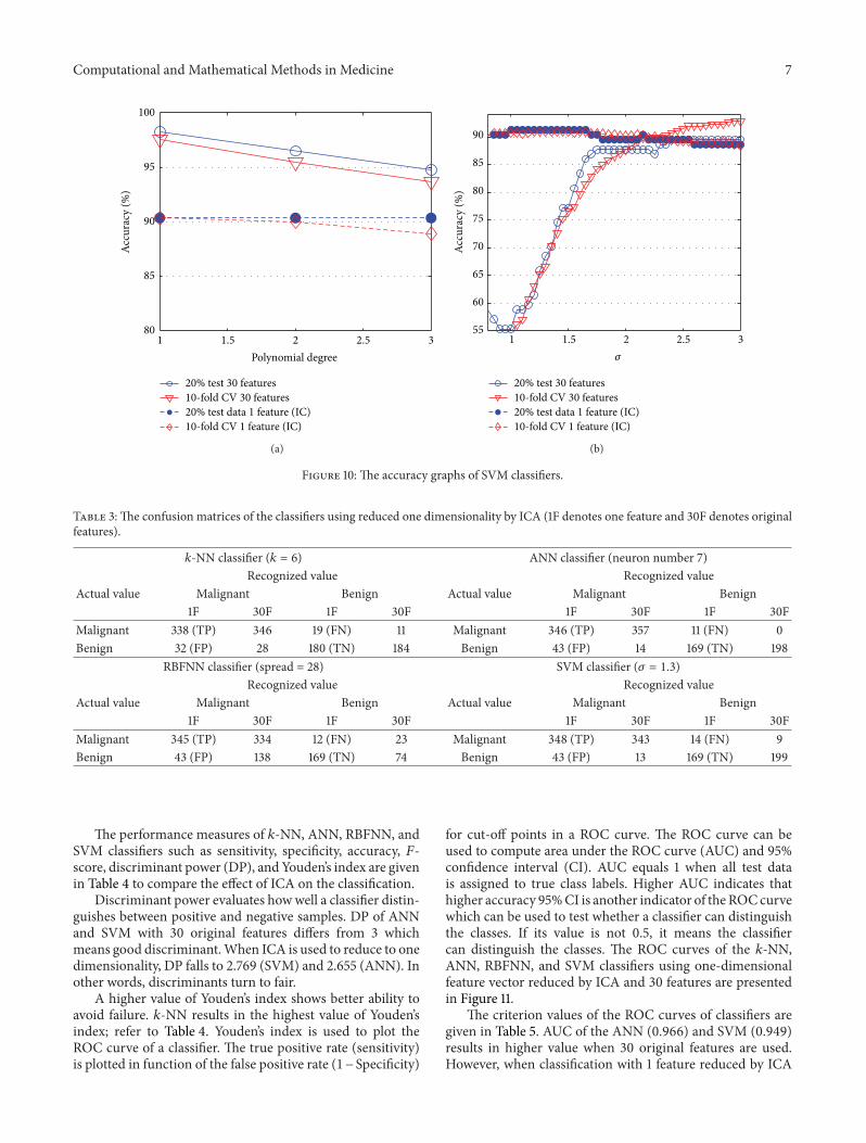
Accuracy evaluation of SVM has been computed forkernel functions including linear, polynomial, and RBF withkernel function parameters such as RBF sigma value for RBFkernel and polynomial degree for polynomial kernel. Theaccuracy graph of SVM classifier is presented in Figure 10
10 20 30 40 50
65
70
75
80
85
90
95
Spread value
Accu
racy
(%)
20% test data with 30 features10-fold CV with 30 features20% test data 1 feature (IC)10-fold CV with 1 feature (IC)
Figure 9: The accuracy graph of RBFNN.
where the axes of polynomial degree indicate linear kernelwhen its value equals one.
Generally, SVM classifier with linear kernel providesmore accurate result than polynomial and RBF kernel. Itsaccuracy is 98.25% for 30 features and 90.35% for reduced1 feature when 20% of data is used as test data. In contrastto polynomial kernel, effect of ICA increases the accuracy ofSVM with RBF kernel from 89.47% (30 features) to 91.23% (1feature).When 10-CV is used, the accuracy is decreased from97.54% (30 features, linear kernel) and 95.25% (30 features,RBFkernel) to 90.33%and 90.86% (reduced 1 feature by ICA).
𝑘-NN, ANN, RBFNN, and SVM have been tested andtrained to find outmaximumaccuracy adjusting their param-eter. The performance measures such as accuracy, specificity,sensitivity, 𝐹-score, Youden’s index, and discriminant powerof the classifiers are compared to each other. The parametersof the classifiers which provide maximum accuracy areselected to be compared to the other classifiers. In additionto these performance measures, the ROC curve of threeclassifiers is plotted to enhance visuality of the comparison.
10-CV and one-dimensional feature vector reduced byICA are used to compare the performances of classifiers. Ininput data of classifiers, the test data are compared to theoriginal class label to find out TP, TN, FP, and FN values.These values for classifiers are given in the form of confusionmatrix in Table 3.
RBFNN classification using 30 original features providesworse performance than reduced one-dimensional featurevector; refer to Table 3. The other classification used with 30features has slightly higher true values when compared toclassification with one feature reduced by ICA.
Computational and Mathematical Methods in Medicine 7
80
85
90
95
100
Polynomial degree
Accu
racy
(%)
20% test 30 features10-fold CV 30 features20% test data 1 feature (IC)10-fold CV 1 feature (IC)
1 1.5 2 2.5 3
(a)
55
60
65
70
75
80
85
90
Accu
racy
(%)
20% test 30 features10-fold CV 30 features20% test data 1 feature (IC)10-fold CV 1 feature (IC)
1 1.5 2 2.5 3
𝜎
(b)
Figure 10: The accuracy graphs of SVM classifiers.
Table 3:The confusion matrices of the classifiers using reduced one dimensionality by ICA (1F denotes one feature and 30F denotes originalfeatures).
𝑘-NN classifier (𝑘 = 6) ANN classifier (neuron number 7)Recognized value Recognized value
Actual value Malignant Benign Actual value Malignant Benign1F 30F 1F 30F 1F 30F 1F 30F
Malignant 338 (TP) 346 19 (FN) 11 Malignant 346 (TP) 357 11 (FN) 0Benign 32 (FP) 28 180 (TN) 184 Benign 43 (FP) 14 169 (TN) 198
RBFNN classifier (spread = 28) SVM classifier (𝜎 = 1.3)Recognized value Recognized value
Actual value Malignant Benign Actual value Malignant Benign1F 30F 1F 30F 1F 30F 1F 30F
Malignant 345 (TP) 334 12 (FN) 23 Malignant 348 (TP) 343 14 (FN) 9Benign 43 (FP) 138 169 (TN) 74 Benign 43 (FP) 13 169 (TN) 199
The performance measures of 𝑘-NN, ANN, RBFNN, andSVM classifiers such as sensitivity, specificity, accuracy, 𝐹-score, discriminant power (DP), and Youden’s index are givenin Table 4 to compare the effect of ICA on the classification.
Discriminant power evaluates howwell a classifier distin-guishes between positive and negative samples. DP of ANNand SVM with 30 original features differs from 3 whichmeans good discriminant.When ICA is used to reduce to onedimensionality, DP falls to 2.769 (SVM) and 2.655 (ANN). Inother words, discriminants turn to fair.
A higher value of Youden’s index shows better ability toavoid failure. 𝑘-NN results in the highest value of Youden’sindex; refer to Table 4. Youden’s index is used to plot theROC curve of a classifier. The true positive rate (sensitivity)is plotted in function of the false positive rate (1−Specificity)
for cut-off points in a ROC curve. The ROC curve can beused to compute area under the ROC curve (AUC) and 95%confidence interval (CI). AUC equals 1 when all test datais assigned to true class labels. Higher AUC indicates thathigher accuracy 95%CI is another indicator of theROCcurvewhich can be used to test whether a classifier can distinguishthe classes. If its value is not 0.5, it means the classifiercan distinguish the classes. The ROC curves of the 𝑘-NN,ANN, RBFNN, and SVM classifiers using one-dimensionalfeature vector reduced by ICA and 30 features are presentedin Figure 11.
The criterion values of the ROC curves of classifiers aregiven in Table 5. AUC of the ANN (0.966) and SVM (0.949)results in higher value when 30 original features are used.However, when classification with 1 feature reduced by ICA

8 Computational and Mathematical Methods in Medicine
Table 4: The comparison of ICA algorithm’s effect on the classifiers’ performance measures (sensitivity, specificity, accuracy, and 𝐹-score in%).
Measures 𝑘-NN ANN RBFNN SVM (RBF K.)1F 30F 1F 30F 1F 30F 1F 30F
𝐹-score 92.98 94.65 92.76 98.07 92.61 80.57 93.04 96.21DP 2.539 2.912 2.655 InF 2.606 1.131 2.769 3.267Υ 0.795 0.839 0.766 0.934 0.763 0.284 0.772 0.899Accuracy 91.03 93.14 90.5 97.53 90.49 87.17 90.86 95.25Specificity 84.9 87.26 79.71 93.39 79.71 34.9 79.71 93.86Sensitivity 94.67 96.63 96.91 100 96.63 93.55 97.47 96.07
Table 5: Criterion values of the ROC curves of 𝑘-NN, ANN, RBFNN, and SVM.
Criterion 𝑘-NN ANN RBFNN SVM1F 30F 1F 30F 1F 30F 1F 30F
AUC 0.880 0.911 0.879 0.956 0.881 0.877 0.879 0.94595% CI 0.86–0.92 0.89–0.94 0.85–0.91 0.94–0.98 0.85–0.91 0.85–0.91 0.85–0.91 0.92–0.97
0 0.2 0.4 0.6 0.8 10.2
0.3
0.4
0.5
0.6
0.7
0.8
0.9
1
True
pos
itive
rate
(sen
sitiv
ity)
ANN (1 IC)RBFNN (1 IC)SVM RBF (1 IC)
ANN (30 features)RBFNN (30 features)SVM RBF (30 features)
False positive rate (1 − specificity)
k-NN (1 IC)
k-NN (30 features)
Figure 11: The ROC curves of 𝑘-NN, ANN, RBFNN, and SVMclassifiers.
is evaluated, 𝑘-NN (0.897) and SVM (0.885) result in higherAUC. It means 𝑘-NN and SVM classifiers using reduced onefeature distinguish samples more correctly.
Table 5 shows that the accuracy of the 𝑘-NN (91.03%) isbetter than the accuracy of ANN,RBFNN, and SVM(90.50%,90.49%, and 90.86%). Generally, one feature reduced by ICA
Table 6: CPU time for classification.
Classifier Partitioning IC (seconds) 30 features (seconds)
𝑘-NN 20% 8.02 8.3110-CV 13.52 14.77
ANN 20% 11.12 13.910-CV 76.72 118.21
RBFNN 20% 14.9 20.0310-CV 90.49 129.84
SVM (poly) 20% 7.17 7.2810-CV 7.47 9.13
SVM (RBF) 20% 9.02 43.3010-CV 10.72 19.05
decreases the accuracy of 𝑘-NN, ANN, and SVM. However,it increases the accuracy of RBFNN.
The aforementioned classification methods are analyzedin terms of computing time given in Table 6 to comparethe computational complexities to the classifications with theoriginal 30 features.
The proposed methods have lower computing time whencompared to classification of the original dataset. In caseof neural network classifications with 30 features, networkconstructions consume highly more time than classificationwith one IC. The measured durations of 13.9 and 20.03seconds are decreased to 11.12 and 14.9 seconds when ANNand RBFN with 20% partitioning are deployed. Particularly,the effect of using IC as feature on complexity is existedwhen 10-CV is selected. The consumed time of the ANN andRBFNN is decreased from 118.21 and 129.84 seconds to 76.72and 90.49 seconds, respectively. In addition, ICA decreasescomputational times of the SVM and 𝑘-NN classifications,but the rates are less than the neural networks.
5. Discussions
Sensitivity/specificity indicates the proportion of actual pos-itives/negatives which are correctly identified. While use of

Computational and Mathematical Methods in Medicine 9
Table 7: Comparison of the methods and accuracy of previous studies and this study.
Author Method Feature number Accuracy Sensitivity
Krishnan et al. [36]40% test data, SVM (poly.) 30 92.62% 92.69%40% test data, SVM (RBF) 93.72% 94.50%
Bagui et al. [37]64% test data, 𝑘-RNN 30 96.00% 95.09%64% test data, 𝑘-RNN Best 3 98.10% 98.05%
Sweilam et al. [38]PSO + SVM 30 93.52% 91.52%QPSO + SVM 93.06% 90.00%
Mangasarian et al. [39] 10-CV, MSM-T Best 3 97.50% —
Mert et al. [40]10-CV, PNN 3 (2IC + DWT) 96.31% 98.88%LOO, PNN 97.01% 97.78%
Zheng et al. [41] 𝐾-SVM 6 97.38% —
This study
10-CV, 𝑘-NN
1 feature reduced by ICA
91.03% 94.67%40% test, 𝑘-NN 92.56% 94.02%10-CV, ANN 90.50% 96.91%40% test, ANN 90.89% 97.00%10-CV, RBFNN 90.49% 96.63%40% test, RBFNN 89.98% 96.01%10-CV, SVM (linear) 90.33% 96.35%40% test, SVM (linear) 90.01% 95.00%10-CV, SVM (quadratic) 89.98% 95.24%40% test, SVM (quadratic) 91.01% 96.42%10-CV, SVM (RBF) 90.86% 97.47%40% test, SVM (RBF) 91.03% 97.56%
one-dimensional feature vector reduced by ICA decreasesaccuracy slightly, it increases sensitivity values of SVMand RBFNN classifiers. The maximum sensitivity measurebelongs to SVM with RBF kernel when one feature is used.The graph of the effect of ICA on sensitivity measures ofclassifiers is shown in Figure 12.
Sensitivity refers successfully identified malignant sam-ples in cancer classification. Thus, higher sensitivity meanshigher diagnostic capability of malignant tumors and it canbe used to help physicians to diagnose cancerous mass morecorrectly. The accuracy and sensitivity measures of previousclassification studies and this study on WDBC dataset aregiven in Table 7 to compare the effect of feature reductionusing ICA. It should be noted that the studies onWDBCdifferfrom studies onWBC dataset which consists of 699 instanceswith 10 attributes.
Higher number of features used to classify breast canceras benign and malignant resultsin slightly higher accuracy.Feature reduction into one using ICA decreases the accuracyof 𝑘-NN, ANN, and SVM slightly. However, it increases theaccuracy of RBFNN and the sensitivity values of SVM andRBFNN.
Referring to Table 7, the sensitivity measures of the clas-sifiers used with one-dimensional feature vector reduced byICA in this study perform better than the other studies. How-ever, accuracy rates of the proposed classifications (90.53% ±0.34) are lower than the previous methods (94.93% ± 2.07).The study of WDBC data creators [39] set has the highestaccuracy (97.50%) using multisurface method tree (MSM-T) with 3 selected features. Similarly, hybrid methods are
90
91
92
93
94
95
96
97
98
99
100
ANN RBFNN SVM (RBF K.)
Sens
itivi
ty (%
)
1F (IC)30 features
k-NN
Figure 12: Sensitivity measures of the classifiers.
more successful than the others. Breast cancer classificationsusing probabilistic neural network (PNN)with hybrid featurereduction using discrete wavelet transform (DWT) and ICA[40] or classification using SVM with 6-dimensional featurespace obtained by 𝐾-means algorithm [41] have accuracyrates of 96.31% and 97.38% for 10-CV. Particularly, SVMbasedstudies [36, 38] with 30 features have near scores to our one-dimensional results.

10 Computational and Mathematical Methods in Medicine
6. Conclusions
In this study, the effect of dimensionality reduction usingindependent component analysis (ICA) on breast cancerdecision support systems with several classifiers such asartificial neural network (ANN), 𝑘-nearest neighbor (𝑘-NN),radial basis function neural network (RBFNN), and supportvector machine (SVM) is investigated. The results of theapplied original thirty features ofWisconsin diagnostic breastcancer (WDBC) are compared with the reduced one dimen-sion by ICA. The accuracy rates of the classifications withthirty original features except RBFNNhave slightly decreasedfrom 97.53%, 91.03%, and 95.25% to 90.5%, 91.03%, and90.86%, respectively. However, the one-dimensional featurevector causes RBFNN classifier to be more distinguishingwith the increased accuracy from 87.17% to 90.49%. Fur-thermore, the sensitivity rates which define the successfullyrecognized malignant samples are increased from 93.5% to96.63% for RBFNN and from 96.07% to 97.47% for SVM,while the others have slight decrease at the rate between0.96% and 3.09%. If the objective is to increase the rate of thesuccessfully identifiedmalignant breast cancer using RBFNNor decrease computational complexity without loss of thehigh accuracy rate, feature reduction applying ICA can be ahigh performance solution.
Conflict of Interests
The authors declare that they have no conflict of interests.
Acknowledgment
This work was supported by the Istanbul University ScientificResearch Projects, Project numbers YADOP-6987, 36196,38262, 42330, and 35830.
References
[1] I. Christoyianni, E. Dermatas, and G. Kokkinakis, “Fast detec-tion of masses in computer-aided mammography,” IEEE SignalProcessing Magazine, vol. 17, no. 1, pp. 54–64, 2000.
[2] N. Salim, Medical Diagnosis Using Neural Network, Faculty ofInformation Technology University, 2013, http://www.gener-ation5.org/content/2004/MedicalDiagnosis.asp.
[3] A. Tartar, N. Kilic, and A. Akan, “Classification of pulmonarynodules by using hybrid features,” Computational and Math-ematical Methods in Medicine, vol. 2013, Article ID 148363, 11pages, 2013.
[4] N. Kilic, O.N.Ucan, andO.Osman, “Colonic polyp detection inCT colonography with fuzzy rule based 3D template matching,”Journal of Medical Systems, vol. 33, no. 1, pp. 9–18, 2009.
[5] A.Mert, N. Kilic, andA.Akan, “Evaluation of bagging ensemblemethod with time-domain feature extraction for diagnosing ofarrhythmia beats,” Neural Computing and Applications, vol. 24,no. 2, pp. 317–326, 2014.
[6] R. W. Brause, “Medical analysis and diagnosis by neural net-works,” in Proceedings of the 2nd International Symposium onMedical Data Analysis (ISMDA ’01), pp. 1–13, Madrid, Spain,October 2001.
[7] T. S. Subashini, V. Ramalingam, and S. Palanivel, “Breast massclassification based on cytological patterns using RBFNN andSVM,” Expert Systems with Applications, vol. 36, no. 3, pp. 5284–5290, 2009.
[8] M. N. Gurcan, H.-P. Chan, B. Sahiner, L. Hadjiiski, N. Petrick,and M. A. Helvie, “Optimal neural network architecture selec-tion: improvement in computerized detection of microcalcifi-cations,” Academic Radiology, vol. 9, no. 4, pp. 420–429, 2002.
[9] A. P. Dhawan, Y. Chitre, C. Bonasso, and K. Wheeler, “Radial-basis-function based classification of mammographic micro-calcifications using texture features,” in Proceedings of the 17thIEEE Engineering in Medicine and Biology Annual Conference,pp. 535–536, September 1995.
[10] A. T.Azar and S.A. El-Said, “Superior neuro-fuzzy classificationsystems,” Neural Computing and Applications, vol. 23, no. 1,supplement, pp. 55–72, 2012.
[11] M. Jia, C. Zhao, F. Wang, and D. Niu, “A new method for deci-sion on the structure of RBF neural network,” in Proceedings ofthe 2006 International Conference on Computational Intelligenceand Security, pp. 147–150, November 2006.
[12] J. K. Sing, S. Thakur, D. K. Basu, M. Nasipuri, and M. Kundu,“High-speed face recognition using self-adaptive radial basisfunction neural networks,” Neural Computing & Applications,vol. 18, no. 8, pp. 979–990, 2009.
[13] R. Huang, L. Law, and Y. Cheung, “An experimental study: onreducingRBF input dimension by ICAandPCA,” inProceedingsof the 2002 International Conference on Machine Learning andCybernetics, vol. 4, pp. 1941–1945, November 2002.
[14] V. N. Vapnik, Statistical Learning Theory, John Wiley & Sons,New York, NY, USA, 1989.
[15] M. F. Akay, “Support vector machines combined with featureselection for breast cancer diagnosis,” Expert Systems withApplications, vol. 36, no. 2, pp. 3240–3247, 2009.
[16] B. Wang, H. Huang, and X. Wang, “A support vector machinebased MSM model for financial short-term volatility forecast-ing,” Neural Computing and Applications, vol. 22, no. 1, pp. 21–28, 2013.
[17] M. Pontil and A. Verri, “Support vector machines for 3D objectrecognition,” IEEE Transactions on Pattern Analysis andMachine Intelligence, vol. 20, no. 6, pp. 637–646, 1998.
[18] J. Zhou, G. Su, C. Jiang, Y. Deng, and C. Li, “A face andfingerprint identity authentication system based onmulti-routedetection,” Neurocomputing, vol. 70, no. 4–6, pp. 922–931, 2007.
[19] E. Gumus, N. Kilic, A. Sertbas, and O. N. Ucan, “Evaluationof face recognition techniques using PCA, wavelets and SVM,”Expert Systems with Applications, vol. 37, no. 9, pp. 6404–6408,2010.
[20] S. Kara, A. Guven, and S. Icer, “Classification of macular andoptic nerve disease by principal component analysis,” Comput-ers in Biology and Medicine, vol. 37, no. 6, pp. 836–841, 2007.
[21] A. Hyvarinen and E. Oja, “Independent component analysis:algorithms and applications,” Neural Networks, vol. 13, no. 4-5,pp. 411–430, 2000.
[22] M. P. S. Chawla, “A comparative analysis of principal compo-nent and independent component techniques for electrocardio-grams,” Neural Computing and Applications, vol. 18, no. 6, pp.539–556, 2009.
[23] S. D. Villalba and P. Cunningham, “An evaluation of dimensionreduction techniques for one-class classification,” ArtificialIntelligence Review, vol. 27, no. 4, pp. 273–294, 2007.

Computational and Mathematical Methods in Medicine 11
[24] W. H. Wolberg, W. N. Street, and O. L. Mangasarian, “Machinelearning techniques to diagnose breast cancer from image-processed nuclear features of fine needle aspirates,” CancerLetters, vol. 77, no. 2-3, pp. 163–171, 1994.
[25] K. H. Liu, B. Li, Q. Q. Wu, J. Zhang, J. X. Du, and G. Y. Liu,“Microarray data classification based on ensemble independentcomponent selection,” Computers in Biology and Medicine, vol.39, no. 11, pp. 953–960, 2009.
[26] 2013, http://research.ics.tkk.fi/ica/fastica/.[27] J. Bilski, “The UD RLS algorithm for training feedforward neu-
ral networks,” International Journal of Applied Mathematics andComputer Science, vol. 15, pp. 115–123, 2005.
[28] N. Sivri, N. Kilic, and O. N. Ucan, “Estimation of stream tem-perature in Firtina Creek (Rize-Turkiye) using artificial neuralnetwork model,” Journal of Environmental Biology, vol. 28, no.1, pp. 67–72, 2007.
[29] O. A. Abdalla, M. H. Zakaria, S. Sulaiman, andW. F.W. Ahmad,“A comparison of feed-forward back-propagation and radialbasis artificial neural networks: A Monte Carlo study,” in Pro-ceedings of the International Symposium in Information Technol-ogy (ITSim ’10), vol. 2, pp. 994–998, Kuala Lumpur, Malaysia,June 2010.
[30] B. E. Boser, I.M.Guyon, andV.N.Vapnik, “A training algorithmfor optimal margin classifiers,” in Proceedings of the 5th AnnualACM Workshop on Computational Learning Theory, pp. 144–152, July 1992.
[31] V.N. Vapnik,TheNature of Statistical LearningTheory, Springer,New York, NY, USA, 1995.
[32] R. Courant and D. Hilbert, Methods of Mathematical Physics,Wiley, New York, NY, USA, 1953.
[33] W. J. Youden, “Index for rating diagnostic tests,” Cancer, vol. 3,no. 1, pp. 32–35, 1950.
[34] L. L. Pesce and C. E. Metz, “Reliable and computationallyefficient maximum-likelihood estimation of proper binormalROC curves,” Academic Radiology, vol. 14, no. 7, pp. 814–829,2007.
[35] J. Hamidzadeh, R. Monsefi, and H. S. Yazdi, “DDC: distance-based decision classifier,”Neural Computing&Applications, vol.21, no. 7, pp. 1697–1707, 2012.
[36] A. M. Krishnan, S. Banerjee, C. Chakraborty, and A. K. Ray,“Statistical analysis of mammographic features and its classi-fication using support vector machine,” Expert Systems withApplications, vol. 37, no. 1, pp. 470–478, 2010.
[37] S. C. Bagui, S. Bagui, K. Pal, and N. R. Pal, “Breast cancer detec-tion using rank nearest neighbor classification rules,” PatternRecognition, vol. 36, no. 1, pp. 25–34, 2003.
[38] N. H. Sweilam, A. A. Tharwat, and N. K. Abdel Moniem, “Sup-port vectormachine for diagnosis cancer disease: a comparativestudy,” Egyptian Informatics Journal, vol. 11, no. 2, pp. 81–92,2010.
[39] O. L. Mangasarian, W. N. Street, and W. H. Wolberg, “Breastcancer diagnosis and prognosis via linear programming,”Oper-ations Research, vol. 43, no. 4, pp. 570–577, 1995.
[40] A. Mert, N. Kılıc, and A. Akan, “An improved hybrid featurereduction for increased breast cancer diagnostic performance,”Biomedical Engineering Letters, vol. 4, no. 3, pp. 285–291, 2014.
[41] B. Zheng, S. W. Yoon, and S. S. Lam, “Breast cancer diagnosisbased on feature extraction using a hybrid of K-means andsupport vector machine algorithms,” Expert Systems with Appli-cations, vol. 41, no. 4, pp. 1476–1482, 2014.

Submit your manuscripts athttp://www.hindawi.com
Stem CellsInternational
Hindawi Publishing Corporationhttp://www.hindawi.com Volume 2014
Hindawi Publishing Corporationhttp://www.hindawi.com Volume 2014
MEDIATORSINFLAMMATION
of
Hindawi Publishing Corporationhttp://www.hindawi.com Volume 2014
Behavioural Neurology
EndocrinologyInternational Journal of
Hindawi Publishing Corporationhttp://www.hindawi.com Volume 2014
Hindawi Publishing Corporationhttp://www.hindawi.com Volume 2014
Disease Markers
Hindawi Publishing Corporationhttp://www.hindawi.com Volume 2014
BioMed Research International
OncologyJournal of
Hindawi Publishing Corporationhttp://www.hindawi.com Volume 2014
Hindawi Publishing Corporationhttp://www.hindawi.com Volume 2014
Oxidative Medicine and Cellular Longevity
Hindawi Publishing Corporationhttp://www.hindawi.com Volume 2014
PPAR Research
The Scientific World JournalHindawi Publishing Corporation http://www.hindawi.com Volume 2014
Immunology ResearchHindawi Publishing Corporationhttp://www.hindawi.com Volume 2014
Journal of
ObesityJournal of
Hindawi Publishing Corporationhttp://www.hindawi.com Volume 2014
Hindawi Publishing Corporationhttp://www.hindawi.com Volume 2014
Computational and Mathematical Methods in Medicine
OphthalmologyJournal of
Hindawi Publishing Corporationhttp://www.hindawi.com Volume 2014
Diabetes ResearchJournal of
Hindawi Publishing Corporationhttp://www.hindawi.com Volume 2014
Hindawi Publishing Corporationhttp://www.hindawi.com Volume 2014
Research and TreatmentAIDS
Hindawi Publishing Corporationhttp://www.hindawi.com Volume 2014
Gastroenterology Research and Practice
Hindawi Publishing Corporationhttp://www.hindawi.com Volume 2014
Parkinson’s Disease
Evidence-Based Complementary and Alternative Medicine
Volume 2014Hindawi Publishing Corporationhttp://www.hindawi.com
Related Documents