1 BONE AND CARTILAGE LIA DAMAYANTI Department of Histology - FMUI

Welcome message from author
This document is posted to help you gain knowledge. Please leave a comment to let me know what you think about it! Share it to your friends and learn new things together.
Transcript
1
BONE AND CARTILAGELIA DAMAYANTI
Department of Histology - FMUI

2
Agenda
Introduction Cartilage
Development of cartilage Growth and repair Components of cartilage Type of Cartilage
3
Agenda
Bone Components of bone Development of the bone Microarchitexture of the bone

4
Introduction
Cartilage and bone Specialized connective tissue
Cartilage A firm pliable matrix Resist mechanical stress
Bone The hardest tissues of the body Resists stresses place upon it
Comprise the skeleton Function in supporting the body The growth start early in the fetus and continues after birth

5
Cartilage
Avascular Regenerate poorly
6
Cartilage
Function: subordinate role to bone in support and protection of
the body shock absorber
has the specialized property a highly resilient tissue capable of bearing considering weight having some rigidity giving remarkable flexibility to the body

7
Development of cartilage
Developmentbegins at the 5 prenatal weekMesenchyme cells →chondroblast, interlaced
with the connective fibers → deposit extra-cellular matrix → chondrocytes, imprisoned in lacunae
embryonic skeleton → hyaline type Degenerates →replaced by the bone
8
Growth
postnatal epiphyseal growth center in long bones articular surface of movable joint open and closedcartilage rings along the larger larger
respiratory passageways Costosternal junctions between ribs and sternum

9
Growth
Cartilage An avascular tissue Receive the nutrien by diffusion
through its matrix 2 mechanisms of cartilage growth
Interstitial (endogenous) growth Through mitosis of the imprisoned
chondrocyte Multiple to form a nest cells called as
isogenous cells Expansion of lacunae housing of the
cells Growing of the interstitial matrix
10
Growth
Appositional (exogenous) growth Occurs when a cartilage structure increases
in size by new cartilage being deposited on its surface
Dependent on the presence of undifferentiated primitive cells on the surface of the cartilage which differentiate to become chondroblast which lay down new cartilage
Interstitial growth begins earlier cease early in life
Appositional growth throughout life
11
Composed by Perichondrium Extra-cellular matrix Cartilage cells
12
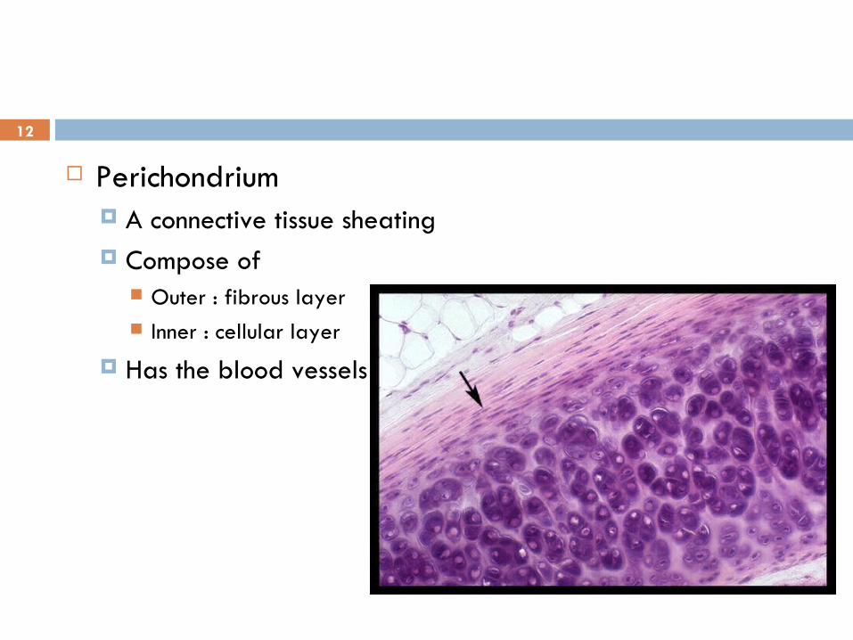
Perichondrium A connective tissue sheating Compose of
Outer : fibrous layer Inner : cellular layer
Has the blood vessels

13
Extracellular matrix Divided into 2 regions
Territorial matrix Surround the lacunae Poor in collagen and rich in
chondroitin sulfate (basophilic and intense staining with Periodic acid-Schiff)
Inter-territorial matrix Bulk of matrix Rich in type II collagen and
poorer in proteoglycans

14
Cartilage cells Chondrogenic cells
spindle-shape, narrow cells Cytoplasm is sparse with small golgi apparatus, a few
mitochondria, rough endoplasmic reticulum (RER) and an abundance of free ribosome
differentiate into chondroblast Chondroblast
derived from mesenchyme within the center of chondrofication chondrogenic cells of the inner cellular layers of the
perichondrium
15
histology appearance plump, basophilic cells a rich network of RER, a well-developed Golgi complex, numerous
mitochondria Produce and deposit extracellular matrix
Chondrocyte Mature chondroblast surrounded by matrix Entrapped singly or in group in the lacunae near the periphery are ovoid deeper in the cartilage are more rounded

16
Cartilage
3 types, based on the fibers present in the matrix Hyaline cartilage Elastic cartilage Fibrocartilage
17
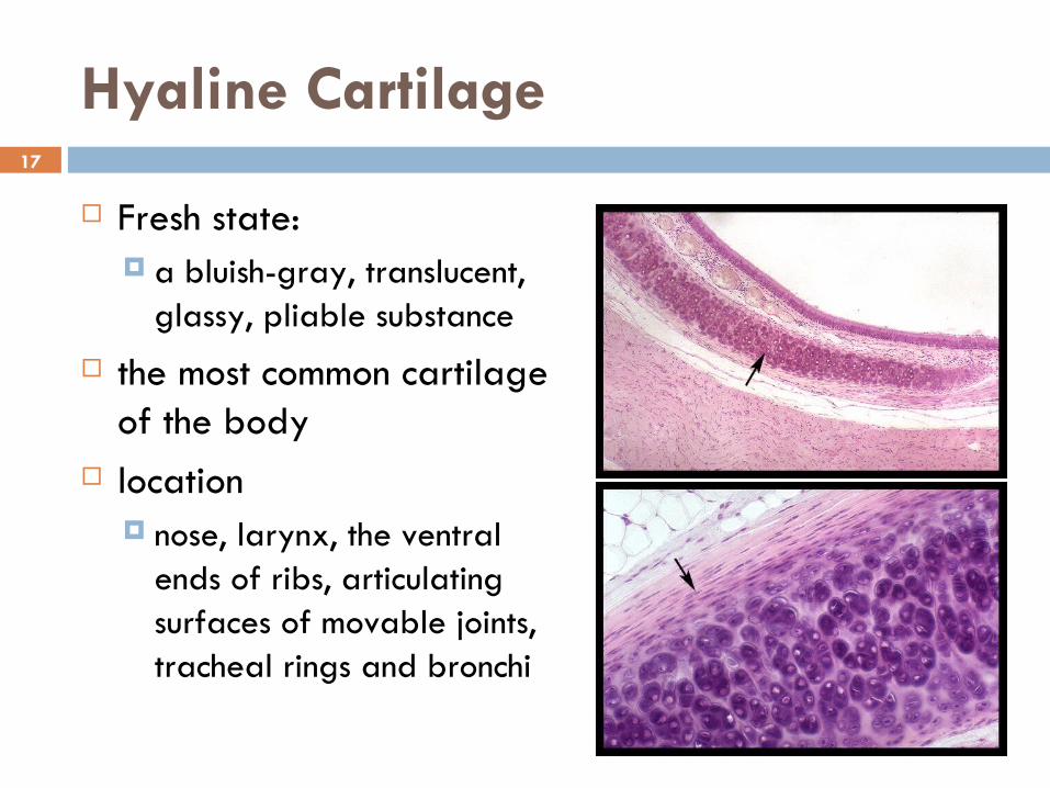
Hyaline Cartilage
Fresh state: a bluish-gray, translucent,
glassy, pliable substance the most common cartilage
of the body location
nose, larynx, the ventral ends of ribs, articulating surfaces of movable joints, tracheal rings and bronchi

18
Hyaline Cartilage
Matrix Fiber →Collagen fiber
Mostly type II Type IX, X, and XI Not visible under light microscopy
Proteoglycans Aggrecans
Large proteoglycans Composed of protein core
Glycosaminoglycan molecule (chondroitin 4-sulphate, chondroitin-6 sulphate and heparan sulfate) are covalently linked
Link to hyaluronic acid

19
Glycosaminoglycan form electrostatic bonds with the collagen
Glycoprotein Large molecule Has binding site for collagen type II, chondroitin-4 and 6 sulfates,
hyaluronic acid and integrin of chondroblast and chondrocyte
20
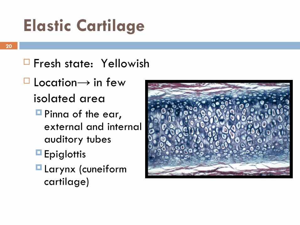
Elastic Cartilage
Fresh state: Yellowish Location→ in few
isolated areaPinna of the ear,
external and internal auditory tubes
Epiglottis Larynx (cuneiform
cartilage)
21
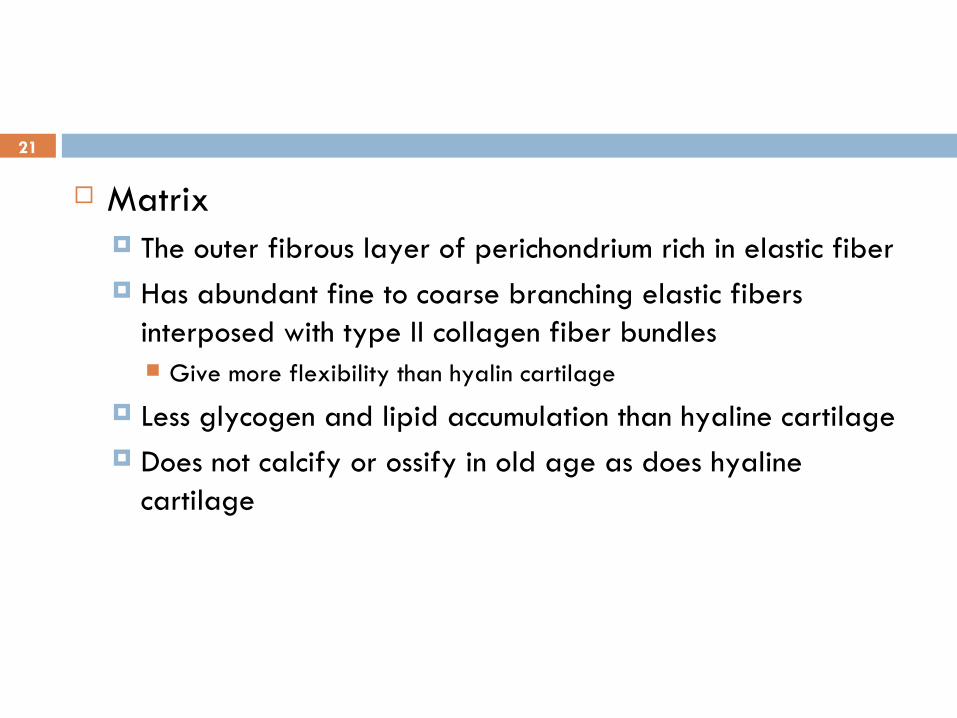
Matrix The outer fibrous layer of perichondrium rich in elastic fiber Has abundant fine to coarse branching elastic fibers
interposed with type II collagen fiber bundles Give more flexibility than hyalin cartilage
Less glycogen and lipid accumulation than hyaline cartilage Does not calcify or ossify in old age as does hyaline
cartilage

22
Cartilage cells The cells are more closely packed and usually are
found singly in lacunae Chondrocytes more abundant and larger than those
of hyaline cartilage

23
Hyaline vs Elastic Cartilage

24
Fibrocartilage
Location: Best seen in intervertebral disks Other articular disks: knee, mandible, sternoclavicular
joints For resistance to compression, durability, tensile
strength Develop from dense fibrous tissue richly with
fibroblast Fibroblast with separated matrix differentiate into
chondroblasts→ fibrocartilage
25
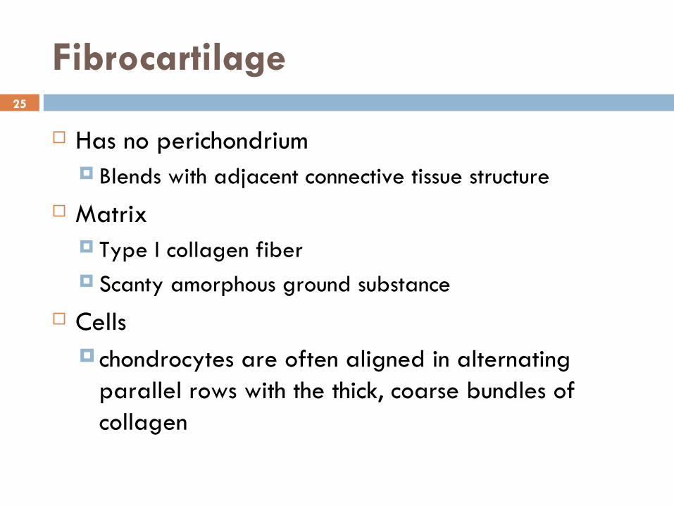
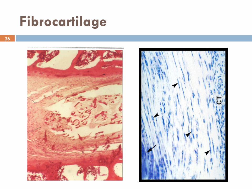
Fibrocartilage
Has no perichondrium Blends with adjacent connective tissue structure
Matrix Type I collagen fiber Scanty amorphous ground substance
Cells chondrocytes are often aligned in alternating
parallel rows with the thick, coarse bundles of collagen
26
Fibrocartilage

27
Bone
A specialized connective tissue whose extracellular matrix is calcified, incarcerating the cells that secreted it
the hardest substance of the body Highly vascularized Dynamic tissue Regenerate completely
28
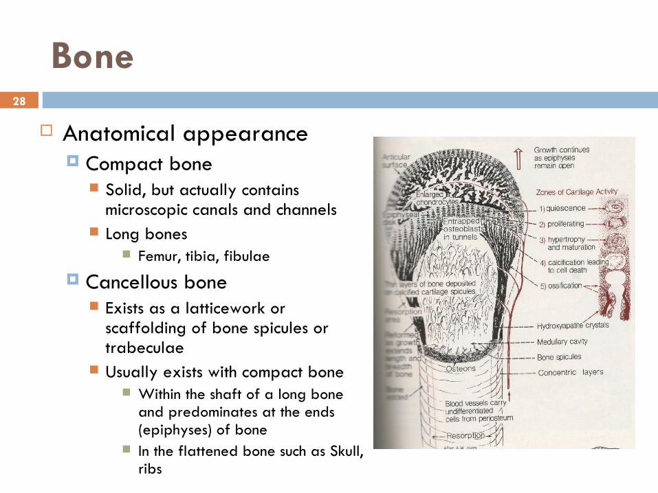
Bone
Anatomical appearance Compact bone
Solid, but actually contains microscopic canals and channels
Long bones Femur, tibia, fibulae
Cancellous bone Exists as a latticework or
scaffolding of bone spicules or trabeculae
Usually exists with compact bone Within the shaft of a long bone
and predominates at the ends (epiphyses) of bone
In the flattened bone such as Skull, ribs
29
Histological appearancePeriosteum
The layer covered the external surface of bone The cells of bone
Osteogenic (osteoprogenitor) cells Osteoblasts Osteocytes Osteoclasts
The bone matrix

30
Periosteum A tough, vascular, fibrous layer Cover the bone except over the
articular surfaces Has 2 layers
The outer layer Largely collagenous fibers and
small component of elastic fibers
The inner (Osteogenic) layer
31
The cells of bone Osteoprogenitor cells
Located in the inner cellular layer of periosteum Lining haversian canals Endosteum
Derived from embryonic mesenchyme Can proliferate and differentiate
Osteoblast Chondroblast (low oxygen tension)
Histological appearance Spindle-shape and has a pale staining oval nucleus Scant pale staining cytoplasm
32
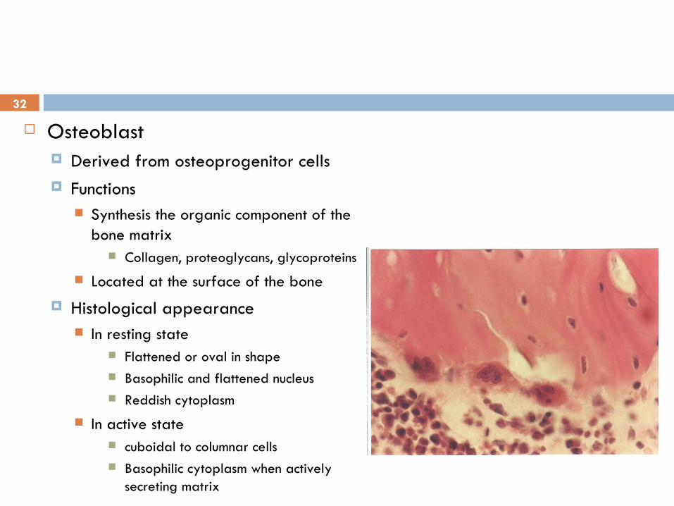
Osteoblast Derived from osteoprogenitor cells Functions
Synthesis the organic component of the bone matrix
Collagen, proteoglycans, glycoproteins Located at the surface of the bone
Histological appearance In resting state
Flattened or oval in shape Basophilic and flattened nucleus Reddish cytoplasm
In active state cuboidal to columnar cells Basophilic cytoplasm when actively
secreting matrix
33
Osteoblast Differentiation Induced by
Hormone Parathyroid hormone
Microenviroment Fibronectin Collagen Procollagen peptides Proteoglycans
Has the receptor for parathyroid hormone on their cell membrane Stimulate osteoblast to secrete osteoclast stimulating factor which activates
osteoclst to resorb bone

34
Osteocytes Mature bone cells Derived from osteoblast Housed in the lacunae within the calcified bony matrix Gap junction Histological appearance
The flattened cells Nucleus is flattenned Cytoplasm is poor in organelles
35
Osteocytes Mature bone cells Derived from osteoblast Housed in the lacunae within the calcified bony matrix Gap junction Histological appearance
The flattened cells Nucleus is flattenned Cytoplasm is poor in organelles

36
Osteoclast The precursor for osteoclast
originates in the bone marrow Has the receptor for osteoclast-
stimulating factor and calcitonin Histological appearance
A large, motile, multinucleated cells Has an acidophilic cytoplasm Occupy the shallow depressions
called as Howship,s lacunae Functions
Resorbing the bone in bone remodelling

37
Matrix of the bone Inorganic constituents
Mostly composed by calcium and phosphorus
Magnesium, sodium, potassium, carbonate
Organic constituents Almost exclusively is collagen type I Glysaminoglican, proteoglycan,
glycoprotein Osteocalcin, osteopontin Bone sialoprotein

38
Developmental of bone
It always arises from a preexisting tissue which it eventually replaces
2 types Intracartilagenous (endochondral) ossification Intramembraneous (desmal) ossification
Please help yourself for the details

39
Intracartilagenous calcification
40
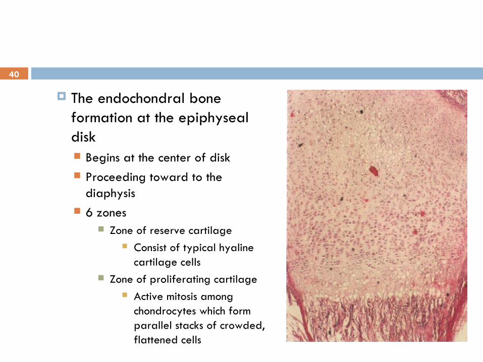
The endochondral bone formation at the epiphyseal disk Begins at the center of disk Proceeding toward to the
diaphysis 6 zones
Zone of reserve cartilage Consist of typical hyaline
cartilage cells Zone of proliferating cartilage
Active mitosis among chondrocytes which form parallel stacks of crowded, flattened cells
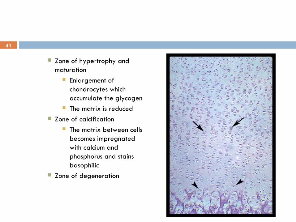
41
Zone of hypertrophy and maturation
Enlargement of chondrocytes which accumulate the glycogen
The matrix is reduced Zone of calcification
The matrix between cells becomes impregnated with calcium and phosphorus and stains basophilic
Zone of degeneration
42
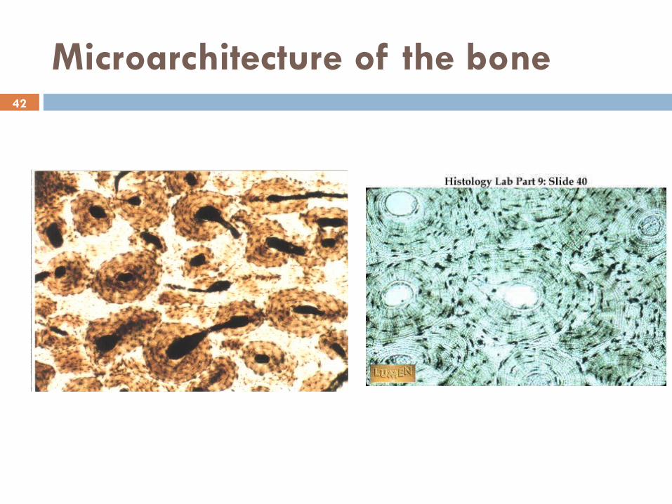
Microarchitecture of the bone

43
Microarchitecture of the bone
Mature bone Lamellae bone
Lamellae of Havers (Primary lamellae) surrounding the central canal (Canal of Havers)
The osteocytes are sequestered in lacunae located within or between the lamellae
The canal contains nerve fiber, loose connective tissue and flatenned osteogenic cells
The protoplasmic processes of the osteocytes enter the tiny channels that radiate from the lacunae, called as canaliculi to communicate with the adjacent osteocytes

44
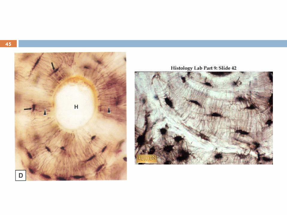
Microarchitecture of the bone
Canal of Volkmann The canals that communicate one
canal of Havers with the other Penetrate the lamellae to join the
haversian canals where anastomoses of their respective blood vessel occurred
Interstitial lamellae The remnants of older, partially
reabsored haversian system , disrupted during the remodelling of the bone
Outer and inner circumferential lamellae
45
46
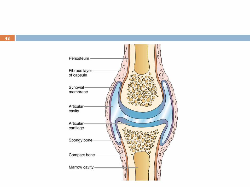
Joints
Basic joint components are:1. Bone2. Hyaline Cartilage (articular cartilage): covered the
bone persistently3. Dense collagen tissues (joint supporting tissue) Classification:
SynarthrosisDiarthrosis

47
Joint supporting tissues
Mostly dens collagen connective tissues Regenerate fairly good but not as good as bone
48
49
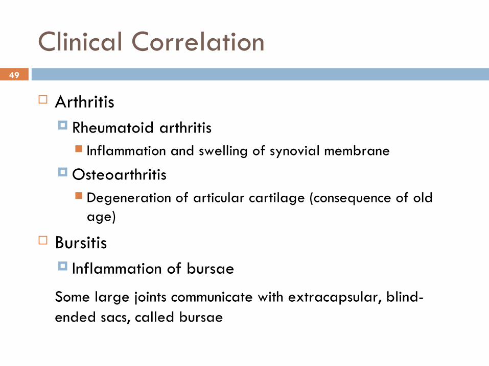
Clinical Correlation
Arthritis Rheumatoid arthritis
Inflammation and swelling of synovial membrane Osteoarthritis
Degeneration of articular cartilage (consequence of old age)
Bursitis Inflammation of bursae
Some large joints communicate with extracapsular, blind-ended sacs, called bursae
Related Documents