Blood DNA methylation biomarkers predict clinical reactivity in food-sensitized infants David Martino, PhD, a,b,d,f Thanh Dang, PhD, a,b Alexandra Sexton-Oates, BSc, b Susan Prescott, MBBS, FRACP, PhD, a,c,f Mimi L. K. Tang, MBBS, PhD, FRACP, FRCPA, FAAAAI, a,b,d,e Shyamali Dharmage, MBBS, MSc, MD, PhD, a,g Lyle Gurrin, PhD, a,g Jennifer Koplin, PhD, a,g Anne-Louise Ponsonby, MD, PhD, a,b,d Katrina J. Allen, MBBS, FRACP, FAAAAI, PhD, a,b,d,f,h * and Richard Saffery, PhD, b,d * for the HealthNuts study investigators Parkville, Melbourne, and Perth, Australia, and Manchester, United Kingdom Background: The diagnosis of food allergy (FA) can be challenging because approximately half of food-sensitized patients are asymptomatic. Current diagnostic tests are excellent makers of sensitization but poor predictors of clinical reactivity. Thus oral food challenges (OFCs) are required to determine a patient’s risk of reactivity. Objective: We sought to discover genomic biomarkers of clinical FA with utility for predicting food challenge outcomes. Methods: Genome-wide DNA methylation (DNAm) profiling was performed on blood mononuclear cells from volunteers who had undergone objective OFCs, concurrent skin prick tests, and specific IgE tests. Fifty-eight food-sensitized patients (aged 11-15 months) were assessed, half of whom were clinically reactive. Thirteen nonallergic control subjects were also assessed. Reproducibility was assessed in an additional 48 samples by using methylation data from an independent population of patients with clinical FA. Results: Using a supervised learning approach, we discovered a DNAm signature of 96 CpG sites that predict clinical outcomes. Diagnostic scores were derived from these 96 methylation sites, and cutoffs were determined in a sensitivity analysis. Methylation biomarkers outperformed allergen-specific IgE and skin prick tests for predicting OFC outcomes. FA status was correctly predicted in the replication cohort with an accuracy of 79.2%. Conclusion: DNAm biomarkers with clinical utility for predicting food challenge outcomes are readily detectable in blood. The development of this technology in detailed follow-up studies will yield highly innovative diagnostic assays. (J Allergy Clin Immunol 2015;nnn:nnn-nnn.) Key words: Food allergy, epigenetics, DNA methylation profiling, Infinium, methylation profiling, epigenetic epidemiology, allergy epigenetics, biomarkers, shrunken centroids Food allergy (FA) affects up to 10% of children in the most affected countries 1 and has become a substantial public health concern. Increasing rates of FA among young children have coincided with an increase in potentially life-threatening anaphylaxis. 2 Laboratory testing for detection of allergen- specific IgE (sIgE) is widely used in the diagnosis of IgE-mediated FA, which offers proof of sensitization; however, further evidence of clinical manifestations on exposure is required to make a definitive diagnosis. 3 This is because the majority of children with a positive skin prick test (SPT) response or sIgE test result are not allergic to that food. 4 At present, there are no reliable biomarkers for clinical FA, and the oral food challenge (OFC) test is still required to clarify clinical reactivity. OFCs are the gold standard method to diagnose FA, but can be dangerous primarily because of the risk of inducing anaphylaxis in an allergic subject. As a result of this, OFCs are often not performed, potentially leading to overdiagnosis of FA or unnecessary avoidance programs. 4 There is a need to improve current allergy testing practices that can help make a more spe- cific diagnosis regarding a patient’s risk of reactivity on exposure. Identifying biomarkers of clinical allergy could prove extremely useful in the absence of a clinical history or if the child has yet to be exposed to a particular food or has undergone a lengthy avoid- ance program. Epigenetic biomarkers of disease risk or exposure have already generated broad interest because these factors can mediate genetic or environmental risk. 5 In diseases underpinned by complex gene-environment interactions, epigenetics is centrally placed as a key mediator. 6 In a previous study we reported DNA methylation (DNAm) differences associated with develop- ment of IgE-mediated FA in early life. 7 Extending this research, in the present study we generate genome-wide DNAm profiles from volunteers recruited to the HealthNuts study undergoing From a the Centre for Food and Allergy Research, Murdoch Childrens Research Institute, Parkville; b Murdoch Childrens Research Institute, Royal Children’s Hospital, Parkville; c the School of Paediatrics and Child Health, University of Western Australia, Perth; d the University of Melbourne Department of Paediatrics; e the Department of Allergy and Immunology, Royal Children’s Hospital, Parkville; f In-FLAME, the International Inflammation Network, World Universities Network (WUN); g the School of Population and Global Health, University of Melbourne; and h the Institute of Inflammation and Repair, University of Manchester. *These author contributed equally to this work. Supported by the CASS Foundation (SM/11/3647/RMR:DSA), the DHB Foundation (CT21242), and the NHMRC (1072752). Disclosure of potential conflict of interest: This study was funded by the CASS Foundation, DHB Foundation, and the National Health and Medical Research Council, Australia’s leading expert body promoting the development and maintenance of public and individual health standards. S. Prescott has received compensation for board membership from Danone Nutricia and the Nestl e Nutrition Institute, has received payment for delivering lectures from Danone Nutricia and the Nestl e Nutrition Institute, and receives royalties from UpToDate. M. L. K. Tang has received compensation for board membership from Danone Nutricia and the Nestl e Nutrition Institute; has received payment for delivering lectures from Danone Nutricia and the Nestl e Nutrition Institute; and has received compensation for travel and other meeting-related expenses from APARPI. A.-L. Ponsonby is employed by the NHMRC, from which she has also received or has grants pending. K. J. Allen has received consultancy fees from Nutricia, Danone, Nestl e, Aspen, and Alphapharm. The rest of the authors declare that they have no relevant conflicts of interest. Received for publication September 1, 2014; revised November 28, 2014; accepted for publication December 29, 2014. Corresponding author: Katrina J. Allen, MBBS, FRACP, FAAAAI, PhD, Gastro and Food Allergy, Murdoch Childrens Research Institute, Flemington Rd, Parkville, Melbourne, Australia. E-mail: [email protected]. 0091-6749/$36.00 Ó 2015 American Academy of Allergy, Asthma & Immunology http://dx.doi.org/10.1016/j.jaci.2014.12.1933 1

Welcome message from author
This document is posted to help you gain knowledge. Please leave a comment to let me know what you think about it! Share it to your friends and learn new things together.
Transcript

Blood DNA methylation biomarkers predict clinicalreactivity in food-sensitized infants
David Martino, PhD,a,b,d,f Thanh Dang, PhD,a,b Alexandra Sexton-Oates, BSc,b Susan Prescott, MBBS, FRACP, PhD,a,c,f
Mimi L. K. Tang, MBBS, PhD, FRACP, FRCPA, FAAAAI,a,b,d,e Shyamali Dharmage, MBBS, MSc, MD, PhD,a,g
Lyle Gurrin, PhD,a,g Jennifer Koplin, PhD,a,g Anne-Louise Ponsonby, MD, PhD,a,b,d
Katrina J. Allen,MBBS, FRACP, FAAAAI, PhD,a,b,d,f,h* andRichard Saffery, PhD,b,d* for theHealthNuts study investigators
Parkville, Melbourne, and Perth, Australia, and Manchester, United Kingdom
Background: The diagnosis of food allergy (FA) can bechallenging because approximately half of food-sensitizedpatients are asymptomatic. Current diagnostic tests areexcellent makers of sensitization but poor predictors of clinicalreactivity. Thus oral food challenges (OFCs) are required todetermine a patient’s risk of reactivity.Objective: We sought to discover genomic biomarkers ofclinical FA with utility for predicting food challenge outcomes.Methods: Genome-wide DNA methylation (DNAm) profilingwas performed on blood mononuclear cells from volunteers whohad undergone objective OFCs, concurrent skin prick tests, andspecific IgE tests. Fifty-eight food-sensitized patients (aged 11-15months) were assessed, half of whom were clinically reactive.Thirteen nonallergic control subjects were also assessed.Reproducibility was assessed in an additional 48 samples byusing methylation data from an independent population ofpatients with clinical FA.Results: Using a supervised learning approach, we discovered aDNAm signature of 96 CpG sites that predict clinical outcomes.
From athe Centre for Food and Allergy Research, Murdoch Childrens Research Institute,
Parkville; bMurdoch Childrens Research Institute, Royal Children’s Hospital,
Parkville; cthe School of Paediatrics and Child Health, University of Western
Australia, Perth; dthe University of Melbourne Department of Paediatrics;ethe Department of Allergy and Immunology, Royal Children’s Hospital, Parkville;fIn-FLAME, the International Inflammation Network, World Universities Network
(WUN); gthe School of Population and Global Health, University of Melbourne;
and hthe Institute of Inflammation and Repair, University of Manchester.
*These author contributed equally to this work.
Supported by the CASS Foundation (SM/11/3647/RMR:DSA), the DHB Foundation
(CT21242), and the NHMRC (1072752).
Disclosure of potential conflict of interest: This study was funded by the CASS
Foundation, DHBFoundation, and the National Health andMedical Research Council,
Australia’s leading expert body promoting the development andmaintenance of public
and individual health standards. S. Prescott has received compensation for board
membership from Danone Nutricia and the Nestl�e Nutrition Institute, has received
payment for delivering lectures from Danone Nutricia and the Nestl�e Nutrition
Institute, and receives royalties from UpToDate. M. L. K. Tang has received
compensation for board membership from Danone Nutricia and the Nestl�e Nutrition
Institute; has received payment for delivering lectures from Danone Nutricia and the
Nestl�e Nutrition Institute; and has received compensation for travel and other
meeting-related expenses from APARPI. A.-L. Ponsonby is employed by the
NHMRC, from which she has also received or has grants pending. K. J. Allen has
received consultancy fees from Nutricia, Danone, Nestl�e, Aspen, and Alphapharm.
The rest of the authors declare that they have no relevant conflicts of interest.
Received for publication September 1, 2014; revised November 28, 2014; accepted for
publication December 29, 2014.
Corresponding author: Katrina J. Allen, MBBS, FRACP, FAAAAI, PhD, Gastro and
Food Allergy, Murdoch Childrens Research Institute, Flemington Rd, Parkville,
Melbourne, Australia. E-mail: [email protected].
0091-6749/$36.00
� 2015 American Academy of Allergy, Asthma & Immunology
http://dx.doi.org/10.1016/j.jaci.2014.12.1933
Diagnostic scores were derived from these 96 methylation sites,and cutoffs were determined in a sensitivity analysis.Methylationbiomarkers outperformed allergen-specific IgE and skin pricktests for predicting OFC outcomes. FA status was correctlypredicted in the replication cohort with an accuracy of 79.2%.Conclusion: DNAm biomarkers with clinical utility forpredicting food challenge outcomes are readily detectable inblood. The development of this technology in detailed follow-upstudies will yield highly innovative diagnostic assays. (J AllergyClin Immunol 2015;nnn:nnn-nnn.)
Key words: Food allergy, epigenetics, DNA methylation profiling,Infinium, methylation profiling, epigenetic epidemiology, allergyepigenetics, biomarkers, shrunken centroids
Food allergy (FA) affects up to 10% of children in the mostaffected countries1 and has become a substantial public healthconcern. Increasing rates of FA among young children havecoincided with an increase in potentially life-threateninganaphylaxis.2 Laboratory testing for detection of allergen-specific IgE (sIgE) is widely used in the diagnosis ofIgE-mediated FA, which offers proof of sensitization; however,further evidence of clinical manifestations on exposure isrequired to make a definitive diagnosis.3 This is because themajority of children with a positive skin prick test (SPT) responseor sIgE test result are not allergic to that food.4
At present, there are no reliable biomarkers for clinical FA, andthe oral food challenge (OFC) test is still required to clarifyclinical reactivity. OFCs are the gold standard method to diagnoseFA, but can be dangerous primarily because of the risk of inducinganaphylaxis in an allergic subject. As a result of this, OFCs areoften not performed, potentially leading to overdiagnosis of FA orunnecessary avoidance programs.4 There is a need to improvecurrent allergy testing practices that can help make a more spe-cific diagnosis regarding a patient’s risk of reactivity on exposure.Identifying biomarkers of clinical allergy could prove extremelyuseful in the absence of a clinical history or if the child has yet tobe exposed to a particular food or has undergone a lengthy avoid-ance program.
Epigenetic biomarkers of disease risk or exposure have alreadygenerated broad interest because these factors can mediategenetic or environmental risk.5 In diseases underpinned bycomplex gene-environment interactions, epigenetics is centrallyplaced as a key mediator.6 In a previous study we reportedDNA methylation (DNAm) differences associated with develop-ment of IgE-mediated FA in early life.7 Extending this research,in the present study we generate genome-wide DNAm profilesfrom volunteers recruited to the HealthNuts study undergoing
1
J ALLERGY CLIN IMMUNOL
nnn 2015
2 MARTINO ET AL
Abbreviations used
DNAm: D
NA methylationFA: F
ood allergyFDR: F
alse discovery rateFS: F
ood sensitizationMAP: M
itogen-activated proteinOFC: O
ral food challengeROC: R
eceiver operating characteristicsIgE: A
llergen-specific IgESPT: S
kin prick testTreg: R
egulatory Tlaboratory tests for food sensitization (FS) and objective OFCs.We examined the medical utility of DNAm to yield clinicallyrelevant biomarkers of OFC outcomes for different FA pheno-types. We chose to study egg allergy as the most common form ofFA in early childhood1 with the highest rate of resolution,8 as wellas peanut allergy, which is less likely to resolve9 and morecommonly associated with both anaphylaxis and mortality.10
Using machine-learning approaches that have proved success inbiomarker translation in the cancer space, we report CpG sitesat which DNAm levels vary according to clinical FA status.Development of this technology in detailed follow-up studiescould yield highly innovative diagnostic testing.
METHODS
Sample populationWe obtained blood samples from the HealthNuts study in Melbourne,
Australia, a population-based study of infant FA.11 These samples were
collected from 11- to 15-month-old infants who underwent SPTs to egg white,
peanut, and 1 of 2 other foods (cow’s milk or shrimp) and had undergone
objective OFCs yielding definitive phenotyping data.12 Although egg allergy
is the most common form of FA in early childhood,1 it has the highest rate of
resolution,8 whereas peanut allergy is unlikely to resolve9 and is more
commonly associated with both anaphylaxis and mortality.10 All infants in
the HealthNuts study underwent SPTs to egg white, peanut, sesame, and 1
of 2 other foods (cow’s milk or shrimp). Those with detectable SPT-induced
wheal reactions (wheal size 1 mm or larger than that elicited by the negative
control) were invited to the HealthNuts research clinic at Melbourne’s Royal
Children’s Hospital within the next 4 to 8 weeks for a repeat SPTand a formal
OFC, which were undertaken irrespective of SPT-induced wheal size and used
predetermined challenge criteria.12 Blood was collected into a sodium heparin
tube (Sarstedt, Newton, NC) 1 to 2 hours after the last OFC dose (see below).
Ethical approval was obtained from the Office for Children HREC (reference
no. CDF/07/492), the Department of Human Services HREC (reference no.
10/07), and Royal Children’s Hospital HREC (reference no. 27047).
Predefined criteria for a positive OFC resultThe criterion for a positive food challenge result was at least 1 of the
following signs present during the OFC: 3 or more concurrent noncontact
hives (urticarial lesion) lasting for more than 5 minutes, perioral or periorbital
angioedema, vomiting (excluding immediate postingestion gagging/
vomiting) or evidence of anaphylaxis, as defined by the Australian Society
of Clinical Allergy and Immunology (evidence of circulatory or respiratory
compromise) within 2 hours of the last OFC dose.12
Purification of mononuclear cells and nucleic
extractionPBMCs were isolated within 2 hours of collection by means of density
gradient centrifugation and cryopreserved in RPMI with 15% dimethyl
sulfoxide in FCS. DNAwas collected from PBMCs by using Qiagen AllPrep
Kits (Qiagen, Doncaster, Victoria, Australia), according to the manufacturer’s
instructions. The purity and concentration of DNA were assessed by using
spectrophotometry. Flow cytometry was performed in parallel to estimate cell
counts.
Flow cytometryQuantitation of total CD41 T-cell numbers and total CD41 regulatory T
(Treg) cell numbers was performed on all blood samples as a gold standard
to assess potential heterogeneity in patients’ blood cell components (see
Fig E2 in this article’s Online Repository at www.jacionline.org). Cells
were stained with fluorochrome-conjugatedmonoclonal or isotype control an-
tibodies in 100-mL staining volumes for 30 minutes at room temperature. For
intracellular staining, cells were subsequently permeabilized, fixed, and
stained with forkhead box protein 3–phycoerythrin antibody or isotype con-
trol, according to the manufacturer’s instructions (BD Biosciences, San
Jose, Calif). All flow cytometric data were acquired on a 10-color LSR II
(BD Biosciences) and analyzed with FACSDiva version 8.2 software by using
well-defined gating strategies. Compensation experiments were performed
with positive and negative control beads (BD Biosciences). The same
compensation settings were used for each flow cytometric analysis. Data
were captured from 2 3 105 or more cells to obtain 75,000 or more viable
CD41 lymphocytes. The Treg cell population was characterized as
CD41CD251 forkhead box protein 3–positive cells.
DNAm analysisTo quantitate DNAm levels, 500 ng of genomic DNA derived from
patients’ PBMCs was bisulfite treated with the Human Genomic Signatures
MethylEasy Xceed kit (Genetic Signatures, North Ryde, New South Wales,
Australia), according to the manufacturer’s instructions. Successful
conversion was verified in all samples by using an in-house bisulfite-
specific PCR assay, as previously described.13 Bisulfite-treated DNA was
submitted to the Australian Genome Research Facility (Parkville, Melbourne,
Australia) as a single randomized batch for hybridization to Illumina Infinium
HumanMethylation450 arrays (Illumina, San Diego, Calif).
Preprocessing of microarray dataRaw .iDAT files were preprocessed by using the Minfi package14 from the
Bioconductor Project (http://www.bioconductor.org) in the R statistical
environment (http://cran.r-project.org/, version 3.0.2). Quality assessment of
control probes on the array indicated high-quality data with excellent
performance of control probes in all samples. The Minfi package was used
for array preprocessing with the stratified quantile normalization method.
Technical bias attributable to different probe chemistries between type I and
type II probes was adjusted in this procedure (see Fig E1 in this article’s
Online Repository at www.jacionline.org). Cell counts were estimated empir-
ically fromDNAm data by using the EstimateCellComposition function in the
Minfi package and compared with flow cytometric data collected in parallel
(see Fig E2 in this article’s Online Repository at www.jacionline.org). Sex
calls were obtained from probe intensities on the sex chromosomes and
checked against the recorded sexes to rule out any sample mix ups during
processing. Probes on the X and Y chromosomes were then removed to
eliminate sex bias, as were poorly performing probes with a signal detection
P value call of greater than .01 in 1 or more samples. Probes previously
demonstrated to potentially cross-hybridize nonspecifically in the genome
were also removed.15 Probes containing a single nucleotide polymorphism
at the single-base extension site with a minor allele frequency of less than
0.05 were removed. Methylation percentages were estimated as b values as
follows:
b5Methylated=ðUnmethylated=Methylated � 100Þ:
They were then used to develop the classifier model. This data set has been
submitted to the Gene Expression Omnibus and is available under accession
number GSE59999.

TABLE I. Sample groupings for HealthNuts study blood
samples
Major group Subgroup Clinical outcome
FA Egg (n 5 15) SPT >2 mm to egg, unequivocal allergic
reaction during OFC
Peanut (n 5 14) SPT >2 mm to peanut, unequivocal
allergic reaction during OFC
FS Egg (n 5 14) SPT >2 mm to egg, no allergic reaction
during OFC
Peanut (n 5 15) SPT >2 mm to peanut, no allergic reaction
during OFC
NA — (n 5 13) Negative SPT response to peanut, egg,
sesame, and cow’s milk; no allergic
reaction during OFC to these allergens
J ALLERGY CLIN IMMUNOL
VOLUME nnn, NUMBER nn
MARTINO ET AL 3
Feature selectionWe used the shrunken centroids classifier method of Tibshirani et al16 to
develop a classifier model based on patients’ methylation profiles. Themethod
computes a standardized centroid equivalent to a reference profile for each
group (FA and FS). This is the average DNAm for each CpG in each class
divided by the within-class SD for that CpG. Classification takes place by
comparing the methylation profile of a new sample with each of these
reference class centroids. The reference centroid closest in squared distance
to an individual patient sample is the predicted class for the new sample.
Posterior probabilities were estimated for each prediction call to estimate
confidence in the prediction. The classifier model was built on a training set
consisting of 80% of the sample population, and performance was validated
in the reserved 20% that was previously unseen by the model. Ten-fold
cross-validations were performed on the training samples and classification
errors were estimated for a range of CpGs included in the model to determine
the optimal number of CpGs in the predictionmodel. Theminimumnumber of
CpGs that produce no misclassification errors was determined empirically
from the training data. The false discovery rate (FDR) was estimated by using
the q value method of Storey and Tibshirani.17 The model was validated on
the unseen 20% of samples and performance was recorded. Gene sets
enrichment analysis was performed by using the method of Subramanian
et al18 through the Broad Institute molecular signatures database (http://
www.broadinstitute.org/gsea) with an FDR q value of 0.05.
Replication studyReproducibility was assessed in a publicly available data set previously
generated for the Illumina Human Methylation 450k array derived from total
CD41 T cells (GSE34639). This study includes genome-wide DNAm from 12
patients with FA and 12 age-matched nonsensitized control subjects.7 In this
study a pediatric allergist had determined allergic status based on clinical
outcomes at the 12-month physical assessment, case history, and allergy
testing. The FA group consisted of patients with FA and IgE mostly to hen’s
egg (n5 11/12), cow’s milk (n5 1/12), or peanut (n5 2/12). FAwas defined
by clear immediate symptoms (1-2 hours) on exposure to egg, milk, or peanut
(including anaphylaxis, angioedema, or urticaria) and IgE-mediated
sensitivity confirmed by a positive SPT response of 3 mm or greater than
that elicited by the negative control at 12 months of age. Matched DNAm
data derived from total CD41 T cells were available at 2 time points, birth
and 12-months, and more detailed experimental procedures can be found in
the study by Martino et al.7
Serum IgE measuresWhole-peanut and egg white IgE levels were measured in the primary
cohort by using the ImmunoCAP System FEIA (Phadia AB, Uppsala,
Sweden).
RESULTS
Clinical features of the sample populationFor this study, we selected subjects monosensitized to either
egg or peanut to represent both the common and severe forms ofFA, respectively. Genome-scale methylation analysis wasperformed on PBMCs from 58 food-sensitized subjects (aged11-15 months), half of whom were clinically reactive based onOFC results. Thirteen nonsensitized nonallergic control subjectswere also assessed. We divided patients into major subgroupsaccording to their food challenge outcome, as shown in Table I.
Nonallergic control subjects had a negative SPT response witha negative OFC outcome. Patients with FS has a positive SPTresponse (either egg or peanut) and a negative OFC outcome(either egg or peanut). Patients with FA had a positive SPTresponse (either egg or peanut) and an unequivocal allergicreaction during OFCs (either egg or peanut). By using these
definitions, the study cohort consisted of 71 samples in total,comprising subgroups of patients with egg sensitization (n5 14),egg allergy (n 5 15), peanut sensitization (n 5 15), or peanutallergy (n5 14) or nonatopic subjects (n5 13). With this samplesize, we had approximately 80% power to detect between-groupdifferences in methylation of 4% or greater, assuming 99% of thegenome is not differentially methylated. Parallel-flow cytometriccell counts were obtained from ex vivo blood samples to assess forconfounding effects of blood cell composition on methylationprofiles (see Fig E2). The DNAm assay was carried out with theIllumina Human Methylation 450k array, as previously describedby us.7,13
Development of a methylation signature predictive
of challenge outcomesAfter data preprocessing and quality control, we first identified
probes most variably methylated between our subgroups ofinterest (egg allergy, egg sensitization, peanut allergy, and peanutsensitization) usingANOVA, and then from these probes, we builta classifier model trained to predict food challenge outcomes.Nonallergic subjects were excluded from building the classifiermodel because these subjects would not be seen in the clinic andtherefore were not of clinical relevance; however, they werereintroduced for the subsequent sensitivity analysis. Theapproach is outlined in Fig 1. In total, 4485 probes variedsignificantly between clinical subgroups of interest (egg allergy,egg sensitization, peanut allergy, and peanut sensitization;P < .01, ANOVA). We used multidimensional scaling analysisto cluster the 58 samples according to the 4485 variable probes.All samples clustered according to their FA phenotype,suggesting these probes correlate with the clinical phenotypesof interest (Fig 1, A). We then trained a nearest shrunken centroidclassifier to predict the food challenge outcome group (FA or FS)to which each patient belonged. The 58 samples were partitionedinto a training set consisting of 80% of the sample population formodel building (n 5 22 for the FA group and n 5 24 for the FSgroup) and a validation set consisting of 7 patients with FA and5 patients with FS. Further independent validation was assessedsubsequently in a separate population. Using a 10-foldcross-validation on the training data set, we developed a classifiermodel based on 649 CpG sites, a threshold at whichcross-validation errors were minimized at 2.2% in the trainingdata set (Fig 1, B). The median FDR (q value) was calculated
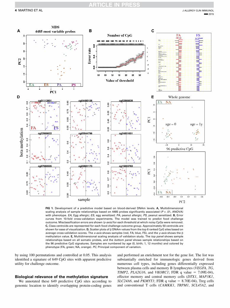
FIG 1. Development of a predictive model based on blood-derived DNAm levels. A, Multidimensional
scaling analysis of sample relationships based on 4485 probes significantly associated (P < .01, ANOVA)
with phenotype. EA, Egg allergic; ES, egg sensitized; PA, peanut allergic; PS, peanut sensitized. B, Errorcurves from 10-fold cross-validation experiments. The model was trained to predict food challenge
outcome. Misclassification errors are shown (y-axis) for each threshold at which noisy CpGs were removed.
C, Class centroids are represented for each food challenge outcome group. Approximately 50 centroids are
shown for ease of visualization. D, Scatter plots of b DNAm values from the top 6 ranked CpG sites based on
average cross-validation scores. The x-axis shows samples (red, FA; blue, FS), and the y-axis shows the b
methylation value. E, Multidimensional scaling analysis of validation study. The top panel shows sample
relationships based on all somatic probes, and the bottom panel shows sample relationships based on
the 96 predictive CpG signatures. Samples are numbered by age (0, birth; 1, 12 months) and colored by
phenotype (FA, green; NA, orange). PC, Principal component of variation.
J ALLERGY CLIN IMMUNOL
nnn 2015
4 MARTINO ET AL
by using 100 permutations and controlled at 0.05. This analysisidentified a signature of 649 CpG sites with apparent predictiveutility for challenge outcome.
Biological relevance of the methylation signatureWe annotated these 649 predictive CpG sites according to
genomic location to identify overlapping protein-coding genes
and performed an enrichment test for the gene list. The list wassubstantially enriched for immunologic genes derived fromnumerous cell types, including genes differentially expressedbetween plasma cells and memory B lymphocytes (VEGFA, TG,TIMP2, PLA2G16, and VKORC1; FDR q value 5 7.09E-04),effector memory and central memory cells (DTX1, MAP3K1,SLC24A6, and PKMYT1; FDR q value 5 8.70E-04), Treg cellsand conventional T cells (CAMKK1, TRPM1, SCL45A2, and
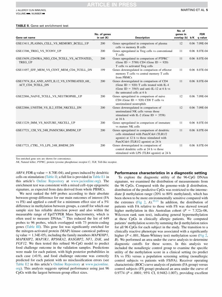
TABLE II. Gene set enrichment test
Gene set name
No. of genes
in set (K) Description
No. of
genes in
overlap (k) k/K
FDR
q value
GSE13411_PLASMA_CELL_VS_MEMORY_BCELL_UP 200 Genes upregulated in comparison of plasma
cells vs memory B cells
12 0.06 7.09E-04
GSE13306_TREG_VS_TCONV_UP 200 Genes upregulated in Treg cells vs conventional
T cells
11 0.06 8.07E-04
GSE15659_CD45RA_NEG_CD4_TCELL_VS_ACTIVATED_
TREG_UP
200 Genes upregulated in comparison of PTPRC2
(Gene ID 5 5788) CD4 (Gene ID 5 920)
T cells vs activated Treg cells
11 0.06 8.07E-04
GSE11057_EFF_MEM_VS_CENT_MEM_CD4_TCELL_DN 199 Genes downregulated in comparison of effector
memory T cells vs central memory T cells
from PBMCs
11 0.06 8.07E-04
GSE17974_IL4_AND_ANTI_IL12_VS_UNTREATED_6H_
ACT_CD4_TCELL_DN
200 Genes downregulated in comparison of CD4
(Gene ID 5 920) T cells treated with IL-4
(Gene ID 5 3565) and anti–IL-12 at 6 h vs
the untreated cells at 6 h
11 0.06 8.07E-04
GSE22886_NAIVE_TCELL_VS_NEUTROPHIL_UP 200 Genes upregulated in comparison of naive
CD4 (Gene ID 5 920) CD8 T cells vs
unstimulated neutrophils
12 0.06 7.09E-04
GSE22886_UNSTIM_VS_IL2_STIM_NKCELL_DN 200 Genes downregulated in comparison of
unstimulated NK cells versus those
stimulated with IL-2 (Gene ID 5 3558)
at 16 h
12 0.06 7.09E-04
GSE13229_IMM_VS_MATURE_NKCELL_UP 200 Genes upregulated in comparison of immature
vs mature NK cells
11 0.06 8.07E-04
GSE17721_12H_VS_24H_PAM3CSK4_BMDM_UP 200 Genes upregulated in comparison of dendritic
cells stimulated with Pam3Csk4 (TLR1/2
agonist) at 12 h vs those stimulated with
Pam3Csk4 (TLR1/2 agonist) at 24 h
11 0.06 8.07E-04
GSE17721_CTRL_VS_LPS_24H_BMDM_DN 200 Genes downregulated in comparison of
control dendritic cells at 24 h vs those
stimulated with LPS (TLR4 agonist) at 24 h
11 0.06 8.07E-04
Ten enriched gene sets are shown for convenience.
NK, Natural killer; PTPRC, protein tyrosine phosphotase receptor C; TLR, Toll-like receptor.
J ALLERGY CLIN IMMUNOL
VOLUME nnn, NUMBER nn
MARTINO ET AL 5
ARF4; FDR q-value5 8.70E-04), and genes induced by dendriticcells on stimulation (Table II; a full list is provided in Table E1 inthis article’s Online Repository at www.jacionline.org). Theenrichment test was consistent with a mixed cell–type epigeneticsignature, as expected from data derived from whole PBMCs.
We next ranked the 649 probes according to their absolutebetween-group difference for our main outcomes of interest (FAvs FS) and applied a cutoff for a minimum effect size of a 5%difference in methylation between groups, a cutoff for which oursample size has reliable detection power and also within themeasurable range of EpiTYPER Mass Spectrometry, which isoften used to measure DNAm.19 This reduced the list of 649probes to 96 probes, which overlapped with 73 protein-codinggenes (Table III). This gene list was significantly enriched forthe mitogen-activated protein (MAP) kinase canonical pathway(q value5 1.34E-05), including the family membersMAPK8IP1,MAPK8IP2, MAP3K1, RPS6KA2, RASGRP2, NF1, ZAK, andFGF12. We then tested this refined 96-CpG model to predictfood challenge outcome in the validation samples. Predictionswere made for each patient, with high posterior probabilities ineach case (>0.9), and food challenge outcome was correctlypredicted for each patient with no misclassification errors (seeTable E2 in this article’s Online Repository at www.jacionline.org). This analysis suggests optimal performance using just 96CpGs with the largest between-group effect sizes.
Performance characteristics in a diagnostic settingTo explore the diagnostic utility of the 96-CpG DNAm
signature, we examined the distribution of measurements fromthe 96 CpGs. Compared with the genome-wide b distribution,distribution of the predictive CpGs was restricted to the interme-diate b methylation range (20% to 80% methylated), which hasbeen shown to be more environmentally sensitive compared withthe extremes (Fig 2, A).20,21 In addition, the distribution ofpatients with FA relative to those with FS was skewed towardhigher methylation in this Australian cohort (P 5 7.322e-06,Wilcoxon rank sum test), indicating general hypermethylationat these CpGs in clinically allergic patients. We computedpatients’ methylation scores by summing the bmethylation ratiosfor all 96 CpGs for each subject in the study. The transition to aclinically reactive phenotype was associated with a significantlyhigher (P < .001, Mann-Whitney test) methylation score (Fig 2,B). We performed an area under the curve analysis to determinediagnostic cutoffs for these scores. In this analysis weincluded the nonallergic control group to examine the specificutility of the methylation score in a clinical setting (to predictFA vs FS) versus a population screening setting (nonallergiccontrol subjects vs patients with FS/FA). Receiver operatingcharacteristic (ROC) curve analysis of patients (FA group) versuscontrol subjects (FS group) produced an area under the curve of0.9774 (P < .0001; 95% CI, 0.9482-1.007), providing excellent
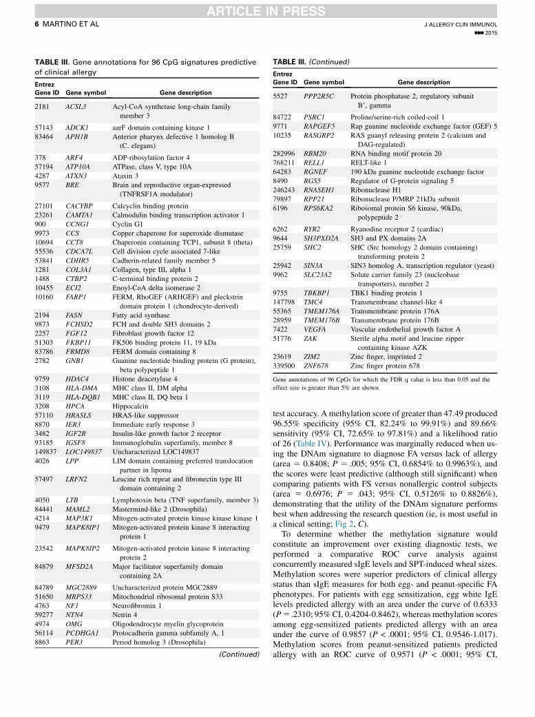
TABLE III. Gene annotations for 96 CpG signatures predictive
of clinical allergy
Entrez
Gene ID Gene symbol Gene description
2181 ACSL3 Acyl-CoA synthetase long-chain family
member 3
57143 ADCK1 aarF domain containing kinase 1
83464 APH1B Anterior pharynx defective 1 homolog B
(C. elegans)
378 ARF4 ADP-ribosylation factor 4
57194 ATP10A ATPase, class V, type 10A
4287 ATXN3 Ataxin 3
9577 BRE Brain and reproductive organ-expressed
(TNFRSF1A modulator)
27101 CACYBP Calcyclin binding protein
23261 CAMTA1 Calmodulin binding transcription activator 1
900 CCNG1 Cyclin G1
9973 CCS Copper chaperone for superoxide dismutase
10694 CCT8 Chaperonin containing TCP1, subunit 8 (theta)
55536 CDCA7L Cell division cycle associated 7-like
53841 CDHR5 Cadherin-related family member 5
1281 COL3A1 Collagen, type III, alpha 1
1488 CTBP2 C-terminal binding protein 2
10455 ECI2 Enoyl-CoA delta isomerase 2
10160 FARP1 FERM, RhoGEF (ARHGEF) and pleckstrin
domain protein 1 (chondrocyte-derived)
2194 FASN Fatty acid synthase
9873 FCHSD2 FCH and double SH3 domains 2
2257 FGF12 Fibroblast growth factor 12
51303 FKBP11 FK506 binding protein 11, 19 kDa
83786 FRMD8 FERM domain containing 8
2782 GNB1 Guanine nucleotide binding protein (G protein),
beta polypeptide 1
9759 HDAC4 Histone deacetylase 4
3108 HLA-DMA MHC class II, DM alpha
3119 HLA-DQB1 MHC class II, DQ beta 1
3208 HPCA Hippocalcin
57110 HRASLS HRAS-like suppressor
8870 IER3 Immediate early response 3
3482 IGF2R Insulin-like growth factor 2 receptor
93185 IGSF8 Immunoglobulin superfamily, member 8
149837 LOC149837 Uncharacterized LOC149837
4026 LPP LIM domain containing preferred translocation
partner in lipoma
57497 LRFN2 Leucine rich repeat and fibronectin type III
domain containing 2
4050 LTB Lymphotoxin beta (TNF superfamily, member 3)
84441 MAML2 Mastermind-like 2 (Drosophila)
4214 MAP3K1 Mitogen-activated protein kinase kinase kinase 1
9479 MAPK8IP1 Mitogen-activated protein kinase 8 interacting
protein 1
23542 MAPK8IP2 Mitogen-activated protein kinase 8 interacting
protein 2
84879 MFSD2A Major facilitator superfamily domain
containing 2A
84789 MGC2889 Uncharacterized protein MGC2889
51650 MRPS33 Mitochondrial ribosomal protein S33
4763 NF1 Neurofibromin 1
59277 NTN4 Netrin 4
4974 OMG Oligodendrocyte myelin glycoprotein
56114 PCDHGA1 Protocadherin gamma subfamily A, 1
8863 PER3 Period homolog 3 (Drosophila)
(Continued)
TABLE III. (Continued)
Entrez
Gene ID Gene symbol Gene description
5527 PPP2R5C Protein phosphatase 2, regulatory subunit
B9, gamma
84722 PSRC1 Proline/serine-rich coiled-coil 1
9771 RAPGEF5 Rap guanine nucleotide exchange factor (GEF) 5
10235 RASGRP2 RAS guanyl releasing protein 2 (calcium and
DAG-regulated)
282996 RBM20 RNA binding motif protein 20
768211 RELL1 RELT-like 1
64283 RGNEF 190 kDa guanine nucleotide exchange factor
8490 RGS5 Regulator of G-protein signaling 5
246243 RNASEH1 Ribonuclease H1
79897 RPP21 Ribonuclease P/MRP 21kDa subunit
6196 RPS6KA2 Ribosomal protein S6 kinase, 90kDa,
polypeptide 2
6262 RYR2 Ryanodine receptor 2 (cardiac)
9644 SH3PXD2A SH3 and PX domains 2A
25759 SHC2 SHC (Src homology 2 domain containing)
transforming protein 2
25942 SIN3A SIN3 homolog A, transcription regulator (yeast)
9962 SLC23A2 Solute carrier family 23 (nucleobase
transporters), member 2
9755 TBKBP1 TBK1 binding protein 1
147798 TMC4 Transmembrane channel-like 4
55365 TMEM176A Transmembrane protein 176A
28959 TMEM176B Transmembrane protein 176B
7422 VEGFA Vascular endothelial growth factor A
51776 ZAK Sterile alpha motif and leucine zipper
containing kinase AZK
23619 ZIM2 Zinc finger, imprinted 2
339500 ZNF678 Zinc finger protein 678
Gene annotations of 96 CpGs for which the FDR q value is less than 0.05 and the
effect size is greater than 5% are shown.
J ALLERGY CLIN IMMUNOL
nnn 2015
6 MARTINO ET AL
test accuracy. A methylation score of greater than 47.49 produced96.55% specificity (95% CI, 82.24% to 99.91%) and 89.66%sensitivity (95% CI, 72.65% to 97.81%) and a likelihood ratioof 26 (Table IV). Performance was marginally reduced when us-ing the DNAm signature to diagnose FA versus lack of allergy(area 5 0.8408; P 5 .005; 95% CI, 0.6854% to 0.9963%), andthe scores were least predictive (although still significant) whencomparing patients with FS versus nonallergic control subjects(area 5 0.6976; P 5 .043; 95% CI, 0.5126% to 0.8826%),demonstrating that the utility of the DNAm signature performsbest when addressing the research question (ie, is most useful ina clinical setting; Fig 2, C).
To determine whether the methylation signature wouldconstitute an improvement over existing diagnostic tests, weperformed a comparative ROC curve analysis againstconcurrently measured sIgE levels and SPT-induced wheal sizes.Methylation scores were superior predictors of clinical allergystatus than sIgE measures for both egg- and peanut-specific FAphenotypes. For patients with egg sensitization, egg white IgElevels predicted allergy with an area under the curve of 0.6333(P5 .2310; 95% CI, 0.4204-0.8462), whereas methylation scoresamong egg-sensitized patients predicted allergy with an areaunder the curve of 0.9857 (P < .0001; 95% CI, 0.9546-1.017).Methylation scores from peanut-sensitized patients predictedallergy with an ROC curve of 0.9571 (P < .0001; 95% CI,

FIG 2. Sensitivity and specificity analysis of DNAm patient scores. A, Distribution of methylation ratios for
the 96 CpG signatures stratified by phenotype. B, Box plots of total DNAm scores showing medians and
ranges (statistical analysis using the Man-Whitney test). C, ROC curve analysis of DNAm scores for
predicting clinical allergy between groups. D, Performance comparisons of DNAm scores against serum
IgE measures. E, Performance comparisons of DNAm scores against egg wheal size among
egg-sensitized patients. F, Performance comparisons of DNAm scores against peanut SPT wheal sizes
among peanut-sensitized patients.
J ALLERGY CLIN IMMUNOL
VOLUME nnn, NUMBER nn
MARTINO ET AL 7

TABLE IV. Sensitivity and specificity analysis of methylation scores from DNAm signature
Cutoff Sensitivity (%) 95% CI Specificity (%) 95% CI Likelihood ratio
>46.71 100 88.06% to 100.0% 72.41 52.76% to 87.27% 3.625
>46.86 96.55 82.24% to 99.91% 75.86 56.46% to 89.70% 4
>47.03 93.1 77.23% to 99.15% 79.31 60.28% to 92.01% 4.5
>47.12 93.1 77.23% to 99.15% 86.21 68.34% to 96.11% 6.75
>47.34 89.66 72.65% to 97.81% 89.66 72.65% to 97.81% 8.667
>47.49 89.66 72.65% to 97.81% 96.55 82.24% to 99.91% 26
>47.66 82.76 64.23% to 94.15% 96.55 82.24% to 99.91% 24
>47.75 82.76 64.23% to 94.15% 100 88.06% to 100.0%
FIG 3. Starburst plot of patient methylation scores for the discovery (left) and replication (right) cohorts.Concentric numerals denote sample numbers, and patients’ scores are shown on the vertical axis in
boldface. Methylation scores derived from CD41 T cells (red) were consistently lower than scores derived
from total PBMCS (blue); however, differences between phenotype classes were conserved in each cohort.
Solid circles are visual guides for diagnostic cutoffs determined by means of sensitivity analysis. Note that
y-axis scales differ in the 2 plots.
J ALLERGY CLIN IMMUNOL
nnn 2015
8 MARTINO ET AL
0.8897-1.025) compared with a peanut IgE test ROC curve of0.8722 (P 5 .00108; 95% CI, 0.7422-1.022; Fig 2, D).
We have also compared performance characteristics of themethylation test against SPTs for prediction of clinical allergy, andthe methylation test was superior to both egg and peanutSPT-induced wheal size (Fig 2, E and F). For patients with eggsensitization, egg SPTwheal size predicted allergywith an area un-der the curve of 0.56 (P5.6123; 95%CI, 0.3308-0.7951), whereasmethylation scores among egg-sensitized patients predictedallergy with an area under the curve of 0.9405 (P < .0018; 95%CI, 0.8386-1.042). Methylation scores from peanut-sensitizedpatients predicted allergy with an ROC curve of 0.9571(P < .0001; 95% CI, 0.8897-1.025) compared with the peanutIgE test ROC curve of 0.9780 (P 5 .0005; 95% CI, 0.9228-1.033). These results also held truewhen the analysiswas restrictedto patients with a wheal size of 3 mm or greater (data not shown).
Assessment of reproducibility in an independent
cohortWe next assessed independent reproducibility in a publicly
available DNAm data set obtained from a West Australian cohort
of patients with FA and nonallergic control subjects previouslygenerated by our laboratory.7 This data set comprised 48 samplesin total, 12 FA and 12 nonallergic control samples, assayed at 2time points (birth and 12 months). FA status was determinedwhen infants were 12 months old based on prior evidence ofclinical reactivity in case histories and objective SPT responses.DNAm data were collected at birth and 12 months from totalCD41 T cells, as published previously.7 We applied multidimen-sional scaling analysis to assess sample relationships based on ourpredictive methylation signature. Multidimensional scalinganalysis of all probes or the top 1000 most variable probes didnot discriminate the FA group from the nonallergic control groupin this data set, suggesting the principal components of variationare driven by unknown effects. However, when this analysis wasrestricted to the 96 predictive CpG methylation signatures, thefirst principal component of variation between groups was dueto phenotype (Fig 1, E). We generated methylation scores foreach sample in this cohort using the 96 predictive CpGs andexamined the distribution of scores between the West Australianand Melbourne cohorts. It was clear that methylation scoresderived from total CD41 T cells were consistently lower thanscores derived from PBMCs; however, differences in methylation

FIG 4. Diagnosing FA with a combination of IgE and methylation testing.
The number of challenges required was determined by using 95% positive
predictive values (PPV) for skin tests (>2 mm and <4mm for egg and >2mm
and <8 mm for peanut) and sIgE tests (>0.35 kUA/L and <1.17 kUA/L for egg
and >0.35 kUA/L and <15 kUA/L for peanut). Methylation predictions for
challenge outcome were based on a cutoff of greater than 47.5 (95%
negative predictive value). Error rates are shown.
J ALLERGY CLIN IMMUNOL
VOLUME nnn, NUMBER nn
MARTINO ET AL 9
scores between phenotypes were still conserved (Fig 3). Thisdifference in methylation profiles because of cell type meantthat the diagnostic cutoffs determined for total PBMCs wouldneed to be recalibrated on total CD41 T cells to performpredictions. Performing ROC curve analysis on T cell–derivedmethylation scores produced an area under the curve of 0.8368(P < .001; 95% CI, 0.7139-0.9598). A sensitivity analysis sug-gested a score of greater than 45.52 resulted in a test sensitivityof 87.5% (95% CI, 67.64% to 97.34%) and a specificity of70.83% (95% CI, 48.91% to 87.38%), with a likelihood ratio of3.0. The difference in diagnostic cutoffs between the PBMC-derived and T cell–derived patients’ scores is shown in Fig 2.
We performed an unsupervised classification of samples in theT-cell data set by assigning patients with a methylation score ofgreater than 45.52 to the nonallergic group and compared theseassignments with the true phenotype. In total, 10 of 48 sampleswere misclassified, resulting in a true error rate of 20.8%.Furthermore, we show that the model can be retrained on theT-cell data and still yield accurate predictions.
We partitioned the T-cell data into a 20:80 split, reserving thebulk of samples for predictions. The model was retrained on 10samples (n 5 5 for the FA group and n 5 5 for the nonallergiccontrol group) by using all 96 predictive CpGs, and predictionswere successful for 34 of the 38 remaining samples (n5 19 for theFA group and n5 19 for the nonallergic control group). Posteriorprobabilities were high (>0.9) for correctly classified samples butranged from 0.4 to 0.6 for misclassified samples. Although thisreplication occurred in a supervised fashion, it demonstrates theflexibility of the model.
Potential diagnostic utility of combining SPTs,
specific IgE tests, and DNAm testsFinally, we explored how DNAm biomarkers might be
combined with existing tests to aid clinical diagnosis. Fig 4 showsthe number of OFCs in HealthNuts study subjects that would need
be undertaken to test for clinical reactivity based on a situation inwhich the current 95% positive predictive value thresholds forSPTs and sIgE tests were used in combination to define athreshold for such testing.22,23 Based on these criteria, an OFCwould be recommended in 16 children to unequivocallydetermine their clinical reactivity. The 95% positive predictivevalue methylation score for these 16 children correctly predictstheir challenge outcome (Fig 4), demonstrating utility in thesesubjects that would otherwise be difficult to classify by using acombination of SPT and sIgE results.
DISCUSSIONBiomarkers of FA are urgently needed to develop more
powerful diagnostic tools that identify bona fide FA against abackground of FS. Here we have developed a multivariateclassifier model with utility for predicting food challengeoutcomes. Different subtypes of FA exist, and we have shownthat pattern-finding computational methods perform extremelywell when prior knowledge of these subtypes are available. Thestrength of this approach is rooted in the definitive food challengeoutcome data, which are best used with supervised classificationmethods24 that can be used to build models to diagnose FA infuture patients. By selecting monosensitized patients, we knewa priori which patients belong to which subgroup. This isimportant because it facilitated selection of clinically significantCpGs before model building. Our model was then built on theassumption that different subgroups of FA are likely to shareoverlapping pathogenic pathways involved in atopy and clinicalallergy. Conceptually, this is not unreasonable, and we havedemonstrated that common methylation pathways can be usedto make accurate predictions of allergy status for different FAphenotypes. It is noteworthy that the sole canonical pathwayenriched in the list of genes associated with CpGs most predictiveof food challenge outcome is theMAP kinase pathway. This is thesecond FA cohort in which we have reported an associationbetween clinical FA and DNAm in MAP kinase signaling genes;the former was discovered in the West Australian cohort.7 Thestrength of this association at present is unclear because differentMAP kinase genes were implicated in each cohort, althoughcollectively, the data suggest a shared pathway. The upstreamregulators of this pathway are currently unknown but warrantfurther investigation.
The enrichment tests for the predictive CpG model built on theMelbourne cohort are consistent with amixed cell–type signature,as would be expected from total mononuclear cells. Thisrepresents proof of principle that peripheral blood potentiallyoffers a noninvasive, easily accessible, safe, and widelyapplicable diagnostic approach. The translational significanceof this is further demonstrated by the diagnostic utility ofcomposite methylation scores, which represent a substantialimprovement over current diagnostic tests. Replication of thefindings presented here will be important to validate the clinicaleffectiveness of DNAm diagnostics. A direct comparison of theapproach described here with emerging component-resolveddiagnostic tests, such as Ara h 2, will be important to identifyan approach with the most clinical utility. This will also allow aninvestigation of whether a combined approach, using both DNAmand other measures, is of even greater clinical utility.
There are a growing number of multivariate diagnostic assays(‘‘gene signatures’’) marketed as diagnostic/prognostic tests.25
J ALLERGY CLIN IMMUNOL
nnn 2015
10 MARTINO ET AL
The reproducible accuracy of patient classification demonstratedin the replication cohort was particularly encouraging. In total,79.2% of samples were correctly diagnosed in an unsupervisedmanner by using the 96-CpG predictive model, despite thesemethylation data being derived from a different cell type, and inthe absence of the ‘‘sensitized but tolerant’’ group, which isexpected to produce the most accurate classification (ROC: FSvs FA 5 0.9774 compared with ROC: FA vs nonallergiccontrol 5 0.8404). Therefore we anticipate better accuracy willbe obtained when the test is applied in the intended clinicalsetting. Collectively, our data demonstrate reproducibility despitechallenges of differ cell types and population.
Although our approach is highly novel, with tremendouspotential for development into a clinically relevant diagnostictest, it has several limitations that require substantial additionalinvestigation. For example, it is not currently clear how applicableour methylation signature might be as a prognostic marker of FAresolution (acquired tolerance). Furthermore, the signaturereported here is likely to require refinement after furtherinvestigation in larger sample sizes.
Other caveats include the cross-sectional analysis and therestricted scope of FA phenotypes investigated here. The datapresented here pertain to age-matched, single-sensitized patients,and further testing is required in larger data sets of different agedchildren with multiple FSs. Furthermore, larger cohorts ofsingle-sensitized subjects will be better suited to address anymechanistic differences between egg and peanut FA.
Biomarkers of clinical allergy that supplement existing patientmanagement strategies will help avoid unnecessary foodchallenges in food-sensitized subjects. This could reduce waitingtimes in the immediate term and substantially alleviate the currentburden on the public health system, at least in part. We believefurther development of the approaches demonstrated here canlead to powerful new tools for diagnosing and treating allergy andother immune-related diseases.
We thank all the parents and children who participated in the study.
Additional HealthNuts study investigators include Melanie Matheson,
Melissa Wake, Anne-Adrian Lowe, David Hill, and Rachel Peters.
Clinical implications: Through validation in 2 separate studypopulations, this article provides proof of principle thatDNAm biomarkers can be used for novel diagnostic testing inpatients with FA.
REFERENCES
1. Osborne NJ, Koplin JJ, Martin PE, Gurrin LC, Lowe AJ, Matheson MC, et al.
Prevalence of challenge-proven IgE-mediated food allergy using population-
based sampling and predetermined challenge criteria in infants. J Allergy Clin
Immunol 2011;127:668-76, e1-2.
2. Prescott S, Allen KJ. Food allergy: riding the second wave of the allergy epidemic.
Pediatr Allergy Immunol 2011;22:155-60.
3. Ito K. Diagnosis of food allergies: the impact of oral food challenge testing. Asia
Pac Allergy 2013;3:59-69.
4. Santos AF, Lack G. Food allergy and anaphylaxis in pediatrics: update 2010-2012.
Pediatr Allergy Immunol 2012;23:698-706.
5. Rakyan VK, Down TA, Balding DJ, Beck S. Epigenome-wide association studies
for common human diseases. Nat Rev Genet 2011;12:529-41.
6. Martino D, Kesper DA, Amarasekera M, Harb H, Renz H, Prescott S. Epigenetics
in immune development and in allergic and autoimmune diseases. J Reprod
Immunol 2014;104-105:43-8.
7. Martino D, Joo JE, Sexton-Oates A, Dang T, Allen K, Saffery R, et al.
Epigenome-wide association study reveals longitudinally stable DNA methylation
differences in CD41 T cells from children with IgE-mediated food allergy.
Epigenetics 2014;9:998-1006.
8. Peters RL, Dharmage SC, Gurrin LC, Koplin JJ, Ponsonby AL, Lowe AJ, et al. The
natural history and clinical predictors of egg allergy in the first 2 years of life: a
prospective, population-based cohort study. J Allergy Clin Immunol 2014;133:
485-91.
9. Ho MH, Wong WH, Heine RG, Hosking CS, Hill DJ, Allen KJ. Early clinical
predictors of remission of peanut allergy in children. J Allergy Clin Immunol
2008;121:731-6.
10. Pumphrey RS, Gowland MH. Further fatal allergic reactions to food in the United
Kingdom, 1999-2006. J Allergy Clin Immunol 2007;119:1018-9.
11. Osborne NJ, Koplin JJ, Martin PE, Gurrin LC, Thiele L, Tang ML, et al. The
HealthNuts population-based study of paediatric food allergy: validity, safety
and acceptability. Clin Exp Allergy 2010;40:1516-22.
12. Koplin JJ, Tang ML, Martin PE, Osborne NJ, Lowe AJ, Ponsonby AL, et al.
Predetermined challenge eligibility and cessation criteria for oral food challenges
in the HealthNuts population-based study of infants. J Allergy Clin Immunol 2012;
129:1145-7.
13. Martino D, Loke YJ, Gordon L, Ollikainen M, Cruickshank MN, Saffery R, et al.
Longitudinal, genome-scale analysis of DNA methylation in twins from birth to 18
months of age reveals rapid epigenetic change in early life and pair-specific effects
of discordance. Genome Biol 2013;14:R42.
14. Aryee MJ, Jaffe AE, Corrada-Bravo H, Ladd-Acosta C, Feinberg AP, Hansen
KD, et al. Minfi: a flexible and comprehensive Bioconductor package for the
analysis of Infinium DNA Methylation microarrays. Bioinformatics 2014;30:
1363-9.
15. Chen YA, Lemire M, Choufani S, Butcher DT, Grafodatskaya D, Zanke BW,
et al. Discovery of cross-reactive probes and polymorphic CpGs in the
Illumina Infinium HumanMethylation450 microarray. Epigenetics 2013;8:
203-9.
16. Tibshirani R, Hastie T, Narasimhan B, Chu G. Diagnosis of multiple cancer types
by shrunken centroids of gene expression. Proc Natl Acad Sci U S A 2002;99:
6567-72.
17. Storey JD, Tibshirani R. Statistical significance for genomewide studies. Proc Natl
Acad Sci U S A 2003;100:9440-5.
18. Subramanian A, Tamayo P, Mootha VK, Mukherjee S, Ebert BL, Gillette MA,
et al. Gene set enrichment analysis: a knowledge-based approach for
interpreting genome-wide expression profiles. Proc Natl Acad Sci U S A 2005;
102:15545-50.
19. Coolen MW, Statham AL, Gardiner-Garden M, Clark SJ. Genomic profiling of
CpG methylation and allelic specificity using quantitative high-throughput mass
spectrometry: critical evaluation and improvements. Nucleic Acids Res 2007;35:
e119.
20. Novakovic B, Yuen RK, Gordon L, Penaherrera MS, Sharkey A, Moffett A, et al.
Evidence for widespread changes in promoter methylation profile in human
placenta in response to increasing gestational age and environmental/stochastic
factors. BMC Genomics 2011;12:529.
21. Oh JE, Chambwe N, Klein S, Gal J, Andrews S, Gleason G, et al. Differential
gene body methylation and reduced expression of cell adhesion and
neurotransmitter receptor genes in adverse maternal environment. Transl
Psychiatry 2013;3:e218.
22. Peters RL, Allen KJ, Dharmage SC, Tang ML, Koplin JJ, Ponsonby AL, et al. Skin
prick test responses and allergen-specific IgE levels as predictors of peanut, egg,
and sesame allergy in infants. J Allergy Clin Immunol 2013;132:874-80.
23. Dang TD, Tang M, Choo S, Licciardi PV, Koplin JJ, Martin PE, et al. Increasing
the accuracy of peanut allergy diagnosis by using Ara h 2. J Allergy Clin Immunol
2012;129:1056-63.
24. Bair E, Tibshirani R. Semi-supervised methods to predict patient survival from
gene expression data. PLoS Biol 2004;2:E108.
25. McCarthy JJ, McLeod HL, Ginsburg GS. Genomic medicine: a decade of
successes, challenges, and opportunities. Sci Transl Med 2013;5:189sr4.
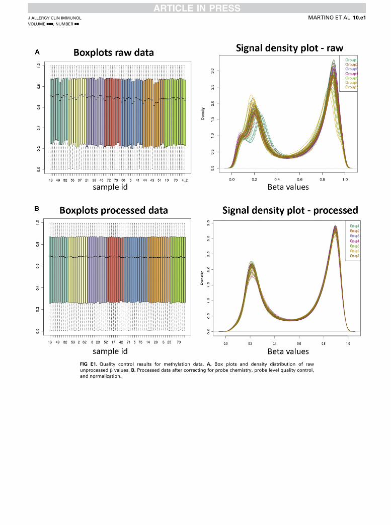
FIG E1. Quality control results for methylation data. A, Box plots and density distribution of raw
unprocessed b values. B, Processed data after correcting for probe chemistry, probe level quality control,
and normalization.
J ALLERGY CLIN IMMUNOL
VOLUME nnn, NUMBER nn
MARTINO ET AL 10.e1

FIG E2. Assessment of confounding because of cellular heterogeneity of PBMC samples. A, Correlation
between flow cytometry–measured total CD41 counts and in silico–estimated CD41 counts using cell
type–predictive CpGs determined by using the method of Houseman. Correlations between gold standard
and data-driven methods were highly comparable. B, Between-group comparisons of cell composition in
PBMC fractions. By using in silico estimates for blood cell subsets, there was no evidence of differential
methylation between primary outcome groups because of cell type proportion. C, Cell counts for each
subtype are shown. Statistical analysis was done by using the Mann-Whitney U test.
J ALLERGY CLIN IMMUNOL
nnn 2015
10.e2 MARTINO ET AL

TABLE E1. Gene set enrichment results
Gene symbol Gene description Gene set name Gene set description
VEGFA Vascular endothelial growth factor A GSE13411_PLASMA_CELL_VS_MEMORY_BCELL_UP Genes upregulated in comparison of plasma cells vs memory B cells
TG Thyroglobulin GSE13411_PLASMA_CELL_VS_MEMORY_BCELL_UP Genes upregulated in comparison of plasma cells vs memory B cells
TIMP2 TIMP metallopeptidase inhibitor 2 GSE13411_PLASMA_CELL_VS_MEMORY_BCELL_UP Genes upregulated in comparison of plasma cells vs memory B cells
PLA2G16 Phospholipase A2, group XVI GSE13411_PLASMA_CELL_VS_MEMORY_BCELL_UP Genes upregulated in comparison of plasma cells vs memory B cells
COL3A1 Collagen, type III, alpha 1 GSE13411_PLASMA_CELL_VS_MEMORY_BCELL_UP Genes upregulated in comparison of plasma cells vs memory B cells
VKORC1 Vitamin K epoxide reductase complex, subunit 1 GSE13411_PLASMA_CELL_VS_MEMORY_BCELL_UP Genes upregulated in comparison of plasma cells vs memory B cells
LMF1 Lipase maturation factor 1 GSE13411_PLASMA_CELL_VS_MEMORY_BCELL_UP Genes upregulated in comparison of plasma cells vs memory B cells
ELL2 Elongation factor, RNA polymerase II, 2 GSE13411_PLASMA_CELL_VS_MEMORY_BCELL_UP Genes upregulated in comparison of plasma cells vs memory B cells
PSRC1 Proline/serine-rich coiled-coil 1 GSE13411_PLASMA_CELL_VS_MEMORY_BCELL_UP Genes upregulated in comparison of plasma cells vs memory B cells
HIST1H2BI Histone cluster 1, H2bi GSE13411_PLASMA_CELL_VS_MEMORY_BCELL_UP Genes upregulated in comparison of plasma cells vs memory B cells
HIST1H2AI Histone cluster 1, H2ai GSE13411_PLASMA_CELL_VS_MEMORY_BCELL_UP Genes upregulated in comparison of plasma cells vs memory B cells
TRIM29 Tripartite motif containing 29 GSE13411_PLASMA_CELL_VS_MEMORY_BCELL_UP Genes upregulated in comparison of plasma cells vs memory B cells
HLA-DMA MHC, class II, DM alpha GSE17721_CPG_VS_GARDIQUIMOD_16H_BMDM_UP Genes upregulated in comparison of DCs stimulated with CpG DNA
(TLR9 agonist) at 16 h vs DCs stimulated with Gardiquimod
(TLR7 agonist) at 16 h
SMARCE1 SWI/SNF related, matrix associated, actin
dependent regulator of chromatin, subfamily
e, member 1
GSE17721_CPG_VS_GARDIQUIMOD_16H_BMDM_UP Genes upregulated in comparison of DCs stimulated with CpG DNA
(TLR9 agonist) at 16 h vs DCs stimulated with Gardiquimod
(TLR7 agonist) at 16 h
CD209 CD209 molecule GSE17721_CPG_VS_GARDIQUIMOD_16H_BMDM_UP Genes upregulated in comparison of DCs stimulated with CpG DNA
(TLR9 agonist) at 16 h vs DCs stimulated with Gardiquimod
(TLR7 agonist) at 16 h
PER3 Period homolog 3 (Drosophila) GSE17721_CPG_VS_GARDIQUIMOD_16H_BMDM_UP Genes upregulated in comparison of DCs stimulated with CpG DNA
(TLR9 agonist) at 16 h vs DCs stimulated with Gardiquimod
(TLR7 agonist) at 16 h
HIST3H2A Histone cluster 3, H2a GSE17721_CPG_VS_GARDIQUIMOD_16H_BMDM_UP Genes upregulated in comparison of DCs stimulated with CpG DNA
(TLR9 agonist) at 16 h vs DCs stimulated with Gardiquimod
(TLR7 agonist) at 16 h
ANGPT1 Angiopoietin 1 GSE17721_CPG_VS_GARDIQUIMOD_16H_BMDM_UP Genes upregulated in comparison of DCs stimulated with CpG DNA
(TLR9 agonist) at 16 h vs DCs stimulated with Gardiquimod
(TLR7 agonist) at 16 h
DHX16 DEAH (Asp-Glu-Ala-His) box polypeptide 16 GSE17721_CPG_VS_GARDIQUIMOD_16H_BMDM_UP Genes upregulated in comparison of DCs stimulated with CpG DNA
(TLR9 agonist) at 16 h vs DCs stimulated with Gardiquimod
(TLR7 agonist) at 16 h
FAM50B Family with sequence similarity 50, member B GSE17721_CPG_VS_GARDIQUIMOD_16H_BMDM_UP Genes upregulated in comparison of DCs stimulated with CpG DNA
(TLR9 agonist) at 16 h vs DCs stimulated with Gardiquimod
(TLR7 agonist) at 16 h
DCLK1 Doublecortin-like kinase 1 GSE17721_CPG_VS_GARDIQUIMOD_16H_BMDM_UP Genes upregulated in comparison of DCs stimulated with CpG DNA
(TLR9 agonist) at 16 h vs DCs stimulated with Gardiquimod
(TLR7 agonist) at 16 h
ATP11A ATPase, class VI, type 11A GSE17721_CPG_VS_GARDIQUIMOD_16H_BMDM_UP Genes upregulated in comparison of DCs stimulated with CpG DNA
(TLR9 agonist) at 16 h vs DCs stimulated with Gardiquimod
(TLR7 agonist) at 16 h
(Continued)
JALLERGYCLIN
IMMUNOL
VOLUMEnnn,NUMBERnn
MARTIN
OETAL
10.e3
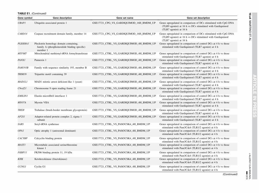
TABLE E1. (Continued)
Gene symbol Gene description Gene set name Gene set description
UBAP1 Ubiquitin associated protein 1 GSE17721_CPG_VS_GARDIQUIMOD_16H_BMDM_UP Genes upregulated in comparison of DCs stimulated with CpG DNA
(TLR9 agonist) at 16 h vs DCs stimulated with Gardiquimod
(TLR7 agonist) at 16 h
CARD14 Caspase recruitment domain family, member 14 GSE17721_CPG_VS_GARDIQUIMOD_16H_BMDM_UP Genes upregulated in comparison of DCs stimulated with CpG DNA
(TLR9 agonist) at 16 h vs DCs stimulated with Gardiquimod
(TLR7 agonist) at 16 h
PLEKHA1 Pleckstrin homology domain containing,
family A (phosphoinositide binding specific)
member 1
GSE17721_CTRL_VS_GARDIQUIMOD_4H_BMDM_UP Genes upregulated in comparison of control DCs at 4 h vs those
stimulated with Gardiquimod (TLR7 agonist) at 4 h
MTFMT Mitochondrial methionyl-tRNA formyltransferase GSE17721_CTRL_VS_GARDIQUIMOD_4H_BMDM_UP Genes upregulated in comparison of control DCs at 4 h vs those
stimulated with Gardiquimod (TLR7 agonist) at 4 h
PANX1 Pannexin 1 GSE17721_CTRL_VS_GARDIQUIMOD_4H_BMDM_UP Genes upregulated in comparison of control DCs at 4 h vs those
stimulated with Gardiquimod (TLR7 agonist) at 4 h
FAM193B Family with sequence similarity 193, member B GSE17721_CTRL_VS_GARDIQUIMOD_4H_BMDM_UP Genes upregulated in comparison of control DCs at 4 h vs those
stimulated with Gardiquimod (TLR7 agonist) at 4 h
TRIM39 Tripartite motif containing 39 GSE17721_CTRL_VS_GARDIQUIMOD_4H_BMDM_UP Genes upregulated in comparison of control DCs at 4 h vs those
stimulated with Gardiquimod (TLR7 agonist) at 4 h
MAD1L1 MAD1 mitotic arrest deficient-like 1 (yeast) GSE17721_CTRL_VS_GARDIQUIMOD_4H_BMDM_UP Genes upregulated in comparison of control DCs at 4 h vs those
stimulated with Gardiquimod (TLR7 agonist) at 4 h
C9orf21 Chromosome 9 open reading frame 21 GSE17721_CTRL_VS_GARDIQUIMOD_4H_BMDM_UP Genes upregulated in comparison of control DCs at 4 h vs those
stimulated with Gardiquimod (TLR7 agonist) at 4 h
EMILIN1 Elastin microfibril interfacer 1 GSE17721_CTRL_VS_GARDIQUIMOD_4H_BMDM_UP Genes upregulated in comparison of control DCs at 4 h vs those
stimulated with Gardiquimod (TLR7 agonist) at 4 h
MYO7A Myosin VIIA GSE17721_CTRL_VS_GARDIQUIMOD_4H_BMDM_UP Genes upregulated in comparison of control DCs at 4 h vs those
stimulated with Gardiquimod (TLR7 agonist) at 4 h
TREH Trehalase (brush-border membrane glycoprotein) GSE17721_CTRL_VS_GARDIQUIMOD_4H_BMDM_UP Genes upregulated in comparison of control DCs at 4 h vs those
stimulated with Gardiquimod (TLR7 agonist) at 4 h
AP2S1 Adaptor-related protein complex 2, sigma 1
subunit
GSE17721_CTRL_VS_GARDIQUIMOD_4H_BMDM_UP Genes upregulated in comparison of control DCs at 4 h vs those
stimulated with Gardiquimod (TLR7 agonist) at 4 h
SARS Seryl-tRNA synthetase GSE17721_CTRL_VS_PAM3CSK4_4H_BMDM_UP Genes upregulated in comparison of control DCs at 4 h vs those
stimulated with Pam3Csk4 (TLR1/2 agonist) at 4 h
OPA1 Optic atrophy 1 (autosomal dominant) GSE17721_CTRL_VS_PAM3CSK4_4H_BMDM_UP Genes upregulated in comparison of control DCs at 4 h vs those
stimulated with Pam3Csk4 (TLR1/2 agonist) at 4 h
CACYBP Calcyclin binding protein GSE17721_CTRL_VS_PAM3CSK4_4H_BMDM_UP Genes upregulated in comparison of control DCs at 4 h vs those
stimulated with Pam3Csk4 (TLR1/2 agonist) at 4 h
MAST1 Microtubule associated serine/threonine
kinase 1
GSE17721_CTRL_VS_PAM3CSK4_4H_BMDM_UP Genes upregulated in comparison of control DCs at 4 h vs those
stimulated with Pam3Csk4 (TLR1/2 agonist) at 4 h
FKBP11 FK506 binding protein 11, 19 kDa GSE17721_CTRL_VS_PAM3CSK4_4H_BMDM_UP Genes upregulated in comparison of control DCs at 4 h vs those
stimulated with Pam3Csk4 (TLR1/2 agonist) at 4 h
KHK Ketohexokinase (fructokinase) GSE17721_CTRL_VS_PAM3CSK4_4H_BMDM_UP Genes upregulated in comparison of control DCs at 4 h vs those
stimulated with Pam3Csk4 (TLR1/2 agonist) at 4 h
CCNG1 Cyclin G1 GSE17721_CTRL_VS_PAM3CSK4_4H_BMDM_UP Genes upregulated in comparison of control DCs at 4 h vs those
stimulated with Pam3Csk4 (TLR1/2 agonist) at 4 h
(Continued)
JALLERGYCLIN
IMMUNOL
nnn2015
10.e4
MARTIN
OETAL

TABLE E1. (Continued)
Gene symbol Gene description Gene set name Gene set description
FAM149B1 Family with sequence similarity 149, member B1 GSE17721_CTRL_VS_PAM3CSK4_4H_BMDM_UP Genes upregulated in comparison of control DCs at 4 h vs those
stimulated with Pam3Csk4 (TLR1/2 agonist) at 4 h
KIAA1967 KIAA1967 GSE17721_CTRL_VS_PAM3CSK4_4H_BMDM_UP Genes upregulated in comparison of control DCs at 4 h vs those
stimulated with Pam3Csk4 (TLR1/2 agonist) at 4 h
SLC23A2 Solute carrier family 23 (nucleobase transporters),
member 2
GSE17721_POLYIC_VS_PAM3CSK4_1H_BMDM_DN Genes downregulated in comparison of DCs stimulated with
poly(I:C) (TLR3 agonist) at 1 h vs DCs stimulated with
Pam3Csk4 (TLR1/2 agonist) at 1 h
RPS6KA2 Ribosomal protein S6 kinase, 90kDa, polypeptide 2 GSE17721_POLYIC_VS_PAM3CSK4_1H_BMDM_DN Genes downregulated in comparison of DCs stimulated with
poly(I:C) (TLR3 agonist) at 1 h vs DCs stimulated with
Pam3Csk4 (TLR1/2 agonist) at 1 h
RELL1 RELT-like 1 GSE17721_POLYIC_VS_PAM3CSK4_1H_BMDM_DN Genes downregulated in comparison of DCs stimulated with
poly(I:C) (TLR3 agonist) at 1 h vs DCs stimulated with
Pam3Csk4 (TLR1/2 agonist) at 1 h
RYR1 Ryanodine receptor 1 (skeletal) GSE17721_POLYIC_VS_PAM3CSK4_1H_BMDM_DN Genes downregulated in comparison of DCs stimulated with
poly(I:C) (TLR3 agonist) at 1 h vs DCs stimulated with
Pam3Csk4 (TLR1/2 agonist) at 1 h
FHOD3 Formin homology 2 domain containing 3 GSE17721_POLYIC_VS_PAM3CSK4_1H_BMDM_DN Genes downregulated in comparison of DCs stimulated with
poly(I:C) (TLR3 agonist) at 1 h vs DCs stimulated with
Pam3Csk4 (TLR1/2 agonist) at 1 h
PRIM2 Primase, DNA, polypeptide 2 (58kDa) GSE17721_POLYIC_VS_PAM3CSK4_1H_BMDM_DN Genes downregulated in comparison of DCs stimulated with
poly(I:C) (TLR3 agonist) at 1 h vs DCs stimulated with
Pam3Csk4 (TLR1/2 agonist) at 1 h
RAP1B RAP1B, member of RAS oncogene family GSE17721_POLYIC_VS_PAM3CSK4_1H_BMDM_DN Genes downregulated in comparison of DCs stimulated with
poly(I:C) (TLR3 agonist) at 1 h vs DCs stimulated with
Pam3Csk4 (TLR1/2 agonist) at 1 h
USP13 Ubiquitin specific peptidase 13 (isopeptidase T-3) GSE17721_POLYIC_VS_PAM3CSK4_1H_BMDM_DN Genes downregulated in comparison of DCs stimulated with
poly(I:C) (TLR3 agonist) at 1 h vs DCs stimulated with
Pam3Csk4 (TLR1/2 agonist) at 1 h
TFF1 Trefoil factor 1 GSE17721_POLYIC_VS_PAM3CSK4_1H_BMDM_DN Genes downregulated in comparison of DCs stimulated with
poly(I:C) (TLR3 agonist) at 1 h vs DCs stimulated with
Pam3Csk4 (TLR1/2 agonist) at 1 h
RTBDN Retbindin GSE17721_POLYIC_VS_PAM3CSK4_1H_BMDM_DN Genes downregulated in comparison of DCs stimulated with
poly(I:C) (TLR3 agonist) at 1 h vs DCs stimulated with
Pam3Csk4 (TLR1/2 agonist) at 1 h
INTS12 Integrator complex subunit 12 GSE17721_POLYIC_VS_PAM3CSK4_1H_BMDM_DN Genes downregulated in comparison of DCs stimulated with
poly(I:C) (TLR3 agonist) at 1 h vs DCs stimulated with
Pam3Csk4 (TLR1/2 agonist) at 1 h
DDX39A DEAD (Asp-Glu-Ala-Asp) box polypeptide 39A GSE22886_NAIVE_TCELL_VS_NEUTROPHIL_UP Genes upregulated in comparison of naive CD4 (Gene ID 5 920)
CD8 T cells vs unstimulated neutrophils
CCT8 Chaperonin containing TCP1, subunit 8 (theta) GSE22886_NAIVE_TCELL_VS_NEUTROPHIL_UP Genes upregulated in comparison of naive CD4 (Gene ID 5 920)
CD8 T cells vs unstimulated neutrophils
CD7 CD7 molecule GSE22886_NAIVE_TCELL_VS_NEUTROPHIL_UP Genes upregulated in comparison of naive CD4 (Gene ID 5 920)
CD8 T cells vs unstimulated neutrophils
(Continued)
JALLERGYCLIN
IMMUNOL
VOLUMEnnn,NUMBERnn
MARTIN
OETAL
10.e5

TABLE E1. (Continued)
Gene symbol Gene description Gene set name Gene set description
NCL Nucleolin GSE22886_NAIVE_TCELL_VS_NEUTROPHIL_UP Genes upregulated in comparison of naive CD4 (Gene ID 5 920)
CD8 T cells vs unstimulated neutrophils
C7orf23 Chromosome 7 open reading frame 23 GSE22886_NAIVE_TCELL_VS_NEUTROPHIL_UP Genes upregulated in comparison of naive CD4 (Gene ID 5 920)
CD8 T cells vs unstimulated neutrophils
BCKDHA Branched chain keto acid dehydrogenase E1,
alpha polypeptide
GSE22886_NAIVE_TCELL_VS_NEUTROPHIL_UP Genes upregulated in comparison of naive CD4 (Gene ID 5 920)
CD8 T cells vs unstimulated neutrophils
RCN2 Reticulocalbin 2, EF-hand calcium binding
domain
GSE22886_NAIVE_TCELL_VS_NEUTROPHIL_UP Genes upregulated in comparison of naive CD4 (Gene ID 5 920)
CD8 T cells vs unstimulated neutrophils
EIF2AK1 Eukaryotic translation initiation factor 2-alpha
kinase 1
GSE22886_NAIVE_TCELL_VS_NEUTROPHIL_UP Genes upregulated in comparison of naive CD4 (Gene ID 5 920)
CD8 T cells vs unstimulated neutrophils
GMPS Guanine monophosphate synthetase GSE22886_NAIVE_TCELL_VS_NEUTROPHIL_UP Genes upregulated in comparison of naive CD4 (Gene ID 5 920)
CD8 T cells vs unstimulated neutrophils
ATP5D ATP synthase, H1 transporting, mitochondrial
F1 complex, delta subunit
GSE22886_NAIVE_TCELL_VS_NEUTROPHIL_UP Genes upregulated in comparison of naive CD4 (Gene ID 5 920)
CD8 T cells vs unstimulated neutrophils
ATP1A1 ATPase, Na1/K1 transporting, alpha 1
polypeptide
GSE22886_UNSTIM_VS_IL2_STIM_NKCELL_DN Genes downregulated in comparison of unstimulated NK cells vs
those stimulated with IL-2 (Gene ID 5 3558) at 16 h
LTB Lymphotoxin beta (TNF superfamily, member 3) GSE22886_UNSTIM_VS_IL2_STIM_NKCELL_DN Genes downregulated in comparison of unstimulated NK cells vs
those stimulated with IL-2 (Gene ID 5 3558) at 16 h
GEM GTP binding protein overexpressed in skeletal
muscle
GSE22886_UNSTIM_VS_IL2_STIM_NKCELL_DN Genes downregulated in comparison of unstimulated NK cells vs
those stimulated with IL-2 (Gene ID 5 3558) at 16 h
WDR12 WD repeat domain 12 GSE22886_UNSTIM_VS_IL2_STIM_NKCELL_DN Genes downregulated in comparison of unstimulated NK cells vs
those stimulated with IL-2 (Gene ID 5 3558) at 16 h
HNRNPC Heterogeneous nuclear ribonucleoprotein C
(C1/C2)
GSE22886_UNSTIM_VS_IL2_STIM_NKCELL_DN Genes downregulated in comparison of unstimulated NK cells vs
those stimulated with IL-2 (Gene ID 5 3558) at 16 h
PTPN7 Protein tyrosine phosphatase, non-receptor type 7 GSE22886_UNSTIM_VS_IL2_STIM_NKCELL_DN Genes downregulated in comparison of unstimulated NK cells vs
those stimulated with IL-2 (Gene ID 5 3558) at 16 h
FASN Fatty acid synthase GSE22886_UNSTIM_VS_IL2_STIM_NKCELL_DN Genes downregulated in comparison of unstimulated NK cells vs
those stimulated with IL-2 (Gene ID 5 3558) at 16 h
ACACA Acetyl-CoA carboxylase alpha GSE22886_UNSTIM_VS_IL2_STIM_NKCELL_DN Genes downregulated in comparison of unstimulated NK cells vs
those stimulated with IL-2 (Gene ID 5 3558) at 16 h
PAICS Phosphoribosylaminoimidazole carboxylase,
phosphoribosylaminoimidazole
succinocarboxamide synthetase
GSE22886_UNSTIM_VS_IL2_STIM_NKCELL_DN Genes downregulated in comparison of unstimulated NK cells vs
those stimulated with IL-2 (Gene ID 5 3558) at 16 h
NAA16 N(alpha)-acetyltransferase 16, NatA auxiliary
subunit
GSE26669_CD4_VS_CD8_TCELL_IN_MLR_COSTIM_
BLOCK_UP
Genes upregulated in comparison of untreated CD4 (Gene ID 5 920)
T cells vs CD8 T cells treated with leukocyte costimulatory
blockade antibodies
CERS4 Ceramide synthase 4 GSE26669_CD4_VS_CD8_TCELL_IN_MLR_COSTIM_
BLOCK_UP
Genes upregulated in comparison of untreated CD4 (Gene ID 5 920)
T cells vs CD8 T cells treated with leukocyte costimulatory
blockade antibodies
BRE Brain and reproductive organ-expressed
(TNFRSF1A modulator)
GSE26669_CD4_VS_CD8_TCELL_IN_MLR_COSTIM_
BLOCK_UP
Genes upregulated in comparison of untreated CD4 (Gene ID 5 920)
T cells vs CD8 T cells treated with leukocyte
costimulatory blockade antibodies
(Continued)
JALLERGYCLIN
IMMUNOL
nnn2015
10.e6
MARTIN
OETAL
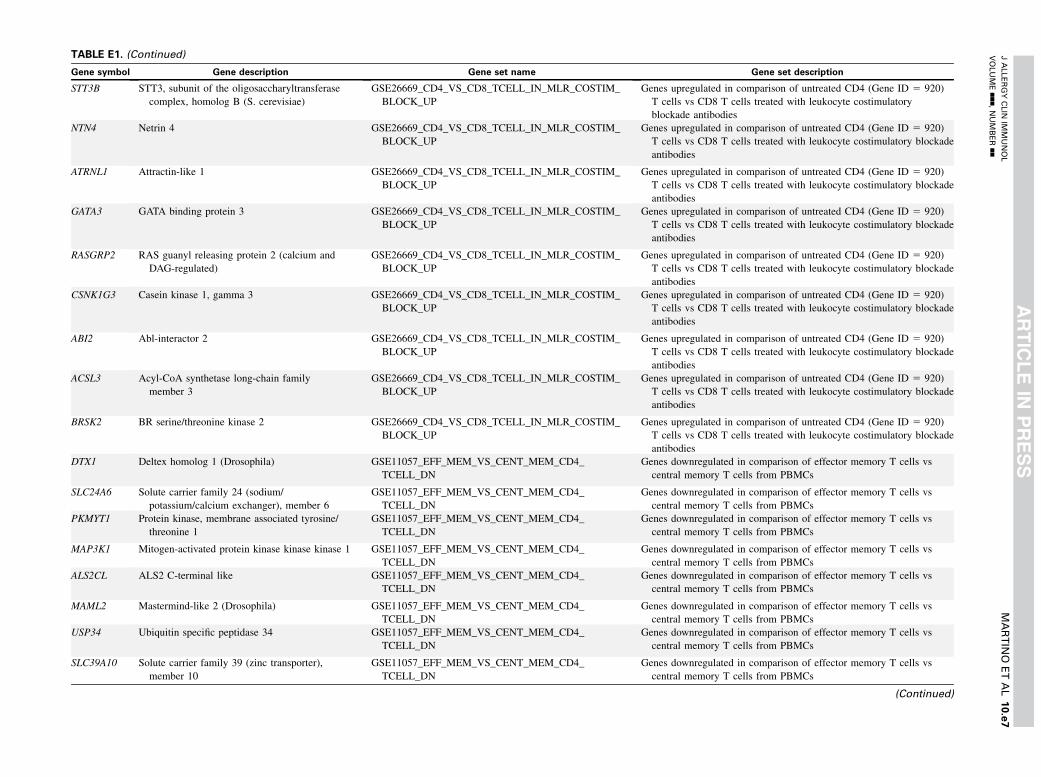
TABLE E1. (Continued)
Gene symbol Gene description Gene set name Gene set description
STT3B STT3, subunit of the oligosaccharyltransferase
complex, homolog B (S. cerevisiae)
GSE26669_CD4_VS_CD8_TCELL_IN_MLR_COSTIM_
BLOCK_UP
Genes upregulated in comparison of untreated CD4 (Gene ID 5 920)
T cells vs CD8 T cells treated with leukocyte costimulatory
blockade antibodies
NTN4 Netrin 4 GSE26669_CD4_VS_CD8_TCELL_IN_MLR_COSTIM_
BLOCK_UP
Genes upregulated in comparison of untreated CD4 (Gene ID 5 920)
T cells vs CD8 T cells treated with leukocyte costimulatory blockade
antibodies
ATRNL1 Attractin-like 1 GSE26669_CD4_VS_CD8_TCELL_IN_MLR_COSTIM_
BLOCK_UP
Genes upregulated in comparison of untreated CD4 (Gene ID 5 920)
T cells vs CD8 T cells treated with leukocyte costimulatory blockade
antibodies
GATA3 GATA binding protein 3 GSE26669_CD4_VS_CD8_TCELL_IN_MLR_COSTIM_
BLOCK_UP
Genes upregulated in comparison of untreated CD4 (Gene ID 5 920)
T cells vs CD8 T cells treated with leukocyte costimulatory blockade
antibodies
RASGRP2 RAS guanyl releasing protein 2 (calcium and
DAG-regulated)
GSE26669_CD4_VS_CD8_TCELL_IN_MLR_COSTIM_
BLOCK_UP
Genes upregulated in comparison of untreated CD4 (Gene ID 5 920)
T cells vs CD8 T cells treated with leukocyte costimulatory blockade
antibodies
CSNK1G3 Casein kinase 1, gamma 3 GSE26669_CD4_VS_CD8_TCELL_IN_MLR_COSTIM_
BLOCK_UP
Genes upregulated in comparison of untreated CD4 (Gene ID 5 920)
T cells vs CD8 T cells treated with leukocyte costimulatory blockade
antibodies
ABI2 Abl-interactor 2 GSE26669_CD4_VS_CD8_TCELL_IN_MLR_COSTIM_
BLOCK_UP
Genes upregulated in comparison of untreated CD4 (Gene ID 5 920)
T cells vs CD8 T cells treated with leukocyte costimulatory blockade
antibodies
ACSL3 Acyl-CoA synthetase long-chain family
member 3
GSE26669_CD4_VS_CD8_TCELL_IN_MLR_COSTIM_
BLOCK_UP
Genes upregulated in comparison of untreated CD4 (Gene ID 5 920)
T cells vs CD8 T cells treated with leukocyte costimulatory blockade
antibodies
BRSK2 BR serine/threonine kinase 2 GSE26669_CD4_VS_CD8_TCELL_IN_MLR_COSTIM_
BLOCK_UP
Genes upregulated in comparison of untreated CD4 (Gene ID 5 920)
T cells vs CD8 T cells treated with leukocyte costimulatory blockade
antibodies
DTX1 Deltex homolog 1 (Drosophila) GSE11057_EFF_MEM_VS_CENT_MEM_CD4_
TCELL_DN
Genes downregulated in comparison of effector memory T cells vs
central memory T cells from PBMCs
SLC24A6 Solute carrier family 24 (sodium/
potassium/calcium exchanger), member 6
GSE11057_EFF_MEM_VS_CENT_MEM_CD4_
TCELL_DN
Genes downregulated in comparison of effector memory T cells vs
central memory T cells from PBMCs
PKMYT1 Protein kinase, membrane associated tyrosine/
threonine 1
GSE11057_EFF_MEM_VS_CENT_MEM_CD4_
TCELL_DN
Genes downregulated in comparison of effector memory T cells vs
central memory T cells from PBMCs
MAP3K1 Mitogen-activated protein kinase kinase kinase 1 GSE11057_EFF_MEM_VS_CENT_MEM_CD4_
TCELL_DN
Genes downregulated in comparison of effector memory T cells vs
central memory T cells from PBMCs
ALS2CL ALS2 C-terminal like GSE11057_EFF_MEM_VS_CENT_MEM_CD4_
TCELL_DN
Genes downregulated in comparison of effector memory T cells vs
central memory T cells from PBMCs
MAML2 Mastermind-like 2 (Drosophila) GSE11057_EFF_MEM_VS_CENT_MEM_CD4_
TCELL_DN
Genes downregulated in comparison of effector memory T cells vs
central memory T cells from PBMCs
USP34 Ubiquitin specific peptidase 34 GSE11057_EFF_MEM_VS_CENT_MEM_CD4_
TCELL_DN
Genes downregulated in comparison of effector memory T cells vs
central memory T cells from PBMCs
SLC39A10 Solute carrier family 39 (zinc transporter),
member 10
GSE11057_EFF_MEM_VS_CENT_MEM_CD4_
TCELL_DN
Genes downregulated in comparison of effector memory T cells vs
central memory T cells from PBMCs
(Continued)
JALLERGYCLIN
IMMUNOL
VOLUMEnnn,NUMBERnn
MARTIN
OETAL
10.e7
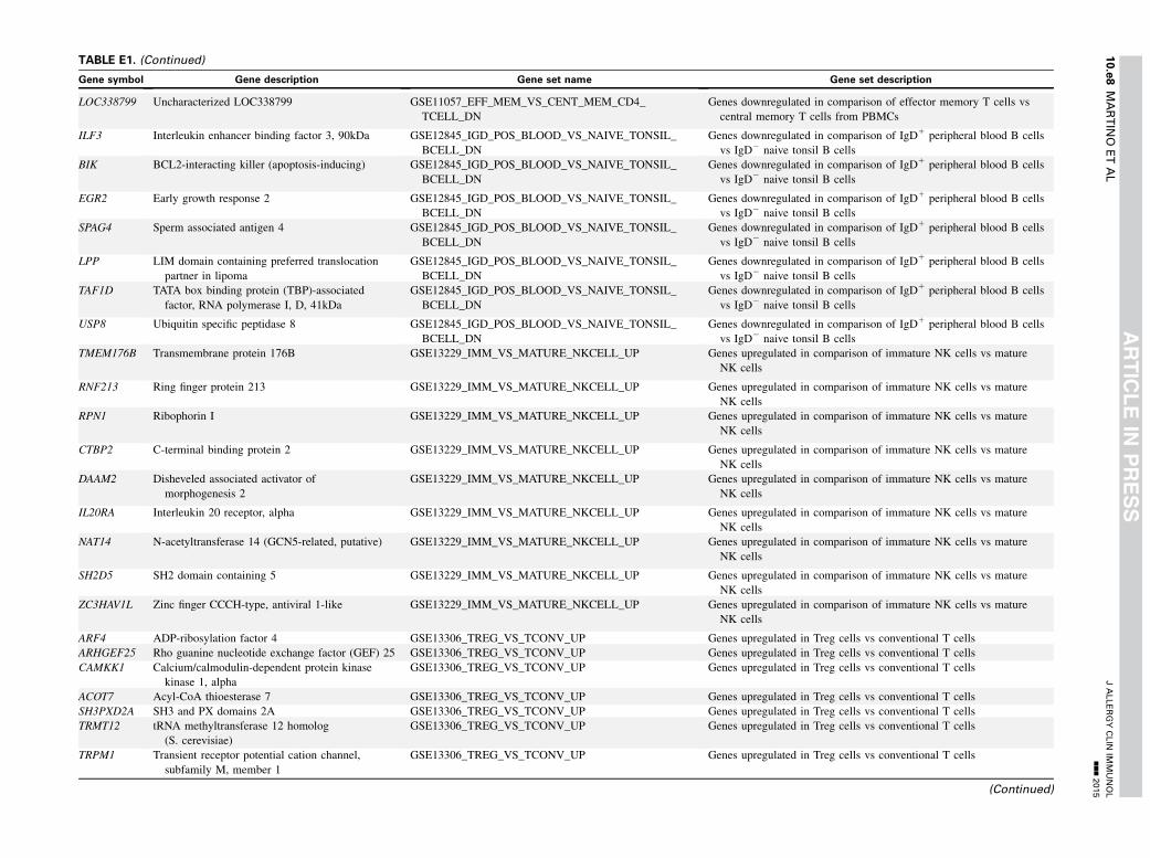
TABLE E1. (Continued)
Gene symbol Gene description Gene set name Gene set description
LOC338799 Uncharacterized LOC338799 GSE11057_EFF_MEM_VS_CENT_MEM_CD4_
TCELL_DN
Genes downregulated in comparison of effector memory T cells vs
central memory T cells from PBMCs
ILF3 Interleukin enhancer binding factor 3, 90kDa GSE12845_IGD_POS_BLOOD_VS_NAIVE_TONSIL_
BCELL_DN
Genes downregulated in comparison of IgD1 peripheral blood B cells
vs IgD2 naive tonsil B cells
BIK BCL2-interacting killer (apoptosis-inducing) GSE12845_IGD_POS_BLOOD_VS_NAIVE_TONSIL_
BCELL_DN
Genes downregulated in comparison of IgD1 peripheral blood B cells
vs IgD2 naive tonsil B cells
EGR2 Early growth response 2 GSE12845_IGD_POS_BLOOD_VS_NAIVE_TONSIL_
BCELL_DN
Genes downregulated in comparison of IgD1 peripheral blood B cells
vs IgD2 naive tonsil B cells
SPAG4 Sperm associated antigen 4 GSE12845_IGD_POS_BLOOD_VS_NAIVE_TONSIL_
BCELL_DN
Genes downregulated in comparison of IgD1 peripheral blood B cells
vs IgD2 naive tonsil B cells
LPP LIM domain containing preferred translocation
partner in lipoma
GSE12845_IGD_POS_BLOOD_VS_NAIVE_TONSIL_
BCELL_DN
Genes downregulated in comparison of IgD1 peripheral blood B cells
vs IgD2 naive tonsil B cells
TAF1D TATA box binding protein (TBP)-associated
factor, RNA polymerase I, D, 41kDa
GSE12845_IGD_POS_BLOOD_VS_NAIVE_TONSIL_
BCELL_DN
Genes downregulated in comparison of IgD1 peripheral blood B cells
vs IgD2 naive tonsil B cells
USP8 Ubiquitin specific peptidase 8 GSE12845_IGD_POS_BLOOD_VS_NAIVE_TONSIL_
BCELL_DN
Genes downregulated in comparison of IgD1 peripheral blood B cells
vs IgD2 naive tonsil B cells
TMEM176B Transmembrane protein 176B GSE13229_IMM_VS_MATURE_NKCELL_UP Genes upregulated in comparison of immature NK cells vs mature
NK cells
RNF213 Ring finger protein 213 GSE13229_IMM_VS_MATURE_NKCELL_UP Genes upregulated in comparison of immature NK cells vs mature
NK cells
RPN1 Ribophorin I GSE13229_IMM_VS_MATURE_NKCELL_UP Genes upregulated in comparison of immature NK cells vs mature
NK cells
CTBP2 C-terminal binding protein 2 GSE13229_IMM_VS_MATURE_NKCELL_UP Genes upregulated in comparison of immature NK cells vs mature
NK cells
DAAM2 Disheveled associated activator of
morphogenesis 2
GSE13229_IMM_VS_MATURE_NKCELL_UP Genes upregulated in comparison of immature NK cells vs mature
NK cells
IL20RA Interleukin 20 receptor, alpha GSE13229_IMM_VS_MATURE_NKCELL_UP Genes upregulated in comparison of immature NK cells vs mature
NK cells
NAT14 N-acetyltransferase 14 (GCN5-related, putative) GSE13229_IMM_VS_MATURE_NKCELL_UP Genes upregulated in comparison of immature NK cells vs mature
NK cells
SH2D5 SH2 domain containing 5 GSE13229_IMM_VS_MATURE_NKCELL_UP Genes upregulated in comparison of immature NK cells vs mature
NK cells
ZC3HAV1L Zinc finger CCCH-type, antiviral 1-like GSE13229_IMM_VS_MATURE_NKCELL_UP Genes upregulated in comparison of immature NK cells vs mature
NK cells
ARF4 ADP-ribosylation factor 4 GSE13306_TREG_VS_TCONV_UP Genes upregulated in Treg cells vs conventional T cells
ARHGEF25 Rho guanine nucleotide exchange factor (GEF) 25 GSE13306_TREG_VS_TCONV_UP Genes upregulated in Treg cells vs conventional T cells
CAMKK1 Calcium/calmodulin-dependent protein kinase
kinase 1, alpha
GSE13306_TREG_VS_TCONV_UP Genes upregulated in Treg cells vs conventional T cells
ACOT7 Acyl-CoA thioesterase 7 GSE13306_TREG_VS_TCONV_UP Genes upregulated in Treg cells vs conventional T cells
SH3PXD2A SH3 and PX domains 2A GSE13306_TREG_VS_TCONV_UP Genes upregulated in Treg cells vs conventional T cells
TRMT12 tRNA methyltransferase 12 homolog
(S. cerevisiae)
GSE13306_TREG_VS_TCONV_UP Genes upregulated in Treg cells vs conventional T cells
TRPM1 Transient receptor potential cation channel,
subfamily M, member 1
GSE13306_TREG_VS_TCONV_UP Genes upregulated in Treg cells vs conventional T cells
(Continued)
JALLERGYCLIN
IMMUNOL
nnn2015
10.e8
MARTIN
OETAL
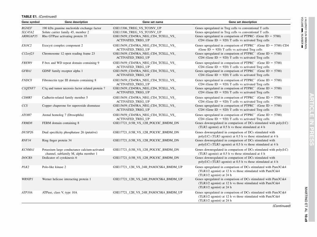
TABLE E1. (Continued)
Gene symbol Gene description Gene set name Gene set description
RGNEF 190 kDa guanine nucleotide exchange factor GSE13306_TREG_VS_TCONV_UP Genes upregulated in Treg cells vs conventional T cells
SLC45A2 Solute carrier family 45, member 2 GSE13306_TREG_VS_TCONV_UP Genes upregulated in Treg cells vs conventional T cells
ARHGAP35 Rho GTPase activating protein 35 GSE15659_CD45RA_NEG_CD4_TCELL_VS_
ACTIVATED_TREG_UP
Genes upregulated in comparison of PTPRC- (Gene ID 5 5788)
CD4 (Gene ID 5 920) T cells vs activated Treg cells
EXOC2 Exocyst complex component 2 GSE15659_CD45RA_NEG_CD4_TCELL_VS_
ACTIVATED_TREG_UP
Genes upregulated in comparison of PTPRC2 (Gene ID 5 5788) CD4
(Gene ID 5 920) T cells vs activated Treg cells
C12orf23 Chromosome 12 open reading frame 23 GSE15659_CD45RA_NEG_CD4_TCELL_VS_
ACTIVATED_TREG_UP
Genes upregulated in comparison of PTPRC2 (Gene ID 5 5788)
CD4 (Gene ID 5 920) T cells vs activated Treg cells
FBXW9 F-box and WD repeat domain containing 9 GSE15659_CD45RA_NEG_CD4_TCELL_VS_
ACTIVATED_TREG_UP
Genes upregulated in comparison of PTPRC2 (Gene ID 5 5788)
CD4 (Gene ID 5 920) T cells vs activated Treg cells
GFRA1 GDNF family receptor alpha 1 GSE15659_CD45RA_NEG_CD4_TCELL_VS_
ACTIVATED_TREG_UP
Genes upregulated in comparison of PTPRC2 (Gene ID 5 5788)
CD4 (Gene ID 5 920) T cells vs activated Treg cells
FNDC8 Fibronectin type III domain containing 8 GSE15659_CD45RA_NEG_CD4_TCELL_VS_
ACTIVATED_TREG_UP
Genes upregulated in comparison of PTPRC2 (Gene ID 5 5788)
CD4 (Gene ID 5 920) T cells vs activated Treg cells
C1QTNF7 C1q and tumor necrosis factor related protein 7 GSE15659_CD45RA_NEG_CD4_TCELL_VS_
ACTIVATED_TREG_UP
Genes upregulated in comparison of PTPRC2 (Gene ID 5 5788)
CD4 (Gene ID 5 920) T cells vs activated Treg cells
CDHR5 Cadherin-related family member 5 GSE15659_CD45RA_NEG_CD4_TCELL_VS_
ACTIVATED_TREG_UP
Genes upregulated in comparison of PTPRC2 (Gene ID 5 5788)
CD4 (Gene ID 5 920) T cells vs activated Treg cells
CCS Copper chaperone for superoxide dismutase GSE15659_CD45RA_NEG_CD4_TCELL_VS_
ACTIVATED_TREG_UP
Genes upregulated in comparison of PTPRC2 (Gene ID 5 5788)
CD4 (Gene ID 5 920) T cells vs activated Treg cells
ATOH7 Atonal homolog 7 (Drosophila) GSE15659_CD45RA_NEG_CD4_TCELL_VS_
ACTIVATED_TREG_UP
Genes upregulated in comparison of PTPRC2 (Gene ID 5 5788)
CD4 (Gene ID 5 920) T cells vs activated Treg cells
FRMD8 FERM domain containing 8 GSE17721_0.5H_VS_12H_POLYIC_BMDM_DN Genes downregulated in comparison of DCs stimulated with poly(I:C)
(TLR3 agonist) at 0.5 h vs those stimulated at 4 h
DUSP26 Dual specificity phosphatase 26 (putative) GSE17721_0.5H_VS_12H_POLYIC_BMDM_DN Genes downregulated in comparison of DCs stimulated with
poly(I:C) (TLR3 agonist) at 0.5 h vs those stimulated at 4 h
RNF34 Ring finger protein 34 GSE17721_0.5H_VS_12H_POLYIC_BMDM_DN Genes downregulated in comparison of DCs stimulated with
poly(I:C) (TLR3 agonist) at 0.5 h vs those stimulated at 4 h
KCNMA1 Potassium large conductance calcium-activated
channel, subfamily M, alpha member 1
GSE17721_0.5H_VS_12H_POLYIC_BMDM_DN Genes downregulated in comparison of DCs stimulated with poly(I:C)
(TLR3 agonist) at 0.5 h vs those stimulated at 4 h
DOCK6 Dedicator of cytokinesis 6 GSE17721_0.5H_VS_12H_POLYIC_BMDM_DN Genes downregulated in comparison of DCs stimulated with
poly(I:C) (TLR3 agonist) at 0.5 h vs those stimulated at 4 h
PLK2 Polo-like kinase 2 GSE17721_12H_VS_24H_PAM3CSK4_BMDM_UP Genes upregulated in comparison of DCs stimulated with Pam3Csk4
(TLR1/2 agonist) at 12 h vs those stimulated with Pam3Csk4
(TLR1/2 agonist) at 24 h
WRNIP1 Werner helicase interacting protein 1 GSE17721_12H_VS_24H_PAM3CSK4_BMDM_UP Genes upregulated in comparison of DCs stimulated with Pam3Csk4
(TLR1/2 agonist) at 12 h vs those stimulated with Pam3Csk4
(TLR1/2 agonist) at 24 h
ATP10A ATPase, class V, type 10A GSE17721_12H_VS_24H_PAM3CSK4_BMDM_UP Genes upregulated in comparison of DCs stimulated with Pam3Csk4
(TLR1/2 agonist) at 12 h vs those stimulated with Pam3Csk4
(TLR1/2 agonist) at 24 h
(Continued)
JALLERGYCLIN
IMMUNOL
VOLUMEnnn,NUMBERnn
MARTIN
OETAL
10.e9
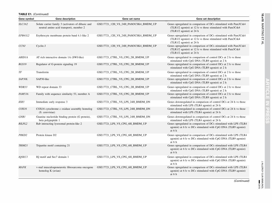
TABLE E1. (Continued)
Gene symbol Gene description Gene set name Gene set description
SLC3A2 Solute carrier family 3 (activators of dibasic and
neutral amino acid transport), member 2
GSE17721_12H_VS_24H_PAM3CSK4_BMDM_UP Genes upregulated in comparison of DCs stimulated with Pam3Csk4
(TLR1/2 agonist) at 12 h vs those stimulated with Pam3Csk4
(TLR1/2 agonist) at 24 h
EPB41L2 Erythrocyte membrane protein band 4.1-like 2 GSE17721_12H_VS_24H_PAM3CSK4_BMDM_UP Genes upregulated in comparison of DCs stimulated with Pam3Csk4
(TLR1/2 agonist) at 12 h vs those stimulated with Pam3Csk4
(TLR1/2 agonist) at 24 h
CCNJ Cyclin J GSE17721_12H_VS_24H_PAM3CSK4_BMDM_UP Genes upregulated in comparison of DCs stimulated with Pam3Csk4
(TLR1/2 agonist) at 12 h vs those stimulated with Pam3Csk4
(TLR1/2 agonist) at 24 h
ARID1A AT rich interactive domain 1A (SWI-like) GSE17721_CTRL_VS_CPG_2H_BMDM_UP Genes upregulated in comparison of control DCs at 2 h vs those
stimulated with CpG DNA (TLR9 agonist) at 2 h
RGS19 Regulator of G-protein signaling 19 GSE17721_CTRL_VS_CPG_2H_BMDM_UP Genes upregulated in comparison of control DCs at 2 h vs those
stimulated with CpG DNA (TLR9 agonist) at 2 h
TF Transferrin GSE17721_CTRL_VS_CPG_2H_BMDM_UP Genes upregulated in comparison of control DCs at 2 h vs those
stimulated with CpG DNA (TLR9 agonist) at 2 h
SAP30L SAP30-like GSE17721_CTRL_VS_CPG_2H_BMDM_UP Genes upregulated in comparison of control DCs at 2 h vs those
stimulated with CpG DNA (TLR9 agonist) at 2 h
WDR33 WD repeat domain 33 GSE17721_CTRL_VS_CPG_2H_BMDM_UP Genes upregulated in comparison of control DCs at 2 h vs those
stimulated with CpG DNA (TLR9 agonist) at 2 h
FAM53A Family with sequence similarity 53, member A GSE17721_CTRL_VS_CPG_2H_BMDM_UP Genes upregulated in comparison of control DCs at 2 h vs those
stimulated with CpG DNA (TLR9 agonist) at 2 h
IER3 Immediate early response 3 GSE17721_CTRL_VS_LPS_24H_BMDM_DN Genes downregulated in comparison of control DCs at 24 h vs those
stimulated with LPS (TLR4 agonist) at 24 h
COX16 COX16 cytochrome c oxidase assembly homolog
(S. cerevisiae)
GSE17721_CTRL_VS_LPS_24H_BMDM_DN Genes downregulated in comparison of control DCs at 24 h vs those
stimulated with LPS (TLR4 agonist) at 24 h
GNB1 Guanine nucleotide binding protein (G protein),
beta polypeptide 1
GSE17721_CTRL_VS_LPS_24H_BMDM_DN Genes downregulated in comparison of control DCs at 24 h vs those
stimulated with LPS (TLR4 agonist) at 24 h
RILPL2 Rab interacting lysosomal protein-like 2 GSE17721_LPS_VS_CPG_6H_BMDM_UP Genes upregulated in comparison of DCs stimulated with LPS (TLR4
agonist) at 6 h vs DCs stimulated with CpG DNA (TLR9 agonist)
at 6 h
PRKD2 Protein kinase D2 GSE17721_LPS_VS_CPG_6H_BMDM_UP Genes upregulated in comparison of DCs stimulated with LPS (TLR4
agonist) at 6 h vs DCs stimulated with CpG DNA (TLR9 agonist)
at 6 h
TRIM21 Tripartite motif containing 21 GSE17721_LPS_VS_CPG_6H_BMDM_UP Genes upregulated in comparison of DCs stimulated with LPS (TLR4
agonist) at 6 h vs DCs stimulated with CpG DNA (TLR9 agonist)
at 6 h
IQSEC1 IQ motif and Sec7 domain 1 GSE17721_LPS_VS_CPG_6H_BMDM_UP Genes upregulated in comparison of DCs stimulated with LPS (TLR4
agonist) at 6 h vs DCs stimulated with CpG DNA (TLR9 agonist)
at 6 h
MAFK v-maf musculoaponeurotic fibrosarcoma oncogene
homolog K (avian)
GSE17721_LPS_VS_CPG_6H_BMDM_UP Genes upregulated in comparison of DCs stimulated with LPS (TLR4
agonist) at 6 h vs DCs stimulated with CpG DNA (TLR9 agonist)
at 6 h
(Continued)
JALLERGYCLIN
IMMUNOL
nnn2015
10.e10
MARTIN
OETAL

TABLE E1. (Continued)
Gene symbol Gene description Gene set name Gene set description
TADA2A Transcriptional adaptor 2A GSE17974_IL4_AND_ANTI_IL12_VS_UNTREATED_6H_
ACT_CD4_TCELL_DN
Genes downregulated in comparison of CD4 (Gene ID 5 920) T cells
treated with IL-4 (Gene ID 5 3565) and anti–IL-12 at 6 h vs
untreated cells at 6 h
TOX2 TOX high mobility group box family member 2 GSE17974_IL4_AND_ANTI_IL12_VS_UNTREATED_6H_
ACT_CD4_TCELL_DN
Genes downregulated in comparison of CD4 (Gene ID 5 920) T cells
treated with IL-4 (Gene ID 5 3565) and anti–IL-12 at 6 h vs
untreated cells at 6 h
C2CD3 C2 calcium-dependent domain containing 3 GSE17974_IL4_AND_ANTI_IL12_VS_UNTREATED_6H_
ACT_CD4_TCELL_DN
Genes downregulated in comparison of CD4 (Gene ID 5 920) T cells
treated with IL-4 (Gene ID 5 3565) and anti–IL-12 at 6 h vs
untreated cells at 6 h
NEDD4 Neural precursor cell expressed, developmentally
down-regulated 4
GSE17974_IL4_AND_ANTI_IL12_VS_UNTREATED_6H_
ACT_CD4_TCELL_DN
Genes downregulated in comparison of CD4 (Gene ID 5 920) T cells
treated with IL-4 (Gene ID 5 3565) and anti–IL-12 at 6 h vs
untreated cells at 6 h
MAP3K13 Mitogen-activated protein kinase kinase kinase 13 GSE17974_IL4_AND_ANTI_IL12_VS_UNTREATED_6H_
ACT_CD4_TCELL_DN
Genes downregulated in comparison of CD4 (Gene ID 5 920) T cells
treated with IL-4 (Gene ID 5 3565) and anti–IL-12 at 6 h vs
untreated cells at 6 h
PDGFA Platelet-derived growth factor alpha polypeptide GSE17974_IL4_AND_ANTI_IL12_VS_UNTREATED_6H_
ACT_CD4_TCELL_DN
Genes downregulated in comparison of CD4 (Gene ID 5 920) T cells
treated with IL-4 (Gene ID 5 3565) and anti–IL-12 at 6 h vs
untreated cells at 6 h
COL6A3 Collagen, type VI, alpha 3 GSE17974_IL4_AND_ANTI_IL12_VS_UNTREATED_6H_
ACT_CD4_TCELL_DN
Genes downregulated in comparison of CD4 (Gene ID 5 920) T cells
treated with IL-4 (Gene ID 5 3565) and anti–IL-12 at 6 h vs
untreated cells at 6 h
KIAA1199 KIAA1199 GSE17974_IL4_AND_ANTI_IL12_VS_UNTREATED_6H_
ACT_CD4_TCELL_DN
Genes downregulated in comparison of CD4 (Gene ID 5 920) T cells
treated with IL-4 (Gene ID 5 3565) and anti–IL-12 at 6 h vs
untreated cells at 6 h
LRCH1 Leucine-rich repeats and calponin homology (CH)
domain containing 1
GSE17974_IL4_AND_ANTI_IL12_VS_UNTREATED_6H_
ACT_CD4_TCELL_DN
Genes downregulated in comparison of CD4 (Gene ID 5 920) T cells
treated with IL-4 (Gene ID 5 3565) and anti–IL-12 at 6 h vs
untreated cells at 6 h
TMEM154 Transmembrane protein 154 GSE20366_EX_VIVO_VS_DEC205_CONVERSION_
NAIVE_CD4_TCELL_UP
Genes upregulated in comparison of TconvLP vs DEC-Pept CD252
(see Table E1 for details)
ZBTB24 Zinc finger and BTB domain containing 24 GSE20366_EX_VIVO_VS_DEC205_CONVERSION_
NAIVE_CD4_TCELL_UP
Genes upregulated in comparison of TconvLP vs DEC-Pept CD252(see Table E1 for details)
CHST11 Carbohydrate (chondroitin 4) sulfotransferase 11 GSE20366_EX_VIVO_VS_DEC205_CONVERSION_
NAIVE_CD4_TCELL_UP
Genes upregulated in comparison of TconvLP vs DEC-Pept CD252(see Table E1 for details)
TMEM176A Transmembrane protein 176A GSE20366_EX_VIVO_VS_DEC205_CONVERSION_
NAIVE_CD4_TCELL_UP
Genes upregulated in comparison of TconvLP vs DEC-Pept CD252(see Table E1 for details)
DC, Dendritic cell; LPS, lipopolysaccharide; NK, natural killer; poly(I:C), polyinosinic-polycytidylic acid; PTPRC, protein tyrosine phosphotase receptor C; Tconv, conventional T; TLR, Toll-like receptor.
JALLERGYCLIN
IMMUNOL
VOLUMEnnn,NUMBERnn
MARTIN
OETAL
10.e11
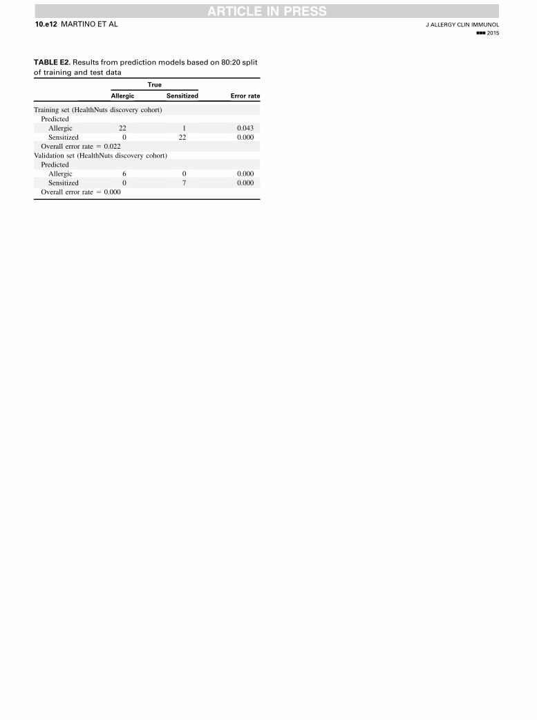
TABLE E2. Results from prediction models based on 80:20 split
of training and test data
True
Error rateAllergic Sensitized
Training set (HealthNuts discovery cohort)
Predicted
Allergic 22 1 0.043
Sensitized 0 22 0.000
Overall error rate 5 0.022
Validation set (HealthNuts discovery cohort)
Predicted
Allergic 6 0 0.000
Sensitized 0 7 0.000
Overall error rate 5 0.000
J ALLERGY CLIN IMMUNOL
nnn 2015
10.e12 MARTINO ET AL
Related Documents