ORIGINAL RESEARCH ARTICLE published: 24 July 2014 doi: 10.3389/fchem.2014.00054 Bis-indole derivatives with antitumor activity turn out to be specific ligands of human telomeric G-quadruplex Jussara Amato 1 , Nunzia Iaccarino 1 , Bruno Pagano 1 , Rita Morigi 2 , Alessandra Locatelli 2 , Alberto Leoni 2 , Mirella Rambaldi 2 , Pasquale Zizza 3 , Annamaria Biroccio 3 , Ettore Novellino 1 and Antonio Randazzo 1 * 1 Department of Pharmacy, University of Naples “Federico II”, Naples, Italy 2 Department of Pharmacy and Biotechnology (FaBiT), University of Bologna, Bologna, Italy 3 Experimental Chemotherapy Laboratory, Regina Elena National Cancer Institute, Rome, Italy Edited by: Asier Unciti-Broceta, The University of Edinburgh, UK Reviewed by: Marc Vendrell, University of Edinburgh, UK Juan Jose Diaz-Mochon, Universidad de Granada, Spain *Correspondence: Antonio Randazzo, Department of Pharmacy, University of Naples “Federico II,” Via D. Montesano 49, Napoli, 80131, Italy e-mail: [email protected] Bis-indolinone derivatives having either 2,6-disubstituted pyridine core (1a and 1b) or 1,10-disubstituted phenanthroline core (2a and 2b), already known to have antitumor activity, have been tested as potential G-quadruplex binders. Compounds 2a and 2b are able to selectively stabilize G-quadruplex over duplex DNA, and also to discriminate among different G-quadruplex structures, having a particular affinity for the parallel form of the human telomeric G-quadruplex. Both compounds are also able to induce telomeric DNA damage that may explain the activity of these compounds. Keywords: G-quadruplex, anticancer activity, telomeric damage, thermal stabilization, phenanthroline derivatives, pyridine derivatives INTRODUCTION G-quadruplexes (G4) are four-stranded nucleic acid structures that spontaneously form within G-rich sequences of DNA and RNA in the presence of cations (Bochman et al., 2012). The recent unambiguous evidence of G4 formation in living cells has increased the enthusiasm and has propelled numerous inves- tigations in this field (Biffi et al., 2013). Several experiments have located G4-forming sequences in different critical positions of the human genome, mainly at the telomeric and gene pro- moter level (Bochman et al., 2012). In particular, the telomeric regions at the chromosome ends play a critical role in the reg- ulation of cellular proliferation. They are made up by 2–20 kb of double-stranded TTAGGG repeats and feature a 3 single- stranded overhang of 50–500 nucleotides (Wright et al., 1997). Parallel to normal cells proliferation, telomeres get gradually shorter, triggering irreversible cellular growth arrest (senescence) (Harley et al., 1990; Price, 1999). A telomere maintenance mech- anism is provided by the six-membered protein complex called shelterin and by telomerase. The latter adds copies of the repeated motif to the end of the single-stranded overhang. This enzyme is transcriptionally repressed in most differentiated human somatic cells while being overexpressed in about 85% of cancer cells (Kim, 1997; Shay and Wright, 2005). In the remaining 15% of human tumors, telomere lengthening is obtained by a different mecha- nism known as alternative lengthening of telomere (ALT) (Fajkus et al., 2005). In both cases, telomeres are maintained to a sta- ble length with consequent senescence circumvention and cellular immortalization. It has been shown that the 3 G-rich single- stranded overhang of the human telomeric DNA can adopt G4 structures and that the formation of the G-quadruplexes inhibits telomerase activity in vitro (Zahler et al., 1991). Furthermore, it has also been demonstrated that molecules that stabilize telomeric G-quadruplexes increase the inhibition of the telomerase (Sun et al., 1997) and lead to telomeric protein uncapping, which, in turn, leads to the onset of DNA damage responses and cel- lular apoptosis. This has opened a new drug intervention field in anticancer therapy. Several different classes of ligands that target G4 DNA have been developed (Granzhan et al., 2010; Monchaud et al., 2010; Ohnmacht and Neidle, 2014). A num- ber of these have been identified by our research group and most of them were discovered in order to target the grooves of the G4 structures (Cosconati et al., 2009, 2010, 2012; Pagano et al., 2010; Petraccone et al., 2011; Di Leva et al., 2013). On the other hand, several other research groups have developed molecules characterized by an extended planar aromatic scaffold, which is generally able to stack on the external G-tetrads of the G4. Compounds having a central pyridine (like, for example, pyridostatin and 360A) (Granotier et al., 2005; Rodriguez et al., 2008) or 1,10-phenanthroline (like, for example, PhenDC3, and PhenDC6) moieties (Dhamodharan et al., 2012) belong to this latter group. Recently, some of us have synthesized and successfully tested very similar molecules as antitumor agents: the bis-indolinone derivatives with the 2,6-disubstituted pyridine core (1a and 1b) as well as the same derivatives with the 1,10-disubstituted phenan- throline core (2a and 2b)(Figure 1)(Andreani et al., 2008, 2010). Interestingly, the structural similarities of these compounds with the mentioned G4 binders inspired us a further investigation in order to evaluate the G4 binding properties of 1a,b and 2a,b, and possibly to propose a potential mode of action of these derivatives capable to explain their antitumor activity. In particular, in this paper we report the results of the binding studies of compounds www.frontiersin.org July 2014 | Volume 2 | Article 54 | 1

Welcome message from author
This document is posted to help you gain knowledge. Please leave a comment to let me know what you think about it! Share it to your friends and learn new things together.
Transcript

ORIGINAL RESEARCH ARTICLEpublished: 24 July 2014
doi: 10.3389/fchem.2014.00054
Bis-indole derivatives with antitumor activity turn out to bespecific ligands of human telomeric G-quadruplexJussara Amato1, Nunzia Iaccarino1, Bruno Pagano1, Rita Morigi2, Alessandra Locatelli 2,
Alberto Leoni2, Mirella Rambaldi2, Pasquale Zizza3, Annamaria Biroccio3, Ettore Novellino1 and
Antonio Randazzo1*
1 Department of Pharmacy, University of Naples “Federico II”, Naples, Italy2 Department of Pharmacy and Biotechnology (FaBiT), University of Bologna, Bologna, Italy3 Experimental Chemotherapy Laboratory, Regina Elena National Cancer Institute, Rome, Italy
Edited by:
Asier Unciti-Broceta, The Universityof Edinburgh, UK
Reviewed by:
Marc Vendrell, University ofEdinburgh, UKJuan Jose Diaz-Mochon,Universidad de Granada, Spain
*Correspondence:
Antonio Randazzo, Department ofPharmacy, University of Naples“Federico II,” Via D. Montesano 49,Napoli, 80131, Italye-mail: [email protected]
Bis-indolinone derivatives having either 2,6-disubstituted pyridine core (1a and 1b) or1,10-disubstituted phenanthroline core (2a and 2b), already known to have antitumoractivity, have been tested as potential G-quadruplex binders. Compounds 2a and 2b areable to selectively stabilize G-quadruplex over duplex DNA, and also to discriminate amongdifferent G-quadruplex structures, having a particular affinity for the parallel form of thehuman telomeric G-quadruplex. Both compounds are also able to induce telomeric DNAdamage that may explain the activity of these compounds.
Keywords: G-quadruplex, anticancer activity, telomeric damage, thermal stabilization, phenanthroline derivatives,
pyridine derivatives
INTRODUCTIONG-quadruplexes (G4) are four-stranded nucleic acid structuresthat spontaneously form within G-rich sequences of DNA andRNA in the presence of cations (Bochman et al., 2012). Therecent unambiguous evidence of G4 formation in living cellshas increased the enthusiasm and has propelled numerous inves-tigations in this field (Biffi et al., 2013). Several experimentshave located G4-forming sequences in different critical positionsof the human genome, mainly at the telomeric and gene pro-moter level (Bochman et al., 2012). In particular, the telomericregions at the chromosome ends play a critical role in the reg-ulation of cellular proliferation. They are made up by 2–20 kbof double-stranded TTAGGG repeats and feature a 3′ single-stranded overhang of 50–500 nucleotides (Wright et al., 1997).Parallel to normal cells proliferation, telomeres get graduallyshorter, triggering irreversible cellular growth arrest (senescence)(Harley et al., 1990; Price, 1999). A telomere maintenance mech-anism is provided by the six-membered protein complex calledshelterin and by telomerase. The latter adds copies of the repeatedmotif to the end of the single-stranded overhang. This enzyme istranscriptionally repressed in most differentiated human somaticcells while being overexpressed in about 85% of cancer cells (Kim,1997; Shay and Wright, 2005). In the remaining 15% of humantumors, telomere lengthening is obtained by a different mecha-nism known as alternative lengthening of telomere (ALT) (Fajkuset al., 2005). In both cases, telomeres are maintained to a sta-ble length with consequent senescence circumvention and cellularimmortalization. It has been shown that the 3′ G-rich single-stranded overhang of the human telomeric DNA can adopt G4structures and that the formation of the G-quadruplexes inhibitstelomerase activity in vitro (Zahler et al., 1991). Furthermore, it
has also been demonstrated that molecules that stabilize telomericG-quadruplexes increase the inhibition of the telomerase (Sunet al., 1997) and lead to telomeric protein uncapping, which,in turn, leads to the onset of DNA damage responses and cel-lular apoptosis. This has opened a new drug intervention fieldin anticancer therapy. Several different classes of ligands thattarget G4 DNA have been developed (Granzhan et al., 2010;Monchaud et al., 2010; Ohnmacht and Neidle, 2014). A num-ber of these have been identified by our research group andmost of them were discovered in order to target the grooves ofthe G4 structures (Cosconati et al., 2009, 2010, 2012; Paganoet al., 2010; Petraccone et al., 2011; Di Leva et al., 2013). Onthe other hand, several other research groups have developedmolecules characterized by an extended planar aromatic scaffold,which is generally able to stack on the external G-tetrads of theG4. Compounds having a central pyridine (like, for example,pyridostatin and 360A) (Granotier et al., 2005; Rodriguez et al.,2008) or 1,10-phenanthroline (like, for example, PhenDC3, andPhenDC6) moieties (Dhamodharan et al., 2012) belong to thislatter group.
Recently, some of us have synthesized and successfully testedvery similar molecules as antitumor agents: the bis-indolinonederivatives with the 2,6-disubstituted pyridine core (1a and 1b) aswell as the same derivatives with the 1,10-disubstituted phenan-throline core (2a and 2b) (Figure 1) (Andreani et al., 2008, 2010).Interestingly, the structural similarities of these compounds withthe mentioned G4 binders inspired us a further investigation inorder to evaluate the G4 binding properties of 1a,b and 2a,b, andpossibly to propose a potential mode of action of these derivativescapable to explain their antitumor activity. In particular, in thispaper we report the results of the binding studies of compounds
www.frontiersin.org July 2014 | Volume 2 | Article 54 | 1

Amato et al. Bis-indole derivatives as G-quadruplex ligands
FIGURE 1 | Chemical structures. Chemical structures of compounds 1a,b
and 2a,b.
1a,b and 2a,b with different G-quadruplex topologies, along withtheir capability to induce telomeric damage.
MATERIALS AND METHODSOLIGONUCLEOTIDESAll synthetic oligonucleotides have been purchased by Biomers(Germany), purified employing standard HPLC protocols andchecked for their integrity by MALDI mass spectrometry. Inparticular, the following DNA sequences have been used forthe experiments: two different truncations of human telomericDNA sequence, namely 5′-TAGGGTTAGGGTTAGGGTTAGGG-3′ (tel23) and 5′-TTAGGGTTAGGGTTAGGGTTAGGGTT-3′(tel26); two sequences from the promoter region of thec-kit oncogene, namely 5′-AGGGAGGGCGCTGGGAGGAGGG-3′ (ckit1) and 5′-CGGGCGGGCGCGAGGGAGGGG-3′ (ckit2);the self-complementary duplex-forming Dickerson dodecamer5′-CGCGAATTCGCG-3′ (ds12).
PREPARATION OF THE SAMPLEG-quadruplexes were prepared in the appropriate buffer (10 mMLi3PO4, 50 mM KCl, pH 7.0 for ckit2; 10 mM Li3PO4, 100 mMKCl, pH 7.0 for all the other oligonucleotides) at 10 μM sin-gle strand concentration, unless otherwise stated. The solutionshave been annealed by heating at 90◦C for 5 min, and graduallycooling to room temperature overnight. The concentration of alloligonucleotides was measured at 260 nm by UV measurement at90◦C using the appropriate molar extinction coefficients. Parallelarrangement of tel23 oligonucleotide was obtained as reportedin the literature (Renciuk et al., 2009), by annealing of 10 mMsingle strand oligonucleotide in 10 mM Li3PO4, 100 mM KCl, pH
7.0. After annealing, the concentrated DNA solution was kept at4◦C for 24 h before dilution. After dilution (necessary for spec-troscopic measurements), the concentration of the sample wasrefined by measuring absorption at 260 nm, using a molar extinc-tion coefficient appropriate for these conditions. To verify that thedilution did not alter the species in solution, CD spectral changeswith time were checked, without any appreciable change observedover the period of time required to complete the experiments.
CIRCULAR DICHROISM (CD) SPECTROSCOPYCD spectra and CD melting curves of oligonucleotides wererecorded on a Jasco J-715 spectropolarimeter equipped with aJasco PTC-423S Peltier temperature controller. CD spectra wererecorded in the wavelength range 230–360 nm at 20◦C, with ascan rate of 100 nm/min, a response time of 1 s and a band-width of 1 nm. All the spectra were averaged over 3 scans. Bufferbaseline was subtracted from each spectrum. The DNA concen-tration was 10 μM (as single strand) and ligand stock solutionwas 1.5 mM in DMSO. DNA/ligand mixtures were obtained byadding 4 molar equiv. of ligands (40 μM). CD melting were per-formed in the temperature range 20–100◦C, at the heating rateof 1◦C/min by following changes of the CD signal at the wave-lengths of maximum variations upon oligonucleotide folding.The melting temperatures were determined from fit of meltingcurves using two state transition model implemented in Origin8.0 program. Each melting experiment was performed at leastthree times.
GEL ELECTROPHORESISNative gel electrophoresis analysis was carried out on 15% poly-acrylamide gel at 5◦C, which was run in 1×TB (pH 7.5) buffersupplemented with 50 mM KCl. An oligonucleotide concentra-tion of 50 μM was used for each sample. Various amounts (2–4equiv) of ligands 1a,b and 2a,b were incubated with DNA at 25◦Cfor 1 h before loading. Prior to loading the mixtures onto the gel,1 μL of glycerol solution (60% v/v) was added. The total volumeloaded in each well was 10 μL.
MOLECULAR DOCKINGThe crystal structure of the 23-mer human telomeric G-quadruplex DNA 5′-TAGGGTTAGGGTTAGGGTTAGGG-3′bound to a tetra-substituted naphthalene diimide ligand(PDB code 3CDM) was used as the target for docking studies(Parkinson et al., 2008). The ligand was removed from the struc-ture to leave empty binding sites. The parallel topology of thestructure results in accessible external 5′ and 3′ planar G-tetradsurfaces, defined as the grid box, being the potential binding sitesfor the ligands. The size of the box was constrained to 15 × 15×15 Å in the x, y, and z dimensions. After optimizing the ligandsand assigning partial atomic charges, docking calculations wereperformed with AutoDock4.0 program using Lamarckian geneticalgorithm (Morris et al., 2009). Grid maps were generated foreach atom type in the ligand using AutoGrid. An active sitebox was created with a grid spacing of 0.375 Å. The maximumnumber of energy evaluations was set to 1.0 × 10⁶, the maximumnumber of genetic algorithm operations was set to 2.7 × 10⁴,the number of individuals in population was set to 150, the rate
Frontiers in Chemistry | Medicinal and Pharmaceutical Chemistry July 2014 | Volume 2 | Article 54 | 2
Amato et al. Bis-indole derivatives as G-quadruplex ligands
of mutation and crossover were set to 0.02 and 0.8, respectively.When searching the conformational and orientational spaces of aligand with rotatable bonds having full flexibility, the structure ofthe G-quadruplex was kept rigid. 20 independent dockings werecarried out to evaluated different ligand poses.
CELLS AND CULTURE CONDITIONSBJ fibroblasts expressing hTERT plus SV40 early region (BJ-HELT) were obtained as previously reported (Salvati et al., 2010).The cell line was grown in Dulbecco Modified Eagle Medium (D-MEM, Invitrogen Carlsbad, CA, USA) supplemented with 10%fetal calf serum, 2 mM L-glutamin and antibiotics.
IMMUNOFLUORESCENCEImmunofluorescence (IF) was performed as previously described(Salvati et al., 2007). Briefly, cells were fixed in 2% formalde-hyde and permeabilized in PBS plus 0.25% Triton X-100 for5 min at room temperature. For immunolabeling, cells were incu-bated with primary antibody for 2 h at room temperature, washedtwice in PBS and finally incubated with the secondary antibod-ies for 1 h. The following antibodies were used: rabbit policlonalanti-TRF1 antibody (Abcam Ltd.; Cambridge UK); mouse mono-clonal anti-γH2AX antibody (Upstate, Lake Placid, NY); TRITC-conjugated Goat anti-Rabbit, FITC-conjugated Goat anti Mouse(Jackson Immunoresearch, Suffolk, UK). Nuclei were immunos-tained with DAPI. Fluorescence signals were recorded by usinga Leica DMIRE2 microscope equipped with a Leica DFC 350FXcamera and elaborated by Leica FW4000 deconvolution software(Leica, Solms, Germany). For quantitative analysis of γH2AX pos-itivity, 200 cells on triplicate slices were scored. For TIF analysis,a single plane was analyzed and 30 γH2AX-positive cells werescored. Cells with at least 4 co-localizations (γH2AX /TRF1) wereconsidered as TIF-positive.
STATISTICAL ANALYSISThe biological experiments have been repeated three times andthe obtained results are presented as means ± standard devia-tion (SD). Significant changes were assessed by using Student’st-test for unpaired data, and P-values < 0.05 (∗) were consideredsignificant.
RESULTS AND DISCUSSIONTARGET SELECTIONIn order to investigate the G-quadruplex binding properties ofcompounds 1a,b and 2a,b (Figure 1), a number of G-quadruplexforming sequences were selected for this investigation. In par-ticular, we focused our attention on both telomeric and non-telomeric sequences able to form G-quadruplexes and havingdifferent folding topologies.
As far as telomeric DNA is concerned, it is well known that,in the presence of K+, it can fold into a variety of G-quadruplextopologies depending on experimental conditions and length ofthe sequences (Dai et al., 2008). Since this can have impor-tant implications in drug discovery, we selected telomeric DNAtruncations and experimental conditions such as to have threedifferent folding topologies, which possess most of the structuralfeatures of numerous folding topologies of telomeric DNA. Thus,
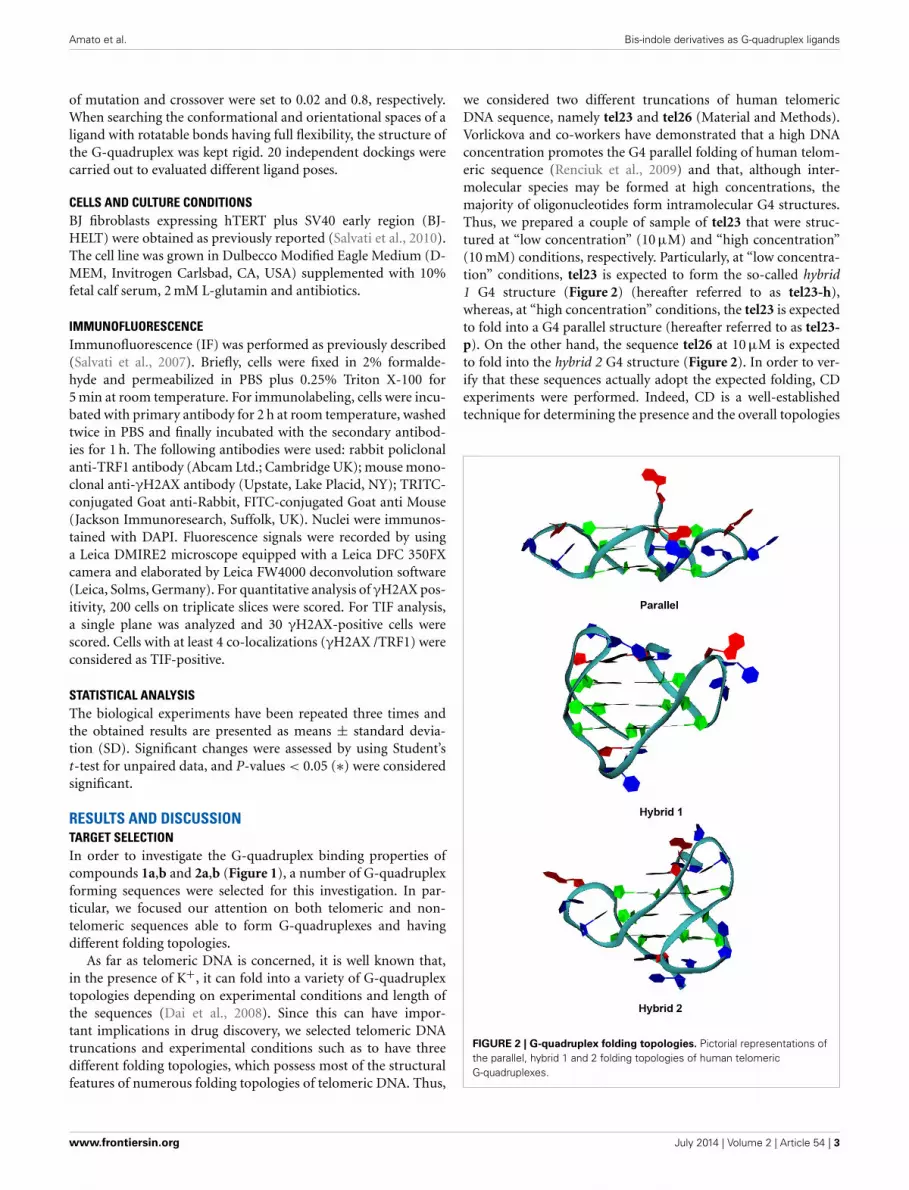
we considered two different truncations of human telomericDNA sequence, namely tel23 and tel26 (Material and Methods).Vorlickova and co-workers have demonstrated that a high DNAconcentration promotes the G4 parallel folding of human telom-eric sequence (Renciuk et al., 2009) and that, although inter-molecular species may be formed at high concentrations, themajority of oligonucleotides form intramolecular G4 structures.Thus, we prepared a couple of sample of tel23 that were struc-tured at “low concentration” (10 μM) and “high concentration”(10 mM) conditions, respectively. Particularly, at “low concentra-tion” conditions, tel23 is expected to form the so-called hybrid1 G4 structure (Figure 2) (hereafter referred to as tel23-h),whereas, at “high concentration” conditions, the tel23 is expectedto fold into a G4 parallel structure (hereafter referred to as tel23-p). On the other hand, the sequence tel26 at 10 μM is expectedto fold into the hybrid 2 G4 structure (Figure 2). In order to ver-ify that these sequences actually adopt the expected folding, CDexperiments were performed. Indeed, CD is a well-establishedtechnique for determining the presence and the overall topologies
FIGURE 2 | G-quadruplex folding topologies. Pictorial representations ofthe parallel, hybrid 1 and 2 folding topologies of human telomericG-quadruplexes.
www.frontiersin.org July 2014 | Volume 2 | Article 54 | 3

Amato et al. Bis-indole derivatives as G-quadruplex ligands
of G4 structures (Masiero et al., 2010; Karsisiotis et al., 2011;Randazzo et al., 2013), although it should be noted that the inter-pretation of CD spectra requires spectra of well characterizedG4 structures for comparison. The tel26 sequence showed a CDspectrum having two positive bands at 290 and 268 nm, and aweak negative band at around 240 nm (Figure S1, SupplementaryMaterial). These data are consistent with a hybrid 2 G4 foldingtopology. Very similar CD spectrum was obtained for tel23-h,indicating also in this case an antiparallel G4 folding topology(namely hybrid 1) (Figure S1, Supplementary Material). Tel23-pactually adopts a parallel conformation, having positive bandaround 265 nm and a negative band around 240 nm in the CDspectrum (Figure S1, Supplementary Material).
As mentioned above, we also took into consideration twonon-telomeric sequences (see Materials and Methods) and bothof them were analyzed by CD spectroscopy. Particularly, weprepared the promoter DNA sequence ckit1, which exists as aparallel structure (Phan et al., 2007), having a characteristic pos-itive band at 262 nm and a negative band at 240 nm in the CDspectrum (Figure S1, Supplementary Material). The other targetG-quadruplex considered is that formed by the ckit2 sequence,that exists in a dimeric parallel-stranded conformation (Kuryavyiet al., 2010) as indicated by a major positive band at 262 nm anda negative band at 240 nm.
Finally, in order to evaluate the selectivity of the ligands for theG4 over the duplex DNA, we used as target also the Dickersonduplex-forming dodecamer (ds12).
BINDING ANALYSISCircular dichroism (CD) studies were performed to explore thepotential of the new ligands to alter the native folding topol-ogy of the investigated G-quadruplexes, inducing a particularconformation. Thus, the bis-indolinone derivatives with the 2,6-disubstituted pyridine core (1a,b), as well as the derivativeswith the 1,10-disubstituted phenanthroline core (2a,b) were syn-thesized as previously described (Andreani et al., 2008, 2010).Upon addition of an excess of ligands (4 equivalents relativeto the DNA), no relevant variations of DNA chiroptical sig-nal were observed for all the analyzed structures (Figure S1,Supplementary Material), thus suggesting an overall conservationof the G4 structures as well as of their architectures. Analogously,the interaction between the ligands and the Dickerson duplex-forming dodecamer (ds12) was also investigated by CD spec-troscopy to evaluate their effect on the reference DNA duplex. The
CD spectra of ds12 in the absence and in presence of ligands insolution were almost superimposable (Figure S1, SupplementaryMaterial), thus suggesting that the investigated compounds donot alter the duplex structure.
Then, the DNA-stabilizing properties of the compounds wereevaluated by measuring the ligand-induced change in the meltingtemperature (�Tm) of the various G4-forming sequences as wellas of the duplex-forming sequence in CD melting experiments(Giancola and Pagano, 2013). All the thermal denaturationswere monitored at the wavelengths of maximum CD intensity.In particular, the melting profiles of the parallel G4 structureswere recorded at 264 nm (tel23-p) and 262 nm (ckit1, ckit2),while the thermal denaturations of the hybrid-type G4s weremonitored at 289 nm (tel23-h) and 290 nm (tel26) (Figure S2,Supplementary Material). Instead, CD melting curves of ds12duplex were recorded at 280 nm. Ligands 1a and 1b did notincrease significantly the stability of any G4 DNAs as well as ofduplex (Table 1). On the other hand, ligands 2a and 2b enhancedthe stability of the parallel telomeric G4 tel23-p by 5.5 and 15.5◦C,respectively (Figure 3 and Table 1). Very interestingly, the sameligands showed to induce only a slight increase (up to 3.0◦C) ofthermal stability of all the other investigated G4s and none forthe duplex. These results highlight the fact that ligands 2a and
FIGURE 3 | CD melting experiments. Normalized CD melting curves oftel23-p in the absence (solid) and in presence of 4 molar equiv of ligands1a (dash), 1b (dot), 2a (dash dot), and 2b (dash dot dot). Tm values arelisted in Table 1.
Table 1 | Melting temperatures.
Ligands Tm(◦C)
ckit1 ckit2 tel23-p tel23-h tel26 ds12
No ligand 70.5 (±0.2) 72.5 (±0.2) 68.5 (±0.3) 64.5 (±0.2) 66.5 (±0.2) 64.0 (±0.3)
1a 71.0 (±0.3) 75.5 (±0.2) 71.5 (±0.2) 64.5 (±0.3) 66.5 (±0.3) 64.5 (±0.2)
1b 71.0 (±0.2) 74.0 (±0.3) 68.5 (±0.3) 64.5 (±0.2) 66.5 (±0.3) 62.5 (±0.2)
2a 71.0 (±0.2) 75.0 (±0.2) 74.0 (±0.3) 65.0 (±0.2) 66.5 (±0.3) 64.5 (±0.3)
2b 72.0 (±0.3) 75.0 (±0.3) 84.0 (±0.3) 64.5 (±0.3) 66.5 (±0.2) 64.5 (±0.2)
Melting temperatures of G-quadruplex and duplex DNAs with and without ligands measured by CD melting experiments.
Frontiers in Chemistry | Medicinal and Pharmaceutical Chemistry July 2014 | Volume 2 | Article 54 | 4

Amato et al. Bis-indole derivatives as G-quadruplex ligands
FIGURE 4 | Nondenaturing PAGE. Nondenaturing PAGE of humantelomeric DNA tel23-p (50 μM) with increasing equivalents of ligands (2and 4 eq.) at 5◦C. Lanes 1 and 2: bromophenol blue; lane 3: tel23-p alone;lane 4: [tel23-p+1a] 1:2 mixture; lane 5: [tel23-p+1a] 1:4 mixture; lane 6:[tel23-p+1b] 1:2 mixture; lane 7: [tel23-p+1b] 1:4 mixture; lane 8:[tel23-p+2a] 1:2 mixture; lane 9: [tel23-p+2a] 1:4 mixture; lane 10:[tel23-p+2b] 1:2 mixture; lane 11: [tel23-p+2b] 1:4 mixture; lane 12:bromophenol blue.
2b not only selectively stabilize G4 over duplex DNA, but alsodiscriminate among different G-quadruplex structures.
Nondenaturing gel electrophoresis experiments were per-formed on tel23-p, that is the G-quadruplex more stabilized bythe ligands. In particular, the experiments were performed beforeand after the addition of the ligands, to confirm the presenceof the intramolecular G4 structure as major conformation insolution. As shown in Figure 4, tel23-p moves essentially as sin-gle band in the gel, thus suggesting the absence of high-orderstructures. Moreover, the addiction of ligands did not have anypronounced effect on the G4 mobility. This clearly indicatesthat (i) all investigated ligands do not induce DNA dimeriza-tion/oligomerization, (ii) in agreement with CD results theseligands do not promote any G4 conformational change.
BIOLOGICAL AND MOLECULAR ACTIVITYThe two molecules that were able to significantly increase thethermal stability of the telomeric G4 were further investigatedfrom biological point of view. In particular, we evaluated if themechanism through which the two bis-indole derivatives 2a and2b exert their antitumor activity is due to their ability to bind theG4 DNA structures. Thus, human transformed fibroblasts (BJ-EHLT) were exposed for 24 h to different concentrations of thetwo compounds and activation of DNA damage response (DDR)was evaluated by immunofluorescence. As shown in Figure 5,both ligands, even if at different extents, induced the phospho-rylation of H2AX, a hallmark of DDR at almost all the drugdoses tested (Thiriet and Hayes, 2005). Specifically, treatment
FIGURE 5 | Analysis of DNA damage response by bis-indole derivatives
2a and 2b. Transformed BJ-EHLT fibroblasts were grown for 24 h inabsence (−) or in presence of the indicated concentrations of compound 2a
or 2b. DNA damage response was evaluated by immunofluorescence (IF)analysis by using an anti-γH2AX antibody (green) and DAPI (blue) was usedto mark nuclei. (A) Representative images of IF analysis. Images wereacquired by using a Leica Deconvolution microscope (magnification 20×).(B) Quantification of γH2AX-positive BJ-EHLT fibroblasto from (A).Histograms show the mean values ± SD of at least three independentexperiments. p-values were calculated using the student t-test (∗p < 0.05;∗∗p < 0.005).
with compound 2a produced a dose-dependent effect with aninduction of γH2AX-positive cells starting from 1 μM (about30%) and reaching about 70% of positive cells at 5 μM concen-tration (Figure 5). Interestingly, exposure of BJ-EHLT to 0.5 μMof 2b was already sufficient to induce a potent phosphorylationof H2AX (more than 50% of positive cells), percentage that doesnot further enhanced with the increase of the dosage (Figure 5).Altogether, these results suggest that the chemical substituentsintroduced in the tested molecules can determine a differentaffinity of the two ligands for the target.
To evaluate whether γH2AX was phosphorylated in responseto dysfunctional telomeres, the most effective drug concentra-tions of both compounds were tested by double immunofluores-cence (IF). The analysis performed by deconvolution microscopyrevealed that both compounds induced γH2AX foci that colo-calized with TRF1, an effective marker for telomeres, generatingthe so-called telomere-dysfunction induced foci (TIFs) (Takaiet al., 2003) (Figure 6), clearly indicating that the tested com-pounds caused telomere localized damage. Consistent with thesedata, results from quantitative analysis revealed that both 2aand 2b significantly increased the percentage of cells with more
www.frontiersin.org July 2014 | Volume 2 | Article 54 | 5
Amato et al. Bis-indole derivatives as G-quadruplex ligands
FIGURE 6 | Analysis of telomere damage by bis-indole derivatives 2a
and 2b. BJ-EHLT fibroblasts were grown in absence (−) or in presence ofthe bis-indole derivatives 2a and 2b at the reported concentration. Upon24 h, each sample was processed for IF analysis by using antibodies againstγH2AX (green) and TRF1 (red) to mark DNA damage and telomeres,respectively. DAPI staining was used to mark nuclei. (A) Representative IFimages acquired by using a Leica Deconvolution microscope (magnification63×). Enlarged views of TIFs are reported on the right of each picture.(B,C) Quantification of TIF-positive cells (B) and mean of TIFs per nucleus(C) from IF experiments reported in (A). Data are means ± SD of threeindependent experiments. p-values were calculated using the student t-test(∗∗p < 0.005; ∗∗∗p < 0.001).
than four γH2AX/TRF1 colocalizations (Pearson’s correlationcoefficient ≥0.45), with a mean of about 6 TIFs per nucleus(Figure 6).
Notably, while at the lowest doses of 2a, the majority of γH2AXspots colocalized with TRF1, at the highest concentrations animportant fraction of the damage was not localized at the telom-eres. On the contrary, compounds induced both telomeric andnot telomeric DNA damage even at low drug doses (0.5 μM con-centration). This is in line with recent data in which it has beendemonstrated that G-quadruplex structures have been found inthe promoters of several genes involved in cancer processes (e.g.,
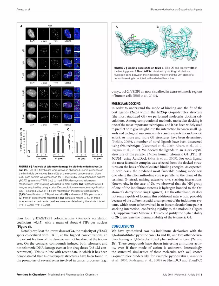
FIGURE 7 | Binding pose of 2b on tel23-p. Side (A) and top-view (B) ofthe binding pose of 2b on tel23-p obtained by docking calculations.Hydrogen bond between the indolinone moiety and the O4′ atom of adeoxyribose ring is depicted with a dashed black line.
c-myc, bcl-2, VEGF) an now visualized in extra-telomeric regionsof human cells (Biffi et al., 2013).
MOLECULAR DOCKINGIn order to understand the mode of binding and the fit of thebest ligands (2a,b) within the tel23-p G-quadruplex structure(the most stabilized G4) we performed molecular docking cal-culations. Among computational methods, molecular docking isone of the most important techniques, and it has been widely usedto predict or to give insight into the interaction between small lig-ands and biological macromolecules (such as proteins and nucleicacids). As more and more G4 structures have been determined(Neidle, 2009), a number of novel ligands have been discoveredusing this technique (Cosconati et al., 2009; Alcaro et al., 2012;Pagano et al., 2012). We docked the ligands to an X-ray crystalstructure of the parallel 23-mer human telomeric G4 (PDB ID3CDM) using AutoDock (Morris et al., 2009). For each ligand,the most favorable complex was selected from the docked struc-tures on the basis of the calculated binding energies. As expected,in both cases, the predicted most favorable binding mode wasone where the phenanthroline core is parallel to the plane of theterminal G-tetrad, making extensive π–π stacking interactions.Noteworthy, in the case of 2b, we observed that the NH groupof one of the indolinone systems is hydrogen bonded to the O4′atom of a deoxyribose ring (Figure 7). On the other hand, 2a doesnot seem capable of forming this additional interaction, probablybecause of the different spatial arrangement of the indolinone sys-tems, which seem to be involved in an intramolecular lone pair-πstacking interaction, conferring rigidity to the molecule (FigureS3, Supplementary Material). This could justify the higher abilityof 2b to increase the thermal stability of the telomeric G4.
CONCLUSIONSWe have synthesized two bis-indolinone derivatives with the2,6-disubstituted pyridine core (1a and 1b) and two other deriva-tives having a 1,10-disubstituted phenanthroline core (2a and2b). These compounds have shown interesting antitumor activ-ity, even if their mode of action is unknown. Interestingly,the structural similarities of these molecules with well-knownG-quadruplex binders like for example pyridostatin (Granotieret al., 2005; Rodriguez et al., 2008) or PhenDC3 and PhenDC6
Frontiers in Chemistry | Medicinal and Pharmaceutical Chemistry July 2014 | Volume 2 | Article 54 | 6
Amato et al. Bis-indole derivatives as G-quadruplex ligands
(Dhamodharan et al., 2012) suggested us that also these moleculescould actually bind G-quadruplexes and this interaction mayberesponsible of their antitumor activity. Compounds 2a and 2bactually are the only two compounds able to selectively stabilizeG4 over duplex DNA and also to discriminate among different G-quadruplex structures, having a particular affinity for the parallelhuman telomeric G-quadruplex tel23-p. Docking calculationshave indicated potential binding modes for those compounds,providing possible explanations of the different affinities andactivities, and therefore laying the basis for the development ofnew ligands. All together these results represent the proof of con-cept that 2a and 2b interact and stabilize the G4 structures bothin vitro and in cellulo, and therefore that they could be consideredas the lead compounds for developing new anticancer drugs.
ACKNOWLEDGEMENTThis work was supported by the “Italian Association for CancerResearch” (MFAG 11947 and IG 14150).
SUPPLEMENTARY MATERIALThe Supplementary Material for this article can be found onlineat: http://www.frontiersin.org/journal/10.3389/fchem.2014.
00054/abstract
REFERENCESAlcaro, S., Costa, G., Distinto, S., Moraca, F., Ortuso, F., Parrotta, L., et al. (2012).
The polymorphisms of DNA G-quadruplex investigated by docking experi-ments with telomestatin enantiomers. Curr. Pharm. Des. 18, 1873–1879. doi:10.2174/138161212799958495
Andreani, A., Burnelli, S., Granaiola, M., Leoni, A., Locatelli, A., Morigi, R.,et al. (2008). Antitumor activity of bis-indole derivatives. J. Med. Chem. 51,4563–4570. doi: 10.1021/jm800194k
Andreani, A., Burnelli, S., Granaiola, M., Leoni, A., Locatelli, A., Morigi, R., et al.(2010). Antitumor activity and COMPARE analysis of bis-indole derivatives.Bioorg. Med. Chem. 18, 3004–3011. doi: 10.1016/j.bmc.2010.03.063
Biffi, G., Tannahill, D., McCafferty, J., and Balasubramanian, S. (2013).Quantitative visualization of DNA G-quadruplex structures in human cells. Nat.Chem. 5, 182–186. doi: 10.1038/nchem.1548
Bochman, L. N., Paeschke, K., and Zakian, V. A. (2012). DNA secondary structures:stability and function of G-quadruplex structures. Nat. Rev. Gen. 13, 770–780.doi: 10.1038/nrg3296
Cosconati, S., Marinelli, L., Trotta, R., Virno, A., De Tito, S., Romagnoli, R., et al.(2010). Structural and conformational requisites in DNA quadruplex groovebinding: another piece to the puzzle. J. Am. Chem. Soc. 132, 6425–6433. doi:10.1021/ja1003872
Cosconati, S., Marinelli, L., Trotta, R., Virno, A., Mayol, L., Novellino, E., et al.(2009). Tandem application of virtual screening and NMR experiments in thediscovery of brand new DNA quadruplex groove binders. J. Am. Chem. Soc. 131,16336–16337. doi: 10.1021/ja9063662
Cosconati, S., Rizzo, A., Trotta, R., Pagano, B., Iachettini, S., De Tito, S., et al.(2012). Shooting for selective druglike G-quadruplex binders: evidence fortelomeric DNA damage and tumor cell death. J. Med. Chem. 55, 9785–9792.doi: 10.1021/jm301019w
Dai, J., Carver, M., and Yang, D. (2008). Polymorphism of humantelomeric quadruplex structures. Biochimie 90, 1172–1183. doi:10.1016/j.biochi.2008.02.026
Dhamodharan, V., Harikrishna, S., Jagadeeswaran, C., Halder, K., andPradeepkumar, P. I. (2012). Selective G-quadruplex DNA stabilizing agentsbased on bisquinolinium and bispyridinium derivatives of 1,8-naphthyridine.J. Org. Chem. 77, 229–242. doi: 10.1021/jo201816g
Di Leva, F. S., Zizza, P., Cingolani, C., D’Angelo, C., Pagano, B., Amato, J., et al.(2013). Exploring the chemical space of G-quadruplex binders: discovery of anovel chemotype targeting the human telomeric sequence. J. Med. Chem. 56,9646–9654. doi: 10.1021/jm401185b
Fajkus, J., Sýkorová, E., and Leitch, A. R. (2005). Telomeres in evolution andevolution of telomeres. Chromosome Res. 13, 469–479. doi: 10.1007/s10577-005-0997-2
Giancola, C., and Pagano, B. (2013). Energetics of ligand binding to G-quadruplexes. Top Curr. Chem. 330, 211–242. doi: 10.1007/128_2012_347
Granotier, C., Pennarun, G., Riou, L., Hoffschir, F., Gauthier, L. R., De Cian,A., et al. (2005). Preferential binding of a G-quadruplex ligand to humanchromosome ends. Nucleic Acids Res. 33, 4182–4190. doi: 10.1093/nar/gki722
Granzhan, A., Monchaud, D., Saettel, N., Guédin, A., Mergny, J. L., and Teulade-Fichou, M. P. (2010). “One ring to bind them all”-Part II: identification ofpromising G-quadruplex ligands by screening of cyclophane-type macrocycles.J. Nucleic Acids 2010:460561. doi: 10.4061/2010/460561
Harley, C. B., Futcher, A. B., and Greider, C. W. (1990). Telomeres shorten duringageing of human fibroblasts. Nature 345, 458–460. doi: 10.1038/345458a0
Karsisiotis, A. I., Hessari, N. M., Novellino, E., Spada, G. P., Randazzo, A., andWebba da Silva, M. (2011). Topological characterization of nucleic acid G-quadruplexes by UV absorption and circular dichroism. Angew. Chem. Int. Ed.Engl. 50, 10645–10648. doi: 10.1002/anie.201105193
Kim, N. W. (1997). Clinical implications of telomerase in cancer. Eur. J. Cancer 33,781–786. doi: 10.1016/S0959-8049(97)00057-9
Kuryavyi, V., Phan, A. T., and Patel, D. J. (2010). Solution structures of all parallel-stranded monomeric and dimeric G-quadruplex scaffolds of the human c-kit2promoter. Nucleic Acids Res. 38, 6757–6773. doi: 10.1093/nar/gkq558
Masiero, S., Trotta, R., Pieraccini, S., De Tito, S., Perone, R., Randazzo, A.,et al. (2010). A non-empirical chromophoric interpretation of CD spectraof DNA G-quadruplex structures. Org. Biomol. Chem. 8, 2683–2692. doi:10.1039/c003428b
Monchaud, D., Granzhan, A., Saettel, N., Guédin, A., Mergny, J. L., and Teulade-Fichou, M. P. (2010). “One ring to bind them all”-part I: the efficiency ofthe macrocyclic scaffold for G-quadruplex DNA recognition. J. Nucleic Acids2010:525862. doi: 10.4061/2010/525862
Morris, G. M., Huey, R., Lindstrom, W., Sanner, M. F., Belew, R. K., Goodsell, D. S.,et al. (2009). AutoDock4 and AutoDockTools4: automated docking with selec-tive receptor flexibility. J. Comput. Chem. 30, 2785–2791. doi: 10.1002/jcc.21256.
Neidle, S. (2009). The structures of quadruplex nucleic acids and their drugcomplexes. Curr. Opin. Struct. Biol. 19, 239–250. doi: 10.1016/j.sbi.2009.04.001
Ohnmacht, S. A., and Neidle, S. (2014). Small-molecule quadruplex-targeted drug discovery. Bioorg. Med. Chem. Lett. 24, 2602–2612. doi:10.1016/j.bmcl.2014.04.029
Pagano, B., Cosconati, S., Gabelica, V., Petraccone, L., De Tito, S., Marinelli, L.,et al. (2012). State-of-the-art methodologies for the discovery and characteri-zation of DNA G-quadruplex binders. Curr. Pharm. Des. 18, 1880–1899. doi:10.2174/138161212799958332
Pagano, B., Fotticchia, I., De Tito, S., Mattia, C. A., Mayol, L., Novellino, E., et al.(2010). Selective binding of distamycin a derivative to G-quadruplex structure[d(TGGGGT)]4. J. Nucleic Acids 2010:247137. doi: 10.4061/2010/247137
Parkinson, G. N., Cuenca, F., and Neidle, S. (2008). Topology conservation andloop flexibility in quadruplex-drug recognition: crystal structures of inter- andintramolecular telomeric DNA quadruplex-drug complexes. J. Mol. Biol. 381,1145–1156. doi: 10.1016/j.jmb.2008.06.022.
Petraccone, L., Fotticchia, I., Cummaro, A., Pagano, B., Ginnari-Satriani, L., Haider,S., et al. (2011). The triazatruxene derivative azatrux binds to the parallel formof the human telomeric G-quadruplex under molecular crowding conditions:biophysical and molecular modeling studies. Biochimie 93, 1318–1327. doi:10.1016/j.biochi.2011.05.017
Phan, A. T., Kuryavyi, V., Burge, S., Neidle, S., and Patel, D. P. (2007). Structure ofan unprecedented G-quadruplex scaffold in the human c-kit promoter. J. Am.Chem. Soc. 129, 4386–4392. doi: 10.1021/ja068739h
Price, C. M. (1999). Telomeres and telomerase: broad effects on cell growth. Curr.Opin. Genet. Dev. 9, 218–224. doi: 10.1016/S0959-437X(99)80032-X
Randazzo, A., Spada, G. P., and Webba da Silva, M. (2013). Circulardichroism of quadruplex structures. Top. Curr. Chem. 330, 67–86. doi:10.1007/128_2012_331
Renciuk, D., Kejnovska, I., Skolakova, P., Bednarova, K., Motlova, J., andVorlickova, M. (2009). Arrangements of human telomere DNA quadruplex inphysiologically relevant K+ solutions. Nucleic Acids Res. 37, 6625–6634. doi:10.1093/nar/gkp701
Rodriguez, R., Muller, S., Yeoman, J. A., Trentesaux, C., Riou, J.-F., andBalasubramanian, S. (2008). A novel small molecule that alters shelterin
www.frontiersin.org July 2014 | Volume 2 | Article 54 | 7

Amato et al. Bis-indole derivatives as G-quadruplex ligands
integrity and triggers a DNA-damage response at telomeres. J. Am. Chem. Soc.130, 15758–15759. doi: 10.1021/ja805615w
Salvati, E., Leonetti, C., Rizzo, A., Scarsella, M., Mottolese, M., Galati, R., et al.(2007). Telomere damage induced by the G-quadruplex ligand RHPS4 has anantitumor effect. J. Clin. Invest. 117, 3236–3247. doi: 10.1172/JCI32461
Salvati, E., Scarsella, M., Porru, M., Rizzo, A., Iachettini, S., Tentori, L., et al.(2010). PARP1 activated at telomeres upon G4 stabilization: possible target fortelomere-based therapy. Oncogene 29, 6280-6293. doi: 10.1038/onc.2010.344
Shay, J. W., and Wright, W. E. (2005). Senescence and immortalization:role of telomeres and telomerase. Carcinogenesis 26, 867–874. doi:10.1093/carcin/bgh296
Sun, D., Thompson, B., Cathers, B. E., Salazar, M., Kerwin, S. M., Trent, J. O.,et al. (1997). Inhibition of human telomerase by a G-quadruplex-interactivecompound. J. Med. Chem. 40, 2113–2116. doi: 10.1021/jm970199z
Takai, H., Smogorzewska, A., and de Lange, T. (2003). DNA damage foci at dys-functional telomeres. Curr. Biol. 13, 1549-1556. doi: 10.1016/S0960-9822(03)00542-6
Thiriet, C., and Hayes, J. J. (2005). Chromatin in need of a fix: phosphoryla-tion of H2AX connects chromatin to DNA repair. Mol. Cell 18, 617-622. doi:10.1016/j.molcel.2005.05.008
Wright, W. E., Tesmer, V. M., Huffman, K. E., Levene, S. D., and Shay, J. W. (1997).Normal human chromosomes have long G-rich telomeric overhangs at one end.Genes Dev. 11, 2801–2809. doi: 10.1101/gad.11.21.2801
Zahler, A. M., Williamson, J. R., Cech, T. R., and Prescott, D. M. (1991). Inhibitionof telomerase by G-quartet DNA structures. Nature 350, 718–720. doi:10.1038/350718a0
Conflict of Interest Statement: The authors declare that the research was con-ducted in the absence of any commercial or financial relationships that could beconstrued as a potential conflict of interest.
Received: 03 June 2014; paper pending published: 17 June 2014; accepted: 04 July 2014;published online: 24 July 2014.Citation: Amato J, Iaccarino N, Pagano B, Morigi R, Locatelli A, Leoni A, RambaldiM, Zizza P, Biroccio A, Novellino E and Randazzo A (2014) Bis-indole derivatives withantitumor activity turn out to be specific ligands of human telomeric G-quadruplex.Front. Chem. 2:54. doi: 10.3389/fchem.2014.00054This article was submitted to Medicinal and Pharmaceutical Chemistry, a section ofthe journal Frontiers in Chemistry.Copyright © 2014 Amato, Iaccarino, Pagano, Morigi, Locatelli, Leoni, Rambaldi,Zizza, Biroccio, Novellino and Randazzo. This is an open-access article distributedunder the terms of the Creative Commons Attribution License (CC BY). The use, dis-tribution or reproduction in other forums is permitted, provided the original author(s)or licensor are credited and that the original publication in this journal is cited, inaccordance with accepted academic practice. No use, distribution or reproduction ispermitted which does not comply with these terms.
Frontiers in Chemistry | Medicinal and Pharmaceutical Chemistry July 2014 | Volume 2 | Article 54 | 8
Related Documents