Ecotoxicology and Environmental Safety 53 (2002) 331–347 Biotransformation, genotoxic, and histopathological effects of environmental contaminants in European eel (Anguilla anguilla L.) Ma´rio Pacheco* and Maria Ana Santos Animal Physiology/Ecotoxicology Sector, Biology Department, University of Aveiro, Aveiro 3810-193, Portugal Received 7 May 2000; received in revised form 30 March 2001; accepted 30 April 2001 Abstract A prolonged toxicity study was carried out in young European eel (Anguilla anguilla L.) to evaluate the effects of environmental contaminants, namely, two individual standard compounds, benzo[a]pyrene (BaP) and dehydroabietic acid (DHAA), and a complex mixture, bleached kraft pulp mill effluent (BKPME). Fish were exposed to BaP (0.22, 0.45, and 0.9 mM) and BKPME (3.12%, 6.25%, and 12.5% (v/v)) for 3, 7, and 30 days and to DHAA (0.07, 0.15, and 0.30 mM) for 3, 7, 30, 90, and 180 days. The biomarkers include biotransformation and genotoxicity indicators, such as total ethoxyresorufin O-deethylase (EROD) activity and frequency of erythrocytic nuclear abnormalities (ENAs), respectively. Hematological dynamics was assessed as frequency of immature erythrocytes (IEs). Histopathological examinations were carried out for the highest concentrations and for 30 days and longer exposures. Total EROD increases significantly only after 180 days of DHAA exposure. However, significant ENA induction was generally observed during exposure to all contaminants tested. Nevertheless, some of the ENA results suggest an altered genotoxic response, which may arise either from short-term exposures to the highest contaminant levels or long-term exposures to the lowest contaminant levels. IE frequency decreased significantly after 30 days of exposure to 0.45 mM BaP and 180 days of exposure to the entire DHAA concentration range. Increased density of pigmented macrophage aggregates in 30-day BaP- and BKPME-exposed fish as well as in 90- and 180-day DHAA-exposed fish confirmed histopathological liver alterations. Bile accumulation in hepatocytes after BaP treatment, cytoplasmic vacuolization and cell atrophy following DHAA exposure, as well as liver loss of parenchymal cells in BKPME-exposed fish, were also detected. Dispersed necrosis and focal inflammation were observed in the livers of all treated groups. Fish exposed to DHAA and BKPME showed skin and gill disruption as well as kidney Malpighian corpuscle alterations. All 30-day-treated groups revealed intense spleen hemosiderosis, indicating increased erythrophagia. This splenic effect may be strongly correlated with the observed disappearance of ENAs. Neoplastic lesions were not found. A multibiomarker strategy, which includes EROD, ENA, and IE assays as well as histopathological studies, contributed to a better understanding of the global toxic process. r 2002 Elsevier Science (USA). All rights reserved. Keywords: Anguilla; Ethoxyresorufin O-deethylase; Genotoxicity; Histopathology; Pulp mill effluent; Resin acids; Benzo[a]pyrene 1. Introduction Fish respond to environmental pollutants by altering/ adapting their metabolic functions. Among the variety of biomarkers adopted in ecotoxicological investiga- tions, there is notable interest in parameters related to enzymatic xenobiotic detoxification and activation (biotransformation). In this context, cytochrome P450- dependent monooxygenase induction, measured as ethoxyresorufin O-deethylase (EROD) activity, has been widely used as a sensitive indicator of exposure to environmental contaminants (Addison et al., 1994; Martel et al., 1994; Pacheco and Santos, 1999). The activation of certain xenobiotics results in the produc- tion of potentially damaging oxyradicals, which react with many biological molecules including DNA (Di Guilio et al., 1989). Thus, DNA integrity can be affected. Considering the risks associated with DNA damage, the implementation of genotoxicity biomarkers such as DNA adducts (Harvey et al., 1997), DNA strand breaks (Everaarts, 1995), micronuclei (Jaylet et al., 1986) and erythrocytic nuclear abnormalities (ENAs) (Pacheco and Santos, 1996, 1999) is of particular importance. *Corresponding author. Fax: 351-234-426-408. E-mail address: [email protected] (M. Pacheco). 0147-6513/02/$ - see front matter r 2002 Elsevier Science (USA). All rights reserved. PII:S0147-6513(02)00017-9

Welcome message from author
This document is posted to help you gain knowledge. Please leave a comment to let me know what you think about it! Share it to your friends and learn new things together.
Transcript
Ecotoxicology and Environmental Safety 53 (2002) 331–347
Biotransformation, genotoxic, and histopathological effects ofenvironmental contaminants in European eel (Anguilla anguilla L.)
Mario Pacheco* and Maria Ana Santos
Animal Physiology/Ecotoxicology Sector, Biology Department, University of Aveiro, Aveiro 3810-193, Portugal
Received 7 May 2000; received in revised form 30 March 2001; accepted 30 April 2001
Abstract
A prolonged toxicity study was carried out in young European eel (Anguilla anguilla L.) to evaluate the effects of environmental
contaminants, namely, two individual standard compounds, benzo[a]pyrene (BaP) and dehydroabietic acid (DHAA), and a complex
mixture, bleached kraft pulp mill effluent (BKPME). Fish were exposed to BaP (0.22, 0.45, and 0.9 mM) and BKPME (3.12%,
6.25%, and 12.5% (v/v)) for 3, 7, and 30 days and to DHAA (0.07, 0.15, and 0.30 mM) for 3, 7, 30, 90, and 180 days. The biomarkers
include biotransformation and genotoxicity indicators, such as total ethoxyresorufin O-deethylase (EROD) activity and frequency
of erythrocytic nuclear abnormalities (ENAs), respectively. Hematological dynamics was assessed as frequency of immature
erythrocytes (IEs). Histopathological examinations were carried out for the highest concentrations and for 30 days and longer
exposures. Total EROD increases significantly only after 180 days of DHAA exposure. However, significant ENA induction was
generally observed during exposure to all contaminants tested. Nevertheless, some of the ENA results suggest an altered genotoxic
response, which may arise either from short-term exposures to the highest contaminant levels or long-term exposures to the lowest
contaminant levels. IE frequency decreased significantly after 30 days of exposure to 0.45 mM BaP and 180 days of exposure to the
entire DHAA concentration range. Increased density of pigmented macrophage aggregates in 30-day BaP- and BKPME-exposed
fish as well as in 90- and 180-day DHAA-exposed fish confirmed histopathological liver alterations. Bile accumulation in
hepatocytes after BaP treatment, cytoplasmic vacuolization and cell atrophy following DHAA exposure, as well as liver loss of
parenchymal cells in BKPME-exposed fish, were also detected. Dispersed necrosis and focal inflammation were observed in the
livers of all treated groups. Fish exposed to DHAA and BKPME showed skin and gill disruption as well as kidney Malpighian
corpuscle alterations. All 30-day-treated groups revealed intense spleen hemosiderosis, indicating increased erythrophagia. This
splenic effect may be strongly correlated with the observed disappearance of ENAs. Neoplastic lesions were not found. A
multibiomarker strategy, which includes EROD, ENA, and IE assays as well as histopathological studies, contributed to a better
understanding of the global toxic process.
r 2002 Elsevier Science (USA). All rights reserved.
Keywords: Anguilla; Ethoxyresorufin O-deethylase; Genotoxicity; Histopathology; Pulp mill effluent; Resin acids; Benzo[a]pyrene
1. Introduction
Fish respond to environmental pollutants by altering/adapting their metabolic functions. Among the varietyof biomarkers adopted in ecotoxicological investiga-tions, there is notable interest in parameters related toenzymatic xenobiotic detoxification and activation(biotransformation). In this context, cytochrome P450-dependent monooxygenase induction, measured asethoxyresorufin O-deethylase (EROD) activity, has been
widely used as a sensitive indicator of exposure toenvironmental contaminants (Addison et al., 1994;Martel et al., 1994; Pacheco and Santos, 1999). Theactivation of certain xenobiotics results in the produc-tion of potentially damaging oxyradicals, which reactwith many biological molecules including DNA (DiGuilio et al., 1989). Thus, DNA integrity can beaffected. Considering the risks associated with DNAdamage, the implementation of genotoxicity biomarkerssuch as DNA adducts (Harvey et al., 1997), DNA strandbreaks (Everaarts, 1995), micronuclei (Jaylet et al., 1986)and erythrocytic nuclear abnormalities (ENAs) (Pachecoand Santos, 1996, 1999) is of particular importance.
*Corresponding author. Fax: 351-234-426-408.
E-mail address: [email protected] (M. Pacheco).
0147-6513/02/$ - see front matter r 2002 Elsevier Science (USA). All rights reserved.
PII: S 0 1 4 7 - 6 5 1 3 ( 0 2 ) 0 0 0 1 7 - 9
Disturbance of living processes at the molecular andsubcellular levels of biological organization by xenobio-tics frequently leads to cell injury resulting in degen-erative and neoplastic diseases in target organs.Therefore, histopathological biomarkers have beenproven to be useful complementary indicators of toxicityin fish organs (Schwaiger et al., 1996; Teh et al., 1997).The information provided by each biomarker indivi-
dually is of limited relevance, as there is a considerablelikelihood of misinterpretation. According to Mooreand Simpson (1992), it must be emphasized thatbiomarkers are best used as selected batteries of testsrather than individually. Thus, interlinking of the resultsobtained at different biological levels, such as molecular(biotransformation metabolism), subcellular (genotoxi-city expression), and cellular/tissue (histopathologicaleffects) levels, may lead to a substantial improvement inthe knowledge of integrated fish toxic response.The environmental contaminants tested in the present
study include two individual chemicals—benzo[a]pyrene(BaP), a polycyclic aromatic hydrocarbon (PAH), anddehydroabietic acid (DHAA), a resin acid—as well as acomplex mixture, bleached kraft pulp mill effluent(BKPME). BaP, a PAH commonly found in contami-nated environments, is recognized as a biotransforma-tion inducer (Wolkers et al., 1996; Pacheco and Santos,1998) with strong genotoxic and carcinogenic potentialin fish (Hendricks et al., 1985; Hawkins et al., 1990;Pacheco and Santos, 1997). Effluents from the woodprocessing industry are one of the most significantsources of aquatic pollution worldwide. Resin acids areimportant BKPME components and DHAA in parti-cular is considered a major poison to fish. Previousstudies have indicated that either DHAA or BKPME asa whole induce alterations at different biological levels,namely, biotransformation activation (Martel et al.,1994; Pacheco and Santos, 1999) and genotoxicity(Pacheco and Santos, 1997, 1999).The study described here on the toxicity of environ-
mental pollutants in young specimens of Anguilla anguilla
L. after prolonged exposure had two main objectives:(i) the assessment of BaP, DHAA, and BKPME effects astotal EROD activity, alterations in immature erythrocytefrequency and ENA induction, as well as their histo-pathological effects on different fish target organs (skin,gills, kidney, spleen and liver); (ii) an attempt to relate thepreceeding biomarkers to improve our understanding ofthe fish toxic response as an integrated process.
2. Materials and methods
2.1. Test contaminants
BaP was obtained from Sigma Chemical Company(USA). DHAA was obtained from Helix Biotech
Corporation (Canada). The neutralized and secondarilytreated BKPME was collected from a pulp and papermill sewage outlet (Portucel - Cacia, Aveiro, Portugal).This mill produces bleached kraft pulp and paper, usingEucalyptus globulus and Pinus pinaster (75% and 25%,respectively) as wood supply.
2.2. Fish
European eel (A. anguilla) young specimens, with anaverage weight of 0.2 g (glass eels), were caught inOctober at the Mondego River mouth, Figueira da Foz.They were transported to the laboratory and allowed toacclimate (2 weeks) to controlled conditions, i.e., inrecirculating, filtered, well-aerated, and dechlorinatedtap water, at 201C, under a natural photoperiod. Fishwere fed three times a week, either in the recovery periodor during the experiments.
2.3. Experimental design
Fish exposure took place in 15-L aquaria, containing70 fish each, under the previously described conditions.A 2-cm layer of washed sand was placed on theaquarium bottom. The initial/nominal exposure con-centrations were 0.22, 0.45, and 0.9 mM for BaP, 0.07,0.15, and 0.3 mM for DHAA and 3.12%, 6.25%, and12.5% (v/v) for BKPME. The concentration rangesselected were based on our previous studies (Pachecoand Santos, 1997, 1999). Half of the test solution in eachaquarium was renewed weekly. Fish sampling wascarried out at 3, 7, 30, 90, and 180 days. However,considerable mortality was observed after 30 daysexposure in BaP and BKPME experiments, preventingcontinuation of the experiment beyond that exposurelength.Fish were killed by decapitation. At each sampling
point, five fish were collected and immediately frozen inliquid nitrogen for homogenization and microsomalisolation, and another five eels were collected for bloodsampling. Additionally, histology was carried out at 30days and longer exposures where three eels werecollected from test groups at highest concentrations.BaP and DHAA added to the experimental aquaria
were previously dissolved in dimethyl sulfoxide(DMSO). The DMSO concentration in water (0.033%)has no detectable cytogenetic or toxic effect. The sameDMSO level was maintained in the control aquarium.The BKPME control aquarium contained only cleanwater.
2.4. EROD assay
The whole eel’s body was used for microsomalisolation and EROD activity measurement. Microsomeswere obtained according to the methods of Lange et al.
M. Pacheco, M.A. Santos / Ecotoxicology and Environmental Safety 53 (2002) 331–347332
(1993) and Monod and Vindimian (1991) as adapted byPacheco and Santos (1997). Total EROD activity wasmeasured as described by Burke and Mayer (1974), andthe results were expressed as pmol/min/mg of micro-somal protein. Microsomal protein content is deter-mined according to the Biuret method (Gornall et al.,1949).
2.5. Mutagenicity testing
Mutagenicity was tested using the ENA assay,according to the procedure adapted by Pacheco andSantos (1996). Each group’s final result was presented asthe mean value (%) of the sums for all the individuallesions observed and scored in 1000 mature erythrocytesper fish blood smear.
2.6. Immature erythrocytes (IE) frequency
IEs were scored on each 1000-erythrocyte (matureand immature) sample per fish. Results are presented asa frequency (%) obtained using the expression
IE frequency ð%Þ ¼No: IEs
ðNo: IEsþNo: MEsÞ� 1000
(ME=mature erythrocytes).
2.7. Histopathology
The whole fish body was fixed in Bouin’s solution androutinely embedded in paraffin for light microscopy.The section thickness was 5 mm. Sections were stainedwith hematoxylin and eosin (HE). Histological criteriaemployed in lesion classification were derived fromcriteria previously established for fish by Hibiya (1982)and Myers et al. (1987).
2.8. Statistical evaluation
Means7standard errors (SE) were calculated for eachexperimental group. Data were analyzed for significanceof differences between control and each exposed groupaccording to the two-tailed Student’s t-test (Bailey,1959).
3. Results
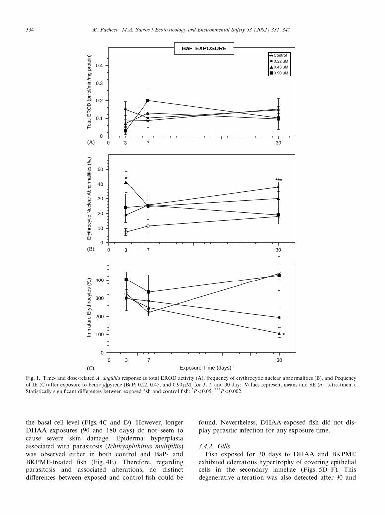
3.1. EROD response
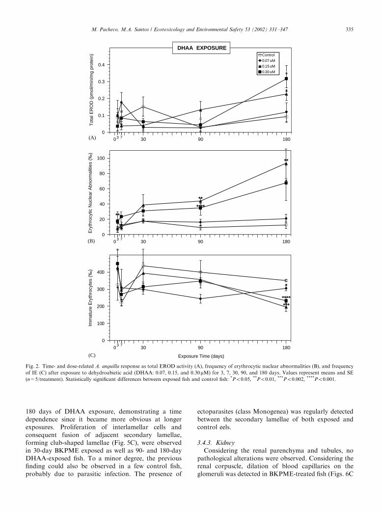
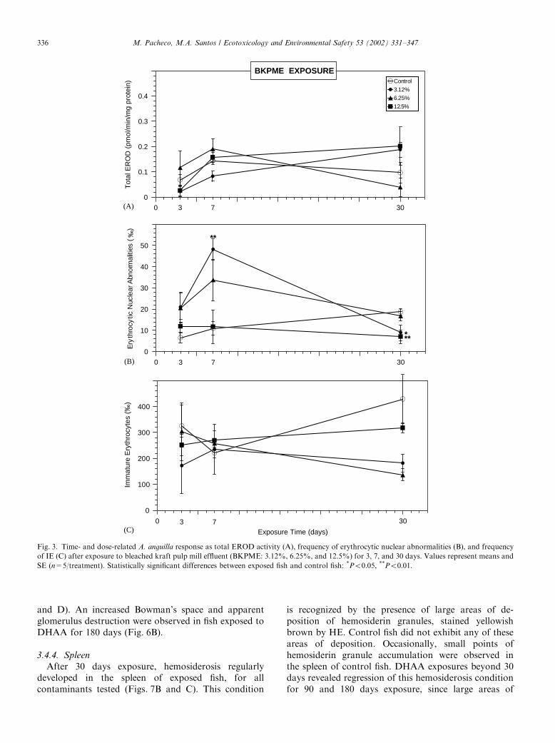
BaP and BKPME exposures did not induce anysignificant alteration in the total EROD activity(Figs. 1A and 3A). Regarding the DHAA experiment,exposures from 3 to 90 days also did not reveal anysignificant increase (Fig. 2A). However, after 180 daysexposure a clear dose–response relation was observed,
and a significant increase was noted for 0.15 and0.30 mM (Po0:05).
3.2. ENA response
The results of the BaP experiment displayed asignificant ENA increase after 3 days exposure to0.45 mM (Po0:05), followed by an accentuated declineafter 7 days exposure (Fig. 1B). At 30 days exposure, thehighest BaP concentrations remained incapable of ENAelevation, whereas the lowest concentration (0.22 mM)induced a significant ENA increase (Po0:002). Further-more, at this exposure length 0.9 mM BaP presented anENA value significantly lower than 0.22 mM (Po0:05).The analysis of genotoxic response to DHAA
demonstrated that, after 3 days exposure, ENA fre-quency was significantly elevated by the highestconcentration (0.30 mM; Po0:01) (Fig. 2B), whereas 7and 30 days exposure did not produce any statisticalsignificant alteration, despite an evident general ENAincrease. At 90 days exposure, the highest DHAAconcentrations induced a significant ENA increase(Po0:01 for 0.15 mM and Po0:001 for 0.30 mM). From90 up to 180 days exposure, ENA frequency was notablyincreased by 0.15 and 0.30 mM DHAA (Po0:01 andPo0:05; respectively).The ENA results of the BKPME experiment (Fig. 3B)
demonstrated a significant increase for 7 days exposureto the lowest concentration (3.12%, Po0:01). Addi-tionally, the highest BKPME concentration (12.5%)exhibited the lowest ENA frequencies along the 30 daysexposure. Furthermore, at 30 days exposure all BKPMEconcentrations displayed ENA levels below the control,being particularly significant for 3.12% and 12.5%(Po0:05 and Po0:01; respectively).
3.3. Hematological dynamics (IE frequency)
The IE frequency (Figs. 1C, 2C and 3C) was generallydecreased by all tested contaminants after 30 days andlonger exposures. However, this decrease was statisti-cally significant only for 30 days exposure to 0.45 mMBaP (Po0:05) and for 180 days exposure to all DHAAconcentrations (Po0:05; Po0:002; and Po0:001;respectively for 0.07, 0.15, and 0.30 mM).
3.4. Histological examination
Exposures to 0.9 mM BaP, 0.3 mMDHAA, and 12.5%BKPME revealed the following histological findings.
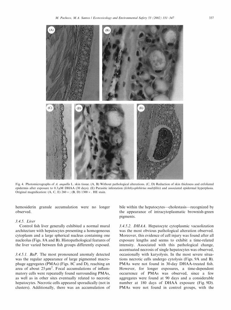
3.4.1. Skin
Thirty-day exposures to DHAA and BKPME causedevident skin disruption. Accentuated reduction of skinthickness and large portions of exfoliated epidermiswere observed. Tissue destruction, frequently, reaches
M. Pacheco, M.A. Santos / Ecotoxicology and Environmental Safety 53 (2002) 331–347 333
the basal cell level (Figs. 4C and D). However, longerDHAA exposures (90 and 180 days) do not seem tocause severe skin damage. Epidermal hyperplasiaassociated with parasitosis (Ichthyophthirius multifiliis)was observed either in both control and BaP- andBKPME-treated fish (Fig. 4E). Therefore, regardingparasitosis and associated alterations, no distinctdifferences between exposed and control fish could be
found. Nevertheless, DHAA-exposed fish did not dis-play parasitic infection for any exposure time.
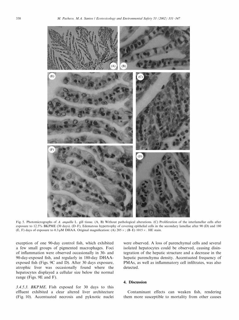
3.4.2. Gills
Fish exposed for 30 days to DHAA and BKPMEexhibited edematous hypertrophy of covering epithelialcells in the secondary lamellae (Figs. 5D–F). Thisdegenerative alteration was also detected after 90 and
0 5 10 15 20 25 30
AA concentration ( M)
0
0.1
0.2
0.3
0.4
Tot
al E
RO
D (
pmol
/min
/mg
prot
ein) Control
0.22 uM
0.45 uM
0.90 uM
0 5 10 15 20 25 30
0
10
20
30
40
50
Ery
thro
cytic
Nuc
lear
Abn
orm
aliti
es (
‰)
***
3 7
3 7
BaP EXPOSURE
*
0 5 10 15 20 25 30
Exposure Time (days)
0
100
200
300
400
Imm
atur
e E
ryth
rocy
tes
(‰)
3 7
*
(A)
(B)
(C)
Fig. 1. Time- and dose-related A. anguilla response as total EROD activity (A), frequency of erythrocytic nuclear abnormalities (B), and frequency
of IE (C) after exposure to benzo[a]pyrene (BaP: 0.22, 0.45, and 0.90mM) for 3, 7, and 30 days. Values represent means and SE (n=5/treatment).
Statistically significant differences between exposed fish and control fish: *Po0:05; ***Po0:002:
M. Pacheco, M.A. Santos / Ecotoxicology and Environmental Safety 53 (2002) 331–347334
180 days of DHAA exposure, demonstrating a timedependence since it became more obvious at longerexposures. Proliferation of interlamellar cells andconsequent fusion of adjacent secondary lamellae,forming club-shaped lamellae (Fig. 5C), were observedin 30-day BKPME exposed as well as 90- and 180-dayDHAA-exposed fish. To a minor degree, the previousfinding could also be observed in a few control fish,probably due to parasitic infection. The presence of
ectoparasites (class Monogenea) was regularly detectedbetween the secondary lamellae of both exposed andcontrol eels.
3.4.3. Kidney
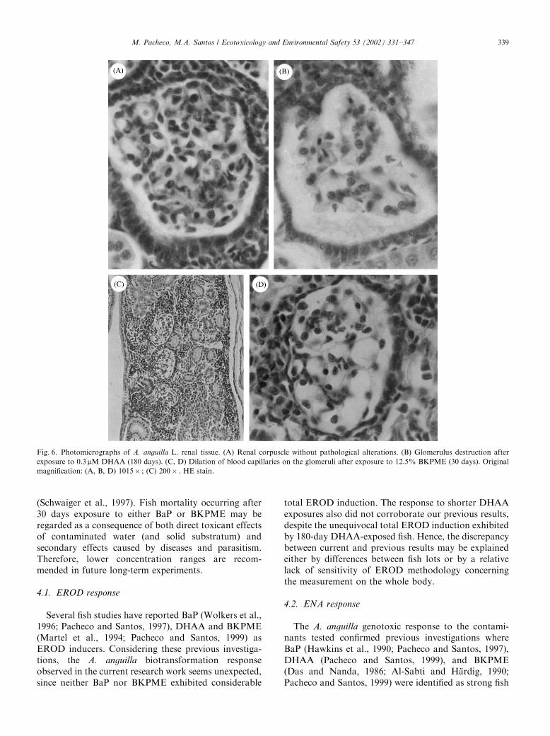
Considering the renal parenchyma and tubules, nopathological alterations were observed. Considering therenal corpuscle, dilation of blood capillaries on theglomeruli was detected in BKPME-treated fish (Figs. 6C
0 10 20 30 40 50 60 70 80 90 100 110 120 130 140 150 160 170 180
AA concentration ( M)
0
0.1
0.2
0.3
0.4
Tot
al E
RO
D (
pmol
/min
/mg
prot
ein) Control
0.07 uM
0.15 uM
0.30 uM
0 10 20 30 40 50 60 70 80 90 100 110 120 130 140 150 160 170 180
0
20
40
60
80
100
Ery
thro
cytic
Nuc
lear
Abn
orm
aliti
es (
‰)
**
******
**
3 7
3 7
DHAA EXPOSURE
*
*
*
0 10 20 30 40 50 60 70 80 90 100 110 120 130 140 150 160 170 180
Exposure Time (days)
0
100
200
300
400
Imm
atur
e E
ryth
rocy
tes
(‰)
****
3 7
***
*
(A)
(B)
(C)
Fig. 2. Time- and dose-related A. anguilla response as total EROD activity (A), frequency of erythrocytic nuclear abnormalities (B), and frequency
of IE (C) after exposure to dehydroabietic acid (DHAA: 0.07, 0.15, and 0.30mM) for 3, 7, 30, 90, and 180 days. Values represent means and SE
(n=5/treatment). Statistically significant differences between exposed fish and control fish: *Po0:05; **Po0:01; ***Po0:002; ****Po0:001:
M. Pacheco, M.A. Santos / Ecotoxicology and Environmental Safety 53 (2002) 331–347 335
and D). An increased Bowman’s space and apparentglomerulus destruction were observed in fish exposed toDHAA for 180 days (Fig. 6B).
3.4.4. Spleen
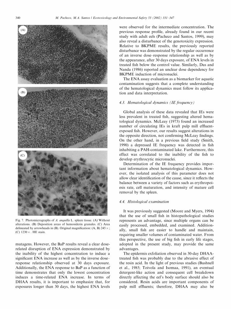
After 30 days exposure, hemosiderosis regularlydeveloped in the spleen of exposed fish, for allcontaminants tested (Figs. 7B and C). This condition
is recognized by the presence of large areas of de-position of hemosiderin granules, stained yellowishbrown by HE. Control fish did not exhibit any of theseareas of deposition. Occasionally, small points ofhemosiderin granule accumulation were observed inthe spleen of control fish. DHAA exposures beyond 30days revealed regression of this hemosiderosis conditionfor 90 and 180 days exposure, since large areas of
0 5 10 15 20 25 30
AA concentration ( M)
0
0.1
0.2
0.3
0.4
Tot
al E
RO
D (
pmol
/min
/mg
prot
ein) Control
3.12%
6.25%
12.5%
0 5 10 15 20 25 30
0
10
20
30
40
50
Ery
thro
cytic
Nuc
lear
Abn
orm
aliti
es (
)
**
3 7
‰
BKPME EXPOSURE
3 7
***
0 5 10 15 20 25 30
Exposure Time (days)
0
100
200
300
400
Imm
atur
e E
ryth
rocy
tes
(‰)
3 7
(A)
(B)
(C)
Fig. 3. Time- and dose-related A. anguilla response as total EROD activity (A), frequency of erythrocytic nuclear abnormalities (B), and frequency
of IE (C) after exposure to bleached kraft pulp mill effluent (BKPME: 3.12%, 6.25%, and 12.5%) for 3, 7, and 30 days. Values represent means and
SE (n=5/treatment). Statistically significant differences between exposed fish and control fish: *Po0:05; **Po0:01:
M. Pacheco, M.A. Santos / Ecotoxicology and Environmental Safety 53 (2002) 331–347336
hemosiderin granule accumulation were no longerobserved.
3.4.5. Liver
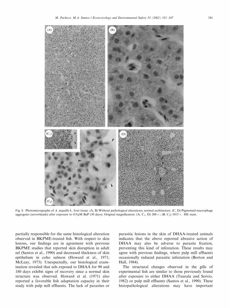
Control fish liver generally exhibited a normal muralarchitecture with hepatocytes presenting a homogeneouscytoplasm and a large spherical nucleus containing onenucleolus (Figs. 8A and B). Histopathological features ofthe liver varied between fish groups differently exposed.
3.4.5.1. BaP. The most pronounced anomaly detectedwas the regular appearance of large pigmented macro-phage aggregates (PMAs) (Figs. 8C and D), reaching anarea of about 25 mm2. Focal accumulations of inflam-matory cells were repeatedly found surrounding PMAs,as well as in other sites eventually related to necrotichepatocytes. Necrotic cells appeared sporadically (not inclusters). Additionally, there was an accumulation of
bile within the hepatocytes—cholestasis—recognized bythe appearance of intracytoplasmatic brownish-greenpigments.
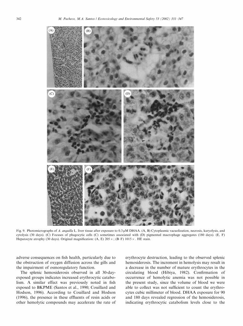
3.4.5.2. DHAA. Hepatocyte cytoplasmic vacuolizationwas the most obvious pathological alteration observed.Moreover, this evidence of cell injury was found after allexposure lengths and seems to exhibit a time-relatedintensity. Associated with this pathological change,accentuated necrosis of single hepatocytes was observed,occasionally with karyolysis. In the most severe situa-tions necrotic cells undergo cytolysis (Figs. 9A and B).PMAs were not found in 30-day DHAA-treated fish.However, for longer exposures, a time-dependentoccurrence of PMAs was observed, since a fewaggregates were found at 90 days and a considerablenumber at 180 days of DHAA exposure (Fig. 9D).PMAs were not found in control groups, with the
Fig. 4. Photomicrographs of A. anguilla L. skin tissue. (A, B) Without pathological alterations. (C, D) Reduction of skin thickness and exfoliated
epidermis after exposure to 0.3mM DHAA (30 days). (E) Parasitic infestation (Ichthyophthirius multifiliis) and associated epidermal hyperplasia.
Original magnification: (A, C, E) 260� ; (B, D) 1300� . HE stain.
M. Pacheco, M.A. Santos / Ecotoxicology and Environmental Safety 53 (2002) 331–347 337
exception of one 90-day control fish, which exhibiteda few small groups of pigmented macrophages. Fociof inflammation were observed occasionally in 30- and90-day-exposed fish, and regularly in 180-day DHAA-exposed fish (Figs. 9C and D). After 30 days exposure,atrophic liver was occasionally found where thehepatocytes displayed a cellular size below the normalrange (Figs. 9E and F).
3.4.5.3. BKPME. Fish exposed for 30 days to thiseffluent exhibited a clear altered liver architecture(Fig. 10). Accentuated necrosis and pyknotic nuclei
were observed. A loss of parenchymal cells and severalisolated hepatocytes could be observed, causing disin-tegration of the hepatic structure and a decrease in thehepatic parenchyma density. Accentuated frequency ofPMAs, as well as inflammatory cell infiltrates, was alsodetected.
4. Discussion
Contaminant effects can weaken fish, renderingthem more susceptible to mortality from other causes
Fig. 5. Photomicrographs of A. anguilla L. gill tissue. (A, B) Without pathological alterations. (C) Proliferation of the interlamellar cells after
exposure to 12.5% BKPME (30 days). (D–F), Edematous hypertrophy of covering epithelial cells in the secondary lamellae after 90 (D) and 180
(E, F) days of exposure to 0.3 mM DHAA. Original magnification: (A) 205� ; (B–E) 1015� . HE stain.
M. Pacheco, M.A. Santos / Ecotoxicology and Environmental Safety 53 (2002) 331–347338
(Schwaiger et al., 1997). Fish mortality occurring after30 days exposure to either BaP or BKPME may beregarded as a consequence of both direct toxicant effectsof contaminated water (and solid substratum) andsecondary effects caused by diseases and parasitism.Therefore, lower concentration ranges are recom-mended in future long-term experiments.
4.1. EROD response
Several fish studies have reported BaP (Wolkers et al.,1996; Pacheco and Santos, 1997), DHAA and BKPME(Martel et al., 1994; Pacheco and Santos, 1999) asEROD inducers. Considering these previous investiga-tions, the A. anguilla biotransformation responseobserved in the current research work seems unexpected,since neither BaP nor BKPME exhibited considerable
total EROD induction. The response to shorter DHAAexposures also did not corroborate our previous results,despite the unequivocal total EROD induction exhibitedby 180-day DHAA-exposed fish. Hence, the discrepancybetween current and previous results may be explainedeither by differences between fish lots or by a relativelack of sensitivity of EROD methodology concerningthe measurement on the whole body.
4.2. ENA response
The A. anguilla genotoxic response to the contami-nants tested confirmed previous investigations whereBaP (Hawkins et al., 1990; Pacheco and Santos, 1997),DHAA (Pacheco and Santos, 1999), and BKPME(Das and Nanda, 1986; Al-Sabti and Hardig, 1990;Pacheco and Santos, 1999) were identified as strong fish
Fig. 6. Photomicrographs of A. anguilla L. renal tissue. (A) Renal corpuscle without pathological alterations. (B) Glomerulus destruction after
exposure to 0.3 mM DHAA (180 days). (C, D) Dilation of blood capillaries on the glomeruli after exposure to 12.5% BKPME (30 days). Original
magnification: (A, B, D) 1015� ; (C) 200� . HE stain.
M. Pacheco, M.A. Santos / Ecotoxicology and Environmental Safety 53 (2002) 331–347 339
mutagens. However, the BaP results reveal a clear dose-related disruption of ENA expression demonstrated bythe inability of the highest concentration to induce asignificant ENA increase as well as by the inverse dose–response relationship observed at 30 days exposure.Additionally, the ENA response to BaP as a function oftime demonstrates that only the lowest concentrationinduces a time-related ENA increase. In terms ofDHAA results, it is important to emphasize that, forexposures longer than 30 days, the highest ENA levels
were observed for the intermediate concentration. Theprevious response profile, already found in our recentstudy with adult eels (Pacheco and Santos, 1999), mayalso reveal a disturbance of the genotoxicity expression.Relative to BKPME results, the previously reporteddisturbance was demonstrated by the regular occurrenceof an inverse dose–response relationship as well as bythe appearance, after 30 days exposure, of ENA levels intreated fish below the control value. Similarly, Das andNanda (1986) reported an unclear dose dependency forBKPME induction of micronuclei.The ENA assay evaluation as a biomarker for aquatic
contamination suggests that a complete understandingof the hematological dynamics must follow its applica-tion and data interpretation.
4.3. Hematological dynamics (IE frequency)
Global analysis of these data revealed that IEs wereless prevalent in treated fish, suggesting altered hema-tological dynamics. McLeay (1973) found an increasednumber of circulating IEs in kraft pulp mill effluent-exposed fish. However, our results suggest alterations inthe opposite direction, not confirming McLeay findings.On the other hand, in a previous field study (Smith,1990) a depressed IE frequency was detected in fishinhabiting a PAH-contaminated lake. Furthermore, thiseffect was correlated to the inability of the fish todevelop erythrocytic micronuclei.Determination of the IE frequency provides impor-
tant information about hematological dynamics. How-ever, the isolated analysis of this parameter does notallow clear identification of the cause, since it reflects thebalance between a variety of factors such as erythropoi-esis rate, cell maturation, and intensity of mature cellremoval by the spleen.
4.4. Histological examination
It was previously suggested (Moore and Myers, 1994)that the use of small fish in histopathological studiesrepresents an advantage, since multiple organs can beeasily processed, embedded, and examined. Addition-ally, small fish are easier to handle and maintain,requiring smaller volumes of contaminated water. Fromthis perspective, the use of big fish in early life stages,adopted in the present study, may provide the sameadvantages.The epidermis exfoliation observed in 30-day DHAA-
treated fish was probably due to the abrasive effect ofthe resin acid. In the light of previous studies (Bushnellet al., 1985; Toivola and Isomaa, 1991), an eventualdetergent-like action and consequent cell breakdowndirectly affecting the eel’s body surface should also beconsidered. Resin acids are important components ofpulp mill effluents; therefore, DHAA may also be
Fig. 7. Photomicrographs of A. anguilla L. spleen tissue. (A) Without
alterations. (B) Deposition areas of hemosiderin granules. (C) Area
delineated by arrowheads in (B). Original magnification: (A, B) 245� ;
(C) 1230� . HE stain.
M. Pacheco, M.A. Santos / Ecotoxicology and Environmental Safety 53 (2002) 331–347340
partially responsible for the same histological alterationobserved in BKPME-treated fish. With respect to skinlesions, our findings are in agreement with previousBKPME studies that reported skin disruption in adulteel (Santos et al., 1990) and decreased thickness of skinepithelium in coho salmon (Howard et al., 1971;McLeay, 1973). Unexpectedly, our histological exam-ination revealed that eels exposed to DHAA for 90 and180 days exhibit signs of recovery since a normal skinstructure was observed. Howard et al. (1971) alsoreported a favorable fish adaptation capacity in theirstudy with pulp mill effluents. The lack of parasites or
parasitic lesions in the skin of DHAA-treated animalsindicates that the above reported abrasive action ofDHAA may also be adverse to parasite fixation,preventing this kind of infestation. These results mayagree with previous findings, where pulp mill effluentsoccasionally reduced parasitic infestation (Borton andHall, 1984).The structural changes observed in the gills of
experimental fish are similar to those previously foundafter exposure to either DHAA (Tuurala and Soivio,1982) or pulp mill effluents (Santos et al., 1990). Thesehistopathological alterations may have important
Fig. 8. Photomicrographs of A. anguilla L. liver tissue. (A, B) Without pathological alterations; normal architecture. (C, D) Pigmented macrophage
aggregates (arrowheads) after exposure to 0.9mM BaP (30 days). Original magnification: (A, C1, D) 200� ; (B, C2) 1015� . HE stain.
M. Pacheco, M.A. Santos / Ecotoxicology and Environmental Safety 53 (2002) 331–347 341
adverse consequences on fish health, particularly due tothe obstruction of oxygen diffusion across the gills andthe impairment of osmoregulatory function.The splenic hemosiderosis observed in all 30-day-
exposed groups indicates increased erythrocytic catabo-lism. A similar effect was previously noted in fishexposed to BKPME (Santos et al., 1990; Couillard andHodson, 1996). According to Couillard and Hodson(1996), the presence in these effluents of resin acids orother hemolytic compounds may accelerate the rate of
erythrocyte destruction, leading to the observed splenichemosiderosis. The increment in hemolysis may result ina decrease in the number of mature erythrocytes in thecirculating blood (Hibiya, 1982). Confirmation ofoccurrence of hemolytic anemia was not possible inthe present study, since the volume of blood we wereable to collect was not sufficient to count the erythro-cytes cubic millimeter of blood. DHAA exposure for 90and 180 days revealed regression of the hemosiderosis,indicating erythrocytic catabolism levels close to the
Fig. 9. Photomicrographs of A. anguilla L. liver tissue after exposure to 0.3mMDHAA. (A, B) Cytoplasmic vacuolization, necrosis, karyolysis, and
cytolysis (30 days). (C) Focuses of phagocytic cells (C) sometimes associated with (D) pigmented macrophage aggregates (180 days). (E, F)
Hepatocyte atrophy (30 days). Original magnification: (A, E) 205� ; (B–F) 1015� . HE stain.
M. Pacheco, M.A. Santos / Ecotoxicology and Environmental Safety 53 (2002) 331–347342
control level. As previously reported for skin disruption,this fact may be interpreted as a tendency for splenicrecovery. An alternative hypothesis should be presented,concerning the development of a degenerative processthat induces a decrease in the spleen’s capacity toremove aged and abnormal erythrocytes, due toprogressive exhaustion of the metabolic steps involvedin that function. However, the significant ENA increaseand iron removal from the spleen do not provide strongsupport for the last suggestion.The renal glomerular injury that was observed in the
present 30-day BKPME and 180-day DHAA exposures
might impair glomerular filtration. Histological altera-tions in the renal tubules of BKPME-exposed eels havepreviously been detected (Santos et al., 1990).The increased density of hepatic PMAs generally
found in treated fish reflects important liver lesions.PMAs are focal accumulations of pigmented macro-phages, which may contain four types of brownpigments: melanin, lipofuscin, ceroid, and hemosiderin.According to Myers et al. (1987), this alteration is oftenassociated with a degenerative-necrotic condition. Morerecently, Couillard and Hodson (1996) studied theproliferation of PMAs in BKPME-exposed fish and
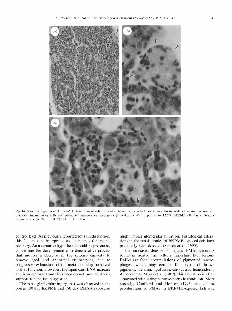
Fig. 10. Photomicrographs of A. anguilla L. liver tissue revealing altered architecture, decreased parenchyma density, isolated hepatocytes, necrosis,
pyknosis, inflammatory cells and pigmented macrophage aggregates (arrowheads) after exposure to 12.5% BKPME (30 days). Original
magnification: (A) 245� ; (B, C) 1230� . HE stain.
M. Pacheco, M.A. Santos / Ecotoxicology and Environmental Safety 53 (2002) 331–347 343
suggested that several known toxic effects of theseeffluents could contribute to this response, includingCYP1A induction, toxic hemolysis, and impairedmacrophage function. These statements are also applic-able to splenic hemosiderosis. The same authorssuggested PMA density as a useful marker of BKPMEtoxicity. In experimental groups where the highestPMA densities were found a considerable portion ofthe hepatic parenchyma was occupied by these aggre-gates. Consequently, the liver’s biotransformation ca-pacity may have been substantially reduced. Thatreduction is probably involved in the decreased resis-tance to contaminants and survival levels displayed inthe current study.The cholestasis recognizable in the liver of BaP-
treated fish is a manifestation of a pathophysiologicalcondition, attributable to a failure of the metabolism orexcretion of bile pigments. Excretion of bilirubindepends on its water solubility obtained throughconjugation with glucuronic acid. This conjugation iscatalyzed by the enzyme UDP-glucuronyl transferase. Adecreased capacity to glucuronidate bilirubin may be theprimary reason for this liver dysfunction (Nikinmaa andOikari, 1982). This decrease may be related either to aconjugation enzyme inhibition or to exhaustion of theconjugation process, particularly in terms of activatedglucuronic acid.The separation between adjacent hepatocytes ob-
served in the livers of BKPME-treated fish, probably
related to cell necrosis, indicates degeneration ofstructural proteins in the hepatocyte membrane, whichcommonly maintain the hepatic parenchyma as acompact homogeneous tissue. The time-related livervacuolization detected in DHAA-exposed fish signalsan intracellular degenerative process suggestive ofunderlying metabolic disorders. This anomaly was pre-viously noted in resin acid-treated fish (Mattsoff andOikari, 1987) and BKPME-treated fish (Axelsson andNorrgren, 1991). The hepatocyte pyknosis and necrosisdetected in our liver examination may be regarded asnonspecific lesions since they were generally found in alltreated groups. Several investigators previously relatedthis cellular deterioration to BKPME exposure (Santoset al., 1990; Axelsson and Norrgren, 1991).The previously demonstrated carcinogenicity (hepatic
and non-hepatic) of BaP (Hendricks et al., 1985;Hawkins et al., 1990) and BKPME (Smith and Rokosh,1989; Kinae et al., 1990) could not be confirmed by ourdata, probably due to the short exposures. With respectto DHAA, prolonged exposure up to 180 days did notinduce neoplastic lesions, suggesting the absence ofcarcinogenic potential or a need for longer exposures.
4.5. Interaction of biomarkers
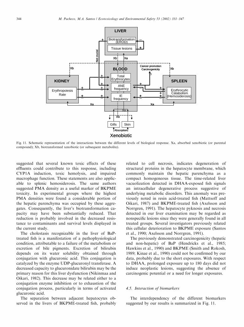
The interdependency of the different biomarkerssuggested by our results is summarized in Fig. 11.
LIVER
ANEfrequency
IEfrequency
+_
KIDNEY
ErythropoiesisRate
SPLEEN
ErythrocyticCatabolism
(Hemosiderosis)
+_
+_ +_ _ _+
TotalErythrocytes
+++_+
+
+
Tissue lesions
Biotransformation(EROD)
+
_
Cancer promotionCarcinogenicity
?
Not
cru
cial
_
+
Xenobiotic
SkinGills
XaXa
Xa
Absorption
Xb
Xb Xb
Xb
Xa
BLOOD
Fig. 11. Schematic representation of the interactions between the different levels of biological response. Xa, absorbed xenobiotic (or parental
compound); Xb, biotransformed xenobiotic (or subsequent metabolite).
M. Pacheco, M.A. Santos / Ecotoxicology and Environmental Safety 53 (2002) 331–347344
Considering that, in the vast majority of points intime, ENA induction was not associated with apreceding or simultaneous EROD induction, we suggestthat this genotoxic response does not depend on a veryhigh biotransformation activity. However, the highestENA levels were observed when fish also exhibit thehighest EROD activity levels, suggesting that, particu-larly for DHAA, a strong biotransformation activitymay potentiate the expression of genotoxicity. Ourresults also indicate that the eventual interdependencebetween the previous responses may vary with thecontaminant.Concerning the 30-day exposure, parallel analysis of
ENA and spleen histological data demonstrates that thedisturbances observed in ENA expression coincide withan accentuated hemosiderosis. Therefore, the hypothesisthat ENA expression was disturbed by increasederythrocytic catabolism seems to be confirmed. Wesuggest that the increment in cell removal may be asplenic response to the increased frequency of circulat-ing abnormal erythrocytes. This idea is based on theassumption that nuclear abnormalities induce functionaldisorders with a consequent expression on the cellmembrane, allowing their identification by the spleenand consequent catabolism. Previously, other authors(Das and Nanda, 1986; Brunetti et al., 1988) suggested adose-related cytotoxicity, which killed the erythrocytesinstead of forming a nonlethal nuclear lesion, explainthe impairment in expression of nuclear abnormalities.Therefore, to explain the simultaneous occurrence ofsplenic hemosiderosis and disturbances in ENA expres-sion, the conjugation of both mechanisms should alsobe accepted, namely, in the presence of xenobiotics withhemolytic and genotoxic properties.Our IE results for the 30-day exposures to the highest
concentrations do not refute the hypothesis that ENAexpression was also conditioned by a reduced erythro-poiesis rate. This effect may result either from inhibitionof DNA synthesis (Williams and Metcalfe, 1992) orfrom direct inhibition of erythropoiesis (Dinnen et al.,1988). With respect to the unchanged erythropoiesisrate, the increased removal of mature erythrocytesshould lead to an increase in IE frequency. However,our IE results revealed the opposite tendency, indicat-ing, therefore, the probable occurrence of simultaneouserythropoiesis inhibition. With respect to longer DHAAexposures, ENAs seem to be more easily expressedconcomitantly with regression of splenic hemosiderosis.Considering this regression and the observed IEdecrease, inhibition of erythropoiesis seems to beevident as a result of the prolonged DHAA exposure.This inhibition was not enough to prevent ENAexpression but may justify the low ENA levels observedat the highest DHAA concentration.Our data suggest that genotoxic manifestations are
not necessarily followed by induction of neoplasms.
Similarly, Metcalfe et al. (1995) detected BKPMEmutagenic activity without consequent carcinogenicity.However, these authors suggested that the effluentmight have neoplasm-promoting activity.
5. Conclusions
Our results support the importance of prolonged fishstudies in the evaluation of toxicity of environmentalcontaminants. The capacity of all contaminants testedto induce several organ and tissue lesions as well asgenotoxic effects was demonstrated. Additionally, it canbe stated that:
i. A. anguilla (glass eel) seems to be a good organismto carry out long-term studies.
ii. ENA assay was confirmed as an effective andsuitable genotoxic biomarker also applicable tolong-term exposures. However, its use may beinadequate for situations of intense contaminationsince some disturbances of genotoxicity expressionmay occur. These disturbances were confirmed todepend on the balance between increased splenicerythrocytic catabolism and erythropoiesis ratereduction. Furthermore, that balance may varywith the contaminant, concentration, and exposurelength.
iii. Our histopathological findings showed the useful-ness of nonneoplastic lesions as indicators of fishexposure to pollutants.
iv. The combination of biochemical, genetic, andhistological biomarkers provides useful and sensi-tive tools in the investigation of sublethal effectsinduced by environmental contaminants. Further-more, this strategy contributes to the establishmentof a causal relationship between contaminantexposure and various biological responses.
Acknowledgments
This research was supported by the program PRAXISXXI (Contract PCNA/C/BIA/175-96) and by theAveiro University Research Institute/CZCM.
References
Addison, R.F., Willis, D.E., Zinck, M.E., 1994. Liver microsomal
mono-oxygenase induction in winter flounder (Pseudopleuronectes
americanus) from a gradient of sediment PAH concentrations at
Sydney Harbour, Nova Scotia. Mar. Environ. Res. 37, 283–296.
Al-Sabti, K., Hardig, J., 1990. Micronucleus test in fish for monitoring
the genotoxic effects of industrial waste products in the Baltic Sea,
Sweden. Comp. Biochem. Physiol. C 97, 179–182.
M. Pacheco, M.A. Santos / Ecotoxicology and Environmental Safety 53 (2002) 331–347 345
Axelsson, B., Norrgren, L., 1991. Parasite frequency and liver
anomalies in tree-spined stickleback, Gasterosteus aculeatus (L.),
after long-term exposure to pulp mill effluents in marine
mesocosms. Arch. Environ. Contam. Toxicol. 21, 505–513.
Bailey, N.T., 1959. Statistical Methods in Biology. English University
Press, London.
Borton, D.L., Hall, T.J., 1984. Observations of the conditions of
organs and tissues of fish exposed to biologically treated kraft mill
effluent. Technical Bulletin No. 419, National Council of the Paper
Industry for Air and Stream Improvement, New York, 124pp.
Brunetti, R., Majone, F., Gola, I., Beltrame, C., 1988. The
micronucleus test: examples of application to marine ecology.
Mar. Ecol. Prog. Ser. 44, 65–68.
Burke, M.D., Mayer, R.T., 1974. Ethoxyresorufin: direct fluorometric
assay of a microsomal-O-deethylation which is preferentially
inducible by 3-methylcholanthrene. Drug. Metab. Dispos. 2,
583–588.
Bushnell, P.G., Nikinma, M., Oikari, A., 1985. Metabolic effects
of dehydroabietic acid on rainbow trout erythrocytes. Comp.
Biochem. Physiol. C 81, 391–394.
Couillard, C.M., Hodson, P.V., 1996. Pigmented macrophage aggre-
gates: a toxic response in fish exposed to bleached-kraft mill
effluent? Environ. Toxicol. Chem. 15, 1844–1854.
Das, R.K., Nanda, N.K., 1986. Induction of micronuclei in peripheral
erythrocytes of the fish, Heteropneustes fossilis, by mitomycin C
and paper mill effluent. Mutat. Res. 175, 67–71.
Di Guilio, R.T., Washburn, P.C., Wenning, R.J., Winston, G.W.,
Jewell, C.S., 1989. Biochemical responses in aquatic animals: a
review of determinants of oxidative stress. Environ. Toxicol. Chem.
8, 1103–1123.
Dinnen, R.D., Tomlinson, S.M., Hart, D., Chopra, C., Heddle, J.A.,
1988. Application of a micronucleus assay to the peripheral blood
cells of rainbow trout, Salmo gairdneri. Can. Tech. Rep. Fish.
Aquat. Sci. 1607, 69–78.
Everaarts, J.M., 1995. Dna integrity as a biomarker of marine
pollution: strand breaks in seastar (Asterias rubens) and dab
(Limanda limanda). Mar. Pollut. Bull. 31, 431–438.
Gornall, A.C., Bardawill, C.J., David, M.M., 1949. Determination of
serum proteins by means of the biuret reaction. J. Biol. Chem. 177,
751–766.
Harvey, J.S., Lyons, B.P., Waldock, M., Parry, J.M., 1997. The
application of the 32P-postlabelling assay to aquatic biomonitoring.
Mutat. Res. 38, 77–88.
Hawkins, W.E., Walker, W.W., Overstreet, R.M., Lytle, J.S., Lytle,
T.F., 1990. Carcinogenic effects of some polycyclic aromatic
hydrocarbons on the Japanese medaka and guppy in waterborne
exposures. Sci. Total Environ. 94, 155–167.
Hendricks, J.D., Meyers, T.R., Shelton, D.W., Casteel, J.L., Bailey,
G.S., 1985. Hepatocarcinogenicity of benzo[a]pyrene to rainbow
trout by dietary exposure and intraperitoneal injection. J. Natl.
Cancer Inst. 74, 839–850.
Hibiya, T., 1982. An Atlas of Fish Histology—Normal and
Pathological Features. Kodansha, Tokyo.
Howard, T.E., McLeay, D.J.,Walden, C.C., 1971. Sub-lethal effects
of bleached kraft mill effluents to fish. CPAR Project Report 9-2,
Pulp and Paper Pollution Abatement, Ottawa, Canada, 63pp.
Jaylet, A., Deparis, P., Ferrier, V., Grinfeld, S., Siboulet, R., 1986. A
new micronucleus test using peripheral blood erythrocytes of the
newt Pleurodeles waltl to detect mutagens in fresh-water pollution.
Mutat. Res. 164, 245–257.
Kinae, N., Yamashita, M., Tomita, I., Kimura, I., Ishida, H., Kumai,
H., Nakamura, G., 1990. A possible correlation between environ-
mental chemicals and pigment cell neoplasia in fish. Sci. Total
Environ. 94, 143–153.
Lange, U., Danischewski, D., Siebers, D., 1993. Regional variability
and sexual differences in ethoxyresorufin O-deethylase activities
and cytochrome P450 concentrations in the liver of mature dab
(Limanda limanda L.) in the German Bight. In: Braumbeck, T.,
Hunke, W., Segner, H. (Eds.), Fish Ecotoxicology and Ecophysiol-
ogy. VCH Verlag Chemie, Weinheim.
Martel, P.H., Kovacs, T.G., O’Connor, B.I., Voss, R.H., 1994. A
survey of pulp and paper mill effluents for their potential to induce
mixed function oxidase enzyme activity in fish. Water Res. 28,
1835–1844.
Mattsoff, L., Oikari, A., 1987. Acute hyperbilirubinaemia in rainbow
trout (Salmo gairdneri) caused by resin acid. Comp. Biochem.
Physiol. C 88, 263–268.
McLeay, D.J., 1973. Effects of a 12-hr and 25-day exposure to
kraft pulp mill effluent on the blood and tissues of juvenile
coho salmon (Oncorhynchus kisutch). J. Fish. Res. Board Can. 30,
395–400.
Metcalfe, C.D., Nanni, M.E., Scully, N.M., 1995. Carcinogenicity and
mutagenicity testing of extracts from bleached kraft mill effluent.
Chemosphere 30, 1085–1095.
Monod, G., Vindimian, E., 1991. Effect of storage conditions and
subcellular fractionation of fish and cytochrome P-450-dependent
enzymatic activities used for the monitoring of water pollution.
Water Res. 25, 173–177.
Moore, M.J., Myers, M.S., 1994. Pathobiology of chemical-associated
neoplasia in fish. In: Malins, D.C., Ostrander, G.K. (Eds.),
Aquatic Toxicology: Molecular, Biochemical, and Cellular
Perspectives. CRC Press, Lewis Publishers, Boca Raton, FL,
pp. 327–386.
Moore, M.N., Simpson, M.G., 1992. Molecular and cellular pathology
in environment impact assessment. Aquat. Toxicol. 22, 313.
Myers, M.S., Rhodes, L.D., McCain, B.B., 1987. Pathologic anatomy
and patterns of occurrence of hepatic neoplasms, putative
preneoplastic lesions, and other idiopathic hepatic conditions in
English sole (Parophrys vetulus) from Puget Sound, Washington.
J. Natl. Cancer Inst. 78 (2), 333–363.
Nikinmaa, M., Oikari, A., 1982. Physiological changes in trout (Salmo
gairdneri) during a short-term exposure to resin acids and during
recovery. Toxicol. Lett. 14, 103–110.
Pacheco, M., Santos, M.A., 1996. Induction of micronuclei and
nuclear abnormalities in the erythrocytes of Anguilla anguilla L.
exposed either to cyclophosphamide or to bleached kraft pulp mill
effluent. Fresenius Environ. Bull. 5, 746–751.
Pacheco, M., Santos, M.A., 1997. Induction of erod activity and
genotoxic effects by polycyclic aromatic hydrocarbons and resin
acids on the juvenile eel (Anguilla anguilla L.). Ecotoxicol. Environ.
Saf. 38, 252–259.
Pacheco, M., Santos, M.A., 1998. Anguilla anguilla L. liver EROD and
erythrocytic nuclear abnormalities induction by cyclophosphamide
and PAHs. Ecotoxicol. Environ. Saf. 40, 71–76.
Pacheco, M., Santos, M.A., 1999. Biochemical and genetic responses
of adult eel (Anguilla anguilla L.) to resin acids and pulp mill
effluent: laboratory and field experiments. Ecotoxicol. Environ.
Saf. 42, 81–93.
Santos, M.A., Pires, F., Hall, A., 1990. Metabolic effects of kraft mill
effluents on the eel Anguilla anguilla L. Ecotoxicol. Environ. Saf.
20, 10–19.
Schwaiger, J., Fent, K., Stecher, H., Ferling, H., Negele, R.-D., 1996.
Effects of sublethal concentrations of triphenyltriacetate on rain-
bow trout (Oncorhynchus mykiss). Arch. Environ. Contam.
Toxicol. 30, 327–334.
Schwaiger, J., Wanke, R., Adam, S., Pawert, M., Honnen, W.,
Triebskorn, R., 1997. The use of histopathological indicators to
evaluate contaminant-related stress in fish. J. Aquat. Ecosystem
Stress Recovery 6, 75–86.
Smith, I.R., 1990. Erythrocytic micronuclei in wild fish from Lakes
Superior and Ontario that have pollution-associated neoplasia.
J. Great Lakes Res. 16 (1), 139–142.
M. Pacheco, M.A. Santos / Ecotoxicology and Environmental Safety 53 (2002) 331–347346

Smith, I., Rokosh, D.A., 1989. An epidemiological study of a white
sucker (Catostomus commersoni) population inhabiting the
Kaministiquia River, Thunder Bay, Ontario. Proceedings of the
10th Annual Meeting of Society of Environmental Toxicology
Chemistry, Toronto, Ont., Canada, October 28–November 2, 1989,
p. 262.
Teh, S.J., Adams, S.M., Hinton, D.E., 1997. Histopathologic
biomarkers in feral freshwater fish populations exposed to different
types of contaminants stress. Aquat. Toxicol. 37, 51–70.
Toivola, D.M., Isomaa, B., 1991. Effects of dehydroabietic acid on the
erythrocyte membrane. Chem.–Biol. Interact. 79, 65–78.
Tuurala, H., Soivio, A., 1982. Structural and circulatory changes in the
secondary lamellae of Salmo gairdneri gills after sublethal exposure
to dehydroabietic acid and zinc. Aquat. Toxicol. 2, 21–29.
Williams, R.C., Metcalfe, C.D., 1992. Development of an in vivo
hepatic micronucleus assay with rainbow trout. Aquat. Toxicol. 23,
193–202.
Wolkers, J., J�rgensen, E.H., Nijmeijer, S.M., Witkamp, R.F., 1996.
Time-dependent induction of two distinct hepatic cytochrome
P4501A catalytic activities at low temperatures in Arctic char
(Salvelinus alpinus) after oral exposure to benzo[a]pyrene. Aquat.
Toxicol. 35, 127–138.
M. Pacheco, M.A. Santos / Ecotoxicology and Environmental Safety 53 (2002) 331–347 347
Related Documents