153 4 Biotransformation in Fishes Daniel Schlenk, Malin Celander, Evan Gallagher, Stephen George, Margaret James, Seth Kullman, Peter van den Hurk, and Kristie Willett CONTENTS Introduction ............................................................................................................................................ 154 Phase I Reactions ................................................................................................................................... 154 Oxidation ...................................................................................................................................... 154 Cytochrome P450 Family of Drug Metabolizing Enzymes .............................................. 154 Flavin-Containing Monooxygenases .................................................................................. 175 Monoamine Oxidases ......................................................................................................... 178 Alcohol and Aldehyde Dehydrogenases ............................................................................ 178 Peroxidases ......................................................................................................................... 179 Aldehyde Oxidase............................................................................................................... 179 Reductases .................................................................................................................................... 179 DT-Diaphorase .................................................................................................................... 179 Azo- and Nitroreductases ................................................................................................... 179 Hydrolysis..................................................................................................................................... 180 Epoxide Hydrolase.............................................................................................................. 180 Carboxylesterases ............................................................................................................... 182 Phase II Enzymes................................................................................................................................... 183 UDP-Glucuronosyltransferases .................................................................................................... 183 Overview ............................................................................................................................. 183 UGT Gene Structure........................................................................................................... 184 Reactions and Substrate Specificity ................................................................................... 185 Enzymology of Piscine UGTs............................................................................................ 187 Tissue Distribution .............................................................................................................. 188 Regulation of UGTs ........................................................................................................... 188 Inhibition of UGTs ............................................................................................................. 189 Glutathione S-Transferases .......................................................................................................... 190 Overview ............................................................................................................................. 190 GST Gene Structure ........................................................................................................... 191 Reactions and Substrate Specificity ................................................................................... 193 Fish GST and Oxidative Stress .......................................................................................... 194 Tissue Distribution .............................................................................................................. 196 Regulation of GSTs ............................................................................................................ 197 Inhibition of GSTs .............................................................................................................. 199 Sulfotransferase ............................................................................................................................ 199 Overview ............................................................................................................................. 199 Gene Structure of SULT..................................................................................................... 201 Piscine SULTs..................................................................................................................... 203 Reactions and Substrate Specificity ................................................................................... 203 Tissue Distribution .............................................................................................................. 204 Regulation of SULT............................................................................................................ 204 Inhibition of SULT ............................................................................................................. 205 Activation of SULT ............................................................................................................ 205

Welcome message from author
This document is posted to help you gain knowledge. Please leave a comment to let me know what you think about it! Share it to your friends and learn new things together.
Transcript
153
4
Biotransformation in Fishes
Daniel Schlenk, Malin Celander, Evan Gallagher, Stephen George, Margaret James, Seth Kullman, Peter van den Hurk, and Kristie Willett
CONTENTS
Introduction ............................................................................................................................................ 154Phase I Reactions................................................................................................................................... 154
Oxidation ...................................................................................................................................... 154Cytochrome P450 Family of Drug Metabolizing Enzymes .............................................. 154Flavin-Containing Monooxygenases .................................................................................. 175Monoamine Oxidases ......................................................................................................... 178Alcohol and Aldehyde Dehydrogenases ............................................................................ 178Peroxidases ......................................................................................................................... 179Aldehyde Oxidase............................................................................................................... 179
Reductases .................................................................................................................................... 179DT-Diaphorase .................................................................................................................... 179Azo- and Nitroreductases ................................................................................................... 179
Hydrolysis..................................................................................................................................... 180Epoxide Hydrolase.............................................................................................................. 180Carboxylesterases ............................................................................................................... 182
Phase II Enzymes................................................................................................................................... 183UDP-Glucuronosyltransferases .................................................................................................... 183
Overview............................................................................................................................. 183UGT Gene Structure........................................................................................................... 184Reactions and Substrate Specificity ................................................................................... 185Enzymology of Piscine UGTs............................................................................................ 187Tissue Distribution.............................................................................................................. 188Regulation of UGTs ........................................................................................................... 188Inhibition of UGTs ............................................................................................................. 189
Glutathione S-Transferases .......................................................................................................... 190Overview............................................................................................................................. 190GST Gene Structure ........................................................................................................... 191Reactions and Substrate Specificity ................................................................................... 193Fish GST and Oxidative Stress .......................................................................................... 194Tissue Distribution.............................................................................................................. 196Regulation of GSTs ............................................................................................................ 197Inhibition of GSTs.............................................................................................................. 199
Sulfotransferase ............................................................................................................................ 199Overview............................................................................................................................. 199Gene Structure of SULT..................................................................................................... 201Piscine SULTs..................................................................................................................... 203Reactions and Substrate Specificity ................................................................................... 203Tissue Distribution.............................................................................................................. 204Regulation of SULT............................................................................................................ 204Inhibition of SULT ............................................................................................................. 205Activation of SULT ............................................................................................................ 205
154
The Toxicology of Fishes
Amino Acid Conjugation ............................................................................................................. 205Overview............................................................................................................................. 205Enzymes Specificity, Regulation, and Inhibition ............................................................... 206
Acetylation ................................................................................................................................... 206Overview............................................................................................................................. 206Enzyme Specificity, Regulation, and Inhibition................................................................. 207
Toxicological Relevance ........................................................................................................................ 207Benzo(
a
)pyrene ............................................................................................................................ 207Aflatoxin B
1
.................................................................................................................................. 209Organophosphate Esters and Carbamates.................................................................................... 213
Conclusions ............................................................................................................................................ 215References .............................................................................................................................................. 216
Introduction
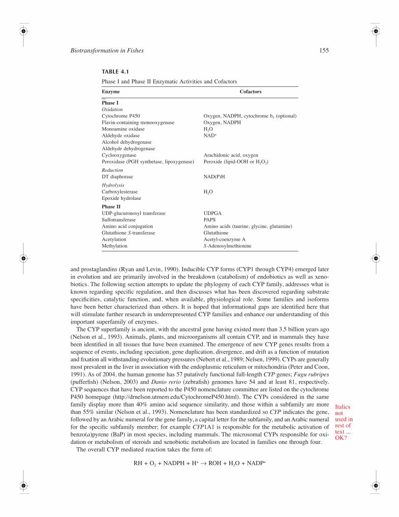
Biotransformation is a two-phase process catalyzed primarily through enzymatic reactions that oftenradically alter the chemistry of nonpolar lipophilic chemicals to polar water-soluble metabolites pre-dominately leading to detoxification and elimination of the parent compounds. Unfortunately, the alter-ation of chemistry required for enhanced polarity often creates reactive intermediates throughbioactivation, which can be more biologically hazardous than the initial parent compounds. The phaseI process either adds or exposes polar atoms within a xeno- or endobiotic compound. Three generalphase I reactions include oxidation, reduction, and hydrolysis (Table 4.1). When polarity has beenenhanced through phase I reactions, phase II reactions generally attempt to further enhance polaritythrough conjugation of the phase I product with a bulky polar endogenous molecule. Alternatively, phaseII reactions may protect against bioactivation by masking functional groups (i.e., amines) prone toreactive intermediate formation with groups that likely provide steric hindrance (i.e., methyl, acetyl)rather than augmented polarity (Table 4.1).
Phase I Reactions
Oxidation
Various enzymes are involved in the oxidation of xeno- and endobiotic compounds. Dehydrogenases oxidizesubstrates transferring electrons to an electron-deficient acceptor that is typically an essential cofactor forcatalysis (e.g., NAD
+
). Oxygenases catalyze the incorporation of molecular oxygen into molecules, andwater is the source of oxygen for oxidases. Peroxidases derive oxygen from peroxide cofactors.
Cytochrome P450 Family of Drug Metabolizing Enzymes
Overview
The most dominant enzyme system responsible for oxidation processes in phase I biotransformation isthe cytochrome P450 monooxygenases. The cytochrome P450s (CYPs) constitute a superfamily of heme-containing proteins that catalyze biological oxidation and reduction reactions. Klingenberg (1958) andGarfinkel (1958) first reported that hepatic microsomes contain a pigment that binds carbon monooxidewith an unusual visible absorption maximum at 450 nm in its CO-reduced difference spectrum. Omuraand Sato (1962) discovered that this pigment was a b-type cytochrome and called it cytochrome P450.The hepatic microsomal CYP system has broad substrate specificity and is responsible for oxidativemetabolism of many structurally diverse endogenous and xenobiotic compounds. CYP enzymes areimportant for converting lipophilic foreign chemicals into more water-soluble products for excretionand, hence, detoxification. On the other hand, CYP enzymes catalyze the conversion of certain com-pounds such as polycyclic aromatic hydrocarbons (PAHs) and nitrosamines into more toxic intermediates.Constitutive CYP forms that appeared early in evolution are involved in biosynthesis (anabolism) ofendogenous substances such as steroids, fatty acids, vitamins, bile acids, leukotrienes, thromboxanes,
Biotransformation in Fishes
155
and prostaglandins (Ryan and Levin, 1990). Inducible CYP forms (CYP1 through CYP4) emerged laterin evolution and are primarily involved in the breakdown (catabolism) of endobiotics as well as xeno-biotics. The following section attempts to update the phylogeny of each CYP family, addresses what isknown regarding specific regulation, and then discusses what has been discovered regarding substratespecificities, catalytic function, and, when available, physiological role. Some families and isoformshave been better characterized than others. It is hoped that informational gaps are identified here thatwill stimulate further research in underrepresented CYP families and enhance our understanding of thisimportant superfamily of enzymes.
The CYP superfamily is ancient, with the ancestral gene having existed more than 3.5 billion years ago(Nelson et al., 1993). Animals, plants, and microorganisms all contain CYP, and in mammals they havebeen identified in all tissues that have been examined. The emergence of new CYP genes results from asequence of events, including speciation, gene duplication, divergence, and drift as a function of mutationand fixation all withstanding evolutionary pressures (Nebert et al., 1989; Nelsen, 1999). CYPs are generallymost prevalent in the liver in association with the endoplasmic reticulum or mitochondria (Peter and Coon,1991). As of 2004, the human genome has 57 putatively functional full-length
CYP
genes;
Fugu rubripes
(pufferfish) (Nelson, 2003) and
Danio rerio
(zebrafish) genomes have 54 and at least 81, respectively.CYP sequences that have been reported to the P450 nomenclature committee are listed on the cytochromeP450 homepage (http://drnelson.utmem.edu/CytochromeP450.html). The CYPs considered in the samefamily display more than 40% amino acid sequence similarity, and those within a subfamily are morethan 55% similar (Nelson et al., 1993). Nomenclature has been standardized so
CYP
indicates the gene,followed by an Arabic numeral for the gene family, a capital letter for the subfamily, and an Arabic numeralfor the specific subfamily member; for example
CYP
1A1 is responsible for the metabolic activation ofbenzo(
a
)pyrene (BaP) in most species, including mammals. The microsomal CYPs responsible for oxi-dation or metabolism of steroids and xenobiotic metabolism are located in families one through four.
The overall CYP mediated reaction takes the form of:
RH + O
2
+ NADPH + H
+
→
ROH + H
2
O + NADP
+
TABLE 4.1
Phase I and Phase II Enzymatic Activities and Cofactors
Enzyme Cofactors
Phase I
Phase I
Oxidation
Cytochrome P450 Oxygen, NADPH, cytochrome b
5
(optional)Flavin-containing monooxygenase Oxygen, NADPHMonoamine oxidase H
2
OAldehyde oxidaseAlcohol dehydrogenaseAldehyde dehydrogenase
NAD
+
Cyclooxygenase Arachidonic acid, oxygenPeroxidase (PGH synthetase, lipoxygenase) Peroxide (lipid-OOH or H
2
O
2
)
Reduction
DT diaphorase NAD(P)H
Hydrolysis
CarboxylesteraseEpoxide hydrolase
H
2
O
Phase II
UDP-glucuronosyl transferase UDPGASulfotransferase PAPSAmino acid conjugation Amino acids (taurine, glycine, glutamine)Glutathione
S
-transferase GlutathioneAcetylation Acetyl-coenzyme AMethylation
S
-Adenosylmethionine
Italicsnotused inrest oftext ...OK?
156
The Toxicology of Fishes
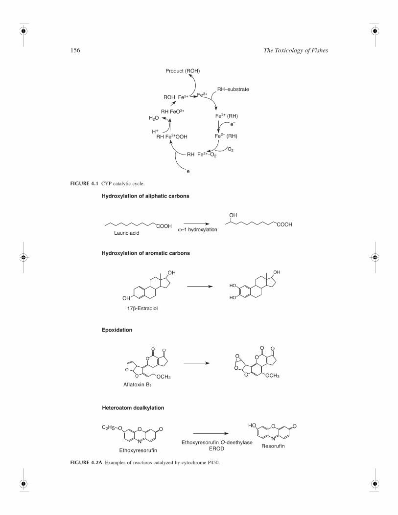
FIGURE 4.1
CYP catalytic cycle.
FIGURE 4.2A
Examples of reactions catalyzed by cytochrome P450.
Product (ROH)
RH–substrate
ROH Fe3+
RH FeO3+
RH Fe2+–O2
H+RH Fe2+OOH Fe2+ (RH)
Fe3+ (RH)H2O
Fe3+
O2
e–
e–
Hydroxylation of aliphatic carbons
Hydroxylation of aromatic carbons
Epoxidation
Heteroatom dealkylation
Lauric acid
Aflatoxin B1
EthoxyresorufinResorufin
Ethoxyresorufin O-deethylase EROD
17β-Estradiol
ω–1 hydroxylationCOOH COOH
OH
OHOH
HO
HO
OO O
O
OO
O OHO
N
OO O
N
OO
O
O O
OCH3
C2H5
OCH3
OH
Biotransformation in Fishes
157
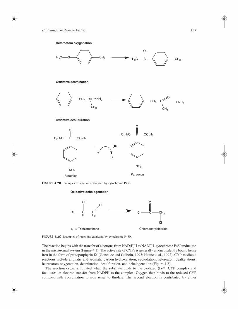
The reaction begins with the transfer of electrons from NAD(P)H to NADPH–cytochrome P450 reductasein the microsomal system (Figure 4.1). The active site of CYPs is generally a noncovalently bound hemeiron in the form of protoporphyrin IX (Gonzalez and Gelboin, 1993; Henne et al., 1992). CYP-mediatedreactions include aliphatic and aromatic carbon hydroxylation, epoxidation, heteroatom dealkylations,heteroatom oxygenation, deamination, desulfuration, and dehalogenation (Figure 4.2).
The reaction cycle is initiated when the substrate binds to the oxidized (Fe
+3
) CYP complex andfacilitates an electron transfer from NADPH to the complex. Oxygen then binds to the reduced CYPcomplex with coordination to iron
trans
to thiolate. The second electron is contributed by either
FIGURE 4.2B
Examples of reactions catalyzed by cytochrome P450.
FIGURE 4.2C
Examples of reactions catalyzed by cytochrome P450.
Heteroatom oxygenation
Oxidative deamination
Oxidative desulfuration
Parathion Paraoxon
H3C CH3
CH2
NO2
NO2
CH2
C2H5O OC2H5
C2H5O OC2H5P
PS
H3C CH3
+ NH3
NH2CH
CH3CH3
S
O
O
O
O
C
S
S
S
CH2H2H
Cl
C
O
ClCC
Cl
Cl
Cl
Cl
Oxidative dehalogenation
Chloroacetylchloride1,1,2–Trichloroethane
158
The Toxicology of Fishes
NADPH–cytochrome P450 reductase or NADH–cytochrome b
5
reductase. The next step involves cleav-age of the oxygen–oxygen bond, uptake of two protons, and release of water. Oxygen is inserted intothe substrate through generation of hydroxyl and carbon free radicals. Finally, dissociation of ROHrestores the P450 to the initial ferric state (Parkinson, 2001). In addition, the peroxide shunt can allowa peroxy compound to substitute for oxygen in substrate oxidation.
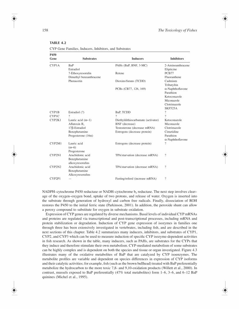
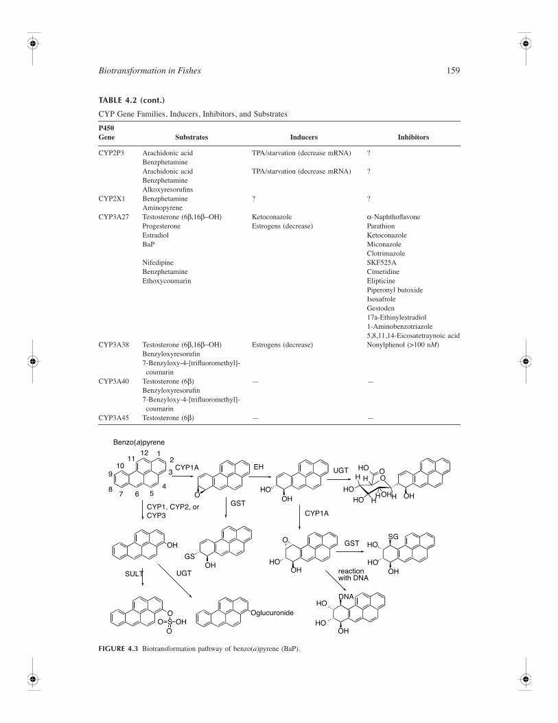
Expression of CYP genes are regulated by diverse mechanisms. Basal levels of individual CYP mRNAsand proteins are regulated via transcriptional and post-transcriptional processes, including mRNA andprotein stabilization or degradation. Induction of CYP gene expression of isozymes in families onethrough three has been extensively investigated in vertebrates, including fish, and are described in thenext sections of this chapter. Table 4.2 summarizes many inducers, inhibitors, and substrates of CYP1,CYP2, and CYP3 which can be used to measure induction of specific CYP isozyme-dependent activitiesin fish research. As shown in the table, many inducers, such as PAHs, are substrates for the CYPs thatthey induce and therefore stimulate their own metabolism. CYP-mediated metabolism of some substratescan be highly complex and is dependent on both the species and tissue or organ investigated. Figure 4.3illustrates many of the oxidative metabolites of BaP that are catalyzed by CYP isoenzymes. Themetabolite profiles are variable and dependent on species differences in expression of CYP isoformsand their catalytic activities; for example, fish (such as the brown bullhead) treated with BaP preferentiallymetabolize the hydrocarbon to the more toxic 7,8- and 9,10-oxidation products (Willett et al., 2000). Incontrast, mussels exposed to BaP preferentially (47% total metabolites) form 1–6, 3–6, and 6–12 BaPquinines (Michel et al., 1995).
TABLE 4.2
CYP Gene Families, Inducers, Inhibitors, and Substrates
P450 Gene Substrates Inducers Inhibitors
CYP1A BaPEstradiol
PAHs (BaP, BNF, 3-MC) 2-AminoanthraceneElipticine
7-EthoxyresorufinDimethyl benzanthracene
Retene PCB77Fluoranthene
Phenacetin Dioxins/furans (TCDD) CadmiumTributyltin
PCBs (CB77, 126, 169)
α
-NaphthoflavoneParathionKetoconazoleMiconazoleClotrimazoleSKF525A
CYP1B Estradiol (?) BaP, TCDD ?CYP1C ? ? ?CYP2K1 Lauric acid (
ω−1)
Aflatoxin B
1
17
β
-EstradiolBenzphetamineProgesterone (16
α
)
Diethyldithiocarbamate (activator)BNF (decrease)Testosterone (decrease mRNA)Estrogens (decrease protein)
KetoconazoleMiconazoleClotrimazoleCimetidineParathion
α
-NaphthoflavoneCYP2M1 Lauric acid
(
ω−6)
Progesterone
Estrogens (decrease protein) ?
CYP2N1 Arachidonic acidBenzphetaminealkoxyresorufins
TPA/starvation (decrease mRNA) ?
CYP2N2 Arachidonic acidBenzphetamineAlkoxyresorufins
TPA/starvation (decrease mRNA) ?
CYP2P1 — Fasting/refeed (increase mRNA) ?
Biotransformation in Fishes
159
TABLE 4.2 (cont.)
CYP Gene Families, Inducers, Inhibitors, and Substrates
P450 Gene Substrates Inducers Inhibitors
CYP2P3 Arachidonic acidBenzphetamine
TPA/starvation (decrease mRNA) ?
Arachidonic acidBenzphetamineAlkoxyresorufins
TPA/starvation (decrease mRNA) ?
CYP2X1 BenzphetamineAminopyrene
? ?
CYP3A27 Testosterone (6
β,16β−ΟΗ)
ProgesteroneEstradiolBaP
KetoconazoleEstrogens (decrease)
α
-NaphthoflavoneParathionKetoconazoleMiconazoleClotrimazole
NifedipineBenzphetamineEthoxycoumarin
SKF525ACimetidineElipticinePiperonyl butoxideIsosafroleGestoden17a-Ethinylestradiol1-Aminobenzotriazole5,8,11,14-Eicosatetraynoic acid
CYP3A38 Testosterone (6
β,16β−ΟΗ)
Benzyloxyresorufin7-Benzyloxy-4-[trifluoromethyl]-coumarin
Estrogens (decrease) Nonylphenol (>100 n
M
)
CYP3A40 Testosterone (6
β)
Benzyloxyresorufin7-Benzyloxy-4-[trifluoromethyl]-coumarin
— —
CYP3A45 Testosterone (6
β)
— —
FIGURE 4.3
Biotransformation pathway of benzo(
a
)pyrene (BaP).
Benzo(a)pyrene
CYP1, CYP2, orCYP3
GST
HO
CYP1A
O
12
3
4567
8
910
1112
EH UGTH H
HO
HO
HO HH H OHOH
OO
OH
CYP1A
SULT UGTOH
GS
OH
OH
OHHO
HOSG
OHHO
HODNA
reactionwith DNA
OglucuronideOS OHOO
O
HO
GST
160
The Toxicology of Fishes
CYP1
Phylogeny —
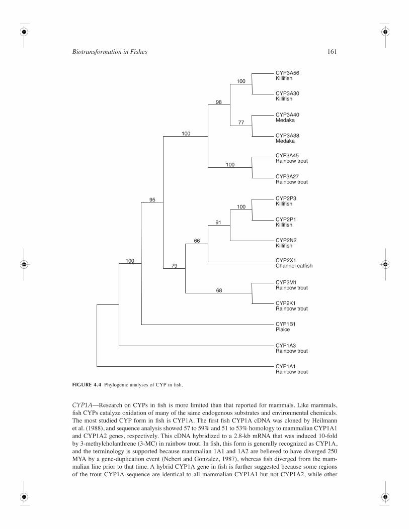
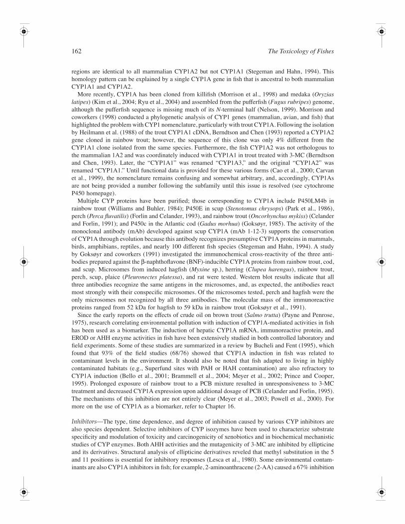
CYP1 split from the CYP2 family approximately 450 million years ago (MYA) (Nelsonet al., 1993) (Figure 4.4). The CYP1 gene family represents one of the most studied families of CYPin fish, primarily with regard to the role that CYP1A subfamily members (see below) play in thebiotransformation of environmentally persistent aromatic hydrocarbons (e.g., TCDD, PAHs) and theirrelationship with disease processes resulting from exposure to compounds of this nature (Stegman andHahn, 1994).
Regulation—
Expression of CYP genes are regulated by diverse mechanisms. Basal levels of individualCYP mRNAs and proteins are regulated via transcriptional and post-transcriptional processes, includingmRNA and protein stabilization or degradation. CYP1 gene expression is induced by structurally diversearomatic hydrocarbons wherein the induction response requires initial binding to the aryl hydrocarbonreceptor (AhR). The AhR is a basic helix–loop–helix DNA-binding protein that has been extensivelycharacterized in laboratory animals and human cell lines. In mammals, the AhR controls the transcriptionof the genes CYP1A1, CYP1A2, CYP1B1, as well as phase II enzymes such as glutathione
S
-transferase,uridine diphosphate–glucuronosyl transferase, and aldehyde-3-dehydrogenase (Safe, 1995). Agonists forthe AhR include both synthetic and naturally occurring compounds. The best characterized ligandsinclude certain PAHs and planar halogenated aromatic hydrocarbons (HAHs), including polychlorinateddibenzo-
p
-dioxins (PCDDs) and -furans (PCDFs), as well as polychlorinated biphenyls (PCBs). PlanarHAHs bind to the AhR in predictable structure–activity relationships. 2,3,7,8-Tetrachlorodibenzo-
p
-dioxin (TCDD) has the highest AhR binding affinity among the HAHs and is also correlated with thehigh toxic potency of this congener (Safe, 1990). Structure–activity relationships are not as clear-cut forthe PAHs because the toxic and genotoxic responses induced by these compounds are not all AhRmediated; however, Billiard and coworkers (2002) found a positive relationship between PAH bindingto the teleost AhR and PAH potency for CYP1A induction. More recently, numerous naturally occurringdietary AhR ligands have been identified, including indole 3-carbinol, indolo-(3,2-b)-carbazole, diben-zoylmethanes, curcumin, and carotinoids (Denison and Nagy, 2003; Jeuken et al., 2003).
Studies with the AhR
–/–
knockout mouse have shown that this protein plays an important role in normalembryonic development (Abbott et al., 1995) and development of the liver and immune system (Fernan-dez-Salguero et al., 1995). The AhR was first identified in 1976 in hepatic cytosol from C57BL/6 mice(Poland et al., 1976). Apparent molecular masses of the photoaffinity-labeled cytosolic AhR are highlyspecies dependent, ranging from 95 kDa for mouse to 124 kDa for hamster (Safe, 1995). The AhR hasbeen identified by photoaffinity labeling in several species of teleosts and elasmobranchs (Hahn et al.,1994), as well as in the fish cell lines PLHC-1 and RTG-2 (Hahn et al., 1993). Nuclear AhR levels foundin the killifish (
Fundulus heteroclitus
) were 203 fmol/mg, which was relatively high compared to mostrodent species (Willett et al., 1995). Whereas mammals have a single AhR gene, two genes (AhR1 andAhR2) have been cloned in killifish (Karchner et al., 1999), zebrafish (Andreasen et al., 2002), andrainbow trout (
Oncorhynchus mykiss
) (Abnet et al., 1999). Four forms of the AhR (AhR1
α
,
β
; AhR2
α
,
β
) are found in Atlantic salmon (
Salmo salar
L.) (Hansson et al., 2004). The presence of AhR in fishindicates that this protein has been conserved for at least 450 million years. For more on AhR-mediatedtoxicities, refer to Chapter 5.
A simplified mechanism of AhR-mediated induction of CYP1A involves a ligand entering the cellwhere it associates with the cytosolic AhR, which exists as a multiprotein complex with two moleculesof heat-shock protein 90 (Hsp90), the X-associated protein 2 (XAP2), and a 23-kDa co-chaperone protein(p23) (Denison and Nagy, 2003). Binding of ligand causes a conformational change that facilitates nucleartranslocation and association with the AhR nuclear translocator (ARNT). The AhR/ARNT heterodimer,in turn, associates with dioxin response elements (DREs; sometimes called AhR response elements, orAhREs) and with various coregulators (Carlson and Perdew, 2002) to initiate transcription and translationof AhR-responsive genes, including CYP1s. Induction of CYP1 protein can be determined at the tran-scriptional (mRNA) level using northern blot or quantitative reverse transcription polymerase chainreaction (RT-PCR) and at the protein level by using western blot, enzyme-linked immunosorbent assays(ELISAs), or immunohistochemistry. Finally, CYP1-dependent catalytic activities such as aryl hydrocar-bon hydroxylase (AHH) or ethoxyresorufin-
O
-deethylase (EROD) can also be used to measure induction.
Biotransformation in Fishes
161
CYP1A—
Research on CYPs in fish is more limited than that reported for mammals. Like mammals,fish CYPs catalyze oxidation of many of the same endogenous substrates and environmental chemicals.The most studied CYP form in fish is CYP1A. The first fish CYP1A cDNA was cloned by Heilmannet al. (1988), and sequence analysis showed 57 to 59% and 51 to 53% homology to mammalian CYP1A1and CYP1A2 genes, respectively. This cDNA hybridized to a 2.8-kb mRNA that was induced 10-foldby 3-methylcholanthrene (3-MC) in rainbow trout. In fish, this form is generally recognized as CYP1A,and the terminology is supported because mammalian 1A1 and 1A2 are believed to have diverged 250MYA by a gene-duplication event (Nebert and Gonzalez, 1987), whereas fish diverged from the mam-malian line prior to that time. A hybrid CYP1A gene in fish is further suggested because some regionsof the trout CYP1A sequence are identical to all mammalian CYP1A1 but not CYP1A2, while other
FIGURE 4.4
Phylogenic analyses of CYP in fish.
CYP3A56Killifish
CYP3A30Killifish
CYP2P3Killifish
CYP2P1Killifish
CYP2N2Killifish
CYP2X1Channel catfish
CYP3A40Medaka
CYP3A38Medaka
CYP3A45Rainbow trout
CYP3A27Rainbow trout
CYP2M1Rainbow trout
CYP2K1Rainbow trout
CYP1A3Rainbow trout
CYP1A1Rainbow trout
CYP1B1Plaice
10079
68
66
91
100
100
100
100
95
98
77
162
The Toxicology of Fishes
regions are identical to all mammalian CYP1A2 but not CYP1A1 (Stegeman and Hahn, 1994). Thishomology pattern can be explained by a single CYP1A gene in fish that is ancestral to both mammalianCYP1A1 and CYP1A2.
More recently, CYP1A has been cloned from killifish (Morrison et al., 1998) and medaka (
Oryziaslatipes
) (Kim et al., 2004; Ryu et al., 2004) and assembled from the pufferfish (
Fugus rubripes
) genome,although the pufferfish sequence is missing much of its
N
-terminal half (Nelson, 1999). Morrison andcoworkers (1998) conducted a phylogenetic analysis of CYP1 genes (mammalian, avian, and fish) thathighlighted the problem with CYP1 nomenclature, particularly with trout CYP1A. Following the isolationby Heilmann et al. (1988) of the trout CYP1A1 cDNA, Berndtson and Chen (1993) reported a CYP1A2gene cloned in rainbow trout; however, the sequence of this clone was only 4% different from theCYP1A1 clone isolated from the same species. Furthermore, the fish CYP1A2 was not orthologous tothe mammalian 1A2 and was coordinately induced with CYP1A1 in trout treated with 3-MC (Berndtsonand Chen, 1993). Later, the “CYP1A1” was renamed “CYP1A3,” and the original “CYP1A2” wasrenamed “CYP1A1.” Until functional data is provided for these various forms (Cao et al., 2000; Carvanet al., 1999), the nomenclature remains confusing and somewhat arbitrary, and, accordingly, CYP1Asare not being provided a number following the subfamily until this issue is resolved (see cytochromeP450 homepage).
Multiple CYP proteins have been purified; those corresponding to CYP1A include P450LM4b inrainbow trout (Williams and Buhler, 1984); P450E in scup (
Stenotomus chrysops
) (Park et al., 1986),perch (
Perca fluvatilis
) (Forlin and Celander, 1993), and rainbow trout (
Oncorhynchus mykiss
) (Celanderand Forlin, 1991); and P450c in the Atlantic cod (
Gadus morhua
) (Goksøyr, 1985). The activity of themonoclonal antibody (mAb) developed against scup CYP1A (mAb 1-12-3) supports the conservationof CYP1A through evolution because this antibody recognizes presumptive CYP1A proteins in mammals,birds, amphibians, reptiles, and nearly 100 different fish species (Stegeman and Hahn, 1994). A studyby Goksøyr and coworkers (1991) investigated the immunochemical cross-reactivity of the three anti-bodies prepared against the
β
-naphthoflavone (BNF)-inducible CYP1A proteins from rainbow trout, cod,and scup. Microsomes from induced hagfish (
Myxine
sp.), herring (
Clupea harengus
), rainbow trout,perch, scup, plaice (
Pleuronectes platessa
), and rat were tested. Western blot results indicate that allthree antibodies recognize the same antigens in the microsomes, and, as expected, the antibodies reactmost strongly with their conspecific microsomes. Of the microsomes tested, perch and hagfish were theonly microsomes not recognized by all three antibodies. The molecular mass of the immunoreactiveproteins ranged from 52 kDa for hagfish to 59 kDa in rainbow trout (Goksøyr et al., 1991).
Since the early reports on the effects of crude oil on brown trout (
Salmo trutta
) (Payne and Penrose,1975), research correlating environmental pollution with induction of CYP1A-mediated activities in fishhas been used as a biomarker. The induction of hepatic CYP1A mRNA, immunoreactive protein, andEROD or AHH enzyme activities in fish have been extensively studied in both controlled laboratory andfield experiments. Some of these studies are summarized in a review by Bucheli and Fent (1995), whichfound that 93% of the field studies (68/76) showed that CYP1A induction in fish was related tocontaminant levels in the environment. It should also be noted that fish adapted to living in highlycontaminated habitats (e.g., Superfund sites with PAH or HAH contamination) are also refractory toCYP1A induction (Bello et al., 2001; Brammell et al., 2004; Meyer et al., 2002; Prince and Cooper,1995). Prolonged exposure of rainbow trout to a PCB mixture resulted in unresponsiveness to 3-MCtreatment and decreased CYP1A expression upon additional dosage of PCB (Celander and Forlin, 1995).The mechanisms of this inhibition are not entirely clear (Meyer et al., 2003; Powell et al., 2000). Formore on the use of CYP1A as a biomarker, refer to Chapter 16.
Inhibitors—
The type, time dependence, and degree of inhibition caused by various CYP inhibitors arealso species dependent. Selective inhibitors of CYP isozymes have been used to characterize substratespecificity and modulation of toxicity and carcinogenicity of xenobiotics and in biochemical mechanisticstudies of CYP enzymes. Both AHH activities and the mutagenicity of 3-MC are inhibited by ellipticineand its derivatives. Structural analysis of ellipticine derivatives reveled that methyl substitution in the 5and 11 positions is essential for inhibitory responses (Lesca et al., 1980). Some environmental contam-inants are also CYP1A inhibitors in fish; for example, 2-aminoanthracene (2-AA) caused a 67% inhibition

Biotransformation in Fishes
163
of BNF-induced EROD activity in channel catfish (
Ictalurus punctatus
) (Watson et al., 1995). In both
in vitro
and
in vivo
experiments, 2-AA was a mechanism-based inhibitor of CYP1A. Similarly, 3,3
′
,4,4
′
-tetrachlorinated biphenyl (PCB 77) at high doses causes competitive inhibition of CYP1A enzymeactivity and decreased induction of CYP1A protein (Gooch et al., 1989; White et al., 1997b). White andcoworkers’ (1997a) data suggested that, in scup, tetrachlorinated biphenyl (TCB) decreased CYP1Aprotein by enhancing protein degradation. TCB also initiated redox cycling through an uncoupling ofCYP1A (Schlezinger and Stegeman, 2001). Finally, in killifish cotreated with BaP and the four-ringPAH fluoranthene (FL), both hepatic EROD activity and CYP1A immunoreactive protein levels weresignificantly inhibited (Willett et al., 2001). Although a covalent interaction between FL and CYP1Awas not detected, the relative composition of DNA adducts changed in cotreated fish, suggesting thatBaP metabolism is significantly affected when fish are co-exposed to PAH mixtures. Antifungal imida-zoles (clotrimazole and ketoconazole) inhibit CYP1A-mediated EROD activities in gizzard shad (
Dor-osoma cepedianum
), rainbow trout, and Atlantic cod (Hasselberg et al., 2004; Hegelund et al., 2004;Levine et al., 1997). Acrylamide, an environmental contaminant, is also an inhibitor of 1A and suggestsfeedback regulation on 1A mRNA transcription (Haasch et al., 1992; Petersen and Lech, 1987).
CYP1B—
Until the mid-1990s, the CYP1 gene family was believed to contain a single subfamily withthe well-known members CYP1A1 and CYP1A2; however, in 1994, the cDNA for CYP1B1 was isolatedfrom TCDD-induced human keratinocyte cells (Sutter et al., 1994). Human CYP1B1 is a single-copygene that is located on chromosome 2. It contains three exons and two introns and generates a 5.2-kbmRNA (Murray et al., 2001). In mammals, CYP1B1 expression is high in vascular endothelial cells,breast, prostate, uterus, epithelial lining of the head and neck, and the adrenal cortex (Nebert et al.,2004). CYP1B1 protein has also been reported in human breast, kidney, lung, brain, and testis tumors(McFadyen et al., 2001; Murray et al., 2001). Recombinant human CYP1B1 is highly active in oxidizingthe potent PAHs BaP and dimethylbenzanthracene (DMBA) to their respective carcinogenic metabolites.Shimada and coworkers (1999) found that CYP1B1 was more active than CYP1A1 in metabolizing BaPto the proximate toxicant BaP-7,8-diol. Their study suggests that species or tissues with less CYP1B1may be less likely to form DNA-reactive PAH metabolites and thereby may be more resistant tocarcinogenesis. This finding is supported by studies with CYP1B1-null mice. Seventy percent of DMBA-treated wild-type mice developed highly malignant lymphomas, whereas the CYP1B1-null mice onlyhad 7.5% cancer incidence (Buters et al., 1999). Likewise, metabolism of DMBA to toxic intermediatesin MCF7 and T47D breast cancer cells is blocked by CYP1B1 antibodies (Angus et al., 1999; Christouet al., 1994).
A significant void in CYP1B1 research exists in nonmammalian species such as fish. There are onlytwo published studies where CYP1B was studied in four fish species: scup, killifish, zebrafish (
Daniorerio
) (Godard et al., 2000), and plaice (Leaver and George, 2000). The plaice CYP1B has two protein-coding exons with similar exon–intron boundaries compared to the human CYP1B1. The amino acidsequence of the plaice has 54% identity with the human CYP1B1 sequence but only 39% identity withthe plaice CYP1A (Leaver and George, 2000). CYP1B was detected in plaice gill by northern blot butdid not appear to be induced by BNF. For carp (
Cyprinus carpio
), two partial CYP1B and two CYP1Csequences have been submitted to GenBank. Similarly, scup genes and the killifish partial sequencerecently have been reclassified CYP1C1 and CYP1C2, suggesting that the genes have less than 55%amino acid identity with either the plaice or mammalian CYP1Bs. The cloned channel catfish and brownbullhead (
Ameiurus nebulosus
) CYP1B genes are 71, 61, and 55% similar, respectively, to carp, plaice,and human CYP1B1 (accession number DQ088663). Currently, none of the CYP1B-dependent metabolicstudies has been done in fish or fish cells, yet fish are commonly used in toxicology and carcinogenicitytesting.
In vivo
and
in vitro
data in catfish do indicate that CYP1B mRNA is induced in gill (but notliver) primary cultured cells and gill, kidney, blood, gonad, and liver following BaP exposure (Butala,unpublished data; Metzger, unpublished data).
In addition to its involvement in PAH metabolism, human CYP1B1 is an estradiol hydroxylaseprimarily at the C-4 position, whereas CYP1A1 and CYP1A2 have activity at the C-2, C-6α, and C-15α positions of estradiol (Hayes et al., 1996). Tumors have been reported in tissues where estradiolwas converted to the 4-hydroxyestradiol metabolite, but tumors did not form where there was primarily
164 The Toxicology of Fishes
2-hydroxylation. Similarly, an elevated ratio of 4-/2-hydroxyestradiol formation in human mammarymicrosomes has been used as a risk factor for malignant breast cancer (Liehr and Ricci, 1996). Catecholestrogens, such as 4-hydroxyestradiol, are capable of undergoing metabolic redox cycling betweenhydroquinone and quinone forms, generating potentially mutagenic free radicals and oxidative stress.In channel catfish the 4-/2-hydroxyestradiol ratio was statistically higher in microsomes from BaP-treatedfish compared to controls (0.2 and 0.04, respectively) (Butala et al., 2004). The shift toward more 4-hydroxyestradiol production in exposed fish suggests induced production of the redox active estrogenmetabolite. As with PAH metabolism, lower levels of CYP1B1 and less associated formation of 4-hydroxyestradiol may be indicative of lower genotoxicity. More studies are necessary to determine thephysiological significance of CYP1B in fish.
CYP2
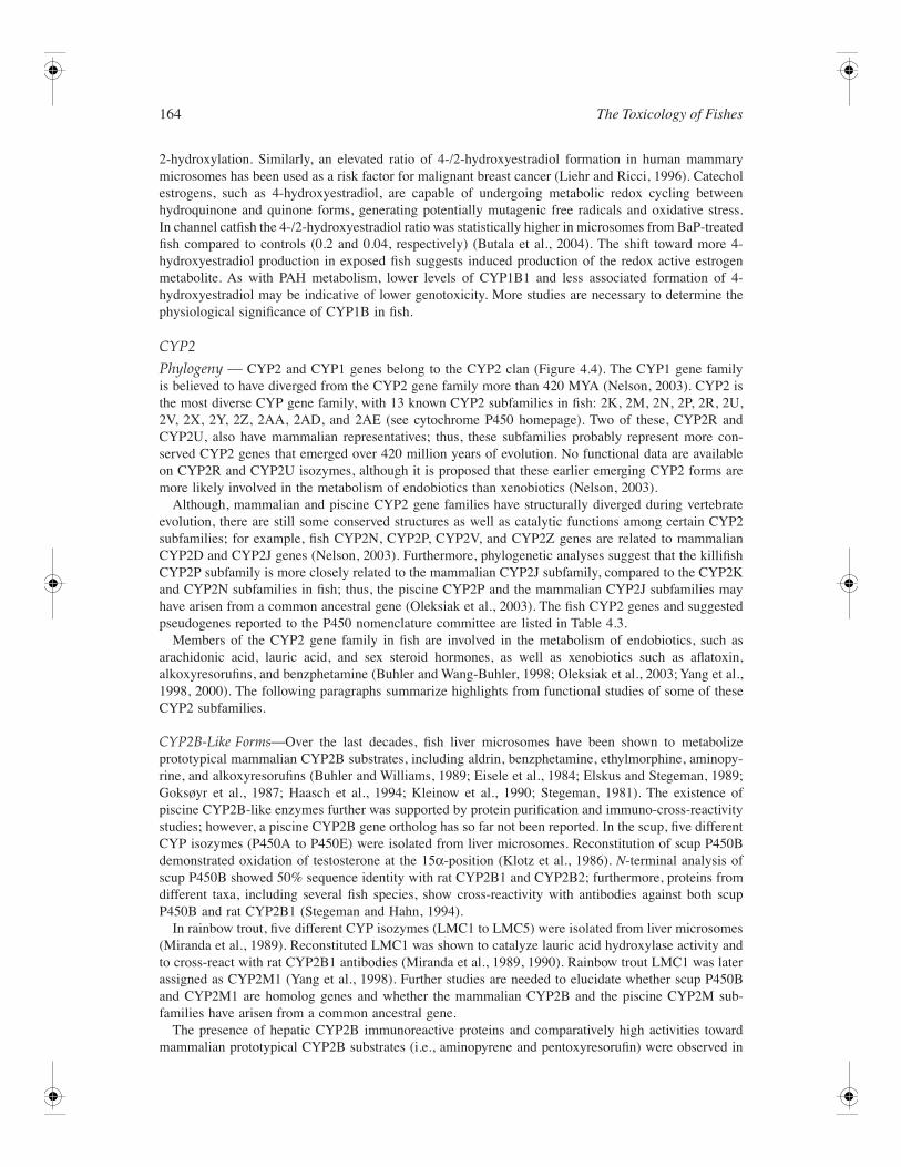
Phylogeny — CYP2 and CYP1 genes belong to the CYP2 clan (Figure 4.4). The CYP1 gene familyis believed to have diverged from the CYP2 gene family more than 420 MYA (Nelson, 2003). CYP2 isthe most diverse CYP gene family, with 13 known CYP2 subfamilies in fish: 2K, 2M, 2N, 2P, 2R, 2U,2V, 2X, 2Y, 2Z, 2AA, 2AD, and 2AE (see cytochrome P450 homepage). Two of these, CYP2R andCYP2U, also have mammalian representatives; thus, these subfamilies probably represent more con-served CYP2 genes that emerged over 420 million years of evolution. No functional data are availableon CYP2R and CYP2U isozymes, although it is proposed that these earlier emerging CYP2 forms aremore likely involved in the metabolism of endobiotics than xenobiotics (Nelson, 2003).
Although, mammalian and piscine CYP2 gene families have structurally diverged during vertebrateevolution, there are still some conserved structures as well as catalytic functions among certain CYP2subfamilies; for example, fish CYP2N, CYP2P, CYP2V, and CYP2Z genes are related to mammalianCYP2D and CYP2J genes (Nelson, 2003). Furthermore, phylogenetic analyses suggest that the killifishCYP2P subfamily is more closely related to the mammalian CYP2J subfamily, compared to the CYP2Kand CYP2N subfamilies in fish; thus, the piscine CYP2P and the mammalian CYP2J subfamilies mayhave arisen from a common ancestral gene (Oleksiak et al., 2003). The fish CYP2 genes and suggestedpseudogenes reported to the P450 nomenclature committee are listed in Table 4.3.
Members of the CYP2 gene family in fish are involved in the metabolism of endobiotics, such asarachidonic acid, lauric acid, and sex steroid hormones, as well as xenobiotics such as aflatoxin,alkoxyresorufins, and benzphetamine (Buhler and Wang-Buhler, 1998; Oleksiak et al., 2003; Yang et al.,1998, 2000). The following paragraphs summarize highlights from functional studies of some of theseCYP2 subfamilies.
CYP2B-Like Forms—Over the last decades, fish liver microsomes have been shown to metabolizeprototypical mammalian CYP2B substrates, including aldrin, benzphetamine, ethylmorphine, aminopy-rine, and alkoxyresorufins (Buhler and Williams, 1989; Eisele et al., 1984; Elskus and Stegeman, 1989;Goksøyr et al., 1987; Haasch et al., 1994; Kleinow et al., 1990; Stegeman, 1981). The existence ofpiscine CYP2B-like enzymes further was supported by protein purification and immuno-cross-reactivitystudies; however, a piscine CYP2B gene ortholog has so far not been reported. In the scup, five differentCYP isozymes (P450A to P450E) were isolated from liver microsomes. Reconstitution of scup P450Bdemonstrated oxidation of testosterone at the 15α-position (Klotz et al., 1986). N-terminal analysis ofscup P450B showed 50% sequence identity with rat CYP2B1 and CYP2B2; furthermore, proteins fromdifferent taxa, including several fish species, show cross-reactivity with antibodies against both scupP450B and rat CYP2B1 (Stegeman and Hahn, 1994).
In rainbow trout, five different CYP isozymes (LMC1 to LMC5) were isolated from liver microsomes(Miranda et al., 1989). Reconstituted LMC1 was shown to catalyze lauric acid hydroxylase activity andto cross-react with rat CYP2B1 antibodies (Miranda et al., 1989, 1990). Rainbow trout LMC1 was laterassigned as CYP2M1 (Yang et al., 1998). Further studies are needed to elucidate whether scup P450Band CYP2M1 are homolog genes and whether the mammalian CYP2B and the piscine CYP2M sub-families have arisen from a common ancestral gene.
The presence of hepatic CYP2B immunoreactive proteins and comparatively high activities towardmammalian prototypical CYP2B substrates (i.e., aminopyrene and pentoxyresorufin) were observed in
Biotransformation in Fishes 165
TABLE 4.3
CYP2 Gene Family in Fish
Subfamily/Gene Species Refs.
CYP2K2K12K22K3-2K52K6-2K8; 2K16-2K222K9-2K112K12-2K15 pseudogenes
Rainbow trout (O. mykiss)Killifish (F. heteroclitus)Rainbow troutZebrafish (D. rerio)Pufferfish (F. rubripes)Pufferfish
Buhler et al. (2000); Cok et al. (1998); Katchamart et al. (2002); Yang et al. (2000)
CYP2M2M1
Rainbow trout Buhler et al. (2000); Cok et al. (1998); Katchamart et al. (2002); Yang et al. (1998)
CYP2N2N12N22N32N4-2N82N9-2N112N13
KillifishKillifishScup (S. chrysops)Butterfly fish (Chaetodon sp.)PufferfishZebrafish
Oleksiak et al. (2000); Peterson and Bain (2004); Schlenk et al. (in press); Vrolijk et al. (1994)
CYP2P2P1-2P32P42P5 pseudogene2P6-2P102P112P
KillifishPufferfishPufferfishZebrafishLargemouth bass (M. salmoides)Atlantic salmon (S. salar)
Oleksiak et al. (2003)
CYP2R2R12R12R2-2R3 pseudogenes
ZebrafishPufferfishPufferfish
—
CYP2U2U12U1
ZebrafishPufferfish
—
CYP2VCYP2V1
Zebrafish —
CYP2XCYP2X12X2-2X42X5 pseudogene2X6-2X11
Catfish (I. punctatus)PufferfishPufferfishZebrafish
Schlenk et al. (2002)
CYPY2Y1-2Y22Y3-2Y4
PufferfishZebrafish
—
CYPZ2ZI-2Z2
Pufferfish —
CYP2AA2AA1-2AA8
— —
CYP2AD2AD1 (formerly 2N12)2AD2-2AD3; 2AD62AD42AD5
PufferfishZebrafishMedaka (O. latipes)Three-spined stickleback (Gasterosteus aculeatus)
—
CYP2AE2AE1
Zebrafish —
Note: Genes, gene fragments, and possible pseudogenes were obtained from the Cytochrome P450 homepage:http://drnelson.utmem.edu/CytochromeP450.html.

166 The Toxicology of Fishes
some tropical fishes from the Bermuda Archipelago; however, great differences were found in aminopy-rene-N-demethylase and pentoxyresorufin-O-depentylase activities among species, with the Bermudachub (Kyphosous sectatrix) and sergeant major (Abudefduf saxatilis) displaying the highest activities. Inaddition, polyclonal antibody (pAb) against rat CYP2B1 cross-reacted most strongly with hepaticmicrosomal proteins in tomtate (Haemulon aurolineatum), pinfish (Lagodan rhomboides), Bermudachub, and sergeant major (Stegeman et al., 1997). The reason for the observed differences in CYP2B-like expression among species is not known. Earlier, it was suggested that natural dietary compoundsmay be causing these differences, as higher CYP2B-like protein levels were observed in butterfly fish(Chaetodon capistratus) that consumed gorgonian (containing high levels of allelochemicals) comparedto butterfly fish that avoided gorgonians (Vrolijk et al., 1994). CYP2N mRNA expression in C. xanthurus(CYP2N7) was significantly higher than in the facultative coralline-feeding butterfly fish C. kleini, C.auriga (CYP2N6), or C. vagabundus, as well as an obligate coralline feeding species (C. punctofasciatus)from the Great Barrier Reef in Australia (Schlenk et al., in press). When each species, including C.xanthurus, was gavaged with gorgonian extracts from Sinnularia maxima for 3 days, with the exceptionof C. punctofasciatus, CYP2N mRNA expression was diminished (Schlenk et al., in press). In theBermuda species investigated, herbivorous fish had higher CYP levels (including CYP2B-like proteins)compared to carnivorous fish (Stegeman et al., 1997). It remains to be shown if natural dietary chemicalsmay act as inducers of CYP2B-like forms in fish or if other mechanisms are involved.
Phenobarbital (PB) and 1,4-bis(2-[3,5-dichloropyridyloxy])benzene (TCPOBOP) are powerful PB-type inducers of CYP2B genes in mammals (Poland et al., 1981). In mammals, induction of CYP2B byPB-type inducers proceeds through activation of the constitutive androstane receptor (CAR) followedby nuclear translocation, dimerization with the retinoid X receptor (RXR), and binding to phenobarbitalresponse elements (PBREMs) in the promoter region of the CYP2B genes (Honkakoski et al., 1997,1998a,b).
In fish, however, an apparent lack of response to PB-type inducers has been observed (Buhler andWilliams, 1989; Eisele et al., 1984; Goksøyr et al., 1987; Haasch et al., 1994; Iwata et al., 2002; Stegeman,1981), although a CAR immunoreactive protein was detected in scup liver cytosol and nucleus usingantibodies against human CAR. No induction of CYP protein levels, including scup P450B, or catalyticactivities were seen in scup injected with TCPOBOB. In fact, TCPOBOB treatment had no effect ontranslocation of the cytosolic CAR-immunoreactive protein in scup liver (Iwata et al., 2002). This studypoints to functional differences, possibly in receptor activation or translocation, between fish and mam-mals. Recently, a single piscine CAR/PXR gene was identified (fr078207) when searching the pufferfishgenome; however, this receptor was more related to PXR family members and hence a probable functionalanalog of PXR (Maglich et al., 2003). Thus, CAR may have diverged from the PXR at a later point invertebrate evolution, or CAR may have been lost in some or all teleost lines (Maglich et al., 2003). Theapparent lack of a piscine CAR receptor may be one explanation for the observed lack of PB-type aswell as diminished CYP3A (see below) induction in fish.
CYP2E-Like Forms—The possible existence of a CYP2E form in fish (Poeciliopsis monacha-lucida)was proposed based on hybridization with a rat CYP2E1 49-base oligonucleotide, antibodies to ratCYP2E1, as well as responsiveness to ethanol treatment. This CYP form was suggested to be involvedin the CYP-mediated dealkylation of the fish carcinogen diethylnitrosamine (Kaplan et al., 1991).Furthermore, hepatic microsomal metabolism of the mammalian CYP2E substrate chlorzoxazone inwinter flounder (Pleuronectes americanus) and in viviparous Poeciliopsis monacha and Poeciliopsisviriosa is indicative of the presence of CYP2E-like enzymes (Kaplan et al., 2001; Wall and Crivello,1998). A piscine CYP2E gene ortholog, however, has so far not been reported.
The CYP2K Subfamily—A CYP protein, denoted LMC2, was isolated from rainbow trout liver (Mirandaet al., 1989). It was subsequently cloned and assigned as CYP2K1 (Buhler et al., 1994). CYP2K1 isone of the dominant CYP forms expressed in liver and trunk kidney, and it displays sexually dimorphicexpression, with higher levels in sexually mature males compared to females (Buhler et al., 1994). Inaddition to liver and trunk kidney, CYP2K1 also is expressed, though at lower levels, in blood cells,upper intestine, head kidney, stomach, heart, gonads, and male muscle (Cok et al., 1998). Heterologous
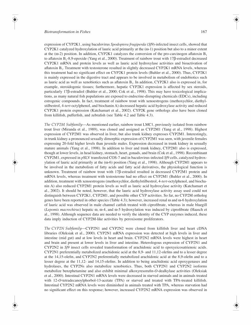
Biotransformation in Fishes 167
expression of CYP2K1, using baculovirus Spodoptera frugiperda (Sf9)-infected insect cells, showed thatCYP2K1 catalyzed hydroxylation of lauric acid primarily at the (ω-1) position but also to a minor extentat the (ω-2) position. In addition, CYP2K1 catalyzes the conversion of the pro-carcinogen aflatoxin B1
to aflatoxin B1-8,9-epoxide (Yang et al., 2000). Treatment of rainbow trout with 17β-estradiol decreasedCYP2K1 mRNA and protein levels as well as lauric acid hydroxylase activities and bioactivation ofaflatoxin B1. Treatment with testosterone resulted in slightly decreased CYP2K1 mRNA levels, whereasthis treatment had no significant effect on CYP2K1 protein levels (Buhler et al., 2000). Thus, CYP2K1is mainly expressed in the digestive tract and appears to be involved in metabolism of endobiotics suchas lauric acid as well as xenobiotics such as aflatoxin B1. In addition, CYP2K1 also is expressed in, forexample, steroidogenic tissues; furthermore, hepatic CYP2K1 expression is affected by sex steroids,particularly 17β-estradiol (Buhler et al., 2000; Cok et al., 1998). This may have toxicological implica-tions, as many natural fish populations are exposed to endocrine-disrupting chemicals (EDCs), includingestrogenic compounds. In fact, treatment of rainbow trout with xenoestrogens (methoxychlor, diethyl-stilbestrol, 4-tert-octylphenol, and biochanin A) decreased hepatic acid hydroxylase activity and reducedCYP2K1 protein expression (Katchamart et al., 2002). CYP2K gene orthologs also have been clonedfrom killifish, pufferfish, and zebrafish (see Table 4.2 and Table 4.3).
The CYP2M Subfamily—As mentioned earlier, rainbow trout LMC1, previously isolated from rainbowtrout liver (Miranda et al., 1989), was cloned and assigned as CYP2M1 (Yang et al., 1998). Highestexpression of CYP2M1 was observed in liver, but also trunk kidney expresses CYP2M1. Interestingly,in trunk kidney a pronounced sexually dimorphic expression of CYP2M1 was seen, with juvenile femalesexpressing 20-fold higher levels than juvenile males. Expression decreased in trunk kidney in sexuallymature animals (Yang et al., 1998). In addition to liver and trunk kidney, CYP2M1 also is expressed,though at lower levels, in head kidney, stomach, heart, gonads, and brain (Cok et al., 1998). RecombinantCYP2M1, expressed in pSLV transfected COS-7 and in baculovirus-infected Sf9 cells, catalyzed hydrox-ylation of lauric acid primarily at the (ω-6) position (Yang et al., 1998). Although CYP2M1 appears tobe involved in the metabolism of fatty acids and fatty acid derivatives, the physiological function isunknown. Treatment of rainbow trout with 17β-estradiol resulted in decreased CYP2M1 protein andmRNA levels, whereas treatment with testosterone had no effect on CYP2M1 (Buhler et al., 2000). Inaddition, treatment with xenoestrogens (methoxychlor, diethylstilbestrol, 4-tert-octylphenol, and biocha-nin A) also reduced CYP2M1 protein levels as well as lauric acid hydroxylase activity (Katchamart etal., 2002). It should be noted, however, that the lauric acid hydroxylase activity assay used could notdistinguish between CYP2K1, CYP2M1, and possible other CYP activities. So far, no CYP2M orthologgenes have been reported in other species (Table 4.3); however, increased renal ω and ω-6 hydroxylationof lauric acid was observed in male channel catfish treated with ciprofibrate, whereas in male bluegill(Lepomis macrochirus) hepatic ω, ω-4, and ω-5 hydroxylation was induced by ciprofibrate (Haasch etal., 1998). Although sequence data are needed to verify the identity of the CYP enzymes induced, thesedata imply induction of CYP2M-like activities by peroxisome proliferators.
The CYP2N Subfamily—CYP2N1 and CYP2N2 were cloned from killifish liver and heart cDNAlibraries (Oleksiak et al., 2000). CYP2N1 mRNA expression was detected at high levels in liver andintestine (mid gut) and at low levels in heart and brain. CYP2N2 mRNA levels were highest in heartand brain and present at lower levels in liver and intestine. Heterologous expression of CYP2N1 andCYP2N2 in Sf9 insect cells revealed transformation of arachidonic acid to epoxyeicosatrienoic acids.CYP2N1 preferentially metabolized arachidonic acid at the 8,9- and 11,12-olefins and to a lesser degreeat the 14,15-olefin, and CYP2N2 preferentially metabolized arachidonic acid at the 8,9-olefin and to alesser degree at the 11,12- and 14,15-olefins. In addition to being arachidonic acid epoxygenases andhydrolases, the CYP2Ns also metabolize xenobiotics. Thus, both CYP2N1 and CYP2N2 isoformsmetabolize benzphetamine and also exhibit minimal alkoxyresorufin-O-dealkylase activities (Oleksiaket al., 2000). Intestinal CYP2N1 mRNA levels were decreased in starved animals and in animals treatedwith 12-O-tetradecanoylphorbol-13-acetate (TPA) or starved and treated with TPA-treated killifish.Intestinal CYP2N2 mRNA levels were diminished in animals treated with TPA, whereas starvation hadno significant effect on this response; however, increased CYP2N2 mRNA expression was observed in
168 The Toxicology of Fishes
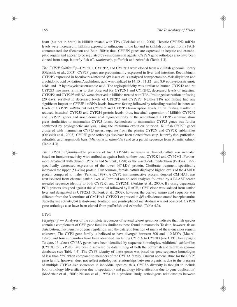
heart (but not in brain) in killifish treated with TPA (Oleksiak et al., 2000). Hepatic CYP2N2 mRNAlevels were increased in killifish exposed to anthracene in the lab and in killifish collected from a PAH-contaminated site (Peterson and Bain, 2004); thus, CYP2N genes are expressed in hepatic and extrahe-patic organs and appear to be regulated by environmental agents. CYP2N gene orthologs also have beencloned from scup, butterfly fish (C. xanthurus), pufferfish and zebrafish (Table 4.3).
The CYP2P Subfamily—CYP2P1, CYP2P2, and CYP2P3 were cloned from a killifish genomic library(Oleksiak et al., 2003). CYP2P genes are predominantly expressed in liver and intestine. RecombinantCYP2P3 expressed in baculovirus-infected Sf9 insect cells catalyzed benzphetamine-N-dealkylation andarachidonic acid oxidation. Arachidonic acid was oxidized to 14,15-, 11,12-, and 8,9-epoxyeicosatrienoicacids and 19-hydroxyeicosatetraenoic acid. The regiospecificity was similar to human CYP2J2 and ratCYP2J3 isozymes. Similar to that observed for CYP2N1 and CYP2N2, decreased levels of intestinalCYP2P2 and CYP2P3 mRNA were observed in killifish treated with TPA. Prolonged starvation or fasting(20 days) resulted in decreased levels of CYP2P2 and CYP2P3. Neither TPA nor fasting had anysignificant impact on CYP2P1 mRNA levels; however, fasting followed by refeeding resulted in increasedlevels of CYP2P1 mRNA but not CYP2P2 and CYP2P3 transcription levels. In rat, fasting resulted inreduced intestinal CYP2J3 and CYP2J4 protein levels; thus, intestinal expression of killifish CYP2P2and CYP2P3 genes and arachidonic acid regiospecificity of the recombinant CYP2P3 isozyme showgreat similarities to mammalian CYP2J forms. Relatedness to mammalian CYP2J genes was furtherconfirmed by phylogenetic analysis, using the minimum evolution criterion. Killifish CYP2P genesclustered with mammalian CYP2J genes, separate from the piscine CYP2N and CYP2K subfamilies(Oleksiak et al., 2003). CYP2P gene orthologs also have been cloned from scup, butterfly fish, pufferfish,zebrafish, and largemouth bass (Micropterus salmoides) and as a partial sequence from Atlantic salmon(Table 4.3).
The CYP2X Subfamily—The presence of two CYP2-like isozymes in channel catfish was indicatedbased on immunoreactivity with antibodies against both rainbow trout CYP2K1 and CYP2M1. Further-more, treatment with ethanol (Perkins and Schlenk, 1998) or the insecticide fenitrothion (Perkins, 1999)specifically decreased expression of the lower (47-kDa) protein. Clofibrate treatment specificallyincreased the upper (51-kDa) protein. Furthermore, female catfish displayed higher levels of the 47-kDaprotein compared to males (Perkins, 1998). A CYP2-immunoreactive protein, denoted CM-HA3, wasnext isolated from channel catfish liver. N-Terminal amino acid analyses followed by a BLAST searchrevealed sequence identity to both CYP2K1 and CYP2M1 (Perkins et al., 2000). By using degeneratePCR primers designed against this N-terminal followed by RACE, a CYP clone was isolated from catfishliver and designated as CYP2X1 (Schlenk et al., 2002); however, the derived amino acid sequence wasdifferent from the N-terminus of CM-HA3. CYP2X1 expressed in Sf9 cells demonstrated benzphetaminedemethylase activity, but testosterone, fenthion, and p-nitrophenol metabolism was not observed. CYP2Xgene orthologs also have been cloned from pufferfish and zebrafish (Table 4.3).
CYP3
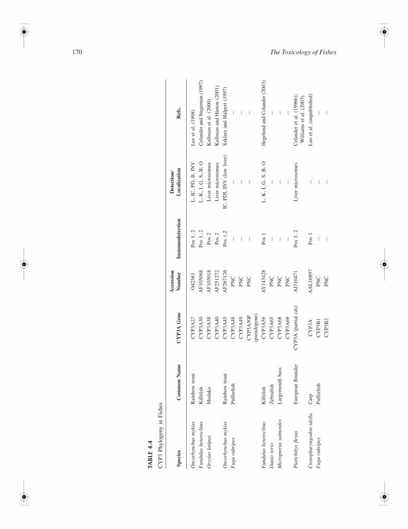
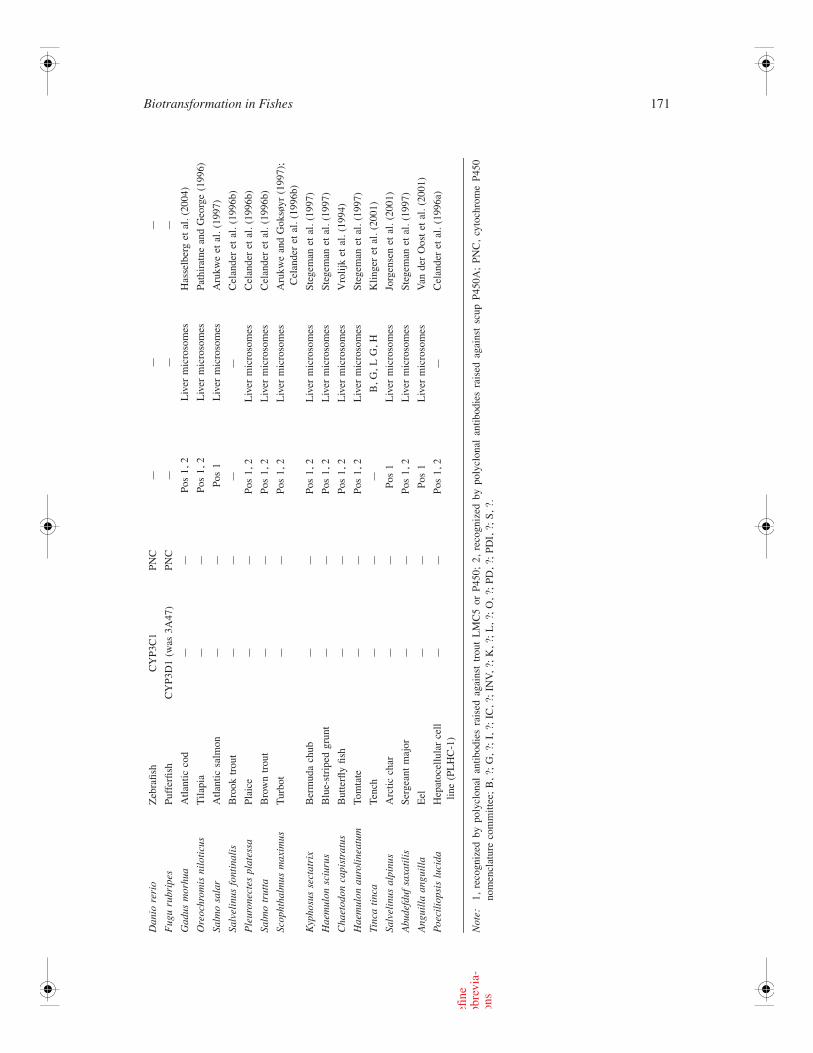
Phylogeny — Analyses of the complete sequences of several teleost genomes indicate that fish speciescontain a complement of CYP gene families similar to those found in mammals. To date, however, tissuedistribution, mechanisms of gene regulation, and the catalytic function of many of these enzymes remainunknown. The CYP3 gene family is believed to have diverged between 800 and 110 MYA (Maurel,1996), and four subfamilies have been identified, including CYP3A to CYP3D (see CYP Home page).To date, 13 teleost CYP3A genes have been identified by sequence homologies. Additional subfamilies(CYP3B to CYP3D) have been discovered by data mining of both the pufferfish and zebrafish genomedatabases (see Table 4.4). The CYP3 identity of these genes was based on gene sequence homologiesof less than 55% when compared to members of the CYP3A family. Current nomenclature for the CYP3gene family, however, does not reflect orthologous relationships between organisms due to the presenceof multiple CYP3A-like sequences in individual species; thus, CYP3A diversity is thought to includeboth orthology (diversification due to speciation) and paralogy (diversification due to gene duplication)(McArthur et al., 2003; Nelson et al., 1996). In a previous study, orthologous relationships between
Biotransformation in Fishes 169
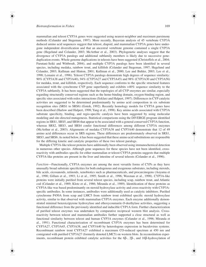
mammalian and teleost CYP3A genes were suggested using nearest-neighbor and maximum parsimonymethods (Celander and Stegeman, 1997). More recently, Bayesian analysis of 45 vertebrate CYP3Adeduced amino acid sequences suggest that teleost, diapsid, and mammalian CYP3A genes have under-gone independent diversification and that an ancestral vertebrate genome contained a single CYP3Agene (Hegelund and Celander, 2003; McArthur et al., 2003). Phylogenetic analyses suggest that thedivergence of CYP3A paralogs and additional subfamily members is likely due to successive gene-duplication events. Whole genome duplications in teleosts have been suggested (Christoffels et al., 2004;Furutani-Seiki and Wittbrodt, 2004), and multiple CYP3A paralogs have been identified in severalspecies, including medaka, rainbow trout, and killifish (Celander and Stegeman, 1997; Hegelund andCelander, 2003; Kullman and Hinton, 2001; Kullman et al., 2000; Lee and Buhler, 2003; Lee et al.,1998; Lemaire et al., 1996). Teleost CYP3A paralogs demonstrate high degrees of sequence similarity:90% (CYP3A38 and CYP3A40), 94% (CYP3A27 and CYP3A45) and 98% (CYP3A30 and CYP3A56)for medaka, trout, and killifish, respectively. Each sequence conforms to the specific structural featuresassociated with the cytochrome CYP gene superfamily and exhibits >40% sequence similarity to theCYP3A subfamily. It has been suggested that the topologies of all CYP enzymes are similar, especiallyregarding structurally conserved regions such as the heme-binding domain, oxygen-binding region, andspecific sites associated with redox interactions (Szklarz and Halpert, 1997). Differences in CYP catalyticactivities are suggested to be determined predominantly by amino acid composition in six substraterecognition sites (SRS1 to SRS6) (Gotoh, 1992). Recently homology models for CYP3A genes havebeen described (Harlow and Halpert, 1998; Yang et al., 1998). Key amino acids associated with CYP3Asubstrate specificity, binding, and regio-specific catalysis have been suggested by using molecularmodeling and site-directed mutagenesis. Statistical comparisons using the DIVERGE program identifiedregions in SRS1, SRS5, and SRS6 that appear to be associated with a general conserved CYP3A function,whereas SRS2, SRS3, and SRS4 confer functional differences among different CYP3A enzymes(McArthur et al., 2003). Alignments of medaka CYP3A38 and CYP3A40 demonstrate that 12 of 49amino acid differences occur in SRS regions. These differences are predominately observed in SRS1,SRS3, and SRS6. As noted below, it has been suggested that these amino acid substitutions are responsiblefor the differing kinetic and catalytic properties of these two teleost paralogs.
Multiple CYP3A-like teleost proteins have additionally been observed using immunochemical detectionin numerous other species. Although gene sequences for these species have not been identified, cross-reactivity with antibodies specific for either mammalian or teleost CYP3A proteins suggests that multipleCYP3A-like proteins are present in the liver and intestine of several teleosts (Celander et al., 1996).
Function—Functionally, CYP3A enzymes are among the most versatile forms of CYPs as they haveunusually broad substrate specificities for both endogenous and exogenous substrates, including steroids,bile acids, eicosanoids, retinoids, xenobiotics such as pharmaceuticals, and procarcinogens (Aoyama etal., 1990; Gillam et al., 1993; Li et al., 1995; Smith et al., 1996; Waxman et al., 1998). CYP3A-likeproteins were initially purified from several teleost species, including scup, rainbow trout, and Atlanticcod (Celander et al., 1989; Klotz et al., 1986; Miranda et al., 1989). Identification of these proteins asCYP3A-like was based predominantly on steroid hydroxylase activity and cross-reactivity with CYP3A-specific antibodies. In some instances, antibodies were additionally used as catalytic inhibitors. Purifiedcytochrome P450A from scup and LMC5 from rainbow trout exhibited specific steroid hydroxylaseactivity, similar to that observed with mammalian CYP3A enzymes. Each enzyme additionally demon-strated minimal benzo(a)pyrene hydroxylase and ethoxycoumarin O-deethylase activities, suggesting afunctional difference from the previously identified and inducible CYP1A form. Further characterizationof purified teleost enzymes was undertaken by comparative reciprocal western blot analysis. Cross-reactivity between teleost and mammalian antibodies further supported a close structural as well asfunctional similarity between teleost and human CYP3A enzymes (Celander et al., 1996; Miranda etal., 1991). Functional characterization of recombinant CYP3A enzymes has been determined forCYP3A27, CYP3A45, CYP3A38, and CYP3A40 by heterologous expression in baculovirus systems.Recombinant rainbow trout CYP3A27 exhibited a maximum CO-reduced spectrum at 450 nm andcomigrated with purified CYP3A27 (formerly denoted LMC5) on western blots. In reconstitution exper-iments, recombinant protein exhibited catalytic activities for the 6β-, 2β-, and 16β-hydroxylation of
170 The Toxicology of Fishes
TAB
LE 4
.4
CY
P3 P
hylo
geny
in
Fish
es
Spe
cies
Com
mon
Nam
eC
YP
3A G
ene
Acc
essi
on
Num
ber
Imm
unod
etec
tion
Det
ecti
on/
Loc
aliz
atio
nR
efs.
Onc
orhy
nchu
s m
ykis
sR
ainb
ow t
rout
CY
P3A
27O
4256
3Po
s 1,
2L
, IC
, PD
, B
, IN
VL
ee e
t al
. (1
998)
Fun
dulu
s he
tero
clit
usK
illifi
shC
YP3
A30
AF1
0506
8Po
s 1,
2L
, K
, I,
G,
S, B
, O
Cel
ande
r and
Ste
gem
an (1
997)
Ory
zias
lat
ipas
Med
aka
CY
P3A
38
AF1
0501
8Po
s 2
Liv
er m
icro
som
esK
ullm
an e
t al
. (2
000)
CY
P3A
40A
F251
272
Pos
2L
iver
mic
roso
mes
Kul
lman
and
Hin
ton
(200
1)
Onc
orhy
nchu
s m
ykis
sR
ainb
ow t
rout
CY
P3A
45A
F267
126
Pos
1.2
IC,
PDI,
IN
V (
low
liv
er)
Szkl
arz
and
Hal
pert
(19
97)
Fug
u ru
brip
esPu
ffer
fish
CY
P3A
48PN
C—
——
CY
P3A
49PN
C—
——
CY
P3A
50P
(pse
udog
ene)
PNC
——
—
Fun
dulu
s he
tero
clit
usK
illifi
shC
YP3
A56
AY
1434
28Po
s 1
L,
K,
I, G
, S,
B,
OH
egel
und
and
Cel
ande
r (2
003)
Dan
io r
erio
Zeb
rafis
hC
YP3
A65
PNC
——
—
Mic
ropt
erus
sal
moi
des
Lar
gem
outh
bas
sC
YP3
A68
PNC
——
—
CY
P3A
69PN
C—
——
Pla
tich
thys
fles
usE
urop
ean
floun
der
CY
P3A
(pa
rtia
l cd
s)A
J310
471
Pos
1, 2
Liv
er m
icro
som
esC
elan
der
et a
l. (1
996b
);
Will
iam
s et
al.
(200
3)
Cte
noph
aryn
godo
n id
ella
Car
pC
YP3
AA
AL
1689
7Po
s 1
—L
ao e
t al
. (u
npub
lishe
d)
Fug
u ru
brip
esPu
ffer
fish
CY
P3B
1PN
C—
——
CY
P3B
2PN
C—
——
Biotransformation in Fishes 171
Dan
io r
erio
Zeb
rafis
hC
YP3
C1
PNC
——
—
Fug
u ru
brip
esPu
ffer
fish
CY
P3D
1 (w
as 3
A47
)PN
C—
——
Gad
us m
orhu
aA
tlant
ic c
od—
—Po
s 1,
2L
iver
mic
roso
mes
Has
selb
erg
et a
l. (2
004)
Ore
ochr
omis
nil
otic
usT
ilapi
a—
—Po
s 1,
2L
iver
mic
roso
mes
Path
irat
ne a
nd G
eorg
e (1
996)
Salm
o sa
lar
Atla
ntic
sal
mon
——
Pos
1L
iver
mic
roso
mes
Aru
kwe
et a
l. (1
997)
Salv
elin
us f
onti
nali
sB
rook
tro
ut—
——
—C
elan
der
et a
l. (1
996b
)
Ple
uron
ecte
s pl
ates
saPl
aice
——
Pos
1, 2
Liv
er m
icro
som
esC
elan
der
et a
l. (1
996b
)
Salm
o tr
utta
Bro
wn
trou
t—
—Po
s 1,
2L
iver
mic
roso
mes
Cel
ande
r et
al.
(199
6b)
Scop
htha
lmus
max
imus
Tur
bot
——
Pos
1, 2
Liv
er m
icro
som
esA
rukw
e an
d G
oksø
yr (
1997
);
Cel
ande
r et
al.
(199
6b)
Kyp
hosu
s se
ctat
rix
Ber
mud
a ch
ub—
—Po
s 1,
2L
iver
mic
roso
mes
Steg
eman
et
al.
(199
7)
Hae
mul
on s
ciur
usB
lue-
stri
ped
grun
t—
—Po
s 1,
2L
iver
mic
roso
mes
Steg
eman
et
al.
(199
7)
Cha
etod
on c
apis
trat
usB
utte
rfly
fish
——
Pos
1, 2
Liv
er m
icro
som
esV
rolij
k et
al.
(199
4)
Hae
mul
on a
urol
inea
tum
Tom
tate
—
—Po
s 1,
2L
iver
mic
roso
mes
Steg
eman
et
al.
(199
7)
Tinc
a ti
nca
Tenc
h—
——
B,
G,
L G
, H
Klin
ger
et a
l. (2
001)
Salv
elin
us a
lpin
usA
rctic
cha
r—
—Po
s 1
Liv
er m
icro
som
es
Jorg
ense
n et
al.
(200
1)
Abu
defd
uf s
axat
ilis
Serg
eant
maj
or
——
Pos
1, 2
Liv
er m
icro
som
esSt
egem
an e
t al
. (1
997)
Ang
uill
a an
guil
laE
el—
—Po
s 1
Liv
er m
icro
som
esV
an d
er O
ost
et a
l. (2
001)
Poec
ilio
psis
luc
ida
Hep
atoc
ellu
lar
cell
line
(PL
HC
-1)
——
Pos
1, 2
—C
elan
der
et a
l. (1
996a
)
Not
e:1,
rec
ogni
zed
by p
olyc
lona
l an
tibod
ies
rais
ed a
gain
st t
rout
LM
C5
or P
450;
2,
reco
gniz
ed b
y po
lycl
onal
ant
ibod
ies
rais
ed a
gain
st s
cup
P450
A;
PNC
, cy
toch
rom
e P4
50no
men
clat
ure
com
mitt
ee;
B,
?; G
, ?;
I,
?; I
C,
?; I
NV
, ?;
K,
?; L
, ?;
O,
?; P
D,
?; P
DI,
?;
S, ?
.
efin
ebb
revi
a-on
s
172 The Toxicology of Fishes
testosterone at 1.48, 0.043, and 0.034 nmol/min/nmol, respectively, as well as dehydrogenation ofnifedipine at 50 pmol/min/nmol (Lee and Buhler, 2002). Although turnover rates were significantlyhigher with the recombinant enzyme, hydroxylase profiles were similar to that observed with purifiedCYP3A27 enzyme. Heterologous expression of the rainbow trout paralog CYP3A45 additionally exhibitstestosterone hydroxylase activity with 6β-hydroxytestosterone as the major metabolite. Activity washigher than that observed with CYP3A27 and was significantly enhanced by the addition of cytochromeb5 to the reconstitution assays (Lee and Buhler, 2003).
Recombinant medaka CYP3A38 and CYP3A40 catalyzed hydroxylation of testosterone, as well asthe O-debenzylation of benzyloxyresorufin (BR) and 7-benzyloxy-4-(trifluoromethyl)-coumarin (BFC);however; efficiencies and specificities were significantly different between the two isoforms. Thus, Km
and Vmax activities based on BFC-O-debenzyloxylase were estimated to be 0.116 and 0.363 µM and 7.95and 7.77 nmol/min/nmol P450 for CYP3A38 and CYP3A40, respectively. Medaka CYP3A38 preferen-tially catalyzed testosterone hydroxylation at the 6β- and 16β-positions, with minor hydroxylation atother positions within the steroid nucleus, whereas CYP3A40 catalysis was predominantly limited tothe 6β- position (Kashiwada, unpublished data). Putative identification of CYP3A SRS1 to SRS6indicated that 12 of the 49 amino acid differences between CYP3A38 and CYP3A40 occur in SRS1,SRS3, and SRS5, previously known to be associated with steroid hydroxylation (Kashiwada, unpublisheddata; Kullman and Hinton, 2001). Functional analysis of teleost and mammalian CYP3A paralogs hasdemonstrated that gene-duplication events are tied to acquisition of new function and that convergentevolution of CYP3A function may be frequent among independent gene copies (McArthur et al., 2003).The physiological role of CYP3A is yet to be determined. Given the robust steroid hydroxylase activity,there is speculation that CYP3A forms play an important part in steroid (hormones as well as bile acids)homeostasis. Although the physiological function of CYP3A is still unknown, the fact that these genesare expressed in tissues that act as barriers to the environment (i.e., digestive and respiratory tracts)together with the broad substrate specificities of CYP3A enzymes suggest that they evolved as biochem-ical defense to prevent bioaccumulation of xenobiotics. In addition, the presence of CYP3A enzymesin steroidogenic tissues implies a role in steroid biotransformation.
Regulation—CYP3A are the major constitutive CYP forms expressed in the liver and intestine of mostmammals and other species, including fish (Celander et al., 1996, 1989; Hegelund and Celander, 2003;Hegelund et al., 2004; Husoy et al., 1994). Numerous studies have demonstrated, however, that consti-tutive expression of CYP3A genes between species, and paralogous CYP3A forms within species, arehighly variable with age, gender, development, tissue localization, and between individuals. Variationsin gene expression may be due to external environmental factors, including temperature, salinity, diet,or other environmental stressors; biological factors, such as circulating hormone levels; or tissue-specificfactors associated with development or reproductive cycle. Depending on species, sexual dimorphicexpression of CYP3A has been observed. Lee et al. (1998) reported higher levels of CYP3A expressionin juvenile female rainbow trout intestine when compared to males. Gender differences were additionallyobserved in kidney and liver but to a lesser degree than in intestine. A similar trend was demonstratedin winter flounder (Pleuronectes americanus) hepatic microsomes and in Atlantic cod, where CYP3Aexpression was sevenfold higher in females than males (Gray et al., 1991; Hasselberg et al., 2004). Incontrast, adult killifish males displayed up to 2.5-fold higher levels of hepatic and extrahepatic CYP3A30and CYP3A56 mRNA and protein levels compared to females (Hegelund and Celander, 2003). Thisfinding was in agreement with higher hepatic testosterone 6β-hydroxylase activities in male killifish(Gray et al., 1991; Stegeman and Woodin, 1984). Other species, including medaka and sexually maturerainbow trout, also exhibit a higher level of CYP3A expression in male liver microsomes compared tofemales (Aoyama et al., 1990; Celander et al., 1989, 1996; Gillam et al., 1993; Gotoh, 1992; Harlowand Halpert, 1998; Kullman and Hinton, 2001; Lee and Buhler, 2003; Li et al., 1995; Smith et al., 1996;Szklarz and Halpert, 1997; Waxman et al., 1998). Variations in CYP3A expression in Atlantic salmonand turbot (Scophthalmus maximus) during reproductive cycles has been reported implying a regulatoryrole of sex steroids on CYP3A expression (Arukwe and Goksøyr, 1997; Larsen et al., 1992). Steroidreproductive hormones and plasma growth hormones have been shown to influence the sexual dimorphicexpression of CYP3A in rodents (Park et al., 1999; Sakuma et al., 2002; Wang and Strobel, 1997).
Biotransformation in Fishes 173
In teleosts, the effects of exposure to reproductive hormones on CYP3A expression differ dependingon species. Numerous studies with rainbow trout, brook trout, medaka, and other fish species havedemonstrated that exposure to 17β-estradiol results in a suppression of total microsomal CYP content.This correlated with decreased CYP3A mRNA expression, CYP protein levels, or steroid hydroxylaseactivities (Buhler et al., 2000; Celander et al., 1989; Pajor et al., 1990). Treatment of male Atlantic codwith 17β-estradiol resulted in an increase in CYP3A protein expression (Hasselberg et al., 2004). Themechanisms of hormonal regulation of CYP genes in teleosts have yet to be determined; however, it ispossible that circulating hormone levels (steroids or growth hormone) are associated with sexual dimor-phic differences in CYP expression in fish.
Numerous pharmaceuticals and xenobiotics have been demonstrated to alter CYP3A gene transcriptionin mammals via binding and transactivation of members of the nuclear receptor family NR1, includingthe pregnane X receptor (PXR), the constitutive androstane receptor (CAR), and the vitamin D receptor(VDR) (Pascussi et al., 2003; Plant and Gibson, 2003). As mentioned above, although Maglich et al.(2003) did not find CAR in the pufferfish genome, they identified a single PXR/CAR gene that wasmore PXR like, indicating CAR may have evolved after divergence of fish or may have been lost insome or all teleosts. To date, PXR and VDR have been identified in few teleost species. Prototypicalmammalian NR1 (PXR) receptor agonists, including dexamethasone (DEX), rifampicin (RIF), andpregnenolone-16α-carbonitrile (PCN), are seemingly less effective at altering teleost CYP3A genetranscription (Celander et al., 1989, 1996a,b; Kullman, unpublished data). In tilapia, PCN-treatmentresulted in twofold induction of CYP3A proteins (Pathiratne and George, 1996). The ligand-bindingregion of PXR was isolated from zebrafish (Kliewer et al., 2002). This region was shown to be activatedby some prototypical mammalian PXR agonists, including nifedipine, phenobarbital, clotrimazole, andsome steroids, but PCN, DEX, and RIF did not activate the fish receptor (Moore et al., 2002). Inductionof hepatic CYP3A expression in vivo by ketoconazole was observed in juvenile rainbow trout and Atlanticcod (Hasselberg et al., 2004; Hegelund et al., 2004). As mentioned above, response to 17β-estradiolresulted in either enhanced or diminished CYP3A expression, depending on the species examined (Buhlerand Wang-Buhler, 1998; Hasselberg et al., 2004; Husoy et al., 1994; Pajor et al., 1990). Furthermore,xenoestrogens also affect CYP3A expression. Male Atlantic cod exposed to alkylphenols showedenhanced hepatic CYP3A expression, whereas treatment with ethinylestradiol and nonylphenol sup-pressed CYP3A expression in Atlantic salmon and Atlantic cod (Arukwe et al., 1997; Hasselberg et al.,2004). Slight increases in hepatic CYP3A expression have been demonstrated in rainbow trout exposedto cortisol, whereas cortisol treatment had no effect on CYP3A protein levels in Arctic char (Salvelinusalpinus) (Celander et al., 1989; Jorgensen et al., 2001).
Although gene sequences for PXR and VDR have been identified in fish (Kliewer et al., 2002), fewfunctional data exist regarding their role in transcriptional activation of CYP3 genes. Identification ofcognate hormone response elements upstream of pufferfish and medaka CYP3A genes suggest thatnuclear receptors may be involved in transcriptional regulation; however, this has yet to be determined(Kullman, pers. commun.). Cloning and analysis of lamprey VDR suggest that this nuclear receptor iscapable of binding and transcriptional activation of the mammalian CYP3A4-DR3 hormone responseelement. These results demonstrate conservation in the DNA binding behavior of an early form of thisreceptor and a possible role in transcriptional activation of CYP genes in fish. (For more on nuclearreceptors in teleosts, see Chapter 7). Modulation of teleost CYP3A expression following xenobioticexposure is highly variable and species dependent.
Studies examining the binding behavior of mammalian PXR have demonstrated transactivation andCYP3A induction by xenoestrogens, including bisphenol A, nonylphenol, DDT, and other organochlorinepesticides (Courmoul et al., 2002; Masuyama et al., 2000; Takeshita et al., 2001; You, 2004). The responseof aquatic species to these and other PXR, CAR and VDR ligands has been minimal; thus, the overallinduction of CYP3A in teleosts appears to be considerably weaker than that reported in mammals.Compared to mammalian species, this suggests that teleost CYP3A expression may be governed byalternative transcriptional mechanisms. Given the recent completion of several teleost genome projects,examination of promoter sequences will shed some light on regulatory elements and transcriptionalcontrol of theses genes. In several studies, discrepancies have been noted between changes in geneexpression and changes in steroid hydroxylase activity. In a study by Hasselberg et al. (2004), exposure

174 The Toxicology of Fishes
of juvenile Atlantic cod to ketoconazole resulted in slight increases in CYP3A expression but markeddecreases in CYP3A-specific catalytic activity. Similar discrepancies have been noted in medaka, forwhich significant decreases in catalytic activity cannot be accounted for by corresponding reductions inCYP3A expression (Kashiwada, unpublished data). As noted in the section on CYP1A, these discrep-ancies between message level and catalytic activity necessitate determination of expression at severallevels prior to making conclusions about regulatory mechanisms.
Localization—Localization of CYP3A expression is similar to that observed in mammalian species withhigh levels of expression found in the liver and intestinal mucosa. Some differences are observed betweenspecific CYP3A paralogs and may represent putative regulatory and functional differences that occurredduring gene duplication events. Immunohistochemical and mRNA analysis of CYP3A27 has demon-strated strong responses in intestinal ceca, proximal descending intestine, and liver, with minor expressionoccurring in brain (Cok et al., 1998; Lee et al., 2001). The rainbow trout paralog CYP3A45 waspredominantly expressed in the gastrointestinal tract, with weak expression occurring in the liver (Leeand Buhler, 2003). In killifish, CYP3A30 and CYP3A56 were coexpressed in intestine and liver, con-sistent with that observed for rainbow trout CYP3A27. In both species, CYP3A expression is prominentin the digestive tract (intestinal mucosa and liver), suggesting a role for these enzymes in first-passmetabolism of xenobiotics. Extrahepatic CYP3A expression is observed in gill, kidney, brain, spleen,and ovary, suggesting a possible role for CYP3A enzymes in the fine-tuning of endogenous substratesat the site of synthesis or action as well as metabolic defense against xenobiotics (Hegelund et al., 2004).
Development—Biotransformation systems for embryonic and fetal tissues of vertebrates are not as wellcharacterized as those for adults. Embryogenesis is a dynamic process that presents a continuouslychanging metabolic profile as enzymes are induced and repressed (Miller et al., 1996). In general,developing organisms lack many CYP forms present in adults; however, in some instances, they containcertain forms that are only expressed during development. These differences in total CYP content mayexplain developmental processes and altered sensitivities to toxic exposures during development. Teleostembryos are often sensitive to xenobiotics, and prolonged exposures result in numerous developmentalmalformations, early mortality, and delayed hatch (Cooke and Hinton, 1999; Villalobos et al., 1996). Thenonspecific onset of many of these aberrations may reflect an inability of embryos to sufficientlymetabolize and detoxify many of these xenobiotics. CYP3A27 cDNA was cloned from both embryonicand adult hepatic mRNA, suggesting this gene is expressed early during rainbow trout development (Leeet al., 1998). Kullman and Hinton (2001) demonstrated that CYP3A38 and CYP3A40 genes are differ-entially regulated during embryonic development. Analysis of CYP3A38 and CYP3A40 mRNA andprotein demonstrated the presence of a single CYP3A transcript for early and late embryonic stages andtwo CYP3A transcripts in larvae and adult liver in medaka. Using gene-specific probes, results demon-strated that CYP3A40 is expressed early in embryonic development and continues throughout adultstages. CYP3A38, however, is tightly suppressed during embryonic development and is only expressedpost-hatch. Given the role of CYP enzymes in maintaining steady-state levels of morphogenic ligands,it is not surprising to find these enzymes in the earliest stages of life. Previous studies have providedstrong evidence for the presence of multiple forms of CYP in the developing fish embryo (Buhler et al.,1997; Chen and Cooper, 1999). In general, fetal CYP forms are present in low levels and exhibit stringenttemporal expression patterns that diminish following birth or hatching (Juchau et al., 1998). CYP3A40mRNA expression was detected as early as stage 11, representing an early multicellular stage of devel-opment. Liver formation does not occur until stage 26, demonstrating that initial CYP3A40 expressionoccurs prior to organogenesis. Functionally, expression of CYP3A40 during early embryonic developmentmay serve multiple purposes including xenobiotic metabolism, hydroxylation of steroid or other mor-phogenic ligands, and metabolism of yolk. As with human CYP3A7, CYP3A may be the only constitu-tively expressed cytochrome P450 during embryogenesis, although some adults do express CYP3A7.
Mechanisms of Stimulation and Inhibition—Mutation and docking studies have demonstrated thatCYP3A proteins have a large substrate-binding pocket in comparison to other members of the CYPsuperfamily (Khan and Halpert, 2000). This large pocket is thought to enable CYP3A enzymes to bind
Biotransformation in Fishes 175
multiple substrate molecules at any given time and results in unusual kinetic behaviors consistent withallosteric interaction. The atypical kinetic behavior of CYP3A enzymes results in either sigmoidal orconvex rate–substrate concentration profiles indicative of positive or negative cooperativity (Houston andKenworthy, 2000). With human CYP3A4, homotropic cooperativity (i.e., one substrate) has been observedwith numerous chemicals and results in an initial lag in the rate–substrate concentration profile, thusgenerating sigmoidal profiles (Harlow and Halpert, 1997). Addition of secondary substrates such as α-naphthoflavone (ANF) can result in modification of testosterone hydroxylation activity, suggesting het-erotropic (two or more substrates) cooperative interaction (Harlow and Halpert, 1997, 1998). Activationof BFC O-debenzyloxylase activity by ANF also was observed in rainbow trout liver microsomes(Hegelund and Celander, unpublished data). In studies with CYP3A38 and CYP3A40, addition of higher(>25 µM) BFC concentrations resulted in a decrease of catalytic activity and a convex rate–substrate plot.This type of deviation from Michaelis–Menten kinetics is indicative of negative homotropic cooperativityand is due to an inability to maintain Vmax at higher substrate concentrations. In separate experiments,nonylphenol was used as a heterotropic effector. Results were biphasic, suggesting that nonylphenol actsas a concentration-dependent cooperative activator and inhibitor of BFC catalysis. The rate substrate plotdemonstrates a sigmoidal curve at low concentrations (below 100 nM), indicative of heterotropic activa-tion; however, at higher concentrations, activity levels are decreased, resulting in a convex rate–substrateplot that is indicative of heterotropic inhibition (Kullman et al., 2004). Rainbow trout and killifish exposedto ketoconazole, a potent antifungal agent, additionally exhibited significant decreases in CYP3A-medi-ated BFC O-debenzyloxylase activity, suggesting that this pharmaceutical is a potent heterotropic inhibitorof CYP3A activity (Hegelund et al., 2004). Alkylphenol additionally inhibited CYP3A activity in Atlanticcod liver microsomes with an IC50 of 100 µM compared to ketoconazole, which had an IC50 in asubmicromolar range (Hasselberg et al., 2004). These studies demonstrate that teleost CYP3A enzymesexhibit unusual kinetic behaviors consistent with allosteric interaction and cannot be described by hyper-bolic kinetic models. Homotropic cooperative inhibition of BFC at high concentrations suggests that theCYP3A protein is capable of binding multiple substrate molecules, which may result in autoactivationor inhibition of catalysis. Interestingly, the addition of nonylphenol, alkylphenol, or ketokonazole resultsin heterotropic cooperative inhibition at environmentally relevant concentrations (Hasselberg et al., 2004;Hegelund et al., 2004). Given the putative role of CYP3A in maintaining the homeostatic balance fornumerous endobiotics, enzymatic activation and inhibition by xenobiotic compounds may represent a(nongenomic) mechanism of altered metabolism and subsequent toxicity.
Flavin-Containing Monooxygenases
OverviewFlavin-containing monooxygenases (FMOs) are a multigene family of enzymes involved in the monoox-ygenation of primarily soft-nucleophilic-heteroatom-containing compounds (see Table 4.5) and someinorganic compounds (Hines et al., 1994; Ziegler, 1988; Ziegler and Mitchell, 1972). Because FMOsare located in the smooth endoplasmic reticulum (microsomes) and require NADPH and oxygen ascofactors for catalysis, FMO-catalyzed reactions were thought at one time to merely be another mixed-function oxidase reaction. FMOs were finally identified as unique enzymatic entities following purifi-cation of the enzyme by Ziegler and Mitchell (1972). Through an elegant series of experiments, Poulsenand Ziegler (1979) showed that FMOs have a distinct reaction mechanism that is not dependent on areductase coenzyme (such as CYP), but they are directly reduced by NADPH. The reduction of theflavin by NADPH then allows binding of molecular oxygen, creating a hydroperoxyflavin that is verysusceptible to nucleophilic attack by soft-nucleophilic-heteroatom-containing compounds and variousinorganic species; thus, compounds with non-delocalized electronic features, such as tertiary amine- andsulfur-containing compounds, are excellent substrates for FMOs (Ziegler, 1988; Ziegler and Mitchell,1972). Numerous substrates have been identified in mammalian systems, but few have been examinedin fish (Schlenk, 1993, 1998).
One of the most surveyed, but more difficult to measure, FMO-catalyzed reactions in fish is that oftrimethylamine oxidase (or more accurately TMA oxygenase) (for reviews, see Baker et al., 1963;Schlenk, 1998). Although FMO has yet to be purified to homogeneity in fish, numerous inhibition and
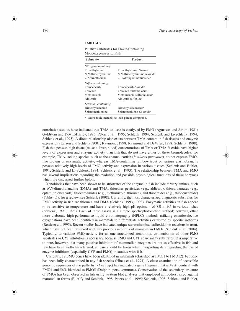
176 The Toxicology of Fishes
correlative studies have indicated that TMA oxidase is catalyzed by FMO (Agutsson and Strom, 1981;Goldstein and Dewitt-Harley, 1973; Peters et al., 1995; Schlenk, 1994; Schlenk and Li-Schlenk, 1994;Schlenk et al., 1995). A direct relationship also exists between TMA content in fish tissues and enzymeexpression (Larsen and Schlenk, 2001; Raymond, 1998; Raymond and DeVries, 1998; Schlenk, 1998).Fish that possess high tissue (muscle, liver, blood) concentrations of TMA or TMA N-oxide have higherlevels of expression and enzyme activity than fish that do not have either of these biomolecules; forexample, TMA-lacking species, such as the channel catfish (Ictalurus punctatus), do not express FMO-like protein or enzymatic activity, whereas TMA-containing rainbow trout or various elasmobranchspossess relatively high levels of FMO activity and expression in various tissues (Schlenk and Buhler,1991; Schlenk and Li-Schlenk, 1994; Schlenk et al., 1993). The relationship between TMA and FMOhas several implications regarding the evolution and possible physiological functions of these enzymeswhich are discussed further below.
Xenobiotics that have been shown to be substrates of the enzyme in fish include tertiary amines, suchas N,N-dimethylanaline (DMA) and TMA; thioether pesticides (e.g., aldicarb); thiocarbamates (e.g.,eptam, thiobencarb); thiocarbamides (e.g., methimizole, thiourea), and thioamides (e.g., thiobenzamide)(Table 4.5); for a review, see Schlenk (1998). Currently, the most characterized diagnostic substrates forFMO activity in fish are thiourea and DMA (Schlenk, 1993, 1998). Enzymatic activities in fish appearto be sensitive to temperature and have a relatively high pH optimum of 8.0 to 9.6 in various fishes(Schlenk, 1993, 1998). Each of these assays is a simple spectrophotometric method; however, othermore elaborate high-performance liquid chromatography (HPLC) methods utilizing enantioselectiveoxygenations have been identified in mammals to differentiate activities catalyzed by specific isoforms(Rettie et al., 1995). Recent studies have indicated unique stereochemical sulfoxidation reactions in trout,which have not been observed with any previous isoforms of mammalian FMOs (Schlenk et al., 2004).Typically, to validate FMO activity for an uncharacterized xenobiotic, co-incubation of other FMOsubstrates or CYP inhibitors is necessary, because FMO and CYP share many substrates. It is imperativeto note, however, that many putative inhibitors of mammalian enzymes are not as effective in fish andfew have been well-characterized, so care should be taken when interpreting data regarding the use ofenzyme inhibitors (especially CYP and FMO) in studies with fish.
Currently, 12 FMO genes have been identified in mammals (classified as FMO1 to FMO12), but nonehas been fully characterized in any fish species (Hines et al., 1994). A close examination of accessiblegenomic sequences of the pufferfish (Fugu sp.) has indicated a gene fragment that is 42% identical withFMO4 and 56% identical to FMO5 (Dolphin, pers. commun.). Conservation of the secondary structureof FMOs has been observed in fish using western blot analyses that employed antibodies raised againstmammalian forms (El-Alfy and Schlenk, 1998; Peters et al., 1995; Schlenk, 1998; Schlenk and Buhler,
TABLE 4.5
Putative Substrates for Flavin-Containing Monooxygenases in Fish
Substrate Product
Nitrogen-containingTrimethylamine Trimethylamine N-oxideN,N-Dimethylaniline N,N-Dimethylaniline N-oxide2-Aminofluorene 2-Hydroxyaminofluorenea
Sulfur -containingThiobencarb Thiobencarb-S-oxidea
Thiourea Thiourea-sulfonic acida
Methimazole Methimazole-sulfonic acida
Aldicarb Aldicarb sulfoxidea
Selenium-containingDimethylselenideSelenomethionine
Dimethylselenoxidea
Selenomethione-Se-oxidea
a More toxic metabolite than parent compound.
Biotransformation in Fishes 177
1993; Schlenk and Li-Schlenk; Schlenk et al., 1995). At least two microsomal proteins in rainbow troutliver were recognized by antibodies raised against porcine FMO1 (Schlenk and Buhler, 1993). Subsequentstudies with antibodies raised against human FMO1 only recognized one of the two bands (Schlenk etal., 2004). Although a faint band was noted in Japanese medaka liver microsomes with anti-FMO3, astronger signal from anti-FMO1 was observed (El-Alfy and Schlenk, 2002); however, catalytic activities(stereochemical analyses) in trout and shark do not correspond to FMO1 or FMO3 but seem to resembleFMO4-like activities, which tends to support genetic observations in Fugu (Schlenk et al., 2004).
Regulation, Function, and Toxicological RelevanceRegulation of FMO expression appears to be extremely complex, with the enzyme often expressed in arandom manner (Baker et al., 1963). As mentioned earlier, FMO has been observed in all marine fishspecies, some euryhaline, and virtually no freshwater species. Strong correlations have been observedbetween FMO1-like proteins, enzymatic activity, and mRNA recognized by FMO1 cDNA in juvenileAtlantic flounder (Platichthys flesus) and turbot (Scophthalmus maximus) (Peters et al., 1995; Schlenket al., 1996a,b), but catalytic activity did not correspond with mRNA expression in sexually mature adultflounder (Schlenk, unpublished data). In fact, although FMO1-like mRNA was observed in all sexuallymature animals, hepatic FMO activity was lacking in more than 40% of the animals, with males havingmore frequent expression. Although evidence suggests that various hormones may modulate the expres-sion of FMO (El-Alfy and Schlenk, 2002; El-Alfy et al., 2002; Schlenk et al., 1997), no consistentinduction of enzyme expression has been observed following xenobiotic treatment. In medaka, estradioldownregulated the hepatic FMO3-like protein but induced the gill FMO1-like form (El-Alfy and Schlenk,2002). Testosterone downregulated both forms of FMO and activity in medaka. Arterial infusion ofcortisol induced expression of FMO1 in rainbow trout gill and liver (El-Alfy et al., 2002). However,infusion of growth hormone failed to alter FMO activity or expression (Schlenk, unpublished data).
As mentioned above, FMO activity and protein in several tissues, particularly the gill, appear to bedirectly correlated with serum osmolality or the salinity regime in which fish resides (Daikoku andSakaguchi, 1990; El-Alfy and Schlenk, 2002; Schlenk, 1998; Schlenk and El-Alfy, 1998; Schlenk et al.,1996a,b). Several hypotheses have been put forth to explain this relationship. Four possibilities are: (1)TMA N-oxide is produced as a cellular defense to prevent the enzyme inactivation by high cellular ion(Na, K) or urea that occurs in fish residing in subartic or subantarctic environments (Raymond, 1998;Raymond and DeVries, 1998); (2) TMA N-oxide is produced to counterbalance high tissue and serumlevels of urea that are present regardless of temperature (e.g., in sharks) (Van Waarde, 1988; Yancey etal., 1982); (3) TMA N-oxide may be produced in muscle of deep-sea gadiform teleosts (which alsoproduce urea) as an adaptive defense against high pressure (Gillett et al., 1997); and (4) TMA N-oxidemay be formed by FMOs as a secondary organic osmolyte in response to shifts in salinity regimes(Schlenk, 1993). Each scenario is interrelated, with the common similarity being hyperosmolality. Insome euryhaline fish, FMO activity and expression are directly related to the salinity regime in whichthe animal resides (Charest et al., 1988; Daikoku and Sakaguchi, 1990; Daikoku et al., 1988; El-Alfyand Schlenk, 2002; Lange and Fugelli, 1965; Schlenk and El-Alfy, 1998; Schlenk et al., 1996a,b). FMOactivity and protein expression is higher in gills and kidneys than liver in several species of euryhalinefish such as the Atlantic flounder (Platichthys flesus), Japanese medaka (Oryzias latipes), and rainbowtrout (Larsen and Schlenk, 2001; Schlenk, 1998; Schlenk and El-Alfy, 1998; Schlenk et al., 1995). Inaddition, FMO activity in trout and medaka is downregulated following steroid treatment, which alsodownregulates osmoregulatory function (i.e., Na+,K+-ATPase) (McCormick, 1995; Schlenk et al., 1997).FMO activity and expression in rapid osmoconformers such as striped bass and tilapia do not respondto changes in salinity (Wang et al., 2001), which is also consistent with mechanisms of Na+,K+-ATPaseregulation in these species. Testing scenarios 1 and 3, recent studies in rainbow trout have indicated thaturea infusion or reductions in temperature induce FMO activity and increase muscular TMAO concen-trations (Larsen and Schlenk, 2001). Given induction following cortisol treatment, several factors maybe involved in FMO regulation in rainbow trout, including stress resulting from alterations in cellularredox potential due to urea or hyperosmotic conditions (El-Alfy et al., 2002; Larsen and Schlenk, 2001).Clearly, more studies are necessary to better understand the regulation of this enzyme system given itsrole in xenobiotic biotransformation and toxicology.
178 The Toxicology of Fishes
The toxicological significance of FMO-catalyzed biotransformation reactions has not been extensivelyexamined in fish. Recent studies with organoselenides and FMO from sharks have indicated that FMOmay be involved in the oxidation and initiation of redox cycling in these species (Schlenk et al., 2003);however, most studies examining the toxicological roles of FMO have examined pesticides. The oxy-genation of the pesticides thiobencarb and eptam in striped bass (Morone saxatilis) by hepatic FMOwas shown to lead to the formation of a reactive-intermediate that covalently bound protein sulfhydralgroups (Cashman et al., 1990; Perkins et al., 1999); however, protein binding was not observed in vivoby thiobencarb. S-Oxygenation of aldicarb to the sulfoxide by FMO significantly increased the inhibitionof acetylcholinesterase in rainbow trout (Oncorhynchus mykiss) (250-fold) and Japanese medaka (Oryziaslatipes) (40-fold) (El-Alfy and Schlenk, 2002; Perkins et al., 1999). Elevated toxicity has been observedin FMO-containing fish which can activate aldicarb to the more potent sulfoxide compared to speciesthat lack FMO and convert aldicarb to the less toxic sulfone or hydrolytic metabolites (Perkins andSchlenk, 2000; Schlenk, 1995).
Enhanced sulfoxidation may possibly explain the enhanced toxicity of aldicarb in higher salinity observedin medaka and trout, as FMO expression has been shown to be directly correlated to salinity in medaka(Larsen and Schlenk, 2001; Schlenk and El-Alfy, 1998). Studies comparing the effects of salinity on aldicarbtoxicity in trout and striped bass indicate that salinity significantly enhances the toxicity of aldicarb in troutbut not in striped bass (Wang et al., 2001). In striped bass, aldicarb sulfoxide formation and FMO expressionwere unchanged by salinity, whereas salinity increased aldicarb sulfoxide formation, cholinesterase inhi-bition, and FMO expression in rainbow trout. Consequently, understanding factors that affect the expressionpatterns of FMO is important when considering species-specific sensitivities to xenobiotics and differentialresponses of organisms to environmental factors such as salinity and temperature regimes.
Monoamine OxidasesMonoamine oxidases catalyze the oxidation and eventual elimination of alpha carbon groups fromsecondary amines. Monoamine oxidases have been characterized in several fish species, with mostoccurring in trout. Given the critical importance in catecholamine metabolism, most studies have focusedon its endogenous role in the neurophysiology of fish. In contrast to terrestrial vertebrates, which havetwo forms of the enzyme (MAO A and MAO B), fish appear to only have a single form that is geneticallydistinct from terrestrial vertebrates. Although no specific studies have examined the role of MAO inxenobiotic biotransformation in fish, the effects of various organic and inorganic pollutants on enzymeactivity has been examined (Senatori et al., 2003).
Alcohol and Aldehyde DehydrogenasesAlcohol dehydrogenase (ADH) catalyzes the oxidation of alcohols to aldehydes, which are subsequentlyconverted to acids by aldehyde dehydrogenase (ALDH). NAD+ is a cofactor for each enzyme. A class 3ADH cDNA was first identified in sea bream, in which its expression was observed in all tissues as wellas eggs and embryos. Expression decreased during early embryonal development but increased fourfoldfrom day 1 to 21 after hatching, indicating that the maternal ADH mRNA is present in the eggs andembryos but diminishes as development occurs, allowing the larval tissue to express its own ADH(Funkenstein and Jakkowiew, 1996). An additional ADH3 cDNA was also identified by RT-PCR inzebrafish (Danio rerio) (Dasmahapatra et al., 2001). Expression of the gene in zebrafish embryos appearedto correspond with temporal variations in zebrafish susceptibility to ethanol toxicity. In cod, an ADHenzyme was purified that displayed structural similarities to ADH3, but functionally it was more likeADH1. Ethanol was an excellent substrate for the purified enzyme, and 4-methylpyrazole was a stronginhibitor (Ki = 0.1 µM) (Danielsson et al., 1992). Allylic and acetylenic alcohols appear to be bioactivatedthrough oxidation by trout liver ADH and may also act as inhibitors (Bradbury and Christensen, 1991).ALDH has been observed in all tissues of numerous fish species (Nagai et al., 1997). Similar to CYP1A,ALDH has been observed in mammals to be regulated by the Ah receptor. Studies in the dab (Limandalimanda), the sea bass (Dicentrarchus labrax), and the rainbow trout failed to observe increases in ALDHactivities following 3-MC or BNF treatment [222]; however, expression was significantly elevated in livertumor tissues from adult rainbow trout treated with aflatoxin (Parker et al., 1993).
no 222ref inoriginalmanuscript

Biotransformation in Fishes 179
PeroxidasesLipoxygenases, cyclooxygenases, and peroxidases such as prostaglandin-H synthetase are typicallyinvolved in the oxidation of arachidonic acid to hydroxy and peroxyeicosatetraenoic acids, which aresubsequently converted to prostaglandins, prostacylins, and thromboxanes. In mammals, each of theseenzymes has been shown to activate various xenobiotics through cooxidation pathways requiring ahydroperoxide as a cofactor. Although the enzymes have been observed in fish, their contributions toxenobiotic biotransformation has not been examined. Examples of lipoxgenases observed in rainbowtrout gills include the 15- and 12-lipoxygenase (German and Berger, 1990; Hsieh et al., 1988). PGHsynthetase has also been observed in gills of freshwater fish (Christ and Van Dorp, 1972).
Aldehyde OxidaseAldehyde oxidase is a molybdozyme that is similar to xanthine oxidase located in the cytosols of liverin mammals and fish. Pyrroles, pyridines, pyrimidines, purines, and aromatic aldehydes derived fromcatecholamine metabolism tend to undergo oxidation reactions; however, under anaerobic conditions,2-hydroxypyrimidine, N1-methylnicotinamide, or butyraldehyde may act as electron donors in livercytosols in fish, leading to substrate reduction under these conditions. Enzyme activity is inhibited bymenadione, β-estradiol, and chlorpromazine. Recent studies in the goldfish (Carassius auratus) and thesea bream (Pagrus major) have demonstrated that aldehyde oxidase catalyzes the reduction of fenthionsulfoxide to the parent sulfide (Kitamura et al., 2003).
Reductases
One of the most common reductases involved in xenobiotic biotransformation is the cytochrome P450reductase, which catalyzes single-electron reductions to substrates prone to accept single electrons(e.g., quinones, cyclic or aromatic amines). Often, this pathway activates heterocyclic compounds toredox cycling intermediates. Limited studies have evaluated the role of this enzyme in biotransfor-mation in fish.
DT-DiaphoraseDT-diaphorase (NAD(P)H:[quinone acceptor] oxidoreductase) plays a significant role in one- and two-electron reduction reactions, particularly in the liver. 4-Nitroquinoline 1-oxide and nitrofurantoin wereboth reduced by DT-diaphorase to genotoxic metabolites in a brown bullhead fibroblast cell line (Has-spieler et al., 1997). A novel dicoumarol-sensitive oxidoreductase that catalyzes the reduction of phenan-threnequinone was purified from gastric cytosol in the channel catfish (Ictalurus punctatus) (Hasspielerand Di Giulio, 1994). Due to its likely AhR-mediated regulation, the enzyme has shown promise as abiomarker in fish (see Chapter 16). Whether or not bifunctional regulation through ARE occurs (as inmammals) is uncertain.
Azo- and NitroreductasesCatalyzing up to three sequential two-electron reductions, nitroreductases play a significant role in thebiotransformation of primarily nitro aromatic compounds to corresponding amino aromatic compounds.When 2-nitrofluorene was incubated with liver microsomes or cytosol of sea bream (Pagrus major) inthe presence of NADPH or 2-hydroxypyrimidine, 2-aminofluorene was formed (Ueda et al., 2002).Hepatic nitroreductases were also observed in catfish and catalyzed the formation of superoxide fromnitrofurantoin, p-nitrobenzoic acid, and m-dinitrobenzene (Washburn and Di Giulio, 1988). Hepatic nitro-and azoreductases were observed in the marine teleosts barracuda (Sphyraena barracuda) and yellowtailsnappers (Ocyurus chrysurus); however, only azo reductase activity was observed in elasmobranches,such as the lemon shark (Negaprion brevirostris) and stingray (Dasyatis americana) (Adamson et al.,1965). The lampricide 3-trifluoromethyl-4-nitrophenol was reduced by nitroreductases to 3-trifluorom-ethyl-4-aminophenol in rainbow trout (Lech and Costrini, 1972).
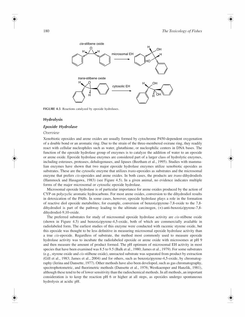
180 The Toxicology of Fishes
Hydrolysis
Epoxide Hydrolase
OverviewXenobiotic epoxides and arene oxides are usually formed by cytochrome P450-dependent oxygenationof a double bond or an aromatic ring. Due to the strain of the three-membered oxirane ring, they readilyreact with cellular nucleophiles such as water, glutathione, or nucleophilic centers in DNA bases. Thefunction of the epoxide hydrolase group of enzymes is to catalyze the addition of water to an epoxideor arene oxide. Epoxide hydrolase enzymes are considered part of a larger class of hydrolytic enzymes,including esterases, proteases, dehalogenases, and lipases (Beetham et al., 1995). Studies with mamma-lian enzymes have shown that two major epoxide hydrolase enzymes utilize xenobiotic epoxides assubstrates. These are the cytosolic enzyme that utilizes trans-epoxides as substrates and the microsomalenzyme that prefers cis-epoxides and arene oxides. In both cases, the products are trans-dihydrodiols(Hammock and Hasagawa, 1983) (see Figure 4.5). In a given animal, no evidence indicates multipleforms of the major microsomal or cytosolic epoxide hydrolase.
Microsomal epoxide hydrolase is of particular importance for arene oxides produced by the action ofCYP on polycyclic aromatic hydrocarbons. For most arene oxides, conversion to the dihydrodiol resultsin detoxication of the PAHs. In some cases, however, epoxide hydrolase plays a role in the formationof reactive diol epoxide metabolites; for example, conversion of benzo(a)pyrene-7,8-oxide to the 7,8-dihydrodiol is part of the pathway leading to the ultimate carcinogen, (+)-anti-benzo(a)pyrene-7,8-dihydrodiol-9,10-oxide.
The preferred substrates for study of microsomal epoxide hydrolase activity are cis-stilbene oxide(shown in Figure 4.5) and benzo(a)pyrene-4,5-oxide, both of which are commercially available inradiolabeled form. The earliest studies of this enzyme were conducted with racemic styrene oxide, butthis epoxide was thought to be less definitive in measuring microsomal epoxide hydrolase activity thana true cis-epoxide. Regardless of substrate, the method most commonly used to measure epoxidehydrolase activity was to incubate the radiolabeled epoxide or arene oxide with microsomes at pH 9and then measure the amount of product formed. The pH optimum of microsomal EH activity in mostspecies that have been examined was 8.5 to 9.5 (Balk et al., 1980; James et al., 1979). For some substrates(e.g., styrene oxide and cis-stilbene oxide), unreacted substrate was separated from product by extraction(Gill et al., 1983; James et al., 2004) and for others, such as benzo(a)pyrene-4,5-oxide, by chromatog-raphy (Jerina and Dansette, 1977). Other methods have also been developed, such as gas chromatography,spectrophotometric, and fluorimetric methods (Dansette et al., 1976; Westkaemper and Hanzlik, 1981),although these tend to be of lower sensitivity than the radiochemical methods. In all methods, an importantconsideration is to keep the reaction pH 6 or higher at all steps, as epoxides undergo spontaneoushydrolysis at acidic pH.
FIGURE 4.5 Reactions catalyzed by epoxide hydrolases.
cis–stilbene oxide
trans–stilbene oxide
microsomal EH
cytosolic EH
H
H
H H
H
H
H
HO
OH
OH
OH
HO
O
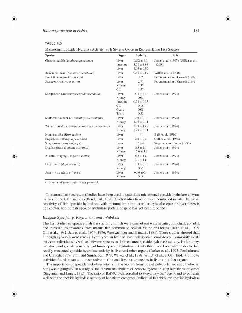
Biotransformation in Fishes 181
In mammalian species, antibodies have been used to quantitate microsomal epoxide hydrolase enzymein liver subcellular fractions (Bend et al., 1978). Such studies have not been conducted in fish. The cross-reactivity of fish epoxide hydrolases with mammalian microsomal or cytosolic epoxide hydrolases isnot known, and no fish epoxide hydrolase protein or gene has yet been reported.
Enzyme Specificity, Regulation, and InhibitionThe first studies of epoxide hydrolase activity in fish were carried out with hepatic, branchial, gonadal,and intestinal microsomes from marine fish common to coastal Maine or Florida (Bend et al., 1978;Gill et al., 1982; James et al., 1974, 1976; Westkaemper and Hanzlik, 1981). These studies showed that,although epoxides were readily hydrolyzed in liver of most fish species, considerable variability existsbetween individuals as well as between species in the measured epoxide hydrolase activity. Gill, kidney,intestine, and gonads generally had lower epoxide hydrolase activity than liver. Freshwater fish also hadreadily measured epoxide hydrolase activity in liver and other organs (Parker et al., 1993; Perdudurandand Cravedi, 1989; Stott and Sinnhuber, 1978; Walker et al., 1978; Willett et al., 2000). Table 4.6 showsactivities found in some representative marine and freshwater species in liver and other organs.
The importance of epoxide hydrolase activity in the biotransformation of polycyclic aromatic hydrocar-bons was highlighted in a study of the in vitro metabolism of benzo(a)pyrene in scup hepatic microsomes(Stegeman and James, 1985). The ratio of BaP-9,10-dihydrodiol to 9-hydroxy-BaP was found to correlatewell with the epoxide hydrolase activity of hepatic microsomes. Individual fish with low epoxide hydrolase
TABLE 4.6
Microsomal Epoxide Hydrolase Activitya with Styrene Oxide in Representative Fish Species
Species Organ Activity Refs.
Channel catfish (Ictalurus punctatus) LiverIntestineLiver
2.62 ± 1.03.78 ± 1.951.03 ± 0.06
James et al. (1997); Willett et al. (2000)
Brown bullhead (Ameiurus nebulosus) Liver 0.85 ± 0.07 Willett et al. (2000)Trout (Oncorhynchus mykiss) Liver 1.2 Perdudurand and Cravedi (1989)Sturgeon (Acipenser baeri) Liver
KidneyGill
2.771.371.37
Perdudurand and Cravedi (1989)
Sheepshead (Archosargus probatocephalus) LiverKidneyIntestineGillOvaryTestis
5.6 ± 2.40.05
0.74 ± 0.330.160.080.52
James et al. (1974)
Southern flounder (Paralichthyes lethostigma) LiverKidney
2.0 ± 0.71.33 ± 0.11
James et al. (1974)
Winter flounder (Pseudopleuronectes americanus) LiverKidney
27.9 ± 15.98.25 ± 6.11
James et al. (1974)
Northern pike (Esox lucius) Liver 4 Balk et al. (1980)English sole (Parophrys vetulus) Liver 2.8 ± 0.2 Collier et al. (1986)Scup (Stenotomus chrysops) Liver 2.6–9 Stegeman and James (1985)Dogfish shark (Squalus acanthias) Liver
Kidney6.3 ± 2.112.6 ± 3.9
James et al. (1974)
Atlantic stingray (Dasyatis sabina) LiverKidney
6.2 ± 1.83.1 ± 1.8
James et al. (1974)
Large skate (Raja ocellata) LiverKidney
1.8 ± 0.20.55
James et al. (1974)
Small skate (Raja erinacea) LiverKidney
0.46 ± 0.40.16
James et al. (1974)
a In units of nmol · min–1 · mg protein–1.
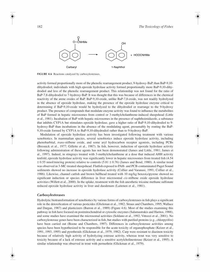
182 The Toxicology of Fishes
activity formed proportionally more of the phenolic rearrangement product, 9-hydroxy-BaP, than BaP-9,10-dihydrodiol; individuals with high epoxide hydrolase activity formed proportionally more BaP-9,10-dihy-drodiol and less of the phenolic rearrangement product. This relationship was not found for the ratio ofBaP-7,8-dihydrodiol to 7-hydroxy-BaP. It was thought that this was because of differences in the chemicalreactivity of the arene oxides of BaP. BaP-9,10-oxide, unlike BaP-7,8-oxide, was not readily hydrolyzedin the absence of epoxide hydrolase, making the presence of the epoxide hydrolase enzyme critical todetermining if BaP-9,10-oxide would be hydrolyzed to the dihydrodiol or rearrange to the 9-hydroxyproduct. The presence of compounds that modulate enzyme activity was found to influence the metabolitesof BaP formed in hepatic microsomes from control or 3-methylcholanthrene-induced sheepshead (Littleet al., 1981). Incubation of BaP with hepatic microsomes in the presence of naphthoimidazole, a substancethat inhibits CYP1A but stimulates epoxide hydrolase, gave a higher ratio of BaP-9,10-dihydrodiol to 9-hydroxy-BaP than incubations in the absence of the modulating agent, presumably by routing the BaP-9,10-oxide formed by CYP1A to BaP-9,10-dihydrodiol rather than to 9-hydroxy-BaP.
Modulation of epoxide hydrolase activity has been investigated following treatment with variousxenobiotics. In mammalian species, several xenobiotics induce epoxide hydrolase activity, includingphenobarbital, trans-stilbene oxide, and some aryl hydrocarbon receptor agonists, including PCBs(Bresnick et al., 1977; Gillette et al., 1987). In fish, however, induction of epoxide hydrolase activityfollowing administration of these agents has not been demonstrated (James and Little, 1981; James etal., 1997). Indeed, in stingrays treated with 3-methylcholanthrene at a dose that induced AHH activitytenfold, epoxide hydrolase activity was significantly lower in hepatic microsomes from treated fish (4.54± 0.55 nmol/min/mg protein) relative to controls (5.81 ± 0.76) (James and Bend, 1980). A similar trendwas observed in 3-MC-treated sheepshead. Flatfish exposed to PAH- and PCB-contaminated Puget Soundsediments showed no increase in epoxide hydrolase activity (Collier and Varanasi, 1991; Collier et al.,1986). Likewise, channel catfish and brown bullhead treated with 10 mg/kg benzo(a)pyrene showed nosignificant induction or species difference in liver microsomal cis-stilbene oxide epoxide hydrolaseactivities (Willett et al., 2000). In the splake, treatment with the fish anesthetic tricaine methane sulfonatereduced epoxide hydrolase activity in liver and duodenum (Laitenen et al., 1981).
CarboxylesterasesHydrolytic biotransformation of xenobiotics by various forms of carboxylesterases in fish plays a significantrole in the detoxification of various pesticides (Glickman et al., 1982; Straus and Chambers, 1995; Wallaceand Dargan, 1987) and plasticizers (Barron et al., 1989) (Figure 4.6). Most of the studies examining thispathway in fish have focused on postmitochondrial or cytosolic enzymes (Salamastrakis and Haritos, 1988),and some studies have examined the microsomal activities (Soldano et al., 1992; Vittozzi et al., 2001). Nocarboxylesterase genes have been characterized in fish, but studies with purified proteins (e.g., chlorpyrifos)have been carried out (Boone and Chambers, 1997). Differences in carboxylesterase activities amongspecies have been hypothesized to be responsible for the acute toxicity of organophosphate (Keizer et al.,1991, 1993, 1995) and pyrethroids (Glickman et al., 1979, 1982). Carp were resistant to diazinon toxicitybecause of relatively high activity of hydrolyzing esterase activity, whereas trout was very sensitive totoxicity because of a lack of esterase activity and a sensitive acetylcholinesterase (Keizer et al., 1995). Asimilar relationship was observed in trout with permethrin (Glickman et al., 1979).
FIGURE 4.6 Reactions catalyzed by carboxylesterases.
O
O CNHMe
Carbaryl 1-Naphthol
OH
+ HOOCNHMe
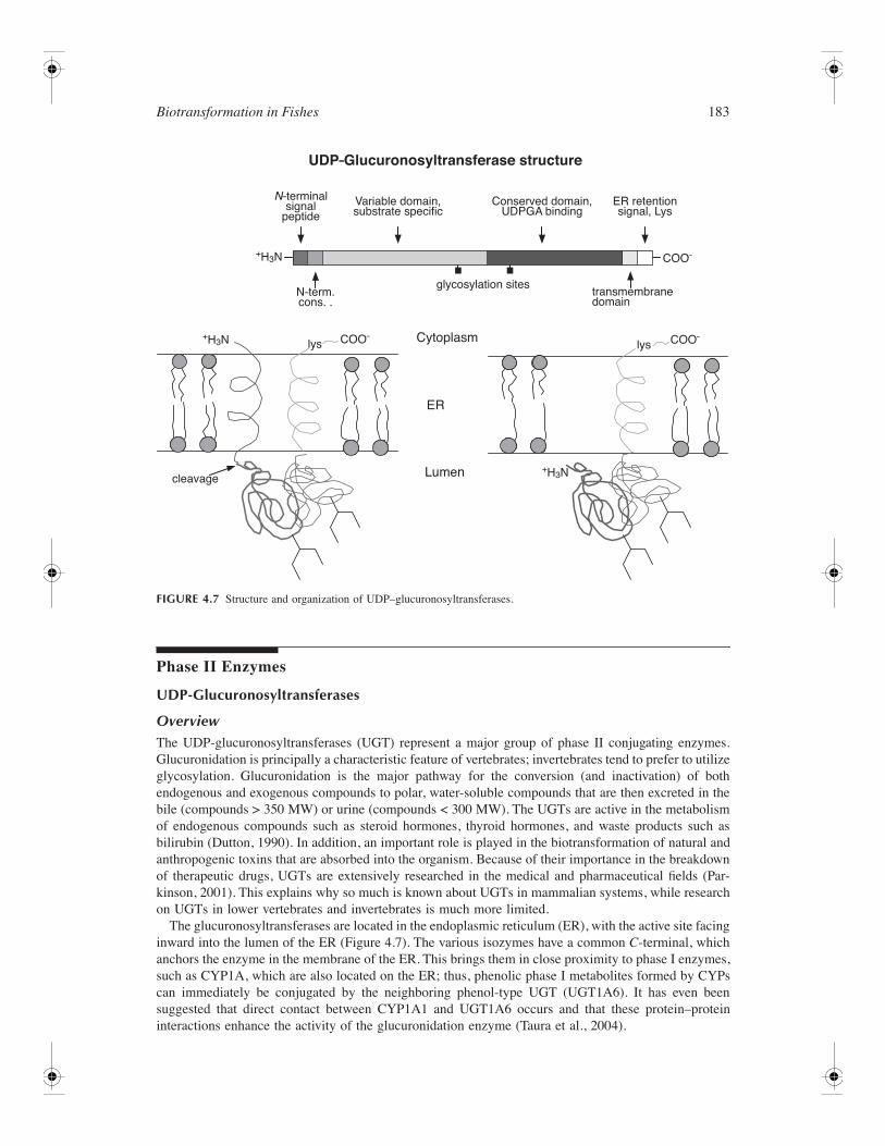
Biotransformation in Fishes 183
Phase II Enzymes
UDP-Glucuronosyltransferases
OverviewThe UDP-glucuronosyltransferases (UGT) represent a major group of phase II conjugating enzymes.Glucuronidation is principally a characteristic feature of vertebrates; invertebrates tend to prefer to utilizeglycosylation. Glucuronidation is the major pathway for the conversion (and inactivation) of bothendogenous and exogenous compounds to polar, water-soluble compounds that are then excreted in thebile (compounds > 350 MW) or urine (compounds < 300 MW). The UGTs are active in the metabolismof endogenous compounds such as steroid hormones, thyroid hormones, and waste products such asbilirubin (Dutton, 1990). In addition, an important role is played in the biotransformation of natural andanthropogenic toxins that are absorbed into the organism. Because of their importance in the breakdownof therapeutic drugs, UGTs are extensively researched in the medical and pharmaceutical fields (Par-kinson, 2001). This explains why so much is known about UGTs in mammalian systems, while researchon UGTs in lower vertebrates and invertebrates is much more limited.
The glucuronosyltransferases are located in the endoplasmic reticulum (ER), with the active site facinginward into the lumen of the ER (Figure 4.7). The various isozymes have a common C-terminal, whichanchors the enzyme in the membrane of the ER. This brings them in close proximity to phase I enzymes,such as CYP1A, which are also located on the ER; thus, phenolic phase I metabolites formed by CYPscan immediately be conjugated by the neighboring phenol-type UGT (UGT1A6). It has even beensuggested that direct contact between CYP1A1 and UGT1A6 occurs and that these protein–proteininteractions enhance the activity of the glucuronidation enzyme (Taura et al., 2004).
FIGURE 4.7 Structure and organization of UDP–glucuronosyltransferases.
N–terminalsignal
peptide
N–term.cons. .
+H3N
+H3N
+H3N
COO–
COO–
Variable domain,substrate specific
glycosylation sites
Cytoplasm
ER
cleavage Lumen
transmembranedomain
Conserved domain,UDPGA binding
ER retentionsignal, Lys
UDP–Glucuronosyltransferase structure
lysCOO–lys
184 The Toxicology of Fishes
Glucuronidation involves the transfer of an activated sugar group to the substrate. This processincreases the water solubility and the molecular weight of the substrates, thus facilitating transport andexcretion. The preferred cofactor for most UGT isoforms is uridine diphosphoglucuronic acid (UDPGA);however, a number of other nucleotide–sugar cofactors have been reported for these enzymes; therefore,the name UDP-glycosyltransferase has been proposed as a better name for this superfamily (Mackenzieet al., 1997). UDPGA is synthesized in the cytosol from uridine-triphosphate and glucose-1-phosphate.Contrary to many phase I reactions, glucuronidation does not require additional energy supply in theform of ATP or NADPH. The synthesis of UDPGA in the cytosol introduces an extra threshold for theglucuronidation reaction rate, as the water-soluble cofactor has to be actively transported through theER membrane to reach the active site on the UGT enzyme. Mechanisms surrounding transport are stillunknown.
UGT Gene StructureMultiple UGT genes have been characterized in mammals (17 in humans) and are subdivided into twofamilies. All UGT1 family members are encoded by a unique single locus gene, which in humans has13 different first exons spanning some 300 kb. They encode the aglycone binding site, which is alter-natively spliced onto identical exons 2 to 5, which encode the C-terminal half of the protein responsiblefor UDP-glucuronic acid binding. Each exon 1 appears to have its own promoter in the 5′ upstreamregion between its transcription start site and the preceding exon 1. Elaboration of the gene appears tohave arisen from duplication into two clusters: (1) a bilirubin cluster (1A1 to 1A5 isoforms), primarilyinvolved in the conjugation of amines, bilirubin, carboxylic acids, and thyroxine, and (2) a phenol cluster,which conjugates planar polyaromatic hydrocarbons (1A6) and bulky phenols (1A7 to 1A13). A similaralthough slightly less complex elaboration of the UGT1A family has been found in rodents and therabbit. In humans and rats, at least one gene of the bilirubin cluster is a pseudogene resulting in reducedconjugation of planar phenols. In contrast, the genes of UGT2 family members, each comprised of sixexons, are located at distinct loci on another chromosome. The UGT2A subfamily has postulated rolesin olfaction, and the products of the UGT2B family genes appear to be particularly involved in bile acidand steroid hormone conjugation. Although many of the enzymes were successfully purified from rodents,the majority of the genes have been heterologously expressed for activity studies, and indeed most ofthe human enzymes are now commercially available.
The gene encoding the major phenol-conjugating isoform of plaice has been cloned (UGT1B1) andfound to share an almost identical five-exon structure with mammalian UGT1 family members (Georgeand Leaver, 2002; George, unpublished data). When heterologously expressed, it displays specificity forplanar phenols such as 1-naphthol and 4-nitrophenol as aglycones but no demonstrable activity withbilirubin or steroids (Leaver et al., unpublished data).
Expressed sequence tags (ESTs) for UGTs have subsequently been cloned from flounder, plaice, andzebrafish. Analysis of the currently published zebrafish EST sequences and the latest assembly of thegenome reveals that at least 14 distinct UGTs are expressed (George and Taylor, 2002; George, unpub-lished data). More putative genes can be identified in the genome; however, they are found on numerouschromosomes (in both mouse and humans, the UGT1 family occurs on one chromosome and the UGT2family on another). The phylogenetic relationships between these expressed zebrafish UGT homologsand the human genes is shown in Figure 4.8. The zebrafish genes are divisible into three groups; thegroup with closest similarity to the characterized UGT1B1 gene comprises two alternatively spliced six-exon genes. Gene 6220 is comprised of six exon 1’s, three of which are known to be expressed (Figure4.9). The products arise by alternative splicing of the primary transcripts. Inspection of the nucleotidesequences of the intervening introns between the multiple exons 1’s reveals the presence of peroxisomalproliferator response elements (PPREs) and xenobiotic response elements (XREs), which would supportthe plaice induction data. A further three zebrafish genes are comprised of six exons and divide withthe mammalian UGT2 family, thus inferring the closest homology. Interestingly, gene 4649 also appearsto exhibit alternative splicing. Remarkably, unlike mammals, at least four of the zebrafish putative UGTtranscripts appear to be the products of intronless genes; these may not be UGTs but may be relatedglycosyltransferases.
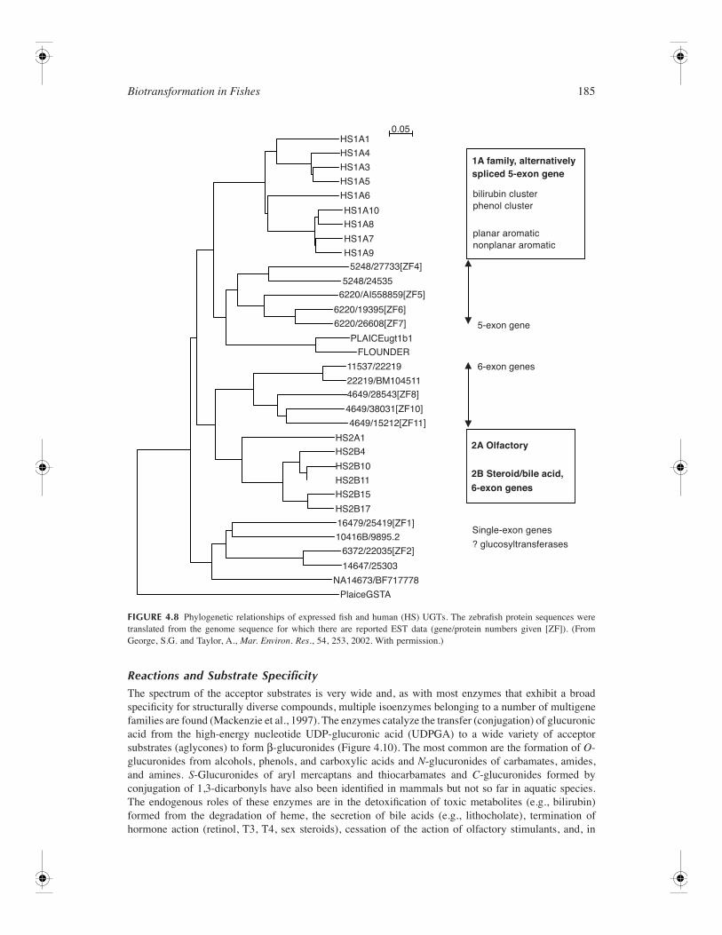
Biotransformation in Fishes 185
Reactions and Substrate SpecificityThe spectrum of the acceptor substrates is very wide and, as with most enzymes that exhibit a broadspecificity for structurally diverse compounds, multiple isoenzymes belonging to a number of multigenefamilies are found (Mackenzie et al., 1997). The enzymes catalyze the transfer (conjugation) of glucuronicacid from the high-energy nucleotide UDP-glucuronic acid (UDPGA) to a wide variety of acceptorsubstrates (aglycones) to form β-glucuronides (Figure 4.10). The most common are the formation of O-glucuronides from alcohols, phenols, and carboxylic acids and N-glucuronides of carbamates, amides,and amines. S-Glucuronides of aryl mercaptans and thiocarbamates and C-glucuronides formed byconjugation of 1,3-dicarbonyls have also been identified in mammals but not so far in aquatic species.The endogenous roles of these enzymes are in the detoxification of toxic metabolites (e.g., bilirubin)formed from the degradation of heme, the secretion of bile acids (e.g., lithocholate), termination ofhormone action (retinol, T3, T4, sex steroids), cessation of the action of olfactory stimulants, and, in
FIGURE 4.8 Phylogenetic relationships of expressed fish and human (HS) UGTs. The zebrafish protein sequences weretranslated from the genome sequence for which there are reported EST data (gene/protein numbers given [ZF]). (FromGeorge, S.G. and Taylor, A., Mar. Environ. Res., 54, 253, 2002. With permission.)
HS1A1
HS1A4
HS1A3
HS1A5
HS1A6
HS1A10
HS1A8
HS1A7
HS1A95248/27733[ZF4]
6220/AI558859[ZF5]
6220/19395[ZF6]
6220/26608[ZF7]
4649/28543[ZF8]
4649/38031[ZF10]
4649/15212[ZF11]
16479/25419[ZF1]
6372/22035[ZF2]
NA14673/BF717778
10416B/9895.2
14647/25303
HS2A1
HS2B4
HS2B10
HS2B11
HS2B15
HS2B17
11537/22219
22219/BM104511
PLAICEugt1b1
PlaiceGSTA
FLOUNDER
5248/24535
5-exon gene
6-exon genes
0.05
2A Olfactory
2B Steroid/bile acid,
6-exon genes
Single-exon genes
? glucosyltransferases
1A family, alternativelyspliced 5-exon gene
bilirubin clusterphenol cluster
planar aromaticnonplanar aromatic
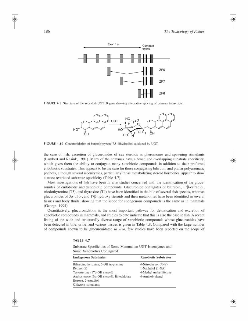
186 The Toxicology of Fishes
the case of fish, excretion of glucuronides of sex steroids as pheromones and spawning stimulants(Lambert and Resink, 1991). Many of the enzymes have a broad and overlapping substrate specificity,which gives them the ability to conjugate many xenobiotic compounds in addition to their preferredendobiotic substrates. This appears to be the case for those conjugating bilirubin and planar polyaromaticphenols, although several isoenzymes, particularly those metabolizing steroid hormones, appear to showa more restricted substrate specificity (Table 4.7).
Most investigations of fish have been in vivo studies concerned with the identification of the glucu-ronides of endobiotic and xenobiotic compounds. Glucuronide conjugates of bilirubin, 17β-estradiol,triodothyronine (T3), and thyroxine (T4) have been identified in the bile of several fish species, whereasglucuronides of 3α-, 3β-, and 17β-hydroxy steroids and their metabolites have been identified in severaltissues and body fluids, showing that the scope for endogenous compounds is the same as in mammals(George, 1994).
Quantitatively, glucuronidation is the most important pathway for detoxication and excretion ofxenobiotic compounds in mammals, and studies to date indicate that this is also the case in fish. A recentlisting of the wide and structurally diverse range of xenobiotic compounds whose glucuronides havebeen detected in bile, urine, and various tissues is given in Table 4.8. Compared with the large numberof compounds shown to be glucuronidated in vivo, few studies have been reported on the scope of
FIGURE 4.9 Structure of the zebrafish UGT1B gene showing alternative splicing of primary transcripts.
FIGURE 4.10 Glucuronidation of benzo(a)pyrene 7,8-dihydrodiol catalyzed by UGT.
TABLE 4.7
Substrate Specificities of Some Mammalian UGT Isoenzymes and Some Xenobiotics Conjugated
Endogenous Substrates Xenobiotic Substrates
Bilirubin, thyroxine, 5-OH tryptamineRetinol (?)Testosterone (17β-OH steroid)Androsterone (3α-OH steroid), lithochlolateEstrone, 2-estradiolOlfactory stimulants
4-Nitrophenol (4NP)1-Naphthol (1-NA)4-Methyl umbelliferone4-Aminobiphenyl
Exon 1ʼs Commonexons
ZF5
ZF7
ZF6
HOUGT
HOHO
HOOH OHOH
H H
HH H
OO
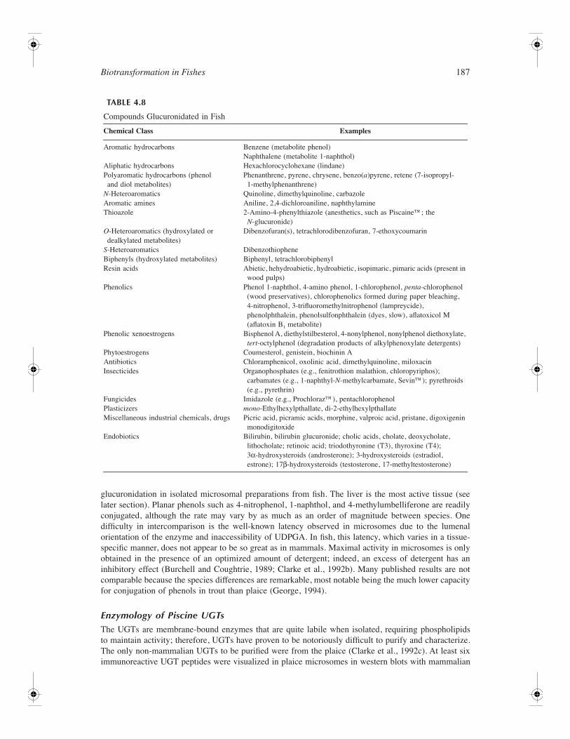
Biotransformation in Fishes 187
glucuronidation in isolated microsomal preparations from fish. The liver is the most active tissue (seelater section). Planar phenols such as 4-nitrophenol, 1-naphthol, and 4-methylumbelliferone are readilyconjugated, although the rate may vary by as much as an order of magnitude between species. Onedifficulty in intercomparison is the well-known latency observed in microsomes due to the lumenalorientation of the enzyme and inaccessibility of UDPGA. In fish, this latency, which varies in a tissue-specific manner, does not appear to be so great as in mammals. Maximal activity in microsomes is onlyobtained in the presence of an optimized amount of detergent; indeed, an excess of detergent has aninhibitory effect (Burchell and Coughtrie, 1989; Clarke et al., 1992b). Many published results are notcomparable because the species differences are remarkable, most notable being the much lower capacityfor conjugation of phenols in trout than plaice (George, 1994).
Enzymology of Piscine UGTsThe UGTs are membrane-bound enzymes that are quite labile when isolated, requiring phospholipidsto maintain activity; therefore, UGTs have proven to be notoriously difficult to purify and characterize.The only non-mammalian UGTs to be purified were from the plaice (Clarke et al., 1992c). At least siximmunoreactive UGT peptides were visualized in plaice microsomes in western blots with mammalian
TABLE 4.8
Compounds Glucuronidated in Fish
Chemical Class Examples
Aromatic hydrocarbons Benzene (metabolite phenol)Naphthalene (metabolite 1-naphthol)
Aliphatic hydrocarbons Hexachlorocyclohexane (lindane)Polyaromatic hydrocarbons (phenol and diol metabolites)
Phenanthrene, pyrene, chrysene, benzo(a)pyrene, retene (7-isopropyl-1-methylphenanthrene)
N-Heteroaromatics Quinoline, dimethylquinoline, carbazoleAromatic amines Aniline, 2,4-dichloroaniline, naphthylamineThioazole 2-Amino-4-phenylthiazole (anesthetics, such as Piscaine™; the
N-glucuronide)O-Heteroaromatics (hydroxylated or dealkylated metabolites)
Dibenzofuran(s), tetrachlorodibenzofuran, 7-ethoxycoumarin
S-Heteroaromatics DibenzothiopheneBiphenyls (hydroxylated metabolites) Biphenyl, tetrachlorobiphenylResin acids Abietic, hehydroabietic, hydroabietic, isopimaric, pimaric acids (present in
wood pulps)Phenolics Phenol 1-naphthol, 4-amino phenol, 1-chlorophenol, penta-chlorophenol
(wood preservatives), chlorophenolics formed during paper bleaching, 4-nitrophenol, 3-trifluoromethylnitrophenol (lampreycide), phenolphthalein, phenolsulfonphthalein (dyes, slow), aflatoxicol M (aflatoxin B1 metabolite)
Phenolic xenoestrogens Bisphenol A, diethylstilbesterol, 4-nonylphenol, nonylphenol diethoxylate, tert-octylphenol (degradation products of alkylphenoxylate detergents)
Phytoestrogens Coumesterol, genistein, biochinin AAntibiotics Chloramphenicol, oxolinic acid, dimethylquinoline, miloxacinInsecticides Organophosphates (e.g., fenitrothion malathion, chloropyriphos);
carbamates (e.g., 1-naphthyl-N-methylcarbamate, Sevin™); pyrethroids (e.g., pyrethrin)
Fungicides Imidazole (e.g., Prochloraz™), pentachlorophenolPlasticizers mono-Ethylhexylpthallate, di-2-ethylhexylpthallateMiscellaneous industrial chemicals, drugs Picric acid, picramic acids, morphine, valproic acid, pristane, digoxigenin
monodigitoxideEndobiotics Bilirubin, bilirubin glucuronide; cholic acids, cholate, deoxycholate,
lithocholate; retinoic acid; triodothyronine (T3), thyroxine (T4); 3α-hydroxysteroids (androsterone); 3-hydroxysteroids (estradiol, estrone); 17β-hydroxysteroids (testosterone, 17-methyltestosterone)
188 The Toxicology of Fishes
antisera, indicating common structural epitopes (Clarke et al., 1992a). From purification studies, at leastfive UGTs have been identified in plaice liver (George, 1994), and the phenol conjugating isoform thatwas purified to homogeneity displayed a very high activity toward planar phenols and no measurableactivity toward bilirubin or steroids. The bilirubin-conjugating isoform appeared to exhibit both bilirubin-and phenol-conjugating activity as found in mammals. Reviews of earlier studies on glucuronidation infish can be found in Clarke et al. (1991) and George (1994). The picture that emerges from these reviewsis that UGT isoforms are found in a wide variety of fish species and that specific enzymatic activitiesare found for bilirubin, steroid and thyroid hormones, and phenolic compounds. As in mammaliansystems, a diverse group of xenobiotic compounds has been identified as substrates for UGTs, amongwhich are chlorinated phenols, aromatic hydrocarbon metabolites, phthalates, aflatoxin, pesticides, andantibiotics. Comparison of UGT activities among species was found to be a problematic task primarilydue to the non-optimization of detergent concentrations in assays in most published studies. The enzymesalso display maximal activity at 37˚C (Clarke et al., 1992b); thus, corrections for temperature should beused for intercomparisons.
Tissue DistributionIn fish species, UGT activity is usually high in liver and intestine, but measurable activities have alsobeen found in gill, kidney, and muscle tissue (Clarke et al., 1991; George et al., 1998; James et al., 1998;Singh et al., 1996). UGTs play an important role in gonadal tissues; in addition to the regulation ofsteroid hormones, UGTs play a role in the production of sex pheromones in fish (Lambert and Resink,1991). Testosterone UGT activity is present in liver, testis, and intestine (Clarke et al., 1992b), andglucuronidation of pregnenolone and androstenedione has been demonstrated in vitro with testicularpreparations (Andersson, 1992). Glucuronidation of bilirubin is confined to liver in both plaice andsalmon.
Regulation of UGTsCoregulation of both CYPs and UGTs occurs in mammals, and prototypical inducers such as clofibrate,PAHs, phenobarbital, and pregnenolone 16α-carbonitrile (PCN) differentially induce expression of bothCYP and UGT isoforms. The degree of upregulation of UGT activity is generally some two- to threefold.UGT1A1 (bilirubin conjugation) is induced by the hyperlipidemic agent (and peroxisomal proliferator)clofibrate. In common with a number of phase I and II genes, UGT1A6 (planar phenol conjugation) isinduced by interaction of the Ah receptor with an XRE in its promoter region. This coregulation ofCYP1A and UGT1A6 in mammals and CYP1A and UGT1B1 in fish has been shown to facilitatedetoxification of PAHs such as BaP. Mammalian CYP2B and CYP3A and UGT1A genes are also inducedvia a nuclear pregnane X receptor and a constitutive androstane receptor. Interestingly, these recognitionmotifs are also present in the zebrafish UGT6220 gene (George, unpublished data). Mammalian steroidUGTs are induced by PCN. Several reports indicate a modest induction of phenol UGT activity in fishfrom polluted environments and after experimental PAH exposure. To study the induction of AhR-activated enzymes, two PAHs are often used: 3-methylcholanthrene (3-MC) and β-naphthoflavone (BNF).Both compounds have been used as CYP1A and phenol UGT inducers in a number of fish species (Table4.9). Maximal induction of CYP1A is usually found around 3 days after single treatment, but phenolUGT induction appears to be slower, with a maximum induction occurring around 8 days after treatment.In general, induction of EROD activity can be up to 250-fold, but UGT activity is never induced morethan 3-to 6-fold. Two species, cod (Gadus morhua) and gilthead sea bream (Sparus aurata), displayedlittle or no response to AhR ligands as phenol UGT inducers (Goksøyr et al., 1987; Pretti et al., 2001).It must be noted, however, that multiple isoforms in mammals, including the constitutive steroid isoforms,also conjugate 4-nitrophenol. Immunoblot and northern blot analyses of xenobiotic-treated plaice haveshown that induction of UGTs appears to be tissue specific (Clarke et al., 1992a). Treatment with thePAH (3-MC) increased phenol-conjugating activity and a 56-kDa immunoreactive peptide in liver byapproximately 1.7-fold. The bifunctional PAH-type inducer BNF caused an induction (approximatelythree- to fourfold) of UGT1B1 mRNA only in intestine (Leaver et al., unpublished data). Althoughclofibrate did not appear to induce phenol- or bilirubin-conjugating activities (Clarke et al., 1992a),
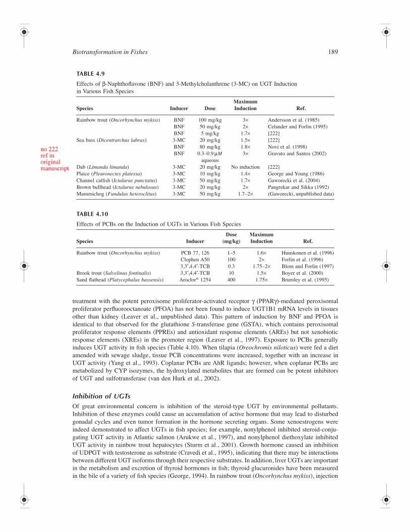
Biotransformation in Fishes 189
treatment with the potent peroxisome proliferator-activated receptor γ (PPARγ)-mediated peroxisomalproliferator perfluorooctanoate (PFOA) has not been found to induce UGT1B1 mRNA levels in tissuesother than kidney (Leaver et al., unpublished data). This pattern of induction by BNF and PFOA isidentical to that observed for the glutathione S-transferase gene (GSTA), which contains peroxisomalproliferator response elements (PPREs) and antioxidant response elements (AREs) but not xenobioticresponse elements (XREs) in the promoter region (Leaver et al., 1997). Exposure to PCBs generallyinduces UGT activity in fish species (Table 4.10). When tilapia (Oreochromis niloticus) were fed a dietamended with sewage sludge, tissue PCB concentrations were increased, together with an increase inUGT activity (Yang et al., 1993). Coplanar PCBs are AhR ligands; however, when coplanar PCBs aremetabolized by CYP isozymes, the hydroxylated metabolites that are formed can be potent inhibitorsof UGT and sulfotransferase (van den Hurk et al., 2002).
Inhibition of UGTsOf great environmental concern is inhibition of the steroid-type UGT by environmental pollutants.Inhibition of these enzymes could cause an accumulation of active hormone that may lead to disturbedgonadal cycles and even tumor formation in the hormone secreting organs. Some xenoestrogens wereindeed demonstrated to affect UGTs in fish species; for example, nonylphenol inhibited steroid-conju-gating UGT activity in Atlantic salmon (Arukwe et al., 1997), and nonylphenol diethoxylate inhibitedUGT activity in rainbow trout hepatocytes (Sturm et al., 2001). Growth hormone caused an inhibitionof UDPGT with testosterone as substrate (Cravedi et al., 1995), indicating that there may be interactionsbetween different UGT isoforms through their respective substrates. In addition, liver UGTs are importantin the metabolism and excretion of thyroid hormones in fish; thyroid glucuronides have been measuredin the bile of a variety of fish species (George, 1994). In rainbow trout (Oncorhynchus mykiss), injection
TABLE 4.9
Effects of β-Naphthoflavone (BNF) and 3-Methylcholanthrene (3-MC) on UGT Induction in Various Fish Species
Species Inducer DoseMaximum Induction Ref.
Rainbow trout (Oncorhynchus mykiss) BNF 100 mg/kg 3× Andersson et al. (1985)BNF 50 mg/kg 2× Celander and Forlin (1995)BNF 5 mg/kg 1.7× [222]
Sea bass (Dicentrarchus labrax) 3-MC 20 mg/kg 1.5× [222]BNF 80 mg/kg 1.8× Novi et al. (1998)BNF 0.3–0.9 µM
aqueous3× Gravato and Santos (2002)
Dab (Limanda limanda) 3-MC 20 mg/kg No induction [222]Plaice (Pleuronectes platessa) 3-MC 10 mg/kg 1.4× George and Young (1986)Channel catfish (Ictalurus punctatus) 3-MC 50 mg/kg 1.7× Gaworecki et al. (2004)Brown bullhead (Ictalurus nebulosus) 3-MC 20 mg/kg 2× Pangrekar and Sikka (1992)Mummichog (Fundulus heteroclitus) 3-MC 50 mg/kg 1.7–2× (Gaworecki, unpublished data)
TABLE 4.10
Effects of PCBs on the Induction of UGTs in Various Fish Species
Species InducerDose
(mg/kg)Maximum Induction Ref.
Rainbow trout (Oncorhynchus mykiss) PCB 77, 126 1–5 1.6× Huuskonen et al. (1996)Clophen A50 100 2× Forlin et al. (1996)3,3′,4,4′-TCB 0.3 1.75–2× Blom and Forlin (1997)
Brook trout (Salvelinus fontinalis) 3,3′,4,4′-TCB 10 1.5× Boyer et al. (2000)Sand flathead (Platycephalus bassensis) Aroclor® 1254 400 1.75× Brumley et al. (1995)
no 222ref inoriginalmanuscript
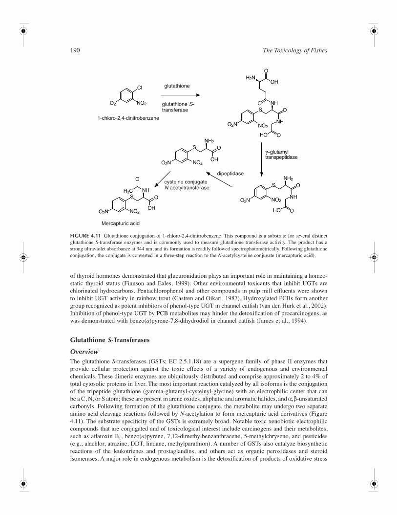
190 The Toxicology of Fishes
of thyroid hormones demonstrated that glucuronidation plays an important role in maintaining a homeo-static thyroid status (Finnson and Eales, 1999). Other environmental toxicants that inhibit UGTs arechlorinated hydrocarbons. Pentachlorophenol and other compounds in pulp mill effluents were shownto inhibit UGT activity in rainbow trout (Castren and Oikari, 1987). Hydroxylated PCBs form anothergroup recognized as potent inhibitors of phenol-type UGT in channel catfish (van den Hurk et al., 2002).Inhibition of phenol-type UGT by PCB metabolites may hinder the detoxification of procarcinogens, aswas demonstrated with benzo(a)pyrene-7,8-dihydrodiol in channel catfish (James et al., 1994).
Glutathione S-Transferases
OverviewThe glutathione S-transferases (GSTs; EC 2.5.1.18) are a supergene family of phase II enzymes thatprovide cellular protection against the toxic effects of a variety of endogenous and environmentalchemicals. These dimeric enzymes are ubiquitously distributed and comprise approximately 2 to 4% oftotal cytosolic proteins in liver. The most important reaction catalyzed by all isoforms is the conjugationof the tripeptide glutathione (gamma-glutamyl-cysteinyl-glycine) with an electrophilic center that canbe a C, N, or S atom; these are present in arene oxides, aliphatic and aromatic halides, and α,β-unsaturatedcarbonyls. Following formation of the glutathione conjugate, the metabolite may undergo two separateamino acid cleavage reactions followed by N-acetylation to form mercapturic acid derivatives (Figure4.11). The substrate specificity of the GSTs is extremely broad. Notable toxic xenobiotic electrophiliccompounds that are conjugated and of toxicological interest include carcinogens and their metabolites,such as aflatoxin B1, benzo(a)pyrene, 7,12-dimethylbenzanthracene, 5-methylchrysene, and pesticides(e.g., alachlor, atrazine, DDT, lindane, methylparathion). A number of GSTs also catalyze biosyntheticreactions of the leukotrienes and prostaglandins, and others act as organic peroxidases and steroidisomerases. A major role in endogenous metabolism is the detoxification of products of oxidative stress
FIGURE 4.11 Glutathione conjugation of 1-chloro-2,4-dinitrobenzene. This compound is a substrate for several distinctglutathione S-transferase enzymes and is commonly used to measure glutathione transferase activity. The product has astrong ultraviolet absorbance at 344 nm, and its formation is readily followed spectrophotometrically. Following glutathioneconjugation, the conjugate is converted in a three-step reaction to the N-acetylcysteine conjugate (mercapturic acid).
CI
NO2O2
O2N
O2N
O2N
O2N
H2N
H3C
NO2
NO2
NO2
NO2
NH2
NH2
NH
HO
OH
OH HO
NH
NHNH
OH
O
γ–glutamyltranspeptidase
O
O
O
OO
O
O
O
S
S
S
S
1–chloro–2,4–dinitrobenzene
glutathione S–transferase
cysteine conjugateN-acetyltransferase
glutathione
Mercapturic acid
dipeptidase
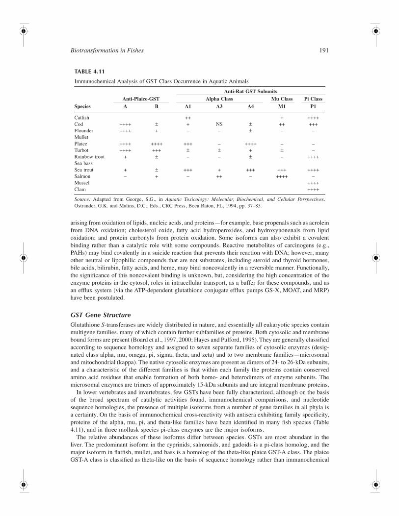
Biotransformation in Fishes 191
arising from oxidation of lipids, nucleic acids, and proteins—for example, base propenals such as acroleinfrom DNA oxidation; cholesterol oxide, fatty acid hydroperoxides, and hydroxynonenals from lipidoxidation; and protein carbonyls from protein oxidation. Some isoforms can also exhibit a covalentbinding rather than a catalytic role with some compounds. Reactive metabolites of carcinogens (e.g.,PAHs) may bind covalently in a suicide reaction that prevents their reaction with DNA; however, manyother neutral or lipophilic compounds that are not substrates, including steroid and thyroid hormones,bile acids, bilirubin, fatty acids, and heme, may bind noncovalently in a reversible manner. Functionally,the significance of this noncovalent binding is unknown, but, considering the high concentration of theenzyme proteins in the cytosol, roles in intracellular transport, as a buffer for these compounds, and asan efflux system (via the ATP-dependent glutathione conjugate efflux pumps GS-X, MOAT, and MRP)have been postulated.
GST Gene StructureGlutathione S-transferases are widely distributed in nature, and essentially all eukaryotic species containmultigene families, many of which contain further subfamilies of proteins. Both cytosolic and membranebound forms are present (Board et al., 1997, 2000; Hayes and Pulford, 1995). They are generally classifiedaccording to sequence homology and assigned to seven separate families of cytosolic enzymes (desig-nated class alpha, mu, omega, pi, sigma, theta, and zeta) and to two membrane families—microsomaland mitochondrial (kappa). The native cytosolic enzymes are present as dimers of 24- to 26-kDa subunits,and a characteristic of the different families is that within each family the proteins contain conservedamino acid residues that enable formation of both homo- and heterodimers of enzyme subunits. Themicrosomal enzymes are trimers of approximately 15-kDa subunits and are integral membrane proteins.
In lower vertebrates and invertebrates, few GSTs have been fully characterized, although on the basisof the broad spectrum of catalytic activities found, immunochemical comparisons, and nucleotidesequence homologies, the presence of multiple isoforms from a number of gene families in all phyla isa certainty. On the basis of immunochemical cross-reactivity with antisera exhibiting family specificity,proteins of the alpha, mu, pi, and theta-like families have been identified in many fish species (Table4.11), and in three mollusk species pi-class enzymes are the major isoforms.
The relative abundances of these isoforms differ between species. GSTs are most abundant in theliver. The predominant isoform in the cyprinids, salmonids, and gadoids is a pi-class homolog, and themajor isoform in flatfish, mullet, and bass is a homolog of the theta-like plaice GST-A class. The plaiceGST-A class is classified as theta-like on the basis of sequence homology rather than immunochemical
TABLE 4.11
Immunochemical Analysis of GST Class Occurrence in Aquatic Animals
SpeciesAnti-Plaice-GST
Anti-Rat GST SubunitsAlpha Class Mu Class Pi Class
A B A1 A3 A4 M1 P1
Catfish ++ + ++++Cod ++++ ± + NS ± ++ +++Flounder ++++ + – – ± – –MulletPlaice ++++ ++++ +++ – ++++ – –Turbot ++++ +++ ± ± + ± –Rainbow trout + ± – – ± – ++++Sea bassSea trout + ± +++ + +++ +++ ++++Salmon – + – ++ – ++++ –Mussel ++++Clam ++++
Source: Adapted from George, S.G., in Aquatic Toxicology: Molecular, Biochemical, and Cellular Perspectives.Ostrander, G.K. and Malins, D.C., Eds., CRC Press, Boca Raton, FL, 1994, pp. 37–85.
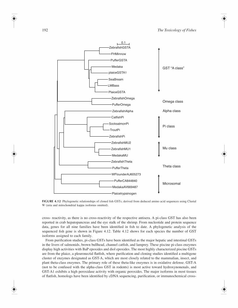
192 The Toxicology of Fishes
cross- reactivity, as there is no cross-reactivity of the respective antisera. A pi-class GST has also beenreported in crab hepatopancreas and the eye stalk of the shrimp. From nucleotide and protein sequencedata, genes for all nine families have been identified in fish to date. A phylogenetic analysis of thesequenced fish gene is shown in Figure 4.12. Table 4.12 shows for each species the number of GSTisoforms assigned to each family.
From purification studies, pi-class GSTs have been identified as the major hepatic and intestinal GSTsin the livers of salmonids, brown bullhead, channel catfish, and lamprey. These piscine pi-class enzymesdisplay high activities with BaP epoxides and diol epoxides. The most highly characterized piscine GSTsare from the plaice, a pleuronectid flatfish, where purification and cloning studies identified a multigenecluster of enzymes designated as GST-A, which are most closely related to the mammalian, insect, andplant theta-class enzymes. The primary role of these theta-like enzymes is in oxidative defense. GST-A(not to be confused with the alpha-class GST in rodents) is most active toward hydroxynonenals, andGST-A1 exhibits a high peroxidase activity with organic peroxides. The major isoforms in most tissuesof flatfish, homologs have been identified by cDNA sequencing, purification, or immunochemical cross-
FIGURE 4.12 Phylogenetic relationships of cloned fish GSTs; derived from deduced amino acid sequences using ClustalW (zeta and mitochondrial kappa isoforms omitted).
GST “A class”
Omega class
Alpha class
Pi class
Mu class
Theta class
Microsomal
ZebrafishGSTA
ZebrafishOmega
ZebrafishAlpha
CatfishPi
SocksalmonPi
TroutPi
ZebrafishPi
ZebrafishMU2
ZebrafishMU1
ZebrafishTheta
PufferTheta
PufferCA844840
MedakaAV669487
Plaicetrypsinogen
WFlounderAJ605273
MedakaMU
PufferOmega
PufferGSTA
plaiceGSTA1
PlaiceGSTA
SeaBream
LMBass
Medaka
FHMinnow
0.1
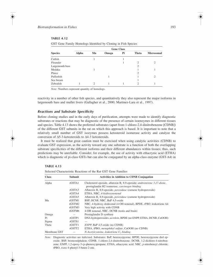
Biotransformation in Fishes 193
reactivity in a number of other fish species, and quantitatively they also represent the major isoforms inlargemouth bass and mullet livers (Gallagher et al., 2000; Martinez-Lara et al., 1997).
Reactions and Substrate SpecificityBefore cloning studies and in the early days of purification, attempts were made to identify diagnosticsubstrates or reactions that may be diagnostic of the presence of certain isoenzymes in different tissuesand species. Table 4.13 shows the preferred substrates (apart from 1-chloro-2,4-dinitrobenzene [CDNB])of the different GST subunits in the rat on which this approach is based. It is important to note that arelatively small number of GST isozymes possess ketosteroid isomerase activity and catalyze theconversion of ∆5-3-ketosteroids to ∆4-3 ketosteroids.
It must be realized that great caution must be exercised when using catalytic activities (CDNB) toevaluate GST expression, as the activity toward any one substrate is a function of both the overlappingsubstrate specificities of the different isoforms and their different abundances within tissues; thus, suchpredictions may be unreliable. Consider, for example, the use of activity with ethacrynic acid (ETHA)which is diagnostic of pi-class GSTs but can also be conjugated by an alpha-class enzyme (GST-A4) in
TABLE 4.12
GST Gene Family Homologs Identified by Cloning in Fish Species
SpeciesGene Class
Alpha Mu Omega Pi Theta Microsomal
Catfish 1 1Flounder 1 2 2Largemouth bass 2Medaka 1 1 1Plaice 2Pufferfish 1 1 2Sea bream 1Zebrafish 1 2 1 1 2 1
Note: Numbers represent quantity of homologs.
TABLE 4.13
Selected Characteristic Reactions of the Rat GST Gene Families
Class Subunit Activities in Addition to CDNB Conjugation
Alpha rGSTA1 Cholesterol epoxide, aflatoxin B1 8,9-epoxide; androsterone 3,17-dione, prostaglandin H2 isomerase, carcinogen binding
rGSTA3 Aflatoxin B1 8,9-epoxide; peroxidase (cumene hydroperoxide)rGSTA4 ETHA, NBC, 4-hydroxynonenalrGSTA5 Aflatoxin B1 8,9-epoxide, peroxidase (cumene hydroperoxide)
Mu rGSTM1 BSP, DCNB, NBC, BaP 4,5-oxiderGSTM2 NBC, 4-hydroxy dodecenal (4-OH nonenal), BPDE, tPBO, leukotriene A4rGSTM3 Very high activity with CDNBrGSTM6 4-OH nonenal, NBC, DCNB (testis and brain)
Omega — Prostaglandin D synthasePi rGSTP1 DNA hydroperoxides, acrolein, BPDE (no ENPP, ETHA, DCNB, CuOOH)Sigma rGSTS1 —Theta rGSTT1 ENPP, BaP 4,5-oxide (no CDNB)
rGSTT2 ETHA, tPBO, menaphthyl sulfate, CuOOH (no CDNB)Membrane GST — N-Acetylcysteine, leukotriene C4 binding
Note: Diagnostic activities are italicized. Substrates: BaP, benzo(a)pyrene; BPDE, benzo(a)pyrene diol ep-oxide; BSP, bromosulphalein; CDNB, 1-chloro-2,4-dinitrobenzene; DCNB, 1,2-dichloro-4-nitroben-zene; ENPP, 1,2-epoxy-3-(p-phenoxy)propane; ETHA, ethacrynic acid; NBC, p-nitrobenzyl chloride;tPBO, trans-4-phenyl-3-buten-2-one.
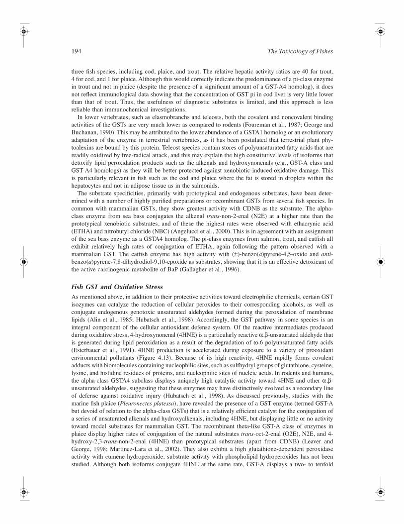
194 The Toxicology of Fishes
three fish species, including cod, plaice, and trout. The relative hepatic activity ratios are 40 for trout,4 for cod, and 1 for plaice. Although this would correctly indicate the predominance of a pi-class enzymein trout and not in plaice (despite the presence of a significant amount of a GST-A4 homolog), it doesnot reflect immunological data showing that the concentration of GST pi in cod liver is very little lowerthan that of trout. Thus, the usefulness of diagnostic substrates is limited, and this approach is lessreliable than immunochemical investigations.
In lower vertebrates, such as elasmobranchs and teleosts, both the covalent and noncovalent bindingactivities of the GSTs are very much lower as compared to rodents (Foureman et al., 1987; George andBuchanan, 1990). This may be attributed to the lower abundance of a GSTA1 homolog or an evolutionaryadaptation of the enzyme in terrestrial vertebrates, as it has been postulated that terrestrial plant phy-toalexins are bound by this protein. Teleost species contain stores of polyunsaturated fatty acids that arereadily oxidized by free-radical attack, and this may explain the high constitutive levels of isoforms thatdetoxify lipid peroxidation products such as the alkenals and hydroxynonenals (e.g., GST-A class andGST-A4 homologs) as they will be better protected against xenobiotic-induced oxidative damage. Thisis particularly relevant in fish such as the cod and plaice where the fat is stored in droplets within thehepatocytes and not in adipose tissue as in the salmonids.
The substrate specificities, primarily with prototypical and endogenous substrates, have been deter-mined with a number of highly purified preparations or recombinant GSTs from several fish species. Incommon with mammalian GSTs, they show greatest activity with CDNB as the substrate. The alpha-class enzyme from sea bass conjugates the alkenal trans-non-2-enal (N2E) at a higher rate than theprototypical xenobiotic substrates, and of these the highest rates were observed with ethacrynic acid(ETHA) and nitrobutyl chloride (NBC) (Angelucci et al., 2000). This is in agreement with an assignmentof the sea bass enzyme as a GSTA4 homolog. The pi-class enzymes from salmon, trout, and catfish allexhibit relatively high rates of conjugation of ETHA, again following the pattern observed with amammalian GST. The catfish enzyme has high activity with (±)-benzo(a)pyrene-4,5-oxide and anti-benzo(a)pyrene-7,8-dihydrodiol-9,10-epoxide as substrates, showing that it is an effective detoxicant ofthe active carcinogenic metabolite of BaP (Gallagher et al., 1996).
Fish GST and Oxidative StressAs mentioned above, in addition to their protective activities toward electrophilic chemicals, certain GSTisozymes can catalyze the reduction of cellular peroxides to their corresponding alcohols, as well asconjugate endogenous genotoxic unsaturated aldehydes formed during the peroxidation of membranelipids (Alin et al., 1985; Hubatsch et al., 1998). Accordingly, the GST pathway in some species is anintegral component of the cellular antioxidant defense system. Of the reactive intermediates producedduring oxidative stress, 4-hydroxynonenal (4HNE) is a particularly reactive α,β-unsaturated aldehyde thatis generated during lipid peroxidation as a result of the degradation of ω-6 polyunsaturated fatty acids(Esterbauer et al., 1991). 4HNE production is accelerated during exposure to a variety of prooxidantenvironmental pollutants (Figure 4.13). Because of its high reactivity, 4HNE rapidly forms covalentadducts with biomolecules containing nucleophilic sites, such as sulfhydryl groups of glutathione, cysteine,lysine, and histidine residues of proteins, and nucleophilic sites of nucleic acids. In rodents and humans,the alpha-class GSTA4 subclass displays uniquely high catalytic activity toward 4HNE and other α,β-unsaturated aldehydes, suggesting that these enzymes may have distinctively evolved as a secondary lineof defense against oxidative injury (Hubatsch et al., 1998). As discussed previously, studies with themarine fish plaice (Pleuronectes platessa), have revealed the presence of a GST enzyme (termed GST-Abut devoid of relation to the alpha-class GSTs) that is a relatively efficient catalyst for the conjugation ofa series of unsaturated alkenals and hydroxyalkenals, including 4HNE, but displaying little or no activitytoward model substrates for mammalian GST. The recombinant theta-like GST-A class of enzymes inplaice display higher rates of conjugation of the natural substrates trans-oct-2-enal (O2E), N2E, and 4-hydroxy-2,3-trans-non-2-enal (4HNE) than prototypical substrates (apart from CDNB) (Leaver andGeorge, 1998; Martinez-Lara et al., 2002). They also exhibit a high glutathione-dependent peroxidaseactivity with cumene hydroperoxide; substrate activity with phospholipid hydroperoxides has not beenstudied. Although both isoforms conjugate 4HNE at the same rate, GST-A displays a two- to tenfold
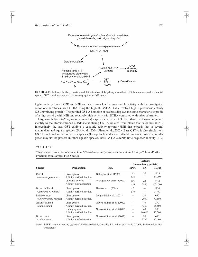
Biotransformation in Fishes 195
higher activity toward O2E and N2E and also shows low but measurable activity with the prototypicalxenobiotic substrates, with ETHA being the highest. GST-A1 has a fivefold higher peroxidase activity(25 µm/min/mg protein). The purified GST-A homolog of sea bass displays the same characteristic profileof a high activity with N2E and relatively high activity with ETHA compared with other substrates.
Largemouth bass (Micropterus salmoides) expresses a liver GST that shares extensive sequenceidentity to the aforementioned 4HNE-metabolizing GST-A isolated from plaice that detoxifies 4HNE.Interestingly, the bass GST exhibits a catalytic activity toward 4HNE that exceeds that of severalmammalian and aquatic species (Doi et al., 2004; Pham et al., 2002). Bass GST-A is also similar to aGST form found in two other fish species (European flounder and fathead minnow); however, similargenes may not be present in other aquatic species. Bass GST-A exhibits little sequence identity (21%
FIGURE 4.13 Pathway for the generation and detoxification of 4-hydroxynonenal (4HNE). In mammals and certain fishspecies, GST constitutes a protective pathway against 4HNE injury.
TABLE 4.14
The Catalytic Properties of Glutathione S-Transferase in Cytosol and Glutathione Affinity-Column-Purified Fractions from Several Fish Species
Species Preparation Ref.
Activity(nmol/min/mg protein)
BPDE EA CDNB
Catfish (Ictalurus punctatus)
Liver cytosolAffinity purified fraction
Gallagher et al. (1996) 5.3128
37—
132524.000
Intestinal cytosolAffinity purified fraction
Gadagbui and James (2000) 8.3453
652880
1010107, 000
Brown bullhead (Ameiurus nebulosus)
Liver cytosolAffinity purified fraction
Henson et al. (2001) <5310
——
11305,700
Rainbow trout (Oncorhynchus mykiss)
Liver cytosolAffinity purified fraction
Melgar Riol et al. (2001) ——
202030
63077,180
Atlantic salmon (Salmo salar)
Liver cytosolAffinity purified fraction
Novoa-Valinas et al. (2002) ——
704350
29016,600
Kidney cytosolAffinity purified fraction
Novoa-Valinas et al. (2002) ——
6010,620
20037,500
Brown trout (Salmo trutta)
Liver cytosolAffinity purified fraction
Novoa-Valinas et al. (2002) ——
903790
65027,690
Note: BPDE, (+)-anti-benzo(a)pyrene-7,8-dihydrodiol-9,10-oxide; EA, ethacrynic acid; CDNB, 1-chloro-2,4-dini-trobenzene.
(O2–, H2O2, HO•).
Generation of reactive oxygen species
Release toxic α, βunsaturated aldehydes4 hydroxynonenal, 4HNE
Exposure to metals, pyrollizidine alkaloids, pesticides, peroxidized oils, toxic algae, fatty diet
Lipid peroxidation
OO
H GST,ADHALDH
Protein and DNAdamage
Liverdisease,mortality
Detoxification

196 The Toxicology of Fishes
or less) to several mammalian GSTs, including the rat alpha-class GST form rGSTA4-4, which, asdiscussed, has a high catalytic efficiency toward conjugation of GSH with 4HNE. The presence of thisinteresting GST in several fish species suggests an important conservation of function that likely protectsfish lipid membranes against the deleterious effects of oxidative injury.
Tissue DistributionWith the exception of the mammalian theta-class enzymes, all GSTs conjugate the prototypical substrate1-chloro,-2,4-dinitrobenzene (CDNB) with greatest activity (Figure 4.11). CDNB conjugating activityhas been observed in all fish species examined to date (Table 4.14) and occurs in multiple tissues (George,1994). Because CDNB is a general substrate and the rate of conjugation can vary between isoforms byup to two orders of magnitude, comparison of total CDNB conjugating activity in livers of differentspecies, for example, is probably of little relevance in toxicological evaluation of environmentally relevantxenobiotics, as it will bear little relationship to the rate of metabolism of other compounds which maybe isoform specific. Functional comparisons of activity toward individual toxicants is perhaps moremeaningful from a toxicological viewpoint. Whereas kidney is the most active toward styrene-7,8-oxideand BaP-4,5-oxide relative to other tissues in the little skate (Raja erinacea) (Bend et al., 1978; Fouremanet al., 1987; Gill et al., 1982), this finding must be placed in the context of relevance to the animal ascompounds of this type may not be present systemically. They will more likely be taken up in the dietor generated by metabolism in the intestine or liver and conjugated in these tissues.
In general, most in vitro laboratory investigations of GST–CDNB conjugating activities in fish tissuesuse assay conditions and substrate concentrations similar to those proposed for mammals: 1 mM GSHand 1 mM CDNB) (Habig and Jakoby, 1981). Kinetic studies, however, have demonstrated that thesesubstrate concentrations may not be at saturation with respect to initial rate kinetics for many isoformsbecause the Km values differ by up to two orders of magnitude. Because the enzymes contain bothelectrophile and nucleophile binding sites, GST detoxification rates are determined by the concentrationof electrophilic substrate and by the concentrations of nucleophilic cosubstrate (GSH). Kinetic studies inlargemouth bass and brown bullheads in the presence of variable electrophile concentrations suggest thatin vitro saturation of hepatic GST–CDNB conjugation occurs at higher electrophile concentrations inbrown bullheads than in largemouth bass (Gallagher et al., 2000). Such an observation is consistent witha higher capacity for brown bullheads to detoxify electrophilic GST substrates under conditions of highenvironmental exposure. These observations are important under the prototypical conditions of environ-mental chemical exposure when the amount of chemical reaching the liver is relatively low and the rateof in vivo clearance is directly proportional to the amount of chemical concentrations in vivo. Also, asalluded to earlier, it is important to consider that the rate of GST–CDNB activity may not be reflectiveof rates of GST conjugation of environmentally relevant GST substrates, such as pesticides or epoxidecarcinogens. For example, a comparison of the rates of hepatic GST–CDNB conjugation by starry flounderand English sole are not correlated with the rates of hepatic GST conjugation of (+)-7β,8α-dihydroxy-9α,10α-oxy-7,8,9,10-tetrahydrobenzo(a)pyrene (BPDE) in those species. Specifically, starry floundercatalyze the rate of CDNB conjugation at initial rates threefold higher than is observed in English sole,whereas GST-BPDE activities are threefold higher in English sole as compared to starry flounder (Gal-lagher et al., 1998). Thus, specific GST conjugation of environmental chemicals in fish must be quantitatedunder assay conditions where the conjugation of the chemical can be monitored directly.
Although glutathione conjugation is generally associated with detoxication, small halogenated alkenes,such as dibromoethane, are activated by glutathione conjugation. In mammals, theta-class GST areparticularly efficient in the conversion of dibromoethane to the carcinogenic sulfonium ion metabolite,as shown in Figure 4.14 (Thier et al., 1996). It was shown that a small fish species, the medaka, wasquite susceptible to the development of liver cancer following exposure to ethylenedibromide and thata form of GST in liver was increased in the exposed fish, suggesting the presence of a theta-like GSTin the medaka (Hawkins et al., 1998). This has been confirmed with cloning studies. Other fish species,such as the pleuronectid flatfish, bass, and mullet, have been shown to have theta-like GST in their livers(Gallagher et al., 2000; Leaver et al., 1993; Martinez-Lara, 1997). It is tempting to speculate that thismay account for the high tumor incidence observed in flatfish from some polluted environments.
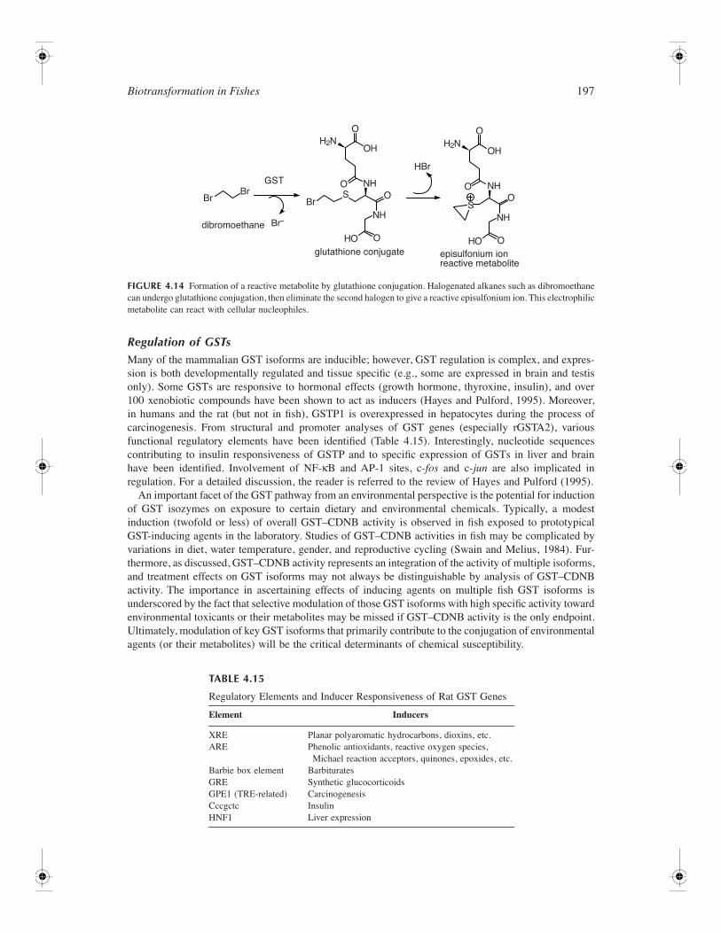
Biotransformation in Fishes 197
Regulation of GSTsMany of the mammalian GST isoforms are inducible; however, GST regulation is complex, and expres-sion is both developmentally regulated and tissue specific (e.g., some are expressed in brain and testisonly). Some GSTs are responsive to hormonal effects (growth hormone, thyroxine, insulin), and over100 xenobiotic compounds have been shown to act as inducers (Hayes and Pulford, 1995). Moreover,in humans and the rat (but not in fish), GSTP1 is overexpressed in hepatocytes during the process ofcarcinogenesis. From structural and promoter analyses of GST genes (especially rGSTA2), variousfunctional regulatory elements have been identified (Table 4.15). Interestingly, nucleotide sequencescontributing to insulin responsiveness of GSTP and to specific expression of GSTs in liver and brainhave been identified. Involvement of NF-κB and AP-1 sites, c-fos and c-jun are also implicated inregulation. For a detailed discussion, the reader is referred to the review of Hayes and Pulford (1995).
An important facet of the GST pathway from an environmental perspective is the potential for inductionof GST isozymes on exposure to certain dietary and environmental chemicals. Typically, a modestinduction (twofold or less) of overall GST–CDNB activity is observed in fish exposed to prototypicalGST-inducing agents in the laboratory. Studies of GST–CDNB activities in fish may be complicated byvariations in diet, water temperature, gender, and reproductive cycling (Swain and Melius, 1984). Fur-thermore, as discussed, GST–CDNB activity represents an integration of the activity of multiple isoforms,and treatment effects on GST isoforms may not always be distinguishable by analysis of GST–CDNBactivity. The importance in ascertaining effects of inducing agents on multiple fish GST isoforms isunderscored by the fact that selective modulation of those GST isoforms with high specific activity towardenvironmental toxicants or their metabolites may be missed if GST–CDNB activity is the only endpoint.Ultimately, modulation of key GST isoforms that primarily contribute to the conjugation of environmentalagents (or their metabolites) will be the critical determinants of chemical susceptibility.
FIGURE 4.14 Formation of a reactive metabolite by glutathione conjugation. Halogenated alkanes such as dibromoethanecan undergo glutathione conjugation, then eliminate the second halogen to give a reactive episulfonium ion. This electrophilicmetabolite can react with cellular nucleophiles.
TABLE 4.15
Regulatory Elements and Inducer Responsiveness of Rat GST Genes
Element Inducers
XRE Planar polyaromatic hydrocarbons, dioxins, etc.ARE Phenolic antioxidants, reactive oxygen species,
Michael reaction acceptors, quinones, epoxides, etc.Barbie box element BarbituratesGRE Synthetic glucocorticoidsGPE1 (TRE-related) CarcinogenesisCccgctc InsulinHNF1 Liver expression
GST
BrBr
Br
HBr
Br–dibromoethane
SS
OO O
O
O
OH
HO
NH
NH
OHO
NH
H2N
O
O
OH
episulfonium ionreactive metabolite
glutathione conjugate
NH
H2N
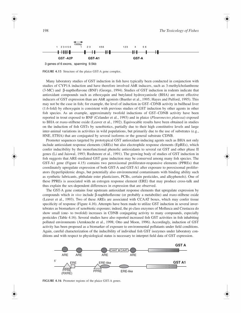
198 The Toxicology of Fishes
Many laboratory studies of GST induction in fish have typically been conducted in conjunction withstudies of CYP1A induction and have therefore involved AhR inducers, such as 3-methylcholanthrene(3-MC) and β-napthoflavone (ΒNF) (George, 1994). Studies of GST induction in rodents indicate thatantioxidant compounds such as ethoxyquin and butylated hydroxyanisole (BHA) are more effectiveinducers of GST expression than are AhR agonists (Buetler et al., 1995; Hayes and Pulford, 1995). Thismay not be the case in fish; for example, the level of induction in GST–CDNB activity in bullhead liver(1.6-fold) by ethoxyquin is consistent with previous studies of GST induction by other agents in otherfish species. As an example, approximately twofold inductions of GST–CDNB activity have beenreported in trout exposed to ΒNF (Celander et al., 1993) and in plaice (Pleuronectes platessa) exposedto BHA or trans-stilbene oxide (Leaver et al., 1992). Equivocable results have been obtained in studieson the induction of fish GSTs by xenobiotics, partially due to their high constitutive levels and largeinter-animal variations in activities in wild populations, but primarily due to the use of substrates (e.g.,HNE, ETHA) that are conjugated by several isoforms or the general substrate CDNB.
Promoter sequences targeted by prototypical GST antioxidant-inducing agents such as BHA not onlyinclude antioxidant response elements (AREs) but also electrophile response elements (EpREs), whichconfer inducibility by the monofunctional phenolic antioxidants to several rat GST and other phase IIgenes (Li and Jaiswal, 1993; Rushmore et al., 1991). The growing body of studies of GST induction infish suggests that ARE-mediated GST gene induction may be conserved among many fish species. TheGST-A1 gene (Figure 4.15) contains two peroxisomal proliferator-responsive elements (PPREs) thatcoordinately upregulate expression of both GST-A and GST-A1 after exposure to peroxisomal prolifer-ators (hyperlipidemic drugs, but potentially also environmental contaminants with binding ability suchas synthetic lubricants, phthalate ester plasticizers, PCBs, certain pesticides, and alkyphenols). One ofthese PPREs is associated with an estrogen response element (ERE) that may produce cross-talk andthus explain the sex-dependent differences in expression that are observed.
The GST-A gene contains four upstream antioxidant response elements that upregulate expression bycompounds which in vivo include β-naphthoflavone (or probably a metabolite) and trans-stilbene oxide(Leaver et al., 1993). Two of these AREs are associated with CCAAT boxes, which may confer tissuespecificity of response (Figure 4.16). Attempts have been made to utilize GST induction in several inver-tebrates as biomarkers of xenobiotic exposure; indeed, the pi-class enzymes of Mollusca and Crustacea doshow small (one- to twofold) increases in CDNB conjugating activity to many compounds, especiallypesticides (Table 4.16). Several studies have also reported increased fish GST activities in fish inhabitingpolluted environments (Armknecht et al., 1998; Otto and Moon, 1996). Accordingly, induction of GSTactivity has been proposed as a biomarker of exposure to environmental pollutants under field conditions.Again, careful characterization of the inducibility of individual fish GST isozymes under laboratory con-ditions and with respect to physiological status is necessary to interpret field data of GST expression.
FIGURE 4.15 Structure of the plaice GST-A gene complex.
FIGURE 4.16 Promoter regions of the plaice GST-A genes.
GST –A2P GST–A1 GST–A
1000bp1 2 3 4 5 6
3 genes of 6 exons, spanning 9.5kb
1 2 3 4 5 6 1 2 3 4 5 6
ARE ARE
PPRE(RXRE)
PPRE ERE–like
ARE ARE
GST A
GST A1
CAAT CAAT
5ʼERE ERE–like
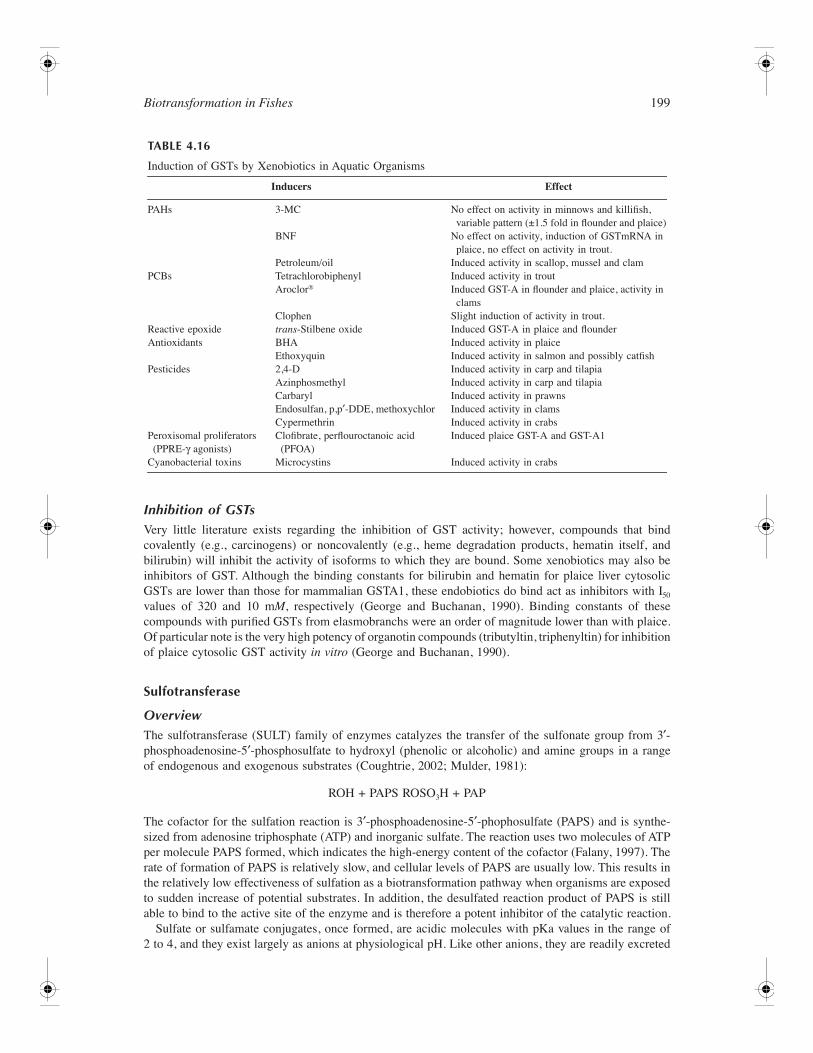
Biotransformation in Fishes 199
Inhibition of GSTsVery little literature exists regarding the inhibition of GST activity; however, compounds that bindcovalently (e.g., carcinogens) or noncovalently (e.g., heme degradation products, hematin itself, andbilirubin) will inhibit the activity of isoforms to which they are bound. Some xenobiotics may also beinhibitors of GST. Although the binding constants for bilirubin and hematin for plaice liver cytosolicGSTs are lower than those for mammalian GSTA1, these endobiotics do bind act as inhibitors with I50
values of 320 and 10 mM, respectively (George and Buchanan, 1990). Binding constants of thesecompounds with purified GSTs from elasmobranchs were an order of magnitude lower than with plaice.Of particular note is the very high potency of organotin compounds (tributyltin, triphenyltin) for inhibitionof plaice cytosolic GST activity in vitro (George and Buchanan, 1990).
Sulfotransferase
OverviewThe sulfotransferase (SULT) family of enzymes catalyzes the transfer of the sulfonate group from 3′-phosphoadenosine-5′-phosphosulfate to hydroxyl (phenolic or alcoholic) and amine groups in a rangeof endogenous and exogenous substrates (Coughtrie, 2002; Mulder, 1981):
ROH + PAPS ROSO3H + PAP
The cofactor for the sulfation reaction is 3′-phosphoadenosine-5′-phophosulfate (PAPS) and is synthe-sized from adenosine triphosphate (ATP) and inorganic sulfate. The reaction uses two molecules of ATPper molecule PAPS formed, which indicates the high-energy content of the cofactor (Falany, 1997). Therate of formation of PAPS is relatively slow, and cellular levels of PAPS are usually low. This results inthe relatively low effectiveness of sulfation as a biotransformation pathway when organisms are exposedto sudden increase of potential substrates. In addition, the desulfated reaction product of PAPS is stillable to bind to the active site of the enzyme and is therefore a potent inhibitor of the catalytic reaction.
Sulfate or sulfamate conjugates, once formed, are acidic molecules with pKa values in the range of2 to 4, and they exist largely as anions at physiological pH. Like other anions, they are readily excreted
TABLE 4.16
Induction of GSTs by Xenobiotics in Aquatic Organisms
Inducers Effect
PAHs 3-MC No effect on activity in minnows and killifish, variable pattern (±1.5 fold in flounder and plaice)
BNF No effect on activity, induction of GSTmRNA in plaice, no effect on activity in trout.
Petroleum/oil Induced activity in scallop, mussel and clamPCBs Tetrachlorobiphenyl Induced activity in trout
Aroclor® Induced GST-A in flounder and plaice, activity in clams
Clophen Slight induction of activity in trout.Reactive epoxide trans-Stilbene oxide Induced GST-A in plaice and flounderAntioxidants BHA Induced activity in plaice
Ethoxyquin Induced activity in salmon and possibly catfishPesticides 2,4-D Induced activity in carp and tilapia
Azinphosmethyl Induced activity in carp and tilapiaCarbaryl Induced activity in prawnsEndosulfan, p,p′-DDE, methoxychlor Induced activity in clamsCypermethrin Induced activity in crabs
Peroxisomal proliferators (PPRE-γ agonists)
Clofibrate, perflouroctanoic acid (PFOA)
Induced plaice GST-A and GST-A1
Cyanobacterial toxins Microcystins Induced activity in crabs
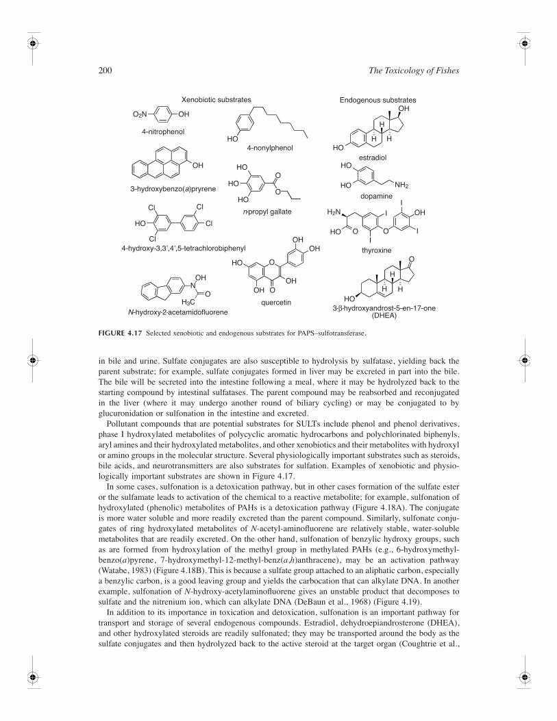
200 The Toxicology of Fishes
in bile and urine. Sulfate conjugates are also susceptible to hydrolysis by sulfatase, yielding back theparent substrate; for example, sulfate conjugates formed in liver may be excreted in part into the bile.The bile will be secreted into the intestine following a meal, where it may be hydrolyzed back to thestarting compound by intestinal sulfatases. The parent compound may be reabsorbed and reconjugatedin the liver (where it may undergo another round of biliary cycling) or may be conjugated to byglucuronidation or sulfonation in the intestine and excreted.
Pollutant compounds that are potential substrates for SULTs include phenol and phenol derivatives,phase I hydroxylated metabolites of polycyclic aromatic hydrocarbons and polychlorinated biphenyls,aryl amines and their hydroxylated metabolites, and other xenobiotics and their metabolites with hydroxylor amino groups in the molecular structure. Several physiologically important substrates such as steroids,bile acids, and neurotransmitters are also substrates for sulfation. Examples of xenobiotic and physio-logically important substrates are shown in Figure 4.17.
In some cases, sulfonation is a detoxication pathway, but in other cases formation of the sulfate esteror the sulfamate leads to activation of the chemical to a reactive metabolite; for example, sulfonation ofhydroxylated (phenolic) metabolites of PAHs is a detoxication pathway (Figure 4.18A). The conjugateis more water soluble and more readily excreted than the parent compound. Similarly, sulfonate conju-gates of ring hydroxylated metabolites of N-acetyl-aminofluorene are relatively stable, water-solublemetabolites that are readily excreted. On the other hand, sulfonation of benzylic hydroxy groups, suchas are formed from hydroxylation of the methyl group in methylated PAHs (e.g., 6-hydroxymethyl-benzo(a)pyrene, 7-hydroxymethyl-12-methyl-benz(a,h)anthracene), may be an activation pathway(Watabe, 1983) (Figure 4.18B). This is because a sulfate group attached to an aliphatic carbon, especiallya benzylic carbon, is a good leaving group and yields the carbocation that can alkylate DNA. In anotherexample, sulfonation of N-hydroxy-acetylaminofluorene gives an unstable product that decomposes tosulfate and the nitrenium ion, which can alkylate DNA (DeBaun et al., 1968) (Figure 4.19).
In addition to its importance in toxication and detoxication, sulfonation is an important pathway fortransport and storage of several endogenous compounds. Estradiol, dehydroepiandrosterone (DHEA),and other hydroxylated steroids are readily sulfonated; they may be transported around the body as thesulfate conjugates and then hydrolyzed back to the active steroid at the target organ (Coughtrie et al.,
FIGURE 4.17 Selected xenobiotic and endogenous substrates for PAPS–sulfotransferase.
Xenobiotic substrates
4–nitrophenol
4–nonylphenol
3–hydroxybenzo(a)pryrene
n-propyl gallate
thyroxine
dopamine
estradiol
Endogenous substrates
4–hydroxy–3,3ʼ,4ʼ,5–tetrachlorobiphenyl
3-β-hydroxyandrost–5–en–17–one(DHEA)N–hydroxy-2-acetamidofluorene
quercetin
O2N OH
HOHO
HO
HO
HO
H
H
H
H
HH
HO
HO
HO
OH
OH
OHOH
OH
OH
OH
OH
O
O O
O
I
II
I
N
O
O
O
OHO
HO
H3C
H2N
NH2
HO Cl
Cl
Cl
Cl
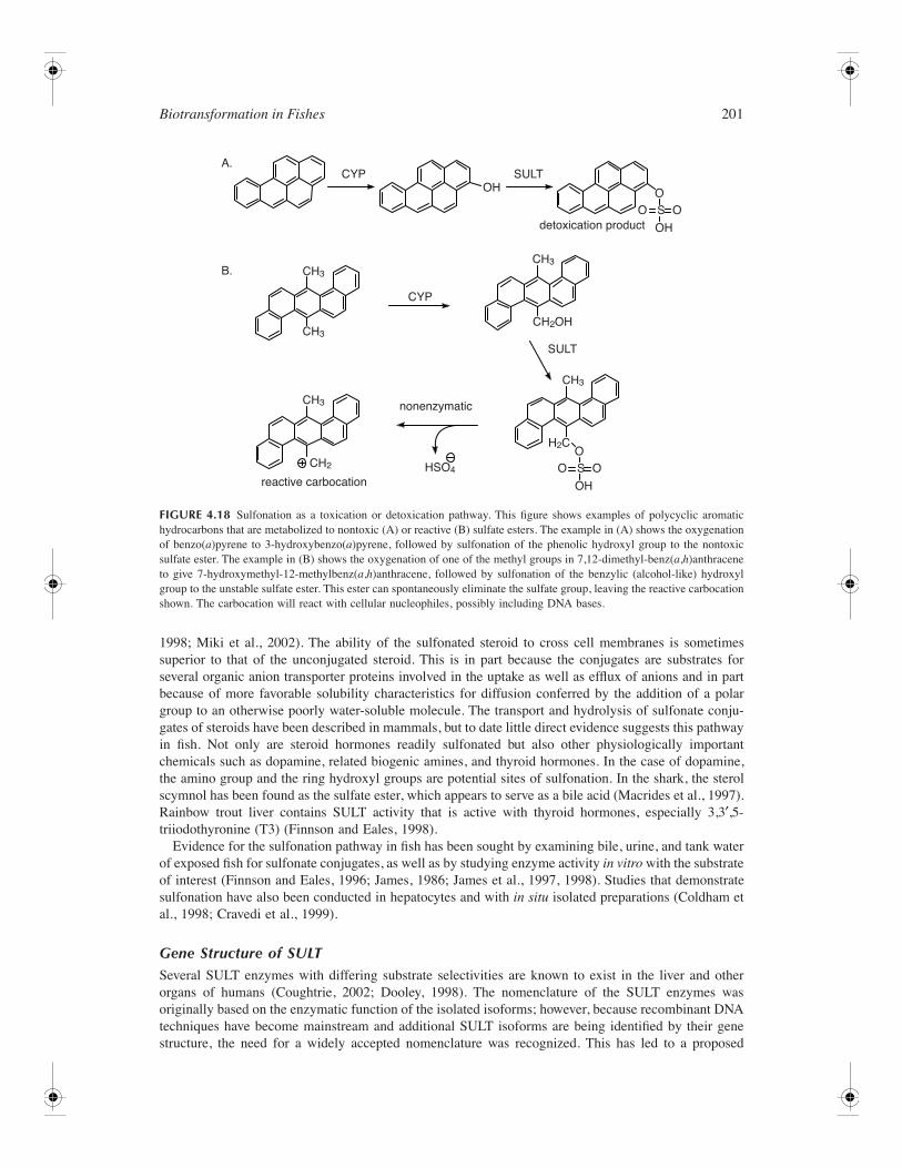
Biotransformation in Fishes 201
1998; Miki et al., 2002). The ability of the sulfonated steroid to cross cell membranes is sometimessuperior to that of the unconjugated steroid. This is in part because the conjugates are substrates forseveral organic anion transporter proteins involved in the uptake as well as efflux of anions and in partbecause of more favorable solubility characteristics for diffusion conferred by the addition of a polargroup to an otherwise poorly water-soluble molecule. The transport and hydrolysis of sulfonate conju-gates of steroids have been described in mammals, but to date little direct evidence suggests this pathwayin fish. Not only are steroid hormones readily sulfonated but also other physiologically importantchemicals such as dopamine, related biogenic amines, and thyroid hormones. In the case of dopamine,the amino group and the ring hydroxyl groups are potential sites of sulfonation. In the shark, the sterolscymnol has been found as the sulfate ester, which appears to serve as a bile acid (Macrides et al., 1997).Rainbow trout liver contains SULT activity that is active with thyroid hormones, especially 3,3′,5-triiodothyronine (T3) (Finnson and Eales, 1998).
Evidence for the sulfonation pathway in fish has been sought by examining bile, urine, and tank waterof exposed fish for sulfonate conjugates, as well as by studying enzyme activity in vitro with the substrateof interest (Finnson and Eales, 1996; James, 1986; James et al., 1997, 1998). Studies that demonstratesulfonation have also been conducted in hepatocytes and with in situ isolated preparations (Coldham etal., 1998; Cravedi et al., 1999).
Gene Structure of SULTSeveral SULT enzymes with differing substrate selectivities are known to exist in the liver and otherorgans of humans (Coughtrie, 2002; Dooley, 1998). The nomenclature of the SULT enzymes wasoriginally based on the enzymatic function of the isolated isoforms; however, because recombinant DNAtechniques have become mainstream and additional SULT isoforms are being identified by their genestructure, the need for a widely accepted nomenclature was recognized. This has led to a proposed
FIGURE 4.18 Sulfonation as a toxication or detoxication pathway. This figure shows examples of polycyclic aromatichydrocarbons that are metabolized to nontoxic (A) or reactive (B) sulfate esters. The example in (A) shows the oxygenationof benzo(a)pyrene to 3-hydroxybenzo(a)pyrene, followed by sulfonation of the phenolic hydroxyl group to the nontoxicsulfate ester. The example in (B) shows the oxygenation of one of the methyl groups in 7,12-dimethyl-benz(a,h)anthraceneto give 7-hydroxymethyl-12-methylbenz(a,h)anthracene, followed by sulfonation of the benzylic (alcohol-like) hydroxylgroup to the unstable sulfate ester. This ester can spontaneously eliminate the sulfate group, leaving the reactive carbocationshown. The carbocation will react with cellular nucleophiles, possibly including DNA bases.
nonenzymatic
reactive carbocationHSO4
CH3
H2C
CH3CH3
CH3
CH3
CH2
CH2OH
OOSO
OH
OOSO
OHdetoxication product
SULT
SULT
OH
CYP
CYPA.
B.
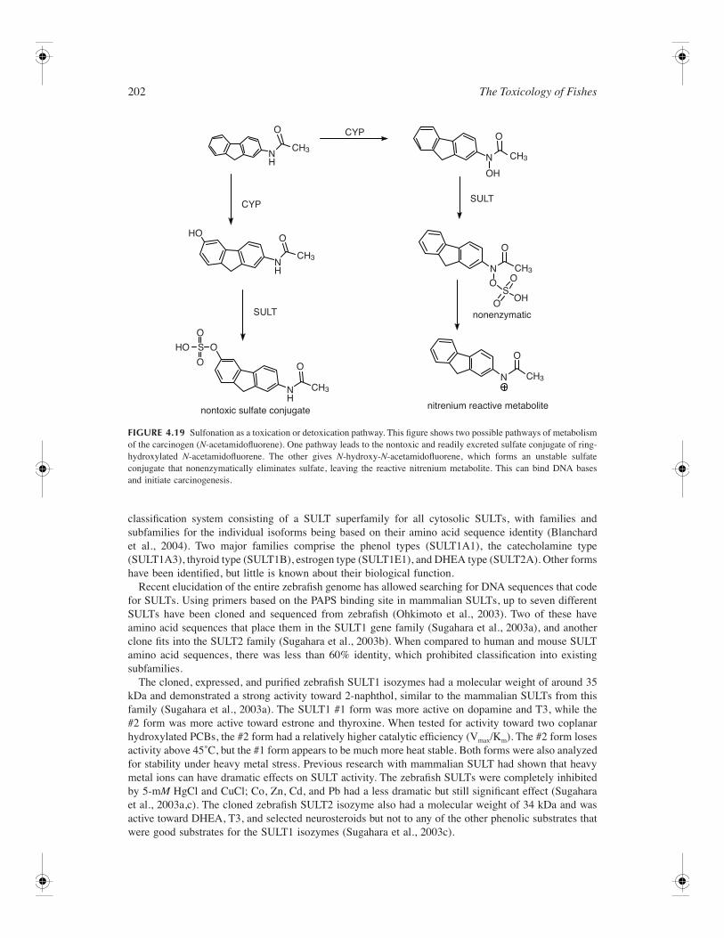
202 The Toxicology of Fishes
classification system consisting of a SULT superfamily for all cytosolic SULTs, with families andsubfamilies for the individual isoforms being based on their amino acid sequence identity (Blanchardet al., 2004). Two major families comprise the phenol types (SULT1A1), the catecholamine type(SULT1A3), thyroid type (SULT1B), estrogen type (SULT1E1), and DHEA type (SULT2A). Other formshave been identified, but little is known about their biological function.
Recent elucidation of the entire zebrafish genome has allowed searching for DNA sequences that codefor SULTs. Using primers based on the PAPS binding site in mammalian SULTs, up to seven differentSULTs have been cloned and sequenced from zebrafish (Ohkimoto et al., 2003). Two of these haveamino acid sequences that place them in the SULT1 gene family (Sugahara et al., 2003a), and anotherclone fits into the SULT2 family (Sugahara et al., 2003b). When compared to human and mouse SULTamino acid sequences, there was less than 60% identity, which prohibited classification into existingsubfamilies.
The cloned, expressed, and purified zebrafish SULT1 isozymes had a molecular weight of around 35kDa and demonstrated a strong activity toward 2-naphthol, similar to the mammalian SULTs from thisfamily (Sugahara et al., 2003a). The SULT1 #1 form was more active on dopamine and T3, while the#2 form was more active toward estrone and thyroxine. When tested for activity toward two coplanarhydroxylated PCBs, the #2 form had a relatively higher catalytic efficiency (Vmax/Km). The #2 form losesactivity above 45˚C, but the #1 form appears to be much more heat stable. Both forms were also analyzedfor stability under heavy metal stress. Previous research with mammalian SULT had shown that heavymetal ions can have dramatic effects on SULT activity. The zebrafish SULTs were completely inhibitedby 5-mM HgCl and CuCl; Co, Zn, Cd, and Pb had a less dramatic but still significant effect (Sugaharaet al., 2003a,c). The cloned zebrafish SULT2 isozyme also had a molecular weight of 34 kDa and wasactive toward DHEA, T3, and selected neurosteroids but not to any of the other phenolic substrates thatwere good substrates for the SULT1 isozymes (Sugahara et al., 2003c).
FIGURE 4.19 Sulfonation as a toxication or detoxication pathway. This figure shows two possible pathways of metabolismof the carcinogen (N-acetamidofluorene). One pathway leads to the nontoxic and readily excreted sulfate conjugate of ring-hydroxylated N-acetamidofluorene. The other gives N-hydroxy-N-acetamidofluorene, which forms an unstable sulfateconjugate that nonenzymatically eliminates sulfate, leaving the reactive nitrenium metabolite. This can bind DNA basesand initiate carcinogenesis.
nonenzymatic
nontoxic sulfate conjugate nitrenium reactive metabolite
CH3
CH3
CH3
CH3
CH3
CH3
OO
O
O OS
O
O
OOH
CYP
CYPSULT
SULT
NH
NH
NH
N
N
N
OH
HO
HO OS
O
O O
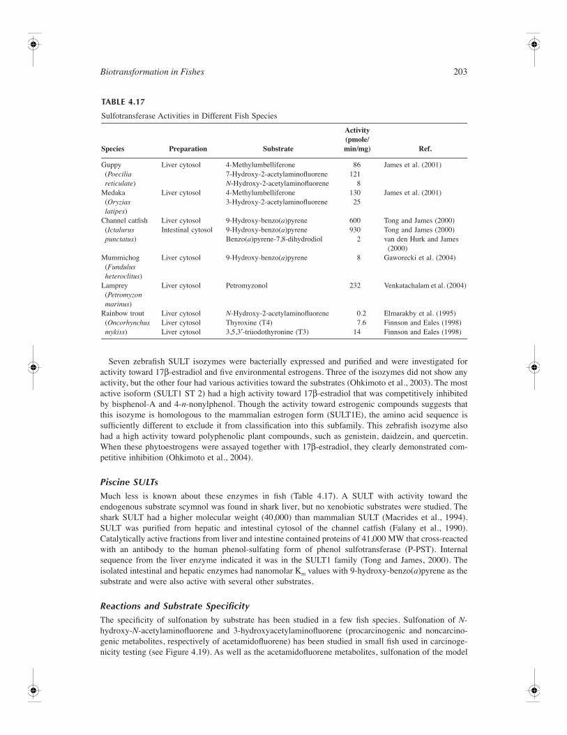
Biotransformation in Fishes 203
Seven zebrafish SULT isozymes were bacterially expressed and purified and were investigated foractivity toward 17β-estradiol and five environmental estrogens. Three of the isozymes did not show anyactivity, but the other four had various activities toward the substrates (Ohkimoto et al., 2003). The mostactive isoform (SULT1 ST 2) had a high activity toward 17β-estradiol that was competitively inhibitedby bisphenol-A and 4-n-nonylphenol. Though the activity toward estrogenic compounds suggests thatthis isozyme is homologous to the mammalian estrogen form (SULT1E), the amino acid sequence issufficiently different to exclude it from classification into this subfamily. This zebrafish isozyme alsohad a high activity toward polyphenolic plant compounds, such as genistein, daidzein, and quercetin.When these phytoestrogens were assayed together with 17β-estradiol, they clearly demonstrated com-petitive inhibition (Ohkimoto et al., 2004).
Piscine SULTsMuch less is known about these enzymes in fish (Table 4.17). A SULT with activity toward theendogenous substrate scymnol was found in shark liver, but no xenobiotic substrates were studied. Theshark SULT had a higher molecular weight (40,000) than mammalian SULT (Macrides et al., 1994).SULT was purified from hepatic and intestinal cytosol of the channel catfish (Falany et al., 1990).Catalytically active fractions from liver and intestine contained proteins of 41,000 MW that cross-reactedwith an antibody to the human phenol-sulfating form of phenol sulfotransferase (P-PST). Internalsequence from the liver enzyme indicated it was in the SULT1 family (Tong and James, 2000). Theisolated intestinal and hepatic enzymes had nanomolar Km values with 9-hydroxy-benzo(a)pyrene as thesubstrate and were also active with several other substrates.
Reactions and Substrate SpecificityThe specificity of sulfonation by substrate has been studied in a few fish species. Sulfonation of N-hydroxy-N-acetylaminofluorene and 3-hydroxyacetylaminofluorene (procarcinogenic and noncarcino-genic metabolites, respectively of acetamidofluorene) has been studied in small fish used in carcinoge-nicity testing (see Figure 4.19). As well as the acetamidofluorene metabolites, sulfonation of the model
TABLE 4.17
Sulfotransferase Activities in Different Fish Species
Species Preparation Substrate
Activity (pmole/min/mg) Ref.
Guppy (Poecilia reticulate)
Liver cytosol 4-Methylumbelliferone 86 James et al. (2001)7-Hydroxy-2-acetylaminofluorene 121N-Hydroxy-2-acetylaminofluorene 8
Medaka (Oryzias latipes)
Liver cytosol 4-Methylumbelliferone 130 James et al. (2001)3-Hydroxy-2-acetylaminofluorene 25
Channel catfish (Ictalurus punctatus)
Liver cytosol 9-Hydroxy-benzo(a)pyrene 600 Tong and James (2000)Intestinal cytosol 9-Hydroxy-benzo(a)pyrene 930 Tong and James (2000)
Benzo(a)pyrene-7,8-dihydrodiol 2 van den Hurk and James (2000)
Mummichog (Fundulus heteroclitus)
Liver cytosol 9-Hydroxy-benzo(a)pyrene 8 Gaworecki et al. (2004)
Lamprey (Petromyzon marinus)
Liver cytosol Petromyzonol 232 Venkatachalam et al. (2004)
Rainbow trout (Oncorhynchus mykiss)
Liver cytosol N-Hydroxy-2-acetylaminofluorene 0.2 Elmarakby et al. (1995)Liver cytosol Thyroxine (T4) 7.6 Finnson and Eales (1998)Liver cytosol 3,5,3′-triiodothyronine (T3) 14 Finnson and Eales (1998)

204 The Toxicology of Fishes
compound 4-methylumbelliferone was examined. Sulfonation of N-hydroxy-N-acetamidofluorene wasdemonstrated in control and 2-acetamidofluorene (AAF)-treated guppy, indicating that the pathway tothe reactive ultimate carcinogen was intact (James et al., 2001). SULT activity was higher in guppy thanmedaka, suggesting a possible biochemical basis for the higher sensitivity of guppy to the carcinogeniceffects of AAF. Rainbow trout, a species that is relatively insensitive to the carcinogenicity of AAF,formed very little N-hydroxy-2-acetylaminofluorene (N-OH-AAF) or N-OH-AAF sulfate (Steward etal., 1995).
Sulfonation of several xenobiotic substrates has been studied in the channel catfish. These include3,7- and 9-hydroxybenzo(a)pyrene, benzo(a)pyrene-7,8-dihydrodiol, 2-naphthol, acetaminophen, 6-hydroxymethylbenzo(a)pyrene, 7-hydroxymethyl-12-methylbenz(a,h)anthracene, and p-nitrophenol.Higher activity was found with the phenolic substrates than the alcoholic substrates (Tong and James,2000). In contrast to the benzo(a)pyrene phenols, the dihydrodiol procarcinogen benzo(a)pyrene-7,8-dihydrodiol was a very poor substrate for sulfation in the catfish intestine or liver (van den Hurk andJames, 2000). Others have shown in mammalian systems that BaP-7,8-dihydrodiol inhibits SULT but isa poor substrate (Rap and Duffel, 1992). In vivo studies with the southern flounder, however, have shownthat the sulfate conjugate of BaP-7,8-dihydrodiol was formed to some extent and was excreted morerapidly than the glucuronide (Pritchard and Bend, 1984).
The only other fish SULT that has been extensively studied for substrate selectivity is a recently clonedzebrafish enzyme. The expressed zebrafish SULT had high activity with flavonoids, isoflavonoids, andother phenolic natural products such as chlorogenic acid, catechin, epicatechin, and quercetin, as wellas with phenolic xenobiotics such as nonylphenol and naphthol (Sugahara et al., 2003a). The zebrafishSULT was also active with dopamine, thyroxine, estrone, and dehydroepiandrosterone. Studies ofcatalytic efficiency (Vmax/Km) showed that the endogenous substrate dopamine was a relatively poorsubstrate for the expressed SULT enzyme compared with the natural product n-propyl gallate (Sugaharaet al., 2003a).
Tissue DistributionMost studies of sulfonation have focused on activity in the liver, but in the catfish the intestine wasshown to be as active as liver in catalyzing the sulfonation of several substrates, including hydroxylatedbenzo(a)pyrenes, dopamine, and 6-hydroxymethylbenzo(a)pyrene (James et al., 1997; Tong and James,2000). Demonstration of activity in the intestine is important in the context of first-pass metabolism ofdietary carcinogens, which are often present at low levels. When low concentrations of substrate arepresent in ingested material, biotransformation in the intestine becomes more important. The capacityof the intestine for metabolism is fairly low, because the liver is a larger organ, but if only small amountsof xenobiotic are present, as is the case in many environmental samples, the intestine and other extra-hepatic organs that are portals of entry, such as gills and skin, become important sites of first-passmetabolism.
Regulation of SULTNot enough studies of SULT in fish have been conducted to have a good understanding of the numberof SULTs present or their regulation by endogenous or exogenous factors. In mammalian species, SULTsare not induced by exposure to PAHs, and there are reports that AhR agonists such as PAHS repressSULT activity (Runge-Morris and Wilusz, 1994). The few studies that have been conducted in fish showno clear evidence of an effect of PAH-type inducing agents on SULT activity (James et al., 1997). Incatfish exposed to β-naphthoflavone, there was a slight loss of activity with 7-hydroxybenzo(a)pyrenebut no effect with the 3- and 9-hydroxy-BaP substrates. Evidence suggests that agents such as gluco-corticoids and related substances cause a modest upregulation of SULT in mammals (Runge-Morris,1998) and that tamoxifen upregulates SULT1A in intestine and liver of rats (Maiti and Chen, 2003), butthis has not been demonstrated in fish.
Recent investigations into the expression of phenol-type SULT in mummichog (Fundulus heteroclitus)has revealed that this species has a remarkably low expression of this enzyme (Gaworecki et al., 2004).The activity toward 3-hydroxybenzo(a)pyrene was very low compared to a variety of other fish species
Biotransformation in Fishes 205
(Gaworecki et al., unpublished data). In addition, a polyclonal antibody that was generated againstchannel catfish phenol-type SULT did not recognize any SULT protein in mummichog samples. Despitethe low expression of phenol-type SULT in mummichog from clean reference sites, animals from theAtlantic Wood site in the Elizabeth River in Virginia, a site heavily contaminated with PAHs and othercontaminants, showed significantly increased SULT activity (Gaworecki et al., 2004). Treatment withsingle doses of 3-methylcholanthrene or benzo(a)pyrene failed to induce the protein; thus, contrary toAhR-mediated CYP1A and UGT induction, SULT upregulation does not appear to be linked to AhRactivation. This is not unusual, as GST induction also requires chronic exposure (James et al., 1979).
Inhibition of SULTSulfotransferase is subject to inhibition from a variety of sources. In vitro, PAP derived from thecosubstrate or sulfonate donor molecule PAPS inhibits SULT activity. Many bulky substrates inhibittheir own sulfonation; for example, in catfish intestinal cytosol 3-hydroxybenzo(a)pyrene inhibited itsown metabolism at concentrations above 0.5 µM (Tong and James, 2000). The nonsubstrate β-naph-thoflavone inhibited SULT activity with 3-, 7-, and 9-hydroxybenzo(a)pyrene in channel catfish intestinaland hepatic cytosol, with an IC50 of 48.0 ± 3.0 µM (James et al., 1997). A natural flavonoid, quercetin,was a potent inhibitor of sulfation of 3-, 7-, and 9-hydroxybenzo(a)pyrene in channel catfish intestinalcytosol, with an IC50 of less than 1 µM (van den Hurk and James, 2000). Although pentachlorophenolwas reported to inhibit phenol sulfation in mammals, it was a poor inhibitor of the sulfonation ofhydroxylated benzo(a)pyrenes in catfish and had an IC50 of about 100 µM (van den Hurk and James,2000). Recent studies showed that several hydroxylated PCBs inhibited estrogen SULT in mammaliansystems with nanomolar IC50 values. Studies of the effects of hydroxylated PCB on the sulfonation ofhydroxylated benzo(a)pyrenes in catfish intestinal cytosol showed that several of these PCB metabolitesinhibited SULT, with low to high micromolar IC50 values (van den Hurk et al., 2002). Several metalcations, including mercury, cobalt, zinc, cadmium, copper, and lead, were shown to inhibit zebrafishSULT, although quite high concentrations (5 mM) were tested (Sugahara et al., 2003c).
Sulfonation of thyroid hormones was studied in liver cytosol from rainbow trout (Oncorhynchusmykiss) (Finnson and Eales, 1998). Based on optimum pH, thermal stability, and specific inhibitors, theinvestigators concluded that it is most likely that only one isozyme form is responsible for the sulfonationof thyroid hormones in trout, in contrast to the multiple isozyme forms found in mammals. No informationis available yet on the inhibiting effects of hydroxylated PCBs on thyroid hormone sulfonation, as hasbeen reported for mammalian systems (Schuur et al., 1998).
Activation of SULTIn vitro, SULT activity with some substrates can be increased by the addition of Mg2+ and others byincorporation of BSA into the assay vial. This phenomenon was also observed with the isolated catfishSULT (Tong and James, 2000) and zebrafish SULT (Sugahara et al., 2003a). The in vivo significanceof this activation is unclear (Konishi-Imamura et al., 1995).
Amino Acid Conjugation
OverviewA relatively little studied pathway of phase II metabolism is the conjugation of carboxylic acids withamino acids. This pathway results in the formation of an amide bond between the carboxylate and theamino group of the amino acid (Figure 4.20). The resulting conjugate is an anion at physiological pHand is generally a better substrate for organic anion transporter proteins in kidney than the unconjugatedxenobiotic; thus, amino acid conjugation facilitates the excretion of xenobiotic carboxylic acids (Jamesand Pritchard, 1987). The amino acid utilized for this pathway in all fish species studied to date is taurine(James, 1976). It has been suggested that glycine conjugates (the major metabolites of benzoic acids inmammals) are metabolites of p-aminobenzoate in fish (James, 1986), but no solid evidence supports thispathway in fish. The enzymes responsible for amino acid conjugation are found in liver and kidney
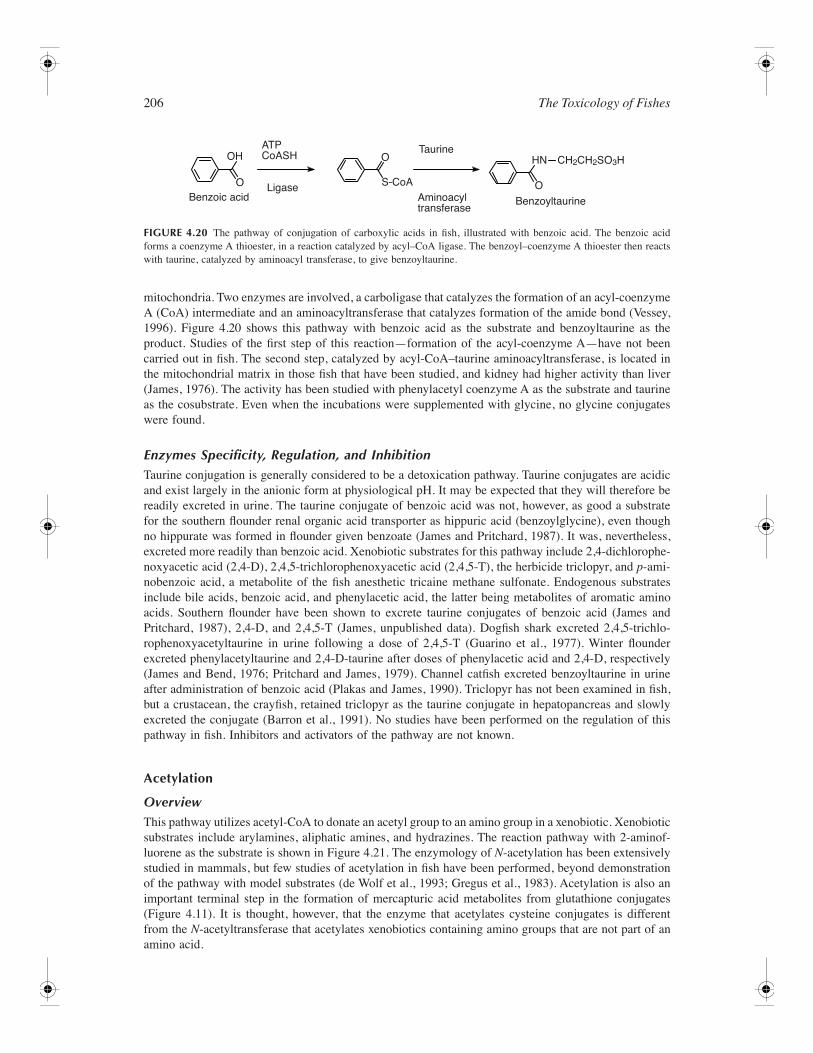
206 The Toxicology of Fishes
mitochondria. Two enzymes are involved, a carboligase that catalyzes the formation of an acyl-coenzymeA (CoA) intermediate and an aminoacyltransferase that catalyzes formation of the amide bond (Vessey,1996). Figure 4.20 shows this pathway with benzoic acid as the substrate and benzoyltaurine as theproduct. Studies of the first step of this reaction—formation of the acyl-coenzyme A—have not beencarried out in fish. The second step, catalyzed by acyl-CoA–taurine aminoacyltransferase, is located inthe mitochondrial matrix in those fish that have been studied, and kidney had higher activity than liver(James, 1976). The activity has been studied with phenylacetyl coenzyme A as the substrate and taurineas the cosubstrate. Even when the incubations were supplemented with glycine, no glycine conjugateswere found.
Enzymes Specificity, Regulation, and InhibitionTaurine conjugation is generally considered to be a detoxication pathway. Taurine conjugates are acidicand exist largely in the anionic form at physiological pH. It may be expected that they will therefore bereadily excreted in urine. The taurine conjugate of benzoic acid was not, however, as good a substratefor the southern flounder renal organic acid transporter as hippuric acid (benzoylglycine), even thoughno hippurate was formed in flounder given benzoate (James and Pritchard, 1987). It was, nevertheless,excreted more readily than benzoic acid. Xenobiotic substrates for this pathway include 2,4-dichlorophe-noxyacetic acid (2,4-D), 2,4,5-trichlorophenoxyacetic acid (2,4,5-T), the herbicide triclopyr, and p-ami-nobenzoic acid, a metabolite of the fish anesthetic tricaine methane sulfonate. Endogenous substratesinclude bile acids, benzoic acid, and phenylacetic acid, the latter being metabolites of aromatic aminoacids. Southern flounder have been shown to excrete taurine conjugates of benzoic acid (James andPritchard, 1987), 2,4-D, and 2,4,5-T (James, unpublished data). Dogfish shark excreted 2,4,5-trichlo-rophenoxyacetyltaurine in urine following a dose of 2,4,5-T (Guarino et al., 1977). Winter flounderexcreted phenylacetyltaurine and 2,4-D-taurine after doses of phenylacetic acid and 2,4-D, respectively(James and Bend, 1976; Pritchard and James, 1979). Channel catfish excreted benzoyltaurine in urineafter administration of benzoic acid (Plakas and James, 1990). Triclopyr has not been examined in fish,but a crustacean, the crayfish, retained triclopyr as the taurine conjugate in hepatopancreas and slowlyexcreted the conjugate (Barron et al., 1991). No studies have been performed on the regulation of thispathway in fish. Inhibitors and activators of the pathway are not known.
Acetylation
OverviewThis pathway utilizes acetyl-CoA to donate an acetyl group to an amino group in a xenobiotic. Xenobioticsubstrates include arylamines, aliphatic amines, and hydrazines. The reaction pathway with 2-aminof-luorene as the substrate is shown in Figure 4.21. The enzymology of N-acetylation has been extensivelystudied in mammals, but few studies of acetylation in fish have been performed, beyond demonstrationof the pathway with model substrates (de Wolf et al., 1993; Gregus et al., 1983). Acetylation is also animportant terminal step in the formation of mercapturic acid metabolites from glutathione conjugates(Figure 4.11). It is thought, however, that the enzyme that acetylates cysteine conjugates is differentfrom the N-acetyltransferase that acetylates xenobiotics containing amino groups that are not part of anamino acid.
FIGURE 4.20 The pathway of conjugation of carboxylic acids in fish, illustrated with benzoic acid. The benzoic acidforms a coenzyme A thioester, in a reaction catalyzed by acyl–CoA ligase. The benzoyl–coenzyme A thioester then reactswith taurine, catalyzed by aminoacyl transferase, to give benzoyltaurine.
CH2CH2SO3H
O
ATPCoASH
O
OOH
S–CoA
Taurine
Aminoacyltransferase
BenzoyltaurineBenzoic acidLigase
HN
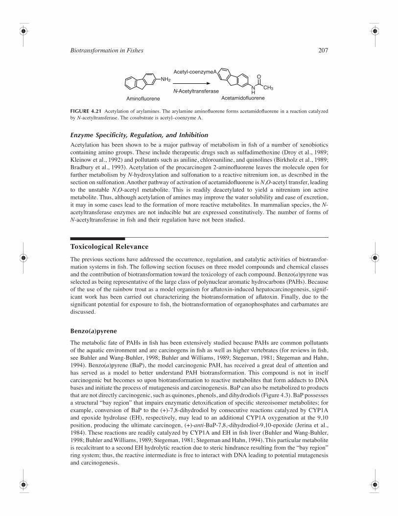
Biotransformation in Fishes 207
Enzyme Specificity, Regulation, and InhibitionAcetylation has been shown to be a major pathway of metabolism in fish of a number of xenobioticscontaining amino groups. These include therapeutic drugs such as sulfadimethoxine (Droy et al., 1989;Kleinow et al., 1992) and pollutants such as aniline, chloroaniline, and quinolines (Birkholz et al., 1989;Bradbury et al., 1993). Acetylation of the procarcinogen 2-aminofluorene leaves the molecule open forfurther metabolism by N-hydroxylation and sulfonation to a reactive nitrenium ion, as described in thesection on sulfonation. Another pathway of activation of acetamidofluorene is N,O-acetyl transfer, leadingto the unstable N,O-acetyl metabolite. This is readily deacetylated to yield a nitrenium ion activemetabolite. Thus, although acetylation of amines may improve the water solubility and ease of excretion,it may in some cases lead to the formation of more reactive metabolites. In mammalian species, the N-acetyltransferase enzymes are not inducible but are expressed constitutively. The number of forms ofN-acetyltransferase in fish and their regulation have not been studied.
Toxicological Relevance
The previous sections have addressed the occurrence, regulation, and catalytic activities of biotransfor-mation systems in fish. The following section focuses on three model compounds and chemical classesand the contribution of biotransformation toward the toxicology of each compound. Benzo(a)pyrene wasselected as being representative of the large class of polynuclear aromatic hydrocarbons (PAHs). Becauseof the use of the rainbow trout as a model organism for aflatoxin-induced hepatocarcinogenesis, signif-icant work has been carried out characterizing the biotransformation of aflatoxin. Finally, due to thesignificant potential for exposure to fish, the biotransformation of organophosphates and carbamates arediscussed.
Benzo(a)pyrene
The metabolic fate of PAHs in fish has been extensively studied because PAHs are common pollutantsof the aquatic environment and are carcinogens in fish as well as higher vertebrates (for reviews in fish,see Buhler and Wang-Buhler, 1998; Buhler and Williams, 1989; Stegeman, 1981; Stegeman and Hahn,1994). Benzo(a)pyrene (BaP), the model carcinogenic PAH, has received a great deal of attention andhas served as a model to better understand PAH biotransformation. This compound is not in itselfcarcinogenic but becomes so upon biotransformation to reactive metabolites that form adducts to DNAbases and initiate the process of mutagenesis and carcinogenesis. BaP can also be metabolized to productsthat are not directly carcinogenic, such as quinones, phenols, and dihydrodiols (Figure 4.3). BaP possessesa structural “bay region” that impairs enzymatic detoxification of specific stereoisomer metabolites; forexample, conversion of BaP to the (+)-7,8-dihydrodiol by consecutive reactions catalyzed by CYP1Aand epoxide hydrolase (EH), respectively, may lead to an additional CYP1A oxygenation at the 9,10position, producing the ultimate carcinogen, (+)-anti-BaP-7,8,-dihydrodiol-9,10-epoxide (Jerina et al.,1984). These reactions are readily catalyzed by CYP1A and EH in fish liver (Buhler and Wang-Buhler,1998; Buhler and Williams, 1989; Stegeman, 1981; Stegeman and Hahn, 1994). This particular metaboliteis recalcitrant to a second EH hydrolytic reaction due to steric hindrance resulting from the “bay region”ring system; thus, the reactive intermediate is free to interact with DNA leading to potential mutagenesisand carcinogenesis.
FIGURE 4.21 Acetylation of arylamines. The arylamine aminofluorene forms acetamidofluorene in a reaction catalyzedby N-acetyltransferase. The cosubstrate is acetyl–coenzyme A.
Acetyl–coenzymeA
N–AcetyltransferaseAcetamidofluoreneAminofluorene
CH3
NH2O
NH
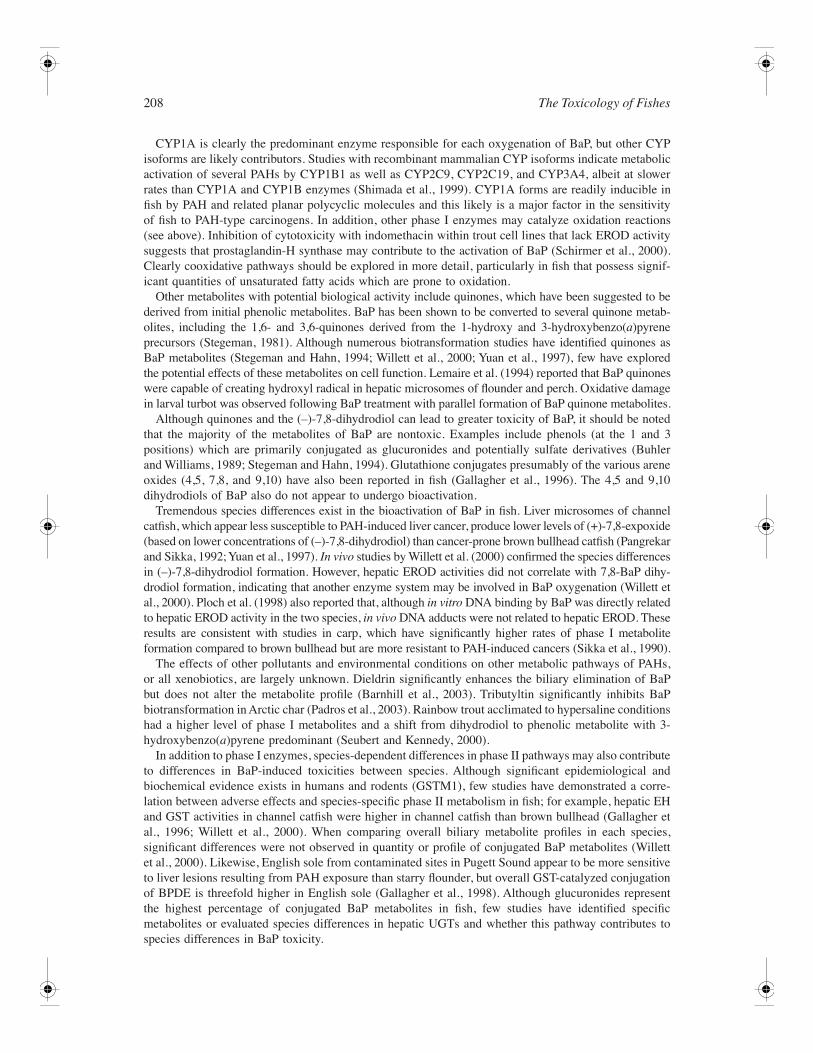
208 The Toxicology of Fishes
CYP1A is clearly the predominant enzyme responsible for each oxygenation of BaP, but other CYPisoforms are likely contributors. Studies with recombinant mammalian CYP isoforms indicate metabolicactivation of several PAHs by CYP1B1 as well as CYP2C9, CYP2C19, and CYP3A4, albeit at slowerrates than CYP1A and CYP1B enzymes (Shimada et al., 1999). CYP1A forms are readily inducible infish by PAH and related planar polycyclic molecules and this likely is a major factor in the sensitivityof fish to PAH-type carcinogens. In addition, other phase I enzymes may catalyze oxidation reactions(see above). Inhibition of cytotoxicity with indomethacin within trout cell lines that lack EROD activitysuggests that prostaglandin-H synthase may contribute to the activation of BaP (Schirmer et al., 2000).Clearly cooxidative pathways should be explored in more detail, particularly in fish that possess signif-icant quantities of unsaturated fatty acids which are prone to oxidation.
Other metabolites with potential biological activity include quinones, which have been suggested to bederived from initial phenolic metabolites. BaP has been shown to be converted to several quinone metab-olites, including the 1,6- and 3,6-quinones derived from the 1-hydroxy and 3-hydroxybenzo(a)pyreneprecursors (Stegeman, 1981). Although numerous biotransformation studies have identified quinones asBaP metabolites (Stegeman and Hahn, 1994; Willett et al., 2000; Yuan et al., 1997), few have exploredthe potential effects of these metabolites on cell function. Lemaire et al. (1994) reported that BaP quinoneswere capable of creating hydroxyl radical in hepatic microsomes of flounder and perch. Oxidative damagein larval turbot was observed following BaP treatment with parallel formation of BaP quinone metabolites.
Although quinones and the (–)-7,8-dihydrodiol can lead to greater toxicity of BaP, it should be notedthat the majority of the metabolites of BaP are nontoxic. Examples include phenols (at the 1 and 3positions) which are primarily conjugated as glucuronides and potentially sulfate derivatives (Buhlerand Williams, 1989; Stegeman and Hahn, 1994). Glutathione conjugates presumably of the various areneoxides (4,5, 7,8, and 9,10) have also been reported in fish (Gallagher et al., 1996). The 4,5 and 9,10dihydrodiols of BaP also do not appear to undergo bioactivation.
Tremendous species differences exist in the bioactivation of BaP in fish. Liver microsomes of channelcatfish, which appear less susceptible to PAH-induced liver cancer, produce lower levels of (+)-7,8-expoxide(based on lower concentrations of (–)-7,8-dihydrodiol) than cancer-prone brown bullhead catfish (Pangrekarand Sikka, 1992; Yuan et al., 1997). In vivo studies by Willett et al. (2000) confirmed the species differencesin (–)-7,8-dihydrodiol formation. However, hepatic EROD activities did not correlate with 7,8-BaP dihy-drodiol formation, indicating that another enzyme system may be involved in BaP oxygenation (Willett etal., 2000). Ploch et al. (1998) also reported that, although in vitro DNA binding by BaP was directly relatedto hepatic EROD activity in the two species, in vivo DNA adducts were not related to hepatic EROD. Theseresults are consistent with studies in carp, which have significantly higher rates of phase I metaboliteformation compared to brown bullhead but are more resistant to PAH-induced cancers (Sikka et al., 1990).
The effects of other pollutants and environmental conditions on other metabolic pathways of PAHs,or all xenobiotics, are largely unknown. Dieldrin significantly enhances the biliary elimination of BaPbut does not alter the metabolite profile (Barnhill et al., 2003). Tributyltin significantly inhibits BaPbiotransformation in Arctic char (Padros et al., 2003). Rainbow trout acclimated to hypersaline conditionshad a higher level of phase I metabolites and a shift from dihydrodiol to phenolic metabolite with 3-hydroxybenzo(a)pyrene predominant (Seubert and Kennedy, 2000).
In addition to phase I enzymes, species-dependent differences in phase II pathways may also contributeto differences in BaP-induced toxicities between species. Although significant epidemiological andbiochemical evidence exists in humans and rodents (GSTM1), few studies have demonstrated a corre-lation between adverse effects and species-specific phase II metabolism in fish; for example, hepatic EHand GST activities in channel catfish were higher in channel catfish than brown bullhead (Gallagher etal., 1996; Willett et al., 2000). When comparing overall biliary metabolite profiles in each species,significant differences were not observed in quantity or profile of conjugated BaP metabolites (Willettet al., 2000). Likewise, English sole from contaminated sites in Pugett Sound appear to be more sensitiveto liver lesions resulting from PAH exposure than starry flounder, but overall GST-catalyzed conjugationof BPDE is threefold higher in English sole (Gallagher et al., 1998). Although glucuronides representthe highest percentage of conjugated BaP metabolites in fish, few studies have identified specificmetabolites or evaluated species differences in hepatic UGTs and whether this pathway contributes tospecies differences in BaP toxicity.
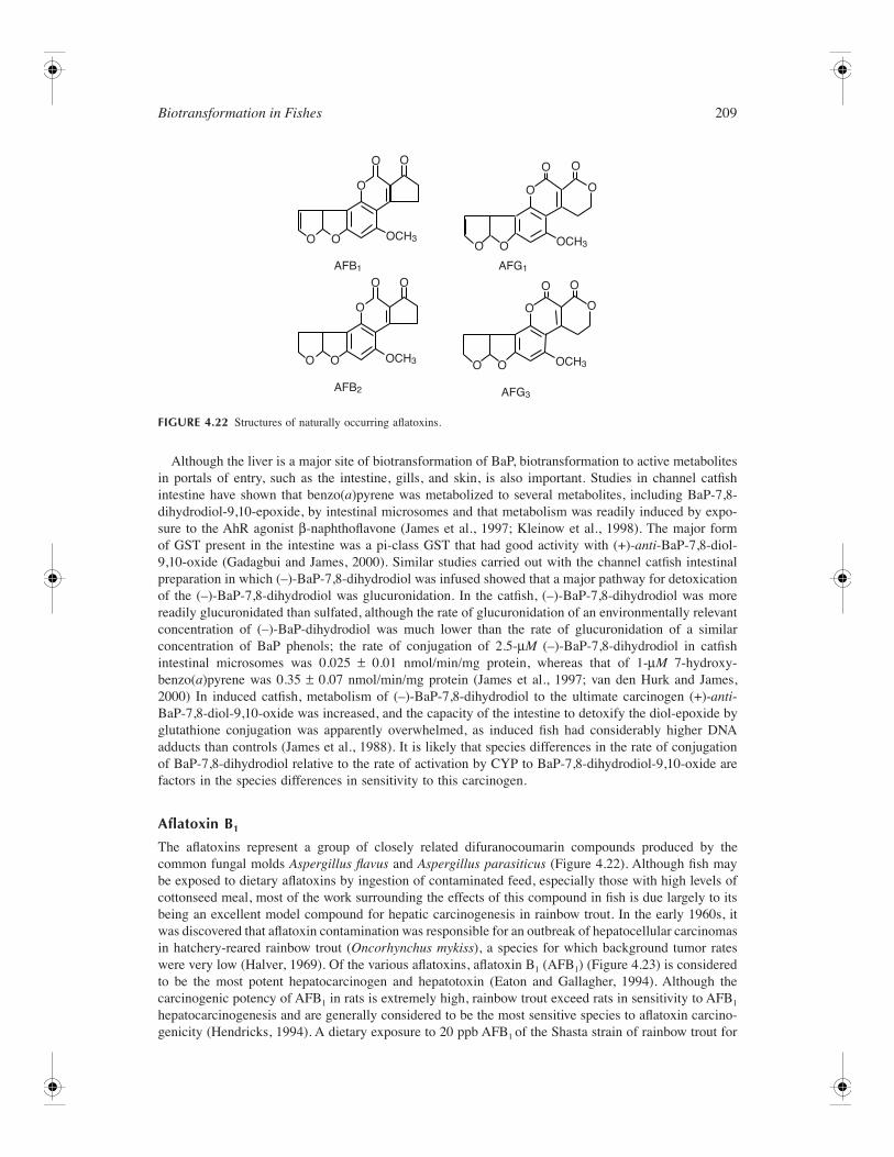
Biotransformation in Fishes 209
Although the liver is a major site of biotransformation of BaP, biotransformation to active metabolitesin portals of entry, such as the intestine, gills, and skin, is also important. Studies in channel catfishintestine have shown that benzo(a)pyrene was metabolized to several metabolites, including BaP-7,8-dihydrodiol-9,10-epoxide, by intestinal microsomes and that metabolism was readily induced by expo-sure to the AhR agonist β-naphthoflavone (James et al., 1997; Kleinow et al., 1998). The major formof GST present in the intestine was a pi-class GST that had good activity with (+)-anti-BaP-7,8-diol-9,10-oxide (Gadagbui and James, 2000). Similar studies carried out with the channel catfish intestinalpreparation in which (–)-BaP-7,8-dihydrodiol was infused showed that a major pathway for detoxicationof the (–)-BaP-7,8-dihydrodiol was glucuronidation. In the catfish, (–)-BaP-7,8-dihydrodiol was morereadily glucuronidated than sulfated, although the rate of glucuronidation of an environmentally relevantconcentration of (–)-BaP-dihydrodiol was much lower than the rate of glucuronidation of a similarconcentration of BaP phenols; the rate of conjugation of 2.5-µM (–)-BaP-7,8-dihydrodiol in catfishintestinal microsomes was 0.025 ± 0.01 nmol/min/mg protein, whereas that of 1-µM 7-hydroxy-benzo(a)pyrene was 0.35 ± 0.07 nmol/min/mg protein (James et al., 1997; van den Hurk and James,2000) In induced catfish, metabolism of (–)-BaP-7,8-dihydrodiol to the ultimate carcinogen (+)-anti-BaP-7,8-diol-9,10-oxide was increased, and the capacity of the intestine to detoxify the diol-epoxide byglutathione conjugation was apparently overwhelmed, as induced fish had considerably higher DNAadducts than controls (James et al., 1988). It is likely that species differences in the rate of conjugationof BaP-7,8-dihydrodiol relative to the rate of activation by CYP to BaP-7,8-dihydrodiol-9,10-oxide arefactors in the species differences in sensitivity to this carcinogen.
Aflatoxin B1
The aflatoxins represent a group of closely related difuranocoumarin compounds produced by thecommon fungal molds Aspergillus flavus and Aspergillus parasiticus (Figure 4.22). Although fish maybe exposed to dietary aflatoxins by ingestion of contaminated feed, especially those with high levels ofcottonseed meal, most of the work surrounding the effects of this compound in fish is due largely to itsbeing an excellent model compound for hepatic carcinogenesis in rainbow trout. In the early 1960s, itwas discovered that aflatoxin contamination was responsible for an outbreak of hepatocellular carcinomasin hatchery-reared rainbow trout (Oncorhynchus mykiss), a species for which background tumor rateswere very low (Halver, 1969). Of the various aflatoxins, aflatoxin B1 (AFB1) (Figure 4.23) is consideredto be the most potent hepatocarcinogen and hepatotoxin (Eaton and Gallagher, 1994). Although thecarcinogenic potency of AFB1 in rats is extremely high, rainbow trout exceed rats in sensitivity to AFB1
hepatocarcinogenesis and are generally considered to be the most sensitive species to aflatoxin carcino-genicity (Hendricks, 1994). A dietary exposure to 20 ppb AFB1 of the Shasta strain of rainbow trout for
FIGURE 4.22 Structures of naturally occurring aflatoxins.
O O
O
O O
O O
O
O
O
OCH3
OCH3
AFB1
AFB2
O O
O O
O O
O O
O
O O
O
OCH3
OCH3
AFG3
AFG1
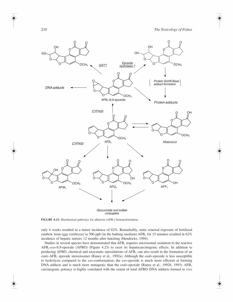
210 The Toxicology of Fishes
only 4 weeks resulted in a tumor incidence of 62%. Remarkably, static renewal exposure of fertilizedrainbow trout eggs (embryos) to 500 ppb (in the bathing medium) AFB1 for 15 minutes resulted in 62%incidence of hepatic tumors 12 months after hatching (Hendricks, 1994).
Studies in several species have demonstrated that AFB1 requires microsomal oxidation to the reactiveAFB1-exo-8,9-epoxide (AFBO) (Figure 4.23) to exert its hepatocarcinogenic effects. In addition toproducing AFBO, chemical and enzymatic epoxidations of AFB1 can also result in the formation of anendo-AFB1 epoxide stereoisomer (Raney et al., 1992a). Although the endo-epoxide is less susceptibleto hydrolysis compared to the exo-conformation, the exo-epoxide is much more efficient at formingDNA adducts and is much more mutagenic than the endo-epoxide (Raney et al., 1992b, 1993). AFB1
carcinogenic potency is highly correlated with the extent of total AFBO–DNA adducts formed in vivo
FIGURE 4.23 Biochemical pathways for aflatoxin (AFB1) biotransformation.
AFB1–8,9–epoxide
Aflatoxicol
OH
OH
OCH3OCH3
OCH3
OCH3
OCH3OCH3
OCH3
AFB1
AFQ1AFM1AFP1
O O
O
O
OO
O GST?
CYP450
CYP450
Glucuronide and sulfateconjugates
DNA adducts
Protein adducts
Protein Schiff–Baseadduct formation
Epoxidehydrolase ?
O
O O
O O
O
O
OO
OO
O
OO
OO
O
O O
O
O
O
O O
O O
O
O O
O
O
O
OH
OH
O
OH
OH
OH
GS
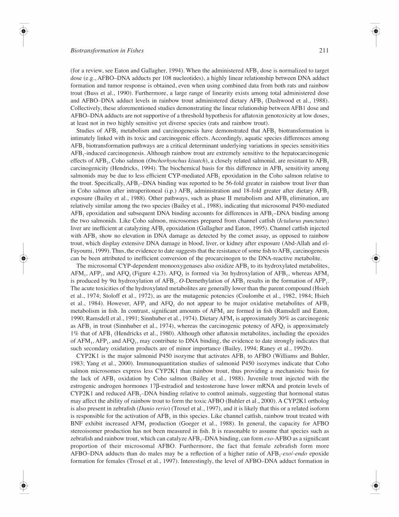
Biotransformation in Fishes 211
(for a review, see Eaton and Gallagher, 1994). When the administered AFB1 dose is normalized to targetdose (e.g., AFBO–DNA adducts per 108 nucleotides), a highly linear relationship between DNA adductformation and tumor response is obtained, even when using combined data from both rats and rainbowtrout (Buss et al., 1990). Furthermore, a large range of linearity exists among total administered doseand AFBO–DNA adduct levels in rainbow trout administered dietary AFB1 (Dashwood et al., 1988).Collectively, these aforementioned studies demonstrating the linear relationship between AFB1 dose andAFBO–DNA adducts are not supportive of a threshold hypothesis for aflatoxin genotoxicity at low doses,at least not in two highly sensitive yet diverse species (rats and rainbow trout).
Studies of AFB1 metabolism and carcinogenesis have demonstrated that AFB1 biotransformation isintimately linked with its toxic and carcinogenic effects. Accordingly, aquatic species differences amongAFB1 biotransformation pathways are a critical determinant underlying variations in species sensitivitiesAFB1-induced carcinogenesis. Although rainbow trout are extremely sensitive to the hepatocarcinogeniceffects of AFB1, Coho salmon (Onchorhynchus kisutch), a closely related salmonid, are resistant to AFB1
carcinogenicity (Hendricks, 1994). The biochemical basis for this difference in AFB1 sensitivity amongsalmonids may be due to less efficient CYP-mediated AFB1 epoxidation in the Coho salmon relative tothe trout. Specifically, AFB1–DNA binding was reported to be 56-fold greater in rainbow trout liver thanin Coho salmon after intraperitoneal (i.p.) AFB1 administration and 18-fold greater after dietary AFB1
exposure (Bailey et al., 1988). Other pathways, such as phase II metabolism and AFB1 elimination, arerelatively similar among the two species (Bailey et al., 1988), indicating that microsomal P450-mediatedAFB1 epoxidation and subsequent DNA binding accounts for differences in AFB1–DNA binding amongthe two salmonids. Like Coho salmon, microsomes prepared from channel catfish (Ictalurus punctatus)liver are inefficient at catalyzing AFB1 epoxidation (Gallagher and Eaton, 1995). Channel catfish injectedwith AFB1 show no elevation in DNA damage as detected by the comet assay, as opposed to rainbowtrout, which display extensive DNA damage in blood, liver, or kidney after exposure (Abd-Allah and el-Fayoumi, 1999). Thus, the evidence to date suggests that the resistance of some fish to AFB1 carcinogenesiscan be been attributed to inefficient conversion of the procarcinogen to the DNA-reactive metabolite.
The microsomal CYP-dependent monooxygenases also oxidize AFB1 to its hydroxylated metabolites,AFM1, AFP1, and AFQ1 (Figure 4.23). AFQ1 is formed via 3α hydroxylation of AFB1, whereas AFM1
is produced by 9α hydroxylation of AFB1. O-Demethylation of AFB1 results in the formation of AFP1.The acute toxicities of the hydroxylated metabolites are generally lower than the parent compound (Hsiehet al., 1974; Stoloff et al., 1972), as are the mutagenic potencies (Coulombe et al., 1982, 1984; Hsiehet al., 1984). However, AFP1 and AFQ1 do not appear to be major oxidative metabolites of AFB1
metabolism in fish. In contrast, significant amounts of AFM1 are formed in fish (Ramsdell and Eaton,1990; Ramsdell et al., 1991; Sinnhuber et al., 1974). Dietary AFM1 is approximately 30% as carcinogenicas AFB1 in trout (Sinnhuber et al., 1974), whereas the carcinogenic potency of AFQ1 is approximately1% that of AFB1 (Hendricks et al., 1980). Although other aflatoxin metabolites, including the epoxidesof AFM1, AFP1, and AFQ1, may contribute to DNA binding, the evidence to date strongly indicates thatsuch secondary oxidation products are of minor importance (Bailey, 1994; Raney et al., 1992b).
CYP2K1 is the major salmonid P450 isozyme that activates AFB1 to AFBO (Williams and Buhler,1983; Yang et al., 2000). Immunoquantitation studies of salmonid P450 isozymes indicate that Cohosalmon microsomes express less CYP2K1 than rainbow trout, thus providing a mechanistic basis forthe lack of AFB1 oxidation by Coho salmon (Bailey et al., 1988). Juvenile trout injected with theestrogenic androgen hormones 17β-estradiol and testosterone have lower mRNA and protein levels ofCYP2K1 and reduced AFB1–DNA binding relative to control animals, suggesting that hormonal statusmay affect the ability of rainbow trout to form the toxic AFBO (Buhler et al., 2000). A CYP2K1 orthologis also present in zebrafish (Danio rerio) (Troxel et al., 1997), and it is likely that this or a related isoformis responsible for the activation of AFB1 in this species. Like channel catfish, rainbow trout treated withBNF exhibit increased AFM1 production (Goeger et al., 1988). In general, the capacity for AFBOstereoisomer production has not been measured in fish. It is reasonable to assume that species such aszebrafish and rainbow trout, which can catalyze AFB1–DNA binding, can form exo-AFBO as a significantproportion of their microsomal AFBO. Furthermore, the fact that female zebrafish form moreAFBO–DNA adducts than do males may be a reflection of a higher ratio of AFB1-exo/-endo epoxideformation for females (Troxel et al., 1997). Interestingly, the level of AFBO–DNA adduct formation in
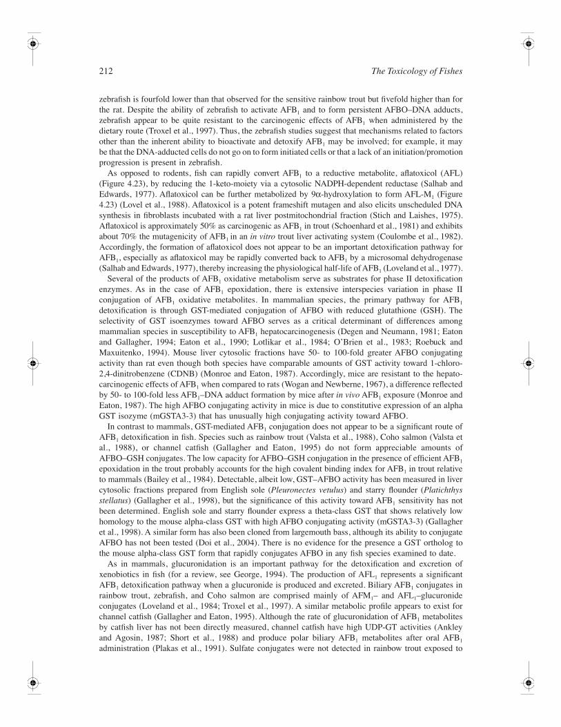
212 The Toxicology of Fishes
zebrafish is fourfold lower than that observed for the sensitive rainbow trout but fivefold higher than forthe rat. Despite the ability of zebrafish to activate AFB1 and to form persistent AFBO–DNA adducts,zebrafish appear to be quite resistant to the carcinogenic effects of AFB1 when administered by thedietary route (Troxel et al., 1997). Thus, the zebrafish studies suggest that mechanisms related to factorsother than the inherent ability to bioactivate and detoxify AFB1 may be involved; for example, it maybe that the DNA-adducted cells do not go on to form initiated cells or that a lack of an initiation/promotionprogression is present in zebrafish.
As opposed to rodents, fish can rapidly convert AFB1 to a reductive metabolite, aflatoxicol (AFL)(Figure 4.23), by reducing the 1-keto-moiety via a cytosolic NADPH-dependent reductase (Salhab andEdwards, 1977). Aflatoxicol can be further metabolized by 9α-hydroxylation to form AFL-M1 (Figure4.23) (Lovel et al., 1988). Aflatoxicol is a potent frameshift mutagen and also elicits unscheduled DNAsynthesis in fibroblasts incubated with a rat liver postmitochondrial fraction (Stich and Laishes, 1975).Aflatoxicol is approximately 50% as carcinogenic as AFB1 in trout (Schoenhard et al., 1981) and exhibitsabout 70% the mutagenicity of AFB1 in an in vitro trout liver activating system (Coulombe et al., 1982).Accordingly, the formation of aflatoxicol does not appear to be an important detoxification pathway forAFB1, especially as aflatoxicol may be rapidly converted back to AFB1 by a microsomal dehydrogenase(Salhab and Edwards, 1977), thereby increasing the physiological half-life of AFB1 (Loveland et al., 1977).
Several of the products of AFB1 oxidative metabolism serve as substrates for phase II detoxificationenzymes. As in the case of AFB1 epoxidation, there is extensive interspecies variation in phase IIconjugation of AFB1 oxidative metabolites. In mammalian species, the primary pathway for AFB1
detoxification is through GST-mediated conjugation of AFBO with reduced glutathione (GSH). Theselectivity of GST isoenzymes toward AFBO serves as a critical determinant of differences amongmammalian species in susceptibility to AFB1 hepatocarcinogenesis (Degen and Neumann, 1981; Eatonand Gallagher, 1994; Eaton et al., 1990; Lotlikar et al., 1984; O’Brien et al., 1983; Roebuck andMaxuitenko, 1994). Mouse liver cytosolic fractions have 50- to 100-fold greater AFBO conjugatingactivity than rat even though both species have comparable amounts of GST activity toward 1-chloro-2,4-dinitrobenzene (CDNB) (Monroe and Eaton, 1987). Accordingly, mice are resistant to the hepato-carcinogenic effects of AFB1 when compared to rats (Wogan and Newberne, 1967), a difference reflectedby 50- to 100-fold less AFB1–DNA adduct formation by mice after in vivo AFB1 exposure (Monroe andEaton, 1987). The high AFBO conjugating activity in mice is due to constitutive expression of an alphaGST isozyme (mGSTA3-3) that has unusually high conjugating activity toward AFBO.
In contrast to mammals, GST-mediated AFB1 conjugation does not appear to be a significant route ofAFB1 detoxification in fish. Species such as rainbow trout (Valsta et al., 1988), Coho salmon (Valsta etal., 1988), or channel catfish (Gallagher and Eaton, 1995) do not form appreciable amounts ofAFBO–GSH conjugates. The low capacity for AFBO–GSH conjugation in the presence of efficient AFB1
epoxidation in the trout probably accounts for the high covalent binding index for AFB1 in trout relativeto mammals (Bailey et al., 1984). Detectable, albeit low, GST–AFBO activity has been measured in livercytosolic fractions prepared from English sole (Pleuronectes vetulus) and starry flounder (Platichthysstellatus) (Gallagher et al., 1998), but the significance of this activity toward AFB1 sensitivity has notbeen determined. English sole and starry flounder express a theta-class GST that shows relatively lowhomology to the mouse alpha-class GST with high AFBO conjugating activity (mGSTA3-3) (Gallagheret al., 1998). A similar form has also been cloned from largemouth bass, although its ability to conjugateAFBO has not been tested (Doi et al., 2004). There is no evidence for the presence a GST ortholog tothe mouse alpha-class GST form that rapidly conjugates AFBO in any fish species examined to date.
As in mammals, glucuronidation is an important pathway for the detoxification and excretion ofxenobiotics in fish (for a review, see George, 1994). The production of AFL1 represents a significantAFB1 detoxification pathway when a glucuronide is produced and excreted. Biliary AFB1 conjugates inrainbow trout, zebrafish, and Coho salmon are comprised mainly of AFM1– and AFL1–glucuronideconjugates (Loveland et al., 1984; Troxel et al., 1997). A similar metabolic profile appears to exist forchannel catfish (Gallagher and Eaton, 1995). Although the rate of glucuronidation of AFB1 metabolitesby catfish liver has not been directly measured, channel catfish have high UDP-GT activities (Ankleyand Agosin, 1987; Short et al., 1988) and produce polar biliary AFB1 metabolites after oral AFB1
administration (Plakas et al., 1991). Sulfate conjugates were not detected in rainbow trout exposed to
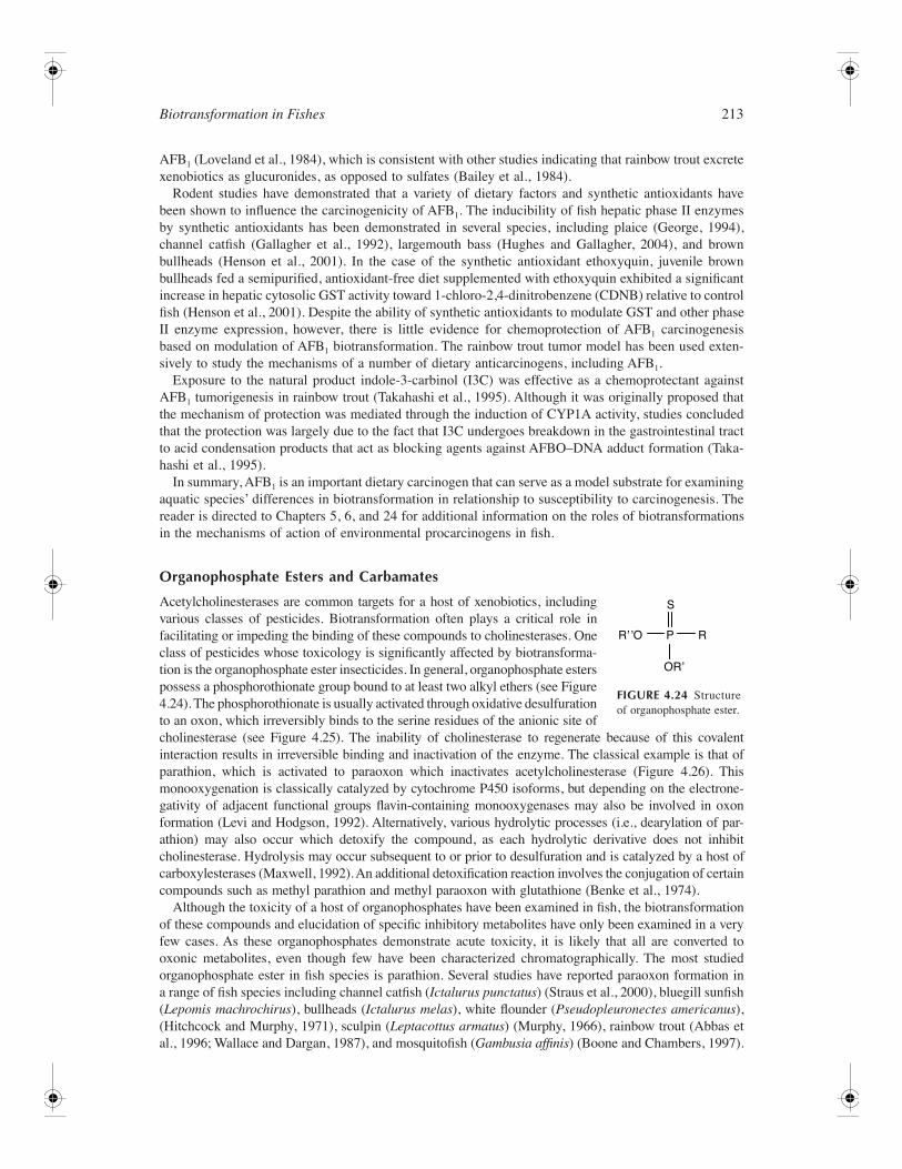
Biotransformation in Fishes 213
AFB1 (Loveland et al., 1984), which is consistent with other studies indicating that rainbow trout excretexenobiotics as glucuronides, as opposed to sulfates (Bailey et al., 1984).
Rodent studies have demonstrated that a variety of dietary factors and synthetic antioxidants havebeen shown to influence the carcinogenicity of AFB1. The inducibility of fish hepatic phase II enzymesby synthetic antioxidants has been demonstrated in several species, including plaice (George, 1994),channel catfish (Gallagher et al., 1992), largemouth bass (Hughes and Gallagher, 2004), and brownbullheads (Henson et al., 2001). In the case of the synthetic antioxidant ethoxyquin, juvenile brownbullheads fed a semipurified, antioxidant-free diet supplemented with ethoxyquin exhibited a significantincrease in hepatic cytosolic GST activity toward 1-chloro-2,4-dinitrobenzene (CDNB) relative to controlfish (Henson et al., 2001). Despite the ability of synthetic antioxidants to modulate GST and other phaseII enzyme expression, however, there is little evidence for chemoprotection of AFB1 carcinogenesisbased on modulation of AFB1 biotransformation. The rainbow trout tumor model has been used exten-sively to study the mechanisms of a number of dietary anticarcinogens, including AFB1.
Exposure to the natural product indole-3-carbinol (I3C) was effective as a chemoprotectant againstAFB1 tumorigenesis in rainbow trout (Takahashi et al., 1995). Although it was originally proposed thatthe mechanism of protection was mediated through the induction of CYP1A activity, studies concludedthat the protection was largely due to the fact that I3C undergoes breakdown in the gastrointestinal tractto acid condensation products that act as blocking agents against AFBO–DNA adduct formation (Taka-hashi et al., 1995).
In summary, AFB1 is an important dietary carcinogen that can serve as a model substrate for examiningaquatic species’ differences in biotransformation in relationship to susceptibility to carcinogenesis. Thereader is directed to Chapters 5, 6, and 24 for additional information on the roles of biotransformationsin the mechanisms of action of environmental procarcinogens in fish.
Organophosphate Esters and Carbamates
Acetylcholinesterases are common targets for a host of xenobiotics, includingvarious classes of pesticides. Biotransformation often plays a critical role infacilitating or impeding the binding of these compounds to cholinesterases. Oneclass of pesticides whose toxicology is significantly affected by biotransforma-tion is the organophosphate ester insecticides. In general, organophosphate esterspossess a phosphorothionate group bound to at least two alkyl ethers (see Figure4.24). The phosphorothionate is usually activated through oxidative desulfurationto an oxon, which irreversibly binds to the serine residues of the anionic site ofcholinesterase (see Figure 4.25). The inability of cholinesterase to regenerate because of this covalentinteraction results in irreversible binding and inactivation of the enzyme. The classical example is that ofparathion, which is activated to paraoxon which inactivates acetylcholinesterase (Figure 4.26). Thismonooxygenation is classically catalyzed by cytochrome P450 isoforms, but depending on the electrone-gativity of adjacent functional groups flavin-containing monooxygenases may also be involved in oxonformation (Levi and Hodgson, 1992). Alternatively, various hydrolytic processes (i.e., dearylation of par-athion) may also occur which detoxify the compound, as each hydrolytic derivative does not inhibitcholinesterase. Hydrolysis may occur subsequent to or prior to desulfuration and is catalyzed by a host ofcarboxylesterases (Maxwell, 1992). An additional detoxification reaction involves the conjugation of certaincompounds such as methyl parathion and methyl paraoxon with glutathione (Benke et al., 1974).
Although the toxicity of a host of organophosphates have been examined in fish, the biotransformationof these compounds and elucidation of specific inhibitory metabolites have only been examined in a veryfew cases. As these organophosphates demonstrate acute toxicity, it is likely that all are converted tooxonic metabolites, even though few have been characterized chromatographically. The most studiedorganophosphate ester in fish species is parathion. Several studies have reported paraoxon formation ina range of fish species including channel catfish (Ictalurus punctatus) (Straus et al., 2000), bluegill sunfish(Lepomis machrochirus), bullheads (Ictalurus melas), white flounder (Pseudopleuronectes americanus),(Hitchcock and Murphy, 1971), sculpin (Leptacottus armatus) (Murphy, 1966), rainbow trout (Abbas etal., 1996; Wallace and Dargan, 1987), and mosquitofish (Gambusia affinis) (Boone and Chambers, 1997).
FIGURE 4.24 Structureof organophosphate ester.
S
P
ORʼ
Rʼ ̓O R
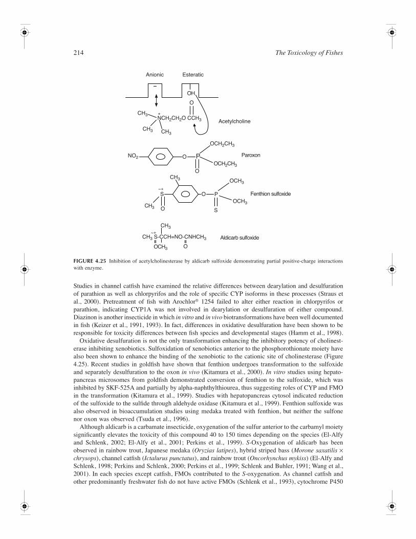
214 The Toxicology of Fishes
Studies in channel catfish have examined the relative differences between dearylation and desulfurationof parathion as well as chlorpyrifos and the role of specific CYP isoforms in these processes (Straus etal., 2000). Pretreatment of fish with Arochlor® 1254 failed to alter either reaction in chlorpyrifos orparathion, indicating CYP1A was not involved in dearylation or desulfuration of either compound.Diazinon is another insecticide in which in vitro and in vivo biotransformations have been well documentedin fish (Keizer et al., 1991, 1993). In fact, differences in oxidative desulfuration have been shown to beresponsible for toxicity differences between fish species and developmental stages (Hamm et al., 1998).
Oxidative desulfuration is not the only transformation enhancing the inhibitory potency of cholinest-erase inhibiting xenobiotics. Sulfoxidation of xenobiotics anterior to the phosphorothionate moiety havealso been shown to enhance the binding of the xenobiotic to the cationic site of cholinesterase (Figure4.25). Recent studies in goldfish have shown that fenthion undergoes transformation to the sulfoxideand separately desulfuration to the oxon in vivo (Kitamura et al., 2000). In vitro studies using hepato-pancreas microsomes from goldfish demonstrated conversion of fenthion to the sulfoxide, which wasinhibited by SKF-525A and partially by alpha-naphthylthiourea, thus suggesting roles of CYP and FMOin the transformation (Kitamura et al., 1999). Studies with hepatopancreas cytosol indicated reductionof the sulfoxide to the sulfide through aldehyde oxidase (Kitamura et al., 1999). Fenthion sulfoxide wasalso observed in bioaccumulation studies using medaka treated with fenthion, but neither the sulfonenor oxon was observed (Tsuda et al., 1996).
Although aldicarb is a carbamate insecticide, oxygenation of the sulfur anterior to the carbamyl moietysignificantly elevates the toxicity of this compound 40 to 150 times depending on the species (El-Alfyand Schlenk, 2002; El-Alfy et al., 2001; Perkins et al., 1999). S-Oxygenation of aldicarb has beenobserved in rainbow trout, Japanese medaka (Oryzias latipes), hybrid striped bass (Morone saxatilis ×chrysops), channel catfish (Ictalurus punctatus), and rainbow trout (Oncorhynchus mykiss) (El-Alfy andSchlenk, 1998; Perkins and Schlenk, 2000; Perkins et al., 1999; Schlenk and Buhler, 1991; Wang et al.,2001). In each species except catfish, FMOs contributed to the S-oxygenation. As channel catfish andother predominantly freshwater fish do not have active FMOs (Schlenk et al., 1993), cytochrome P450
FIGURE 4.25 Inhibition of acetylcholinesterase by aldicarb sulfoxide demonstrating partial positive-charge interactionswith enzyme.
CH3 S–CCH=NO–CNHCH3
CH3
CH3
CH3
OCH3
OCH3
OCH2CH3
NCH2CH2O CCH3
OCH2CH3
OCH3 O
O
O
O
O
O
OH
+~
+
+
~
Aldicarb sulfoxide
Fenthion sulfoxide
Paroxon
Acetylcholine
Anionic Esteratic
S
S
P
P
CH3
CH3
NO2
CH3
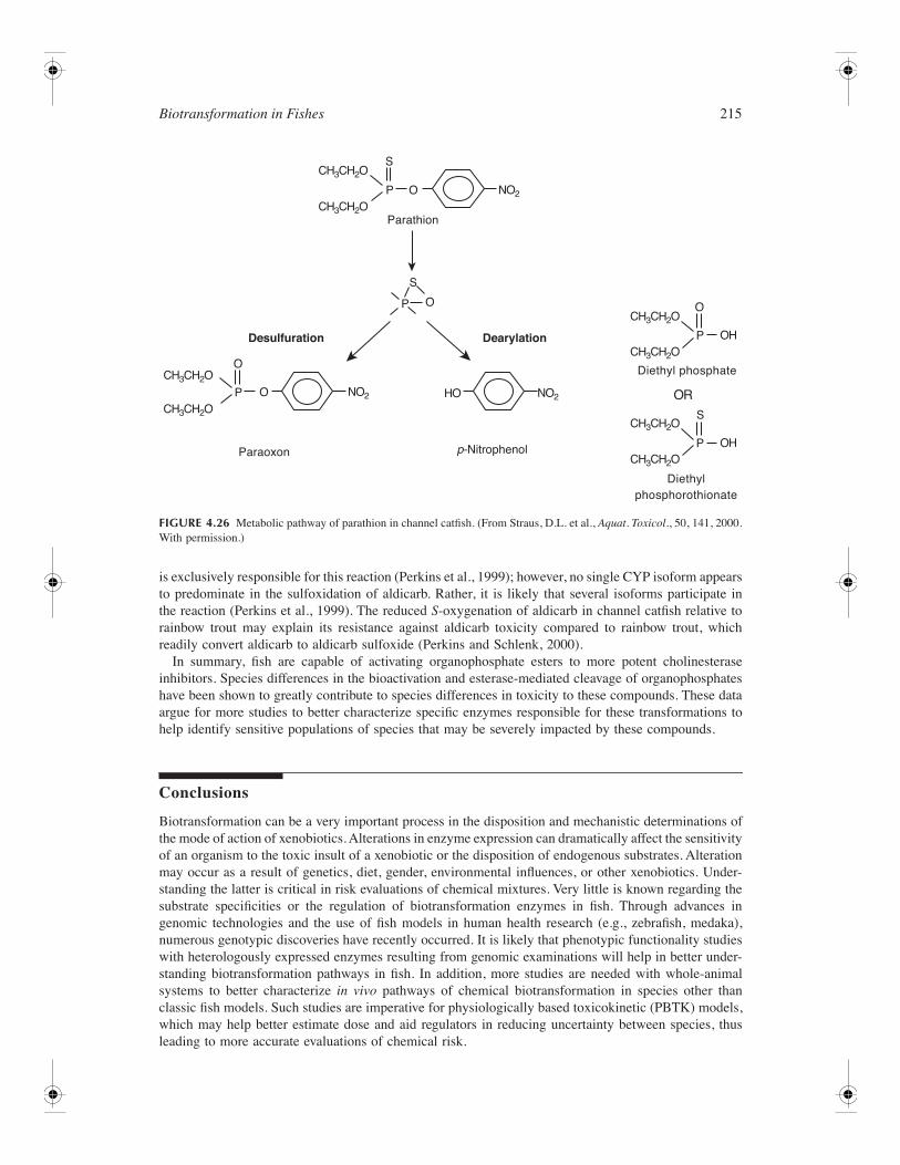
Biotransformation in Fishes 215
is exclusively responsible for this reaction (Perkins et al., 1999); however, no single CYP isoform appearsto predominate in the sulfoxidation of aldicarb. Rather, it is likely that several isoforms participate inthe reaction (Perkins et al., 1999). The reduced S-oxygenation of aldicarb in channel catfish relative torainbow trout may explain its resistance against aldicarb toxicity compared to rainbow trout, whichreadily convert aldicarb to aldicarb sulfoxide (Perkins and Schlenk, 2000).
In summary, fish are capable of activating organophosphate esters to more potent cholinesteraseinhibitors. Species differences in the bioactivation and esterase-mediated cleavage of organophosphateshave been shown to greatly contribute to species differences in toxicity to these compounds. These dataargue for more studies to better characterize specific enzymes responsible for these transformations tohelp identify sensitive populations of species that may be severely impacted by these compounds.
Conclusions
Biotransformation can be a very important process in the disposition and mechanistic determinations ofthe mode of action of xenobiotics. Alterations in enzyme expression can dramatically affect the sensitivityof an organism to the toxic insult of a xenobiotic or the disposition of endogenous substrates. Alterationmay occur as a result of genetics, diet, gender, environmental influences, or other xenobiotics. Under-standing the latter is critical in risk evaluations of chemical mixtures. Very little is known regarding thesubstrate specificities or the regulation of biotransformation enzymes in fish. Through advances ingenomic technologies and the use of fish models in human health research (e.g., zebrafish, medaka),numerous genotypic discoveries have recently occurred. It is likely that phenotypic functionality studieswith heterologously expressed enzymes resulting from genomic examinations will help in better under-standing biotransformation pathways in fish. In addition, more studies are needed with whole-animalsystems to better characterize in vivo pathways of chemical biotransformation in species other thanclassic fish models. Such studies are imperative for physiologically based toxicokinetic (PBTK) models,which may help better estimate dose and aid regulators in reducing uncertainty between species, thusleading to more accurate evaluations of chemical risk.
FIGURE 4.26 Metabolic pathway of parathion in channel catfish. (From Straus, D.L. et al., Aquat. Toxicol., 50, 141, 2000.With permission.)
NO2
CH3CH2O
CH3CH2O
CH3CH2O
CH3CH2O
CH3CH2O
CH3CH2O
CH3CH2O
CH3CH2O
P
P
P
P
O
O
O
OH
P OH
S
S
S
OR
DearylationDesulfuration
Paraoxon
Parathion
p–Nitrophenol
Diethylphosphorothionate
Diethyl phosphateO
O NO2
NO2
HO
216 The Toxicology of Fishes
References
Abbas, R. et al., Toxicokinetics of parathion and paraoxon in rainbow trout after intravascular administrationand water exposure, Toxicol. Appl. Pharmacol., 136, 194, 1996.
Abbott, B. D., Birnbaum, L. S., and Perdew, G. H., Developmental expression of two members of a new classof transcription factors: I. Expression of aryl hydrocarbon receptor in the C57BL/6N mouse embryo,Develop. Dynam., 204, 133, 1995.
Abd-Allah, G. A. and el-Fayoumi, I., A comparative evaluation of aflatoxin B genotoxicity in fish modelsusing the Comet assay, Mutat. Res., 446, 181, 1999.
Abnet, C. C. et al., Two forms of aryl hydrocarbon receptor type 2 in rainbow trout (Oncorhynchus mykiss),J. Biol. Chem., 274, 15159, 1999.
Adamson, R. H. et al., Comparative biochemistry of drug metabolism by azo and nitro reductase, Proc. Natl.Acad. Sci., 54, 1386, 1965.
Agustsson, I. and Strom, A.R., Biosynthesis and turnover of trimethylamine oxide in the teleost cod Gadusmorhua, J. Biol. Chem., 256, 8045, 1981.
Alin P., Danielson U. H., and Mannervik, B., 4-Hydroxyalk-2-enals are substrates for glutathione transferase,FEBS Lett., 179, 267, 1985.
Andersson, T., Pesonen, M., and Johansson C., Differential induction of cytochrome P-450-dependent monoox-ygenase, epoxide hydrolase, glutathione transferase and UDP glucuronosyl transferase activities in the liverof the rainbow trout by beta-naphthoflavone or Clophen A50, Biochem. Pharmacol., 34, 3309, 1985.
Andersson, T., Subcellular fractionation of rainbow trout gonads with emphasis on microsomal enzymesinvolved in steroid metabolism, Cell Tissue Res., 268, 479, 1992.
Andreasen, E. A., Hahn, M. E., Heideman, W., Peterson, R. E., and Tanguay, R. L., The zebrafish (Daniorerio) aryl hydrocarbon receptor type 1 is a novel vertebrate receptor, Mol. Pharmacol., 62, 234, 2002.
Angelucci, S. Sacchetta, P., Moio, P., Melino, S., Petruzzelli, R. Gervasi, P., and Di Ilio, C., Purification andcharacterization of glutathione transferases from the sea bass (Dicentrarchus labrax) liver, Arch. Biochem.Biophys., 373, 435, 2000.
Angus, W. G., Larsen, M. C., and Jefcoate, C. R., Expression of CYP1A1 and CYP1B1 depends on cell-specificfactors in human breast cancer cell lines: role of estrogen receptor status, Carcinogenesis, 20, 947, 1999.
Ankley, G. T. and Agosin, M., Comparative aspects of hepatic UDP-glucuronosyltransferases and glutathioneS-transferases in bluegill and channel catfish, Comp. Biochem. Physiol., 87B, 671, 1987.
Aoyama, T. et al., Five of twelve forms of vaccinia virus-expressed human hepatic cytochrome P450 meta-bolically activate aflatoxin B1, Proc. Natl. Acad. Sci., 87, 4790, 1990.
Armknecht, S.L., Kaattari, S.L., and Van Veld, P.A., An elevated glutathione S-transferase in creosote-resistantmummichog (Fundulus heteroclitus). Aquat. Toxicol., 41, 1, 1998.
Arukwe, A. and Goksøyr, A., Changes in three hepatic cytochrome P450 subfamilies during a reproductivecycle in turbot (Scophthalmus maximus L.), J. Exp. Zool., 277, 313, 1997.
Arukwe, A., Forlin, L., and Goksøyr, A., Xenobiotic and steroid biotransformation enzymes in Atlantic salmon(Salmo salar) liver treated with an estrogenic compound, 4-nonylphenol. Environ. Toxicol. Chem., 16,2576, 1997.
Bailey, G. S. et al., The sensitivity of rainbow trout and other fish to carcinogens, Drug Metab. Disp., 15,725, 1984.
Bailey, G. S. et al., Aflatoxin B1 carcinogenesis and its relation to DNA adduct formation and adduct persistencein sensitive and resistant salmonid fish, Carcinogenesis, 9, 1919, 1988.
Bailey, G. S., Role of aflatoxin-DNA adducts in the cancer process, in The Toxicology of Aflatoxins: HumanHealth, Veterinary and Agricultural Significance, Eaton, D. L. and Groopman, J. D., Eds., Academic Press,New York, 1994, pp. 137–148.
Baker, J. R., Streuempler, A., and Chaykin, S., A comparative study of trimethylamine-N-oxide biosynthesis,Biochim. Biophys. Acta, 71, 58, 1963.
Balk, L. et al., Initial characterization of drug-metabolizing systems in the liver of the Northern pike, Esoxlucius, Drug Metab. Disp., 8, 98, 1980.
Barnhill, M. L. et al., Dieldrin stimulates biliary excretion of 14C-benzo(a)pyrene polar metabolites but doesnot change the biliary metabolite profile in rainbow trout (Oncorhynchus mykiss), Toxicol. Sci., 75, 249,2003.
Biotransformation in Fishes 217
Barron, M. G., Hansen, S. C., and Ball, T., Pharmacokinetics and metabolism of triclopyr in the crayfish(Procambarus clarki), Drug Metab. Disp., 19, 163, 1991.
Barron, M. G., Schultz, I. R., and Hayton, W. L., Presystemic branchial metabolism limits di-2-ethylhexylphthalate accumulation in fish, Toxicol. Appl. Pharmacol., 98, 49, 1989.
Beetham, J. K. et al., Gene evolution of epoxide hydrolases and recommended nomenclature, DNA Cell Biol.,14, 61, 1995.
Bello, S. M. et al., Acquired resistance to Ah receptor agonists in a population of Atlantic killifish (Fundulusheteroclitus) inhabiting a marine superfund site: in vivo and in vitro studies on the inducibility of xenobioticmetabolizing enzymes, Toxicol. Sci., 60, 77, 2001.
Bend, J. R., James, M. O., and Dansette, P. M., In vitro metabolism of xenobiotics in some marine animals,Ann. N.Y. Acad. Sci., 298, 505, 1978.
Benke, G.M. et al., Comparative toxicity, anticholinesterase action, and metabolism of methyl parathion andparathion in sunfish and mice, Toxicol. Appl. Pharmacol., 28, 97, 1974.
Berndtson, A. K. and Chen, T. T., Two unique CYP1 genes are expressed in response to 3-methylcholanthrenetreatment in rainbow trout, Arch. Biochem. Biophys., 310, 187, 1993.
Billiard, S. M. et al., Binding of polycyclic aromatic hydrocarbons (PAHs) to teleost aryl hydrocarbon receptors(ARHs), Comp. Biochem. Physiol., 133, 55, 2002.
Birkholz, D. A., Coutts, R. T., and Hrudey, S. E., Uptake and biotransformation of 6,7-dimethylquinoline and6,8- dimethylquinoline by rainbow trout (Salmo gairdneri), Xenobiotica, 19, 695, 1989.
Blanchard, R. L. et al., A proposed nomenclature system for the cytosolic sulfotransferase (SULT) superfamily,Pharmacogenetics, 14, 199, 2004.
Blom, S. and Forlin, L., Effects of PCB on xenobiotic biotransformation enzyme activities in the liver and21-hydroxylation in the head kidney of juvenile rainbow trout, Aquat. Toxicol., 39, 215, 1997.
Board P. G. et al., Identification, characterization, and crystal structure of the omega class glutathionetransferases, J. Biol. Chem., 275, 24798, 2000.
Board, P. G. et al., Zeta, a novel class of glutathione transferases in a range of species from plants to humans,Biochem. J., 328, 929, 1997.
Boone, J. S. and Chambers, J. E., Biochemical factors contributing to toxicity differences among chlorpyrifos,parathion, and methyl parathion in mosquitofish (Gambusia affinis), Aquat. Toxicol., 39, 333, 1997.
Boyer, P.-M., Ndayibagira, A., and Spear P. A., Dose-dependent stimulation of hepatic retinoic acid hydrox-ylation/oxidation and glucuronidation in brook trout, Salvelinus fontinalis, after exposure to 3,3′, 4, 4′-tetrachlorobiphenyl, Environ. Toxicol. Chem., 19, 700, 2000.
Bradbury, S. P. and Christensen, G. M., Inhibition of alcohol dehydrogenase activity in acetylenic and allylicalcohols: concordance with in vivo electrophile reactivity in fish, Environ. Toxicol. Chem., 10, 1155, 1991.
Bradbury, S. P. et al., Toxicokinetics and metabolism of aniline and 4-chloroaniline in medaka (Oryzias latipes),Toxicol. Appl. Pharmacol., 118, 205, 1993.
Brammell, B. F. et al., Apparent lack of CYP1A response to high PCB body burdens in fish from a chronicallycontaminated PCB site, Mar. Environ. Res., 58, 251, 2004.
Bresnick, E. H. et al., Effect of phenobarbital and 3-methylcholanthrene administration on epoxide hydraselevels in liver microsomes, Biochem. Pharmacol., 26, 891, 1977.
Brumley, C.-M. et al., Validation of biomarkers of marine pollution exposure in sand flathead using Aroclor1254. Aquat. Toxicol., 31, 249, 1995.
Bucheli, T. D. and Fent, K., Induction of cytochrome P450 as a biomarker for environmental contaminationin aquatic ecosystems, Crit. Rev. Environ. Sci. Technol., 25, 201, 1995.
Buetler, T. M. et al., Induction of phase I and phase II drug-metabolizing enzyme mRNA, protein, and activityby BHA, ethoxyquin, and oltipraz, Toxicol. Appl. Pharmacol., 135, 45, 1995.
Buhler, D. R. and Wang-Buhler, J. L., Rainbow trout cytochrome P450s: purification, molecular aspects,metabolic activity, induction and role in environmental monitoring, Comp. Biochem. Physiol., 121C, 107,1998.
Buhler, D. R. and Williams, D. E., Enzymes involved in metabolism of PAH by fishes and other aquaticanimals: oxidative enzymes (or phase I enzymes), in Metabolism of Polycyclic Aromatic Hydrocarbonsin the Aquatic Environment, Varanasi, U., Ed., CRC Press, Boca Raton, FL, 1989, pp. 151–184.
Buhler, D. R. et al., Cloning and sequencing of the major rainbow trout constitutive cytochrome P450(CYP2K1): identification of a new cytochrome P450 gene subfamily and its expression in mature rainbowtrout liver and trunk kidney, Arch. Biochem. Biophys., 312, 45, 1994.
218 The Toxicology of Fishes
Buhler, D. R. et al., Effects of 17β-estradiol and testosterone on hepatic mRNA/protein levels and catalyticactivities of CYP2M1, CYP2K1, and CYP3A27 in rainbow trout (Oncorhynchus mykiss), Toxicol. Appl.Pharmacol., 168, 91, 2000.
Buhler, J.-L. et al., Expression of a constitutive cytochrome P450 (CYP2K1) in livers of rainbow trout(Oncorhynchus mykiss) embryo and sac fry, Aquat. Toxicol., 37, 237, 1997.
Burchell, B. and Coughtrie, M. W., UDP-glucuronosyltransferases, Pharmacol. Therapeut., 43, 261, 1989.Buss, P., Caviezel, M., and Lutz, W. K., Linear dose-response relationship for DNA adducts in rat liver from
chronic exposure to aflatoxin B1, Carcinogenesis, 11, 2133, 1990.Butala, H. et al., Microsomal estrogen metabolism in channel catfish, Mar. Environ. Res., 58, 489, 2004.Buters, J. T. et al., Cytochrome P450 CYP1B1 determines susceptibility to 7,12-dimethybenz(a)anthracene-
induced lymphomas, Proc. Natl. Acad. Sci., 96, 1977, 1999.Cao, Z. et al., Characterization of a CYP1A1 and CYP1A3 gene expression in rainbow trout (Oncorhynchus
mykiss), Aquat. Toxicol., 49, 101, 2000.Carlson, D. B. and Perdew, G. H., A dynamic role for the Ah receptor in cell signaling? Insights from a diverse
group of Ah receptor interacting proteins, J. Biochem. Mol. Toxicol., 16, 317, 2002.Carvan, M. J. et al., Trout CYP1A3 gene: recognition of fish DNA motifs by mouse regulatory proteins, Mar.
Biotechnol., 1, 155, 1999.Cashman, J. R. and Olsen, L. D., S-oxygenation of eptam in hepatic microsomes from fresh and saltwater
striped bass Morone saxatilis. Chem. Res. Toxicol., 2, 392, 1989.Cashman, J. R. et al., S-oxygenation of thiobencarb (Bolero) in hepatic preparations from striped bass (Morone
saxatilis) and mammalian systems, Chem. Res. Toxicol., 3, 433, 1990.Castren, M. and Oikari, A., Changes of the liver UDP-glucuronosyltransferase activity in trout (Salmo gairdneri
Rich.) acutely exposed to selected aquatic toxicants, Comp. Biochem. Physiol., 86C, 357, 1987.Celander M. and Forlin L., Catalytic activity and immunochemical quantification of hepatic cytochrome-P-
450 in beta-naphthoflavone and isosafrol treated rainbow-trout (Oncorhynchus mykiss), Fish Physiol.Biochem., 9, 189, 1991.
Celander M. and Forlin L., Decreased responsiveness of the hepatic cytochrome-P450 1A1 system in rainbowtrout (Oncorhynchus mykiss) after prolonged exposure to PCB, Aquat. Toxicol., 33, 141, 1995.
Celander, M. and Stegeman, J. J., Isolation of a cytochrome P450 3A cDNA sequence (CYP3A30) from themarine teleost Fundulus heteroclitus and phylogenetic analyses of CYP3A genes, Biochem. Biophys. Res.Comm., 236, 306, 1997.
Celander, M., Ronis, M., and Forlin, L., Initial characterization of a constitutive cytochrome P450 isoenzymein rainbow trout liver, Mar. Environ. Res., 28, 9, 1989.
Celander, M. et al., Induction of cytochrome P450 1A1 and conjugating enzymes in rainbow trout (Onco-rhynchus mykiss) liver: a time course study. Comp. Biochem. Physiol., 106C, 343–349, 1993.
Celander, M., Hahn, M. E., and Stegeman, J. J., Cytochromes P450 (CYP) in the Poeciliopsis lucida hepato-cellular carcinoma cell line (PLHC-1): dose- and time-dependent glucocorticoid potentiation of CYP1Ainduction without induction of CYP3A, Arch. Biochem. Biophys., 329, 113, 1996a.
Celander, M. et al., Immunochemical relationships of cytochrome P450-like protein in teleost fish, Fish Physiol.Biochem., 15, 323, 1996b.
Charest, R. P., Chenoweth, M., and Dunn, A., Metabolism of trimethylamines in kelp bass (Paralabrax clathratus)and marine and freshwater pink salmon (Oncorhynchus gorbuscha), J. Comp. Physiol. B., 158, 609, 1988.
Chen, C.-M. and Cooper, K. R., Developmental toxicity and EROD induction in the Japanese medaka (Oryziaslatipes) treated with dioxin congeners, Bull. Environ. Contam. Toxicol., 63, 423, 1999.
Christ, E. J. and Van Dorp, D. A., Comparative aspects of prostaglandin biosynthesis in animal tissues, Biochim.Biophys. Acta, 270, 537, 1972.
Christoffels, A. et al., Fugu genome analysis provides evidence for a whole-genome duplication early duringthe evolution of ray-finned fishes, Mol. Biol. Evol., 21, 1146, 2004.
Christou, M. et al., Co-expression of human CYP1A1 and a human analog of cytochrome P450-EF in responseto 2,3,7,8-tetrachlorodibenzo-p-dioxin in the human mammary carcinoma-derived MCF-7 cells, Carcino-genesis, 15, 725, 1994.
Clarke, D. J., George, S. G., and Burchell, B., Glucuronidation in fish, Aquat. Toxicol., 20, 35, 1991.Clarke, D. J., Burchell, B., and George, S. G., Functional and immunological comparison of hepatic UDP-
glucuronosyltransferases in a piscine and a mammalian species, Comp. Biochem. Physiol., 102B, 425,1992a.
Biotransformation in Fishes 219
Clarke, D. J., Burchell, B., and George, S. G., Differential expression and induction of UDP-glucuronosyl-transferase isoforms in hepatic and extrahepatic tissues of a fish: immunochemical and functional char-acterization, Toxicol. Appl. Pharmacol., 115, 130, 1992b.
Clarke, D. J., George, S. G., and Burchell, B., Multiplicity of UDP-glucuronosyltransferases in fish: purificationand characterization of a phenol UDP-glucuronosyltransferase from the liver of a marine teleost, Pleu-ronectes platessa, Biochem. J., 284, 417, 1992c.
Cok, I. et al., Expression of CYP2M1, CYP2K1, and CYP3A27 in brain, blood, small intestine, and othertissues of rainbow trout, Biochem. Biophys. Res. Comm., 244, 790, 1998.
Coldham, N. G. et al., Biotransformation, tissue distribution, and persistence of 4-nonylphenol residues injuvenile rainbow trout (Oncorhynchus mykiss), Drug Metab. Disp., 26, 347, 1998.
Collier, T. K. and Varanasi, U., Hepatic activities of xenobiotic metabolizing enzymes and biliary levels ofxenobiotics in English sole (Parophrys vetulus) exposed to environmental contaminants, Arch. Environ.Contam. Toxicol., 20, 462, 1991.
Collier, T. K. et al., Xenobiotic metabolizing enzymes in spawning English sole (Parophrys vetulus) exposedto organic-solvent extracts of marine sediments from contaminated and reference areas, Comp. Biochem.Physiol., 84C, 291, 1986.
Cooke, J. B. and Hinton, D. E., Promotion by 17 beta-estradiol and beta-hexachlorocyclohexane of hepato-cellular tumors in medaka, (Oryzias latipes), Aquat. Toxicol., 45, 127, 1999.
Coughtrie, M. W., Sulfation through the looking glass: recent advances in sulfotransferase research for thecurious, Pharmacogenomics, 2, 297, 2002.
Coughtrie, M. W. et al., Biology and function of the reversible sulfation pathway catalysed by humansulfotransferases and sulfatases, Chem. Biol. Interact., 109, 3, 1998.
Coulombe, R. A. et al., Comparative mutagenicity of aflatoxins using a Salmonella/trout hepatic enzymeactivation system, Carcinogenesis, 3, 1261, 1982.
Coulombe, R. A., Wilson, D. W., and Hsieh, D.P.H., Metabolism, DNA binding, and cytotoxicity of aflatoxinB1 in tracheal explants from Syrian hamster, Toxicology, 32, 117, 1984.
Coumoul, X., Diry, M., and Barouki, R., PXR-dependent induction of human CYP3A4 gene expression byorganochlorine pesticides, Biochem. Pharmacol., 64, 1513, 2002.
Cravedi, J.-P. et al., Influence of growth hormone on the hepatic mixed function oxidase and transferasesystems of rainbow trout, Fish Physiol. Biochem., 14, 259, 1995.
Cravedi, J.-P. et al., Biotransformation of pentachlorophenol, aniline and biphenyl in isolated rainbowtrout (Oncorhynchus mykiss) hepatocytes, comparison with in vivo metabolism, Xenobiotica, 29, 499,1999.
Daikoku, T. and Sakaguchi, M., Changes in level of trimethylamine and trimethylamine oxide during adaptationof young eel Anguilla anguilla to a seawater environment, Nippon Suisan Gakkaishi, 56, 1895, 1990.
Daikoku, T., Murata, M., and Sakaguchi, M., Effects of intraperitoneally injected and dietary trimethylamineon the biosynthesis of trimethylamine oxide in relation to seawater adaptation of the eel Anguilla japonicaand the guppy Poecilia reticulata, Comp. Biochem. Physiol., 89A, 261, 1988.
Danielsson, O., Eklund, H., and Joernvall, H., The major piscine liver alcohol dehydrogenase has class-mixedproperties in relation to mammalian alcohol dehydrogenases of classes I and III, Biochemistry, 31, 3751,1992.
Dansette, P. M., DuBois, G. C., and Jerina, D. M., Continuous fluorometric assay of epoxide hydrase activity,Anal. Biochem., 97, 340, 1979.
Dashwood, R. H. et al., Mechanisms of anti-carcinogenesis by indole-3-carbinol: detailed in vivo DNA bindingdose-response studies after dietary administration with aflatoxin B1, Carcinogenesis, 9, 427, 1988.
Dasmahapatra, A. K. et al., Developmental expression of alcohol dehydrogenase (ADH3) in zebrafish (Daniorerio), Biochem. Biophys. Res. Comm., 286, 1082, 2001.
de Wolf, W., Seinen, W., and Hermens, J. L., N-acetyltransferase activity in rainbow trout liver and in vitrobiotransformation of chlorinated anilines and benzenes in fish, Xenobiotica, 23, 1045, 1993.
DeBaun, J. R. et al., Sulfotransferase activation of N-hydroxy-2-acetylaminofluorene in rodent livers suscep-tible and resistant to this carcinogen, Proc. Soc. Exp. Biol. Med., 129, 268, 1968.
Degen, H. H., and Neumann, H. G., Differences in aflatoxin B1- susceptibility of rat and mouse are correlatedwith the capability to inactivate aflatoxin B1-epoxide, Carcinogenesis, 2, 299, 1981.
Denison, M. S., and Nagy, S. R. Activation of the aryl hydrocarbon receptor by structurally diverse exogenousand endogenous chemicals, Ann. Rev. Pharmacol. Toxicol., 43, 309, 2003.

220 The Toxicology of Fishes
Doi, A. M. et al., Molecular cloning and characterization of a glutathione S-transferase from largemouth bass(Micropterus salmoides) liver that is involved in the detoxification of 4-hydroxynonenal, Biochem. Phar-macol., 67, 2129, 2004.
Domanski, T. L. et al., Dual role of human cytochrome P450 3A4 residue Phe-304 in substrate specificityand cooperativity, J. Pharmacol. Exp. Therapeut., 293, 585, 2000.
Dooley, T. P., Molecular biology of the human phenol sulfotransferase gene family, J. Exp. Zool., 282, 223,1998.
Droy, B. F. et al., Influence of ormetoprim on the bioavailability, distribution, and pharmacokinetics ofsulfadimethoxine in rainbow trout (Oncorhynchus mykiss), Comp. Biochem. Physiol., 94C, 303, 1989.
Dutton, G.J., Glucuronidation of Drugs and Other Compounds, CRC Press, Boca Raton, FL. 1990.Eaton, D. L. and Gallagher, E.P., Mechanisms of aflatoxin carcinogenesis, Ann. Rev. Pharmacol. Toxicol., 34,
135, 1994.Eaton, D. L., Ramsdell, H., and Monroe, D. H., Biotransformation as a determinant of species susceptibility
to aflatoxin B1 in in vitro studies in rat, mouse, monkey and human liver, in Microbial Toxins in Foodsand Feeds: Cellular and Molecular Modes of Action, Pohland, A. E., Dowell, V. R. J., and Richard, J. L.,Eds., Plenum Press, New York, 1990, pp. 275–290.
Eisele, T. A. et al., The effects of route of exposure and combined exposure of mixed function oxidase inducersand suppressors on hepatic parameters in rainbow trout (Salmo gairdneri), Aquat. Toxicol., 5, 211, 1984.
El-Alfy, A. and Schlenk, D., Effect of 17β-estradiol and testosterone on the toxicity of aldicarb to Japanesemedaka, Oryzias latipes, Toxicol. Sci., 68, 381, 2002.
El-Alfy, A. and Schlenk, D., Potential mechanisms of the enhancement of aldicarb toxicity to Japanese medaka,Oryzias latipes, at high salinity. Toxicol. Appl. Pharmacol., 152, 175, 1998.
El-Alfy, A. et al., Characterization of salinity enhanced toxicity of aldicarb to Japanese medaka: sexual anddevelopmental differences, Environ. Toxicol. Chem., 20, 2093, 2001.
El-Alfy, A., Larsen, B. K., and Schlenk, D., Effect of cortisol and urea on flavin monooxygenase activity andexpression in rainbow trout, Oncorhynchus mykiss, Mar. Environ. Res., 54, 275, 2002.
Elmarakby, S.-A. et al., Comparative metabolism of 2-acetylaminofluorene by rainbow trout and rat liversubcellular fractions, Aquat. Toxicol., 33, 1, 1995.
Elskus, A. A. and Stegeman, J. J., Further consideration of phenobarbital effects on cytochrome P-450 activityin the killifish, Fundulus heteroclitus, Comp. Biochem. Physiol., 92C, 223, 1989.
Esterbauer, H., Schaur, R. J., and Zollner, H., Chemistry and biochemistry of 4-hydroxynonenal, malonalde-hyde and related aldehydes, Free Radic. Biol. Med., 11, 81, 1991.
Falany, C. N., Enzymology of human cytosolic sulfotransferases, FASEB J., 11, 206, 1997.Falany, C. N. et al., Purification and characterization of human liver phenol-sulfating phenol sulfotransferase,
Arch. Biochem. Biophys., 278, 312, 1990.Fernandez-Salguero, P. et al., Immune system impairment and hepatic fibrosis in mice lacking the dioxin-
binding Ah receptor. Science, 268, 722, 1995.Finnson, K. W. and Eales, J.G., Identification of thyroid hormone conjugates produced by isolated hepatocytes
and excreted in bile of rainbow trout, Oncorhynchus mykiss, Gen. Comp. Endocrinol., 101, 145, 1996.Finnson, K. W. and Eales, J.G., Sulfation of thyroid hormones by liver of rainbow trout, Oncorhynchus mykiss,
Comp. Biochem. Physiol., 120C, 415, 1998.Finnson, K. W. and Eales, J. G., Effect of T(3) treatment and food ration on hepatic deiodination and
conjugation of thyroid hormones in rainbow trout, Oncorhynchus mykiss, Gen. Comp. Endocrinol., 115,379, 1999.
Forlin, L. and Celander, M., Induction of cytochrome P450 1A in teleosts: environmental monitoring inSwedish fresh, brackish and marine waters, Aquat. Toxicol., 26, 41, 1993.
Forlin, L. et al., Effects on UDP glucuronosyl transferase, glutathione transferase, DT-diaphorase and glu-tathione reductase activities in rainbow trout liver after long-term exposure to PCB, Mar. Environ. Res.,42, 213, 1996.
Foureman, G. L. et al., The stereoselectivity of four hepatic glutathione S-transferases purified from a marineelasmobranch (Raja erinacea) with several K-region polycyclic arene oxide substrates, Biochim. Biophys.Acta., 914, 127, 1987.
Funkenstein, B. and Jakowiew, S. B., Molecular cloning of fish alcohol dehydrogenase cDNA, Gene, 174,159, 1996.
Biotransformation in Fishes 221
Furutani-Seiki, M. and Wittbrodt, J., Medaka and zebrafish, an evolutionary twin study, Mech. Develop., 121,629, 2004.
Gadagbui, B. K. and James, M. O., Activities of affinity-isolated glutathione S-transferase (GST) from channelcatfish whole intestine, Aquat. Toxicol., 49, 27, 2000.
Gallagher, E. P. and Eaton, D.L., In vitro metabolism of aflatoxin B1 (AFB1) in channel catfish liver, Toxicol.Appl. Pharmacol., 132, 82, 1995.
Gallagher, E. P., Canada, A. T., and Di Giulio, R. T., The protective role of glutathione in chorothalonil-induced toxicity to channel catfish, Aquat. Toxicol., 23, 155, 1992.
Gallagher, E. P. et al., Channel catfish glutathione S-transferase isoenzyme activity toward (+)-anti-ben-zo(a)pyrene-trans-7,8-dihydrodiol-9m10-epoxide, Aquat. Toxicol., 34, 135, 1996.
Gallagher, E. P. et al., Isolation and cloning of homologous glutathione S-transferase cDNAs from Englishsole and starry flounder liver, Aquat. Toxicol., 44, 171, 1998.
Gallagher E.P. et al., In vitro kinetics of hepatic glutathione S-transferase conjugation in largemouth bass andbrown bullheads, Environ. Toxicol. Chem., 19, 319, 2000.
Garfinkel, D., Studies on pig liver microsomes 1. Enzyme and pigment composition of different microsomalfractions, Arch. Biochem. Biophys., 77, 493, 1958.
Gaworecki, K. M., Rice, C. D., and van den Hurk, P., Induction of phenol-type sulfotransferase and glucu-ronosyltransferase in channel catfish and mummichog, Mar. Environ. Res., 58, 525, 2004.
George, S. G., Enzymology and molecular biology of phase II xenobiotic-conjugating enzymes in fish, inAquatic Toxicology: Molecular, Biochemical and Cellular Perspectives, Ostrander G. K. and Malins, D.C., Eds., CRC Press, Boca Raton, FL, 1994, pp. 37–85.
George, S. G. and Buchanan, G., Isolation, properties and induction of plaice liver cytosolic glutathione-S-transferases. Fish Physiol. Biochem., 8, 437, 1990.
George, S .G. and Leaver, M. J., Allelic variations in the plaice UGT1B1 gene, Mar. Environ. Res., 54, 259,2002.
George, S. G. and Taylor, A., Molecular evidence for multiple UDP-glucuronosyltransferase gene families infish, Mar. Environ. Res., 54, 253, 2002.
George, S. G. and Young, P., The time course of effects of cadmium and 3-methylcholanthrene on activitiesof enzymes of xenobiotic metabolism and metallothionein levels in the plaice, Pleuronectes platessa,Comp. Biochem. Physiol., 83C, 37, 1986.
George, S. G., Leaver, M. J. and Wright, J., Structural studies of a UDP-glucuronosyltransferase gene fromthe plaice (Pleuronectes platessa), Mar. Environ. Res., 46, 33, 1998.
German, J. B. and Berger, R., Formation of 8,15-dihydroxy eicosatetraenoic acid via 15- and 12-lipoxygenasesin fish, Lipids, 25, 849, 1990.
Gill, S. S. et al., Rapid and sensitive enzyme-linked immunosorbent assay for the microsomal epoxidehydrolase, Carcinogenesis, 3, 1307, 1982.
Gill, S. S., Ota, K., and Hammock, B. D., Radiometric assays for mammalian epoxide hydrolases andglutathione S-transferase, Anal. Biochem., 131, 273, 1983.
Gillam, E. M. et al., Expression of modified human cytochrome P450 3A4 in Escherichia coli and purificationand reconstitution of the enzyme, Arch. Biochem. Biophys., 305, 123, 1993.
Gillett, M. B. et al., Elevated levels of trimethylamine oxide in muscles of deep-sea gadiform teleosts: a highpressure adaptation, J. Exp. Zool., 279, 386, 1997.
Gillette, D. M. et al., Comparative toxicology of tetrachlorobiphenyls in mink and rats. I. Changes in hepaticenzyme activity and smooth endoplasmic reticulum volume, Fundam. Appl. Toxicol., 8, 5, 1987.
Glickman, A. H. et al., In vitro metabolism of permethrin isomers by carp and rainbow trout liver microsomes,J. Agric. Food Chem., 27, 1038, 1979.
Glickman, A. H., Weitman, S. D., and Lech, J. J., Differential toxicity of trans-permethrin in rainbow troutand mice. I. Role of biotransformation, Toxicol. Appl. Pharmacol., 66, 153, 1982.
Godard, C. A. et al., Identification of cytochrome P450 1B-like sequences in two teleost fish species (scup,Stenotomus chrysops, and plaice, Pleuronectes platessa) and in a cetacean (striped dolphin, Stenellacoeruleoalba), Mar. Environ. Res., 50, 7, 2000.
Goeger, D. E. et al., Comparative effect of dietary butylated hydroxyanisole and beta-naphthoflavone onaflatoxin B1 metabolism, DNA adduct formation, and carcinogenesis in rainbow trout. Carcinogenesis, 9,1793, 1988 (erratum in Carcinogenesis, 9, 2146, 1988).
222 The Toxicology of Fishes
Goksøyr, A., Purification of hepatic microsomal cytochromes P-450 from β-napthoflavone-treated Atlanticcod (Gadus morhua), a marine teleost fish, Biochim. Biophys. Acta, 840, 409, 1985.
Goksøyr, A. et al., Species characteristics of hepatic xenobiotic and steroid biotransformation systems of twoteleost fish, Atlantic cod (Gadus morhua) and rainbow trout (Salmo gairdneri), Toxicol. Appl. Pharmacol.,89, 347, 1987.
Goksøyr, A. et al., Immunochemical cross-reactivity of β-naphthoflavone-inducible cytochrome P450 (P450IA)in liver microsomes from different fish species and rat, Fish Physiol. Biochem., 9, 1, 1991.
Goldstein, L. and Dewitt-Harley, S., Trimethylamine oxidase of nurse shark liver and its relation to mammalianmixed function amine oxidase, Comp. Biochem. Physiol., 45B, 895, 1973.
Gonzalez, F. J. and Gelboin, H. V., Role of human cytochrome P450s in risk assessment and susceptibility toenvironmentally based disease, J. Toxicol. Environ. Health, 40, 289, 1993.
Gooch, J. W. et al., Effects of ortho- and non-ortho-substituted polychlorinated biphenyl congeners on thehepatic monooxygenase system in the scup (Stenotomus chrysops), Toxicol. Appl. Pharmacol., 98, 422,1989.
Gotoh, O., Substrate recognition sites in cytochrome P450 family 2 (CYP2) proteins inferred from comparativeanalysis of amino acid and coding nucleotide sequences, J. Biol. Chem., 267, 83, 1992.
Gravato, C. and Santos, M. A. Liver phase I and phase II enzymatic induction and genotoxic responses ofbeta-naphthoflavone water-exposed sea bass, Ecotoxicol. Environ. Safety, 52, 62, 2002.
Gray, E. S., Woodin, B. R., and Stegeman, J. J., Sex differences in hepatic monooxygenases in winter flounder(Pseudopleuronectes americanus) and scup (Stenotomus chrysops) and regulation of P450 forms byestradiol, J. Exp. Zool., 259, 330, 1991.
Gregus, Z. et al., Hepatic phase I and phase II biotransformations in quail and trout: comparison to otherspecies commonly used in toxicity testing, Toxicol. Appl. Pharmacol., 67, 430, 1983.
Guarino, A. M., James, M. O., and Bend, J. R., Fate and distribution of the herbicides 2,4-dichlor-phenoxyaceticacid (2,4-D) and 2,4,5-trichlorophenoxyacetic acid (2,4,5-T) in the dogfish shark, Xenobiotica, 7, 623,1977.
Haasch, M. L. et al., Effect of acrylamide monomer on hepatic CYP1A1 monooxygenase induction in rainbowtrout, Comp. Biochem. Physiol., 102C, 281, 1992.
Haasch, M. L. et al., Use of 7-alkoxyphenoxazones, 7-alkoxycoumarins, and 7-alkoxyquinolines as fluorescentsubstrates for rainbow trout hepatic microsomes after treatment with various inducers, Biochem. Phar-macol., 47, 893, 1994.
Haasch, M. L., Henderson, M. C., and Buhler, D. R., Induction of lauric acid hydroxylase activity in catfishand bluegill by peroxisome proliferating agents, Comp. Biochem. Physiol., 121C, 297, 1998.
Habig, W. H. and Jakoby, W. B., Assays for differentiation of glutathione S-transferase, Methods Enzymol.,77, 398, 1981.
Hahn, M. E. et al., Cytochrome P4501A induction and inhibition by 3,3′,4,4′-tetrachlorobiphenyl in an Ahreceptor-containing fish hepatoma cell line (PLHC-1), Aquat. Toxicol., 26, 185, 1993.
Hahn, M. E. et al., Photoaffinity labeling of the Ah receptor: phylogenetic survey of diverse vertebrate andinvertebrate species, Arch. Biochem. Biophys., 310, 218, 1994.
Halver, J. E., Aflatoxicosis and trout hepatoma, in Aflatoxin: Scientific Background, Control and Implications,Goldblatt, L. A., Ed., Academic Press, New York, 1969, pp. 265–306.
Hamm, J. T., Wilson, B. W., and Hinton, D. E., Organophosphate-induced acetylcholinesterase inhibition andembryonic retinal cell necrosis in vivo in the teleost (Oryzias latipes), Neurotoxicology, 19, 853, 1998.
Hammock, B. D. and Hasagawa, L. S., Differential substrate selectivity of murine hepatic cytosolic andmicrosomal epoxide hydrolases, Biochem. Pharmacol., 32, 1155, 1983.
Hansson, M. C. et al., Unprecedented genomic diversity of AhR1 and AhR2 genes in Atlantic salmon (Salmosalar L.), Aquat. Toxicol., 68, 219, 2004.
Harlow, G. R. and Halpert, J. R., Alanine-scanning mutagenesis of a putative substrate recognition site inhuman cytochrome P450 3A4. Role of residues 210 and 211 in flavonoid activation and substrate speci-ficity, J. Biol. Chem., 272, 5396, 1997.
Harlow, G. R. and Halpert, J. R., Analysis of human cytochrome P450 3A4 cooperativity: construction andcharacterization of a site-directed mutant that displays hyperbolic steroid hydroxylation kinetics, Proc.Natl. Acad. Sci., 95, 6636, 1998.
Hasselberg, L. et al., Effects of alkylphenols on CYP1A and CYP3A expression in first spawning Atlanticcod (Gadus morhua), Aquat. Toxicol., 67, 303, 2004.

Biotransformation in Fishes 223
Hasspieler, B. M., and Di Giulio, R. T., Dicoumarol-sensitive NADPH: phenanthrenequinone oxidoreductasein channel catfish (Ictalurus punctatus), Toxicol. Appl. Pharmacol., 125, 184, 1994.
Hasspieler, B. M., Haffner, G. D., and Adeli, K., Roles of DT diaphorase in the genotoxicity of nitroaromaticcompounds in human and fish cell lines, J. Toxicol. Environ. Health, 52, 137, 1997.
Hawkins, W. E. et al., Carcinogenic effects of 1,2-dibromoethane (ethylene dibromide; EDB) in Japanesemedaka (Oryzias latipes), Mutat. Res., 399(2), 221, 1998.
Hayes J. D., Pulford, D. J., The glutathione S-transferase supergene family: regulation of GST and thecontribution of the isoenzymes to cancer chemoprotection and drug resistance, Crit. Rev. Biochem. Mol.Biol., 30, 445, 1995.
Hayes, C. L. et al., 17β-Estradiol hydroxylation catalyzed by human cytochrome P450 1B1, Proc. Natl. Acad.Sci., 93, 9776, 1996.
Hegelund, T. and Celander, M. C., Hepatic versus extrahepatic expression of CYP3A30 and CYP3A56 inadult killifish (Fundulus heteroclitus). Aquat. Toxicol., 64, 277, 2003.
Hegelund, T. et al., Effects of the antifungal imidazole ketoconazole on CYP1A and CYP3A in rainbow troutand killifish, Environ. Toxicol. Chem., 23, 1326, 2004.
Heilmann, L. J. et al., Trout P450IA1: cDNA and deduced protein sequence, expression in liver, and evolu-tionary significance, DNA, 7, 379, 1988.
Hendricks, J. D., Carcinogenicity of aflatoxins in nonmammalian organisms, in The Toxicology of Aflatoxins:Human Health, Veterinary and Agricultural Significance, Eaton, D. L. and Groopman, J. D., Eds.,Academic Press, New York, 1994.
Hendricks, J. D. et al., Carcinogenic response of rainbow trout (Salmo gairdneri) to aflatoxin B1 and synergisticeffect of cyclopropenoid fatty acids, J. Natl. Cancer Inst., 64, 523, 1980.
Henne, K. R. et al., Covalent linkage of prosthetic heme to CYP4 family P450 enzymes, Biochemistry, 40,12925, 2001.
Henson, K. L., Stauffer, G., and Gallagher, E. P., Induction of glutathione S-transferase activity and proteinexpression in brown bullhead (Ameiurus nebulosus) liver by ethoxyquin, Toxicol. Sci., 62, 54, 2001.
Hines, R. N. et al., The mammalian flavin-containing monooxygenases: molecular characterization and reg-ulation of expression, Toxicol. Appl. Pharmacol., 125, 1, 1994.
Hitchcock, M. and Murphy, S. D., Activation of parathion and guthion by mammalian, avian, and piscine liverhomogenates and cell fractions, Toxicol. Appl. Pharmacol., 19, 37, 1971.
Honkakoski, P. and Negishi, M., Characterization of a phenobarbital-responsive enhancer module in mouseP450 CYP2B10 gene, J. Biol. Chem., 272, 14943, 1997.
Honkakoski, P. et al., Activation by diverse xenochemicals of the 51-base-pair phenobarbital-responsiveenhancer module in the CYP2B10 gene, Mol. Pharmacol., 53, 597, 1998a.
Honkakoski, P. et al., The orphan nuclear receptor CAR-retinoid X receptor heterodimer activates the phe-nobarbital-responsive enhancer module of the CYP2B gene, Mol. Cell. Biol., 18, 5652, 1998b.
Houston, J. B. and Kenworthy, K. E., In vitro-in vivo scaling of CYP kinetic data not consistent with theclassical Michaelis-Menten model, Drug Metab. Disp., 28, 246, 2000.
Hsieh, D. P. H. et al., Toxicity of aflatoxin Q1 as evaluated with the chicken embryo and bacterial auxotrophs,Toxicol. Appl. Pharmacol., 30, 237, 1974.
Hsieh, D. P. H., Cullen, J. M., and Ruebner, B. H., Comparative hepatocarcinogenicity of aflatoxins B1 andM1 in the rat, Food Chem., Toxicol., 22, 1027, 1984.
Hsieh, R. J., German, J. B., and Kinsella, J. E., Lipoxygenase in fish tissue: some properties of the 12-lipoxygenase from trout gill, J. Agric. Food Chem., 36, 680, 1988.
Hubatsch, I., Ridderstrom M., and Mannervik, B., Human glutathione transferase A4-4: an alpha class enzymewith high catalytic efficiency in the conjugation of 4-hydroxynonenal and other genotoxic products oflipid peroxidation, Biochem. J., 330, 175, 1998.
Hughes, E. M. and Gallagher, E. P., Effects of beta-naphthoflavone on hepatic biotransformation and glu-tathione biosynthesis in largemouth bass (Micropterus salmoides), Mar. Environ. Res., 58, 675, 2004.
Husoy, A. M. et al., Immunohistochemical localization of CYP1A and CYP3A-like isozymes in hepatic andextrahepatic tissues of Atlantic cod (Gadus morhua L.), a marine fish, Toxicol. Appl. Pharmacol., 129,294, 1994.
Huuskonen, S. et al., Effects of non-ortho-substituted polychlorinated biphenyls (congeners 77 and 126) oncytochrome P4501A and conjugation activities in rainbow trout (Oncorhynchus mykiss), Comp. Biochem.Physiol., 113C, 205, 1996.
224 The Toxicology of Fishes
Iwata, H. et al., Species-specific responses of constitutively active receptor (CAR)–CYP2B coupling: lack ofCYP2B inducer-responsive nuclear translocation of CAR in marine teleost, scup (Stenotomus chrysops),Comp. Biochem. Physiol., 131C, 501, 2002.
James, M. O., Taurine conjugation of carboxylic acids in some marine species, in Conjugation Reactions inDrug Biotransformations, Aitio, A., Ed., Elsevier, Amsterdam, 1976, pp. 121–129.
James, M. O., Xenobiotic conjugation in aquatic species, in Xenobiotic Conjugation Chemistry, Paulson, G.D., Caldwell, J., Hutson, D. H., and Menn, J. J., Eds., American Chemical Society, Washington, D.C., 1986.
James, M. O. and Bend, J. R., Taurine conjugation of 2,4-dichlorophenoxyacetic acid and phenylacetic acidin two marine species, Xenobiotica, 6, 393, 1976.
James, M. O. and Bend, J. R., Polycyclic aromatic hydrocarbon induction of cytochrome P-450-dependentmixed-function oxidases in marine fish, Toxicol. Appl. Pharmacol., 54, 117, 1980.
James, M. O. and Little, P.J., Polyhalogenated biphenyls and phenobarbital: evaluation as inducers of drugmetabolizing enzymes in the sheepshead, Archosargus probatocephalus, Chem. Biol. Interact., 36, 229, 1981.
James, M. O. and Pritchard, J. B., In vivo and in vitro renal metabolism and excretion of benzoic acid by amarine teleost, the southern flounder, Drug Metab. Disp., 15, 665, 1987.
James, M. O., Fouts, J. R., and Bend, J. R., In vitro epoxide metabolism in some marine species, Bull. Mt.Desert Island Biol. Lab., 14, 41, 1974.
James, M. O., Fouts, J. R., and Bend, J. R., Hepatic and extrahepatic metabolism, in vitro, of an epoxide (8-(14) C- styrene oxide) in the rabbit, Biochem. Pharmacol., 25, 187, 1976.
James, M. O. et al., Epoxide hydrase and glutathione S-transferase activities with selected alkene and adreneoxides in several marine species, Chem. Biol. Interact., 25, 321, 1979.
James, M. O., Heard, C. S., Hawkins, W. E., Effect of 3-methylcholanthrene on mono-oxygenase, epoxidehydrolase, and glutathione S-transferase activities in small estuarine and freshwater fish, Aquat. Toxicol.,12, 1, 1988.
James, M. O., Hawkins, W. E., and Walker, W. W., Phase 1 and phase 2 biotransformation and carcinogenicityof 2-acetylaminofluorene in medaka and guppy, Aquat. Toxicol., 28, 79, 1994.
James, M. O. et al., Dietary modulation of phase 1 and phase 2 activities with benzo(a)pyrene and relatedcompounds in the intestine but not the liver of the channel catfish, Ictalurus punctatus, Drug Metab. Disp.,25, 346, 1997.
James, M. O., Sikazwe, D., and Gadagbui, B., Isolation of a pi class glutathione S-transferase (GST) fromcatfish intestinal mucosa, Mar. Environ. Res., 46, 57, 1998.
James, M. O. et al., Intestinal bioavailability and biotransformation of 3-hydroxybenzo(a)pyrene in an isolatedperfused preparation from channel catfish, Ictalurus punctatus, Drug Metab. Disp., 29, 721, 2001.
James, M. O. et al., Increased toxicity of benzo(a)pyrene-7, 8-dihydrodiol in the presence of polychlorobi-phenylols, Mar. Environ. Res., 58, 343, 2004.
Jerina D.M. et al., Identification of the ultimate carcinogenic metabolites of the polycyclic aromatic hydro-carbons: bay-region (R, S)-diol-(S, R)-epoxides, Foreign Compound Metabolism, J. Caldwell and G.Paulson, Eds., London, Taylor and Francis, 1984, pp. 257-270.
Jerina, D. M. and Dansette, P. M., Hepatic microsomal epoxide hydrase: a sensitive radiometric assay forhydration of arene oxides of carcinogenic aromatic hydrocarbons. Mol. Pharmacol., 13, 342, 1977.
Jeuken, A. et al., Activation of the Ah receptor by extracts of dietary herbal supplements, vegetables, andfruits, J. Agric. Food Chem., 51, 5478, 2003.
Jorgensen, E. H. et al., The effect of stress on toxicant-dependent cytochrome p450 enzyme responses in theArctic charr (Salvelinus alpinus), Environ. Toxicol. Chem., 20, 2523, 2001.
Juchau, M. R., Boutelet-Bochan, H., and Huang, Y., Cytochrome P450-dependent biotransformation of xeno-biotics in human and rodent embryonic tissues, Drug Metab. Rev., 30, 541, 1998.
Kaplan, L. A. et al., Nitrosodiethylamine metabolism in the viviparous fish Poeciliopsis: evidence for theexistence of liver P450pj activity and expression, Carcinogenesis, 12, 647, 1991.
Kaplan, L. A., Fielding, E. and Crivello, J. F., The genetic regulation of liver microsomal CYP2E1 activityamong strains of the viviparous fish Poeciliopsis, Comp. Biochem. Physiol., 128C, 143, 2001.
Karchner, S. I., Powell, W. H., and Hahn, M. E., Identification and functional characterization of two highlydivergent aryl hydrocarbon receptors (AhR1 and AhR2) in the teleost Fundulus heteroclitus, J. Biol.Chem., 274, 33814, 1999.
Katchamart, S. et al., Effect of xenoestrogen exposure on the expression of cytochrome P450 isoforms inrainbow trout liver, Environ. Toxicol. Chem., 21, 2445, 2002.
Biotransformation in Fishes 225
Keizer, J., D’Agostino, G., and Vittozzi, L., The importance of biotransformation in the toxicity of xenobioticsto fish. I. Toxicity and bioaccumulation of diazinon in guppy (Poecilia reticulata) and zebra fish (Brachy-danio rerio), Aquat. Toxicol., 21, 239, 1991.
Keizer, J. et al., Comparative diazinon toxicity in guppy and zebra fish: different role of oxidative metabolism,Environ. Toxicol. Chem., 12, 1243, 1993.
Keizer, J. et al., Enzymological differences of AChE and diazinon hepatic metabolism: correlation of in vitrodata with the selective toxicity of diazinon to fish species, Sci. Total Environ., 171, 213, 1995.
Khan, K. K. and Halpert, J. R., Structure–function analysis of human cytochrome P450 3A4 using 7-alkoxycoumarins as active-site probes, Arch. Biochem. Biophys., 373, 335, 2000.
Kim, I.-C. et al., Cloning of cytochrome P450 1A (CYP1A) genes from the hermaphrodite fish Rivulusmarmoratus and the Japanese medaka Oryzias latipes, Mar. Environ. Res., 58, 125, 2004.
Kitamura, S. et al., Interconversion between fenthion and fenthion sulfoxide in goldfish, Carassius auratus,J. Health Sci., 45, 266, 1999.
Kitamura, S. et al., Whole-body metabolism of the organophosphorus pesticide, fenthion, in goldfish, Carassiusauratus, Comp. Biochem. Physiol. 126C, 259, 2000.
Kitamura, S. et al., In vitro metabolism of fenthion and fenthion sulfoxide by liver preparations of sea bream,goldfish and rats, Drug Metab. Disp., 31, 179, 2003.
Kleinow, K. M. et al., A comparison of hepatic P450 induction in rat and trout (Oncorhynchus mykiss): delineationof the site of resistance of fish to phenobarbital-type inducers, Environ. Health Perspect., 71, 105, 1990.
Kleinow, K. M. et al., Pharmacokinetics, bioavailability, distribution, and metabolism of sulfadimethoxine inthe rainbow-trout (Oncorhynchus mykiss), Can. J. Fish. Aquat. Sci., 49, 1070, 1992.
Kleinow, K. M. et al., Bioavailability and biotransformation of benzo(a)pyrene in an isolated perfused in situcatfish intestinal preparation, Environ. Health Perspect., 106, 155, 1998.
Kliewer, S. A., Goodwin, B., and Willson, T. M., The nuclear pregnane X receptor: a key regulator of xenobioticmetabolism Endocrine Rev., 23, 687, 2002.
Klingenberg, M., Pigments of rat liver microsomes, Arch. Biochem. Biophys., 75, 376, 1958.Klinger, W. et al., Cytochrome P450 (P450) isoform expression, P450 concentration, monooxygenase activities,
reactive oxygen species formation, lipid peroxidation, and glutathione content in wild catch carp and tenchliver: influence of a two weeks’ exposure to phenobarbital, Exper. Toxicol. Pathol., 52, 513, 2001.
Klotz, A. V. et al., Cytochrome P-450 isozymes from the marine teleost Stenotomus chrysops: their roles insteroid hydroxylation and the influence of cytochrome b5, Arch. Biochem. Biophys., 249, 326, 1986.
Konishi-Imamura, L. et al., Regulation of arylsulfate sulfotransferase from a human intestinal bacterium bynucleotides and magnesium ion, J. Enzyme Inhib., 8, 233, 1995.
Kullman, S. W. and Hinton, D. E., Identification, characterization, and ontogeny of a second cytochrome P4503A gene from the fresh water teleost medaka (Oryzias latipes), Mol. Reprod. Develop., 58, 149, 2001.
Kullman, S. W., Hamm, J. T., and Hinton, D. E., Identification and characterization of a cDNA encodingcytochrome P450 3A from the fresh water teleost medaka (Oryzias latipes), Arch. Biochem. Biophys.,380, 29, 2000.
Kullman, S. W., Kashiwada, S., and Hinton, D. E., Analysis of medaka cytochrome P450 3A homotropic andheterotropic cooperativity, Mar. Environ. Res., 58, 469, 2004.
Laitenen, M. et al., Tricaine (MS-222) induced modification of the metabolism of foreign compounds in theliver and duodenal mucosa of the splake (Salvelinus fontinalis × Salvelinus namaycush), Acta Pharmacol.Toxicol., 49, 92, 1981.
Lambert, J. G. D. and Resink, J. W., Steroid glucuronides as male pheromones in the reproduction of Africancatfish Clarias gariepinus: a brief review, J. Steroid Biochem. Mol. Biol., 40, 549, 1991.
Lange, R. and Fugelli, K., The osmotic adjustment in the euryhaline teleosts, the flounder, Pleuronectes flesus,and the three-spined stickleback, Gasterosteus aculeatus, Comp. Biochem. Physiol., 15, 283, 1965.
Larsen, B. K. and Schlenk, D., Effect of salinity on flavin-containing monooxygenase expression and activityin rainbow trout (Oncorhynchus mykiss), J. Comp. Physiol. B, 171, 421, 2001.
Larsen, B. K. and Schlenk, D., Effect of urea and low temperature on the expression and activity of flavin-containing monooxygenase in the liver and gill of rainbow trout (Oncorhynchus mykiss), Fish Physiol.Biochem., 25(1), 19–29, 2001.
Larsen, H. E., Celander, M., and Goksøyr, A., The cytochrome-p450 system of Atlantic salmon (Salmo salar).2. Variations in hepatic catalytic activities and isozyme patterns during an annual reproductive-cycle, FishPhysiol. Biochem., 10, 291, 1992.
226 The Toxicology of Fishes
Leaver M. J. and George, S. G., A piscine glutathione S-transferase which efficiently conjugates the end-products of lipid peroxidation, Mar. Environ. Res., 46, 71, 1998.
Leaver, M. J. and George, S. G., A cytochrome P4501B gene from a fish, Pleuronectes platessa, Gene, 256,83, 2000.
Leaver, M. J., Clarke, D. J., and George, S. G., Molecular studies of the phase II xenobiotic conjugativeenzymes of marine Pleuronectid flatfish, Aquatic. Toxicol., 22, 265, 1992.
Leaver, M. J., Scott, K., and George, S. G., Cloning and characterisation of the major hepatic glutathione S-transferase from a marine teleost flatfish, the plaice (Pleuronectes platessa) with structural similarities toplant, insect and mammalian theta class isoenzymes, Biochem. J., 292, 189, 1993.
Leaver, M. J., Wright, J., and George, S. G., Structure and expression of a cluster of glutathione-s-transferasegenes from a marine fish, the plaice (Pleuronectes platessa), Biochem. J., 321, 405, 1997.
Lech, J. J. and Costrini, N. V., In vitro and in vivo metabolism of 3-trifluoromethyl-4-nitrophenol (TFM) inrainbow trout, Comp. Gen. Pharmacol., 3, 160, 1972.
Lee, S. J. and Buhler, D. R., Functional properties of a rainbow trout CYP3A27 expressed by recombinantbaculovirus in insect cells, Drug Metab. Disp., 30, 1406, 2002.
Lee, S. J. and Buhler, D. R., Cloning, tissue distribution, and functional studies of a new cytochrome P4503A subfamily member, CYP3A45, from rainbow trout (Oncorhynchus mykiss) intestinal ceca, Arch.Biochem. Biophys., 412, 77, 2003.
Lee, S. J. et al., Cloning, sequencing, and tissue expression of CYP3A27, a new member of the CYP3Asubfamily from embryonic and adult rainbow trout livers, Arch. Biochem. Biophys., 360, 53, 1998.
Lee, S. J. et al., Immunohistochemical localization and differential expression of cytochrome P450 3A27 inthe gastrointestinal tract of rainbow trout, Toxicol. Appl. Pharmacol., 177, 94, 2001.
Lemaire, P. et al., Stimulation of oxyradical production of hepatic microsomes of flounder (Platichthys flesus)and perch (Perca fluviatilis) by model and pollutant xenobiotics, Arch. Environ. Cont. Toxicol., 26, 191, 1994.
Lemaire, P., Forlin, L., and Livingstone, D. R., Responses of hepatic biotransformation and antioxidantenzymes to CYP1A-inducers (3-methylcholanthrene, β-naphthoflavone) in sea bass (Dicentrarchus la-brax), dab (Limanda limanda), and rainbow trout (Oncorhynchus mykiss), Aquat. Toxicol., 36, 141, 1996.
Lesca, P. et al., Structure–activity relationships in the inhibitory effects of ellipticines on benzo(a)pyrenehydroxylase activity and 3-methylcholanthrene mutagenicity, Biochem. Pharmacol., 29, 3231, 1980.
Levi, P. E. and Hodgson, E., Metabolism of organophosphorus compounds by the flavin-containing monoox-ygenase, in Organophosphates: Chemistry, Fate and Effects, Chambers, J. E. and Levi, P. E., Eds.,Academic Press, San Diego, CA, 1992, pp. 141–154.
Levine, S. L., Czosnyka, H., and Oris, J. T., Effect of the fungicide clotrimazole on the bioconcentration ofbenzo[a]pyrene in gizzard shad (Dorosoma cepedianum): in vivo and in vitro inhibition of cytochromeP4501A activity, Environ. Toxicol. Chem., 16, 306, 1997.
Li, A. P., Kaminski, D. L., and Rasmussen, A., Substrates of human hepatic cytochrome P450 3A4, Toxicol.Appl. Pharmacol., 15, 1, 1995.
Li, Y. and Jaiswal, A. K., Regulation of human NAD(P)H:quinone oxidoreductase gene: role of AP1 bindingsite contained within human antioxidant response element, J. Biol. Chem. 267, 15097, 1992 (erratum inJ. Biol. Chem., 268, 21454, 1993).
Liehr, J. G. and Ricci, M. J., 4-Hydroxylation of estrogens as a marker of human mammary tumors, Proc.Natl. Acad. Sci., 93, 3294, 1996.
Little, P. J. et al., Imidazole derivatives as inhibitors of cytochrome P450-dependent oxidation and activatorsof epoxide hydrolase in hepatic microsomes from a marine fish, Biochem. Pharmacol., 30, 2876, 1981.
Lotlikar, P. D. et al., Modulation of microsome-mediated aflatoxin B1 binding to exogenous and endogenousDNA by cytosolic glutathione S-transferases in rat and hamster livers, Carcinogenesis, 5, 269, 1984.
Loveland, P. M. et al., Formation of aflatoxin B1 from aflatoxicol by rainbow trout (Salmo Gairdneri) liverin vitro, Res. Commun. Chem. Pathol. Pharmacol., 16, 167, 1977.
Loveland, P. M., Nixon, J. E., and Bailey, G. S., Glucuronides in bile of rainbow trout (Salmo gairdneri)injected with [3H]aflatoxin B1 and the effects of dietary β-naphthoflavone, Comp. Biochem. Physiol., 78C,13, 1984.
Loveland, P. M. et al., Comparative metabolism and DNA binding of aflatoxin B1, aflatoxin M1, aflatoxicoland aflatoxicol-M1 in hepatocytes from rainbow trout (Salmo gairdneri), Carcinogenesis, 9, 441, 1988.
Mackenzie, P. I. et al., The UDP glycosyltransferase gene superfamily: recommended nomenclature updatebased on evolutionary divergence, Pharmacogenetics, 7, 255, 1997.
Biotransformation in Fishes 227
Macrides, T. A. et al., A comparison of the hydroxyl radical scavenging properties of the shark bile steroid 5beta-scymnol and plant pycnogenols, Biochem. Mol. Biol. Int., 42, 1249, 1997.
Macrides, T. A. et al., Enzymic sulfation of bile salts: partial purification and characterization of an enzymefrom the liver of the shark Heterodontus portusjacksoni that catalyses the sulfation of the shark bile steroid5 beta-scymnol, Comp. Biochem. Physiol., 107, 461, 1994.
Maglich, J. M. et al., The first completed genome from a teleost fish (Fugu rubripes) adds significant diversityto the nuclear receptor superfamily, Nucl. Acids Res., 31, 4051, 2003.
Maiti, S. and Chen, G. (2003). “Tamoxifen induction of aryl sulfotransferase and hydroxysteroid sulfotrans-ferase in male and female rat liver and intestine.” Drug Metab. Disp., 31(5), 637, 2003.
Martinez-Lara, E. et al., Purification and characterisation of multiple glutathione transferase isoenzymes fromgrey mullet liver, Cell. Mol. Life Sci., 53, 759, 1997.
Martinez-Lara, E., Leaver, M., and George, S. G., Evidence from heterologous expression of glutathione S-transferases A and A1 of the plaice (Pleuronectes platessa) that their endogenous role is in detoxificationof lipid peroxidation products, Mar. Environ. Res., 54, 263, 2002.
Masuyama, H. et al., Endocrine disrupting chemicals, phthalic acid and nonylphenol, activate pregnane Xreceptor-mediated transcription, Mol. Endocrinol., 14, 421, 2000.
Maurel, P., The CYP3 family, in Cytochrome P450: Metabolic and Toxicological Aspects, Ioannides, C., Ed.,CRC Press, Boca Raton, FL, 1996.
Maxwell, D. M., Detoxication of organophosphorus compounds by carboxylesterase in Organophosphates:Chemistry, Fate, and Effects, Chambers, J. E. and Levi, P. E., Eds., Academic Press, San Diego, CA, 1992,pp. 183–227.
McArthur, A. G. et al., Phylogenetic analysis of the cytochrome P450 3 (CYP3) gene family, J. Mol. Evol.,57, 200, 2003.
McCormick, S. D., Hormonal Control of Gill Na+, K+-ATPase and Chloride Cell Function, Academic Press,San Diego, CA, 1995.
McFadyen, M. C. E. et al., Cytochrome P450 CYP1B1 protein expression: a novel mechanism of anticancerdrug resistance, Biochem. Pharmacol., 62, 207, 2001.
McFarland, V. A. et al., Biomarkers of oxidative stress and genotoxicity in livers of field-collected brownbullhead, Ameiurus nebulosus, Arch. Environ. Contam. Toxicol., 37, 236, 1999.
Melgar Riol, M. J. et al., Glutathione S-transferases from rainbow trout liver and freshly isolated hepatocytes:purification and characterization, Comp. Biochem. Physiol., 128C, 227, 2001.
Meyer, J. N. et al., Expression and inducibility of aryl hydrocarbon receptor pathway genes in wild-caughtkillifish (Fundulus heteroclitus) with different contaminant-exposure histories, Environ. Toxicol. Chem.,22, 2337, 2003.
Meyer, J. N., Nacci, D., and Di Giulio, R. T., Cytochrome P4501A (CYP1A) in killifish (Fundulus heteroclitus):heritability of altered expression and relationship to survival in contaminated sediments, Toxicol. Sci., 68,69, 2002.
Michel, X. R., Beasse, C., and Narbonne, J.-F., In vivo metabolism of benzo(a)pyrene in the mussel Mytilusgalloprovincialis, Arch. Environ. Contam. Toxicol., 28, 215, 1995.
Miki, Y. et al., Systemic distribution of steroid sulfatase and estrogen sulfotransferase in human adult andfetal tissues, J. Clin. Endocrinol. Metab., 87, 5760, 2002.
Miller, M. S. et al., Drug metabolic enzymes in developmental toxicology, Fund. Appl. Toxicol., 34, 165, 1996.Miranda, C. L. et al., Purification and characterization of hepatic steroid hydroxylase from untreated rainbow
trout, Arch. Biochem. Biophys., 268, 227, 1989.Miranda, C. L. et al., Immunological characterization of constitutive isozymes of cytochrome P-450 from
rainbow trout: evidence for homology with phenobarbital-induced rat P-450s, Biochim. Biophys. Acta,1037, 155, 1990.
Miranda, C. L. et al., Comparison of rainbow trout and mammalian cytochrome P450 enzymes: evidence forstructural similarity between trout P450 LMC5 and human P450IIIA4, Biochem. Biophys. Res. Comm.,176, 558, 1991.
Monroe, D. H. and Eaton, D. L., Comparative effects of butylated hydroxyanisole on hepatic in vivo DNA bindingand in vitro biotransformation of aflatoxin B1 in the rat and mouse, Toxicol. Appl. Pharmacol., 90, 401, 1987.
Moore, L. B. et al., Pregnane X receptor (PXR), constitutive androstane receptor (CAR), and benzoate Xreceptor (BXR) define three pharmacologically distinct classes of nuclear receptors, Mol. Endocrinol.,16, 977, 2002.
228 The Toxicology of Fishes
Morrison, H. G. et al., Molecular cloning of CYP1A from the estuarine fish Fundulus heteroclitus andphylogenetic analysis of CYP1 genes: update with new species, Comp. Biochem. Physiol., 121C, 231,1998.
Mulder, G. J., Sulfation of Drugs and Related Compounds. CRC Press, Boca Raton, FL, 1981.Murphy, S. D., Liver metabolism and toxicity of thiophosphate insecticides in mammalian, avian, and piscine
species, Proc. Soc. Exp. Biol. Med., 123, 392, 1966.Murray, G. I. et al., Regulation, function and tissue-specific expression of cytochrome P450 CYP1B1, Ann.
Rev. Pharmacol. Toxicol., 41, 297, 2001.Nagai, T. et al., Organ distribution of aldehyde dehydrogenase activity in several fish, Fish. Res., 61, 1047, 1997.Nebert, D. W. and Gonzalez, F. J., P450 genes: structure, evolution, and regulation, Ann. Rev. Biochem., 56,
945, 1987.Nebert, D. W. et al., Evolution of the cytochrome P450 genes, Xenobiotica, 19, 1149, 1989.Nebert, D. W. et al., Role of aryl hydrocarbon receptor-mediated induction of the CYP1 enzymes in environ-
mental toxicity and cancer, J. Biol. Chem., 279, 23847, 2004.Nelson, D. R., Cytochrome P450 and the individuality of species, Arch. Biochem. Biophys., 369, 1, 1999.Nelson, D. R., Comparison of P450s from human and Fugu: 420 million years of vertebrate evolution, Arch.
Biochem. Biophys., 409, 18, 2003.Nelson, D. R. et al., The P450 superfamily: update on new sequences, gene mapping, accession numbers,
early trivial names of enzymes, and nomenclature, DNA Cell. Biol., 12, 1, 1993.Nelson, D. R. et al., The P450 superfamily: update on new sequences, gene mapping, accession numbers, and
nomenclature, Pharmacogenetics, 6, 1, 1996.Novi, S. et al., Biotransformation enzymes and their induction by beta-naphthoflavone in adult sea bass
(Dicentrarchus labrax), Aquat. Toxicol., 41, 63, 1998.Novoa-Valinas, M. C. et al., Comparative study of the purification and characterization of the cytosolic
glutathione S-transferases from two salmonid species: Atlantic salmon (Salmo salar) and brown trout(Salmo trutta), Comp. Biochem. Physiol., 131C, 207, 2002.
O’Brien, K. et al., Metabolic basis of the species difference to aflatoxin B1 induced hepatotoxicity, Biochem.Biophys. Res. Comm., 114, 813, 1983.
Ohkimoto, K. et al., Sulfonation of environmental estrogens by zebrafish cytosolic sulfotransferases, Biochem.Biophys. Res. Comm., 9, 7, 2003.
Ohkimoto, K. et al., Characterization of a zebrafish estrogen-sulfating cytosolic sulfotransferase: inhibitoryeffects and mechanism of action of phytoestrogens, Chem. Biol. Interact., 147, 1, 2004.
Oleksiak, M. F. et al., Identification and regulation of a new vertebrate cytochrome P450 subfamily, theCYP2Ps, and functional characterization of CYP2P3, a conserved arachidonic acid epoxygenase/19-hydroxylase, Arch. Biochem. Biophys., 411, 223, 2003.
Oleksiak, M. F. et al., Identification, functional characterization, and regulation of a new cytochrome P450subfamily, the CYP2Ns, J. Biol. Chem., 275, 2312, 2000.
Omura, T. and Sato, R., A new cytochrome in liver microsomes, J. Biol. Chem.. 237, 1375, 1962.Otto, D. M. and Moon, T. W., Phase I and II enzymes and antioxidant responses in different tissues of brown
bullheads from relatively polluted and non-polluted systems, Arch. Environ. Contam. Toxicol., 31, 141,1996.
Padros, J. et al., Metabolic interactions between low doses of benzo(a)pyrene and tributyltin in Arctic charr(Salvelinus alpinus): a long-term in vivo study, Toxicol. Appl. Pharmacol. 192, 45, 2003.
Pajor, A. M. et al., Feminization of the hepatic microsomal cytochrome P-450 system in brook trout byestradiol, testosterone, and pituitary factors, J. Exp. Zool., 253, 51, 1990.
Pangrekar, J. and Sikka, H. C., Xenobiotic metabolizing enzyme activity in the liver and kidney of the brownbullhead (Ictalurus nebulosus), Mar. Environ. Res., 34, 287, 1992.
Park, S. H. et al., Distinctive roles of STAT5a and STAT5b in sexual dimorphism of hepatic P450 geneexpression: impact of STAT5a gene disruption, J. Biol. Chem., 274, 7421, 1999.
Park, S. S. et al., Monoclonal antibodies to liver microsomal cytochrome P-450E of the marine fish Stenotomuschrysops (scup): cross reactivity with 3-methylcholanthrene induced rat cytochrome P-450, Arch. Biochem.Biophys.. 249, 339, 1986.
Parker, L. M. et al., Biochemical and histological properties of hepatic tumors of rainbow trout, Carcinogenesis,14, 211, 1993.
Biotransformation in Fishes 229
Parkinson, A., Biotransformation of xenobiotics, in Casarett and Doull’s Toxicology: The Basic Science ofPoisons, Klaassen, C. D., Ed., McGraw-Hill, New York, 2001, pp. 133–224.
Pascussi, J. M. et al., The expression of CYP2B6, CYP2C9 and CYP3A4 genes: a tangle of networks ofnuclear and steroid receptors, Biochim. Biophys. Acta, 1619, 243, 2003.
Pathiratne, A. and George, S. G, Comparison of xenobiotic metabolizing enzymes of tilapia with those ofother fish species and interspecies relationships between gene families, Mar. Environ. Res., 42, 293, 1996.
Payne, J. F. and Penrose, W. R., Induction of aryl hydrocarbon (benzo[a]pyrene) hydroxylase in fish bypetroleum, Bull. Environ. Contam. Toxicol., 14, 112, 1975.
Perdudurand, E. F. and Cravedi, J. P., Characterization of xenobiotic metabolizing enzymes in sturgeon(Acipenser baeri), Comp. Biochem. Physiol., 93B, 921, 1989.
Perkins, E. J., Characterization of the Hepatic Cytochrome P450 Monooxygenase System and Its Role in theToxicity of Aldicarb in the Channel Catfish (Ictalurus punctatus), Ph.D. dissertation, University of Mis-sissippi, 1999.
Perkins, E. J. and Schlenk, D., Immunochemical characterization of hepatic cytochrome P450 isozymes inthe channel catfish: assessment of sexual, developmental and treatment-related effects, Comp. Biochem.Physiol., 121C, 305, 1998.
Perkins, E. J. and Schlenk, D., In vivo metabolism, acetylcholinesterase inhibition, and toxicokinetics ofaldicarb in channel catfish (Ictalurus punctatus), Toxicol. Sci., 53, 308, 2000.
Perkins, E. J., El-Alfy, A., and Schlenk D., In vitro sulfoxidation of aldicarb by hepatic microsomes of channelcatfish Ictalurus punctatus, Toxicol. Sci., 48, 67, 1999.
Perkins, E. J., DeBusk, B. C., and Schlenk, D., Isolation of a novel hepatic CYP2-related protein from channelcatfish, Ictalurus punctatus, Fish Physiol. Biochem., 22, 199, 2000.
Peters, L. D. et al., Characterisation of hepatic flavin-containing monooxygenase from the marine teleost,turbot (Scophthalmus maximus L.), Xenobiotica, 25, 121, 1995.
Petersen, D. W. and Lech, J. J., Hepatic effects of acrylamide in rainbow trout. Toxicol. Appl. Pharmacol.,89, 249, 1987.
Peterson, J. S. K. and Bain, L.J., Differential gene expression in anthracene-exposed mummichogs (Fundulusheteroclitus), Aquat. Toxicol., 66, 345, 2004.
Pham, R. T., Gardner, J. L., and Gallagher, E. P., Conjugation of 4-hydroxynonenal by largemouth bass(Micropterus salmoides) glutathione S-transferases, Mar. Environ. Res., 54, 291, 2002.
Plakas, S. M. et al., Tissue disposition and excretion of 14C-labelled aflatoxin B1 after oral administration inchannel catfish, Food Chem. Toxicol., 29, 805, 1991.
Plakas, S. M. and James, M. O., Bioavailability, metabolism, and renal excretion of benzoic-acid in the channelcatfish (Ictalurus punctatus), Drug Metab. Disp., 18, 552, 1990.
Plant, N. J. and Gibson, G. G., Evaluation of the toxicological relevance of CYP3A4 induction, Curr. Opin.Drug Disc. Develop., 6, 50, 2003.
Ploch, S. A. et al., Comparative in vitro and in vivo benzo[a]pyrene–DNA adduct formation and its relationshipto CYP1A activity in two species of ictalurid catfish. Toxicol. Appl. Pharmacol., 149, 90, 1998.
Poland, A., Glover, E., and Kende, A. S., Stereospecific, high affinity binding of 2,3,7,8-tetrachlorodibenzo-p-dioxion by hepatic cytosol: evidence that the binding species is receptor for induction of aryl hydrocarbonhydroxylase, J. Biol. Chem., 251, 4936, 1976.
Poland, A., Mak, I., Glover, E., Species differences in responsiveness to 1,4-bis[2-(3,5-dichloropyridoxy)]ben-zene, a potent phenobarbital-like inducer of microsomal monooxygenase activity, Mol. Pharmacol., 20,442, 1981.
Pong, R. S. and Wogan, G.N., Toxicity and biochemical and fine structural effects of synthetic aflatoxins M1
and B1 in rat liver, J. Natl. Cancer. Inst., 47, 585, 1971.Porter, T. D. and Coon, M. J., Cytochrome P450. Multiplicity of isoforms, substrates, and catalytic and
regulatory mechanisms, J. Biol. Chem., 266, 13469, 1991.Poulsen, L. L. and Ziegler, D. M., The liver microsomal FAD-containing monooxygenase: Spectral charac-
terization and kinetic studies, J. Biol. Chem., 254, 6449, 1979.Powell, W. H. et al., Developmental and tissue-specific expression of AHR1, AHR2, and ARNT2 in dioxin-
sensitive and -resistant populations of the marine fish, Fundulus heteroclitus, Toxicol. Sci., 57, 229, 2000.Pretti, C. et al., Effects of beta-naphthoflavone on the cytochrome P450 system and phase II enzymes in
gilthead seabream (Sparus aurata), Comp. Biochem. Physiol., 130C, 133, 2001.
230 The Toxicology of Fishes
Prince, R. and Cooper, K. R., Comparisons of the effects of 2,3,7,8-tetrachlorodibenzo-p-dioxin on chemicallyimpacted and non-impacted subpopulations of Fundulus heteroclitus. II. Metabolic considerations, Envi-ron. Toxicol. Chem., 14, 589, 1995.
Pritchard, J. B. and Bend, J. R., Mechanism controlling the renal excretion of xenobiotics in fish: effects ofchemical structure. Drug Metab. Rev., 15(4), 655–671, 1984.
Pritchard, J. B. and James, M. O., Factors determining the rate of excretion of anionic xenobiotics, Environ.Health Perspect., 33, 326, 1979.
Ramsdell, H. S. and Eaton, D. L., Species susceptibility to aflatoxin B1 carcinogenesis: comparative kineticsof microsomal biotransformation, Cancer Res., 50, 615, 1990.
Ramsdell, H. S. et al., Bioactivation of aflatoxin B1 by human liver microsomes: role of cytochrome P450IIIA enzymes, Toxicol. Appl. Pharmacol., 108, 436, 1991.
Raney, K. D. et al., Oxidation of aflatoxins and sterigmatocystin by human liver microsomes: significance ofaflatoxin-Q1 as a detoxication product of aflatoxin-B1, Chem. Res. Toxicol., 5, 202, 1992a.
Raney, K. D. et al., The endo-8,9-epoxide of aflatoxin B1: a new metabolite, Chem. Res. Toxicol., 5, 333, 1992b.Raney, V. M., Harris, T. M., and Stone, M. P., DNA conformation mediates aflatoxin B1–DNA binding and
the formation of guanine N7 adducts by aflatoxin B1 8,9-exo-epoxide, Chem. Res. Toxicol., 6, 64, 1993.Rao, S. I. and Duffel, M. W., Inhibition of rat hepatic aryl sulphotransferase IV by dihydrodiol derivatives of
benzo[a]pyrene and naphthalene, Xenobiotica, 22, 247, 1992.Raymond, J. A., Trimethylamine oxide and urea synthesis in rainbow smelt and some other northern fishes,
Physiol. Zool., 71, 515, 1998.Raymond, J. A. and DeVries, A. L., Elevated concentrations and synthetic pathways of trimethylamine oxide
and urea in some teleost fishes of McMurdo Sound, Antarctica, Fish Physiol. Biochem., 18, 387, 1998.Rettie, A. E., Meier, G. P. and Sadeque, A. J. M., Prochiral sulfides as in vitro probes for multiple forms of
the flavin-containing monooxygenase, Chem. Biol. Interact., 96, 3, 1995.Roebuck, B. D. and Maxuitenko, Y. Y. 1994. Biochemical mechanisms and biological implications of the
toxicity of aflatoxins as related to aflatoxin carcinogenesis, in The Toxicology of Aflatoxins: Human Health,Veterinary and Agricultural Significance, Eaton, D. L. and Groopman, J. D., Eds., Academic Press, NewYork, pp. 27–44.
Runge-Morris, M., Regulation of sulfotransferase gene expression by glucocorticoid hormones and xenobioticsin primary rat hepatocyte culture, Chem. Biol. Interact., 109, 315, 1998.
Runge-Morris, M. and Wilusz, J., Suppression of hydroxysteroid sulfotransferase-a gene expression by 3-methylcholanthrene, Toxicol. Appl. Pharmacol., 125, 133, 1994.
Rushmore, T. H., Morton, M. R., and Pickett, C. B., The antioxidant responsive element: activation by oxidativestress and identification of the DNA consensus sequence required for functional activity, J. Biol. Chem.,266, 11632, 1991.
Ryan, D. E. and Levin, W., Purification and characterization of hepatic microsomal cytochrome P-450,Pharmaceut. Therapeut., 45, 153, 1990.
Ryu, J. et al., Molecular cloning of cytochrome P4501A cDNA of medaka (Oryzias latipes) and messengerribonucleic acid regulation by environmental pollutants, Environ. Toxicol. Chem., 23, 1004, 2004.
Safe, S. H., Polychlorinated biphenyls (PCBs), dibenzo-p-dioxins (PCDDs), dibenzofurans (PCDFs) andrelated compounds: environmental and mechanistic considerations which support the development of toxicequivalency factors (TEFs), CRC Crit. Rev. Toxicol., 21, 51, 1990.
Safe, S. H., Modulation of gene expression and endocrine response pathways by 2,3,7,8-tetrachlordibenzo-p-dioxin and related compounds, Pharmaceut. Therapeut., 67, 247, 1995.
Sakuma, T. et al., Regulation of the expression of two female-predominant CYP3A mRNAs (CYP3A41 andCYP3A44) in mouse liver by sex and growth hormones, Arch. Biochem. Biophys., 404, 234, 2002.
Salamastrakis, S. S. and Haritos, A. A., Physiochemical characterization and tissue distribution of multiplemolecular forms of fish (Trachurus trachurus) esterases, Comp. Biochem. Physiol., 91B, 741, 1988.
Salhab, A. S. and Edwards, G. S., Comparative in vitro metabolism of aflatoxicol by liver preparations fromanimals and humans, Cancer Res.. 37, 1016, 1977.
Schirmer, K. et al., Transitory metabolic disruption and cytotoxicity elicited by benzo(a)pyrene in two celllines from rainbow trout liver, J. Biochem. Mol. Toxicol. 14, 262, 2000.
Schlenk, D., A comparison of endogenous and exogenous substrates of the flavin-containing monooxygenasesin aquatic organisms, Aquat. Toxicol., 26, 157, 1993.
Biotransformation in Fishes 231
Schlenk, D., The effect of 2-methylisoborneol on cytochrome P450 expression in the channel catfish (Ictaluruspunctatus), Aquaculture 120, 33, 1994.
Schlenk, D., Use of aquatic organisms as models to determine the in vivo contribution of flavin-containingmonooxygenases to xenobiotic transformation, Mol. Mar. Biol. Biotech., 4, 323, 1995.
Schlenk, D., Occurrence of flavin-containing monooxygenases in non-mammalian eukaryotic organisms,Comp. Biochem. Physiol., 121C, 185, 1998.
Schlenk, D. and Buhler, D. R., Role of flavin-containing monooxygenase in the in vitro biotransformation ofaldicarb in rainbow trout (Oncorhynchus mykiss), Xenobiotica, 21, 1583, 1991.
Schlenk, D. and Buhler, D. R., Immunological characterization of flavin-containing monooxygenases fromthe liver of rainbow trout (Oncorhynchus mykiss): sexual- and age-dependent differences and the effectof trimethylamine on enzyme regulation, Biochim. Biophys. Acta, 1156, 103, 1993.
Schlenk, D. and El-Alfy, A., Expression of branchial flavin-containing monooxygenase is directly correlatedwith salinity-induced aldicarb toxicity in the euryhaline fish (Oryzias latipes), Mar. Environ. Res., 46,103, 1998.
Schlenk, D. and Li-Schlenk, R., Characterization of liver flavin-containing monooxygenase of the smoothdogfish shark (Squalus acanthias) and partial purification of liver flavin-containing monooxygenase ofthe silky shark (Carcharhinus falciformis), Comp. Biochem. Physiol., 109B, 655, 1994.
Schlenk, D. et al., Channel catfish liver monooxygenases: immunological characterization of constitutivecytochromes P450 and the absence of active flavin-containing monooxygenases, Biochem. Pharmacol.,45, 217, 1993.
Schlenk, D. et al., Differential expression and activity of flavin-containing monooxygenases in euryhaline andstenohaline flatfishes, Comp. Biochem. Physiol., 112C, 179, 1995.
Schlenk, D., Peters, L. D., and Livingstone, D. R., Correlation of salinity with flavin-containing monooxyge-nase activity but not cytochrome P450 activity in the euryhaline fish (Platichthys flesus), Biochem.Pharmacol., 52, 815, 1996a.
Schlenk, D., Peters, L. D., and Livingstone, D. R., Down-regulation of piscine flavin containing monooxyge-nase by decreased salinity in euryhaline flounder (Platichthys flesus), Mar. Environ. Res., 42, 339, 1996b.
Schlenk, D., El-Alfy, A., and Buhler, D. R., Downregulation of hepatic flavin-containing monooxygenaseactivity by 17β-estradiol in rainbow trout (Oncorhynchus mykiss), Comp. Biochem. Physiol., 118C, 199,1997.
Schlenk, D. et al., Coning and sequencing of cytochrome P450 2X1 from channel catfish (Ictalurus punctatus),Mar. Environ. Res., 54, 391, 2002.
Schlenk, D., Zubcov, N., and Zubcov E., Effects of salinity on the uptake, biotransformation and toxicity ofdietary seleno-L-methionine to rainbow trout, Toxicol. Sci., 75, 309, 2003.
Schlenk, D., Yeung, C., and Rettie, A., Unique stereoselective sulfoxidation of thioethers indicates novel flavin-containing monooxygenase in liver of rainbow trout, Mar. Environ. Res., 58, 499, 2004.
Schlenk, D., DeBusk, B. C., and Slattery, M., Effects of dietary exposure to Sarcophyton on detoxificationenzyme expression in butterfly fish (Chaetodon sp.) with varied feeding strategies, Comp. Biochem.Physiol., in press.
Schlezinger, J. J. and Stegeman, J. J., Induction and suppression of cytochrome P450 1A by 3,3′,4,4′,5-pentachlorobiphenyl and its relationship to oxidative stress in the marine fish scup (Stenotomus chrysops),Aquat. Toxicol., 52, 101, 2001.
Schoenhard, G. L. et al., Aflatoxicol-induced hepatocellular carcinoma in rainbow trout (Salmo gairdneri) andthe synergistic effects of cyclopropenoid fatty acids, Cancer Res., 41, 1011, 1981.
Schuur, A. G. et al., In vitro inhibition of thyroid hormone sulfation by hydroxylated metabolites of halogenatedaromatic hydrocarbons, Chem. Res. Toxicol., 11, 1075, 1998.
Senatori, O. et al., Monoamine oxidase in teleosts, Biogenic Amines, 17, 199, 2003.Seubert, J. M. and Kennedy, C. J., Benzo(a)pyrene toxicokinetics in rainbow trout (Oncorhynchus mykiss)
acclimated to different salinities. Arch. Environ. Cont. Toxicol. 38, 342, 2000.Shimada, T. et al., Metabolism of benzo(a)pyrene to trans-7, 8-dihydroxy-7, 8-dihydrobenzo(a)pyrene by
recombinant human cytochrome P450 1B1 and purified liver epoxide hydrolase, Chem. Res. Toxicol., 12,623, 1999.
Short, C. R., Flory, W., and Flynn, M., Hepatic drug metabolizing enzyme activity in the channel catfish,Ictalurus punctatus, Comp. Biochem. Physiol., 89C, 153, 1988.
update?
232 The Toxicology of Fishes
Sikka, H. C. et al., Comparative metabolism of benzo(a)pyrene by liver microsomes from brown bullheadand carp. Aquat. Toxicol., 16, 101, 1990.
Singh, J. and Wiebel, F. J., A highly sensitive and rapid fluorometric assay for UDP-glucuronyltransferaseusing 3-hydroxybenzo(a)pyrene as substrate, Anal. Biochem., 98, 394, 1979.
Singh, Y. et al., Trout liver slices for metabolism and toxicity studies, Drug Metab. Disp., 24, 7, 1996.Sinnhuber, R. O. et al., Hepatic carcinogenesis of aflatoxin M1 in rainbow trout (Salmo gairdneri) and its
enhancement by cyclopropene fatty acids, J. Natl. Cancer Inst., 53, 1285, 1974.Smith, B. J. et al., Characterization of the effects of tebufelone on hepatic cytochromes P450 in the beagle
dog, Drug Metab. Disp., 24, 523, 1996.Soldano, S. et al., Xenobiotic metabolizing enzyme systems in test fish. IV. Comparative studies of liver
microsomal and cytosolic hydrolases, Comp. Biochem. Physiol., 101C, 117, 1992.Stegeman, J. J., Polynuclear aromatic hydrocarbons and their metabolism in the marine environment. in
Polycyclic hydrocarbons and cancer., H.V. Gelboin, Ed., Academic Press, New York. Vol. 3 1981, pp 1-60.Stegeman, J. J. and James, M. O., Individual variation in patterns of benzo(a)pyrene metabolism in the marine
fish scup (Stenotomus chrysops), Mar. Environ. Res., 17, 122, 1985.Stegeman, J. J. and Hahn, M. E., Biochemistry and molecular biology of monooxygenases: current perspectives
on forms, functions, and regulation of cytochrome P450 in aquatic species, in Aquatic Toxicology:Molecular, Biochemical, and Cellular Perspectives, Malins, D. C. and Ostrander, G. K., Eds., LewisPublishers, Boca Raton, FL, 1994, pp. 87–206.
Stegeman, J. J. and Woodin, B. R., Differential regulation of hepatic xenobiotic and steroid metabolism inmarine teleost species, Mar. Environ. Res., 14, 422, 1984.
Stegeman, J. J. et al., Cytochromes P450 (CYP) in tropical fishes: catalytic activities, expression of multipleCYP proteins and high levels of microsomal P450 in liver of fishes from Bermuda, Comp. Biochem.Physiol., 116C, 61, 1997.
Steward, A. R. et al., Metabolism of 2-acetylaminofluorene by hepatocytes isolated from rainbow trout, Toxicol.Appl. Pharmacol., 130, 188, 1995.
Stich, H. F. and Laishes, B. A., The response of xeroderma pigmentosum cells and controls to the activatedmycotoxins, aflatoxins, and sterigmatocystin, Int. J. Cancer, 16, 266, 1975.
Stoloff, L. et al., Toxicological study of aflatoxin P1 using the fertile chicken egg, Toxicol. Appl. Pharmacol.,23, 528, 1972.
Stott, W. T. and Sinnhuber, R. O., Dietary protein levels and aflatoxin B metabolism in rainbow trout (Salmogairdneri), J. Environ. Pathol. Toxicol., 2(2), 379, 1978.
Straus, D. L. and Chambers, J. E., Inhibition of acetylcholinesterase and aliesterases of fingerling channelcatfish by chlorpyrifos, parathion, and S,S,S-tributylphosphorotrithioate (DEF), Aquat. Toxicol., 33, 311,1995.
Straus, D. L., Schlenk, D., and Chambers, J. E., Hepatic microsomal desulfuration and dearylation of chlo-rpyrifos and parathion in fingerling channel catfish: lack of effect from Aroclor 1254, Aquat. Toxicol., 50,141, 2000.
Sturm, A. et al., Effects of prochloraz and nonylphenol diethoxylate on hepatic biotransformation enzymes introut: a comparative in vitro/in vivo-assessment using cultured hepatocytes, Aquat. Toxicol., 53, 229, 2001.
Sugahara, T. et al., cDNA cloning, expression, and functional characterization of a zebrafish SULT1 cytosolicsulfotransferase, Arch. Biochem. Biophys., 414, 67, 2003a.
Sugahara, T. et al., Sulfation of hydroxychlorobiphenyls: molecular cloning, expression, and functional char-acterization of zebrafish SULT1 sulfotransferases, Eur. J. Biochem., 270, 2404, 2003b.
Sugahara, T. et al., Sulphonation of dehydroepiandrosterone and neurosteroids: molecular cloning, expression,and functional characterization of a novel zebrafish SULT2 cytosolic sulphotransferase, Biochem. J., 375,785, 2003c.
Sutter, T. R. et al., Complete cDNA sequence of a human dioxin-inducible mRNA identifies a new genesubfamily of cytochrome P450 that maps to chromosome 2, J. Biol. Chem., 269, 13092, 1994.
Swain, L. and Melius, P., Characterization of benzo[a]pyrene metabolites formed by 3-methylcholanthrene-induced goldfish, black bullhead and brown bullhead, Comp. Biochem. Physiol. C, 79, 151, 1984.
Szklarz, G. D. and Halpert, J. R., Molecular modeling of cytochrome P450 3A4, J. Comp. Aided Mol. Design,11, 265, 1997.
Takahashi, N. et al., Induction Of Hepatic CYP1A By Indole-3-Carbinol In Protection against aflatoxin B1
hepatocarcinogenesis in rainbow trout, Food Chem. Toxicol., 33, 841, 1995.
Biotransformation in Fishes 233
Takahashi, N. et al., Mechanisms of indole-3-carbinol (I3C) anticarcinogenesis: inhibition of aflatoxin B1–DNAadduction and mutagenesis by I3C acid condensation products, Food Chem. Toxicol., 33, 851, 1995.
Takeshita, A. et al., Bisphenol-A, an environmental estrogen, activates the human orphan nuclear receptor,steroid and xenobiotic receptor-mediated transcription, Eur. J. Endocrinol., 145, 513, 2001.
Taura, K. et al., Cytochrome P450 1A1 (CYP1A1) inhibitor alpha-naphthoflavone interferes with UDP-glucuronosyltransferase (UGT) activity in intact but not in permeabilized hepatic microsomes from 3-methylcholanthrene-treated rats: possible involvement of UGT–P450 interactions. Biol. Pharmaceut. Bull.,27, 56, 2004.
Thier, R. et al., Human glutathione S-transferase T1-1 enhances mutagenicity of 1,2-dibromoethane, dibro-momethane and 1,2,3,4-diepoxybutane in Salmonella typhimurium, Carcinogenesis, 17, 163, 1996.
Tong, Z. and James, M. O., Purification and characterization of hepatic and intestinal phenol sulfotransferasewith high affinity for benzo[a]pyrene phenols from channel catfish, Ictalurus punctatus, Arch. Biochem.Biophys., 376, 409, 2000.
Troxel, C. M. et al., In vivo aflatoxin B1 metabolism and hepatic DNA adduction to zebrafish (Danio rerio),Toxicol. Appl. Pharmacol., 143, 213, 1997.
Tsuda, T. et al., Accumulation and excretion of fenthion, fenthion sulfoxide and fenthion sulfone by killifish(Oryzias latipes), Comp. Biochem. Physiol., 113C, 45, 1996.
Ueda, O., Kitamura, S., and Ohta, S., Metabolism of 2-nitrofluorene, an environmental pollutant by liverpreparations of sea bream (Pagrus major), Xenobiotica, 32, 667, 2002.
Valsta, L. M., Hendricks, J. D., and Bailey, G. S., The significance of glutathione conjugation for aflatoxinB1 metabolism in rainbow trout and Coho salmon, Food Chem. Toxicol., 26, 129, 1988.
van den Hurk, P. and James, M. O., Sulfation and glucuronidation of benzo[a]pyrene-7,8-dihydrodiol inintestinal mucosa of channel catfish (Ictalurus punctatus), Mar. Environ. Res., 50, 11, 2000.
van den Hurk, P. et al., Hydroxylated polychlorinated biphenyls as inhibitors of the sulfation and glucuronida-tion of 3-hydroxybenzo(a)pyrene, Environ. Health Perspect., 110, 343, 2002.
Van der Oost, R. et al., Biomonitoring of aquatic pollution with feral eel (Anguilla anguilla). II. Biomarkers:pollution-induced biochemical responses, Aquat. Toxicol., 36, 189, 2001.
Van Waarde, A., Biochemistry of non-protein nitrogenous compounds in fish including the use of amino acidsfor anaerobic energy production, Comp. Biochem. Physiol., 91B, 207, 1988.
Venkatachalam, K.-V. et al., Isolation, partial purification, and characterization of a novel petromyzonolsulfotransferase from Petromyzon marinus (lamprey) larval liver, J. Lipid Res., 45, 486, 2004.
Vessey, D. A., Enzymes involved in the formation of amide bonds, in Comprehensive Toxicology, Vol. 3,Guengerich, F. P., Ed., Elsevier, Amsterdam, 1996.
Villalobos, A. S. et al., Dioxin-like properties of a trichloroethylene combustion-generated aerosol, Environ.Health Perspect., 104, 734, 1996.
Vittozzi, L. et al., Mechanistic aspects of organophosphorothionate toxicity in fish and humans, Environ. Int.,26, 125, 2001.
Vrolijk, N. H. et al., Toxicological and ecological implications of biotransformation enzymes in tropical teleostChaetodon capistratus, Mar. Biol., 119, 151, 1994.
Walker, C. H., Bentley, P., and Oesch, F., Phylogenetic distribution of epoxide hydratase in different vertebratespecies, strains and tissues measured using three substrates, Biochim. Biophys. Acta, 539, 427, 1978.
Wall, K. L. and Crivello, J., Chlorzoxazone metabolism by winter flounder liver microsomes: evidence forexistence of a CYP2E1-like isoform in teleosts, Toxicol. Appl. Pharmacol., 151, 98, 1998.
Wallace, K. B. and Dargan, J. E., Intrinsic metabolic clearance of parathion and paraoxon by livers from fishand rodents, Toxicol. Appl. Pharmacol., 90, 235, 1987.
Wang, H. and Strobel, H. W., Regulation of CYP3A9 gene expression by estrogen and catalytic studies usingcytochrome P450 3A9 expressed in Escherichia coli, Arch. Biochem. Biophys., 344, 365, 1997.
Wang, J., Grisle, S., and Schlenk, D., Effects of salinity on aldicarb toxicity to juvenile rainbow trout(Oncorhynchus mykiss) and striped bass (Morone saxatilis × chrysops), Toxicol. Sci., 64, 200, 2001.
Washburn, P. C. and Di Giulio, R. T., Nitroaromatic stimulation of superoxide production in three species offreshwater fish, Mar. Environ. Res., 24, 291, 1988.
Watabe, T., Metabolic activation of 7,12-dimethylbenz[alpha]anthracene (DMBA) and 7- methylbenz[al-pha]anthracene (7-MBA) by rat liver P-450 and sulfotransferase, J. Toxicol. Sci., 8, 119, 1983.
Watson, D. E. et al., Aminoanthracene is a mechanism-based inactivator of CYP1A in channel catfish hepatictissue, Toxicol. Appl. Pharmacol., 135, 208, 1995.
234 The Toxicology of Fishes
Waxman, D. J. et al., Human liver microsomal steroid metabolism: identification of the major microsomalsteroid hormone 6β-hydroxylase cytochrome P450 enzyme, Arch. Biochem. Biophys., 263, 424, 1998.
Westkaemper, R. B. and Hanzlik, R. P., Mechanistic studies of epoxide hydrolase utilizing a continuousspectophotometric assay, Arch. Biochem. Biophys., 208, 195, 1981.
White, R. D. et al., Induction and post-transcriptional suppression of hepatic cytochrome P450 1A1 by 3,3′,4,4′-tetrachlorobiphenyl, Biochem. Pharmacol.. 53, 1029, 1997a.
White, R. D., Shea, D., and Stegeman, J. J., Metabolism of the aryl hydrocarbon receptor agonist 3,3′,4,4′-tetrachlorobiphenyl by the marine fish scup (Stenotomus chrysops) in vivo and in vitro, Drug Metab. Disp.,25, 564, 1997b.
Willett, K. et al., Exposure of killifish to benzo(a)pyrene: comparative metabolism, DNA adduct formationand aryl hydrocarbon (Ah) receptor agonist activities, Comp. Biochem. Physiol., 112B, 93, 1995.
Willett, K. L. et al., Comparative metabolism and excretion of benzo(a)pyrene in two species of Ictaluridcatfish, Toxicol. Sci., 58, 68, 2000.
Willett, K. L. et al., In vivo and in vitro inhibition of CYP1A-dependent activity in Fundulus heteroclitus bythe polynuclear aromatic hydrocarbon (PAH) fluoranthene, Toxicol. Appl. Pharmacol., 177, 264, 2001.
Williams, D. E. and Buhler, D. R., Purified form of cytochrome P-450 from rainbow trout with high activitytoward conversion of aflatoxin B1 to aflatoxin B1-2,3-epoxide, Cancer. Res., 43, 4752, 1983.
Williams, D. E. and Buhler, D. R., Benzo(a)pyrene-hydroxylase catalyzed by purified isozymes of cytochromeP450 from β-naphthoflavone-fed rainbow trout, Biochem. Pharmacol., 33, 3743, 1984.
Williams, T. D. et al., A DNA expression array to detect toxic stress response in European flounder (Platichthysflesus), Aquat. Toxicol., 65, 141, 2003.
Wogan, G. N. and Newberne, P. M., Dose–response characteristics of aflatoxin B1 carcinogenesis in the rat,Cancer Res., 27, 2730, 1967.
Yancey, P. H. et al., Living with water stress: evolution of osmolyte systems, Science, 217, 1214, 1982.Yang, M.-S. et al., Feasibility studies on the use of sewage sludge as supplementary feed for rearing tilapia:
II. PcBs of the treated fish, and biochemical response in the fish liver, Environ. Technol., 14, 1163, 1993.Yang, Y.-H. et al., CYP2M1: cloning, sequencing, and expression of a new cytochrome P450 from rainbow
trout liver with fatty acid (ω-6)-hydroxylation activity, Arch. Biochem. Biophys., 352, 271, 1998.Yang, Y.-H. et al., Heterologous expression of CYP2K1 and identification of the expressed protein (BV-
CYP2K1) as lauric acid (ω-1)-hydroxylase and aflatoxin B1 exo-epoxidase, Drug Metab. Disp., 28, 1279,2000.
You, L., Steroid hormone biotransformation and xenobiotic induction of hepatic steroid metabolizing enzymes,Chem. Biol. Interact., 147, 233, 2004.
Yuan, Z. X. et al., Comparative metabolism of benzo[a]pyrene by liver microsomes of channel catfish andbrown bullhead, Environ. Toxicol. Chem., 16, 835, 1997.
Ziegler, D. M., Flavin-containing monooxygenases: catalytic mechanisms and substrate specificities, DrugMetab. Rev., 19, 1, 1988.
Ziegler, D. M., Recent studies on the structure and function of multisubstrate flavin-containing monooxyge-nases, Ann. Rev. Pharmacol. Toxicol., 33, 179, 1993.
Ziegler, D. M. and Mitchell, C. H., Microsomal oxidase. IV. properties of a mixed function amine oxidaseisolated from pig liver microsomes, Arch. Biochem. Biophys. 150, 116, 1972.
Related Documents


