18-1166-12 Edition AA Dextran

Welcome message from author
This document is posted to help you gain knowledge. Please leave a comment to let me know what you think about it! Share it to your friends and learn new things together.
Transcript
1
18-1166-12
Edition AA
Dextran
2
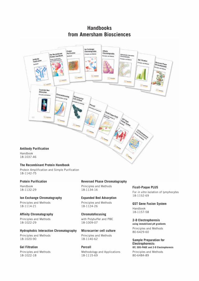
Handbooksfrom Amersham Biosciences
Ficoll-Paque PLUSFor in vitro isolation of lymphocytes18-1152-69
GST Gene Fusion SystemHandbook18-1157-58
2-D Electrophoresisusing immobilized pH gradients
Principles and Methods80-6429-60
Sample Preparation forElectrophoresis:IEF, SDS-PAGE and 2-D Electrophoresis
Principles and Methods80-6484-89
Antibody PurificationHandbook18-1037-46
The Recombinant Protein HandbookProtein Amplification and Simple Purification18-1142-75
Protein PurificationHandbook18-1132-29
Ion Exchange ChromatographyPrinciples and Methods18-1114-21
Affinity ChromatographyPrinciples and Methods18-1022-29
Hydrophobic Interaction ChromatographyPrinciples and Methods18-1020-90
Gel FiltrationPrinciples and Methods18-1022-18
Reversed Phase ChromatographyPrinciples and Methods18-1134-16
Expanded Bed AdsorptionPrinciples and Methods18-1124-26
Chromatofocusingwith Polybuffer and PBE18-1009-07
Microcarrier cell culturePrinciples and Methods18-1140-62
PercollMethodology and Applications18-1115-69
3
Dextranby A.N. de Belder

4
5
Contents
1. Introduction ............................................................................................. 7
2. Structure, physical-chemical properties and reactivity .................................. 92.1. Structure ............................................................................................................. 9
2.2. Physical-chemical properties. .............................................................................. 11
2.3. Reactivity .......................................................................................................... 14
3. Biosynthesis ........................................................................................... 153.1. Dextransucrase (Sucrose: 1,6 a-D-glucan 6-a-glucosyl transferase, ec. 2.4.1.5.) ...... 153.1.1. Purification ........................................................................................................................ 153.1.2. Properties .......................................................................................................................... 163.1.3. Donor substrate specificity .................................................................................................. 16
3.2. Mechanism ........................................................................................................ 17
3.3. Acceptor reactions .............................................................................................. 18
3.4. Branching .......................................................................................................... 20
4. Production of clinical dextran .................................................................. 214.1. Fermentation ..................................................................................................... 21
4.2. Elaboration of dextransucrase .............................................................................. 224.2.1. pH .................................................................................................................................... 224.2.2. Sucrose concentration ......................................................................................................... 224.2.3. Time ................................................................................................................................. 23
4.3. Biosynthesis of dextran ....................................................................................... 234.3.1. pH .................................................................................................................................... 234.3.2. Sucrose concentration ......................................................................................................... 234.3.3. Temperature ...................................................................................................................... 244.3.4. Calcium ............................................................................................................................ 244.3.5. Time ................................................................................................................................. 25
4.4. Clinical fractions ................................................................................................ 264.4.1. Partial acid hydrolysis ......................................................................................................... 264.4.2. Fractionation procedures ..................................................................................................... 26
4.5. Quality assurance ............................................................................................... 27
4.6. Stability in solution ............................................................................................ 28
4.7. Future developments .......................................................................................... 29
5. History of medical applications ................................................................ 315.1. Dextran 70 ........................................................................................................ 31
5.2. Dextran 40 ........................................................................................................ 32
5.3. Dextran 1 .......................................................................................................... 32
5.4. Iron-dextran ....................................................................................................... 32
5.5. Dextran sulfate ................................................................................................... 33
5.6. Diethylaminoethyl dextran (DEAE-dextran) ............................................................ 33
5.7. Perfusion solutions ............................................................................................. 33
5.8. Debrisan ............................................................................................................ 34
5.9. Hyskon .............................................................................................................. 34
6
6. General applications of dextran and its derivates ....................................... 356.1. Dextran fractions ................................................................................................ 35
6.2. Sephadex .......................................................................................................... 36
6.3. Dextran-hemoglobin preparations ......................................................................... 37
6.4. Conjugates of dextran with biologically-active substances ....................................... 37
6.5. Dextran derivates ................................................................................................ 396.5.1. Dextran sulfate ................................................................................................................... 39a. Effects on enzymes ................................................................................................................... 39b. Effects on immune response ...................................................................................................... 40c. Effects on viruses ..................................................................................................................... 40d. Toxicity .................................................................................................................................... 40e. Gene manipulation .................................................................................................................... 416.5.2. DEAE-dextran .................................................................................................................... 416.5.3. Flourescein-labelled dextrans ............................................................................................... 41
6.6. Microcarriers for cell culture ................................................................................ 42
6.7. 99mTc-Dextran ..................................................................................................... 42
7. Dextran analysis ..................................................................................... 43
8. Acknowledgements ................................................................................. 43
References ................................................................................................. 45
Ordering information ................................................................................... 60
7
1. Introduction
A brief search of the Chemical Abstracts database for dextran and dextran-relatedtitles leaves one in little doubt of the prolific interest in this area; an interest whichmay be attributed to the continuing clinical, scientific and technical importance ofdextran and its derivatives.
Currently, more than 1000 publications dealing with dextran appear each year.Reviews in this field are published with impressive regularity (1-25).
Certain limitations had to be imposed on this Review and it has thus been restrictedto dextrans of commercial interest, in particular from Leuconostroc mesenteroidesNRRL B-512(F); other dextrans and the cariogenic glucans are only referred towhere they are of particular relevance. References to dextranases will be confinedsolely to those of interest to the B-512(F) dextran.
Certain abbreviations have been employed throughout:
MWD molecular weight distribution
MW mass average molecular mass
Mn number average molecular mass
MW molecular weight
Dextran 70 a dextran fraction with MW of 70 000
DS degree of substitution

8
9
2. Structure, physical-chemical properties and reactivity
2.1. Structure
The dextran elaborated by Leuconostoc mesenteroides NRRL B-512(F) consists of an a(1�6)-linkedglucan with side chains attached to the 3-positions of the backbone glucose units. From periodate(26, 27) and methylation (28–31) analyses, the degree of branching is estimated as 5%. The value ofthe methylation analysis is that it not only identifies the (1�3) branch linkages but also quantitates them.A systematic study of this analysis in our laboratories, however, has revealed that the estimation of2,4-dimethyl-glucose by gas-liquid chromatography is subject to a rather high coefficient of variation;11-20% depending on the conditions used. The degree of branching is found to decrease on partialacid hydrolysis, although the effect is not dramatic (29).
Dextran Branching (%)
Native dextran 4.6
Dextran 80 3.8
Dextran 10 3.0
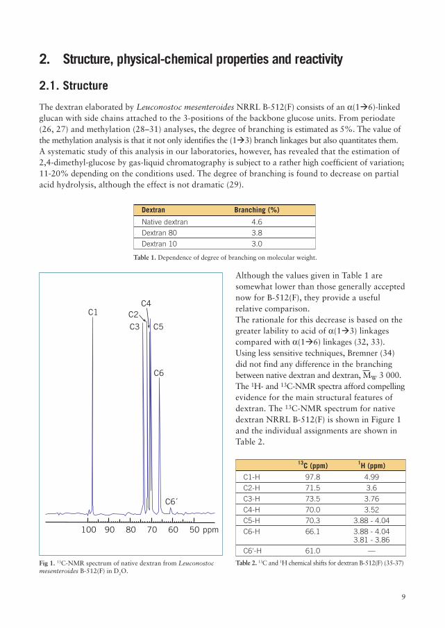
Although the values given in Table 1 aresomewhat lower than those generally acceptednow for B-512(F), they provide a usefulrelative comparison.The rationale for this decrease is based on thegreater lability to acid of a(1�3) linkagescompared with a(1�6) linkages (32, 33).Using less sensitive techniques, Bremner (34)did not find any difference in the branchingbetween native dextran and dextran, MW 3 000.The 1H- and 13C-NMR spectra afford compellingevidence for the main structural features ofdextran. The 13C-NMR spectrum for nativedextran NRRL B-512(F) is shown in Figure 1and the individual assignments are shown inTable 2.
Fig 1. 13C-NMR spectrum of native dextran from Leuconostocmesenteroides B-512(F) in D2O.
Table 1. Dependence of degree of branching on molecular weight.
100 90 80 70 60 50 ppm
C6´
C6
C5
C4
C3C2C1
13C (ppm) 1H (ppm)
C1-H 97.8 4.99
C2-H 71.5 3.6
C3-H 73.5 3.76
C4-H 70.0 3.52
C5-H 70.3 3.88 - 4.04
C6-H 66.1 3.88 - 4.043.81 - 3.86
C6'-H 61.0 —
Table 2. 13C and 1H chemical shifts for dextran B-512(F) (35-37)
10
The signal at 61 ppm, assigned to the C6 atom on the non-reducing glucose-units, is of considerableinterest as it corresponds to the branching (35-36). The downfield signal at 99.5 ppm is tentativelyassigned to the a(1�3) anomeric carbon (36). Studies in our laboratories on the determination ofthe degree of branching of clinical dextran by NMR yields a value of 4.8–5.5 %, depending on theintegrating technique employed (38).
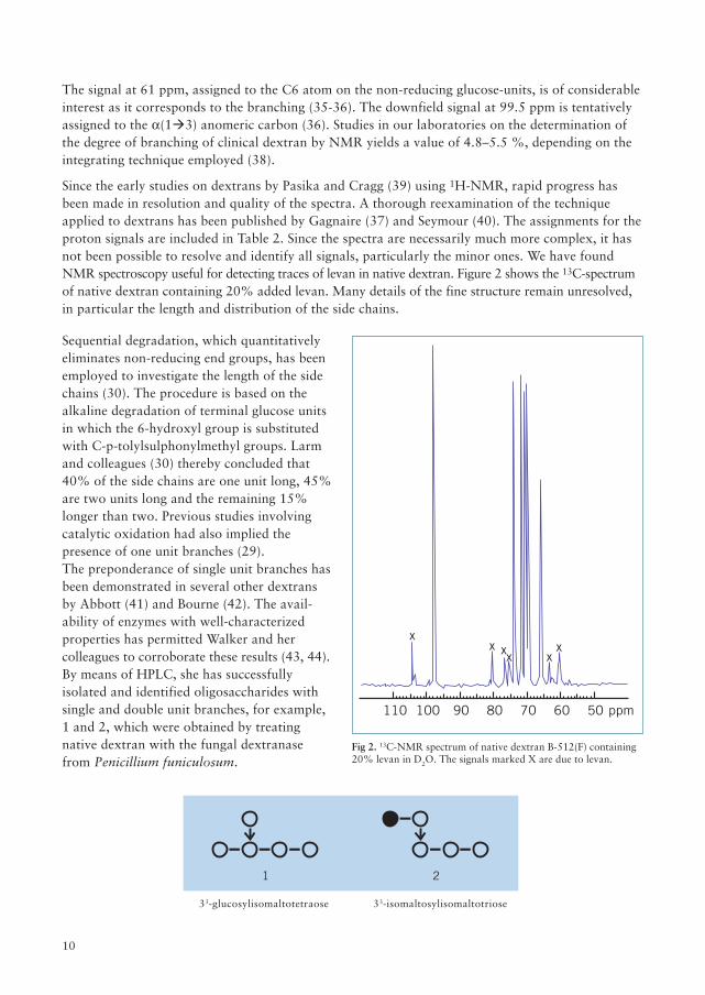
Since the early studies on dextrans by Pasika and Cragg (39) using 1H-NMR, rapid progress hasbeen made in resolution and quality of the spectra. A thorough reexamination of the techniqueapplied to dextrans has been published by Gagnaire (37) and Seymour (40). The assignments for theproton signals are included in Table 2. Since the spectra are necessarily much more complex, it hasnot been possible to resolve and identify all signals, particularly the minor ones. We have foundNMR spectroscopy useful for detecting traces of levan in native dextran. Figure 2 shows the 13C-spectrumof native dextran containing 20% added levan. Many details of the fine structure remain unresolved,in particular the length and distribution of the side chains.
Fig 2. 13C-NMR spectrum of native dextran B-512(F) containing20% levan in D2O. The signals marked X are due to levan.
33-glucosylisomaltotetraose 33-isomaltosylisomaltotriose
Sequential degradation, which quantitativelyeliminates non-reducing end groups, has beenemployed to investigate the length of the sidechains (30). The procedure is based on thealkaline degradation of terminal glucose unitsin which the 6-hydroxyl group is substitutedwith C-p-tolylsulphonylmethyl groups. Larmand colleagues (30) thereby concluded that40% of the side chains are one unit long, 45%are two units long and the remaining 15%longer than two. Previous studies involvingcatalytic oxidation had also implied thepresence of one unit branches (29).The preponderance of single unit branches hasbeen demonstrated in several other dextransby Abbott (41) and Bourne (42). The avail-ability of enzymes with well-characterizedproperties has permitted Walker and hercolleagues to corroborate these results (43, 44).By means of HPLC, she has successfullyisolated and identified oligosaccharides withsingle and double unit branches, for example,1 and 2, which were obtained by treatingnative dextran with the fungal dextranasefrom Penicillium funiculosum.
100110 90 80 70 60 50 ppm
xx xx x
x
1 2
11
A dextran-glucosidase (from Streptococcus mitis) with exo-dextranase activity surprisingly released25% glucose from native dextran B-512(F) (43). The limit dextran obtained had a MW of 20 x 106
and the branching was unchanged indicating no internal cleavage. To account for this amount ofglucose, the side chains longer than two units would need to be at least 33 units long. Our under-standing of the distribution of side chains is also imperfect. Covacevich and Richards (45) byanalysing the distribution of oligosaccharides from the hydrolysate of dextran with an endo-dextranaseconcluded that the branches were not clustered but were distributed in a relatively regular manner.These conclusions were based on a number of assumptions, one of which, that the branches are onlyone unit long, is questionable. Evidence for the presence of long branches has also been adducedfrom studies on the MW and viscosity during the biosynthesis of dextran (46). Further reference tobranching is made in the following section.
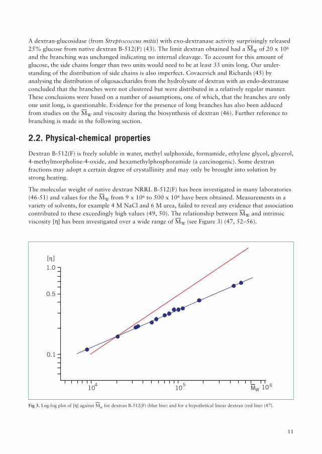
Fig 3. Log-log plot of [h] against MW for dextran B-512(F) (blue line) and for a hypothetical linear dextran (red line) (47).
2.2. Physical-chemical properties
Dextran B-512(F) is freely soluble in water, methyl sulphoxide, formamide, ethylene glycol, glycerol,4-methylmorpholine-4-oxide, and hexamethylphosphoramide (a carcinogenic). Some dextranfractions may adopt a certain degree of crystallinity and may only be brought into solution bystrong heating.
The molecular weight of native dextran NRRL B-512(F) has been investigated in many laboratories(46-51) and values for the MW from 9 x 10⁶ to 500 x 106 have been obtained. Measurements in avariety of solvents, for example 4 M NaCl and 6 M urea, failed to reveal any evidence that associationcontributed to these exceedingly high values (49, 50). The relationship between MW and intrinsicviscosity [h] has been investigated over a wide range of MW (see Figure 3) (47, 52–56).
1.0
0.5
0.1
106MW104
105
[ ]
12
The deviation from linearity at higher MW is ascribed to the increase in branching and thepolymolecularity of the fractions used. Granath (52) obtained the following relationship for dextranin the clinical range.
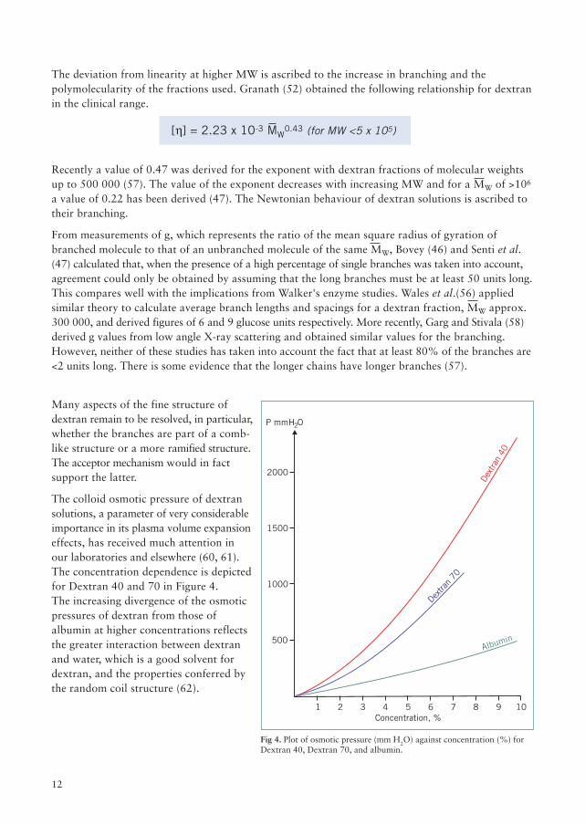
Fig 4. Plot of osmotic pressure (mm H2O) against concentration (%) forDextran 40, Dextran 70, and albumin.
Recently a value of 0.47 was derived for the exponent with dextran fractions of molecular weightsup to 500 000 (57). The value of the exponent decreases with increasing MW and for a MW of >106
a value of 0.22 has been derived (47). The Newtonian behaviour of dextran solutions is ascribed totheir branching.
From measurements of g, which represents the ratio of the mean square radius of gyration ofbranched molecule to that of an unbranched molecule of the same MW, Bovey (46) and Senti et al.(47) calculated that, when the presence of a high percentage of single branches was taken into account,agreement could only be obtained by assuming that the long branches must be at least 50 units long.This compares well with the implications from Walker's enzyme studies. Wales et al.(56) appliedsimilar theory to calculate average branch lengths and spacings for a dextran fraction, MW approx.300 000, and derived figures of 6 and 9 glucose units respectively. More recently, Garg and Stivala (58)derived g values from low angle X-ray scattering and obtained similar values for the branching.However, neither of these studies has taken into account the fact that at least 80% of the branches are<2 units long. There is some evidence that the longer chains have longer branches (57).
[h] = 2.23 x 10⁻³ MW0.43 (for MW <5 x 10⁵)
Many aspects of the fine structure ofdextran remain to be resolved, in particular,whether the branches are part of a comb-like structure or a more ramified structure.The acceptor mechanism would in factsupport the latter.
The colloid osmotic pressure of dextransolutions, a parameter of very considerableimportance in its plasma volume expansioneffects, has received much attention inour laboratories and elsewhere (60, 61).The concentration dependence is depictedfor Dextran 40 and 70 in Figure 4.The increasing divergence of the osmoticpressures of dextran from those ofalbumin at higher concentrations reflectsthe greater interaction between dextranand water, which is a good solvent fordextran, and the properties conferred bythe random coil structure (62).
Concentration, %
500
1 2 3 4 5 6 7 8 9 10
1000
1500
2000De
xtra
n 40
Dextra
n 70
Albumin
P mmH O2
13
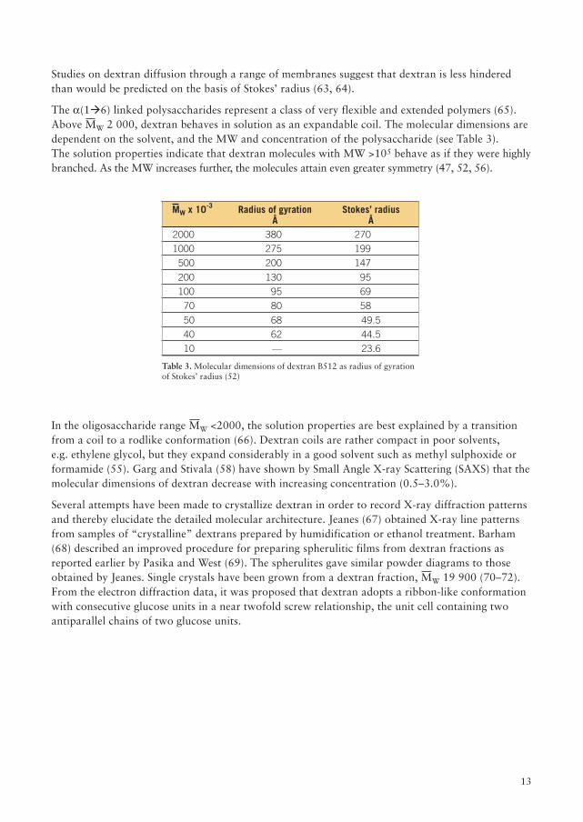
Studies on dextran diffusion through a range of membranes suggest that dextran is less hinderedthan would be predicted on the basis of Stokes’ radius (63, 64).
The a(1�6) linked polysaccharides represent a class of very flexible and extended polymers (65).Above MW 2 000, dextran behaves in solution as an expandable coil. The molecular dimensions aredependent on the solvent, and the MW and concentration of the polysaccharide (see Table 3).The solution properties indicate that dextran molecules with MW >105 behave as if they were highlybranched. As the MW increases further, the molecules attain even greater symmetry (47, 52, 56).
MW x 10-3 Radius of gyration Stokes’ radiusÅ Å
2000 380 270
1000 275 199
500 200 147
200 130 95
100 95 69
70 80 58
50 68 49.5
40 62 44.5
10 — 23.6
Table 3. Molecular dimensions of dextran B512 as radius of gyrationof Stokes’ radius (52)
In the oligosaccharide range MW <2000, the solution properties are best explained by a transitionfrom a coil to a rodlike conformation (66). Dextran coils are rather compact in poor solvents,e.g. ethylene glycol, but they expand considerably in a good solvent such as methyl sulphoxide orformamide (55). Garg and Stivala (58) have shown by Small Angle X-ray Scattering (SAXS) that themolecular dimensions of dextran decrease with increasing concentration (0.5–3.0%).
Several attempts have been made to crystallize dextran in order to record X-ray diffraction patternsand thereby elucidate the detailed molecular architecture. Jeanes (67) obtained X-ray line patternsfrom samples of “crystalline” dextrans prepared by humidification or ethanol treatment. Barham(68) described an improved procedure for preparing spherulitic films from dextran fractions asreported earlier by Pasika and West (69). The spherulites gave similar powder diagrams to thoseobtained by Jeanes. Single crystals have been grown from a dextran fraction, MW 19 900 (70–72).From the electron diffraction data, it was proposed that dextran adopts a ribbon-like conformationwith consecutive glucose units in a near twofold screw relationship, the unit cell containing twoantiparallel chains of two glucose units.
14
2.3. Reactivity
The reactivity of dextran involves primarily a study of the relative reactivities of the secondary,equatorially orientated hydroxyl groups, HO-2, HO-3, and HO-4. A small percentage of thehydroxyl groups in dextran are primary (approx. 1.5%), although this figure increases slightly atlow molecular weights owing to the contribution from the non-reducing end-groups.
Attempts to tritylate the primary hydroxyls selectively in methyl sulphoxide/pyridine have beenreported. Although Rees and coworkers (73) found that some tritylation of secondary hydroxylscould not be avoided, the reaction was used successfully by Larm (30) for studying the length ofthe branches.
Studies on the partial methylation (74) revealed the following relative reactivities k2:k3:k4 = 8:1:3.5.Good agreement between the experimental and theoretical distribution of methyl ethers was onlyobtained when allowance was made for the enhanced reactivity at HO-3 as a result of substitutionat HO-2 or HO-4. Thus, for example, k3 is enhanced by a factor of 5.2 by substitution at both HO-2and HO-4 (74).
As with other glucans, the reactivity at HO-2 towards alkylating agents is higher than at HO-3 orHO-4. This is rationalized in terms of the higher acidity of the HO-2 due to its proximity to theanomeric centre (75). An investigation of the partial methylation of some model compounds, forexample, methyl a- and b-D glucopyranosides and their 6-O-substituted derivatives, indicated thatno special effects need be invoked in the polymers to explain the low reactivity at HO-3 (76).When HO-2 and HO-4 are ionized, the reactivity at HO-3 is depressed. However, substitution atHO-2 or HO-4 abolishes this effect with subsequent increase in reactivity at HO-3 (74).Some caution in applying these generalizations is needed, as the base strength may affect the relativeand absolute reactivities of the hydroxyls (77, 78).
The relative reactivities of the hydroxyl groups towards ethylene oxide closely follows those formethylation (79). At higher degrees of substitution (DS), however, the pattern of substitutionbecomes complex owing to the introduction of primary hydroxyl sites.
Acylations may differ from alkylations in that they may be subject to thermodynamic control owingto migration of the substituents. A study of the partial acetylation of dextran with acetic anhydride/pyridine (80) revealed that k2>k3=k4. This order of reactivity is similar to that shown by methyl b-D-glucopyranoside. When, however, the acylation was conducted in aqueous alkali, the reactivitieswere virtually identical, indicating rearrangement.
An investigation of the distribution of sulfate groups in partially sulfated dextran, using dextran N-4 with asimilar structure to B-512(F), showed that HO-2 was again highest with k2:k3:k4 = 1.6:1.06:1.0 (81). Thepercentage of di-substituted glucose units was surprisingly high even at low DS.
Carbonyl groups may be introduced into dextran by Fenton's reagent (82), methyl sulphoxide/aceticanhydride reagent (83), or by means of aqueous bromine at pH 7 (84). With the latter reagent,oxidation appears to occur mainly at C-2 (21%) and at C-4 (25 %). Similar patterns are obtainedwith methyl a-D-glucopyranoside (85). When the oxidation is performed in the presence of borate,less oxidative cleavage to dicarbonyl moieties results (86).
15
3. Biosynthesis
3.1. Dextransucrase(Sucrose: 1,6 a-D-glucan 6-a-glucosyl transferase, EC. 2.4.1.5.)
3.1.1. Purification
Although the slime-forming properties of filtered extracts of Bacillus mesentericus were first notedby Beijerinck (87) in 1910, the evidence published by Hehre (88, 89) in 1939 and 1941 provided amore rigorous proof of the dextran synthesizing activity present in Leuconostoc extracts. During thenext two decades, a surge of activity led to a rapid expansion in our knowledge of the properties ofdextransucrases. Although a number of refinements in the isolation of the enzyme were made duringthese years, the first attempts to purify the B-512(F) enzyme were described by Ebert and Schenk(90).
Their procedure entailed methanol precipitations, calcium phosphate chromatography and ammoniumsulphate precipitation and gave high activities. Values of 2000 units per milligram (U/mg) residualdextran were reported. The purification of the enzyme was re-examined by Robyt and Walseth (91).Removal of the bound dextran by treatment with an endo-dextranase constituted an important andnovel step in the purification. The enzyme concentrate displayed a specific activity of 53 U/mg (33%recovery) following the final ultrafiltration. Examination of the purified enzyme by PAGE revealedonly two bands, both of which possessed dextransucrase activity. The faster band was assigned to themonomer and the slower to aggregates. On standing, the purified enzyme aggregates (91, 92).Levansucrase and invertase for example were absent from the preparation. The impure enzyme isnot precipitated by ammonium sulphate even at 80% (w/v) (90). Itaya (93) reported that the enzymecould be precipitated directly from the culture by ammonium sulphate provided ovalbumin was added.
The existence of multiple forms of dextransucrase from B-512(F) was established by GPC andelectrophoresis. A component giving a single band on PAGE and isoelectric focusing was isolatedand identified as a dextransucrase. It yielded a dextran very similar to that obtained from a cellculture (92). The purified enzyme rapidly lost activity at 4 °C and even at -15 °C its activity decreasedby 60% over 20 days. The enzyme may be stabilized, however, by addition of dextran but a minimumconcentration of 4 mg/ml is required. The disparity between reports in the literature on the stabilityof dextransucrase preparations is, presumably, attributable to variations in the residual dextranconcentrations. Lyophilized preparations of the purified enzyme appear to be stable indefinitely (92).For the purification of Leuconostoc dextransucrases, a preliminary treatment with dextranase maybe followed by one of several techniques. Polyethyleneglycols (MW from 400–6000) at concentrationsfrom 5% to 40%, respectively, effectively precipitate the enzyme (94).Gel permeation chromatography on Bio-gel A5m or Sepharose™ 6B (94–96) and ion-exchangechromatography on a DEAE-type gel (93–97) have frequently been used. Hydrophobic chromatographyusing O-(phenoxyacetyl)cellulose was found to be advantageous with the B-1355 dextransucrases (94).McCabe and colleagues (98) found that dextransucrase displayed an affinity for Sephadex™. Thistechnique has also been applied Leuconostoc enzymes (99). The purified enzyme, however, does notshown any affinity for Sephadex™ G-100 (92).
16
3.1.2. Properties
The highly purified dextransucrase isolated by Robyt and Walseth (91) appears to be a glycoproteinwith mannose as the preponderant sugar. Only trace amounts of glucose were detected.
The importance of calcium ions for the activity of the enzyme had already been noted by Brock Neelyand Hallmark in 1961 (100). Robyt and Walseth (91) found that the activity of the enzyme whichhad been deactivated by incubation with EDTA could only be fully restored by calcium ions. Theysuggested that the enzyme was a calcium metalloprotein. Similar calcium dependence has beennoted for dextransucrase from Leuconostoc mesenteroides IAM 1046 (93).
Studies on the nature of the catalytic site of the enzyme suggest that polyisophenol phosphates areinvolved (96, 101, 102).
The value (284 000) for the MW of the B-512(F) dextransucrase (90) is almost certainly too high.The preparations used may not have been entirely free from bound dextran and may also have beenaggregated. In a recent study of the B-512(F) enzyme, a value for the monomeric enzyme of 64 000by SDS gel electrophoresis was reported (92). The MW's reported for the Streptococcal enzymeswere 94 000 for S. mutans 6715 (103) and 102 000 for S. sanguis 10558 (96) although forS. mutans HS-6, a value of 170 000 has been quoted (95).
The activity of the enzyme depends, inter alia, on pH. The optimal pH extends over a rather narrowrange, 5.0-5.3. At this pH, the enzyme is also most stable and a partly purified sample was found toretain its activity for at least 24 hours at 2 °C (18, 104, 105).
3.1.3. Donor substrate specificity
Dextransucrase is in many respects a remarkably versatile enzyme. A wide range of products can beformed depending on the nature and concentrations of the donor and acceptor substrates.
It was believed for a long time that sucrose was the only natural substrate for dextran sucrase.However, during the past years, a number of other donors, both natural and synthetic, have beenrecognized. Thus Genghoff and Hehre (106) found that a-D-glucopyranosyl fluoride yields a highmolecular weight dextran in the presence of dextransucrase from Leuconostoc mesenteroides B-512(F)or Streptococcus DS var. Indeed, the kinetic parameters for the reactions were virtually identical tothose for sucrose (107). These findings are of particular interest as they indicate that the synthesisoperates via glucosyl transfer in accordance with the generally agreed mechanism.
Lactulosucrose (O-a-D-galactopyranosyl-(1�4)-O-a-D-fructofuranosyl-(2�1)-a-D-glycopyronoside)is reported to give dextran when incubated with various Leuconostoc dextran sucrases (108).
Recently, Binder and Robyt (109) reported that p-nitrophenyl-a-D-glucopyranoside acted as adonor substrate for dextransucrase (purified preparation) form Leuconostoc mesenteroides B-512(F)and Streptococcus mutans 6715. The products were high MW dextrans and nitrophenylisomaltodextrin glycosides. The rate to transfer was, however, very much less than that for sucrose.
Evidence has been adduced that C-3, C-4 and C-6 are important sites for the binding of sucrose todextransucrase (110).
17
In 1960, Tsuchiya (13) observed that when dextran and dextransucrase were incubated in theabsence of sucrose with suitable acceptors, for example glucose and maltose, the correspondingoligosaccharides were produced. He concluded that even dextran could act as a donor and this hasrecently been reaffirmed by Binder and coworkers (99). By means of a chromatographic study using
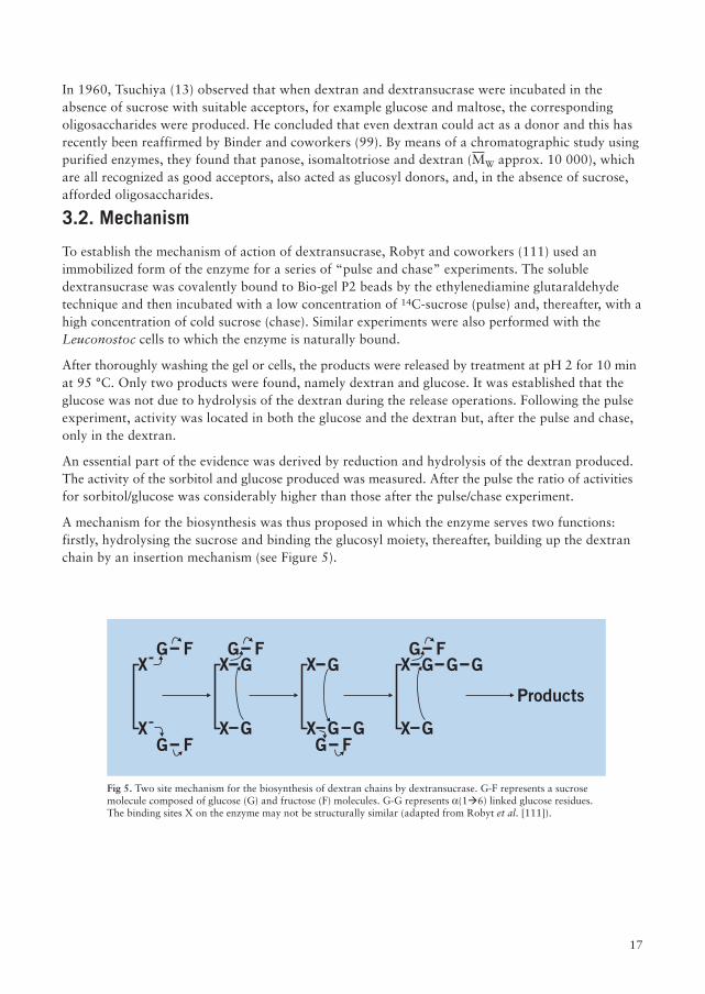
Fig 5. Two site mechanism for the biosynthesis of dextran chains by dextransucrase. G-F represents a sucrosemolecule composed of glucose (G) and fructose (F) molecules. G-G represents a(1�6) linked glucose residues.The binding sites X on the enzyme may not be structurally similar (adapted from Robyt et al. [111]).
purified enzymes, they found that panose, isomaltotriose and dextran (MW approx. 10 000), whichare all recognized as good acceptors, also acted as glucosyl donors, and, in the absence of sucrose,afforded oligosaccharides.
3.2. Mechanism
To establish the mechanism of action of dextransucrase, Robyt and coworkers (111) used animmobilized form of the enzyme for a series of “pulse and chase” experiments. The solubledextransucrase was covalently bound to Bio-gel P2 beads by the ethylenediamine glutaraldehydetechnique and then incubated with a low concentration of 14C-sucrose (pulse) and, thereafter, with ahigh concentration of cold sucrose (chase). Similar experiments were also performed with theLeuconostoc cells to which the enzyme is naturally bound.
After thoroughly washing the gel or cells, the products were released by treatment at pH 2 for 10 minat 95 °C. Only two products were found, namely dextran and glucose. It was established that theglucose was not due to hydrolysis of the dextran during the release operations. Following the pulseexperiment, activity was located in both the glucose and the dextran but, after the pulse and chase,only in the dextran.
An essential part of the evidence was derived by reduction and hydrolysis of the dextran produced.The activity of the sorbitol and glucose produced was measured. After the pulse the ratio of activitiesfor sorbitol/glucose was considerably higher than those after the pulse/chase experiment.
A mechanism for the biosynthesis was thus proposed in which the enzyme serves two functions:firstly, hydrolysing the sucrose and binding the glucosyl moiety, thereafter, building up the dextranchain by an insertion mechanism (see Figure 5).
X
X
G F
G F G F
G FX G
X G
G FX G
X G
X G
X G G
G G
Products
18
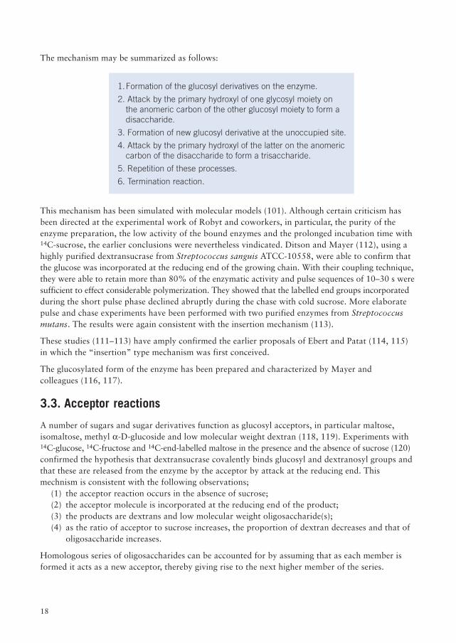
The mechanism may be summarized as follows:
1.Formation of the glucosyl derivatives on the enzyme.
2. Attack by the primary hydroxyl of one glycosyl moiety onthe anomeric carbon of the other glucosyl moiety to form adisaccharide.
3. Formation of new glucosyl derivative at the unoccupied site.
4. Attack by the primary hydroxyl of the latter on the anomericcarbon of the disaccharide to form a trisaccharide.
5. Repetition of these processes.
6. Termination reaction.
This mechanism has been simulated with molecular models (101). Although certain criticism hasbeen directed at the experimental work of Robyt and coworkers, in particular, the purity of theenzyme preparation, the low activity of the bound enzymes and the prolonged incubation time with14C-sucrose, the earlier conclusions were nevertheless vindicated. Ditson and Mayer (112), using ahighly purified dextransucrase from Streptococcus sanguis ATCC-10558, were able to confirm thatthe glucose was incorporated at the reducing end of the growing chain. With their coupling technique,they were able to retain more than 80% of the enzymatic activity and pulse sequences of 10–30 s weresufficient to effect considerable polymerization. They showed that the labelled end groups incorporatedduring the short pulse phase declined abruptly during the chase with cold sucrose. More elaboratepulse and chase experiments have been performed with two purified enzymes from Streptococcusmutans. The results were again consistent with the insertion mechanism (113).
These studies (111–113) have amply confirmed the earlier proposals of Ebert and Patat (114, 115)in which the “insertion” type mechanism was first conceived.
The glucosylated form of the enzyme has been prepared and characterized by Mayer andcolleagues (116, 117).
3.3. Acceptor reactions
A number of sugars and sugar derivatives function as glucosyl acceptors, in particular maltose,isomaltose, methyl a-D-glucoside and low molecular weight dextran (118, 119). Experiments with14C-glucose, 14C-fructose and 14C-end-labelled maltose in the presence and the absence of sucrose (120)confirmed the hypothesis that dextransucrase covalently binds glucosyl and dextranosyl groups andthat these are released from the enzyme by the acceptor by attack at the reducing end. Thismechnism is consistent with the following observations;
(1) the acceptor reaction occurs in the absence of sucrose;(2) the acceptor molecule is incorporated at the reducing end of the product;(3) the products are dextrans and low molecular weight oligosaccharide(s);(4) as the ratio of acceptor to sucrose increases, the proportion of dextran decreases and that of
oligosaccharide increases.
Homologous series of oligosaccharides can be accounted for by assuming that as each member isformed it acts as a new acceptor, thereby giving rise to the next higher member of the series.
19
A more detailed study of the acceptor activity of various sugars was undertaken by Mayer andcoworkers (121). They studied the time course by the glucosyl transfer to these acceptors, andfound that the appearance of the homologues was consistent with the sequential transfer of glucosylunits to the acceptor.
Robyt and Eklund (101) listed 26 known acceptors of varying activity and recently explored thequantitative aspects of acceptor activity in the dextransucrase system. The products were dextranand in certain cases a homologous series of isomalto-oligosaccharides. Glucose, being the product ofthe acceptor reaction of water, was also found in small amounts. Two acceptor products from fructosewere always present in small proportions; leucrose (O-a-D-glucopyranosyl-(1�5)-D-glucopyranse)approx. 2% and isomaltulose (O-a-D-glucopyranosyl-(1�6)-fructofuranose) approx. 1%.The products of an acceptor reaction will, however, depend, inter alia, on the ratio of acceptor tosubstrate (122).
Walker’s findings (123) can now be rationalized in terms of an acceptor mechanism. She found thatwhen 14C-sucrose and an excess of isomalto-oligosaccharides were incubated with dextransucrase,the main product was the next higher homologue with the radioactive label attached to the non-reducing end.
However, the issue has recently become more complicated by the observations that dextran and anumber of D-glucosyl oligosaccharides (e.g. isomaltotriose, panose) may in fact serve both asglycosyl donors and acceptors for dextransucrase (99). Interestingly, they also established that theenzyme could transfer single glycosyl units from dextran to an acceptor molecule.
More recently, Luzio and Mayer (124) have stressed the dependence of the acceptor reaction onconcentration. They reported that at low sucrose concentrations, 0.05 mM to 0.5 mM, the acceptorreaction of water became significant and the hydrolysis of the sucrose and fructose preponderatedover dextran synthesis.
The fact that hydrolysed dextran serves as an efficient acceptor had already been established in the50's (125, 126). Hehre (125) found that a supplement of 1 mg/ml of low MW dextran markedlylowered the MW of the dextran produced by dextransucrase. This finding was developed byHellman and colleagues (126) at Peoria who used a 2% supplement of dextran, MW 17 600, andobtained a 33 % yield of a dextran in the clinical range. They also found that the yields increased asthe MW of the added dextran decreased.
Using 14C-labelled dextran, Mayer and his colleagues (121) found that the MW of the acceptordextran increased significantly during the incubation with dextransucrase, thereby confirming itsrole as an acceptor. Furthermore, the kinetic data identified hydrolyzed dextran (MW approx. 10 000)as one of the most potent acceptors with a Km lower than that of maltose and isomaltose and V 4–5times higher than any other sugar. Their results thus corroborated the earlier findings of Robyt andCorrigan (127). The addition of dextran (MW approx. 70 000) to a purified, dextran-free enzymecaused a 5-fold increase in the activity (129). From kinetic data, it was concluded that the bindingof dextran to the enzyme also increases the affinity of the enzyme for sucrose.
20
3.4. Branching
Attempts to rationalize the branching activities of dextransucrase are still hampered by an incompleteknowledge of branching in native dextran. Ebert and his colleagues (15, 115, 128) had originallyproposed a branching mechanism based on an acceptor reaction whereby growing dextran chains(or oligosaccharide chains) are transferred consecutively to the acceptor dextran by the enzyme,thereby giving rise to a branched dextran. Their hypothesis was based on a study of the productsobtained by adding a radioactive-labelled acceptor dextran of MW approx. 25 000, to purifiedenzyme and sucrose (5%). Enzymes from B-512(F), B-1299 and B-1307 were studied. The MWD ofthe products was examined (although not quantitatively) and the specific activity was also investigated.The results clearly demonstrated that the dextran produced contained one acceptor molecule.However the nature of the new linkage was not established.
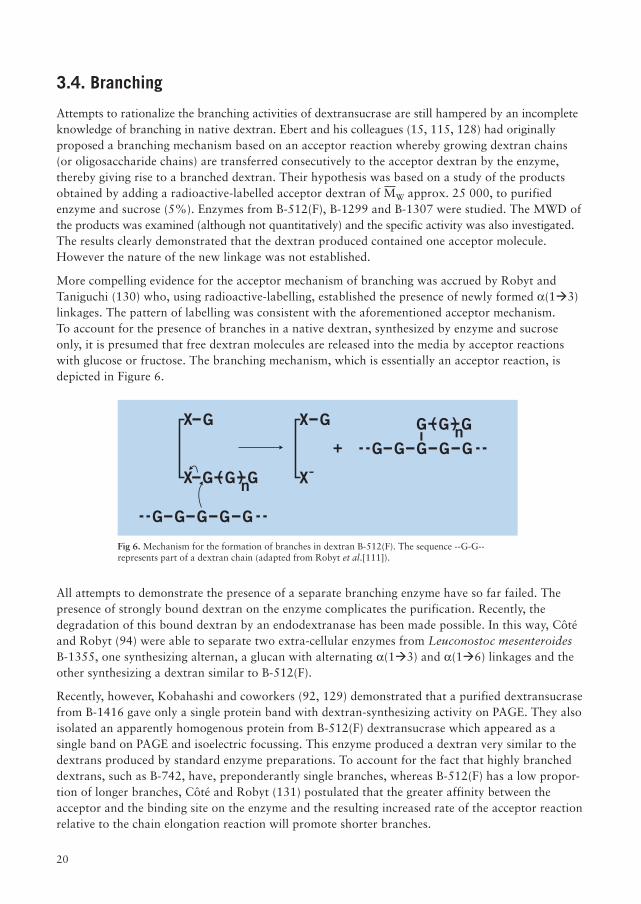
More compelling evidence for the acceptor mechanism of branching was accrued by Robyt andTaniguchi (130) who, using radioactive-labelling, established the presence of newly formed a(1�3)linkages. The pattern of labelling was consistent with the aforementioned acceptor mechanism.To account for the presence of branches in a native dextran, synthesized by enzyme and sucroseonly, it is presumed that free dextran molecules are released into the media by acceptor reactionswith glucose or fructose. The branching mechanism, which is essentially an acceptor reaction, isdepicted in Figure 6.
Fig 6. Mechanism for the formation of branches in dextran B-512(F). The sequence --G-G--represents part of a dextran chain (adapted from Robyt et al.[111]).
All attempts to demonstrate the presence of a separate branching enzyme have so far failed. Thepresence of strongly bound dextran on the enzyme complicates the purification. Recently, thedegradation of this bound dextran by an endodextranase has been made possible. In this way, Côtéand Robyt (94) were able to separate two extra-cellular enzymes from Leuconostoc mesenteroidesB-1355, one synthesizing alternan, a glucan with alternating a(1�3) and a(1�6) linkages and theother synthesizing a dextran similar to B-512(F).
Recently, however, Kobahashi and coworkers (92, 129) demonstrated that a purified dextransucrasefrom B-1416 gave only a single protein band with dextran-synthesizing activity on PAGE. They alsoisolated an apparently homogenous protein from B-512(F) dextransucrase which appeared as asingle band on PAGE and isoelectric focussing. This enzyme produced a dextran very similar to thedextrans produced by standard enzyme preparations. To account for the fact that highly brancheddextrans, such as B-742, have, preponderantly single branches, whereas B-512(F) has a low propor-tion of longer branches, Côté and Robyt (131) postulated that the greater affinity between theacceptor and the binding site on the enzyme and the resulting increased rate of the acceptor reactionrelative to the chain elongation reaction will promote shorter branches.
X G
X n( )G G G
n( )G G G
GG G G G
GG G G G
X
X
G
+
21
4. Production of clinical and technical dextran
Dextran for clinical and technical products is produced in most developed countries throughout theworld. Reliable figures for the annual world production of dextran are not available but an estimatebased on the units of clinical dextran consumed in some countries would lead to a figure in excessof 500 metric tons.
In the West, most producers use the Leuconostoc mesenteroides NRRL B-512(F) or B-512 strain forthe fermentation. In other parts of the world, alternative strains appear to be used (132, 133).
Most major producers of dextran employ a process based on the batchwise culture of Leuconostocin the presence of sucrose. The viscous culture fluid is then precipitated in ethanol or methanol,whereafter the native dextran obtained is hydrolyzed in dilute acid and the desired dextran is isolatedby fractionation. Although the present state of the art offers alternative methods of producingdefined fractions, most producers are still operating a procedure introduced about 35 years ago.In introducing any change, a producer must be convinced that, not only must the new process bemore efficient in man-power and materials, but the final product must conform in every respectwith the medical requirements for safety and efficacy.
The organism, Leuconostoc mesenteroides NRRL B-512(F), is a member of the Lactobacillaceaefamily, genus Leuconostoc and species mesenteroides (134). The organism produces spherical orovoid cells and classifies as a gram-positive facultative anaerobe. Apart from dextran and lactic acid, itproduces, inter alia, carbon dioxide, ethanol, mannitol and acetic acid.
4.1. Fermentation
Although many sugars, for example, glucose invert sugar and maltose, will serve as energy sourcesfor the growth of the bacteria, only sucrose serves to induce dextransucrase production (89, 104,135–137). Thus the culture media are necessarily based on sucrose supplemented with variousnutritional requirements. Nicotinic acid, thiamine, pantothenic acid have been found essential forall Leuconostoc and cystine, glutamic acid, isoleucine and valine for Leuconostoc mesenteroides inparticular (138–141). In practice, these requirements are satisfied by yeast extracts, corn steepliquors, acid hydrolyzed casein or malt extracts generally with the addition of peptone or tryptonebroth (104, 142–145). Jeanes (142) has described the laboratory production of dextran in excellentdetail. The dextrans obtained from cultures using yeast extract, malt extract or liver extract respectively,were similar (145). It is worthy of note that even at concentrations of nutrients one quarter of thosegenerally employed in the fermentations, the yields of dextran were unchanged although longertimes were required (146).
In addition to the presence of various cations, phosphate salts (e.g. sodium dihydrogen phosphate orhydrogen phosphate) at 0.5% are added. For 2% corn steep liquors, Tsuchiya and coworkers (104)found 3.5% potassium dihydrogen phosphate optimal. However, biomass production at 0.1% and1% phosphate were found to be identical. Furthermore the decay in dextransucrase activity between12 and 24 hours was much less with 0.1% phosphate (147).
In the laboratory, shaken cultures are claimed to give consistently higher yields than still cultures (104).
22
4.2. Elaboration of dextransucrase
The factors influencing the elaboration of the enzyme have provided a fertile area of study.
4.2.1. pH
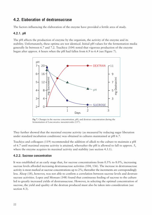
The pH affects the production of enzyme by the organism, the activity of the enzyme and itsstability. Unfortunately, these optima are not identical. Initial pH values for the fermentation mediagenerally lie between 6.7 and 7.2. Tsuchiya (104) noted that vigorous production of the enzymebegan after approx. 6 hours when the pH had fallen from 6.9 to 6.4 (see Figure 7).
They further showed that the maximal enzyme activity (as measured by reducing sugar liberationunder standard incubation conditions) was obtained in cultures maintained at pH 6.7.
Tsuchiya and colleagues (119) recommended the addition of alkali to the culture to maintain a pHof 6.7 until maximal enzyme activity is attained, whereafter the pH is allowed to fall to approx. 5,where the enzyme acquires its maximal activity and stability (see section 4.3.1).
4.2.2. Sucrose concentration
It was established at an early stage that, for sucrose concentrations from 0.5% to 8.0%, increasingsucrose levels afforded increasing dextransucrase activities (104, 136). The increase in dextransucraseactivity is most marked at sucrose concentrations up to 2%; thereafter the increments are correspondinglyless. Alsop (18), however, was not able to confirm a correlation between sucrose levels and dextran-sucrase activities. Lopez and Monsan (148) found that continuous feeding of sucrose to the cultureled to greatly increased yields of dextransucrase. However, in selecting the optimal concentration ofsucrose, the yield and quality of the dextran produced must also be taken into consideration (seesection 4.3).
Fig 7. Changes in the sucrose concentration, pH, and dextran concentration during thefermentation of Leuconostoc mesenteroides (137).
Days
pH
pH
21
3
3
9
5
11
2
3
4
5
6
7
0
4
8
12
16
20
7
SUCROSE
DEXTRAN
mg
Dex
tran
/ml
mg
Suc
rose
/ml
23
4.2.3. Time
Maximum yields of dextransucrase are recorded approx. 6-8 hours after the start of the fermenta-tion (104, 105, 147, 148). Thereafter, the instability of the enzyme of pH >6.2 will contribute to adecline in activity.
4.3. Biosynthesis of dextran
4.3.1. pH
The dextran synthesizing activity of the enzyme exhibits a maximum at pH 5.2, which is consider-ably lower than that for the optimal elaboration of the enzyme in culture (104, 119, 149–151). It is,perhaps, fortuitous that the enzyme also exhibits maximum stability within the pH range 5–6.5(105, 119, 149, 151-153). The stability of the enzyme is, however, dependent on the presence ofother substances, of which high molecular dextran acts as a stabilizer par excellence (154).
As the fermentation proceeds, the pH falls from the initial value, 7.2, to approx. 5 within 20 hoursowing to the liberation of organic acids, thereby, creating an environment favourable for thesynthesis of dextran. Although the periodic neutralization (pH 6.7) of the culture with 5–10 Nsodium hydroxide in order to increase dextransucrase levels has been recommended, there is littleevidence that this also leads to improved yields of high quality dextran (104, 135, 143, 155). HighpH (8) and low temperature (20 °C) appear to favour the formation of levan (3).
4.3.2. Sucrose concentration
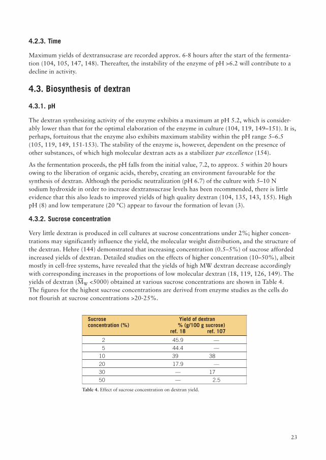
Very little dextran is produced in cell cultures at sucrose concentrations under 2%; higher concen-trations may significantly influence the yield, the molecular weight distribution, and the structure ofthe dextran. Hehre (144) demonstrated that increasing concentration (0.5–5%) of sucrose affordedincreased yields of dextran. Detailed studies on the effects of higher concentration (10–50%), albeitmostly in cell-free systems, have revealed that the yields of high MW dextran decrease accordinglywith corresponding increases in the proportions of low molecular dextran (18, 119, 126, 149). Theyields of dextran (MW <5000) obtained at various sucrose concentrations are shown in Table 4.The figures for the highest sucrose concentrations are derived from enzyme studies as the cells donot flourish at sucrose concentrations >20-25%.
Sucrose Yield of dextranconcentration (%) % (g/100 g sucrose)
ref. 18 ref. 107
2 45.9 —
5 44.4 —
10 39 38
20 17.9 —
30 — 17
50 — 2.5
Table 4. Effect of sucrose concentration on dextran yield.
24
The remaining material consists of mono- and oligo-saccharides and low MW dextran. Precise dataon the molecular weight distributions of these fractions is lacking. A further complicating factor isthe observation that the branching ratio decreases with increasing sucrose concentration. Thus, at asucrose concentration of 30%, the ratio of (1�6) links to non-(1�6) links was 29 whereas at 5%sucrose, the value was 8.5 (156). These values were obtained by periodate oxidation and have notbeen confirmed by modern techniques.
4.3.3. Temperature
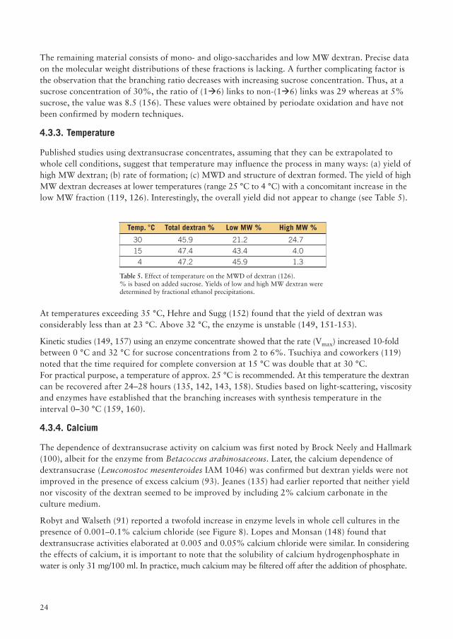
Published studies using dextransucrase concentrates, assuming that they can be extrapolated towhole cell conditions, suggest that temperature may influence the process in many ways: (a) yield ofhigh MW dextran; (b) rate of formation; (c) MWD and structure of dextran formed. The yield of highMW dextran decreases at lower temperatures (range 25 °C to 4 °C) with a concomitant increase in thelow MW fraction (119, 126). Interestingly, the overall yield did not appear to change (see Table 5).
At temperatures exceeding 35 °C, Hehre and Sugg (152) found that the yield of dextran wasconsiderably less than at 23 °C. Above 32 °C, the enzyme is unstable (149, 151-153).
Kinetic studies (149, 157) using an enzyme concentrate showed that the rate (Vmax) increased 10-foldbetween 0 °C and 32 °C for sucrose concentrations from 2 to 6%. Tsuchiya and coworkers (119)noted that the time required for complete conversion at 15 °C was double that at 30 °C.For practical purpose, a temperature of approx. 25 °C is recommended. At this temperature the dextrancan be recovered after 24–28 hours (135, 142, 143, 158). Studies based on light-scattering, viscosityand enzymes have established that the branching increases with synthesis temperature in theinterval 0–30 °C (159, 160).
4.3.4. Calcium
The dependence of dextransucrase activity on calcium was first noted by Brock Neely and Hallmark(100), albeit for the enzyme from Betacoccus arabinosaceous. Later, the calcium dependence ofdextransucrase (Leuconostoc mesenteroides IAM 1046) was confirmed but dextran yields were notimproved in the presence of excess calcium (93). Jeanes (135) had earlier reported that neither yieldnor viscosity of the dextran seemed to be improved by including 2% calcium carbonate in theculture medium.
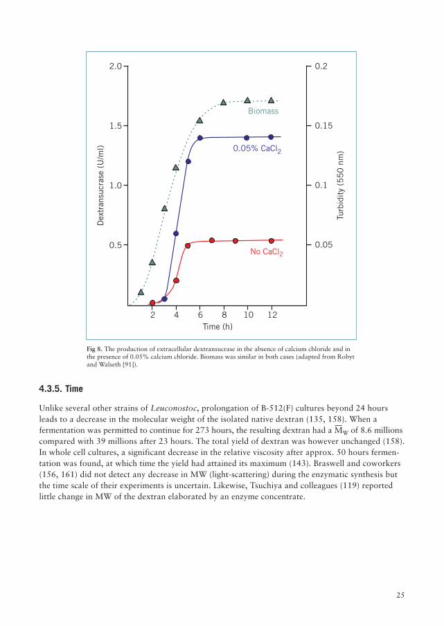
Robyt and Walseth (91) reported a twofold increase in enzyme levels in whole cell cultures in thepresence of 0.001–0.1% calcium chloride (see Figure 8). Lopes and Monsan (148) found thatdextransucrase activities elaborated at 0.005 and 0.05% calcium chloride were similar. In consideringthe effects of calcium, it is important to note that the solubility of calcium hydrogenphosphate inwater is only 31 mg/100 ml. In practice, much calcium may be filtered off after the addition of phosphate.
Temp. °C Total dextran % Low MW % High MW %
30 45.9 21.2 24.7
15 47.4 43.4 4.0
4 47.2 45.9 1.3
Table 5. Effect of temperature on the MWD of dextran (126).% is based on added sucrose. Yields of low and high MW dextran weredetermined by fractional ethanol precipitations.
25
4.3.5. Time
Unlike several other strains of Leuconostoc, prolongation of B-512(F) cultures beyond 24 hoursleads to a decrease in the molecular weight of the isolated native dextran (135, 158). When afermentation was permitted to continue for 273 hours, the resulting dextran had a MW of 8.6 millionscompared with 39 millions after 23 hours. The total yield of dextran was however unchanged (158).In whole cell cultures, a significant decrease in the relative viscosity after approx. 50 hours fermen-tation was found, at which time the yield had attained its maximum (143). Braswell and coworkers(156, 161) did not detect any decrease in MW (light-scattering) during the enzymatic synthesis butthe time scale of their experiments is uncertain. Likewise, Tsuchiya and colleagues (119) reportedlittle change in MW of the dextran elaborated by an enzyme concentrate.
2 4 6 8 10 12
0.050.5
0.11.0
0.151.5
0.22.0
Time (h)
Turb
idit
y (5
50
nm
)
Dex
tran
sucr
ase
(U/m
l)
No CaCl2
0.05% CaCl2
Biomass
Fig 8. The production of extracellular dextransucrase in the absence of calcium chloride and inthe presence of 0.05% calcium chloride. Biomass was similar in both cases (adapted from Robytand Walseth [91]).
26
The fractions most commonly used in medicine have MW of approximately 70 000 or 40 000.In the official monographs, the molecular size specifications are stipulated in terms of the limits forthe average values for the total distribution and of the 10% high and 10% low methanol precipitatedfractions. These values are expressed in terms of either the MW or [h]. For an authoritative reviewsee Nilsson and Söderlund (162). Thus the prime aim of the manufacturer is to achieve maximalyields of dextran meeting these specifications as regards both MWD and purity.
4.4.1. Partial acid hydrolysis
Following the pioneering studies of Ingelman and Halling (163) on the hydrolysis and fractionationof dextran, extensive and detailed studies on the parameters influencing the hydrolysis of nativedextran B-512(F) and the fractionation of the products appeared in 1954-5 (164, 165). It was foundthat only marginal improvements in the maximal yields of clinical dextran fractions could beachieved by varying the nature and concentration of the acid, the time and temperature for thehydrolysis and the nature of the organic solvent used for fractionation. Despite the advent ofpowerful modern techniques for studying distributions, few new studies have appeared.
Basedow et al. (166), studying kinetic aspects of the hydrolysis of narrow fractions (MW 738 000, 72 700,and 4 380) concluded that the rate constant was dependent on the location of the bond in the chain,the bonds at the termini being more reactive to acid hydrolysis than those at the centre.This postulate was confirmed by product analysis using GPC (167).
Jones and coworkers (168) found that the rate constants for isomaltose and dextran are 12.3 h-1
and 3.9 h-1, respectively, implying that the MW indeed influenced the rate of hydrolysis. The rateconstant for dextran corresponded to a degree of hydrolysis of less than 2% (based on bondscleaved/total bonds). When considering the hydrolysis of native dextrans, particularly ones that areonly slightly branched, the contribution of these effects to the product distribution is presumablyminimal in view of the large size of the molecules and the fact that we are dealing with a degree ofhydrolysis of 1.5% (bonds cleaved/total bonds). The hydrolysis conditions are thus chosen primarilywith regard to operational factors and economics.
4.4.2. Fractionation procedures
After hydrolysis, the desired clinical fraction is isolated by fractional precipitations. Wolff andcolleagues (164) found that a clinical fraction could be precipitated between 39–46% ethanol; formethanol slightly higher limits must be used (42-50%). The fractionation requires careful tempera-ture control if good reproducibility is to be obtained. Using the same fractionation conditions, theyield and Mn of the fractions obtained at 20°C were 20% and 32 400, respectively, and at 30 °C,33% and 47 600, respectively (164). The lower yield at 20 °C is due to the fact that more dextranappears in the non-clinical fractions at the lower temperature. The sedimentation of dextran on
4.4. Clinical fractionsThe conversion of native dextran to refined clinical fractions embraces two key operations.
1.The partial hydrolysis of native dextran to products containingappropriate molecular sizes.
2.Fractionation by means of ethanol or methanol to giveclinical dextrans.
27
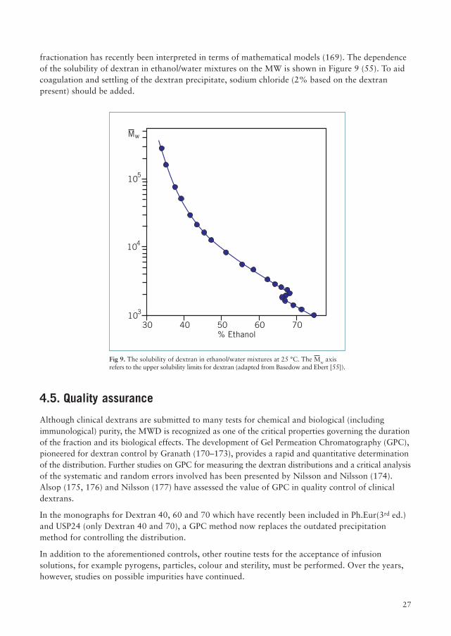
fractionation has recently been interpreted in terms of mathematical models (169). The dependenceof the solubility of dextran in ethanol/water mixtures on the MW is shown in Figure 9 (55). To aidcoagulation and settling of the dextran precipitate, sodium chloride (2% based on the dextranpresent) should be added.
4.5. Quality assurance
Although clinical dextrans are submitted to many tests for chemical and biological (includingimmunological) purity, the MWD is recognized as one of the critical properties governing the durationof the fraction and its biological effects. The development of Gel Permeation Chromatography (GPC),pioneered for dextran control by Granath (170–173), provides a rapid and quantitative determinationof the distribution. Further studies on GPC for measuring the dextran distributions and a critical analysisof the systematic and random errors involved has been presented by Nilsson and Nilsson (174).Alsop (175, 176) and Nilsson (177) have assessed the value of GPC in quality control of clinicaldextrans.
In the monographs for Dextran 40, 60 and 70 which have recently been included in Ph.Eur(3rd ed.)and USP24 (only Dextran 40 and 70), a GPC method now replaces the outdated precipitationmethod for controlling the distribution.
In addition to the aforementioned controls, other routine tests for the acceptance of infusionsolutions, for example pyrogens, particles, colour and sterility, must be performed. Over the years,however, studies on possible impurities have continued.
30 40 50 60 70% Ethanol
103
104
105
Mw
Fig 9. The solubility of dextran in ethanol/water mixtures at 25 °C. The Mw axisrefers to the upper solubility limits for dextran (adapted from Basedow and Ebert [55]).
28
In studies aimed at detecting contaminating macromolecules, Richter (178) was unable to raise PCAreactive antibodies with the centrifuged supernatant of a crude hydrolyzate of native Leuconostocmesenteroides B-512(F) dextran or a purified clinical dextran (MW 70 000) in either rabbits orguinea-pigs and concluded that immunogenic impurities were absent. However, further studiesbased on reversed single radial immunodiffusion (RSRI) using high-titre antisera against the cellular(dextran-free) component of Leuconostoc mesenteroides revealed the presence of antigens in clinicalsamples from several countries (179). Some manufacturers have since then successfully eliminatedthese impurities from their products. The finding that the antigen cross-reacts with yeast mannanindicated that the antigen is, presumably, a mannano-peptide (179). Hedin (180) has examined therole of biologically active contaminants in clinical and other dextrans from the B-512(F) strain bymeans of the rat anaphylatoxin bioassay, the in vitro Limulus test and the rabbit pyrogen assay.Whereas these sensitive tests gave positive reactions in technical grade dextrans, the clinical prod-ucts (Macrodex and Rheomacrodex) did not respond.
Lafrenz (184) investigated the stability of commercial dextrans in various glass and plastic flasksand found that changes in [h] were insignificant over 1 year (20 °C and 40 °C).
Soon after the large scale production of clinical dextran got under way, it became evident that flakestended to form in a small proportion of the bottles. The flake formation was aggravated by freeze/heating cycles but only a few milligrams of the total amount present appeared fo form flakes. Theflakes generally redissolved on heating (185, 186). Studies have confirmed that the flakes have asimilar MWD to the soluble dextran in the bottle.
The tendency for orderly molecular association in dextran has been demonstrated by Jeanes (67)using X-ray diffraction patterns. Crystallinity was enhanced in hydrolysed dextran fractionsparticulary after treatment with 60–70% ethanol. Further evidence for time dependent intermolecularassociation in a Dextran 40 fraction was adduced by Aizawa and coworkers (187) using 1H-NMR,viscosity and IR measurements, albeit using rather high concentrations 10–50%.
4.6. Stability in solution
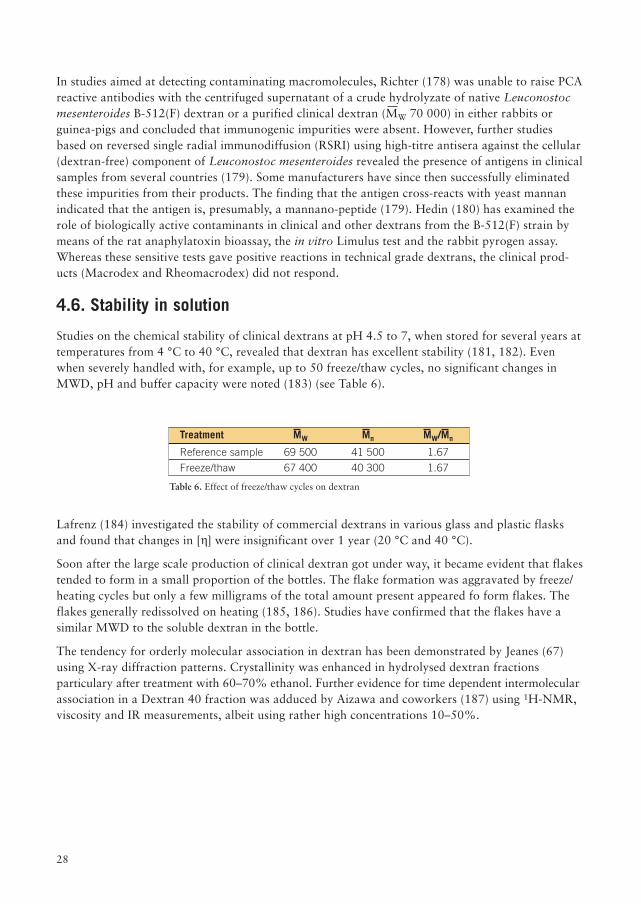
Studies on the chemical stability of clinical dextrans at pH 4.5 to 7, when stored for several years attemperatures from 4 °C to 40 °C, revealed that dextran has excellent stability (181, 182). Evenwhen severely handled with, for example, up to 50 freeze/thaw cycles, no significant changes inMWD, pH and buffer capacity were noted (183) (see Table 6).
Treatment MW Mn MW/Mn
Reference sample 69 500 41 500 1.67
Freeze/thaw 67 400 40 300 1.67
Table 6. Effect of freeze/thaw cycles on dextran
29
4.7. Future developments
Native dextran is generally produced batch-wise in cell cultures. However, a number of alternativeprocesses have been devised, for example (1) the use of cell-free enzyme extracts; (2) fermentationsin the presence of acceptors; (3) continuous processes.
It was discovered some 30 years ago by Hehre (125) and Tsuchiya (188) that a supplement (1 mg/ml)of low MW dextran to the culture fluid markedly affected the MW of the dextran produced.This discovery was further exploited by Tsuchiya and coworkers (119) and Nadel (146). A 33%yield of clinical dextran has been obtained from an enzyme concentrate (126).
Few manufacturers seem to have adopted this technique. The reasons are many; (1) the main clinicalfraction invariably appears to be accompanied by a very high MW fraction; (2) the yields may notexceed those obtained by standard procedures; (3) a separate process for the production of the lowMW dextran must be devised; (4) new safety data to support the change-over must be assembled.
The production of dextran by immobilized dextransucrase has given promising results on a laboratoryscale (148, 151, 155, 189). A clinical dextran has been produced by this technique (155). Studies toimprove the existing methods of depolymerization and fractionation have continued.The reports by Basedow and Ebert and colleagues (166, 190–192) on the effects of shear stress orultrasound, either alone or in combination with acid hydrolysis, are of particular interest in this respect.These authors have also examined the products formed by the action of an endo-dextranase (193).There are indications that these procedures give rise to a less polydispersed product.
Conventional ethanol fractionation techniques are cumbersome and time-consuming. Productionscale ultrafiltration has been available for many years. Unfortunately, this technique also has itslimitations; perhaps the most serious is that dextrans exhibit poor selectivity with these membraneson account of their ability to deform within the pores of the membrane (194). Recently, however,Alsop and coworkers (195) have described a process involving GPC, ultrafiltration and ion ex-change for the efficient fractionation of a dextran hydrolysate.

30
31
5. History of medical applications
5.1. Dextran 70
In the early 1940's, at the same time as Stacey and his associates (196, 197) in Birmingham werestudying bacterial dextrans and Hehre and colleagues (89, 152) in the USA were pursuing the dextranproducing activity of cell-free extracts of Leuconostoc, a young Swedish biochemist, B. Ingelman, atthe Department of Biochemistry and Physical Chemistry, University of Uppsala began probing thepolysaccharides and proteins of sugar beet juice.
One of the critical episodes was the discovery of dextran in an infected sample of the juice. Thisinitiated a series of investigations on the polysaccharide (198). At the end of 1942, a recently qualifiedM.D., A. Grönwall, joined the laboratory to study tuberculin. Considerable effort was beingdevoted at the time to the freeze-drying of blood plasma for military medicine. Within the space ofmonths, Ingelman and Grönwall had stumbled on the idea of using a hydrolyzed dextran as aplasma substitute. After studies on the partial hydrolysis, fractionation, and extensive biologicalstudies, a Swedish pharmaceutical company adopted the project in 1943, and later that year,preliminary clinical trials began. In 1944, under the direction of the surgeon, G. Bohmansson,extensive clinical trials were started at the Regional Hospital in Örebro. The dextran used at thattime was derived from Leuconostoc mesenteroides, strain 7E, and was slightly more branched thanthe present one. By 1947, about four years after the innovation, a 6% solution of a dextran fractionhad been approved for clinical use in Sweden and, shortly thereafter, in the U.K., an achievementthat would be inconceivable under the present regulatory climate.
The product was gradually improved and was designated Dextran 70. Samples of the Swedishproduct were soon tested clinically in the USA. Meanwhile, at the U.S. Department of Agriculture,Northern Regional Research Laboratory at Peoria, Allene Jeanes had been conducting studies ondextrans which were to have an important influence on the course of events (199, 200). Duringthese studies, it had been established that a strain of Leuconostoc mesenteroides isolated from aninfected bottle of root beer by R.G. Benedict and designated NRRL B-512 was a vigorous dextranproducer and its dextran was only slightly branched. In 1949–50, strong interest among U.S.government and medical authorities and advocacy by Drs. Jeanes, Tsuchiya, and Koepsell at Peoria,stimulated a multi-disciplinary research program at the NRRC to provide basic information on theproduction of a synthetic blood volume expander from dextran. Progress was rapid and by the endof 1951, four American companies were producing dextran from the NRRL B-512(F) substrain forclinical purposes.
Numerous other dextrans had been compared but B-512(F) was deemed superior clinically andtechnically as a plasma volume expander. This substrain had been isolated from the original B-512strain in 1948 by W.C. Haynes by selecting colonies with vigorous growth characteristics.The activities of these colonies were followed through repeated growth cycles and the best resultswere obtained from a growth in a Fernbach flask, which thus gave rise to the designation (F) (199).The production of clinical dextrans has since grown steadily throughout the world. Dextran 70 isgenerally marketed as a 6% solution in normal saline and as such continues to maintain its positionworldwide as the plasma volume expander of choice. It is recommended for the treatment of shockor impending shock due, for example, to hemorrhage, burns, surgery or trauma (201–203).Dextran 70 also reduces the risk for thrombosis and numerous studies testify to its value in significantlyreducing the risk of post-operative fatal pulmonary emboli (204, 205).
32
5.2. Dextran 40
The introduction in 1961 of a further dextran product, Dextran 40, following a suggestion byIngelman and Gelin (206, 207), was a direct consequence of earlier observations by Hint andThorsen (208) on the erythrocyte disaggregating properties of dextran of MW <50 000 and theclassic report by Gelin on the circulatory disturbances following trauma and shock (209). Interest inthis product was spurred by the important finding that low MW fractions also impart blood flowimprovement properties by reduction of blood viscosity and inhibition of erythrocyte aggregation(210, 211). Although the optimal disaggregating effect was observed around MW 25 000,a Dextran 40 was finally chosen with due consideration to renal excretion.
The antithrombotic effect of both Dextran 40 and 70, demonstrated experimentally by Gelin andcolleagues (212) and clinically by Koekkenberg (213), provides a prophylactic treatment for deepvenous thrombosis and post-operative fatal pulmonary emboli. A dosage not exceeding 20 ml/kgbody-weight of dextran 40 (10% in normal saline) is recommended during the first 24 hours inpatients undergoing high risk surgery or suffering from high risk trauma
5.3. Dextran 1
With the increased use of clinical dextrans, there followed an increase in the number of reports of dextran-induced anaphylactoid reactions (DIARs). The reported incidence from numerous studies variesfrom 0.03–4.7% (214). As these reactions are sometimes, albeit rarely, life-threatening, a collaborativestudy was conducted between 1968 and 1981 to elucidate the mechanisms underlying DIARs.Since severe reactions were shown to be antibody mediated, the idea of applying the hapten inhibitionprinciple was examined. Following extensive animal and clinical trials, a monovalent haptendextran fraction (MW 1 000) was introduced in 1982. A small volume (20 ml of a 15% solution) wasadministered prior to the Dextran 40 or 70 infusion. Multicentre trials have shown that the incidenceof severe DIARs was thereby considerably reduced (214).
5.4. Iron-dextran
The therapeutic value of colloidal iron preparations was first reported in the 1950's by London andTwigg (215). Numerous attempts have been made to improve these iron preparations (216-218).Thus dextran ([h] 0.05) is first heated with alkali, and is then neutralized in the presence of ferricchloride solution. Studies on this product have revealed that each particle consists of a central ironcore, approximately 3 nm diameter, surrounded by a dextran sheath of approximately 13 nmdiameter (219, 34). The complex is visualized as a particle formed by a protective sheath of dextranattached by terminal metasaccharinic acid units to a b-FeOOH core. A solution of this complexcontaining 5% iron and 20% dextran (Imferon™) is suitable for intramuscular and intravenousinjection for treating iron deficiency anemia. The product is currently used widely for treatinganemia in new-born piglets.
The use of these preparations has been re-examined in humans and a dramatic rise in hemoglobinwas reported following intravenous infusion. The solution is best administered together withglucose solutions (220–222).
33
5.5. Dextran sulfate
Dextran sulfate (dextran hydrogen sulfate, sodium salt) has been tested as a potential substitute forheparin in anticoagulant therapy (223). Following studies by Ingelman (224), Walton and Ricketts(225–227) explored the anticoagulant properties of a wide range of dextran sulfates (MW 7 000 to458 000) and established that the lowest molecular weight products displayed the highest anticoagulantproperties. However, at best this only represented 15% of heparin's activity.
The toxicity of these products had been recognized at an early stage. Nevertheless, a low moleculeweight fraction with MW 7 000 and S, 16% was considered to be qualitatively similar to heparin(228, 229). Preliminary clinical trials were unfortunately discouraging and revealed severe adversereactions, notably, stiff and painful joints, skin eruption, loss of hair and gastro-intestinal symptoms(230). In chronic toxicity studies in animals, retardation in weight gain and osteoporosis withspontaneous fractures were observed (231, 232). It should be noted that the doses in the clinicaltrials corresponded to 1.3 g/day, which is approximately tenfold that used in current heparin therapy.
With the introduction of low-dose heparin therapy for thrombosis prophylaxis in the 70's (233–235),interest in low molecular weight polyanions has been rekindled and reports on interactions withindividual enzyme/inhibitor systems in the coagulation cascade have appeared (236, 237) (see also 6.5.1).
Dextran sulfate immobilized on cellulose has been found to remove LDL cholesterol preferentiallyduring plasmaphoresis (238).
5.6. Diethylaminoethyl dextran (DEAE-dextran)
McKernan and Ricketts (239) originally reported the preparation and properties of DEAE-dextran,A preparation with MW approx. 500 000 has, at a daily oral dose of 2–3 g, been shown to effect areduction in serum cholesterol and triglycerides by 8 and 14% respectively (240). This dose shouldbe compared with the considerably larger doses (4 x 4 g daily) of the insoluble cationic resins (e.g.Cholestyramine™) which must be taken to achieve comparable effects. Further reports on thepharmacology of DEAE-dextran have appeared (241, 242).
5.7. Perfusion solutions
Organ transplantation has in recent years made rapid strides. The need to preserve the viability oftransplant organs is urgent. One of the requirements of a perfusion solution is that it must be iso-osmoticwith the intracellular fluid. The addition of 5–10% low MW dextran has proved beneficial inpreserving isoosmolality and good results have been obtained with kidney, liver, and corneaperfusions (243–247).
34
5.8. Debrisan
Debrisan™ is a wound-cleansing agent prepared in bead form by the emulsion polymerization ofdextran with epichlorohydrin. The product acts by absorbing (approx. 4 ml/g) wound exudate insecreting, infected wounds, ulcers and sores, thereby shortening the healing time. The idea, exemplifyingone of many serendipitous innovations in the pharmaceutical industry, was conceived in 1972 whenUlf Rothman at Malmö General Hospital, Sweden was investigating the proteins in exudate fromapocrine sweat glands in patients suffering from excessive axillary perspiration (248). Exudate wasinitially collected with filter paper but later Sephadex G-25 was used. One patient with badly infectedsores showed a marked improvement after the application of this material. This observation inspiredRothman and his colleagues (249, 250) to initiate further trials on skin wounds and lesions, leading tothe development of a pharmaceutical grade cross-linked dextran (Debrisan) which was launched in 1977.
Numerous reports of trials have appeared during the past decade confirming Debrisan's efficacy inpromoting wound healing (251–253).
5.9. Hyskon
A 32% solution of Dextran 70 stabilized in 10% glucose has proved valuable as a distendingmedium in operative hysteroscopy (254, 255). It has also been shown to prevent the formation ofpost-operative tissue adhesions after tubal or abdominal surgery (256–258).
35
6. General applications of dextran and its derivatives
In this section, examples of known commercial applications of dextran products and examples ofapplications with proven technical or biological effects have been selected. A factor that has restrictedthe widespread commercial use of dextran is the price, which is somewhat higher than most starchand cellulose products on the market. Dextrans are thus most likely to find application in highquality or high technology products.
6.1. Dextran fractions
The demand for technical dextrans from industry has shown a significant increase in the past decade.
Since household sucrose, fruits or fruit beverages could be contaminated with traces of dextrans,ingestion of dextrans, albeit in small amounts, may not be uncommon. Dextran NRRL B-512(F) isdegraded by dextran-splitting bacteria in the human gut and most of the hydrolysis products can beabsorbed to produce a rapid increase in blood sugar and liver glycogen (259–262).
However, in the food industry, where innumerable applications of dextrans in foodstuffs were patentedin the 50's and 60's, no application appears to have been pursued and the mandatory toxicologicalstudies to gain FDA approval were not performed. Hence in 1977, the GRAS (generally recognizedas safe) status of dextrans was deleted (263-265). Dextrans are not permitted in the UK or Europeas foodstuff additives, and dextrans do not seem to have been considered by the Joint FAO/WHOExpert Committee on Foodstuff Additives (JECFA). Dextrans are, however, considered as safe ascomponents of food packaging materials.
Dextran fractions do not appear to be included in the lists of permitted additives (ingredients) forpharmaceutical formulations such as ointments and creams for topical use and tablets and capsulesfor oral use. However, providing the appropriate documentation is presented, there are no a priorireasons why they may not be used. Indeed several products in which a dextran fraction is used as anon-active ingredient are on the market.
Purified dextran fractions with high clarity and low chloride levels find extensive applications in thephotographic industry. Addition of low concentrations of dextran to the silver emulsion is found toenhance significantly the quality of the images. The effect is presumably attributable to the effect ofdextran on the conformation of the gelatin molecules.
Since Albertsson (266) revealed the enormous potential of 2-phase polymer systems, especiallydextran-PEG systems, for the partition of sub-cellular particles and macromolecules, an immensenumber of applications has evolved. These systems offer a means of fractionation beyond the rangeof conventional techniques. Some recent applications are: the separation of peripheral blood cells(267), distinguishing erythrocytes from multiple sclerosis patients (268), the separation of enzymes,for example pullulanase, from Klebsiella pneumoniae cells (269), and the partitioning of murinelymphoblasts (270).
Dextran has been recommended as a cryoprotective agent for human, animal and plant cells (271–273).Thus a mixture of 5% methyl sulphoxide and 9% Dextran 70 was found to afford optimalcryoprotection of human bone marrow committed stem cells (273).
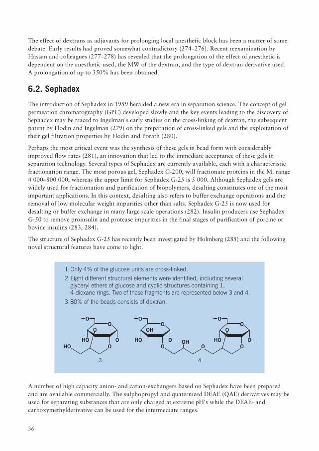
36
The effect of dextrans as adjuvants for prolonging local anesthetic block has been a matter of somedebate. Early results had proved somewhat contradictory (274–276). Recent reexamination byHassan and colleagues (277–278) has revealed that the prolongation of the effect of anesthetic isdependent on the anesthetic used, the MW of the dextran, and the type of dextran derivative used.A prolongation of up to 350% has been obtained.
6.2. Sephadex
The introduction of Sephadex in 1959 heralded a new era in separation science. The concept of gelpermeation chromatography (GPC) developed slowly and the key events leading to the discovery ofSephadex may be traced to Ingelman´s early studies on the cross-linking of dextran, the subsequentpatent by Flodin and Ingelman (279) on the preparation of cross-linked gels and the exploitation oftheir gel filtration properties by Flodin and Porath (280).
Perhaps the most critical event was the synthesis of these gels in bead form with considerablyimproved flow rates (281), an innovation that led to the immediate acceptance of these gels inseparation technology. Several types of Sephadex are currently available, each with a characteristicfractionation range. The most porous gel, Sephadex G-200, will fractionate proteins in the Mr range4 000–800 000, whereas the upper limit for Sephadex G-25 is 5 000. Although Sephadex gels arewidely used for fractionation and purification of biopolymers, desalting constitutes one of the mostimportant applications. In this context, desalting also refers to buffer exchange operations and theremoval of low molecular weight impurities other than salts. Sephadex G-25 is now used fordesalting or buffer exchange in many large scale operations (282). Insulin producers use SephadexG-50 to remove proinsulin and protease impurities in the final stages of purification of porcine orbovine insulins (283, 284).
The structure of Sephadex G-25 has recently been investigated by Holmberg (285) and the followingnovel structural features have come to light.
A number of high capacity anion- and cation-exchangers based on Sephadex have been preparedand are available commercially. The sulphopropyl and quaternized DEAE (QAE) derivatives may beused for separating substances that are only charged at extreme pH's while the DEAE- andcarboxymethylderivative can be used for the intermediate ranges.
1.Only 4% of the glucose units are cross-linked.
2.Eight different structural elements were identified, including severalglyceryl ethers of glucose and cyclic structures containing 1,4-dioxane rings. Two of these fragments are represented below 3 and 4.
3.80% of the beads consists of dextran.
O
O
OO
HOHO
O
O
O
O
OO
HO
O
O
O
OH
OHOHO
O
3 4
37
These gels are now employed commercially for the fractionation of plasma proteins, in particular,human serum albumin, blood clotting factors, immunoglobulin G and haptoglobulin (282) andthese chromatograpic techniques are gradually replacing older batch fractionations.
For the separation according to molecular size in organic media, a hydroxylpropyl derivative ofSephadex LH-20 was developed. This facilitates the separation of lipids, hormones, fatty acids etc.
6.3. Dextran-hemoglobin preparations
Stroma-free hemoglobin would ostensibly appear to provide the ideal blood substitute. Studies haveshown, however, that unmodified hemoglobin is excreted too rapidly by the kidneys (286–287). Thepreparation and properties of dextran-hemoglobin complexes prepared by consecutive reactionswith cyanogen bromide, ethylene diamine, bromoacetylbromide, and finally hemoglobin have beenstudied (288, 289). A complex containing approx. 1:1 molar proportions of hemoglobin to Dextran 20was cleared slowly from the circulation (approx. 20% after 4 hours) (290). It was found, however,that the oxygen is bound much more tightly than in hemoglobin and the oxygen delivery potentialof these complexes needs to be further explored. Although the complexes were found to be non-immunogenic in homologous species (with regard to the hemoglobin) (291), the risks inherent inactivating the immune system to the dextran moiety have yet to be studied. This approach may,nevertheless, enable outdated red cells to be used with advantage (292).
6.4. Conjugates of dextran with biologically-active substances
An account of the research in this area, which embraces the coupling of drugs, enzymes, hormones,and antibodies, would merit a chapter to itself. Molteni (293, 294) and Rogovin (295) have surveyedsome of the fascinating facets of this topic.
The aims of conjugation to dextran are several:
1. to prolong the lifetime of the active component
2. to increase its stability
3. to facilitate targeting of the drug
4. to depress the antigenicity of the protein moiety
The search for mild coupling procedures has provided ground for much ingenuity. The classicalprocedure, reported by Porath and coworkers in 1967, on the activation of polysaccharides withcyanogen bromide is still widely used (296-298). The mechanism is depicted in Figure 10.
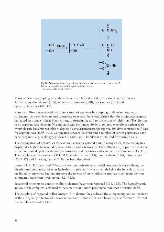
38
Many alternative coupling procedures have since been devised, for example activation via1,1'-carbonyldiimidazole (299), reductive amination (300), isocyanides (301) andcyclic carbonates (302, 303).
Marshall (304) has reviewed the preservation of enzymes by coupling to dextran. Studies onconjugates between dextran and a-amylase or trypsin have established that the conjugates acquireincreased resistance to heat inactivation, to proteinases and to the action of inhibitors. The lifetimeof an asparaginase-dextran 70 conjugate was prolonged 20-fold, in vivo, whereby a patient withlymphoblastic leukemia was able to deplete plasma asparaginase for approx. 100 days compared to 7 daysfor asparaginase itself (305). Conjugates between dextran and a number of serum peptidases havebeen prepared, e.g. carboxypeptidase G2 (306, 307), kallikrein (308), and fibrinolysin (309).
The conjugation of cytostatics to dextran has been explored and, in many cases, these conjugatesdisplayed a high cellular uptake, good activity and less toxicity. These effects are, in part, attributableto the preferential uptake of dextran by lysosomes and the higher endocytic activity of tumour cells (310).The coupling of daunomycin (311, 312), methotrexate (313), daunorubicin (314), mitomycin C(315-317) and 7-deazaguanine (318) has been described.
Larsen (319, 320) has used O-benzoyl dextran derivatives as model compounds for studying thekinetics and mechanism of ester hydrolysis in plasma. It was concluded that the hydrolysis is notmediated by enzymes. Factors affecting the release of metronidazole and naproxen from dextranconjugates have been investigated (321-323).
Successful attempts to couple insulin to dextran have been reported (324, 325). The hypoglycemicaction of the complex is claimed to be superior and more prolonged than that of insulin itself.
The coupling of ragweed pollen Antigen A to dextran has reduced the allergenicity and antigenicityof the allergen by a factor of 7 (on a molar basis). This effect was, however, insufficient to warrantfurther clinical studies (326).
Fig 10. Activation of dextran yielding the intermediate structures 1, carbamate 2,linear imidicarbonate and 3, cyclic imidocarbonate.The latter is the most reactive.
OH
OH CNBrOH
O C N
1
2NHO
O
C
2NH
O OC
3
NHO
CO
39
Before the clinical potential of these devices can be fully exploited, the safety of these compoundsmust be documented.
1.How does the substitution affect the metabolism and toxicity of thecarrier? It is known that even low degrees of substitution may seriouslyretard the metabolism and elimination from the body.
2. Is the immunogencity of the complex enhanced (327)?
3.Can sufficient active substance be coupled to the dextran so thattherapeutic levels can be attained without using excessively highconcentrations of the conjugate?
6.5. Dextran derivatives
6.5.1. Dextran sulfate
Some reference to this substance in connection with its effects on coagulation has already beenmade in section 5.5. Its wide spectrum of biological properties has attracted the attention of manyscientists. The reports may be grouped according to the following biological effects.
a. Enzyme inhibition, activation and release
b.Adjuvant effect on immune response
c. Cellular interactions and response
d.Effect on virus infectivity
This classification is necessarily rather arbitrary as, in many cases, the observed biological effectsare the result of two more of these activities. The studies pertaining to enzymes constitute by far thelargest group with approximately half demonstrating inhibitory effects and half, activating effects.
In each particular instance, it must be stressed that the observed effects are dependent not only onthe MW and DS of the dextran sulfate but also on concentration, pH and ionic strength. Unless statedotherwise, a dextran sulfate with MW of 500 000 and high DS approx. 2 has been used in most of thestudies.
a. Effects on enzymes
Inhibitory effects have been reported for, inter alia, hyaluronidase (328), LH-sensitive adenylatecyclase (329), PGE1-sensitive adenylate cyclase (330, 331), human a-glucosidase (332), ornithinedecarboxylase which plays a key role in cell growth (333), a-amylase and bacterial dextranase (334).
Dextran sulfates are potent non-competitive inhibitors of human leucocyte elastase, an enzyme thatmay be implicated in cartilage degeneration (335). The activity appeared to increase with increasingMW and DS. Reports on the activation of enzymes by dextran sulfates are equally numerous.Whether we are dealing here with the inhibition of an inhibitor in the system is uncertain.
40
Dextran sulfate appears to stimulate the following enzymes, AMP-dependent rabbit musclephosphorylase b (336), tyrosine-3-monoxygenase (337), urokinase (338), calium-dependent ATP-ase(339), and rat brain tryptophan hydroxylase (340).
Numerous studies on the coagulation cascade show that both activation and inhibition may beoperative. Thus dextran sulfate has been found to activate the polymerization of fibrin monomer(341), ATIII (342), conversion of prekallikrein to kallikrein (343–345) and fibrinolysis (346, 347).However, kallikrein (344), the conversion of fibrinogen to fibrin (341) and thrombin (348, 349)appear to be inhibited by dextran sulfate. These effects may be, inter alia, concentration dependent.
b. Effects on immune response
Many reports serve to illustrate that dextran sulfate interferes in many ways with the immuneresponse. Evidence that it is a B-cell mitogen stimulating the proliferation of B cells and thereby theimmune response has been presented (350–354). However, in vivo, dextran sulfate (i.p., 50 mg/kg)has been found to increase the susceptibility of mice to bacterial infection (355–357). These observationshave been ascribed to the toxic effects of sulfate on the mononuclear phagocytes (357, 358). Even atlow concentrations (20 µg/ml), exposure of macrophages to dextran sulfate leads to an accumulationwithin the secondary lysosomes and an inhibition of the phagosome-lysosome interaction fusion(354–361) and may thus interfere with the enzymes responsible for killing bacteria (362). Increasedpermeability of the lymphocytes with excessive leakage of low MW components may also be animportant factor (363–365). Dextran sulfate also induces the mobilization of B and T lymphocytesin vivo (366–369).
c. Effects on viruses
The reports on the effects of dextran sulfate on virus infectivity and growth (as with tumourgrowth) are conflicting. Variable effects are noted with foot and mouth viruses (370).Herpes simplex viruses were inhibited (371). Although dextran sulfate inhibits the release of virionsfrom cells (372), a combination of lipopolysaccharide and dextran sulfate induces their release frommouse spleen cells (373). In vivo, the resistance of experimental animals to viral infection waspotentiated by dextran sulfate (374, 375).
In 1987, the antiviral activity of dextran sulfate against human immunodeficiency virus HIV-1 wasestablished (376, 377). The optimal activity, in vitro, is obtained with a molecular weight (MW) of approx.10 000 and DS 2 (378, 379). Dextran sulfate with MW of approx. 7000 is, however, poorly adsorbedfrom the gut (380) and it is uncertain whether therapeutic concentrations can be obtained by this route.
d. Toxicity
The toxicity of a low MW dextran sulfate, administered intravenously, has been referred to underSection 5.5. More recently attention has been focussed on the toxicity of orally administeredsubstance. Supplements of 0.25% and 0.5% for 82 weeks in rats did not increase the incidence ofinfection or tumours (381). Higher doses (1–10%) resulted in mortality and tumours (382, 383).Apart from dosage, the toxicity is also dependent on MW (384).
Adsorption studies in the gut using a wide range of dextran sulfates (3 000–200 000) suggest only asmall proportion of the dose is absorbed (385–387). Intravenous administration has also beeninvestigated (388). The inhibition of metastasis by dextran sulfate (MW 6–7 x 10³, DS approx. 2) hasbeen tested clinically but the 5 year survival rate in lung cancer was not improved (389, 390). Testsfor mutagenicity and cytogenicity of low MW dextran sulfate proved negative (391).
41
e. Gene manipulation
Finally, some mention should be made of the successful application of dextran sulfate in genemanipulation, where it has become a reagent of particular value for accelerating the hybridizationof DNA fragments (392–394); for accelerating the transfer of DNA fragments from agarose gels(394); and for detecting recombinant mammalian viruses in plaques (395).
6.5.2. DEAE-dextran
The reaction of diethylaminoethyl chloride with dextran in alkali affords a derivative in which threekinds of charged amine groups can be distinguished (396, 397): the single DEAE substituent withpKa 9.2; the tertiary group with pKa 5.5; and the quaternary group on the tandem group which isdissociated at all pH values.
Like the polyanions, the polycations also exhibit a wide range of interesting biological properties.In particular, DEAE-dextrans appear to enhance a number of cellular processes. Many reports (398-401)testify to the enhancement of cellular uptake of viral RNA and also intact virus particles. Considerableinterest has been focussed on the enhanced production of interferon by polyribonucleotidecomplexes in the presence of DEAE-dextran (402–404). These processes appear to operate withoutdetrimental effects on the viability of the cells (398). To what extent these effects are due to theinteraction between the cell surface (net negative charge) and the polycation is uncertain. For mostof these studies, a DEAE-dextran with MW approx. 500 000 and DS 0.5 has been used at concentrationsfrom 10–500 µg/ml.
Reports on its effects on tumour growth are contradictory. Larsen and Thorling (405) found thatincubation of 4 different types of tumour cells with DEAE-dextran prevented the proliferation ofcells in experimental animals. However, Mincheva (406) found no significant effects on metastasisof pulmonary carcinoma in rats. Rice and coworkers (407) reported that repeated injections ofDEAE-dextran (500 µg) in mice induced sarcomas.
6.5.3. Fluorescein-labelled dextrans
Fluorescein isothiocyanate reacts with dextran to give a fluorescent derivative (408) which hasproved useful as a macromolecular tracer in studies of microcirculation and, in particular, vascularpermeability in health and disease.
Techniques developed in the 70’s (409–412) have been exploited to study permeabilities of braincapillaries (413), pancreatic duct (414), cornea (415), retinal vasculature (416), and skeletal muscle (417).
42
6.6. Microcarriers for cell culture
In 1967, van Wezel (418) reported the use of DEAE-Sephadex A-50 as a microcarrier for culturinganchorage-dependent animal cells. However, these microspheres were not ideal for the growth ofcells especially at higher microcarrier concentrations (>2 mg/ml). Subsequent studies (419–422)established that the optimal degree of substitution for cell growth was approx. 1.5 meq/g which wasconsiderably lower than that of the ion-exchanger then available. Several microcarriers based oncross-linked dextran are now available commercially: (1) A microcarrier with DEAE-groups distributedthroughout the matrix (Cytodex™ 1); and (2) a microcarrier to which a layer of collagen is attached(Cytodex™ 3). Microcarrier technology is now employed commercially for the production of,amongst others, viruses, vaccines, and cell products such as interferon.
6.7. 99mTc-Dextran
Technetium appears to complex strongly with dextran and as such has been tested as an agent forlymphoscintigraphy with promising results in the leg, pelvic and para-aortic lymph-nodes (423-425). The radio-labelled pertechnate is first reduced with stannous ion and then added to dextran toyield the technetium complex.
43
7. Dextran analysis
The choice of method will be dictated by the sensitivity required, the nature of the sample and,possibly, by the molecular weight of the dextran present.
In the sugar industry, the alcohol haze method is commonly used (426, 427) but may be replaced bya more specific copper method (428).
Many methods have been devised to measure clinical dextrans in serum and urine. The methodsgenerally involve the removal of proteins by picric acid (429) or trichloroacetic acid (430) followedby the precipitation of dextran with alkaline copper reagent (430), o-toluidine-acetic acid (431), orethanol (429).
Dextran may then be determined by the anthrone reagent (429, 432), turbidimetrically (433),polarimetrically (434) or as reducing sugar after hydrolysis (431). Numerous refinements to thesemethods have appeared in the literature (435-437). A procedure employing glucose oxidase hasbeen described (438).
A specific and sensitive immunochemical determination has been developed which detects dextranconcentrations down to 2.5 µg/ml (439).
8. Acknowledgements
I am greatly indebted to Drs. Allene Jeanes, Kirsti Granath, the late Professor Björn Ingelman andKerstin Nilsson for their invaluable help.

44
45
References
1. Sutherland, W. Trends Biochem. Sci. 4, 55, (1979).
2. Miyazaki, A. Nippon Shishubyo Gakkai Kaishi 20, 276, (1978);through Chem. Abstr. 90, 163374 (1979).
3. Jeanes, A. Am. Chem. Soc., Symposium Ser. 45, 284 (1977);Dextran Bibliography, Misc. Publication 1355, U.S. Department of Agriculture, 1978.
4. Evans, T.H. and Hibbert, H. Advan. Carbohydr. Chem. 2, 204 (1946).
5. Stacey, M. and Ricketts, C.R. in L. Zechmeister, Ed., Fortschr.Chem. Org.Naturstoffe Vol. 8, pp. 28–43 (1951).
6. Ricketts, C.R. in Cook, J. W. and Carruthers, W. Eds., Progr. Org. Chem.Vol. 5, p. 73 (1961).
7. Brock Neely, W. Advan. Carbohydr. Chem. 15, 341 (1960).
8. Jeanes, A. in Mark, H.F. Ed., Encyclopedia of Polymer Science and TechnologyVol. 4, John Wiley & Sons, Inc., New York, pp. 805–824 (1966).
9. Behrens, U. and Ringpfeil, M. Microbielle Polysaccharide, Akademie-Verlag, Berlin, 1964.
10. Murhpy, D.T. and Whistler, R.L. in Whistler, R.L. and BeMiller, J.N. Eds.,Industrial Gums, 2nd ed., Academic Press, Inc., New York, pp. 513–530 (1973).
11. Hehre, E.J. in Nord, F. F. Ed., Advan. Enzymol. Vol. 11, pp. 297–332 (1951).
12. Edelman, J. in Nord, F. F. Ed., Advan. Enzymol. Vol. 17, pp. 189–230 (1956).
13. Tsuchiya, H. Bull. Soc. Chim. Biol. 42, 1777 (1960).
14. Manners, D.J. Bull. Soc. Chim. Biol. 42, 1788 (1960).
15. Ebert, K.H. and Schenk, G. in F. F. Nord, Ed., Advan. Enzymol. Vol. 30, pp. 179–219 (1968).
16. Sidebotham, R.L. Advan. Carbohydr. Chem. Biochem. 30, 371 (1974).
17. Walker, G.J. Int. Rev. Biochem. 16, 75–126 (1978).
18. Alsop, R.M. Prog. Ind. Microbiol. 18, 1 (1983).
19. Jeanes, A. Extracellular Microbial Polysaccharides (Sandford, P. A. andLaskin, A. eds.) American Chemical Soc., Washington, DC, p. 284 (1977).
20. Virnik, A.D. et al. Russ. Chem. Rev. 44, 1280 (1975).
21. Yalpini, M. CRC Crit. Rev. Biotechnol. 3, 375 (1986).
22. Jeanes, A. Mol. Immunol. 23, 999 (1986).
23. Robyt, J.F. Encyclopedia of Polymer Science and Engineering [Vol. 4] (Kroschwitz, J.I.ed.) John Wiley and Sons, New York, p. 752 (1985).
24. Gospodarek, T. Pr. Nauk. Akad. Ekoln. Im Oskara Langego Wroclawiu 313, 5 (1985).
46
25. de Belder, A.N. Ullman´s Encyclopedia of Industrial Chemistry,VCH Verlagsgesellschaft, Weinheim, W. Germany, Vol. A8, p. 449 (1987).
26. Rankin, J.C. and Jeanes, A. J. Am. Chem. Soc. 76, 4435 (1954).
27. Dimler, R.J. et al. J. Am. Chem. Soc. 77, 6568 (1955).
28. Van Cleve, J.W. et al. J. Am. Chem. Soc. 78, 4435 (1951).
29. Lindberg, B. and Svensson, S. Acta Chem. Scand. 22, 1907 (1968).
30. Larm, O. et al. Carbohydr. Res. 20, 39 (1971).
31. Jeanes, A. and Seymour, F.R. Carbohydr. Res. 74, 31 (1971).
32. Wolfrom, M.L. et al. J. Am. Chem. Soc. 73, 595 (1951).
33. Fujimoto, K. et al. Tohoku J. Agr. Res. Sendai 13, 61 (1962).
34. Bremner, I. et al. Carbohydr. Res. 11, 77 (1969).
35. Benesi, A.J. and Gerig, J.T. Carbohydr. Res. 53, 278 (1977).
36. Colson, P. et al. J. Am. Chem. Soc. 96, 8081 (1974).
37. Gagnaire, D. and Vignon, M. Makromol. Chem. 178, 2321 (1977).
38. Agback, H. Unpublished report, Pharmacia (1972).
39. Pasika, W.H. and Cragg, L.H. Can. J. Chem. 41, 293 (1963).
40. Seymour, F.R. et al. Carbohydr. Res. 74, 77 (1979).
41. Abbott, D. et al. J. Chem. Soc. 827, (1966).
42. Bourne, E.J. et al. J. Chem. Soc. 827, (1966).
43. Walker, G.J. and Pulkownik, A. Carbohydr. Res. 29, 1 (1973).
44. Taylor, C., Cheetham, N.W. H. and Walker, G.J. Carbohydr. Res. 137, 18, (1985).
45. Covacevich, M.T. and Richards, G.N. Carbohydr. Res. 54, 311 (1977).
46. Bovey, F.A. J. Polym. Sci. 35, 167 (1959); 35, 183 (1959).
47. Senti, F.R. et al. J. Polym. Sci. 17, 527 (1955).
48. Around, L.H. and Frank, H.P. J. Phys. Chem. 58, 953 (1954).
49. Elias, H.G. Makromol. Chem. 33, 166 (1959).
50. Antonini, E. et al. Biopolymers, 2, 27 (1964).
51. Hellman, N.N. Report on working conference on dextran, Peoria, p. 36 (1951).
52. Granath, K.A. J. Colloid Sci. 13, 308 (1958).
53. Frank, H.P. and Mark, H. Monatsh. 85, 307 (1954).
54. Gekko,K. Biopolymers 10, 1513 (1971).
55. Basedow, A.M. and Ebert, K.H. J. Polym. Sci. 66, 101 (1979).
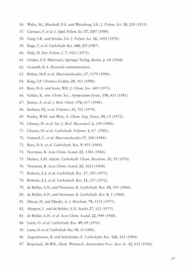
47
56. Wales, M., Marshall, P.A. and Weissberg, S.G. J. Polym. Sci. 10, 229 (1953).
57. Carrasco, F. et al. J. Appl. Polym. Sci. 37, 2087 (1989).
58. Garg, S.K. and Stivala, S.S. J. Polym. Sci. 16, 1419 (1978).
59. Kuge, T. et al. Carbohydr. Res. 160, 205 (1987).
60. Vink, H. Eur. Polym. J. 7, 1411 (1971).
61. Gruber, U.F. Blutersatz, Springer Verlag, Berlin, p. 68 (1968).
62. Granath, K.A. Personal communication.
63. Bohier, M.P. et al. Macromolecules, 17, 1170 (1984).
64. King, S.P. Chemica Scripta, 28, 161 (1988).
65. Rees, D.A. and Scott, W.E. J. Chem. Soc. 469 (1971).
66. Gekko, K. Am. Chem. Soc., Symposium Series, 150, 415 (1981).
67. Jeanes, A. et al. J. Biol. Chem. 176, 617 (1948).
68. Barham, P.J. et al. Polymer, 15, 762 (1974).
69. Pasika, W.M. and West, A. Chem. Eng. News, 50, 13 (1972).
70. Chanzy, H. et al. Int. J. Biol. Macromol. 2, 149 (1980).
71. Chanzy, H. et al. Carbohydr. Polymer 1, 67 (1981).
72. Guizard, C. et al. Macromolecules 17, 100 (1984).
73. Rees, D.A. et al. Carbohydr. Res. 9, 451 (1969).
74. Norrman, B. Acta Chem. Scand. 22, 1381 (1968).
75. Haines, A.H. Advan. Carbohydr. Chem. Biochem. 33, 59 (1976).
76. Norrman, B. Acta Chem. Scand. 22, 1623 (1968).
77. Roberts, E.J. et al. Carbohydr. Res. 17, 393 (1971).
78. Roberts, E.J. et al. Carbohydr. Res. 21, 357 (1972).
79. de Belder, A.N. and Norrman, B. Carbohydr. Res. 10, 391 (1968).
80. de Belder, A.N. and Norrman, B. Carbohydr. Res. 8, 1 (1968).
81. Miyaji, H. and Misaki, A. J. Biochem. 74, 1131 (1973).
82. Ahrgren, L. and de Belder, A.N. Starke 27, 121 (1975).
83. de Belder, A.N. et al. Acta Chem. Scand. 22, 949 (1968).
84. Larm, O. et al. Carbohydr. Res. 49, 69 (1976).
85. Larm, O. et al. Carbohydr. Res. 91, 13 (1981).
86. Augustinsson, B. and Scholander, E. Carbohydr. Res. 126, 161 (1984).
87. Beijerinck, M.W.K. Akad. Wetensch, Amsterdam Proc. Sect. Sc. 12, 635 (1910).
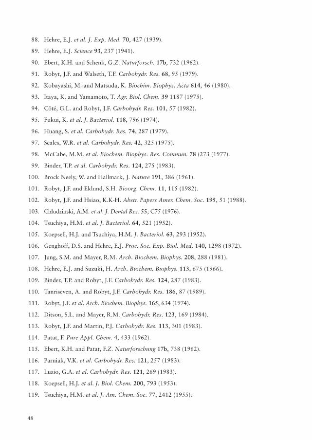
48
88. Hehre, E.J. et al. J. Exp. Med. 70, 427 (1939).
89. Hehre, E.J. Science 93, 237 (1941).
90. Ebert, K.H. and Schenk, G.Z. Naturforsch. 17b, 732 (1962).
91. Robyt, J.F. and Walseth, T.F. Carbohydr. Res. 68, 95 (1979).
92. Kobayashi, M. and Matsuda, K. Biochim. Biophys. Acta 614, 46 (1980).
93. Itaya, K. and Yamamoto, T. Agr. Biol. Chem. 39 1187 (1975).
94. Côté, G.L. and Robyt, J.F. Carbohydr. Res. 101, 57 (1982).
95. Fukui, K. et al. J. Bacteriol. 118, 796 (1974).
96. Huang, S. et al. Carbohydr. Res. 74, 287 (1979).
97. Scales, W.R. et al. Carbohydr. Res. 42, 325 (1975).
98. McCabe, M.M. et al. Biochem. Biophys. Res. Commun. 78 (273 (1977).
99. Binder, T.P. et al. Carbohydr. Res. 124, 275 (1983).
100. Brock Neely, W. and Hallmark, J. Nature 191, 386 (1961).
101. Robyt, J.F. and Eklund, S.H. Bioorg. Chem. 11, 115 (1982).
102. Robyt, J.F. and Hsiao, K.K-H. Abstr. Papers Amer. Chem. Soc. 195, 51 (1988).
103. Chludzinski, A.M. et al. J. Dental Res. 55, C75 (1976).
104. Tsuchiya, H.M. et al. J. Bacteriol. 64, 521 (1952).
105. Koepsell, H.J. and Tsuchiya, H.M. J. Bacteriol. 63, 293 (1952).
106. Genghoff, D.S. and Hehre, E.J. Proc. Soc. Exp. Biol. Med. 140, 1298 (1972).
107. Jung, S.M. and Mayer, R.M. Arch. Biochem. Biophys. 208, 288 (1981).
108. Hehre, E.J. and Suzuki, H. Arch. Biochem. Biophys. 113, 675 (1966).
109. Binder, T.P. and Robyt, J.F. Carbohydr. Res. 124, 287 (1983).
110. Tanriseven, A. and Robyt, J.F. Carbohydr. Res. 186, 87 (1989).
111. Robyt, J.F. et al. Arch. Biochem. Biophys. 165, 634 (1974).
112. Ditson, S.L. and Mayer, R.M. Carbohydr. Res. 123, 169 (1984).
113. Robyt, J.F. and Martin, P.J. Carbohydr. Res. 113, 301 (1983).
114. Patat, F. Pure Appl. Chem. 4, 433 (1962).
115. Ebert, K.H. and Patat, F.Z. Naturforschung 17b, 738 (1962).
116. Parniak, V.K. et al. Carbohydr. Res. 121, 257 (1983).
117. Luzio, G.A. et al. Carbohydr. Res. 121, 269 (1983).
118. Koepsell, H.J. et al. J. Biol. Chem. 200, 793 (1953).
119. Tsuchiya, H.M. et al. J. Am. Chem. Soc. 77, 2412 (1955).
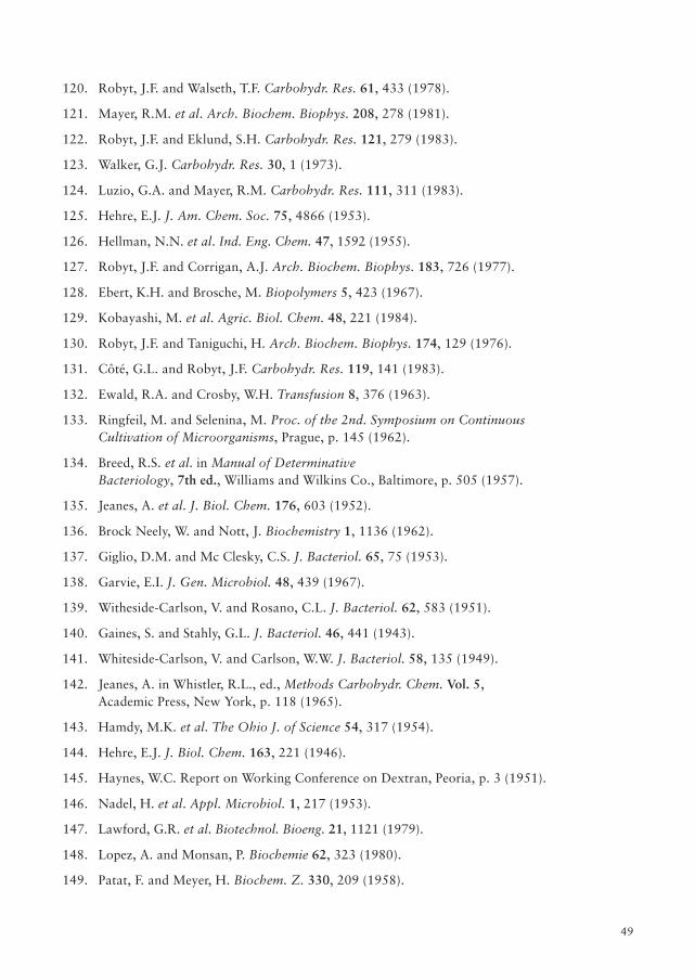
49
120. Robyt, J.F. and Walseth, T.F. Carbohydr. Res. 61, 433 (1978).
121. Mayer, R.M. et al. Arch. Biochem. Biophys. 208, 278 (1981).
122. Robyt, J.F. and Eklund, S.H. Carbohydr. Res. 121, 279 (1983).
123. Walker, G.J. Carbohydr. Res. 30, 1 (1973).
124. Luzio, G.A. and Mayer, R.M. Carbohydr. Res. 111, 311 (1983).
125. Hehre, E.J. J. Am. Chem. Soc. 75, 4866 (1953).
126. Hellman, N.N. et al. Ind. Eng. Chem. 47, 1592 (1955).
127. Robyt, J.F. and Corrigan, A.J. Arch. Biochem. Biophys. 183, 726 (1977).
128. Ebert, K.H. and Brosche, M. Biopolymers 5, 423 (1967).
129. Kobayashi, M. et al. Agric. Biol. Chem. 48, 221 (1984).
130. Robyt, J.F. and Taniguchi, H. Arch. Biochem. Biophys. 174, 129 (1976).
131. Côté, G.L. and Robyt, J.F. Carbohydr. Res. 119, 141 (1983).
132. Ewald, R.A. and Crosby, W.H. Transfusion 8, 376 (1963).
133. Ringfeil, M. and Selenina, M. Proc. of the 2nd. Symposium on ContinuousCultivation of Microorganisms, Prague, p. 145 (1962).
134. Breed, R.S. et al. in Manual of DeterminativeBacteriology, 7th ed., Williams and Wilkins Co., Baltimore, p. 505 (1957).
135. Jeanes, A. et al. J. Biol. Chem. 176, 603 (1952).
136. Brock Neely, W. and Nott, J. Biochemistry 1, 1136 (1962).
137. Giglio, D.M. and Mc Clesky, C.S. J. Bacteriol. 65, 75 (1953).
138. Garvie, E.I. J. Gen. Microbiol. 48, 439 (1967).
139. Witheside-Carlson, V. and Rosano, C.L. J. Bacteriol. 62, 583 (1951).
140. Gaines, S. and Stahly, G.L. J. Bacteriol. 46, 441 (1943).
141. Whiteside-Carlson, V. and Carlson, W.W. J. Bacteriol. 58, 135 (1949).
142. Jeanes, A. in Whistler, R.L., ed., Methods Carbohydr. Chem. Vol. 5,Academic Press, New York, p. 118 (1965).
143. Hamdy, M.K. et al. The Ohio J. of Science 54, 317 (1954).
144. Hehre, E.J. J. Biol. Chem. 163, 221 (1946).
145. Haynes, W.C. Report on Working Conference on Dextran, Peoria, p. 3 (1951).
146. Nadel, H. et al. Appl. Microbiol. 1, 217 (1953).
147. Lawford, G.R. et al. Biotechnol. Bioeng. 21, 1121 (1979).
148. Lopez, A. and Monsan, P. Biochemie 62, 323 (1980).
149. Patat, F. and Meyer, H. Biochem. Z. 330, 209 (1958).
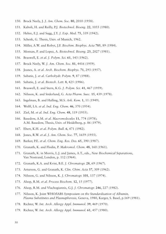
50
150. Brock Neely, J. J. Am. Chem. Soc. 80, 2010 (1958).
151. Kaboli, H. and Reilly, P.J. Biotechnol. Bioeng. 22, 1055 (1980).
152. Hehre, E.J. and Sugg, J.Y. J. Exp. Med. 75, 339 (1942).
153. Schenk, G. Thesis, Univ. of Munich, 1962.
154. Miller, A.W. and Robyt, J.F. Biochim. Biophys. Acta 785, 89 (1984).
155. Monsan, P. and Lopez, A. Biotechnol. Bioeng. 23, 2027 (1981).
156. Braswell, E. et al. J. Polym. Sci. 61, 143 (1962).
157. Brock Neely, W. J. Am. Chem. Soc. 81, 4416 (1959).
158. Jeanes, A. et al. Arch. Biochem. Biophys. 71, 293 (1957).
159. Sabatie, J. et al. Carbohydr. Polym. 9, 87 (1988).
160. Sabatie, J. et al. Biotech. Lett. 8, 425 (1986).
161. Braswell, E. and Stern, K.G. J. Polym. Sci. 41, 467 (1959).
162. Nilsson, K. and Söderlund, G. Acta Pharm. Suec. 15, 439 (1978).
163. Ingelman, B. and Halling, M.S. Ark. Kem. 1, 11 (1949).
164. Wolff, I.A. et al. Ind. Eng. Chem. 46, 370 (1954).
165. Zief, M. et al. Ind. Eng. Chem. 48, 119 (1955).
166. Basedow, A.M. et al. Macromolecules 11, 774 (1978);A.M. Basedow, Thesis, Univ. of Heidelberg, p. 86 (1979).
167. Ebert, K.H. et al. Polym. Bull. 6, 471 (1982).
168. Jones, R.W. et al. J. Am. Chem. Soc. 77, 1659 (1955).
169. Barker, P.E. et al. Chem. Eng. Res. Des. 65, 390 (1987).
170. Granath, K. and Flodin, P. Makromol. Chem. 48, 160 (1961).
171. Granath, K. in Morris, L.J. and James, A.T., eds., New Biochemical Separations,Van Nostrand, London, p. 112 (1964).
172. Granath, K.A. and Kvist, B.E. J. Chromatogr. 28, 69 (1967).
173. Arturson, G. and Granath, K. Clin. Chim. Acta 37, 309 (1962).
174. Nilsson, G. and Nilsson, K. J. Chromatogr. 101, 137 (1974).
175. Alsop, R.M. et al. Process Biochem. 12, 15 (1977).
176. Alsop, R.M. and Vlachogiannis, G.J. J. Chromatogr. 246, 227 (1982).
177. Nilsson, K. Joint WHO/IABS Symposium on the Standardisation of Albumin,Plasma Substitutes and Plasmapheresis, Geneva, 1980, Karger, S. Basel, p.169 (1981).
178. Richter, W. Int. Arch. Allergy Appl. Immunol. 39, 469 (1970).
179. Richter, W. Int. Arch. Allergy Appl. Immunol. 61, 457 (1980).
51
180. Hedin, H. Thesis, Univ. of Uppsala, p. 13 (1977).
181. d'A. Maycock, W. and Ricketts, C.R. Nature 213, 88 (1967).
182. Registration Documentation, Macrodex, Pharmacia, Uppsala.
183. Fure, O.T. et al. Pharm. Acta Helv. 50, 216 (1975).
184. Lafrenz, K. Thesis Univ. of Hamburg, 1969.
185. Second Conference on Artificial Colloidal Agents, Oct. 1963.
186. Cadwallader, D.E. et al. J. Am. Pharm. Assoc. 47, 895 (1958).
187. Aizawa, M. et al. Bull. Chem. Soc. Jap. 49, 2061 (1976).
188. Tsuchiya, H.M. et al. Abstracts of Papers, Am. Chem. Soc. 122nd Meeting, 15A (1952).
189. Monsan, P. et al. Methods in Enzymol. 136, 239 (1987).
190. Basedow, A.M. and Ebert, K.H. Makromol. Chem. 176, 745 (1975).
191. Basedow, A.M. and Ebert, K.H. Polym. Bull. 1, 299 (1979).
192. Basedow, A.M. et al. Makromol. Chem. 179, 2565 (1978).
193. Basedow, A.M. Polym. Bull. 2, 337 (1980).
194. Bohrer, M.P. et al. Macromolecules 17, 1170 (1984).
195. Alsop, R.M. et al. Chem. Eng. (London) 399, 24 (1984).
196. Daker, D. and Stacey, M. J Chem. Soc. 585 (1939).
197. Stacey, M. and Swift, G. J. Chem. Soc. 1555 (1948).
198. Grönwall, A. and Ingelman, B. Vox Sang. 47, 96 (1984).
199. Jeanes, A. Personal communication.
200. Dimler, R.J. 1968 Yearbook of Agriculture, U. S. Gov. Printing Office, p. 314 (1969).
201. Koester, K.H. et al. Lancet 262 (1957).
202. Shoemaker, W.C. Crit. Care Med. 4, 71 (1976).
203. Gruber, U.F. and Siegrist, J. Arch. Klin. Chir. 301, 128 (1962).
204. Bergentz, S.E. World J. Surgery 2, 19 (1978).
205. Bergquist, D. et al. Acta Orthop. Scand. 55, 247 (1984).
206. Gelin, L.E. and Ingelman, B. Acta Chir. Scand. 122, 294 (1961).
207. Gelin, L.E. Rev. Surg. 19, 385 (1962).
208. Thorsen, G. and Hint, H. Acta Chir. Scand. Suppl. 154 (1954).
209. Gelin, L.E. Acta Chir. Scand. 1, 210 (1956).
210. Schmid-Schonbein, H. et al. Hemodilution.Theoretical Basis and Clinical Applications, Karger, Basel, p. 229 (1972).
52
211. Richter, W. Acta Chir. Scand. 131, 1 (1966).
212. Borgstroem, S. et al. Acta Chir. Scand. Suppl. 247 (1959).
213. Koekkenberg, L.J. Bull. Soc. Int. Chir. 5, 501 (1962).
214. Richter, A.W. and Hedin, H.I. Immunol. Today 3, 132 (1982).
215. London, E. and Twigg, G.D. Brit. pat. 748,024 (1956).
216. Alsop, R.M. and Bremner, I. Brit. pat., 1,200,902 (1970).
217. Mueller, A. and Bersin, T. U.S. pat., 3,100,202 (1963).
218. Floramo, N.A. U.S. pat., 3,022,221 (1962).
219. Cox, J.S.G. et al. J. Pharm. Pharmacol. 24, 513 (1972).
220. Kaisi, M. et al. Int. J. Gynecol. Obstet. 26, 235 (1988).
221. Bhatt, R.V. et al. Am. J. Obstet. Gynecol. 94, 1098 (1958).
222. Hunt, E. et al. Am. J. Gastroenterol. 83, 1048 (1988).
223. Groenwall, A., Ingelman, B. and Mosimann, H.Uppsala Lakarforening Forh., 51, 397 (1945).
224. Ingelman, B. Ark. Kemi Min. Geol. 248, 4 (1947).
225. Walton, K.W. Brit. J. Pharmacol. 7, 370 (1952).
226. Ricketts, C.R. Biochem. J. 51, 129 (1952).
227. Ricketts, C.R. and Walton, K.W. Brit. J. Pharmacol. 8, 476 (1953).
228. Ricketts, C.R. and Walton, K.W. Brit. J. Pharmacol. 9, 1 (1954).
229. Ricketts, C.R. Lancet 1004–1010 (1953).
230. Hjort, P. et al. Scand. J. Clin. Lab. Invest. 9, Suppl. 29 (1957).
231. Hint, H.C. and Richter, A. W. Brit. J. Pharmacol. 13, 109 (1958).
232. Ellis, H.A. J. Pathol. Bacteriol. 89, 437 (1965).
233. De Takats, G. J. Am. Med. Assoc. 142, 527 (1950).
234. Sharnoff, J.G. et al. Surg. Gynecol. Obstet. 115, 75 (1962).
235. Sharnoff, J.G. and DeBlazio, G. Lancet 1006–1007 (1970).
236. Suzuki, K. et al. Biochim. Biophys. Acta 585, 416 (1979).
237. Nordenman, B. and Björk, I. VIIth Int. Congress on Thromb. and Haemostasis,London, 1979.
238. Homma, Y. et al. Metabolism 36, 419 (1987).
239. McKernan, W. and Ricketts, C.R. Biochem. J. 76, 117 (1960).
240. Pupita, F. and Barone, A. Int. J. Clin. Pharm. Res. 111, 287 (1983).
241. Soldani, G. et al. Int. J. Obes. 11, 201 (1987).
53
242. Zuccato, E. et al. Pharmacol. Res. Comm. 19, 405 (1987).
243. Manax, W.G. et al. Surgery 57, 528 (1965).
244. Corriere, J.N. et al. Surg. Gynecol. Obstet. 125, 33 (1967).
245. Wustemann, M.C. et al. Scand. J. Urol. Nephrol. 12, 281 (1978).
246. Proctor, E. and Joyce, M. Transplantation 25, 92 (1978).
247. Peyman, G.A. et al. Arch. Ophthalmol. 97, 153 (1979).
248. Rothman, U. Personal communication. 1984.
249. Jacobsson, S. and Rothman, U. I. R. C. S. 11, 3117 (1973).
250. Jacobsson, S. et al. Scand. J. Plast. Reconstr. Surg. 10, 65 (1976).
251. Goode, A.W. et al. Clinical Trials J. 22, 432 (1985).
252. Parulkar, B.G. et al. J. Postgrad. Med. 31, 28 (1985).
253. Åkesson, Å. and Friman, G. Clin. Trials J. 21, 378 (1984).
254. Amin, H.K. and Neuwirth, R.S. Clin. Obstet. Gynecol. 26, 277 (1983).
255. Taylor, P.J. et al. Fertil. Steril. 47, 861 (1987).
256. Blackwell, R.E. and Younger, J.B. Fertil. Steril. 35, 592 (1981).
257. Utian, W.H. S. African Med. J. 58, 204 (1980).
258. Fayez, J.A. and Schneider, P.J. Am. J. Obstet. Gynecol. 157, 1184 (1987).
259. Jeanes, A. Am. Chem. Soc., Symposium Series 15, 336 (1975).
260. Parkinson, T.M. Nature 215, 415 (1967).
261. Bloom, W.L. and Wilhelmi, A.E. Proc. Soc. Exp. Biol. Med. 81, 501 (1952).
262. Booth, A.N. et al. Toxicol. Appl. Pharmacol. 5, 478 (1963).
263. Anderson, D.M.W. Personal communication.
264. Glicksman, M. in M. Glicksman, ed.,Food Hydrocolloids. Vol. 1, CRC Press, Florida, p. 157 (1982).
265. Fed. Registry, 42, FR 59518-21, Nov. 18, 1977.
266. Albertsson, P-A. Thesis, Univ. of Uppsala, 1960.
267. Matsumoto, U. et al. J. Chromatogr. 285, 69 (1984).
268. Van Alstine, J.M. and Brooks, D.E. Clin. Chem. (Winston-Salem, N.C.) 30, 441 (1984).
269. Kula, M.R. et al. Enzyme Eng. 4, 47 (1978).
270. Kessel, D. Biochim. Biophys. Acta 678, 245 (1981).
271. Odavic, R. et al. Experientia 36, 1122 (1980).
272. Ashwood-Smith, M.J. et al. Cryobiology 9, 441 (1972).
54
273. Echlin, P. et al. J. Microsc. (Oxford) 110, 239 (1977).
274. Buckley, F.P. and Fink, B.R. Anesthesiology 51, 5215 (1979).
275. Loder, R.E. Lancet 2, 346 (1960).
276. Rosenblatt, R. Regional Anesth. 6, 108 (1981).
277. Hassan, H.G. et al. Acta Anaesthesiol. Scand. 29, 375 (1985).
278. Hassan, H.G. et al. Acta Anaestesiol. Scand. 29, 380 (1985).
279. Flodin, P. and Ingelman, B. Swed. Pat. 169, 293 (1959).
280. Flodin, P. and Porath, J. U.S. pat., 3,002,823 (1961).
281. Flodin, P. Swed. Pat., 358,894 (1961).
282. Curling, J.M. Separation of plasma proteins (Pharmacia Fine Chemicals) 1983.
283. Cooney, J.C. Bio/Technology (Pharmacia Fine Chemicals) Jan. 1984.
284. The large scale purification of insulin by gel filtration chromatography(Pharmacia Fine Chemicals) 1983.
285. Holmberg, L. Thesis, Univ. of Uppsala, 1983.
286. Rabiner, S.F. et al. J. Exp. Med. 126, 1127 (1967).
287. Bunn, H.F. et al. J. Exp. Med. 129, 909 (1969).
288. Tam, S.C. et al. Can. J. Biochem. 56, 981 (1978).
289. Tam, S.C. et al. Proc. Nat. Acad. Sci. USA 73, 2128 (1976).
290. Blumenstein, J. et al. Progr. Clin. Biol. Res. 13, 205 (1978).
291. Cunnington, P.G. et al. Biochem. J. 193, 261 (1981).
292. Hedlund, B. et al. Progr. Clin. Biol. Res. 122, 71 (1983).
293. Molteni, L. in Bundegaard, H., Hansen, A.B. and Kofod, H., eds.,Optimization of Drug Delivery, Munksgaard, Copenhagen, p. 285 (1982).
294. Molteni, L. Methods in Enzymol. 112, 285 (1985).
295. Rogovin, Z.A. et al. J. Makromol. Sci. A6, 569 (1972).
296. Axén, R. et al. Nature 214, 1302 (1967).
297. Kågedal, L. and Åkerström, S. Acta Chem. Scand. 25, 1855 (1971).
298. Kohn, J. and Wilchek, M. Enzyme Microb. Technol. 4,161 (1982).
299. Bethell, G.S. et al. J. Biol. Chem. 254, 2572 (1979).
300. Larm, O. and Scholander, E. Carbohydr. Res. 58, 249 (1977).
301. Vretblad, P. and Axén, R. Acta Chem. Scand. 27, 269 (1973).
302. Barker, S.A. et al. Carbohydr. Res. 25, 237 (1972).
303. Vandoorne, F. et al. Macromol. Chem. 186, 244 (1985).
55
304. Marshall, J.J. Am. Chem. Soc. Symp. Ser. 12, 125 (1980).
305. Wileman, T. et al. J. Pharm. Pharmacol. 35, 762 (1983).
306. Sherwood, R.F. et al. Biochem. J. 164, 461 (1977).
307. Melton, R.G. et al. Biochem. Pharmacol. 36, 113 (1987).
308. Odya, C.E. et al. Biochem. Pharmacol. 27, 173 (1978).
309. French pat., 2,497,229 through Chem. Abs. 97:133563 (1982).
310. de Duve, C. et al. Biochem. Pharmacol. 23, 2495 (1974).
311. Wilchek, M. Makromol. Chem. Suppl., 2, 207 (1979).
312. Hurwitz, E. et al. Eur. J. Cancer 14, 1213 (1978).
313. Chu, B.C.F. and Whiteley, J.M. Mol. Pharmacol. 17, 382 (1980);Polym. Sci. Technol. 14, 241 (1981).
314. Levi-Schaffer, F. et al. Cancer Treat. Rep. 66, 107 (1982).
315. Honda, K. et al. Saishin Igaku 40, 1844 (1985).
316. Sezaki, H. et al. J. Bioactive and Compatible Polym. 3, 81 (1988).
317. Takakura, Y. et al. Cancer Res. 44, 2505 (1984).
318. Rosemeyer, H. et al. Int. J. Biol. Macromol. 9, 205 (1987).
319. Larsen, C. Advanced Drug Delivery Reviews 3, 103 (1989).
320. Larsen, C. and Johansen, M. Int. J. Pharmaceutics. 27, 205 (1985).
321. Larsen, C. et al. Acta Pharm. Suec. 25, 1 (1988).
322. Harboe, E. et al. Farmaci, Sci. Ed. 16, 73 (1988).
323. Azori, M. et al. Makromol. Chem. 187, 2073 (1986).
324. Armstrong, K. et al. Biochem. Biophys. Res. Commun. 47, 354 (1972).
325. Sakamoto, Y. et al. Biochim. Biophys. Acta 498, 102 (1977).
326. King, T.P. et al. Arch. Biochem. Biophys. 169, 464 (1975).
327. Richter, W. and Kågedal, L. Int. Arch. Allergy Appl. Immunol. 42, 887 (1972).
328. Zimmermann, K. et al. J. Cancer Res. Clin. Oncol. 105, 189 (1983).
329. Amsterdam, A. et al. Biochim. Biophys. Acta 544, 273 (1978).
330. Reches, A. et al. Thromb. Res. 16, 107 (1979).
331. Deschodt- Lanckman, M. et al. Arch. Biochem. Biophys. 208, 1 (1981).
332. Shafit-Zagardo, B. and Turner, B.M. Biochim. Biophys. Acta 659, 7 (1981).
333. Kitani, T. and Fujisawa, H. FEBS. Letters 125, 83 (1981).
334. Ramesch, V. et al. Ind. J. Biochem. Biophys. 19, 17 (1982).
56
335. Lentini, A. et al. Biochem. Int. 10, 221 (1985).
336. Sotiroudis, T.G. et al. Biochem. Biophys. Res. Commun. 90, 234 (1979).
337. Fujisawa, H. et al. Neurol. Neurobiol. 8A, 183 (1984).
338. Matsuo,O. et al. Thromb. Res. 13, 1125 (1978).
339. Kagawa, K. and Tomizawa, S. Biochem. Biophys. Res. Commun. 86, 704 (1979).
340. Kuhn, D.M. et al. Biochem. Pharmacol. 28, 3255 (1979).
341. Iijima, K. et al. Ketsuchi to Myakkan 13, 585; (1982)Chem. Abs. 91: 51945.
342. Kindness, G. et al. IRCS Med. Sci: Libr. Compend. 7, 92 (1979).
343. Scully, M.F. et al. Thromb. Res. 20, 461 (1980).
344. Tankersley, D.L. et al. Blood 62, 448 (1983).
345. Wiggins, R.C. Circ. Res. 58, 595 (1986).
346. Miles, L.A. et al. J. Lab. Clin. Med. 101, 214 (1983).
347. Matsuo, O. and Mihara, H. Rinsho Yahuri 11, 225 (1980); Chem. Abs. 94: 44886.
348. Suzuchi, K. and Hashimoto, S. J. Clin. Pathol. 32, 439 (1979).
349. Petersen, L.C. Eur. J. Biochem 137, 531 (1983).
350. Diamantstein, T. et al. Eur. J. Immunol. 3, 488 (1973).
351. Granstroem, M. et al. Scand. J. Immunol. 7, 277 (1978).
352. Diamantstein, T. et al. Eur. J. Immunol. 1, 302 (1972).
353. Bergstedt-Lindquist, S. et al. Scand. J. Immunol. 15, 439 (1982).
354. Wetzel, G.D. and Kettman, J.R. Cell. Immunol. 61, 176 (1981).
355. Hahn, H. and Bierther, M. 2nd Workshop Conference, Hoechst, Elsevier, N. Y., p. 249 (1974).
356. Hahn, H. Infect. Immun. 10, 1105 (1974).
357. Oka, Y. et al. Microbiol. Immunol. 24, 925 (1980).
358. Hahn, H. and Bierther, M. Infect. Immun. 10, 1110 (1974).
359. Kielian, M.C. and Cohn, Z.A. J. Cell Biol. 93, 875 (1982).
360. Cohn, Z.A. and Parko, E. J. Exp. Med. 125, 213 (1967).
361. Kielian, M.C., Steinman, R.C. and Cohn, Z.A. J. Cell. Biol. 93, 866 (1982).
362. Ginsburg, I., Lahav, M., Neeman, N. and James, J.M.2nd Workshop Conference, Hoechst, Elsevier, N. Y., p. 162 (1974).
363. Kucera, R. and Paulus, H. Arch. Biochem. Biophys. 214, 102 (1982).
364. Chen, J.J. and Jones, M.E. J. Biol. Chem. 254, 2697 (1979).
365. Ataullakhanov, R.I. et al. Byull. Eksp. Biol. Med. 98, 81 (1984).
57
366. Van der Ham, A.C. et al. Cell Tissue Kinet. 10, 387 (1977).
367. Benner, R. et al. Cell Tissue Kinet. 14, 251 (1981).
368. Kettman, J. et al. Cell. Immunol. 88, 129 (1984).
369. Mueller, J. et al. Blut 44, 371 (1982).
370. Abuhab, T.G. Arq. Inst. Biol. (Sao Paulo) 45, 237 (1978).
371. Ebbesen, P. et al. Microbiologica (Bologna) 2, 191 (1979).
372. Totsuka, A. et al. Arch. Virol. 70, 123 (1981).
373. Moroni, C. and Schumann, G. J. Gen. Virol. 38, 497 (1978).
374. Frank, U. et al. Arch. Virol. 58, 259 (1978).
375. Ehlers, B. and Diringer, H. J. Gen. Virol. 8, 1325 (1984).
376. Heno, R. and Kuno, S. Lancet 1, 1379 (1987).
377. Ito, M. et al. Antiviral Res. 9, 361 (1987)
378. Baba, M. et al. Antimicrob. Agents Chemother. 32, 1742 (1988).
379. Nakashima, H. et al. Antiviral Res. 11, 233 (1989).
380. Lorentsen, K.J. et al. Ann. Intern. Med. 111, 561 (1989).
381. Shinpo, K. et al. Yakuri to Chirgo 9, 491 (1981); Chem. Abs. 95: 73478.
382. Hirono, I. et alL. Carcinogenesis 3, 353 (1982).
383. Hirono, I. et al. J. Nat. Cancer Inst. 66, 579 (1981).
384. Hirono, I. et al. Cancer Lett. (Shannon, Irel.) 18, 29 (1983).
385. Kyoichi, K. et al. Biochem. Pharmacol. 27, 1931 (1978).
386. Wada, S. et al. Oyo Yakuri 12, 537 (1976); Chem. Abs. 88: 130699.
387. Yamaguchi, T. et al. Oyo Yakuri 13, 373 (1977); Chem. Abs. 88: 83345.
388. Kagawa, K. and Tomizawa, S. Jap. J. Pharmacol. 29, 723 (1979).
389. Tsubura, E. et al. Prog. Cancer Res. Ther. 5, 367 (1977).
390. Suemasu, K. Cancer Res. 20, 163 (1977).
391. Nagoya, T. et al. Oyo Yakuri 22, 621 (1981); Chem. Abs. 96: 115540.
392. Wahl, G.M. et al. Proc. Nat. Acad. Sci. USA 76, 3683 (1979).
393. Wetmur, J.G. Biopolymers 14, 2517 (1975).
394. Pardue, M.L. and Gall, J.G. Proc. Nat. Acad. Sci. 64, 600 (1969).
395. Villareal, L.P. and Berg, P. Science 196, 183 (1977).
396. McKernan,W.M. and Ricketts, C.R. Biochem. J. 76, 117 (1960).
397. Gubenšek, F. and Lapanje, S. J. Macromol. Sci. 42, 1045 (1968).
58
398. Rigby, P. Nature 221, 968 (1969).
399. Fial, M. and Satzman, B. Appl. Microbiol. 17, 190 (1969).
400. Fox, R.M. et al. Biochemistry 16, 4470 (1977).
401. Kubrusly, F.S. and Koseki, I. Arq. Inst. Biol. (Sao Paolo) 49, 71 (1982);Chem. Abs. 100: 280.
402. Dianzani, F. et al. Ann. N. Y. Acad. Sci. 173, 727 (1970).
403. Tilles, J.G., Proc. Soc. Expt. Biol. Med. 133, 1334 (1970).
404. Walton, J.M., Eur. Pat. Appl., 30,094 through Chem. Abs. 95: 78357 (1981).
405. Larsen, B. and Thorling, E.B. Acta Pathol. Microbiol. Scand. 75, 229 (1969).
406. Mincheva, M. and Karaivanova, M. Onkologiya (Sofia) 17, 189 (1980).
407. de Belder, A.N. and Granath, K. Carbohydr. Res. 30, 375 (1973).
408. de Belder, A.N. and Granath, K. Carbohydr. Res. 30, 375 (1973).
409. Rutili, G. Thesis, Univ. of Uppsala, 1978.
410. Olsson, Y. et al. Acta Neuropathol. 33, 45 (1975).
411. Rutili, G. and Arfors, K-E. Microvasc. Res. 6, 260 (1973).
412. Svensjö, E. Thesis, Univ. of Uppsala, 1978.
413. Tervo, T. et al. Experientia 35, 252 (1979).
414. Cates, M.C. et al. Surgery 104, 137 (1988).
415. Green, K. et al. Acta Ophthalmol. 65, 538 (1987).
416. Lightman, S.L. et al. Arch. Ophthalmol. 105, 844 (1987).
417. Suval, W.D. et al. J. Surg. Res. 42, 550 (1987).
418. van Wezel, A.L. Nature 216, 64 (1967).
419. Horng, C.B. and Mc Limans, W. Biotechnol. Bioeng. 17, 713 (1975).
420. Inooka, S. Tohuku J. Agric. Res. 20, 19 (1969).
421. Levine, D.W. et al. Biotechnol. Bioeng. 21, 821 (1979).
422. Gebb, C. et al. Develop. Biol. Standard. 50, 93 (1982).
423. Henze, E. et al. J. Nucl. Med. 23, 348 (1982).
424. Henze, E. et al. J. Nucl. Med. 23, 923 (1982).
425. Marciano, D. Clin. Nucl. Med. 10, 23 (1985).
426. Nicholson, R.I. and Horsley, M. J. Agric. Food Chem. 7, 640 (1959).
427. Hidi, P. et al. Sugar J. 39, 25 (1976).
428. Clarke, M.A. and Godshall, M.A. J. Assoc. Off. Anal. Chem. 71, 276 (1988).
59
429. Wallenius, G. Acta Soc. Med. Upsal. 59, 69, (1953).
430. Hint, H. and Thorsén, G. Acta Chem. Scand. 1, 808 (1947).
431. Rédei, A. and Nagy, S. Nature 191, 173 (1961).
432. Appel, W. et al. Klin. Chem. Klin. Biochem. 6, 452 (1968).
433. Jacobsson, L. and Hansen, H. Scand. J. Clin. Lab. Invest. 4, 352 (1952).
434. Ingelman, B. Upsala Läkareförhandl. 54, 107 (1949).
435. Craig, A.B. and Waterhouse, C. J. Lab. Clin. Med. 49, 165 (1957).
436. Semple, R.E. et al. Clin. Sci. 17, 511 (1958).
437. Jermyn, M.A. Anal. Biochem. 68, 332 (1975).
438. Gabel, L.F. et al. J. Clin. Chem. Clin. Biochem. 26, 655 (1988).
439. Richter, W. Inter. Arch. Allergy 43, 1 (1972).
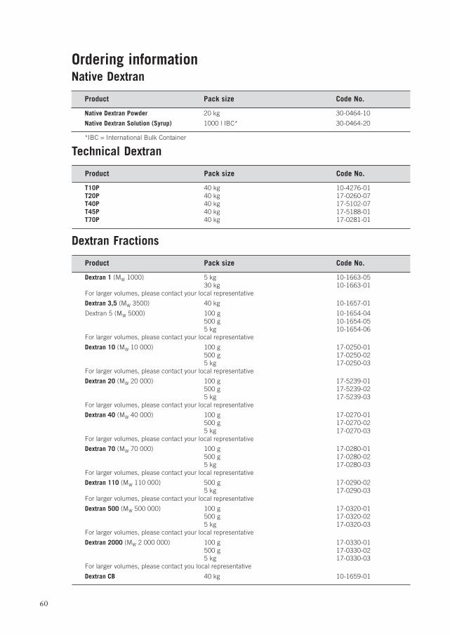
60
Ordering informationNative Dextran
Product Pack size Code No.
Native Dextran Powder 20 kg 30-0464-10
Native Dextran Solution (Syrup) 1000 l IBC* 30-0464-20
*IBC = International Bulk Container
Technical Dextran
Product Pack size Code No.
T10P 40 kg 10-4276-01T20P 40 kg 17-0260-07T40P 40 kg 17-5102-07T45P 40 kg 17-5188-01T70P 40 kg 17-0281-01
Dextran Fractions
Product Pack size Code No.
Dextran 1 (MW 1000) 5 kg 10-1663-0530 kg 10-1663-01
For larger volumes, please contact your local representative
Dextran 3,5 (MW 3500) 40 kg 10-1657-01
Dextran 5 (MW 5000) 100 g 10-1654-04500 g 10-1654-055 kg 10-1654-06
For larger volumes, please contact your local representative
Dextran 10 (MW 10 000) 100 g 17-0250-01500 g 17-0250-025 kg 17-0250-03
For larger volumes, please contact your local representative
Dextran 20 (MW 20 000) 100 g 17-5239-01500 g 17-5239-025 kg 17-5239-03
For larger volumes, please contact your local representative
Dextran 40 (MW 40 000) 100 g 17-0270-01500 g 17-0270-025 kg 17-0270-03
For larger volumes, please contact your local representative
Dextran 70 (MW 70 000) 100 g 17-0280-01500 g 17-0280-025 kg 17-0280-03
For larger volumes, please contact your local representative
Dextran 110 (MW 110 000) 500 g 17-0290-025 kg 17-0290-03
For larger volumes, please contact your local representative
Dextran 500 (MW 500 000) 100 g 17-0320-01500 g 17-0320-025 kg 17-0320-03
For larger volumes, please contact your local representative
Dextran 2000 (MW 2 000 000) 100 g 17-0330-01500 g 17-0330-025 kg 17-0330-03
For larger volumes, please contact you local representative
Dextran CB 40 kg 10-1659-01
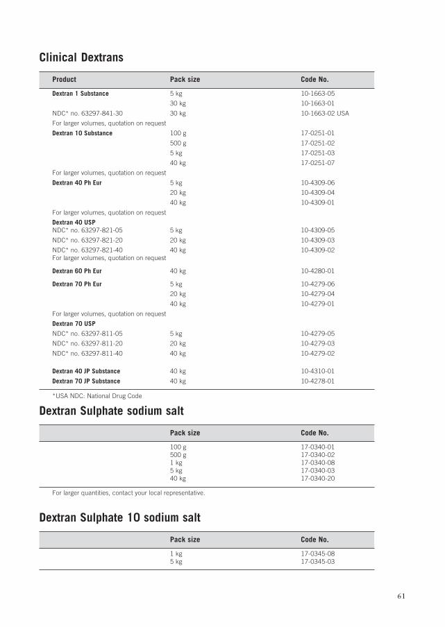
61
Clinical Dextrans
Product Pack size Code No.
Dextran 1 Substance 5 kg 10-1663-05
30 kg 10-1663-01
NDC* no. 63297-841-30 30 kg 10-1663-02 USA
For larger volumes, quotation on request
Dextran 10 Substance 100 g 17-0251-01
500 g 17-0251-02
5 kg 17-0251-03
40 kg 17-0251-07
For larger volumes, quotation on request
Dextran 40 Ph Eur 5 kg 10-4309-06
20 kg 10-4309-04
40 kg 10-4309-01
For larger volumes, quotation on request
Dextran 40 USPNDC* no. 63297-821-05 5 kg 10-4309-05
NDC* no. 63297-821-20 20 kg 10-4309-03
NDC* no. 63297-821-40 40 kg 10-4309-02For larger volumes, quotation on request
Dextran 60 Ph Eur 40 kg 10-4280-01
Dextran 70 Ph Eur 5 kg 10-4279-06
20 kg 10-4279-04
40 kg 10-4279-01
For larger volumes, quotation on request
Dextran 70 USP
NDC* no. 63297-811-05 5 kg 10-4279-05
NDC* no. 63297-811-20 20 kg 10-4279-03
NDC* no. 63297-811-40 40 kg 10-4279-02
Dextran 40 JP Substance 40 kg 10-4310-01
Dextran 70 JP Substance 40 kg 10-4278-01
*USA NDC: National Drug Code
Dextran Sulphate sodium salt
Pack size Code No.
100 g 17-0340-01500 g 17-0340-021 kg 17-0340-085 kg 17-0340-0340 kg 17-0340-20
For larger quantities, contact your local representative.
Dextran Sulphate 10 sodium salt
Pack size Code No.
1 kg 17-0345-085 kg 17-0345-03
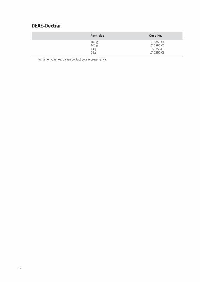
62
DEAE-Dextran
Pack size Code No.
100 g 17-0350-01500 g 17-0350-021 kg 17-0350-095 kg 17-0350-03
For larger volumes, please contact your representative.

63
Percoll, Sephadex, Sepharose, Sephacyl, Ficoll, Ficoll-Paque and Cytodex, are trademarks of Amersham Biosciences Limitedor its subsidiaries.
Amersham Biosciences are trademarks of Amersham plc
All goods and services are sold subject to the terms and conditions of sale of the company withinthe Amersham Biosciences group that supplies them.
A copy of these terms and conditions is available on request.© Amersham Biosciences AB 2003 – All rights reserved.
Amersham Biosciences AB Björkgatan 30, SE-751 84 Uppsala, Sweden
Amersham Biosciences UK Limited Amersham Place, Little Chalfont, Buckinghamshire HP7 9NA, EnglandAmersham Biosciences Inc 800 Centennial Avenue, PO Box 1327, Piscataway, NJ 08855 USA
Amersham Biosciences Europe GmbH Munzinger Strasse 9, D-79111 Freiburg, Germany
Amersham Biosciences KK, Sanken Bldg. 3-25-1, Hyakunincho Shinjuku-ku, Tokyo 169-0073 Japan

64
www.amershambiosciences.com
Related Documents