BIOLOGY FORM ONE NOTES INTRODUCTION TO BIOLOGY What is Biology? Biology is the branch of science that deals with the study of living things. In Greek, Bios means life while Logos means knowledge. Branches of biology There are two main branches: 1. Botany: Study of plants 2. Zoology: Study of animals The others include: 1. Ecology: Study of living things in their surroundings. 2. Genetics: The study of inheritance and variation. 3. Entomology: Study of insects 4. Parasitology: Study of parasites 5. Taxonomy: Study of classification of organisms 6. Microbiology: Study of microscopic organisms 7. Anatomy: Study of structure of cells 8. Cytology: Study of cells 9. Biochemistry: Study of chemical changes inside living organisms Name at least six other smaller branches of biology (6 marks). Importance of Biology 1. Solving environmental problems e.g. Food shortage, poor health services, pollution, misuse of environmental resources etc. 2. Choice of careers e.g. Medicine, Agriculture, public health, Veterinary, Animal husbandry, Horticulture, Dentistry etc. 3. Acquiring scientific skills e.g. observing, identifying, recording, classification, measuring, analyzing, evaluating etc. 4. International co-operation e.g. Development of HIV\AIDS vaccine, fight against severe Acute respiratory Syndrome (SARS), fight to save ozone layer from depletion, management of resources through international depletion. Others

Welcome message from author
This document is posted to help you gain knowledge. Please leave a comment to let me know what you think about it! Share it to your friends and learn new things together.
Transcript
BIOLOGY FORM ONE NOTES
INTRODUCTION TO BIOLOGY
What is Biology?
Biology is the branch of science that deals with the study of living
things. In Greek, Bios means life while Logos means knowledge.
Branches of biology
There are two main branches:
1. Botany: Study of plants
2. Zoology: Study of animals
The others include:
1. Ecology: Study of living things in their surroundings.
2. Genetics: The study of inheritance and variation.
3. Entomology: Study of insects
4. Parasitology: Study of parasites
5. Taxonomy: Study of classification of organisms
6. Microbiology: Study of microscopic organisms
7. Anatomy: Study of structure of cells
8. Cytology: Study of cells
9. Biochemistry: Study of chemical changes inside living
organisms
Name at least six other smaller branches of biology (6 marks).
Importance of Biology
1. Solving environmental problems e.g. Food shortage, poor
health services, pollution, misuse of environmental resources etc.
2. Choice of careers e.g. Medicine, Agriculture, public health,
Veterinary, Animal husbandry, Horticulture, Dentistry etc.
3. Acquiring scientific skills e.g. observing, identifying, recording,
classification, measuring, analyzing, evaluating etc.
4. International co-operation e.g. Development of HIV\AIDS
vaccine, fight against severe Acute respiratory Syndrome
(SARS), fight to save ozone layer from depletion, management of
resources through international depletion.
Others
Help on study of other subjects
Learn what living things are made up of and their bodies
work
Acquire knowledge about plant and animal diseases and
their treatment.
Know the effects of our bodies on drug and substance abuse
and can kill.
Learn about HIV\AIDS diseases and other viral diseases e.g.
its treatment—balanced diets, proper hygiene, spreading,
sexual behavior, cultural practices etc.
List five professional occupations that require the study of biology.
(5 marks)
Characteristics of living things;
1. Nutrition: Process by which living things acquire and utilize
nutrients: plants photosynthesize; animals feed on already
manufactured foods.
2. Respiration: energy-producing process occurring in all the cells
of living things.
3. Gaseous Exchange: where living things take in air (oxygen) and
give out air(carbon iv oxide) across respiratory surfaces.
4. Excretion: Process by which waste or harmful materials resulting
from chemical reactions within cells of living things are
eliminated. Excess of such materials poison living things.
5. Growth and Development: Growth –is the irreversible increase
in size and Mass.—Essential for body function. Development –
Irreversible change in complexity of the structure of living things.
6. Reproduction: Process by which living things give rise to new
individuals of the same kind.
7. Irritability: Is the ability of living things to perceive changes in
their surroundings and respond to them appropriately. E.g.
reaction to changes in temperature, humidity, light, pressure and
to the presence of certain chemicals.
8. Movement: Change in position by either a part or the whole
living thing. Locomotion – Progressive change in position by the
whole living thing. In animals, movement include; swimming,
walking, running, flying. In plants, closing of leaves, folding of
leaves, closing of flowers, growing of shoots towards light etc.
Question
1. List four uses of energy obtained from the process of
respiration. (4 marks).
2. List six characteristics of living things (6 marks).
Collection of specimens
Apparatus used
1. Sweep net: for catching flying insects.
2. Fish net: For trapping small fish and other small water
animals.
3. Pooter:For sucking small animals from rock surfaces and
tree barks.
4. Bait trap: For attracting and trapping small animals e.g.
rats.
5. Pit fall trap: For catching crawling animals.
6. Pair of forceps: picking up small crawling animals e.g.
stinging insects.
7. Specimen bottles: keeping collected specimen. Larger
specimens require large bottles.
8. The magnifying lens: Instrument used to enlarge objects.
Lenses are found in microscope and the hand lens
(magnifier). Its frame is marked e.g. x8 or x10—indicating
how much larger will be the image compared to object.
Precautions during Collection and Observation of specimens
Collect only the number of specimen you need.
Do not harm the specimens during the capture or collection
exercise.
Handle dangerous or injurious specimens with care e.g.
stinging plants or insects i.e. use forceps or hand gloves.
The teacher will immobilize highly mobile animals. (diethyl
ether, formalin, chloroform)
Do not destroy the natural habitat of the specimens.
Practical activity 2
Practical activity 3
Comparison between plants and animals

Plants Animals
1. Green in colour( have
chlorophyll)
1. Lack chlorophyll thus
feed on readymade food.
2. Their cells have
cellulose cell walls.
2. Cells lack cellulose cell
walls.
3. Respond slowly to
changes in the
environment.
3. Respond quickly.
4. Lack specialized
excretory organs.
4. Have complex excretory
organs.
5. Do not move about. 5. Move about in search of
food and water.
6. Growth occurs in shoot
and root tips.(apical
growth)
6.Growth occurs in all body
parts9intercalary growth).
Revision questions
CLASSIFICATION I
INTRODUCTION
Living things are also known as living organisms.
Organisms (forms of life) have distinguishing characteristics and
therefore are grouped.
The Magnifying lens
-Is used for enlarging small objects.
(Diagram)
Procedure of its use
Place the object on the bench.
Move the hand lens from the object to the eye.
An enlarged image is seen.
Drawing magnification = Length of the drawing/ drawing Length
Length of the object/Actual Length
(Diagram)
External features of plants and animals
External features of plants
i) Rhizoids as in moss plant.
ii) Fronds in ferns.
iii) Roots, stems, leave, flowers, seeds, fruits, and cones in higher
plants.
External features of animals
i) Tentacles in hydra
ii) Feathers in birds
iii) Shells in snails
iv) Wings in birds
v) Fur and hair in mammals
vi) Scales and fins in fish
vii) Proglotids in tapeworms
viii) Mammary glands in mammals
ix) Locomotory Structures e.g. limbs in insects
x) Body pigmentation
Practical activity 1
To collect and observe animal specimens
To collect and observe plant specimens
What is classification?
-Is an area of biology that deals with the grouping of living organisms
according to their structure. Organisms with similar structures are put
under one group referred to as a taxon—taxa (plural).
The groupings also consider evolutionary relationships (phylogeny)—
since all living organisms had a common origin at one time.
Taxonomy—Science of classification.
Taxonomist—Biologist who studies taxonomy.
Need for classification.
Reasons
1. To identify living organisms into their correct groups for
reference and study
2. To bring together living organisms with similar characteristics but
separate those with different features.
3. To arrange information of living organisms in an orderly manner.
This avoids chaos and confusion.
4. To understand the evolutionary relationship between different
organisms
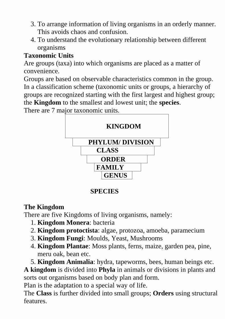
Taxonomic Units
Are groups (taxa) into which organisms are placed as a matter of
convenience.
Groups are based on observable characteristics common in the group.
In a classification scheme (taxonomic units or groups, a hierarchy of
groups are recognized starting with the first largest and highest group;
the Kingdom to the smallest and lowest unit; the species.
There are 7 major taxonomic units.
KINGDOM
PHYLUM/ DIVISION
CLASS
ORDER
FAMILY
GENUS
SPECIES
The Kingdom There are five Kingdoms of living organisms, namely:
1. Kingdom Monera: bacteria
2. Kingdom protoctista: algae, protozoa, amoeba, paramecium
3. Kingdom Fungi: Moulds, Yeast, Mushrooms
4. Kingdom Plantae: Moss plants, ferns, maize, garden pea, pine,
meru oak, bean etc.
5. Kingdom Animalia: hydra, tapeworms, bees, human beings etc.
A kingdom is divided into Phyla in animals or divisions in plants and
sorts out organisms based on body plan and form.
Plan is the adaptation to a special way of life.
The Class is further divided into small groups; Orders using structural
features.
Orders are divided into families using structural features, then
Families into Genera (singular genus) –based on recent common
ancestral features that are less adaptive.
Genus is divided into species i.e. kind of plant, or animal.
Down the hierarchy, the number of organisms in each group decreases
but their similarities increases.
The Species group members naturally interbreed to produce fertile off
springs.
Minor differences are exhibited in the species groups e.g. on colour of
the skin in human beings and varieties of plants.
The groups of the species are termed to as varieties, races or strains.
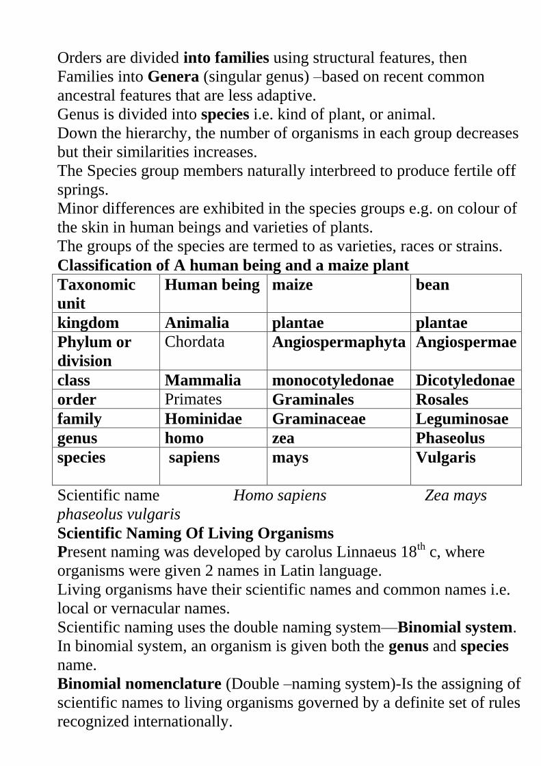
Classification of A human being and a maize plant
Taxonomic
unit
Human being maize bean
kingdom Animalia plantae plantae
Phylum or
division
Chordata Angiospermaphyta Angiospermae
class Mammalia monocotyledonae Dicotyledonae
order Primates Graminales Rosales
family Hominidae Graminaceae Leguminosae
genus homo zea Phaseolus
species sapiens mays Vulgaris
Scientific name Homo sapiens Zea mays
phaseolus vulgaris
Scientific Naming Of Living Organisms
Present naming was developed by carolus Linnaeus 18th c, where
organisms were given 2 names in Latin language.
Living organisms have their scientific names and common names i.e.
local or vernacular names.
Scientific naming uses the double naming system—Binomial system.
In binomial system, an organism is given both the genus and species
name.
Binomial nomenclature (Double –naming system)-Is the assigning of
scientific names to living organisms governed by a definite set of rules
recognized internationally.
Principles of binomial nomenclature
a) The first, genus name, should begin with a capital letter and the
second name, species, should begin or written in small letters e.g.
Lion---- Panthera leo
Leopard----- Panthera pardus
Domestic dog----- Canis farmiliaris
Human being--- Homo sapiens
Maize plant---Zea mays
Lion and Leopard are closely related ---Same genus but distantly
related—different species.
b) The scientific names must be printed in italics in textbooks and
where hand written to be underlined e.g. Panthera leo.
c) The specific name (species) is frequently written with the name of
the scientist who first adequately described and named the
organism e.g.Phaseolus vulgaris i.e. Vulgaris is the scientist who
described and named the bean plant.
d) Biologists should give a Latinized name for a newly described
animal or plant species where Latin name is missing e.g.
Meladogyne kikuyuensis – Is a scientific name of a nematode
from kikuyu.
Aloe kilifiensis --- A member of Aloeceae family from Kilifi
discovery.
Garinsoga parviflora waweruensis --- a member of Macdonald
eye family discovered by Waweru.
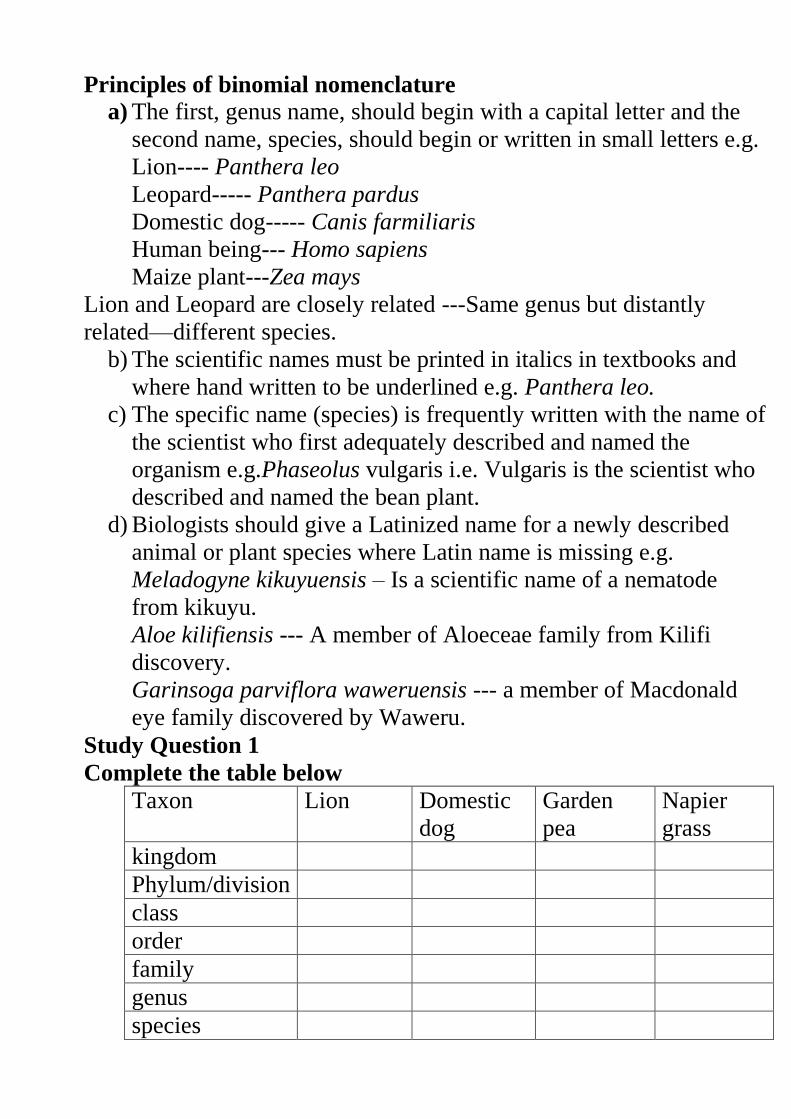
Study Question 1
Complete the table below
Taxon Lion Domestic
dog
Garden
pea
Napier
grass
kingdom
Phylum/division
class
order
family
genus
species
Scientific name --------------------- ------------------------ --
--------------------- ------------------------
Revision Questions:
CLASSIFICATION 1
Review of the magnification lens
Calculating Magnification
External characteristics of plants and animals
Diversity of Living Organisms
Organisms with similar characteristics are placed under one group
called taxon (taxa).
The science of classification is known as taxonomy.
Biologists who study taxonomy are called taxonomists.
Need For Classification
1. Help in identifying living organisms into their correct groups for
reference.
2. It brings together organisms with similar characteristics and
separates those with different features.
3. Help to organize information about living organisms in an orderly
manner avoiding any confusion.
4. Help to understand the evolutionary relationship between
different living organisms.
Historical Background of Classification
Long time ago classification was artificial where living things
were classified as either plants or animals.
Plants were classified as herbs, shrubs and trees.
Animals were further divided into carnivores, herbivores and
omnivores.
Today modern classification uses evolutionary relationships
between living organisms.
Taxonomic Units of Classification
This refers to the groups into which living organisms are placed
in classification.
These units start from the first largest and highest group
(kingdom) to the smallest and lowest unit (species).
There are seven taxonomic units as shown below.
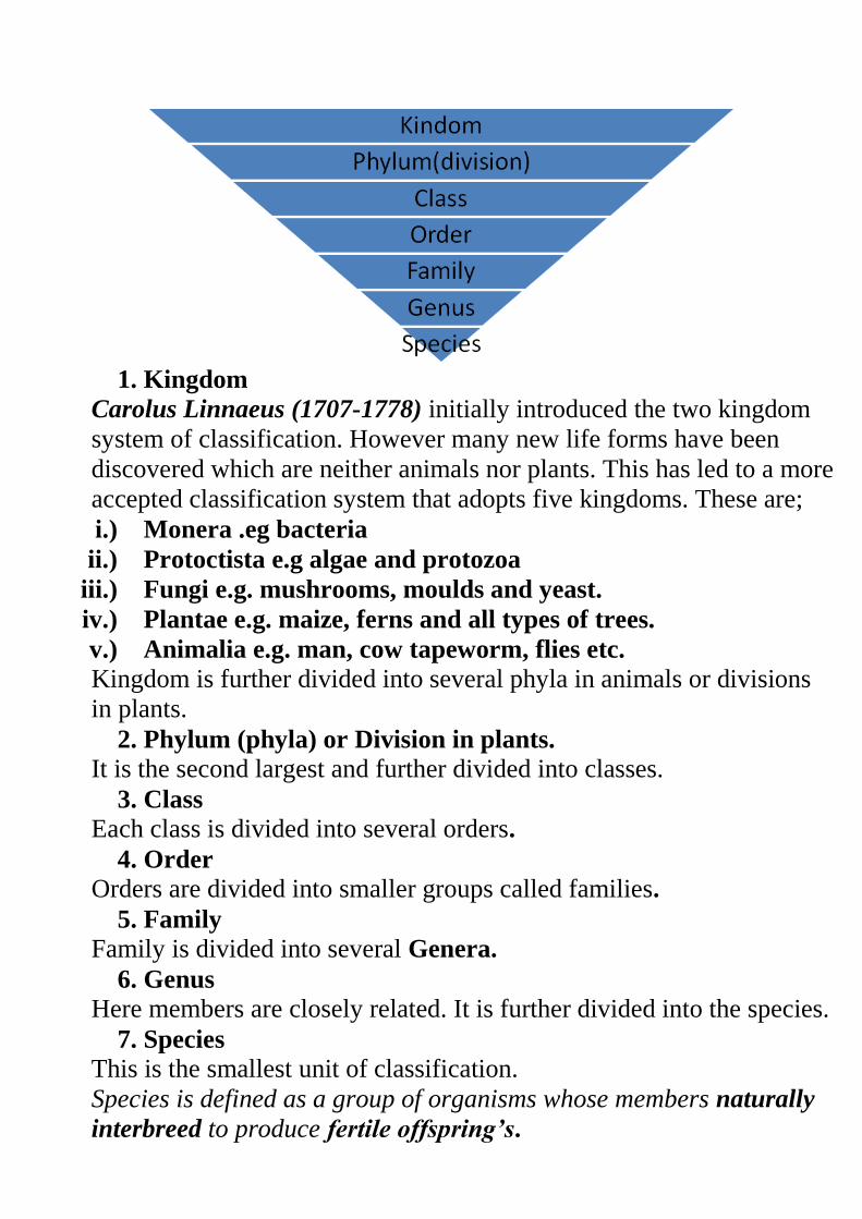
1. Kingdom
Carolus Linnaeus (1707-1778) initially introduced the two kingdom
system of classification. However many new life forms have been
discovered which are neither animals nor plants. This has led to a more
accepted classification system that adopts five kingdoms. These are;
i.) Monera .eg bacteria
ii.) Protoctista e.g algae and protozoa
iii.) Fungi e.g. mushrooms, moulds and yeast.
iv.) Plantae e.g. maize, ferns and all types of trees.
v.) Animalia e.g. man, cow tapeworm, flies etc.
Kingdom is further divided into several phyla in animals or divisions
in plants.
2. Phylum (phyla) or Division in plants.
It is the second largest and further divided into classes.
3. Class
Each class is divided into several orders.
4. Order
Orders are divided into smaller groups called families.
5. Family
Family is divided into several Genera.
6. Genus
Here members are closely related. It is further divided into the species.
7. Species
This is the smallest unit of classification.
Species is defined as a group of organisms whose members naturally
interbreed to produce fertile offspring’s.
Members of a given species have small differences such as skin
colour, height etc.
Classification of Man and Maize plant. ( Table 2.1 Page 15 KLB Bk
1)
Scientific Naming of Living Organisms.
Today organisms are given two names in Latin language. This
was developed by Carolus Linnaeus.
Latin language was used because it was widely spoken during his
time.
In scientific naming, an organism is given the genus and the
species name.
This double naming system is known as Binomial system (two
name System)
Binomial Nomenclature.
This is the double naming system of organisms where organisms are
assigned two names i.e. the generic name and the specific name.
In binomial nomenclature the following rules are observed.
i.) Generic name is written first followed by the specific name. First
letter in the generic name is in capital and the rest are in small
letters. Specific name is written in small letters.
ii.) The two names are underlined separately when handwritten or
italicised when printed.
iii.) Newly discovered species must be given Latinized names.
iv.) Specific name is frequently written with the name of the scientist
who first adequately described and named the organism.
Examples
Revision Questions
CELL PHYSIOLOGY
This is the study of the functions of cell structures.
Membrane Structure and Properties
A membrane is a surface structure which encloses the cell and
organelles. Membranes regulate the flow of materials into out of
the cell or organelle.
Examples of membranes: cell membrane, tonoplast (membrane
surrounding the vacuole), nuclear membrane, mitochondrial
membrane, chloroplast membrane etc.
The Cell Membrane
It has three layers, two protein layers and a phos-pholipid layer
sandwiched in between the two.
Diagram
Properties of Cell Membrane
1. Semi-permeability. – It has small pores allowing for the passage
of molecules of small size into and out of the cell. Cell Wall
however allows all materials to pass through it hence it is referred
to as being Permeable.
2. Sensitivity to Changes in Temperature and pH – Extreme
temperature and pH affects the cell membrane since it has some
protein layers. Such changes alter the structure of the membrane
affecting its normal functioning.
3. Possession of Electric Charges – it has both the negative and
positive charges helping the cell to detect changes in the
environment. These charges also affect the manner in which
substances move in and out of the cell
Physiological Processes
The ability of the cell to control the movement of substances in
and out of the cell is achieved through physiological processes
such as Diffusion, Osmosis and Active Transport.
Diffusion
This is a process by which particles move from a region of high
concentration to a region of low concentration.
Practical Activity 1
To demonstrate diffusion using potassium permanganate (VII)
The difference in concentration of particles between the region of
high concentration and the region of low concentration is known
as the diffusion gradient.
Role of Diffusion in Living Organisms
1. Absorption of Materials
Mineral salts in the soil enter the root by diffusion since their
concentration in the soil is greater than in the root hair cells.
Digested food (glucose and amino acids) diffuse across the wall
of the ileum into the blood for transport to rest of the body.
2. Gaseous Exchange in Plants and Animals
In both plants and animals, respiratory gases (oxygen and Carbon
(IV) oxide) are exchanged through simple diffusion depending on
their concentration gradient.
3. Excretion of Nitrogenous Wastes
4. Transport of Manufactured Food form Leaves to other Plant
Parts.
5. Factors Affecting Diffusion
a) Diffusion Gradient
A greater diffusion gradient between two points increases the rate
of diffusion.
b) Surface Area to Volume Ratio
The higher the ratio the greater the rate of diffusion and the lower
the ratio the lower the rate.
This means that small organisms expose a large surface area to
the surrounding compared to large organisms.
Small organisms therefore depend on diffusion as a means of
transport of foods, respiratory gases and waste products.
Diagrams
c) Thickness of Membranes and Tissues
The thicker the membrane the lower the rate of diffusion because
the distance covered by the diffusing molecules is greater. The
thinner the membrane, the faster the rate.
Size of the Molecules
Small and light molecules diffuse faster than large and heavy
molecules.
d) Temperature
Increase in temperature increases the energy content in molecules
causing them to move faster.
Osmosis
This is the process where solvent molecules (water) move from a
lowly concentrated solution (dilute) to a highly concentrated
solution across a semi-permeable membrane.
Diagram fig 4.6
The highly concentrated solution is known as Hypertonic
Solution.
The lowly concentrated solution is called Hypotonic solution.
Solution of the same concentration are said to be Isotonic.
Osmosis is a special type of diffusion because it involves the
movement of solvent (water) molecules from their region of high
concentration to region of low concentration across a semi
permeable membrane.
Practical activity 2
Practical activity 3
Osmotic Pressure
This is the pressure which needs to be applied to a solution to
prevent the inward flow of water across a semi permeable
membrane. This is the pressure needed to nullify osmosis.
Osmotic pressure is measured using the osmometer.
Osmotic Potential
This is the measure of the pressure a solution would develop to
withdraw water molecules from pure water when separated by a
semi permeable membrane.
Water Relations in Animals
Cell membrane of the animal cell is semi permeable just like the
dialysis/visking tubing.
Cytoplasm contains dissolved sugars and salts in solution form.
If an animal cell e.g. a red blood cell is placed in distilled water
(hypotonic solution), water flows in by osmosis.
The cell would swell up and eventually burst because the cell
membrane is weak. The bursting of the red blood cell when
placed in hypotonic solution is called Haemolysis.
If a similar red blood cell is placed in a hypertonic solution, water
is drawn out of the cell by osmosis. The cell will shrink by a
process called Crenation.
Body fluids surrounding the cells must therefore have same
concentration as to that which is found inside the cell.
Diagrams
Water Relations in Plants
When a plant cell is placed in a hypotonic solution it gains water
by osmosis and distends outwards.
As the cell gains more water, its vacuole enlarges and exerts an
outward pressure called turgor pressure. As more water is drawn
in, the cell becomes firm and rigid and is said to be turgid.
The cell wall in plant cell is rigid and prevents the cell from
bursting unlike the case in animal cells.
The cell wall develops a resistant pressure that pushes towards the
inside. This pressure is equal and opposite the turgor pressure and
is called wall pressure.
Diagrams
When a plant cell is placed in hypertonic solution, water
molecules move out of the cell into the solution by osmosis. The
cell shrinks and becomes flaccid.
If the cell continues to lose more water, plasma membrane pulls
away from the cell wall towards the center.
The process through which plant cells lose water, shrink and
become flaccid is called plasmolysis.
Plasmolysis can be reversed by placing a flaccid cell in distilled
water and this process is called deplasmolysis.
Study Question 5
Practical Activity 4
Wilting
When plants lose water through evaporation and transpiration,
cells lose turgidity, shrink and the plant droops. This is called
wilting.
If water supply from the soil is inadequate, plants do not recover
hence permanent wilting.
Study Question 6
Role of Osmosis in Organisms
1. Absorption of water from the soil
Root hair cells of plants absorb water from the soil by osmosis.
2. Support
Cells of herbaceous plants, which are less woody, absorb
water, become turgid hence support.
3. Opening and closing of the stomata
During the day, guard cells synthesize glucose, draw in water,
become turgid hence open the stomata.
During the night, they lose turgidity since there is no
photosynthesis. As a result, they shrink thus closing the
stomata.
4. Feeding in insectivorous plants
These plants are able to change their turgor pressure on the
leaves which close trapping insects which are digested to
provide the plant with nitrogen.
5. Osmoregulation
In the kidney tubules, water is reabsorbed back to the body by
osmosis.
Factors Affecting Osmosis i.) Concentration of Solutions and Concentration Gradient. The
greater the concentration gradient between two points, the faster
the rate of osmosis.
ii.) Optimum Temperature as long as it does not destroy the
semi-permeability of the membrane.
Active Transport
This is the process that moves substances across cell
membranes against a concentration gradient.
This process requires energy to move these substances across
cell membranes and involves carriers.
Substances such as amino acids, sugar and many ions are taken
in by living organisms through active transport.
Role of Active Transport
i.) Re-absorption of sugars and useful substances by the kidney
ii.) Absorption of some mineral salts by plant roots
iii.) Absorption of digested food from the alimentary canal into
the blood stream
iv.) Accumulation of substances in the body to offset osmotic
imbalance in arid and saline environment
v.) Excretion of waste products from body cells
Factors Affecting Active Transport.
i.) Oxygen concentration.
ii.) Change in pH.
iii.) Glucose concentration.
iv.) Temperature.
v.) Enzyme inhibitors.
NB/ Any factor affecting energy production affect the rate of active
transport.
Revision Questions.
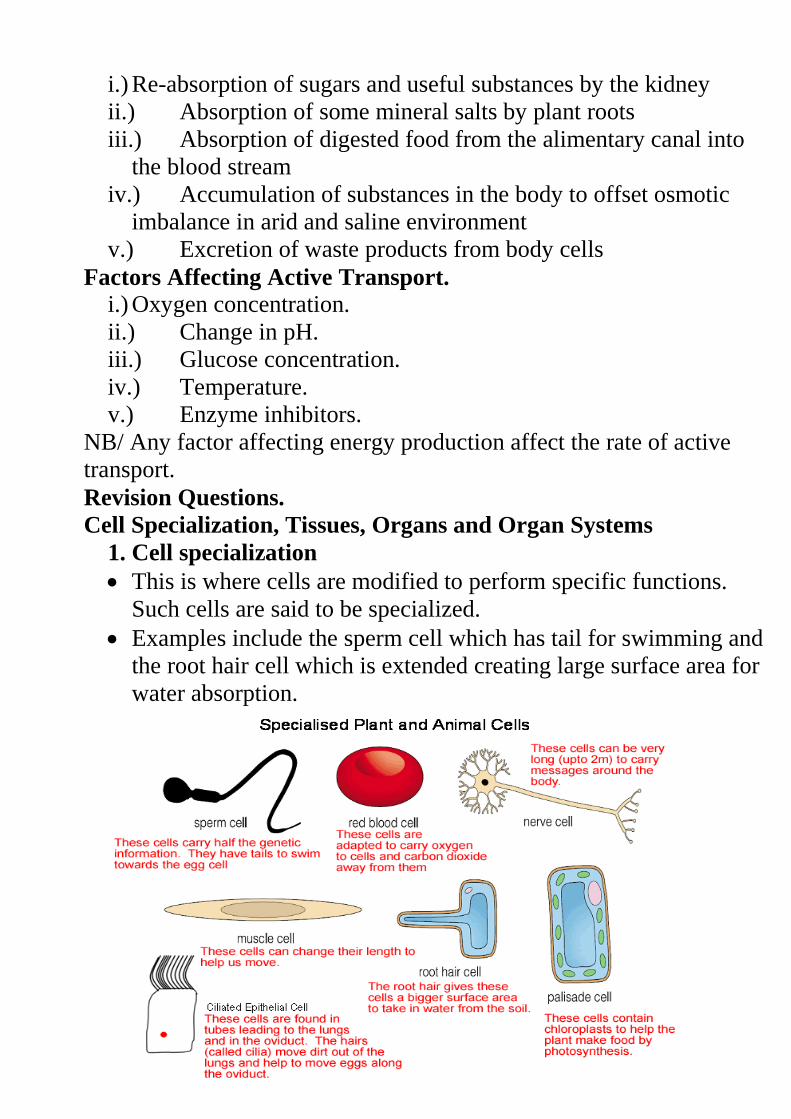
Cell Specialization, Tissues, Organs and Organ Systems
1. Cell specialization
This is where cells are modified to perform specific functions.
Such cells are said to be specialized.
Examples include the sperm cell which has tail for swimming and
the root hair cell which is extended creating large surface area for
water absorption.
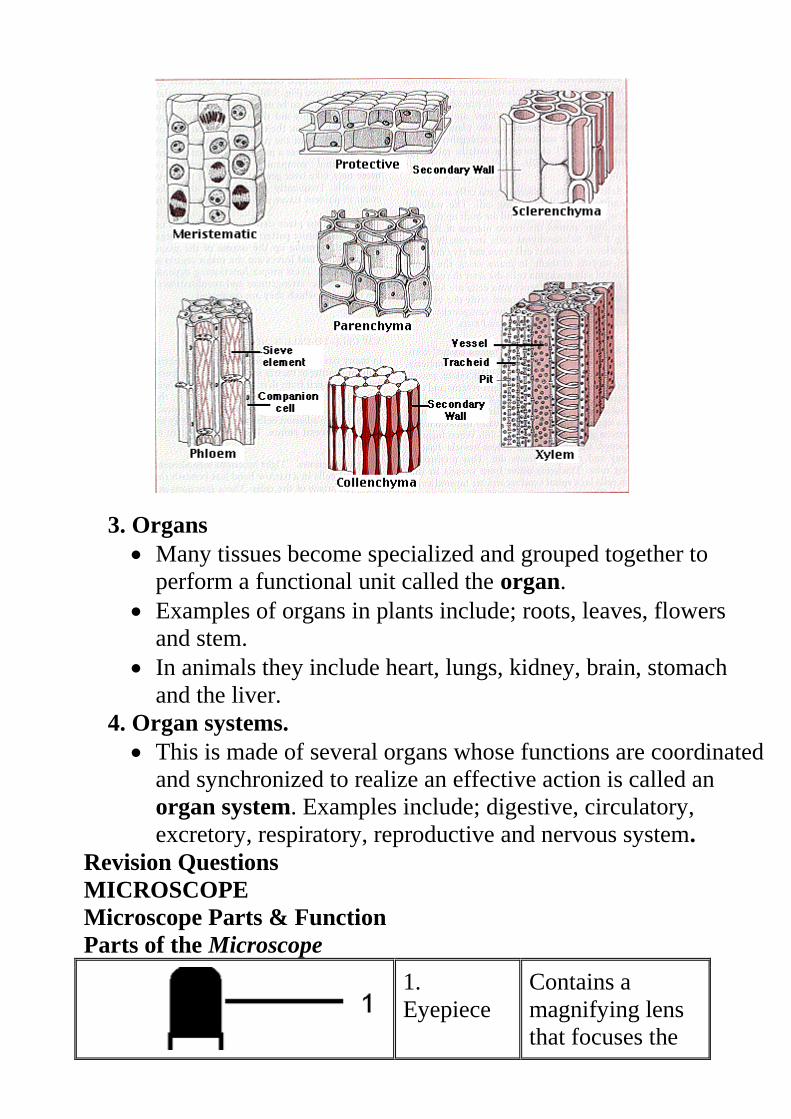
2. Tissues.
These are cells of a particular type that are grouped together to
perform the same function.
Animal tissues include;
- Epithelial tissue – which is a thin continuous layer of cells for
lining and protection of internal and external surfaces.
- Skeletal – it is a bundle of elongated cells with fibres that can
contract. Its contraction and relaxation brings about movement.
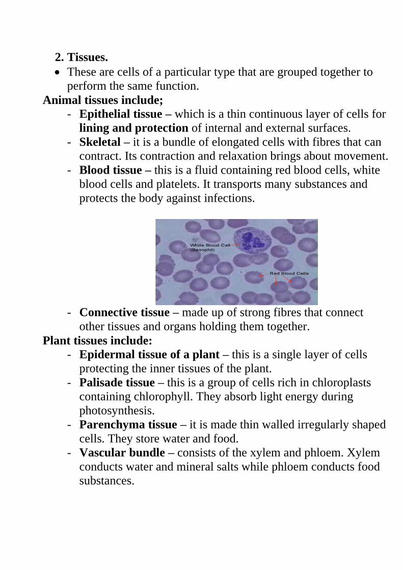
- Blood tissue – this is a fluid containing red blood cells, white
blood cells and platelets. It transports many substances and
protects the body against infections.
- Connective tissue – made up of strong fibres that connect
other tissues and organs holding them together.
Plant tissues include: - Epidermal tissue of a plant – this is a single layer of cells
protecting the inner tissues of the plant.
- Palisade tissue – this is a group of cells rich in chloroplasts
containing chlorophyll. They absorb light energy during
photosynthesis.
- Parenchyma tissue – it is made thin walled irregularly shaped
cells. They store water and food.
- Vascular bundle – consists of the xylem and phloem. Xylem
conducts water and mineral salts while phloem conducts food
substances.
3. Organs
Many tissues become specialized and grouped together to
perform a functional unit called the organ.
Examples of organs in plants include; roots, leaves, flowers
and stem.
In animals they include heart, lungs, kidney, brain, stomach
and the liver.
4. Organ systems.
This is made of several organs whose functions are coordinated
and synchronized to realize an effective action is called an
organ system. Examples include; digestive, circulatory,
excretory, respiratory, reproductive and nervous system.
Revision Questions
MICROSCOPE
Microscope Parts & Function
Parts of the Microscope
1.
Eyepiece
Contains a
magnifying lens
that focuses the
image from the
objective into
your eye.
2. Course
Adjust
For focusing
under low
magnification
3. Fine
Adjust
For focusing
under high
magnification or
low
4. Low
Power
Objective
For large
specimens or
overview
5. High
Power
Objective
For detailed
viewing or small
specimens
6.
Specimen
on glass
slide
What you want to
look at
7. Stage Supports
specimen in
correct location
to lens
8.
Condenser
Focuses the light
on specimen
9.
Diaphragm
(iris or
disc)
Regulates amount
of light and
contrast
10. Light
Source
Illuminates the
specimen for
viewing
Handling and Care of the Microscope
The following rule should be observed:
1. Use both hand when carrying the microscope. One hand should
hold the base and the other holds the limb.
2. Never place the microscope too close to the edge of the bench.
3. Do not touch the mirror and the lenses with the fingers.
4. Clean dirty lenses using soft tissue.
5. Clean other parts using a soft cloth.
6. Do not wet any part of the microscope.
7. Make sure the low power clicks into position in line with the eye
piece before and after use.
8. Always store the microscope in a safe place free from dust and
moisture.
Using the Microscope
1. Place microscope on the bench with the stage facing away from
you.
2. Turn the low power objective lens until it clicks into position.
3. Ensure the diaphragm is fully open.
4. Look through the eyepiece with one eye. Adjust the mirror to
ensure maximum light can pass through.
5. Place the slide containing the specimen on the stage and clip it
into position. Make sure the slide is at the centre of the field of
view.
6. Again look through the eyepiece while adjusting the mirror to
ensure maximum light reach the specimen.
7. Use the coarse adjustment knob to bring the low power objective
lens to the lowest point. While viewing through the eyepiece, turn
the coarse adjustment knob gently until the specimen comes into
focus.
8. Use the fine adjustment knob to bring the image into sharp focus.
9. Make a drawing of what you see.
10. For higher magnification, turn the medium power into
position and adjust the focus using the coarse knob. Use the fine
adjustment knob for sharper focus.
11. For even large magnifications, turn the high power objective
lens into position. In this case use only the fine adjustment knob
to bring details into sharper focus.
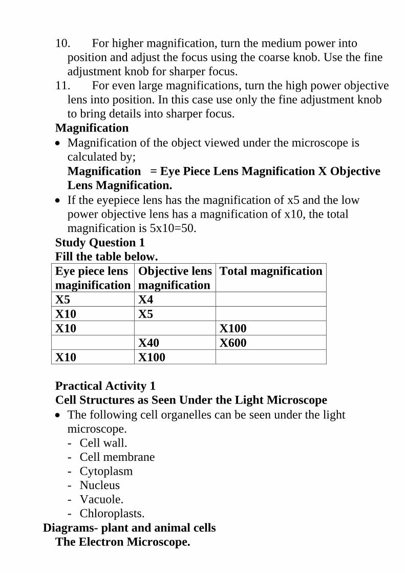
Magnification
Magnification of the object viewed under the microscope is
calculated by;
Magnification = Eye Piece Lens Magnification X Objective
Lens Magnification.
If the eyepiece lens has the magnification of x5 and the low
power objective lens has a magnification of x10, the total
magnification is 5x10=50.
Study Question 1
Fill the table below.
Eye piece lens
maginification
Objective lens
magnification
Total magnification
X5 X4
X10 X5
X10 X100
X40 X600
X10 X100
Practical Activity 1
Cell Structures as Seen Under the Light Microscope
The following cell organelles can be seen under the light
microscope.
- Cell wall.
- Cell membrane
- Cytoplasm
- Nucleus
- Vacuole.
- Chloroplasts.
Diagrams- plant and animal cells
The Electron Microscope.
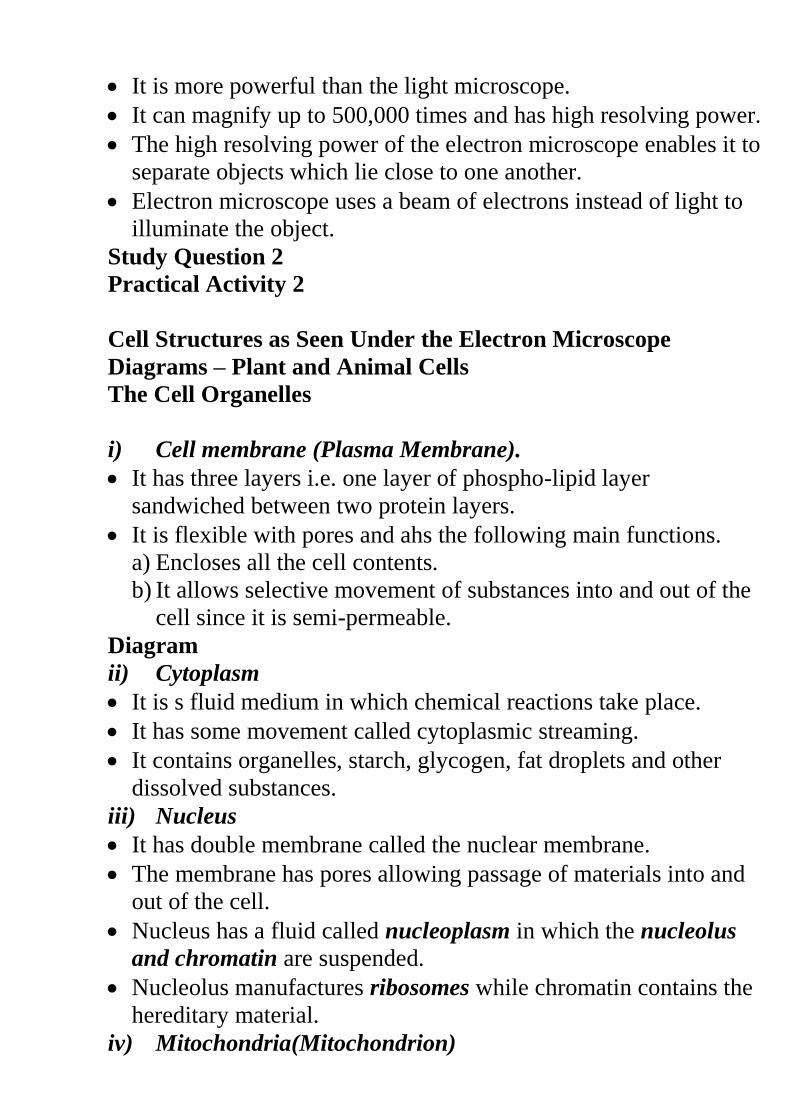
It is more powerful than the light microscope.
It can magnify up to 500,000 times and has high resolving power.
The high resolving power of the electron microscope enables it to
separate objects which lie close to one another.
Electron microscope uses a beam of electrons instead of light to
illuminate the object.
Study Question 2
Practical Activity 2
Cell Structures as Seen Under the Electron Microscope
Diagrams – Plant and Animal Cells
The Cell Organelles
i) Cell membrane (Plasma Membrane).
It has three layers i.e. one layer of phospho-lipid layer
sandwiched between two protein layers.
It is flexible with pores and ahs the following main functions.
a) Encloses all the cell contents.
b) It allows selective movement of substances into and out of the
cell since it is semi-permeable.
Diagram
ii) Cytoplasm
It is s fluid medium in which chemical reactions take place.
It has some movement called cytoplasmic streaming.
It contains organelles, starch, glycogen, fat droplets and other
dissolved substances.
iii) Nucleus
It has double membrane called the nuclear membrane.
The membrane has pores allowing passage of materials into and
out of the cell.
Nucleus has a fluid called nucleoplasm in which the nucleolus
and chromatin are suspended.
Nucleolus manufactures ribosomes while chromatin contains the
hereditary material.
iv) Mitochondria(Mitochondrion)
They are sausage shaped and are the respiratory sites.
Mitochondrion has two membranes. Inner membrane is greatly
folded into cristae to increase the surface area for respiration.
Cells that require a lot of energy have large number of
mitochondria e.g. muscle cell, sperm cell, kidney cell etc.
Diagram
v) Endoplasmic Reticulum (ER)
Some endoplasmic reticulums have granules called Ribosomes on
their surfaces hence referred to as rough endoplasmic reticulum.
Others do not contain ribosomes hence the name smooth
endoplasmic reticulum.
Rough endoplasmic reticulum transport proteins while the
smooth endoplasmic reticulum transports lipids.
Diagrams
vi) Ribosomes
They are spherical in shape and form the site for protein
synthesis.
vii) Lysosomes
They contain lytic enzymes which break down large molecules,
destroy worn out organelles or even the entire cell.
viii) Golgi Bodies (Golgi apparatus)
Their function is to package and transport glyco-proteins.
They are also associated with secretion of synthesized proteins
and carbohydrates.
Diagram
ix) Centrioles
They are rod shaped structures that are used in cell division and in
the formation of cilia and flagella.
Plant cells lack the Centrioles.
x) Chloroplasts
They are egg shaped and contain two membranes.
Chloroplast has chlorophyll which traps light energy to be used
during photosynthesis.
xi) Vacuoles
This are sacs filled with a fluid called cell sap.
Animal cells contain small vacuoles while plant cells have large
vacuoles.
Sap vacuoles store sugars and salts.
Food vacuole store and digest food while contractile vacuoles
excrete unwanted materials from the cell.
xii) Cell wall
It is a rigid outer cover of the plant cells made of cellulose.
It gives the plant cell a definite shape while providing
mechanical support and protection.
Cell wall also allows water, gases and other materials to pass
through it.
Study Question 3
Differences between Plant and Animal Cells
Preparation of Temporary Slides
Practical Activity 3
Estimation of Cell Sizes.
NUTRITION IN PLANTS AND ANIMALS
Nutrition
This is the process by which organisms obtain and Assimilate
nutrients.
There are two modes of nutrition; Autotrophism and
Heterotrophism.
Autotrophism
This is where living organism manufacture its own complex food
substances from simple substances such as carbon (iv) oxide,
water, light or chemical energy.
Where sunlight is used as a source of energy, the process is
referred to as photosynthesis.
Photo means light while synthesis means to make.
Some none green plants make their own food using energy
obtained from certain chemicals through a process called
chemosynthesis.
Organisms that make their own food are referred to as
autotrophs.
Heterotrophism
This is where organisms take in complex food materials such as
carbohydrates, proteins and fats obtained from bodies of plants
and animals.
Organisms that feed on already manufactured foods are called
Heterotrophs.
Autotrophism
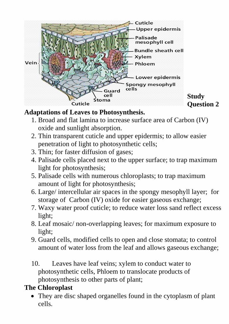
External Structure of a Leaf A leaf is a flattened organ which is attached to the stem or a branch of
a plant.
Diagrams
Parts of a leaf Lamina: This is the flat surface. It is green in colour and contain the
photosynthetic tissue.
Midrib: This is a thick structure running through the middle of the leaf
Veins: They arise from the midrib to forming an extensive network of
veins.
Leaf Apex: This is the tip of the leaf and usually it is pointed.
Petiole: It attaches the leaf to the stem or branch.
In some monocotyledonous plants the leaves are attached to the
stem by the leaf sheath.
Practical Activity 1: To examine the External Features of a
Dicotyledonous and Monocotyledonous leaf
Study Question 1
Internal Structure of a Leaf
Internal structure of the leaf is composed of the following parts.
i.) Cuticle.
It is a thin waterproof and transparent layer that coats the upper
and lower surfaces of the leaf.
It reduces excess water loss and protects the inner tissue of the
plant against mechanical injury.
It also prevents entry of disease causing micro organisms.
Since it is transparent, it allows penetration of light for
photosynthesis.
ii.) Epidermis.
It is a one cell thick tissue on both the upper and lower leaf
surfaces.
It secretes the cuticle and also protects the inner tissues from
mechanical damage and prevents entry of pathogens.
Epidermal cells have no chloroplast except the guard cells.
Guard cells are special bean shaped cells. They have chloroplast
and are able to carry out photosynthesis hence controlling the
opening and closing of the stomata.
Air moves into and out of the leaf through the stomata.
iii.) Palisade layer.
This is layer of cells located beneath the upper epidermis.
It is made of cylindrical shaped cells closely packed together.
They have numerous chloroplasts containing chlorophyll.
Their position and arrangement enables them to receive
maximum light.
iv.) Spongy Mesophyll Layer.
This is below the palisade layer. The cells are irregularly shaped
and loosely packed creating large air spaces in between them.
The air spaces allow gases to diffuse in between the cells. They
contain fewer chloroplasts as compared to the palisade cells.
v.) Leaf Veins.
Each vein is a vascular bundle consisting of xylem and phloem.
Xylem conducts water and mineral salts from the roots to the
leaves while the phloem translocates manufactured food from the
leaves to the rest of the plant.
Study
Question 2
Adaptations of Leaves to Photosynthesis.
1. Broad and flat lamina to increase surface area of Carbon (IV)
oxide and sunlight absorption.
2. Thin transparent cuticle and upper epidermis; to allow easier
penetration of light to photosynthetic cells;
3. Thin; for faster diffusion of gases;
4. Palisade cells placed next to the upper surface; to trap maximum
light for photosynthesis;
5. Palisade cells with numerous chloroplasts; to trap maximum
amount of light for photosynthesis;
6. Large/ intercellular air spaces in the spongy mesophyll layer; for
storage of Carbon (IV) oxide for easier gaseous exchange;
7. Waxy water proof cuticle; to reduce water loss sand reflect excess
light;
8. Leaf mosaic/ non-overlapping leaves; for maximum exposure to
light;
9. Guard cells, modified cells to open and close stomata; to control
amount of water loss from the leaf and allows gaseous exchange;
10. Leaves have leaf veins; xylem to conduct water to
photosynthetic cells, Phloem to translocate products of
photosynthesis to other parts of plant;
The Chloroplast
They are disc shaped organelles found in the cytoplasm of plant
cells.
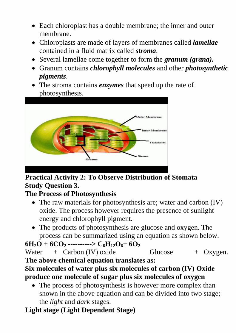
Each chloroplast has a double membrane; the inner and outer
membrane.
Chloroplasts are made of layers of membranes called lamellae
contained in a fluid matrix called stroma.
Several lamellae come together to form the granum (grana).
Granum contains chlorophyll molecules and other photosynthetic
pigments.
The stroma contains enzymes that speed up the rate of
photosynthesis.
Practical Activity 2: To Observe Distribution of Stomata
Study Question 3.
The Process of Photosynthesis
The raw materials for photosynthesis are; water and carbon (IV)
oxide. The process however requires the presence of sunlight
energy and chlorophyll pigment.
The products of photosynthesis are glucose and oxygen. The
process can be summarized using an equation as shown below.
6H2O + 6CO2 ----------> C6H12O6+ 6O2 Water + Carbon (IV) oxide Glucose + Oxygen.
The above chemical equation translates as:
Six molecules of water plus six molecules of carbon (IV) Oxide
produce one molecule of sugar plus six molecules of oxygen
The process of photosynthesis is however more complex than
shown in the above equation and can be divided into two stage;
the light and dark stages.
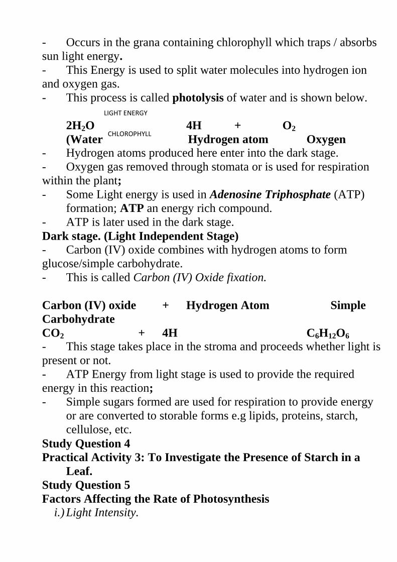
Light stage (Light Dependent Stage)
- Occurs in the grana containing chlorophyll which traps / absorbs
sun light energy.
- This Energy is used to split water molecules into hydrogen ion
and oxygen gas.
- This process is called photolysis of water and is shown below.
2H2O 4H + O2
(Water) Hydrogen atom Oxygen
- Hydrogen atoms produced here enter into the dark stage.
- Oxygen gas removed through stomata or is used for respiration
within the plant;
- Some Light energy is used in Adenosine Triphosphate (ATP)
formation; ATP an energy rich compound.
- ATP is later used in the dark stage.
Dark stage. (Light Independent Stage) - Carbon (IV) oxide combines with hydrogen atoms to form
glucose/simple carbohydrate.
- This is called Carbon (IV) Oxide fixation.
Carbon (IV) oxide + Hydrogen Atom Simple
Carbohydrate
CO2 + 4H C6H12O6
- This stage takes place in the stroma and proceeds whether light is
present or not.
- ATP Energy from light stage is used to provide the required
energy in this reaction;
- Simple sugars formed are used for respiration to provide energy
or are converted to storable forms e.g lipids, proteins, starch,
cellulose, etc.
Study Question 4
Practical Activity 3: To Investigate the Presence of Starch in a
Leaf.
Study Question 5
Factors Affecting the Rate of Photosynthesis
i.) Light Intensity.
LIGHT ENERGY
CHLOROPHYLL
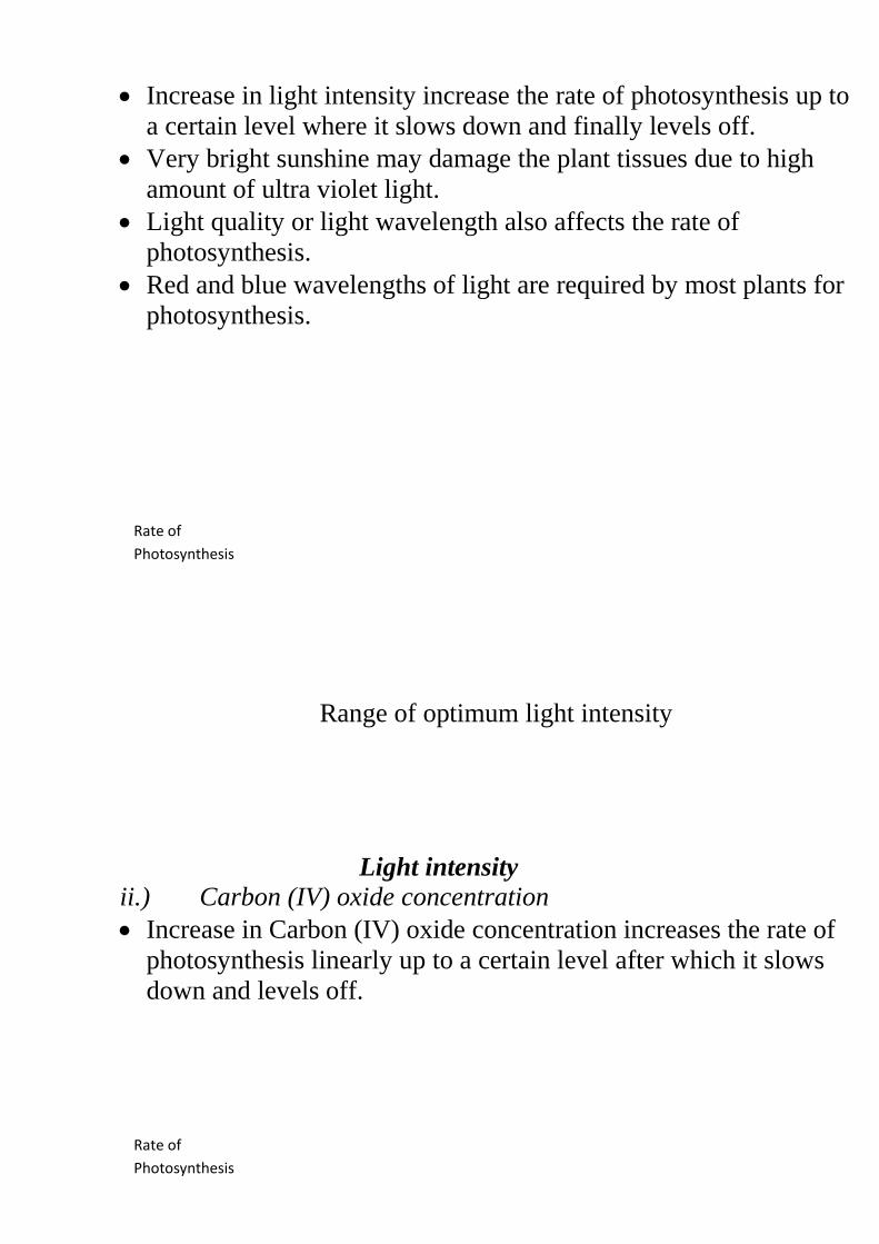
Increase in light intensity increase the rate of photosynthesis up to
a certain level where it slows down and finally levels off.
Very bright sunshine may damage the plant tissues due to high
amount of ultra violet light.
Light quality or light wavelength also affects the rate of
photosynthesis.
Red and blue wavelengths of light are required by most plants for
photosynthesis.
Range of optimum light intensity
Light intensity
ii.) Carbon (IV) oxide concentration
Increase in Carbon (IV) oxide concentration increases the rate of
photosynthesis linearly up to a certain level after which it slows
down and levels off.
Rate of
Photosynthesis
Rate of
Photosynthesis
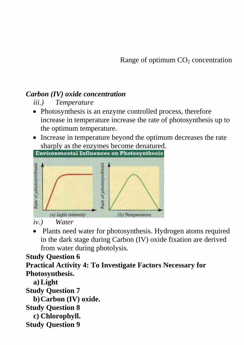
Range of optimum CO2 concentration
Carbon (IV) oxide concentration
iii.) Temperature
Photosynthesis is an enzyme controlled process, therefore
increase in temperature increase the rate of photosynthesis up to
the optimum temperature.
Increase in temperature beyond the optimum decreases the rate
sharply as the enzymes become denatured.
iv.) Water
Plants need water for photosynthesis. Hydrogen atoms required
in the dark stage during Carbon (IV) oxide fixation are derived
from water during photolysis.
Study Question 6
Practical Activity 4: To Investigate Factors Necessary for
Photosynthesis.
a) Light
Study Question 7
b) Carbon (IV) oxide.
Study Question 8
c) Chlorophyll.
Study Question 9
Study Question 10
Practical Activity 5: To Investigate the Gas Produced During
Photosynthesis.
Study Question 11
Chemical Compounds Which Constitute Living Organisms
Cells, tissues and organs are made of chemicals which are
referred to as chemicals of life.
The study of chemical compounds found in living organisms and
reactions in which they take part is called Biochemistry.
Chemicals of life include carbohydrates, lipids and proteins.
a) Carbohydrates
They are compounds of carbon, hydrogen and oxygen in the ratio
of 1:2:1 respectively.
Carbohydrates have a general formula of (CH2O)n where n
represents the number of carbon atoms in a molecule of
carbohydrate.
Carbohydrates are divided into three groups; Monosaccharide’s,
Disaccharides and Polysaccharides.
i) Monosaccharides
They are the simplest carbohydrates and have a general chemical
formula of (CH2O)n where n = 6.
Their chemical formular is therefore C6H12O6. They include;
glucose, fructose, galactose etc.
Properties of Monosaccharides
i) They are soluble in water to form sweet tasting solutions.
ii) They are crystalissable.
iii) They have the reducing property where they reduce copper
sulphate in Benedicts solution to red copper (I) oxide.
Functions
i) They are oxidized to release energy during respiration.
ii) When condensed together, they form polysaccharides such as
starch, cellulose or glycogen.
ii) Disaccharides
They are formed by linking two Monosaccharide molecules
through the process of condensation where a molecule of water is
liberated.
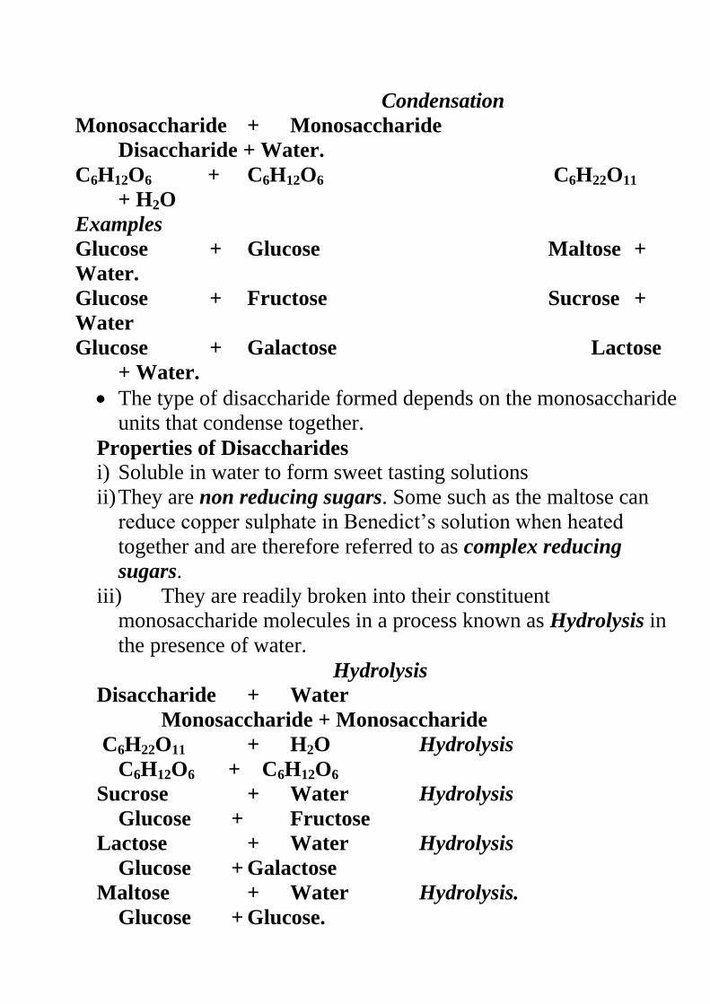
Condensation
Monosaccharide + Monosaccharide
Disaccharide + Water.
C6H12O6 + C6H12O6 C6H22O11
+ H2O
Examples
Glucose + Glucose Maltose +
Water.
Glucose + Fructose Sucrose +
Water
Glucose + Galactose Lactose
+ Water.
The type of disaccharide formed depends on the monosaccharide
units that condense together.
Properties of Disaccharides i) Soluble in water to form sweet tasting solutions
ii) They are non reducing sugars. Some such as the maltose can
reduce copper sulphate in Benedict’s solution when heated
together and are therefore referred to as complex reducing
sugars.
iii) They are readily broken into their constituent
monosaccharide molecules in a process known as Hydrolysis in
the presence of water.
Hydrolysis
Disaccharide + Water
Monosaccharide + Monosaccharide
C6H22O11 + H2O Hydrolysis
C6H12O6 + C6H12O6
Sucrose + Water Hydrolysis
Glucose + Fructose
Lactose + Water Hydrolysis
Glucose + Galactose
Maltose + Water Hydrolysis.
Glucose + Glucose.
Naturally disaccharides are hydrolyzed by enzymes. In the
laboratory, hydrolysis is achieved by boiling them in dilute
Hydrochloric acid.
Functions
They are hydrolyzed by enzymes into monosaccharide’s which
are then oxidized to produce energy.
iii) Polysaccharides.They are made of many monosaccharide
molecules hence are long and more complex.
They have a general formula of (C6H10O5) n; where the value of n
is a very large number.
Examples of polysaccharides
i) Starch
It is present as stored food in plant tissues e.g. maize, wheat,
potatoes, rice etc.
ii) Cellulose
This is the component of the cell wall in plants. Cellulose gives
the plant cells their definite shape.
iii) Glycogen
This is the form in which carbohydrates are stored in animal
tissues. Excess glucose is converted into glycogen for storage in
the liver.
Properties of Polysaccharides i) All are insoluble in water.
ii) Do not have a sweet taste hence are referred to as non-sugars.
Study Question 12
Practical Activity 6: To Carry out Food Tests for
Carbohydrates
i) Starch
ii) Reducing sugars
iii) Non Reducing Sugars
b) Lipids
These are the fats and oils. Fats are found in animals while oils
are found in plants.
Oils are liquid while the fats are solid at room temperature.
They contain carbon, hydrogen and oxygen just like the
carbohydrates. However they contain fewer number of oxygen
atoms than in carbohydrates.
Lipids are made up of three fatty acid molecules and one
molecule of Glycerol.
The nature of a lipid formed, depends on the fatty acids it
contains. Glycerol remains the same in all lipids.
Diagram
Complex lipids are formed through condensation of many lipid
molecules just like in carbohydrates.
Examples of complex lipids include; phospholipids, waxes,
steroids and cholesterol.
Presence of lipids in a food sample is detected using the grease
spot test or emulsion test.
Properties of Lipids
1. When fats are heated they change into liquid while oils solidify
under low temperature.
2. Both fats and oils are insoluble in water. They however dissolve
in organic solvents such as alcohol to form emulsions and
suspensions.
3. Lipids are inert hence can be stored in the tissues of organisms.
Functions of Lipids
i) Source of energy
They give almost twice as much energy as the Monosaccharides.
ii) Source of metabolic water
When oxidized, lipids release more water than Monosaccharides.
Such water is referred to as metabolic water.
iii) Structural compounds
Lipids are constituents of plasma membrane and protoplasm.
iv) Heat insulation
Fats are deposited under the skin of animals forming the adipose
tissue which acts as a heat insulator.
Mammals in the temperate regions have thick adipose tissue to
greatly reduced heat loss.
Thick adipose tissue in aquatic animals helps them to be buoyant
in water.
v) Protection
Fat is deposited around the major organs such as kidney, heart etc
where they act as shock absorber.
Wax in plant cuticles reduces excessive water loss.
Study Question 13
Practical Activity 7: testing for the Presence of Lipids
i) The Grease Spot
ii) The Emulsion Test
c) Proteins
Like carbohydrates and lipids, proteins are compounds of carbon,
hydrogen and oxygen.
In addition they contain nitrogen and sometimes phosphorous
and sulphur.
Some proteins such as haemoglobin contain other elements such
as iron.
Proteins are made up of small units called amino acids. There are
about 20 different types of amino acids.
All amino acids contain the amino group (-NH2) which consists
of hydrogen and nitrogen.
Two amino acids combine to form a dipeptide molecule through
the process of condensation.
The bond between two amino acids is called peptide Bond.
Many amino acids join together to form a long protein chain
called polypeptide chain.
The type and sequence of amino acids contained in such a chain
determine the uniqueness of the protein being formed.
Properties of Proteins i.) They dissolve in water to form colloidal suspensions (not true
solutions) where particles remain suspended in water.
ii.) They are denatured by temperatures above 40 0C. Heat
alters the structure of the protein molecule. Chemicals such as
detergents, acids, bases and organic solvents also denature
proteins.
iii.) They are amphoteric whereby they have both acidic and
basic properties. This property enables them to combine with
non-protein compounds to form conjugated proteins such as
mucus, and haemoglobin. In mucus the non protein compound is
a carbohydrate while in haemoglobin, iron is a non protein.
Functions of Proteins
i.) Structural Functions
Proteins make the framework of living systems e.g. plasma
membrane, connective tissues, muscle fibres, hair, nails,
hooves, skeletal materials etc.
ii.) Metabolic Regulators
These are divided into two
a) Enzymes
Enzymes are organic catalysts which speed up the rate of
metabolic reactions such as respiration, photosynthesis,
digestion etc.
b) Hormones
They are chemical messengers which regulate many body
processes such as growth, reproduction, amount of sugars, salts
and water in the blood etc.
iii.) Source of Energy
Under extreme starvation, proteins are broken down to release
energy.
Study question 14
Practical Activity 8
To Test for Proteins
Enzymes
They are organic catalysts which are protein in nature. They
speed up or slow down the rate of chemical reactions in the
body without themselves being used up.
They are divided into two;
a) Extracellular Enzymes
Extracellular enzymes are produced within the cells but are
used outside the cells which produce them e.g. the digestive
enzymes.
b) Intracellular Enzymes
They are secreted and used within the cells which produce
them e.g. the respiratory enzymes.
Naming of the Enzyme
There are two methods on naming enzymes;
i) Trivial Naming
Enzymes are given names of persons who discovered them.
The names end in -in such as pepsin, trypsin ptyalin etc.
ii) Use of suffix –ase
This is the modern method of naming. The suffix –ase is added
to the substrate (type of food) or the reaction the enzyme
catalyzes.
Example 1
Substrate Enzyme
Carbohydrate Carbohydrase
Starch e.g. amylose Amylase
Sucrose Sucrase
Maltose Maltase
Protein Protease
Lipid Lipase
Example 2
Reaction Enzyme
Hydrolysis Hydrolase
Oxidation Oxidase
Reduction Reductase
Properties of Enzymes
1. They are protein in nature hence are affected by changes in
temperature and pH.
2. They are substrate specific.
3. They are efficient in small amounts as they are not affected by the
reactions they catalyze. They can be used again and again.
4. They are catalysts that speed up the rate cellular reactions and are
not used up in the reactions they catalyses.
5. Most of the enzyme controlled reactions are reversible.
Factors Affecting the Rate of Enzyme Controlled Reactions i.) Temperature
Enzymes are sensitive to changes in temperature and pH since
they are protein in nature.
Enzymes work best within a narrow range of temperature
called the optimum temperature.
Above the optimum temperature, reaction decreases sharply as
the enzymes are denatured.
Most enzymes have optimum temperature between 35-40oC.
Very low temperature inactivates the enzymes hence decrease
rate of reaction.
Diagrams
ii.) pH
Most enzymes have a pH of close to 7.
Some however work best in acidic pH e.g. pepsin while others
work best in alkaline conditions.
As pH changes from the optimum, enzyme activity decreases.
Extreme acidity or alkalinity denatures most enzymes.
Diagrams
iii.) Specificity
Enzymes are specific in nature where a particular enzyme acts
on a particular specific substrate.
For example, sucrose works on sucrose and not any other
substrate.
iv.) Substrate Concentration and Enzyme Concentration.
When substrate concentration increases, the rate of enzyme
reaction also increases upto a certain level.
Further increase does not increase the rate of reaction as all the
active sites of an enzyme are occupied.
When enzyme molecules are increased, the rate of reaction
increases proportionally.
Diagrams
v.) Enzyme Co-factors and Co-enzymes
Co-factors are non protein substances which activates enzymes.
They are required in small quantities and they include metallic
ions such as those of iron, magnesium, zinc, copper etc. Some
are vitamins.
Co-enzymes are non protein molecules that work in association
with particular enzymes. Most co-enzymes are derived from
vitamins.
vi.) Enzyme Inhibitors
Inhibitors compete with the normal substrate for the active sites
and they take up the active site of the enzyme permanently.
There are two types of inhibitors;
a) Competitive Inhibitors
These are chemicals closely related to normal substrate and they
compete for active sites with the normal substrate. They slow
down the rate of reaction.
b) Non Competitive Inhibitors
They do not compete with the substrate. They combine
permanently with enzyme molecules thus blocking the active
sites. They include poisons such as cyanides, mercury and silver-
arsenic compounds.
Importance of Enzymes
Enzymes speed up the rate of cellular reactions and also control
them. This way, they help prevent violent reactions in the cells.
Study Question 15
Practical Activity 9
Study Question 16
Study Question 17
Practical Activity 10
FORM TWO BIOLOGY NOTES
EXCRETION AND HOMEOSTASIS
Excretion-Process by which living organisms separate and eliminate
waste products formed during metabolic processes from the
body. They include; carbon IV oxide, excess water and
mineral salts, nitrogenous wastes etc. accumulation of these
substances may become toxic to cells.
Homeostasis-This is the maintenance of internal environment of cells
under constant conditions E.g. temperature, osmotic
pressure, blood sugar and chemical constituents.
Egestion. - This is the removal of undigested and indigestible
materials from the alimentary Canal of animals.
Secretion. - This is the release of certain useful substances
produced by cells e.g. hormones, Enzymes, sebum, saliva
and mucus.
Excretion in Plants
Plants do not have complex organs for excretion because;
i. There is very little accumulation of toxic wastes such as
nitrogenous wastes.
ii. Some waste products are re-used in the same plant such as Co2,
oxygen and water.
iii. Some of these gases are removed by simple diffusion through the
stomata and lenticels.
iv. Some plants store wastes in their tissues in non-toxic forms such
as nicotine, caffeine, tannins, resins, quinine, morphine etc.
Economic Importance of Plant Excretory Products
i. Tannins – They are deposited in dead tissues of wood and
barks of trees e.g. in acacia and wattle tree. Tannin is used to
treat leather.
ii. Caffeine – it is stored in coffee berries and tea leaves. It is used
as a stimulant.
iii. Quinine – it is stored in the leaves of aloe and bark of cinchona
tree. It is used in the treatment of malaria.
iv. Cocaine – it is obtained from the leaves of coca plant and is
used as an anesthetic.
v. Cannabis – found in the leaves and flowers of Cannabis sativa
(bhang). It is used to manufacture some drugs.
vi. Nicotine – found in leaves of tobacco plant and is used in the
manufacture of insecticides and narcotic drugs. It also
manufactures cigarettes.
vii. Rubber – it is made from latex of rubber plant. It is used in
shoe industry and manufacture of chewing gum.
viii. Colchicines – it is used in plant breeding and treating of cancer.
ix. Pappain- it is obtained from raw paw paw and it is used as a
meat tenderizer.
x. Khat/miraa – it’s chewed and acts as a mild stimulant.
Excretion and Homeostasis in Unicellular Organisms
Most simple organisms such as the protozoa (amoeba and
paramecium) live in aquatic environment.
They depend mainly on diffusion as the means of excretion.
Their bodies have a large surface area to volume ratio providing a
large surface area for gaseous exchange and excretion to take
place by simple diffusion.
Waste products diffuse from the cytoplasm where they are highly
concentrated across the cell membrane into the surrounding water
where their concentration is low.
The organisms also use the contractile vacuole to achieve
excretion.
Amoeba and paramecium live in an aquatic environment that is
hypotonic to their body fluids. Water therefore tends to move into
their cytoplasm by osmosis.
The excess water and dissolved chemicals accumulate in the
contractile vacuole which releases them to the surrounding water.
Diagram
Excretion in Mammals
Mammals have an elaborate excretory system since their bodies
are complex.
The main excretory organs in mammals include; lungs, skin,
kidneys and the liver.
A Structure and Function of the Mammalian Skin
Skin is the largest body organ covering the whole body surface.
It has the following functions.
i. Protection of the underlying tissues from entry of micro-
organisms, physical damage and ultra violet rays from the sun.
ii. Regulation of body temperature.
iii. Excretion of salts, excess water and traces of urea.
iv. Reception of stimuli such as heat, cold, pain, touch and
pressure.
v. Synthesis of vitamin D.
vi. Storage of fats.
Diagram
The skin is made up of two layers;
a) Epidermis (upper and outer layer)
b) The dermis (inner layer)
a) Epidermis (upper and outer layer)
It is made up of three other layers;
i. Cornfield layer.
ii. Granular layer.
iii. Malphigian layer.
i. Cornifield layer
The Cornifield layer of the epidermis consist of dead cells which
form a tough outer coat; that protects the skin against mechanical
damage, bacterial infection and water loss;
ii. Granular layer
It’s the middle layer of the epidermis and is made up of living
cells that give rise to the Cornifield layer.
iii. Malphigian layer
Malphigian layer consists of actively dividing cells that contain
fine granules of melanin; that prevents the skin against ultraviolet
light rays from the sun; melanin gives the skin its colour.
b) The Dermis (inner layer)
It is thicker than the epidermis and consists of the following
structures;
1) Sebaceous glands produce an oily secretion sebum which give
hair its water repelling property; that keeps the epidermis supple
and prevents it from dying
Sebum also prevents bacterial attack due to its antiseptic
property;
2) Has blood vessels; that dilate and contract;
In hot conditions, they dilate; increasing blood flow near the skin
surface enhancing blood flow near the skin surface; minimizing
heat loss;
Blood vessels supply nutrients and oxygen to skin tissues and also
remove waste products and carbon IV oxide.
3) Has Hair follicle ;hairs stand during cold weather thus trapping a
layer of air which prevents heat loss; In hot weather they lie close
to the skin surface; to enhance heat loss to the atmosphere.
4) Have many sensory neurons which detects environmental
changes; increasing sensitivity of the skins.
5) Has subcutaneous layer; contains fat which acts as a heat-
insulating layer and a fuel storage.
6) Lymphatic vessels; they drain excess tissue fluid.
7) Sweat glands; are involved in temperature regulation through loss
of excess heat by the evaporating water.
Sweat also excretes excess water, mineral salts, urea and lactic
acid.
B The Lungs
They are involved with the removal of carbon VI oxide which is
released by cells during their metabolism.
Carbon IV oxide would be toxic if it was left to accumulate in the
tissues.
C Structure and Function of the Kidney
Diagram fig. 4.3; generalized urinary system of a mammal (page 88
KLB)
Mammals have a pair of kidneys which are bean shaped and dark
red in colour.
The kidneys are surrounded by a layer of fat which cushions them
against mechanical injury.
Above each kidney are the adrenal glands which secrete
hormones.
Renal artery supplies blood to the kidneys and the renal vein
removes the blood.
Ureter transports urine from the kidney to the bladder which
temporarily stores the urine.
The mammalian kidney has three distinct regions; cortex, medulla
and pelvis.
Diagram fig. 4.4(a) and 4.4(b) (page 89 KLB)
Cortex
It is the outermost region and is dark red in colour.
Medulla
It is red in colour and extends to form conical structures called
pyramids.
Pyramids open up into the pelvis.
Pelvis
It’s white in colour and narrows down to form the Ureter.
The human kidney contains urinary tubules called the nephrons.
Nephron
It is the basic functional unit of the kidney. Each nephron is made
up two main parts;
Renal tubule
Glomerulus.
Diagram fig. 4.6. The structure of the kidney nephron
The renal tubule has 5 main parts.
1. Bowman’s capsule
2. Proximal convoluted tubule
3. Loop of Henle
4. Distal convoluted tubule
5. Collecting tubule
1. Bowman’s capsule
It is a thin walled and cup shaped structure which contains the
glomeruli.
Glomerulus is a fine network of blood capillaries enclosed by
the Bowman’s capsule.
It is made the afferent and efferent arterioles.
Blood entering the kidney via the renal artery is rich in
nitrogenous wastes such as urea.
Also it has dissolved food substances, plasma proteins, mineral
ions, hormones and oxygen.
Afferent arteriole entering the Glomerulus is wider than the
efferent arteriole leaving it.
This creates extremely high pressure in the Glomerulus
coupled with the fact that renal artery branches directly from
the aorta where blood is at high pressure.
Diagram: structure of the nephron
Due to the high pressure in the glomeruli, the liquid part of the
blood and dissolved substances of low molecular sizes
including urea, glucose, salts and amino acids are forced out of
the Glomerulus into the cavity of the Bowman’s capsule.
The large sized molecules in the plasma such as proteins and
blood cells are not filtered out.
This is because the capillary walls of the Glomerulus and bow
mans capsule have very small pores.
This process is known as ultra-filtration and the filtrate formed
is called glomerular filtrate.
The filtrate then enters the proximal convoluted tubule.
Diagram of ultra-filtration at the Glomerulus
2. Proximal convoluted tubule
As the filtrate flows along the renal tubules, most of the filtered
substances in the glomerular filtrate useful to the body are
selectively reabsorbed back into the blood.
The following substances are actively reabsorbed using energy
in the proximal convoluted tubule; All glucose, Amino acids
and Mineral salts.
The proximal convoluted tubule is adapted in the following
ways for efficient re-absorption of these substances.
i) Presence of mitochondria in the cells lining to provide with
energy required
ii) Cells of the tubule have micro-cilli (infoldings) which
increase surface area for re-absorption.
iii) Tubule is long and coiled to increase the surface area.
iv) Coiling of the tubule reduces the speed of flow of filtrate
giving more time for efficient re-absorption.
v) Tubule is well supplied with blood capillaries.
3. Loop of Henle
This is where particularly sodium chloride is actively
reabsorbed into the blood.
Loop of Henle has counter current flow between the flow of
filtrate and the flow of blood i.e. the filtrate and blood flow in
opposite directions.
The hormone Aldosterone secreted by the adrenal glands
regulate the absorption of sodium salts.
Low content of sodium salts in the blood stimulates adrenal
glands to secret more Aldosterone hormone and therefore more
salts are reabsorbed from the filtrate.
4. Distal convoluted tubule
When the filtrate reaches here, some water is reabsorbed into
the blood by osmosis.
This is made possible by the following;
- Active intake of sodium salt into the blood at the loop of
Henle increases the osmotic potential of the blood.
- The antidiuretic hormone (ADH) secreted by the pituitary
gland. ADH increases the permeability of the tubule and
blood capillaries to water
When there is excess water in the body there is less production
of ADH and less water is reabsorbed hence production of large
amounts of dilute urine.
If the body has lost a lot of water such as through sweating, this
raises the osmotic pressure of blood. Pituitary gland releases
more ADH which increases permeability of the kidney tubules
to water. More water is reabsorbed hence production of little
but concentrated urine.
The distal convoluted tubule has large surface area, it is has a
wall that is one cell thick and is surrounded by may blood
capillaries.
5. Collecting tubule
The filtrate in the collecting tubule becomes the urine and
moves to the collecting duct.
Urine flows into the pelvis via the pyramids and is finally
emptied into the urinary bladder through the ureter. About 1-2
litres of urine are formed in a day.
About 250ml of urine in the urinary bladder initiates the urge
to urinate. The sphincter muscles relax and urine pass.
Urine Composition
Substance %
Composition.
Water 95%
Urea 2%
Uric acid 0.03%
Creatine 0.1%
Salts 1.4%
Ammonia 0.04%
Proteins 0%
Glucose 0%
The quantity and concentration of the urine in animals is affected
by
i) Physiological adaptations.
ii) Habitat of an organism e.g. terrestrial, desert or aquatic.
iii) Structural adaptations of the animals e.g. a desert rat has
long kidney tubules to increase water reabsorption.
Study Questions. Page 93.
Comparison Between Aquatic and Desert Animals
Fresh Water Animals Desert Animals.
i) Have many glomeruli to
increase ultrafiltration.
Few glomeruli to reduce
ultrafiltration.
ii) Short loop oh Henle to reduce
water reabsorption.
Long kidney tubules to increase
water reabsorption.
iii) Produce large quantity of
dilute urine.
Produce small quantity of
concentrated urine.
Comparison of Composition of urine with that of Glomerular
Filtrate and Blood Plasma.
Substance % Composition of;
Plasma Glomerular
Filtrate.
urine
Urea 0.03 0.03 2.0
Uric acid 0.005 0.005 0.03
Ammonia 0.001 0.001 0.004
Glucose 0.1 0.1 0
Amino acids 0.05 0.05 0
Mineral salts 0.70 0.70 1.4
Blood
proteins.
8.00 0 0
Functions of the kidney include: i) Excretion.
ii) Osmoregulation.
iii) Ionic balance.
iv) Regulation of PH
Kidney Diseases i) Nephritis
This is the inflammation of the glomerulus of the kidney. It is
caused by bacteria or infections such as small pox and measles.
Symptoms
Headaches and vomiting
Fever
Passing coloured urine
Presence of proteins in urine
Treatment
Use of antibiotics
ii) Use of just adequate amounts of salts and proteins in diets
Kidney stones
Causes
Lack of vitamins such as vitamin A and inadequate intake of
water
Chemical salts in urine that crystallize to form hard stones.
Symptoms
Increased frequency in passing out urine
Pain and soreness in the upper backside
Difficulty in passing out urine
Fever
Control & Treatment
Seeking medical assistance
Taking a balanced diet with adequate amount of water and
vitamins
Dialysis or artificial washing out of the wastes
Use of laser beam to disintegrate the stones
Kidney transplant
iii) Kidney failure
This is the failure of the kidney to perform as a result of a
drop in blood pressure due to heart failure, haemorrhage or
shock.
If failure is due to other causes, the condition can be
corrected by;
- Kidney dialysis
- Kidney transplant
iv) Albiminuria (Proteins in Urine).
This is also called Proteinuria
Capillaries of the glomerulus lose their ability to be selective
and start allowing blood proteins to pass through into the
kidney tubules. These proteins are released in urine.
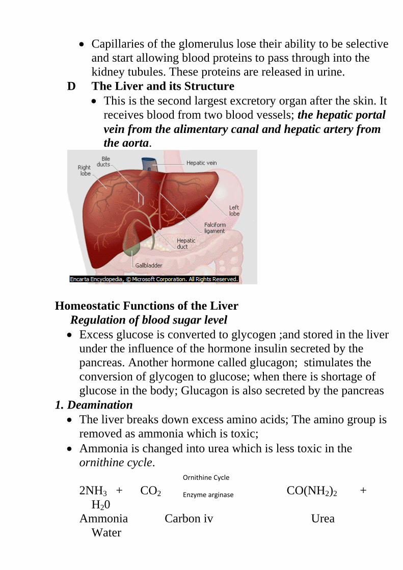
D The Liver and its Structure
This is the second largest excretory organ after the skin. It
receives blood from two blood vessels; the hepatic portal
vein from the alimentary canal and hepatic artery from
the aorta.
Homeostatic Functions of the Liver
Regulation of blood sugar level
Excess glucose is converted to glycogen ;and stored in the liver
under the influence of the hormone insulin secreted by the
pancreas. Another hormone called glucagon; stimulates the
conversion of glycogen to glucose; when there is shortage of
glucose in the body; Glucagon is also secreted by the pancreas
1. Deamination
The liver breaks down excess amino acids; The amino group is
removed as ammonia which is toxic;
Ammonia is changed into urea which is less toxic in the
ornithine cycle.
2NH3 + CO2 CO(NH2)2 +
H20
Ammonia Carbon iv Urea
Water
Ornithine Cycle
Enzyme arginase
(Toxic) Oxide (less toxic)
The remaining carbon skeleton oxidized to carbon IV oxide
and water; this process leads to release of energy. The carbon
skeleton may be converted to glucose to be used during
respiration;
2. Detoxification
Toxic substances are made harmless in the liver e.g.
Ammonia from the process of deamination is converted in the
liver into urea; which is less toxic.
Bacterial toxins are converted to less toxic substances by liver
cells;
Hydrogen peroxide produced by respiring cells is broken down
into water and oxygen which are harmless by the enzyme
catalase found in the liver.
Hydrogen Peroxide Water +
Oxygen
(H2O2) (H2O) (O2)
3. Regulation of plasma proteins
The liver produces most of the proteins found in blood;
fibrinogen and prothrombin which play a role in blood clotting.
Albumin and globulins are also produced by the liver.
Globulins act as antibodies;. Albumin contributes to the
maintenance of osmotic pressure in the body; Non essential
amino acids are synthesized by the liver;
4. Storage of vitamins A, B,D,E and K and mineral salts
The liver store vitamins A, B, D, E and K. Iron released from
the breakdownof erythrocytes is stored in the liver cells; in the
form of a compound called ferritin. The liver therefore is a
good source of these vitamins and iron;
5. Heat production (Thermoregulation)
The various metabolic activities of the liver lead to release of
heat energy; This energy is distributed by the blood to other
Enzyme
Catalase
parts of the body hence contributing to maintenance of constant
body temperature;
6. Inactivation of hormones and drugs
After performing their functions, hormones and drugs are
chemically modified to inactive compounds; The by-products
are eliminated through the kidneys and faeces and via bile;
7. Storage of blood
The large size and high capacity for contraction and expansion
of its veins enables the liver to hold a large volume of blood; It
therefore regulates the volume of blood in the general
circulation depending on the body’s requirements ;
8. Regulation of cholesterol and fat metabolism
When carbohydrates are in short supply in the body, fats in
different parts of the body are mobilized and taken to the liver;
The fats are oxidized to carbon (IV) oxide and water with the
production of energy or modified and sent to tissues for
oxidation;
9. Manufacture of red blood cells in foetus.
Liver Diseases and Disorders
1. Liver Cirrhosis
This is the hardening of the liver tissues due to death of
liver cells.
This is caused by ingestion of toxic chemicals such as
alcohol.
Bacteria, viruses and parasites such as liver flukes can
also cause the disease.
Control
Avoid excess alcohol.
Avoid fatty diets.
Low salt intake
2. Hepatitis
This is a viral disease causing inflammation of the liver.
It is transmitted through contaminated food, milk and
water.
There are two types of hepatitis; Hepatitis A and B.
3. Jaundice
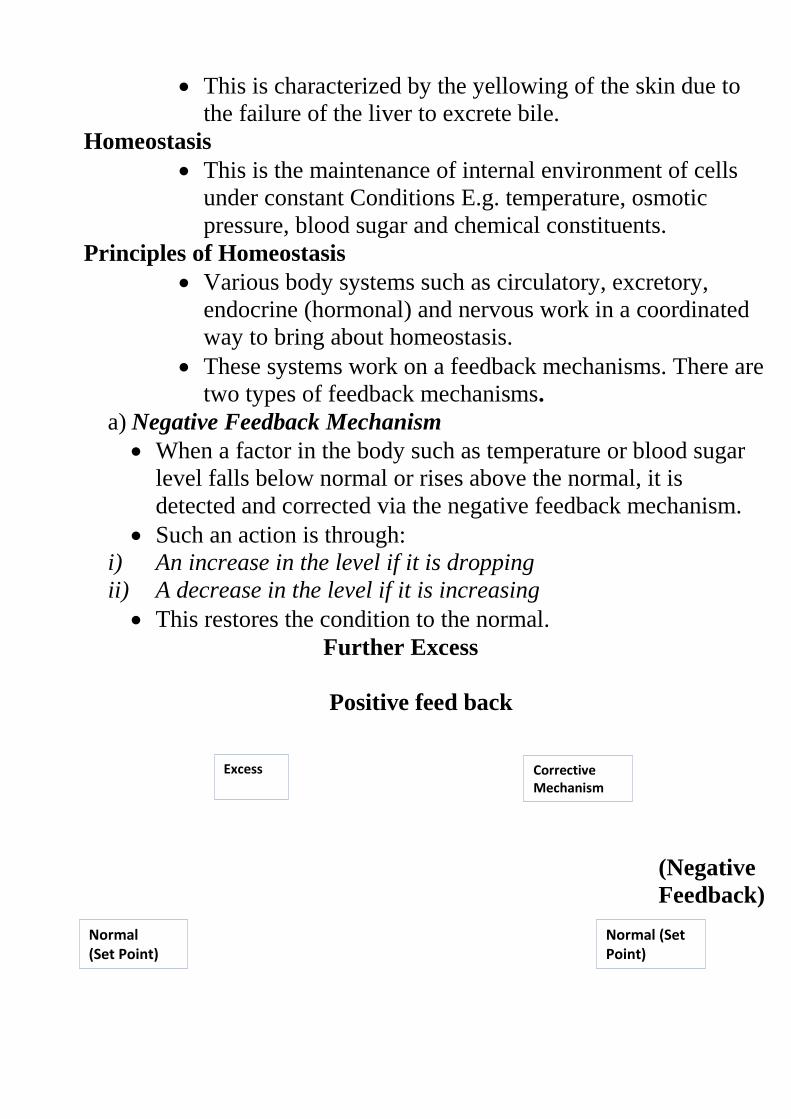
This is characterized by the yellowing of the skin due to
the failure of the liver to excrete bile.
Homeostasis
This is the maintenance of internal environment of cells
under constant Conditions E.g. temperature, osmotic
pressure, blood sugar and chemical constituents.
Principles of Homeostasis
Various body systems such as circulatory, excretory,
endocrine (hormonal) and nervous work in a coordinated
way to bring about homeostasis.
These systems work on a feedback mechanisms. There are
two types of feedback mechanisms.
a) Negative Feedback Mechanism
When a factor in the body such as temperature or blood sugar
level falls below normal or rises above the normal, it is
detected and corrected via the negative feedback mechanism.
Such an action is through:
i) An increase in the level if it is dropping
ii) A decrease in the level if it is increasing
This restores the condition to the normal.
Further Excess
Positive feed back
(Negative
Feedback)
Corrective Mechanism
Excess
Normal (Set Point)
Normal (Set Point)
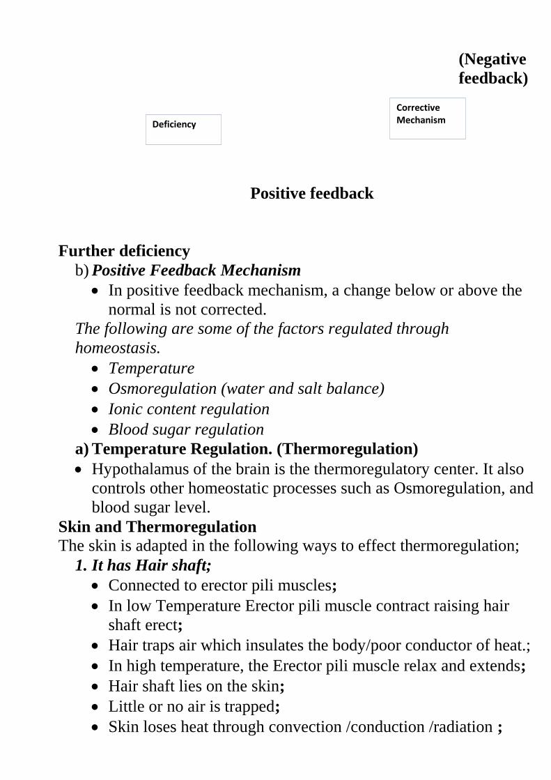
(Negative
feedback)
Positive feedback
Further deficiency
b) Positive Feedback Mechanism
In positive feedback mechanism, a change below or above the
normal is not corrected.
The following are some of the factors regulated through
homeostasis.
Temperature
Osmoregulation (water and salt balance)
Ionic content regulation
Blood sugar regulation
a) Temperature Regulation. (Thermoregulation)
Hypothalamus of the brain is the thermoregulatory center. It also
controls other homeostatic processes such as Osmoregulation, and
blood sugar level.
Skin and Thermoregulation
The skin is adapted in the following ways to effect thermoregulation;
1. It has Hair shaft;
Connected to erector pili muscles;
In low Temperature Erector pili muscle contract raising hair
shaft erect;
Hair traps air which insulates the body/poor conductor of heat.;
In high temperature, the Erector pili muscle relax and extends;
Hair shaft lies on the skin;
Little or no air is trapped;
Skin loses heat through convection /conduction /radiation ;
Corrective Mechanism
Deficiency
1. Blood vessels
In High temperature they vasodilate;
More blood flows near skin surface;
Heat is lost through conduction /convection/ radiation;
In Low temperature they Vasoconstrict;
Little blood flows near the skin;
Less heat or ho heat lost through conduction/convection/
radiation;
Diagrams
3) Sweat gland
In High temperature, Sweating occurs and ( evaporates) and
Carries latent heat of vaporization; cooling the body;
4) Has subcutaneous layer; contains fat which acts as a heat-
insulating layer. Organisms in cold areas have thick subcutaneous
layer for heat insulation.
Homoiotherms and Poikilotherms
Homoiotherms (Endotherms)
They are the animals whose body temperature is maintained at
a constant body temperature despite the wide fluctuations in
the temperature of the external environment e.g. birds and
mammals.
Poikilotherms (Ectotherms)
These are organisms with variable body temperature according
to the temperature of the local atmosphere e.g. in organisms
such as reptiles, amphibians, insects, and fish.
Methods of Regulating Body Temperature in Animals.
i) Metabolic activities of the Body, such as shivering to raise
body temperature.
ii) Insulatory mechanisms such as dilation or constriction of blood
vessels, hair movement etc.
iii) Behavioral activities such as clustering together, burrowing,
basking, hibernation, aestivation, putting on warm clothes etc.
iv) Presence of adaptive features such as hair/fur, subcutaneous
tissue etc.
Hibernation is where animals go into deep sleep for long period of
time due to cold.
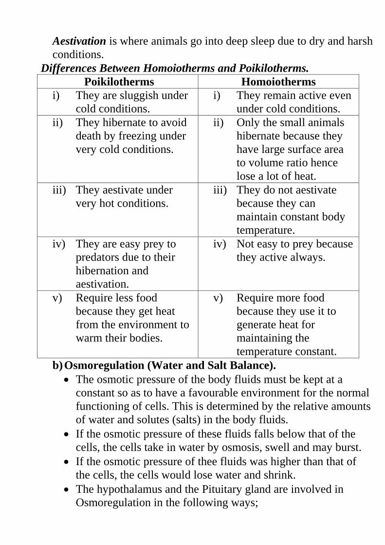
Aestivation is where animals go into deep sleep due to dry and harsh
conditions.
Differences Between Homoiotherms and Poikilotherms.
Poikilotherms Homoiotherms
i) They are sluggish under
cold conditions.
i) They remain active even
under cold conditions.
ii) They hibernate to avoid
death by freezing under
very cold conditions.
ii) Only the small animals
hibernate because they
have large surface area
to volume ratio hence
lose a lot of heat.
iii) They aestivate under
very hot conditions.
iii) They do not aestivate
because they can
maintain constant body
temperature.
iv) They are easy prey to
predators due to their
hibernation and
aestivation.
iv) Not easy to prey because
they active always.
v) Require less food
because they get heat
from the environment to
warm their bodies.
v) Require more food
because they use it to
generate heat for
maintaining the
temperature constant.
b) Osmoregulation (Water and Salt Balance).
The osmotic pressure of the body fluids must be kept at a
constant so as to have a favourable environment for the normal
functioning of cells. This is determined by the relative amounts
of water and solutes (salts) in the body fluids.
If the osmotic pressure of these fluids falls below that of the
cells, the cells take in water by osmosis, swell and may burst.
If the osmotic pressure of thee fluids was higher than that of
the cells, the cells would lose water and shrink.
The hypothalamus and the Pituitary gland are involved in
Osmoregulation in the following ways;
i) When the osmotic pressure of the blood rises due to
dehydration, the hypothalamus is stimulated and sends an
impulse to the pituitary gland which secretes the Antidiuretic
Hormone (ADH) or Vasopressin into the blood. ADH
increases permeability of the kidney tubules to water. More
water is reabsorbed, osmotic pressure of the blood falls hence
production of little but concentrated urine.
ii) When osmotic pressure of the blood falls due to excess water
in the body there is less production of ADH and less water is
reabsorbed hence production of large amounts of dilute urine.
Diabetes Insipidus
This is a condition whereby large quantities of dilute urine are
produced when the pituitary gland fails to produce ADH or
produces it in inadequate amounts. This condition is also
known as Diuresis.
c) Regulation of Ionic Content
Important ions must be regulated within narrow ranges for
efficient functioning of the cells.
Ions are involved in processes such as respiration, protein
synthesis, muscle contraction etc.
The level of sodium ions is regulated by a hormone called
Aldosterone produced by the adrenal glands.
When the level of sodium ions is low in the blood, more
Aldosterone is released which stimulates reabsorption of
sodium ions into the blood.
If sodium ions concentration in the blood rises above the
optimum level, adrenal glands produce less Aldosterone into
the blood and less amounts of sodium ions are reabsorbed.
d) Regulation of Blood Sugar Level.
All sugars such as galactose, lactose and fructose are converted
to glucose.
Glucose is broken down to release energy and excess is
converted into glycogen and stored in the liver or converted
into fats as stored as adipose tissue.
Some glucose flows in general circulation of blood and is
maintained within a narrow range of 90-100mg per 100cm3 of
blood.
The pancreas produces two hormones Insulin and Glucagon
that are responsible for blood sugar regulation.
When there is excess sugar in the blood, insulin is produced
and regulates the blood sugar level by the following;
i) Converts excess glucose into glycogen for storage.
ii) Inhibits conversion of glycogen to glucose.
iii) Converts glucose into fats.
iv) Increases breakdown of glucose to release energy.
When the level of the blood sugar falls, glucagon is secreted
and corrects the situation by the following;
i) Increases the breakdown of glycogen into glucose.
ii) Increases the conversion of fats and proteins into glucose.
iii) Inhibits the conversion of glucose into energy.
NB/. The hormone adrenaline produced by the adrenal glands also
has homeostatic effect on glucose.
It is released during emergencies to avail glucose for fight or flight.
Diabetes Mellitus (Sugar Disease)
This is due to a deficiency in insulin secretion from the
pancreas.
This leads to very high levels of sugar in the blood that cannot
be utilised by cells hence eliminated by kidney in urine.
Symptoms
Presence of glucose in urine
Loss of body weight due to breakdown of fats and proteins
Chronic starvation
Thirst sensation.
Control
Insulin injection into the blood stream
Avoid foods rich in sugars.
Avoid excessive intake of alcohol.
Question
Explain why insulin is not administered orally (through the
mouth)
Revision questions
Gaseous Exchange
This is the process by which respiratory gases (oxygen and
carbon IV oxide) are passed across the respiratory surface.
Gases are exchanged depending on their concentration gradient.
In simple organisms such as amoeba, diffusion is enough to bring
about gaseous exchange.
CO2 diffuses out into the surrounding water while oxygen
diffuses from the water across the plasma membrane into the
amoeba.
Diagram
Importance of Gaseous Exchange
1. Promote oxygen intake for respiration.
2. Facilitate carbon IV oxide removal from the body as a metabolic
waste product.
Gaseous Exchange in Plants
During the day, green plants take in carbon IV for photosynthesis.
Oxygen is given out as a byproduct of photosynthesis and is
released into the atmosphere.
Examples of respiratory Surfaces in Plants
Stomata in leaves
Roots e.g. pneumatophores
Lenticels in woody stems
Structure and Function of the Stomata
They are tiny openings on the leaf surfaces. They are made up of
two guard cells.
Guard cells are the only epidermal cells containing chloroplasts.
They regulate the opening and closing of the stomata.
Adaptations of Guard Cells i) They are bean shaped/sausage shaped.
ii) Contain chloroplast hence can photosynthesize.
iii) Inner walls are thicker while outer wall is thin to facilitate
the opening and closing of stomata.
Diagram
Mechanism of Opening and Closing of Stomata
There are three theories that try to explain how the stomata open
and close.
i) Photosynthetic theory
ii) Starch Sugar inter-conversion Theory. (effect of changes in
pH of guard cells)
iii) Potassium Ion Theory.
i) Photosynthetic theory
During the day, guard cells photosynthesize forming glucose.
This glucose increases the osmotic pressure in the guard cells.
Guard cells draw in water from the neighbouring epidermal cells
and become turgid.
The stoma opens.
During the night, there is no photosynthesis due to absence of
light.
Glucose is converted into starch lowering the osmotic pressure in
the guard cells.
Guard cells lose water and become flaccid closing the stomata.
ii) Starch Sugar inter-conversion Theory. (effect of changes
in pH of guard cells)
This is under the influence of pH in the guard cells.
During the day CO2 is used up during photosynthesis raising the
pH in the guard cells.
In this high pH, enzymes convert more starch into glucose.
Osmotic pressure of the guard cells increases and water enters
into them, making them turgid hence opening the stomata.
During the night, there is no photosynthesis. The level of CO2
increases lowering the pH.
Enzymes become inactivated and starch is not converted into
glucose.
Osmotic pressure of guard cells falls making them to lose water
by osmosis.
Guard cells become flaccid and stoma closes.
Mechanism of Gaseous Exchange in Plants
Oxygen diffuses from the atmosphere where it is more
concentrated into the plant.
CO2 diffuses out as a metabolic waste product along a
concentration gradient into the atmosphere.
a) Gaseous Exchange through the Stomata
Stomata are modified in number of ways depending on the habitat
of the plant.
Xerophytes: These are plants adapted to life in dry areas.
They have less number of stomata that are small in size.
Stomata may be sunken, hairy and in some they open during the
night and close during the day.
Hydrophytes: These are the aquatic plants (water Plants)
They have many stomata that are large in size and mainly found
on the upper leaf surface.
Hydrophytes have the aerenchyma tissue with large air spaces to
store air for gaseous exchange.
Diagrams
Mesophytes: They are plants growing in areas with adequate amounts
of water.
They have a fairly large number of stomata found on both leaf
surfaces.
b) Gaseous Exchange through the Lenticels
They are openings found on woody stems and they are made of
loosely packed cells.
They allow gaseous exchange between the inside of the plant and
the outside by diffusion.
Actual gaseous exchange occurs on some moist cells under the
lenticels.
Diagram
c) Gaseous Exchange through the Roots
Plants like the mangroves growing in muddy salty waters have
specialized aerial breathing roots called pneumatophores.
Pneumatophores rise above the salty water to facilitate gaseous
exchange.
Gaseous Exchange in Animals
Types and Characteristics of Respiratory Surface
Different animals have different respiratory surfaces depending
on the animal’s size, activity and the environment in which it
operates as shown below.
Type of Respiratory
Surface
Environment/Medium
of Operation
Example of
Organism
1. Cell Membrane. Water Amoeba
2. Gill Filaments Water Fish
3. Tracheoles Air Insects
4. Alveoli/Lungs Air Mammals
Birds
Frogs
Reptiles
5. Skin Water Frog
Air Earthworm
6. Buccal Cavity Air Frog
The respiratory surface is the basic unit of any breathing system
upon which actual gaseous exchange occurs by diffusion.
Respiratory surfaces have the following main characteristics.
i) Must have a large surface area.
ii) Must be moist to allow gases to diffuse in solution form.
iii) Have a dense network of blood capillaries for efficient
gaseous exchange.
iv) Have a thin membrane to reduce the diffusion distance.
Gaseous Exchange in Insects
Insects have their gaseous exchange system made of many air tubes
forming the tracheal system.
Tracheal system is made up of spiracles and Tracheoles.
Spiracles are external openings found on both sides of the
abdomen and thorax.
Spiracles have valves to control their opening and closing. They
also have hairs to prevent excessive water loss from the body
tissue.
Spiracles open into tubes called trachea. Trachea is reinforced
with spiral bands of chitin to keep them open.
Trachea subdivides into finer air tubes called Tracheoles.
Tracheoles are in direct contact with body tissues and organs and
they supply individual cells with oxygen.
Tracheoles do not have bands of chitin and therefore they allow
gaseous exchange across their thin moist walls.
Diagram
Mechanism of Gaseous Exchange in the Tracheal System of an
Insect
Air is drawn into and out of the tracheal system by muscular
movement of the abdominal wall.
When spiracle valves are open, air is drawn into the tracheal
system. The valves close and air is forced along the system by
muscle movement.
Oxygen diffuses into the tissue fluid and into the cells.
CO2 diffuses out of the cells and into the tissue fluid then into the
tracheal system.
Gaseous Exchange in Fish
The breathing system of the fish consists of the following;
o Mouth (buccal) cavity.
o Gills.
o Opercular cavity.
o Operculum.
Gills are made of a long curved bone called the gill bar.
Gill filaments arise from one side of the gill bar. They are many
and suspend freely in water providing a large surface area for
gaseous exchange.
Gill rakers arise from the other side of the gill bar. They are teeth
like and they prevent solids present in water from damaging the
delicate gill filaments.
Blood vessels enter the gill bar and branch into the gill filaments
as blood capillaries.
Operculum is found on either side of the body near the head and
it also protects the delicate gills.
Diagram
Mechanism of Gaseous Exchange in the Gills of a Bony Fish
Floor of the mouth cavity is lowered increasing the volume of the
mouth cavity but lowering the pressure.
Water flows into the mouth cavity and the operculum closes.
Operculum on either side bulge outwards without opening. This
increases volume in the gill cavity but the pressure drops.
Water containing dissolved oxygen flows from the mouth cavity
to the gill chamber over the gills.
The mouth closes and the floor of the mouth cavity is raised.
The remaining water in the mouth is forced to flow towards the
gill chamber.
Oxygen diffuses from the water into the blood through the thin
walls of the gill filaments. It combines with haemoglobin for
transportation to all body parts.
CO2 diffuses from the blood into the flowing water.
To ensure maximum gaseous exchange, the water flowing over
the gills and the blood in the gills flows in opposite directions.
This is called counter current flow system and it ensures that at all
the points, concentration of oxygen is always higher in the water
than in the blood.
Diagram
If the water and blood were flowing in the same direction,
gaseous exchange will not be that effective.
Where the oxygen is 50% in water, there is no concentration
gradient because blood also has 50% oxygen concentration.
Diagram
Mechanism of Gaseous Exchange in Amphibians
Amphibians live on both land and water and therefore exhibit the
following methods of gaseous exchange.
1. Gaseous exchange through the lining of the buccal cavity
2. Gaseous exchange through the lungs
3. Gaseous exchange through the skin
a) Gaseous exchange through the mouth (buccal) cavity
Air is taken in or expelled from the mouth cavity by raising and
lowering of the floor mouth.
Lining of the mouth cavity is moist to dissolves oxygen.
There is a rich supply of blood capillaries under the lining of the
mouth cavity. Oxygen diffuses into the blood and is carried by
haemoglobin to all parts of the body.
Carbon IV oxide from the tissues is brought by the blood to the
mouth cavity where diffuses out.
Gaseous exchange through the lungs
The frog has two lungs which are connected to the buccal cavity.
T he inner lining of the lungs is moist, thin and is richly supplied
with blood capillaries.
During inspiration, the floor of the mouth cavity is lowered and
nostrils are open. Air rushes through the open nostrils into the
mouth cavity.
Nostrils close and the floor of the mouth cavity is raised. This
reduces the volume and increase the pressure in the mouth cavity
forcing air into the lungs.
Carbon IV oxide from the tissues diffuse into the lung while the
oxygen from the lungs diffuses into the tissues.
b) Gaseous exchange through the skin
Frogs have a thinner and moist skin than the toads.
There is large network of blood capillaries below the skin to carry
the respiratory gases.
Oxygen from the air and water diffuse through the skin into the
blood stream.
Carbon IV oxide diffuses out of the blood capillaries through the
moist skin into the surrounding water and air.
Mechanism of Gaseous Exchange in Mammals
The following structures are involved in gaseous exchange in
mammals;
- Nose (Nostrils)
- Larynx
- Trachea
- Chest cavity (ribs and intercostals muscles)
- Diaphragm.
a) Nose
It has two openings called nostrils which let in air into the air
passages.
As air moves in the passages, it is warmed and moistened
The lining of the nasal cavity has also the sense organs for smell.
b) Larynx
It is located on top of the trachea
It is called the voice box. It controls the pitch of the voice.
c) Trachea
It is a tube made of rings of cartilage which prevents it from
collapsing during breathing.
Inside it is lined with ciliated epithelium. Cilia beat in waves and
move mucus and foreign particles away from the lungs towards
the pharynx.
As the trachea enters the lungs, it divides into two branches called
Bronchi (Bronchus).
d) Lungs
They are found in the chest cavity and they are enclosed by a
double membrane called the pleural membrane.
The space between the membranes is called the pleural cavity.
Pleural cavity is filled with pleural fluid which reduces friction
making the lungs to move freely in the chest cavity during
breathing.
Diagrams
In the lungs each bronchus divides into small tubes called
bronchioles.
Bronchioles branch further to form air sacs called alveoli
(alveolus)
Alveolus is covered by a fine network of blood capillaries.
The mechanism of breathing
Breathing is achieved by changes in the volume and air pressure
of the thoracic cavity.
Thoracic cavity is enclosed by ribs.
Ribs are covered by intercostals muscles.
The diaphragm is a muscular sheet of tissue below the chest
cavity. It curves upwards in the form of a dome shape.
Breathing mechanism involves two processes.
a) Inspiration (Inhalation) i.e. breathing in.
b) Expiration (Exhalation) i.e. breathing out.
Inspiration (Inhalation) i.e. breathing
This occurs when the volume of thoracic cavity increases and the
pressure decreases.
External intercostals muscles contract while the internal
intercostals muscles relax.
Ribs are pulled upwards and outwards.
Diaphragm flattens increasing the volume of the thoracic cavity
while decreasing the pressure inside it.
Air rushes into the lungs through the nose and trachea inflating
the lungs.
Diagrams page 62
Expiration (Exhalation) i.e. breathing out
Volume of thoracic cavity decreases while pressure increases.
This is brought about by the following;
External intercostals muscles relax while internal ones contract.
Ribs move downwards and inwards.
Diaphragm relaxes and regains its original dome shape.
Volume of the thoracic cavity decrease and pressure increases.
Air is forced out of the lungs through the air passages to the
atmosphere.
Gaseous exchange in the alveolus
Alveoli and blood capillaries are made of very thin walls.
The wall of the alveolus is covered b a film of moisture which
dissolves oxygen in the inhaled air.
Oxygen diffuses through the epithelium of the alveolus, the
capillary wall and through the cell membrane of the red blood
cells.
In the red blood cells it combines with haemoglobin.
Carbon (iv) oxide is more concentrated in the blood capillaries
than in the alveoli.
It therefore diffuses from the capillaries into the alveoli.
Water vapour also passes out of the blood by the same process.
Diagram page 64 KLB
Percentage composition of gases in inhaled and exhaled air
Gas % in inhaled air. % in exhaled air
Oxygen 20 16.9
Carbon (iv) oxide 0.03 4.0
Nitrogen and other gases 79.97 79.97
Regulation of Breathing
This is controlled by a part of the brain called Medulla oblongata.
Factors affecting the rate of breathing in humans
1. Exercise
Breathing rate increases during vigorous activity.
2. Age
Younger people have a faster breathing because their bodies have
more energy demand.
3. Emotions Things like anxiety, fear and fright increases the breathing rate.
4. Temperature
Relatively high temperatures increase the rate of breathing. However,
very high temperatures reduce the breathing rate.
5. Health
If there is fever (high body temperature), the breathing rate increases.
Some respiratory diseases however, make breathing difficult.
Lung Volumes
i) Lung capacity
This is the total amount of air the lungs can hold when
completely filled. The lungs of an adult have a capacity of
about 5,500cm3
ii) Tidal volume
This is the amount of air taken in and out of the lungs during
normal breathing. Tidal volume is about 500cm3
iii) Inspiratory reserve volume
This is an additional volume attained after having a forced
inhalation in addition to the tidal volume. It is about 2000cm3
iv) Inspiratory capacity
This is the tidal volume +Inspiratory reserve volume.
v) Expiratory reserve volume
This is air removed after a forced exhalation. It can be up to
1,300cm3
vi) Vital capacity
This is the deepest possible exhalation. This air can only be
forcibly pushed out of the lungs.
vii) Residual volume
This is the air that normally remains in the lungs after the
deepest exhalation. It is normally about 1,500cm3
Diagram
Diseases of the Respiratory System
i) Asthma It is caused by:
Allergens such as pollen grains, certain foods and drugs
Infections of the lungs by bacteria and viruses
Symptoms
Difficulty in breathing
Wheezing sound when breathing
Treatment and Control
Avoiding the causative agents
Injection of drugs and oral application of pills
Spraying directly into the bronchial tubes with a muscle
relaxant
ii) Bronchitis There are two types; Acute and Chronic
Symptoms
Production of thick greenish or yellowish sputum
Difficulty in breathing
Difficulty in walking and sleeping
Treatment
Seeking early medical assistance
iii) Whooping cough It is caused by a bacterium called Bordetella pertussis.
Symptoms
Prolonged coughing and vomiting
Conjuctival haemorrhage (bleeding)
Convulsions and coma
Severe pneumonia in the bronchioles
Ulcers and heart complications
Emaciation due to repeated vomiting
Treatment
Use of antibiotics
Use of a balanced diet on patients
Control
Children immunization at early age
iv) Pneumonia It is caused by a bacterial called Streptococcus pneumoniae
Symptoms
Coughing
Fever
Chest pains
Deposits of fluids in the lungs
Treatment
Use of antibiotics such as penicillin and sulphonamides
Control
Avoid overcrowding.
Good ventilation in living houses
v) Pulmonary Tuberculosis It is caused by a bacterium called Mycobacterium tuberculosis.
Symptoms
Weight loss
Coughing with blood stained sputum.
Fever
Treatment
Use of antibiotics such as streptomycin
Control
Pasteurization of milk
Immunization using BCG (Bacille Calmette Guerin)
Use of radiography (X-Ray)
vi) Lung cancer Cancer is uncontrolled cell growth in the body causing tumours.
Some general causes
Smoking
Inhalation of cancer causing substances such as asbestos
Exposure to radiations such as X-rays, radioactive substances
such as uranium and substances that alter the genetic
composition of the cell such as mustard gas
Treatment and control
Surgery to remove the tumour
Radiotherapy
Chemotherapy
Use of some drugs
Not smoking
Revision Questions
RESPIRATION
Process by which food substances are chemically broken down in
living cells to release energy, carbon (iv) oxide, water or alcohol.
Respiration takes place mainly in the mitochondria. It has two
membranes, inner and outer.
Inner membrane is folded into projections called cristae. Cristae
provide a large surface area for respiratory enzymes. Respiratory
enzymes are bound to the cristae.
Diagram
Practical Activity 1
To investigate the gas given off when food is burnt.
Types of Respiration
Aerobic Respiration
Anaerobic Respiration.
Aerobic Respiration
Process by which food substances such as glucose are broken
down in the presence of oxygen to release energy, water and
carbon (IV) oxide.
The energy is stored in the form of a chemical substance called
Adenosine Triphosphate (ATP).
This energy is released in small quantities since a lot of heat
energy would burn the body cells. Respiratory
Enzymes
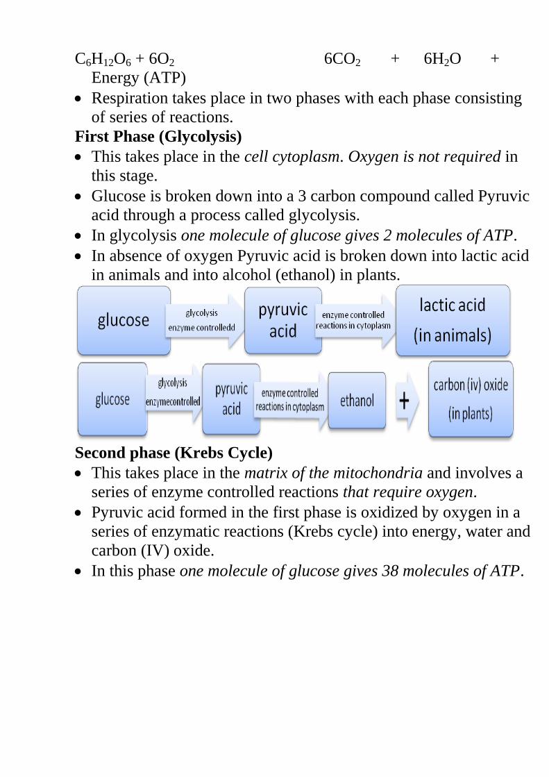
C6H12O6 + 6O2 6CO2 + 6H2O +
Energy (ATP)
Respiration takes place in two phases with each phase consisting
of series of reactions.
First Phase (Glycolysis)
This takes place in the cell cytoplasm. Oxygen is not required in
this stage.
Glucose is broken down into a 3 carbon compound called Pyruvic
acid through a process called glycolysis.
In glycolysis one molecule of glucose gives 2 molecules of ATP.
In absence of oxygen Pyruvic acid is broken down into lactic acid
in animals and into alcohol (ethanol) in plants.
Second phase (Krebs Cycle)
This takes place in the matrix of the mitochondria and involves a
series of enzyme controlled reactions that require oxygen.
Pyruvic acid formed in the first phase is oxidized by oxygen in a
series of enzymatic reactions (Krebs cycle) into energy, water and
carbon (IV) oxide.
In this phase one molecule of glucose gives 38 molecules of ATP.
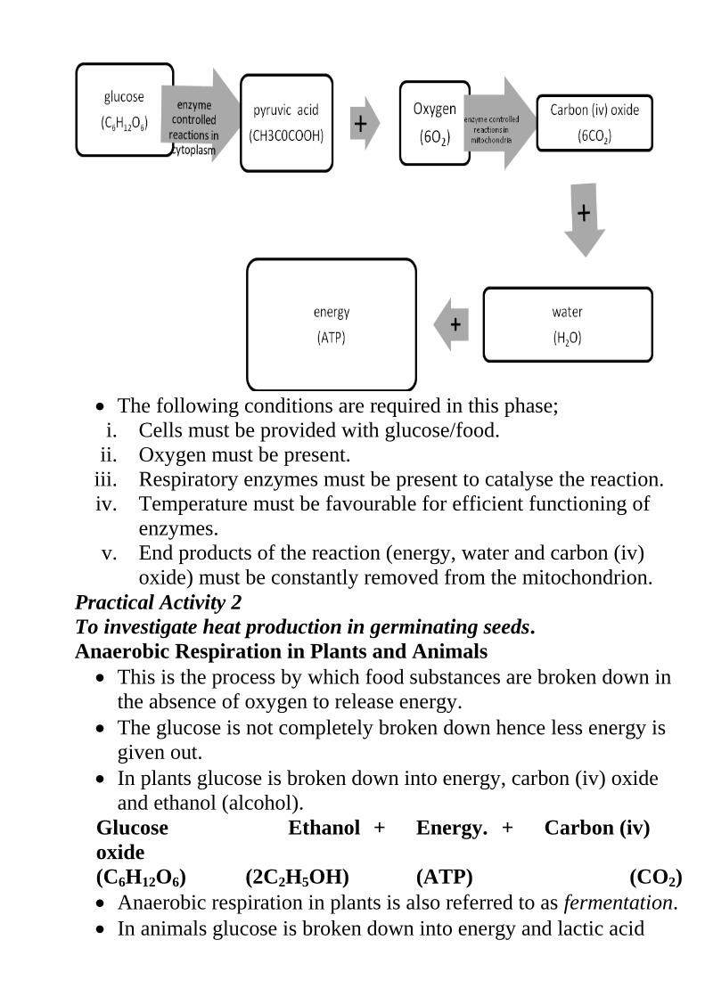
The following conditions are required in this phase;
i. Cells must be provided with glucose/food.
ii. Oxygen must be present.
iii. Respiratory enzymes must be present to catalyse the reaction.
iv. Temperature must be favourable for efficient functioning of
enzymes.
v. End products of the reaction (energy, water and carbon (iv)
oxide) must be constantly removed from the mitochondrion.
Practical Activity 2
To investigate heat production in germinating seeds.
Anaerobic Respiration in Plants and Animals
This is the process by which food substances are broken down in
the absence of oxygen to release energy.
The glucose is not completely broken down hence less energy is
given out.
In plants glucose is broken down into energy, carbon (iv) oxide
and ethanol (alcohol).
Glucose Ethanol + Energy. + Carbon (iv)
oxide
(C6H12O6) (2C2H5OH) (ATP) (CO2)
Anaerobic respiration in plants is also referred to as fermentation.
In animals glucose is broken down into energy and lactic acid
Glucose Lactic acid + Energy.
(C6H12O6) (2C3H6O3) (ATP)
Oxygen Debt
This is oxygen required to get rid of the lactic acid that
accumulates in the body tissues when the oxygen supply is less
than required.
Accumulation of lactic acid causes fatigue and muscle crumps.
Oxygen debt is paid back by breathing more quickly and more
deeply in order to increase oxygen supply such as during recovery
period after a race when a person pants.
When paying back the oxygen debt, lactic acid is oxidized to
energy, water and carbon (iv) oxide or it is taken to the liver and
converted into glycogen.
Application of Anaerobic Respiration i. Baking industry
ii. Beer brewing and distillery industry.
iii. Dairy industry in the production of yoghurt and cheese.
iv. Production of vinegar citric acid, oxalic acid, butyric acid and
some drugs.
v. Production of power alcohol which is used as a substitute for
petrol.
vi. Silage making.
vii. Biogas production.
viii. Making compost manure
Practical Activity 3
To investigate gas produced during fermentation.
Comparison between Aerobic and Anaerobic Respiration
Aerobic Respiration Anaerobic Respiration.
i. Oxygen is required Oxygen not required
ii. High amount of energy is
released as one molecule
of glucose yields 38 ATP
molecules (2880 KJ)
Low amount of energy is
released as one molecule of
glucose yields 2 ATP
molecules (210 KJ)
iii. There is complete
breakdown of the
substrate into carbon (iv)
There is incomplete breakdown
of substrate hence lactic acid or
alcohols are produced.
oxide and water.
iv. End products are energy,
water and carbon (iv)
oxide
End products are energy,
alcohol in plants and lactic acid
in animals.
v. Water molecules are
produced.
Water molecules are not
produced.
vi. Over a short period of
time, energy is not
released faster
Over short period of time,
energy is released faster.
vii. Occurs in the cytoplasm
and in the mitochondrion.
Occurs only in the cell
cytoplasm.
Respiratory Substrates
These are energy rich foods which when oxidized release energy.
They include;
i. Carbohydrates –
They are the main source of energy mainly in the form of simple
sugars such as glucose, fructose and galactose.
They produce about 17KJ (2898/mole) per gram when completely
oxidized.
ii. Fats –
They produce more energy than carbohydrates or proteins. One
gram of fats yields about 38 KJ of energy when completely
oxidized.
They are however not the main substrate because they are not
very soluble in water hence not easily transported to the sites of
respiration. It also requires more oxygen to oxidize one gram of
fats than one gram of glucose.
iii. Proteins –
They are not normally used in respiration unless in cases of
extreme starvation.
One gram of proteins yields 22KJ of energy when completely
oxidized.
Assignment
Where do plants and animals get the following from;
- Carbohydrates.
- Fats
- Proteins
Respiratory Quotient (RQ) and its Significance
RQ is the ratio showing the relationship between the amounts of
carbon (iv) oxide used against the amount of oxygen used in
respiration.
RQ =
RQ varies with the type of substrate being oxidized. For example
carbohydrates have a RQ of 1.0 when fully oxidized, fats have 0.7
and proteins have 0.9.
RQ can therefore be used to indicate the type of substrate being
oxidized and also whether aerobic respiration or anaerobic
respiration is taking place.
RQ is also affected by factors such as age, health of the organism
and the temperature.
Factors Affecting the Rate of Respiration i. Oxygen concentration. When the amount of oxygen increases, the
respiration rate also increases. Decrease in oxygen concentration
will lead to decreased respiration rate.
ii. Substrate concentration. Increase in sugar concentration increases
respiration and vice versa.
iii. Hormones. Presence of some hormones such as adrenaline and
thyroxine in the body increases the rate of respiration.
iv. Surface area to volume ratio (Body size). If the SA/volume ratio
is high, the organism would lose more heat energy. As more heat
is lost to the surrounding more is required to replace the lost
energy hence more respiration.
v. Age. Young people require more energy because their cells are
actively dividing hence respiration rate is higher in them than in
older people.
vi. Occupation. People engaged in heavier tasks have higher rate of
respiration.
vii. Sex. Generally male’s have faster respiration rate than females
due to presence of more muscles in their bodies.
viii. Basal metabolic rate. This is the energy required to maintain
normal body functions such breathing, heartbeat, blood
circulation etc while at rest.
Revision Questions
FORM THREE BIOLOGY NOTES
ECOLOGY
Introduction
Ecology is the study of the interrelationships of organisms to each
other and to their environment (biotic and Abiotic factors).
Autecology; study of single species within a community and how it
relates with both the biotic and Abiotic factors.
Synecology. This is the study of many different species of organisms’
interacting among themselves within an ecosystem.
Ecology helps to address the following issues.
Sustainable food production
Pollution control
Natural resources conservation
Pest and disease control
Population control
Eco-tourism
Prediction of adverse weather conditions
Concepts of ecology
Biosphere/ecosphere. This is the part of the earth and atmosphere
inhabited by living organisms.
Habitat. This is a specific locality with a particular set of
conditions where an organism lives. Habitats can be terrestrial or
aquatic.
Ecological niche. This is the position occupied by an organism in
a habitat. It includes the physical space where an organism is
found and its role in the habitat.
Population. This refers to all members of a given species in
particular habitat.
Community. This refers to all organisms belonging to different
species interacting in the same habitat. Many populations make
up a community.
Ecosystem. This is a natural unit made of biotic and Abiotic
factors whose interactions lead to a self sustaining system. E.g. a
tropical rain forest, a small pond etc.
Biomass. This is the total dry weight of living organisms at a
particular Trophic (feeding) level or per unit area.
Carrying capacity. This is the maximum number of organisms an
area can comfortably support without depletion of the available
resources. E.g. the maximum number of cows a pasture land can
comfortably hold without overgrazing.
Study Question 1
Factors in an Ecosystem
They are divided into two:
1. Abiotic factors or the non living factors
2. Biotic or the living factors
Abiotic Factors
Light. This is required by plants and photosynthetic bacteria to
manufacture food. The sun is the source of light energy. Light
intensity and quality (wavelength) affects the rate of
photosynthesis, flowering and germination in plants, while in
animals it affects migration, hibernation and reproduction. Light
intensity is measured using a Photographic Light meter while a
Seechi disc measures light penetration in water.
Atmospheric pressure. Variation in atmospheric pressure affects
the availability of oxygen and carbon (IV) dioxide in the
atmosphere. These two gases in turn affect the distribution of
living organisms. Low atmospheric pressure increases the rate of
transpiration. Barometer is used to measure it.
Humidity. This is the amount of water vapour in the atmosphere.
It affects the rate of water loss from plants and animals surfaces
through transpiration and sweating respectively. The higher the
humidity the lower the rate of loss and vice versa. It is measured
using the hygrometer.
Salinity. This refers to the salt concentration of the water. This
divides the aquatic environment into marine, estuarine and fresh
water. Only organisms with adaptable osmoregulatory features
can comfortably occupy such habitats. In estuaries, there are
fluctuations of salt concentrations at different times. When the sea
tide is low, the salt concentrations are low due to the greater
diluting effect of the fresh water being discharged. High tide
raises the salt level. Estuarine organisms must therefore be
adapted to cope with such wide salt variations.
Wind. This is moving air. It increases the rate of water loss from
organisms affecting their distribution. It also influences rain
formation. It helps in formation of sand dunes in deserts which
become habitats for the growth of deserts plants. Its an agent of
seed and fruit dispersal
Temperature. This affects the distribution of organisms in any
habitat. Very low temperature may inactivate enzymes while very
high temperatures denature them. Temperature varies due to
seasons, altitude, and latitude and diurnally in hot deserts.
pH (hydrogen ion concentration.)
This is the measure of acidity or alkalinity of water in aquatic
habitats or soil solution. This influences the distribution of plants
and animals in soil and aquatic habitats. Different organisms have
different pH requirements. pH is determined using the pH meter.
Study Question 2
Practical Activity 1
Study Question 3
Biotic Inter-Relationships
Competition
Living organisms compete for resources such as nutrients, space, light
and mates. There are two types of competition.
i.) Inter-specific competition. This is the competition between
individuals of different species for the same resources. For
example. An experiment6 was carried out on two closely related
species of paramecia- Paramecium caudatum and Paramecium
aurelia. It was observed that when each species is grown
separately in controlled cultures with constant food supply, they
show normal population growth. When they are grown together
in the same culture, thre is competition and Paramecium
caudatum is eliminated. See graphs.
However, closely related species can live together without
competition. For example, when Paramecium caudatum and
Paramecium bursoria are grown in the same culture, there is no
competition because each species occupies a different part of the
culture. Similarly, browsers and grazers can occupy same habitat
without competition because they feed at different levels of the same
plants. For example, the zebras eat the softer shoots, followed by the
wild beasts, and the gazelles which eat the fibrous left over of the same
grass.
Study Question 4
ii.) Intra-specific competition. This is the competition between
members of the same species for the same resources.
When there is competition the best adapted organisms survive while
the less adapted ones may die or be forced to migrate.
Predation
This is the relationship where one organism kills another for food and
feed on it either as a whole or a part of it. The predator is the one
which kills while the prey is the one being killed for food.
Predators have various adaptations to enable them to be efficient in
capturing the prey. These include;
Sharp eyesight as in eagles, kites and hawk
Fast flight,
Modified beaks
Strong jaws with carnassial’s teeth as in leopards and lions.
Large claws on strong forelimbs.
Colour camouflage such as the spotted pattern of the leopard
blends well with the background colour of the bushes and trees.
Moving against the wind while stalking the prey. Preys also have
structural and behavioural adaptations. These include:
Swift movement e.g. the antelope and gazelle
Camouflage e.g. in gazelles and stripes of the zebra.
Large eyes on the sides of the head to giving them a wide field of
view
Confrontational display in porcupine
NB/. When the number of the prey increases that of the predators also
increases. An increase in the number of predators leads to a decrease
in the population of the prey. This decrease in prey population leads to
a fall in predator population which in turn gives space for the increase
in the population of the prey. This is the basis of biological control.
See the graph below.
Parasitism
This is the relationship where an organism [parasite] obtains
nutrients from another live organism [host] without killing it. The
parasite obtains food and shelter from the host causing some harmful
effects. Parasites may weaken the host and also transmit diseases
which may kill their host thus reducing their number an d distribution.
There are two types of parasites;
Ecto-parasites
Endo-parasites
Study Question 5
Symbiosis
This is an association between two of different species in which both
benefit. For example the association of colon bacteria with humans
and other animals, especially plant-eating animals, the ox-pecker bird
and the ox etc.
The Rhizobium bacteria help the leguminous plants to fix nitrogen
while the bacteria obtain shelter and carbohydrates from the plants.
Diagram
Saprophytism
This is where organisms obtain nutrients from dead organisms causing
decomposition hence releasing nutrients into the ecosystem.
Saprophytes include the bacteria and fungi.
The Nitrogen Cycle
This refers to the cycling of nitrogen and its compounds in the natural
environment.
Although nitrogen is abundant in the atmosphere as nitrogen gas,
it cannot be utilised by plants. It has to be converted into a form
that can be absorbed by plants through a process called nitrogen
fixation. Nitrogen fixation is done in two ways;
Biological fixation. This can occur in two forms
1. Nitrogen fixation by symbiotic bacteria such as
Rhizobium spp. They are found in the root nodules of
legumes. They convert nitrogen gas into ammonia which is
then converted into nitrates for plant utilisation.
2. Nitrogen fixation by free living bacteria e.g. Clostridium,
Azotobacter, and some algae such as Anabaena, chlorella
and Nostoc. Non-Biological nitrogen fixation. This is done by lightning.
During thunderstorms, lightning energy combines atmospheric
nitrogen gas with oxygen to form nitrous and nitric acid. These
are then converted into nitrates.
Plants absorb nitrates and convert them into plant proteins.
Animals feed on these plants and obtain the proteins. They are
then digested into amino acids and become assimilated into
animal proteins.
When living organisms die, saprophytic bacteria and fungi break
down the proteins in their bodies into ammonia. Nitrifying
bacteria convert this ammonia into nitrates thorough a process
called nitrification. Nitrosomonas and Nitrococcus convert
ammonia into nitrites and Nitrobacter convert nitrites into
Nitrates.
Some soil micro organisms such as Pseudomonas denitrificans
& Thiobacillus denitrificans utilise the oxygen in the nitrates
reducing it to nitrites, ammonia and eventually into nitrogen gas.
This is called de-nitrification.
This reduces the amount of nitrogen available to plants but it
frees the nitrogen so that it becomes available for the cycle to
continue.
Diagram
Practical activity 2
Study question 6
Energy Flow in an Ecosystem
The sun is the natural source of energy. This energy is transferred to
the following feeding levels;
Producers
Primary consumers
Secondary consumers
Tertiary consumers
Quaternary consumers
These feeding levels are called Trophic levels
Decomposers They break down organic materials into simple substances which
are made available for re-use by other organisms. Decomposers are
mainly fungi and bacteria.
Food Chains
This is the representation of energy flow from a producer to other
organisms linearly. Green plants are eaten by herbivores which are
eaten by carnivores.
Producers’ Primary consumers Secondary consumers
Tertiary consumers Quaternary consumers
Some energy is lost as it is moved from one trophic level to the
next. This is through respiration, defecation, excretion and in form
of heat.
Fig. 2.7
Examples
When the decomposers are included in a food chain, they are placed
at the end.
Study Question 7
Food Webs
These are several interconnected food chains. Simple food chains
rarely exist since in any ecosystem, many populations interact.
Examples
Study Question 8
Ecological Pyramids
These give a simplified representation of feeding relationships and
energy flow in an ecosystem. They are of three types.
Pyramid of numbers
Pyramid of biomass
Pyramid of energy
Pyramid of Numbers
There is a progress decrease in the number of organisms as one
move from the producers all the way to the quaternary consumers.
Producers have the greatest number followed in a decreasing order
by primary, secondary, tertiary and quaternary consumers.
Construction of Pyramid of Numbers
i.) Use data provided or collected.
ii.) From the data, identify and draw the most suitable food chain.
iii.) Indicate the numbers at each trophic level in the food chain.
iv.) Choose a suitable scale for the data.
v.) Using the chosen scale draw a horizontal rectangular bar to
represent the number of the producers as the base of the pyramid.
vi.) Progressively draw horizontal bars of the other trophic levels in
their ascending order.
Ensure that the width of the bars is uniform.
Study Question 9
Interpretation of Pyramid of Numbers
Generally the body size of organisms increases at each trophic
level from the base to the apex of the pyramid as their number
decreases.
At each trophic level much energy is lost through respiration,
excretion, sweating, defecation etc. therefore less energy is
transmitted to the succeeding trophic level. Fewer organisms can
therefore be supported.
Inverted pyramid of numbers also exist. For example where one
mango tree supports several monkeys each being fed on by
several fleas.
Pyramid of Biomass
Biomass of an organism is its constant dry weight. In an ecosystem,
the producers have the highest biomass followed in decreasing order
by primary, secondary, tertiary and quaternary consumers.
Study Question 10
Practical activity 3
Study Question 11
Population
Populations change in size, structure and organisation.
Characteristics of a population.
Density. This is the number of individuals per unit area. E.g. 50
gazelles per Km2.
Dispersion. This is the distribution or spread of organisms in a
habitat.
Population growth. This refers to the rate of increase in
numbers.
Population Estimation Methods
Usually a representative sample is used to estimate the population of
organism in a big habitat. A sample is a small number of individuals
taken from the habitat that is a representative of the whole population.
The following methods are used when sampling.
Quadrat method.
Line transect.
Belt transect.
Capture-recapture method.
Adaptations of plants to various Habitats
An adaptation is a change to suit environment: the development of
physical, physiological or behavioural characteristics that allow
organisms to survive and reproduce in their habitats. There are four
main groups of plants namely;
Xerophytes.
Mesophytes.
Hydrophytes.
Halophytes.
Xerophytes
These are plants adapted to survive in the dry habitats. These habitats
have the following characteristics.
i.) Unpredictable and poorly distributed rainfall between 250-
350mm per year.
ii.) Very high day temperatures and very low night temperatures
hence high diurnal temperature range.
iii.) They are very windy.
iv.) Low humidity.
Adaptations of Xerophytes
i.) Shedding of leaves during the dry season to reduce the surface
exposed to transpiration.
ii.) Reduced leaves in size such as in pine or modified into spines as
in cactus. This reduces the surface area over which transpiration
occurs.
iii.) Leaves have a thick waxy cuticle to reduce the rate of
transpiration.
iv.) Some store water in large parenchyma cells contained in
succulent stems and leaves.
v.) Some have reversed stomatal rhythm.
vi.) Sunken stomata
vii.) Folded leaves reduced the surface area.
viii.) Reduced number of stomata
ix.) Some have deep roots to absorb water from deep in the soil.
Others have superficial roots growing horizontally close to the
surface to absorb water after a light
Mesophytes
These are plants growing in well watered areas. Such habitats have the
following general characteristics.
Adequate rainfall; 950-1800mm that is well distributed
throughout the year.
Relatively high humidity
Thick clouds
Moderate to high temperatures
Shallow water table
Less windy
Adaptations of Mesophytes
They show various adaptations depending on where they grow. Some
of these adaptations are for reduction of water loss, others for
increased water, loss and some are also adapted to light conditions.
Forest Ecosystem
i.) Vegetation grows fast to compete for light.
ii.) Trees grow very tall to compete for light.
iii.) Some develop buttress roots or prop roots for extra support such
as the Ficus natalensis.
iv.) Climbers such as lianas support themselves on stems of tall trees
to reach light.
v.) Epiphytes support themselves on the branches of tall trees.
vi.) Others are adapted to carry out photosynthesis under low light
intensity by having many chloroplasts that are sensitive to low
light intensity.
vii.) They show leaf mosaic pattern to minimise overlapping
enhancing trapping of light for photosynthesis.
Those in areas with a lot of water have broad leaves, thin cuticle
and many stomata on both surfaces to encourage high rate of
transpiration.
Those in dry areas have waxy and shiny cuticle to reflect light.
Others are deep rooted to obtain water from deep in the soil.
Hydrophytes
These are plants growing in fresh water either partially or wholly.
Such habitats have the following general characteristics.
Low concentration of dissolved gases such as oxygen
Presence of waves and currents
Inadequate light in water
Adaptations of hydrophytes
i.) Broad leaves with maximum number of stomata on upper leaf
surface providing a large surface are for transpiration.
ii.) They have a large air filled tissue called aerenchyma tissue. The
air reduces the density hence creating buoyancy to the plants and
also aids in gaseous exchange.
iii.) Submerged ones have dissected leaves to offer large surface area
for light absorption required during photosynthesis.
iv.) They have chloroplasts sensitive to low light intensity.
v.) They have poorly developed leaves and lack the root hairs to
reduce water absorption
vi.) Flowers are raised above the water to allow for pollination.
Diagrams
Halophytes
These are plants which are able to tolerate very salty conditions in soil
and marine water. Such habitats have the following general
characteristics.
High concentration of mineral salts
Low concentration of dissolved gases
Low light intensity in marine water
Presence of waves and currents in marine water
Adaptations of Halophytes
i.) They root cells which concentrate a lot of salts to enable them to
absorb water by osmosis.
ii.) Some have salt glands that secrete excess salts.
iii.) Many have water storage tissues.
iv.) Some like the mangroves have breathing roots called
pneumatophores. These rise above the water surface to obtain
oxygen from the atmosphere.
v.) Mangroves growing on mud flats have buttress roots for support.
vi.) Submerged halophytes are adapted to photosynthesise under low
light intensity.
vii.) Their fruits are adapted for dispersal by having aerenchymatous
tissue for air storage to make them buoyant.
CLASSIFICATION II
Classification, in biology is the identification, naming, and grouping of
organisms into a formal system based on similarities such as internal
and external anatomy, physiological functions, genetic makeup, or
evolutionary history.
Study Question 1
General Principles of Classification
Organisms that have similar and common features are grouped
together while those that have different features are grouped
separately.
Taxonomy is the study of grouping of organisms according to their
relationship. There are seven major taxonomic units (taxa).
Kingdom
Phylum (phyla) or Division in plants
Class
Order
Family
Genus
Species
As you move from the kingdom to the species the differences decrease
as the similarities increases.
Species is a group of organisms that can freely interbreed to give rise
to viable/fertile offsprings.
Sometime members of different species may interbreed to give an
offspring which is sterile. E.g. a donkey and a horse can interbreed to
give rise to a mule which is infertile.
Binomial Nomenclature
This is the double naming system of organisms where organisms are
assigned two names i.e. the generic name and the specific name.
Examples In binomial nomenclature the following rules are observed.
v.) Generic name is written first followed by the specific name.
vi.) First letter in the generic name is in capital and the rest are in
small letters.
vii.) Specific name is written in small letters.
viii.) The two names are underlined separately when handwritten or
italicised when printed.
Study Question 2
The Five Kingdoms of Classification
Carolus Linnaeus initially introduced the two kingdom system of
classification. However many new life forms have been discovered
which are neither animals nor plants. This has led to a more accepted
classification system that adopts five kingdoms. These are;
Monera
Protoctista
Fungi
Plantae
Animalia.
Fig. 1.2
1. Kingdom Monera
The kingdom is made up of mainly the bacteria e.g. nitrobacter,
azotobacter. Vibrio cholerae etc.
General characteristics
i.) They are unicellular and microscopic. Some are single cells while
others are in colonies. They have different body shapes.
Fig. 1.4
ii.) Most are heterotrophic, feeding either saprophytically or
parasitically. Some are autotrophic.
iii.) They are prokaryotic i.e. their nuclear material is not enclosed by
a nuclear membrane.
iv.) They have few organelles which are not membrane bound. They
don’t have mitochondria.
v.) They have a cell wall though not made of cellulose.
vi.) They reproduce asexually mainly through binary fission.
vii.) Most of them respire an-aerobically but some respire aerobically.
viii.) Most of them move by use of flagella.
Diagrams
Study question 3
2. Kingdom Protoctista
Examples include paramecium, amoeba, plasmodium,
chlamydomonas, euglena, spirogyra, and trypanosome.
General characteristics
i.) They are eukaryotic whereby their nuclei is bound by a nuclear
membrane.
ii.) Some are heterotrophic while others are autotrophic.
iii.) They have may organelles including mitochondria all of which
are membrane bound.
iv.) They have different body forms; some are unicellular or colonial
while others are multicellular.
v.) Reproduction is mainly asexual by fission, fragmentation or
sporulation. Some reproduce sexually by conjugation.
vi.) They are mobile and move by means of cilia, flagella or
pseudopodia.
vii.) Some may have specialised structures that perform specific
functions such as contractile vacuole for osmoregulation.
Diagrams
Practical Activities 1 and 2
3. Kingdom Fungi
Examples
Saprophytic ones include mushrooms, toadstools, bread moulds,
penicilia, yeast etc.
Parasitic ones cause plant diseases such as wheat rust, potato and
tomato blight and animal diseases such as athlete’s foot and ringworm.
Practical Activities 3
General characteristics
i.) They are eukaryotic.
ii.) Most have cell walls made of chitin but a few have cellulose cell
walls.
iii.) They store food particles in their cytoplasm in the form of
glycogen or oil droplets but not starch.
iv.) The basic unit is the hyphae. Hyphae are thin filaments and many
of them make up structures called mycelium.
v.) Fungi have neither the chloroplasts nor the chlorophyll. They feed
on already manufactured food. Hyphae act as the roots and are
sent into the food material to obtain nutrients. In saprophytic
fungi the hyphae are referred to as rhizoids and in parasitic ones
as haustoria.
vi.) They reproduce sexually (fusion of nuclei in hyphal branches)
and asexually (spores and budding).
Examples
Study Question 4
4. Kingdom Plantae
Study question 5
General Characteristics
i.) They are eukaryotic and multicellular.
ii.) In most their body is differentiated into leaves, stem and roots.
iii.) They reproduce both sexually and asexually.
iv.) Their cells have cellulose cell walls
v.) They have photosynthetic pigment hence are autotrophic.
vi.) Majority have a transport system
vii.) They show alternation of generation.
The kingdom Plantae is divided into three main divisions.
Bryophyta.
Pteridophyta.
Spermatophyta.
A. Division Bryophyta
These are the mosses and the liverworts.
General Characteristics
i.) The lack the vascular system
ii.) Contain chlorophyll and are therefore photosynthetic.
iii.) They have rhizoids for anchorage and water and mineral salts
absorption.
iv.) They show alternation of generations.
v.) Fertilisation depends of availability of water. Male gametes are
produced by the antheridia and female gametes by the
archegonia. vi.) They grow on damp substratum such as walls, rocks and marshes.
vii.) They are thalloid as in liverworts or differentiated into simple
leaf like and stem like structures as in mosses.
Diagrams.
B. Division Pteridophyta
This includes ferns and horsetails.
They are more advanced compared to the bryophytes.
General Characteristics
i.) They have leaves, stems and roots but no flowers.
ii.) They are photosynthetic.
iii.) They have a clearly defined vascular system made of xylem and
phloem.
iv.) They have compound leaves with leaflets called pinna.
v.) On the lower side of mature leaves are the spores bearing
structures (sporangia) which occur in groups called sori (sorus-
singular). see diagram.
vi.) They show alternation of generations where the sporophyte (fern
plant) is the dominant one while the gametophyte is a heart
shaped structure called Prothallus. See diagram.
vii.) They have sexual reproduction which is dependent of water.
Study Question 6
Practical Activity 4
Study Question 7
Practical Activity 5
C. Division Spermatophyta This comprises of all the seed bearing plants.
General Characteristics
i.) They contain chloroplasts hence are photosynthetic.
ii.) The plant body is differentiated into roots, stems, leaves and seed
bearing structures.
iii.) Vascular system is highly developed with xylem tissue consisting
of both xylem vessels and tracheids.
iv.) Sexual reproduction is well defined.
v.) Seeds are produced after fertilisation.
vi.) They show alternation of generation.
The division Spermatophyta is made up of two main subdivisions i.e.
Gymnospermaphyta
Angiospermaphyta
Gymnospermaphyta
General Characteristics
They bear male and female cones.
After fertilisation seeds are borne on the female cones and they
are naked i.e. they are not enclosed in a fruit wall.
They show xerophytic characteristics such as needle like leaves,
rolled leaves, thick waxy cuticle and sunken stomata.
Phloem doesn’t contain companion cells and xylem mainly
consists of tracheids.
This subdivision has three main classes.
Coniferales
Cycadales
Ginkgoales
i) Class Coniferales
These include all the common gymnosperms.
They are found in areas of little water.
They have small needle-shaped leaves with waxy cuticle.
They have cones and most of them are ever green.
Male cones are in form of clusters at the base of the terminal
bud.
Female cones are on lateral buds of young shoots and they
contain naked seeds.
Diagrams.
ii) Class Cycadales
They resemble the palm trees by appearance.
They have long compound leaves which are clustered at the apex
of a thick short un-branched stem.
They bear cones at the apex of the trunk.
iii) Class Ginkgoales
Members here are very rare.
They include the Ginkgo biloba of China.
They are deciduous with fan like leaves.
Angiospermaphyta
General characteristics
Are usually bisexual and flower bearing.
Seeds are enclosed in an ovary which develops into a fruit.
Xylem has tracheids and vessels while the phloem has companion
cells.
They have double fertilisation.
This subdivision is divided into two classes.
Monocotyledonae. – examples
Dicotyledonae. – examples
Class Monocotyledonae Class Dicotyledonae.
They have seeds with one
cotyledon.
Have two cotyledons.
They have narrow-long leaves
with parallel venation.
Broad leaves with reticulate
venation.
Most of their leaves have a
modified petiole to form a
leaf sheath.
Leaves have distinct petioles.
Their stems have scattered
vascular bundle.
Vascular bundles are arranged
to form a concentric ring.
Pith is usually absent. Pith is present.
Vascular cambium is usually
absent hence no secondary
growth.
Vascular cambium is present
hence there is secondary
growth.
They have a fibrous root
system
They have a tap root system
Floral parts are in threes or in
multiples of three.
Floral parts are in fours, fives
or their multiples.
In the root vascular bundles
are arranged in a ring with
phloem and xylem
alternating.
In roots, the xylem is
centrally placed and star
shaped with the phloem
alternating with the arms of
the xylem.
Study question 8
Practical activity 6
ix.) Kingdom Animalia
Study Question 9
General characteristics
i.) Most show locomotion but a few are sessile
ii.) Most reproduce sexually and a few asexually
iii.) They are eukaryotic and multicellular
iv.) All are heterotrophic
v.) Their cells have no cell walls
Kingdom Animalia has nine phyla but only two will be discussed i.e.
Arthropoda and chordata.
Phylum Arthropoda
Practical Activity 7
General Characteristics i.) They are segmented.
ii.) They are bilaterally symmetrical.
iii.) They have open circulatory system where blood flows in open
cavities called haemocoel.
iv.) Head is well developed with eyes, sensory structures and a fairly
developed brain.
v.) Gaseous exchange is through the tracheal system which opens
through the spiracles to the outside. Some aquatic ones use gills.
vi.) Reproduction is mostly sexual with internal fertilization. They
have different sexes.
vii.) They have jointed appendages hence the name arthropoda.
viii.) They have a body covered with exoskeleton made of chitin. This
provides a surface for muscle attachment. It is shed periodically
to allow growth through a process called moulting.
ix.) Most have their body divided into head, thorax and abdomen. In
some, the head and the thorax are fused to form Cephalothorax.
The thorax and the abdomen are all segmented.
The phylum arthropoda is divided into five classes.
Crustacea
Chilopoda
Diplopoda
Arachnida.
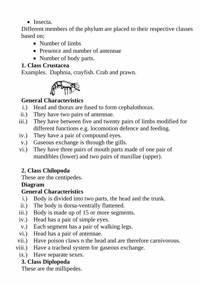
Insecta.
Different members of the phylum are placed to their respective classes
based on;
Number of limbs
Presence and number of antennae
Number of body parts.
1. Class Crustacea
Examples. Daphnia, crayfish. Crab and prawn.
General Characteristics i.) Head and thorax are fused to form cephalothorax.
ii.) They have two pairs of antennae.
iii.) They have between five and twenty pairs of limbs modified for
different functions e.g. locomotion defence and feeding.
iv.) They have a pair of compound eyes.
v.) Gaseous exchange is through the gills.
vi.) They have three pairs of mouth parts made of one pair of
mandibles (lower) and two pairs of maxillae (upper).
2. Class Chilopoda
These are the centipedes.
Diagram
General Characteristics i.) Body is divided into two parts, the head and the trunk.
ii.) The body is dorsa-ventrally flattened.
iii.) Body is made up of 15 or more segments.
iv.) Head has a pair of simple eyes.
v.) Each segment has a pair of walking legs.
vi.) Head has a pair of antennae.
vii.) Have poison claws n the head and are therefore carnivorous.
viii.) Have a tracheal system for gaseous exchange.
ix.) Have separate sexes.
3. Class Diplopoda
These are the millipedes.
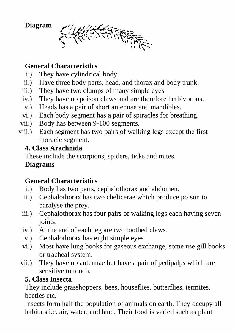
Diagram
General Characteristics i.) They have cylindrical body.
ii.) Have three body parts, head, and thorax and body trunk.
iii.) They have two clumps of many simple eyes.
iv.) They have no poison claws and are therefore herbivorous.
v.) Heads has a pair of short antennae and mandibles.
vi.) Each body segment has a pair of spiracles for breathing.
vii.) Body has between 9-100 segments.
viii.) Each segment has two pairs of walking legs except the first
thoracic segment.
4. Class Arachnida
These include the scorpions, spiders, ticks and mites.
Diagrams
General Characteristics i.) Body has two parts, cephalothorax and abdomen.
ii.) Cephalothorax has two chelicerae which produce poison to
paralyse the prey.
iii.) Cephalothorax has four pairs of walking legs each having seven
joints.
iv.) At the end of each leg are two toothed claws.
v.) Cephalothorax has eight simple eyes.
vi.) Most have lung books for gaseous exchange, some use gill books
or tracheal system.
vii.) They have no antennae but have a pair of pedipalps which are
sensitive to touch.
5. Class Insecta
They include grasshoppers, bees, houseflies, butterflies, termites,
beetles etc.
Insects form half the population of animals on earth. They occupy all
habitats i.e. air, water, and land. Their food is varied such as plant
tissues, animal fluids, dead animals and excretions of animals making
them to be found almost everywhere on earth.
General Characteristics i.) Body is divided into three parts, head, thorax and abdomen.
ii.) Thorax is made up of three segments with three pairs of legs.
Some have one or two pairs of wings on the thorax.
iii.) Head has one pair of antennae.
iv.) They undergo complete or incomplete metamorphosis.
v.) Excretion is through the malpighian tubules which remove uric
acid.
vi.) Gaseous exchange is through the tracheal system but they breathe
through the spiracles.
vii.) The head a pair of compound eyes and several simple eyes.
viii.) Abdomen is made up of 11 or fewer segments. The terminal
segments are modified for reproduction.
ix.) Mouth parts consist of the mandibles, maxillae and labium. The
mouth parts are modified according to their feeding habits such as
sucking, biting, chewing etc.
Assignment
Discuss the economic importance of arthropods.
Study Question 10
Practical Activity
Phylum Chordata
Chordate, common name for animals of the phylum Chordata, which
includes vertebrates as well as some invertebrates that possess, at least
for some time in their lives, a stiff rod called a notochord lying above
the gut. About 43,700 living species are known, making the chordates
the third largest animal phylum.
In animals such as the Amphioxus the notochord persists but in others
it is replaced at later stages of development by the vertebral column.
Members in this phylum inhabit both aquatic (marine and fresh water)
and terrestrial (burrowers and arboreal) environments.
General Characteristics
i.) Members have a notochord at some stage of their development.
ii.) They are bilaterally symmetrical.
iii.) Heart is ventrally placed. Blood flows from the heart through the
arteries and gets back to the heart through the veins.
iv.) They have a post anal tail although it is greatly reduced in some.
v.) They have an endoskeleton.
vi.) They have a closed circulatory system.
vii.) They have visceral clefts where in fish they become the gills in
higher chordates they are only present in the embryo.
viii.) They have a tubular dorsal nerve cord. It develops anteriorly into
brain and posteriorly as the spinal cord. Spinal cord is enclosed
by the vertebral column.
ix.) They have segmented muscle blocks called myotomes on either
side of the body.
The main classes of the phylum chordata are;
Pisces
Amphibia
Reptilia.
Aves.
Mammalia
Pisces
Diagram
These are the fishes. They include those with a skeleton made of
cartilage e.g. shark and those with a bony skeleton such as the tilapia,
Nile perch, lung fish, dog fish, and cat fish etc.
General Characteristics
i.) The move by fins
ii.) Bodies are covered with scales
iii.) Have gills for gaseous exchange in water.
iv.) They don’t have a middle or inner ear.
v.) They have streamlined bodies.
vi.) They have a lateral line for sensitivity.
vii.) Their heart has two main chambers i.e. the auricle and the
ventricle.
viii.) They are poikilothermic/ectothermic.
ix.) Eyes are covered by a nictating membrane.
Amphibia
They include the toads, newts, salamanders and frogs.
The toad is the most advanced amphibian. Its skin is less moist and
therefore uses the lungs more for gaseous exchange. They therefore
stay mostly on land and only return to the ponds during reproduction.
Diagrams
General Characteristics
i.) They have a double circulatory system.
ii.) They have a three chambered heart with two atria and one
ventricle.
iii.) Fertilisation is external and they breed in water.
iv.) Gaseous exchange is through the skin, lungs and gills.
v.) They have two eyes and an eardrum behind the eyes.
vi.) They are ectothermic.
vii.) They have 4 well developed limbs. The hind limbs are more
muscular than the forelimbs.
Reptilia
Examples include tortoise, turtles, snakes, crocodiles, lizards and
chameleons.
General Characteristics
i.) They are ectothermic.
ii.) They have a well developed lung for gaseous exchange.
iii.) They have double circulatory system with the heart having three
chambers i.e. two atria and a partially divided ventricle.
Crocodiles however have a four chambered heart.
iv.) The body is covered with a dry scaly skin reducing desiccation.
v.) Some have four limbs while others don’t have any limbs such as
the snakes.
vi.) Fertilisation is internal. They lay eggs with a leathery shell to
avoid desiccation. Some species of chameleons give birth to
young ones.
Aves
Examples include doves, chicken, hawks, eagles and turkeys.
They are terrestrial and arboreal while some have been adapted for
aquatic life.
General Characteristics
i.) Bodies are covered with feathers for in insulation.
ii.) They have beaks.
iii.) They internal auditory canal/ meatus
iv.) Fertilisation is internal and they lay hard calcareous eggs.
v.) They have lungs for gaseous exchange.
vi.) They have air sacs which store air in them reducing their body
density for flight.
vii.) They are endothermic.
viii.) They have hollow bones.
ix.) They have scales on their hind limbs.
x.) They have double circulatory system with a four chambered
heart.
xi.) The sternum is enlarged to form keel for attachment of flight
muscles.
Mammalia
Study Question 11
Some are arboreal such as the tree squirrels, and some monkeys.
Some are terrestrial either on the surface of the earth or in
tunnels.
Some are aquatic such as the dolphins and whales.
General Characteristics i.) They have double circulatory system
ii.) They have mammary glands hence the name Mammalia.
iii.) Their body is usually covered with fur or hair.
iv.) They have two eternal ears (pinna)
v.) They have sweat glands.
vi.) They have lungs for gaseous exchange.
vii.) They have four limbs.
viii.) They have a diaphragm which separates the body cavity into
thoracic and abdominal cavities..
ix.) The brain is highly developed.
x.) They have seven cervical vertebrae at their neck.
xi.) They are endothermic.
xii.) They have heterodont type of dentition where the teeth are
differentiated into four types, incisors, canines, pre-molars and
molars. The number varies in relation to feeding habits.
Although most mammals give birth to live young ones, some
are egg laying such as the duck billed platypus. After
hatching, the young ones are fed on milk.
Practical Activity 9
Practical Activity 10.
The Dichotomous Key
The word dichotomous means separating into two. I.e. Separation of
different or contradictory things: a separation into two divisions that
differ widely from or contradict each other. As you move down the
key you progress from general characteristics to more specific
characteristics. The last single choice reveals the identity of the
unknown organism.
Rules Used in Constructing a Dichotomous Key i.) Use morphological features as far as possible.
ii.) Start with the major characteristics and proceed to lesser
variations that separate the organisms into smaller groups. E.g. in
leaves start with type of leaf i.e. simple or compound.
iii.) Select a single characteristic at a time and identify it by a number
such as.
Type of leaf
Type of venation
iv.) Use identical forms of words for the two contrasting statements
e.g.
1. a) leaf simple.
b) Leaf compound
2. a) Leaf net veined.
b) Leaf parallel veined.
v.) The statements should always be written in positive form. Where
a negative statement cannot be avoided, the first statement must
be in the positive form e.g.
a) Animal with wings
b) Animal without wings
vi.) Avoid overlapping statements or generalisations such as
Short plants
Tall plants
Be very specific in your description such as
Plant I metre tall and above.
Plant 15cm to 60cm tall.
Some common Features Used For Identification.
In animals
i.) Locomotory structures (legs, wings and fins)
ii.) Antennae, presence and number
iii.) Presence and type of eyes
iv.) Number of body parts
v.) Body segmentation
vi.) Type of skeleton present
vii.) Feeding structures
viii.) Presence of hair, fur, scales or feathers on the body
In plants
Part of
plant
Some characteristics.
Leaf Phylotaxy
Leaf type
Leaf venation
Margin
Lamina
Colour
Flower Inflorescence
Flower shape
Number of floral
Stem Type of stem( woody, herbaceous or fleshy)
Shape (rectangular or cylindrical)
Texture of the stem (smooth or spiny/thorny)
Roots Root system (taproot or fibrous)
Storage roots.
Summary of the Five Kingdoms.
Practical Activities.
Revision Questions.
REPRODUCTION IN PLANTS AND ANIMALS
This is the process by which mature individuals produce
offsprings.
There are two types of reproduction.
1) Sexual reproduction which involves male and female
gametes
Diagram
2) Asexual reproduction where no gametes are involved.
Diagram
Importance of Reproduction
1) Procreation
This ensures that a species does not become extinct.
2) Quality improvement
Reproduction allows for mixing of genetic materials bringing
about variations.
These variations are important tools in the refinement of
quality of offsprings.
Cell Division
Life in all living things start as a single cell as a spore or as a
zygote.
The cells have to divide further to give rise to make cells.
Cell division starts with division of the nucleus (chromosome)
and then the cytoplasm.
Chromosomes
These are microscopic thread like structure within cells that
carries the molecule deoxyribonucleic acid (DNA)—the
hereditary material that influences the development and
characteristics of each organism.
Each chromosome is made up of two parallel strands called
chromatids.
Chromatids are joined together at one point by the centromere.
Diagram
Each cell has a fixed number of chromosomes e.g. each human
body cell has 46 chromosomes.
Chromosomes occur in pairs in the nucleus. A member of each
pair is called homologous chromosomes.
Homologous chromosomes are similar in appearance, size, and
shape but their genetic constitution may be different.
Genes are found along the length of the chromosomes.
Genes are very tiny and made up of a chemical substance called
DNA (De oxy Ribonucleic Acid)
DNA determines the characteristics of the offspring.
There are two types of cell division
i) Mitosis
ii) Meiosis
Mitosis
In this type of cell division, each cell divides into two daughter
cells each having the same number of chromosomes as the parent
cell.
Mitosis occurs in series of stages i.e.
i) Interphase
ii) Prophase
iii) Metaphase.
iv) Anaphase
v) Telophase.
1)Interphase
During this stage the following activities take place within the cell in
preparation of the division.
Synthesis of new cell organelles such as ribosome’s, centrioles,
mitochondria and Golgi apparatus.
Multiplication of genetic material so that each daughter cell will
have same number of chromosomes as the parent cell.
Build up of enough energy stores in form of ATP (Adenosine
Triphosphate) during respiration. This energy is important to see
the cell through the process of division.
At this stage the chromosomes are not clearly visible.
Diagrams
2)Prophase
The following events take place in this stage.
Centrioles separate and move to opposite poles of the cells.
Spindle fibres begin to form
Nuclear membrane begins to break down and nucleolus
disappears.
Chromosomes thicken and shorten and they can be stained easily
hence become visible.
Diagram
3)Metaphase
Nuclear membrane disappears and chromosomes are free in the
cytoplasm.
Spindle fibres lengthen and attach to the centrioles at both poles.
Chromosomes align themselves at the equator and are attached to
the spindle fibres by their centromere.
Diagram
3) Anaphase
Chromatids separate at the centromere and migrate to opposite
poles. This is brought about by the shortening of the spindle
fibres.
Spindle fibres begin to disappear.
In animal cells, cell membrane begins to constrict towards the end
of anaphase.
Diagram
4) Telophase
Chromatids collect together at the two opposite poles of the
spindle.
Nuclear membrane forms around each set of chromatids and are
now referred to as chromosomes.
Cytoplasm divides into two hence the formation of two daughter
cells.
Chromosomes become less distinct.
In animal cells, division of cytoplasm is by constriction of cell
membrane.
In plant cells, a cell plate forms within the cytoplasm and grows to
separate the cell into two.
Diagrams
Significance of Mitosis
i) Forms basis for asexual reproduction e.g. budding and
spore formation.
ii) Causes cell growth when the cells formed increase in
number and size.
iii) Ensures genetic constitution of the offspring is the same
as the parents.
iv) Replaces damaged and dead cells in the body.
Meiosis
This involves two divisions of the parental cell resulting into four
daughter cells.
First meiotic cell division involves the separation of the
homologous chromosomes. It is referred to as Reduction division
because the numbers of chromosomes are reduced by half.
In the second stage, the sister chromatids are separated and it is
referred to as Equatorial division
Each daughter cell has half the number of chromosomes (haploid
n) as the parent cell.
This takes place in the reproductive organs of animals (testis and
ovary) and plants (anthers and ovary).
Meiosis is divided into same series of stages as in mitosis.
The phases are given names as in mitosis but each is followed by
I or II.
First Meiotic Division
Interphase I
The cell prepares for division by the following.
Replication of chromosomes.
Synthesis of new cell organelles.
Build up of energy.
Prophase I
Nucleolus disappears.
Centrioles move to opposite poles.
Chromosomes shorten and thicken becoming more visible.
Homologous chromosomes lie side by side in the process of
synapsis forming pairs called bivalents.
Homologous chromosomes may become coiled around each other
with their chromatids remaining in contact at points called
chiasmata.
NB/. During chiasma formation homologous chromosomes may
exchange genetic material during crossing over. These genetic
exchanges are important because they bring about variations in
offsprings.
Metaphase.I
Nuclear membrane disappears.
Homologous chromosomes as a bivalent move to the equator of
the cell.
Spindle fibres are fully formed and get attached to the
chromosomes at the centromere.
Homologous chromosomes orientate towards different poles.
Diagram
Anaphase I
Homologous chromosomes separate and migrate to the opposite
poles with their centromeres leading. This is brought about by the
shortening of the spindle fibres.
Diagram
Telophase I
Cell divides across the middle when the chromosomes reach the
poles.
At the end of meiosis I homologous chromosomes are separated.
Diagram Second Meiotic Division.
In this stage the sister chromatids are separated from each other.
Interphase II
Cells go into a short interphase.
Prophase II
Chromosomes become shorter and thicker.
New spindle fibres are formed.
Metaphase.II
Chromosomes align at the equator of the cell.
Spindle fibres attach to their centromeres.
Chromosomes orientate themselves towards the opposite poles.
Anaphase II
Sister chromatids separate from each other.
Spindle fibres shorten pulling them to the opposite poles.
Telophase II
Spindle fibres disappears
Nucleolus reappears and nuclear membrane forms around each
set of chromatids.
Chromatids uncoil and become threadlike.
Cytoplasm divides.
Four cells are formed (tetrad).
Each cell has haploid (n) number of chromosomes.
Significance of Meiosis 1. Gamete formation (sperms and ova) forming basis for sexual
reproduction
2. Provides opportunities for genetic variations during crossing over
Similarities between mitosis and meiosis
1. Both take place in plants and animals.
2. Both involve division (multiplication) of cells.
Differences between meiosis and mitosis
Meiosis Mitosis.
1. Homologous
chromosomes associate
with each other.
No association of homologous
chromosomes
2. Takes place in 2 nuclear
divisions.
Takes place in one nuclear
division.
3. 4 daughter cells are
produced each haploid (n)
2 daughter cells are produced
each diploid (2n)
4. Occurs in reproductive
organs leading to gamete
formation.
Occurs in somatic (body) cells
leading to growth.
5. Chiasma formation takes
place leading to crossing
over hence variation
No chiasma formation
therefore no crossing over
hence no variation.
Asexual Reproduction
This is the production of offsprings from a single organism
without fusion of gametes.
This type of reproduction involves mitosis.
Types of Asexual Reproduction
1. Binary fission in amoeba, plasmodium and bacteria
2. Sporulation in rhizopus
3. Budding in yeasts
1. Binary fission in amoeba
When there is enough food and favourable temperature and pH, a
mature amoeba divides into two.
During binary division, in amoeba, internal reorganization of
molecules necessary for structural construction takes place.
Nucleus first divides mitotically (Karyogamy) into two followed
by the division of the cytoplasm (Cytogamy)
Diagrams
2. Sporulation in Rhizopus
This is the formation of spores in substrates like the bread to form
bread moulds
A spore is a microscopic reproductive unit which contains a
nucleus and a small amount of cytoplasm.
Spores are produced by bacteria, most fungi, mosses and ferns.
Rhizopus has a vegetative body called the mycelium.
Mycelium is made up of many branched threads called hyphae.
Horizontal hyphae are called stolons.
Vertically growing ones are called sporangiophore.
Tips of sporangiophore swell up to form the sporangia
(sporangium).
Sporangia are the spore bearing structures. When fully mature,
sporangium wall burst releasing the spores. If spores land on a
suitable medium, they germinate and develop into other rhizopus.
Rhizopus uses structures called rhizoids for anchorage and to
obtain nutrients from the substrate.
Diagrams
Budding in Yeast Under favourable conditions such as plenty of sugar, moisture, oxygen
and optimum temperature, the yeast cell reproduces asexually by
budding.
A projection of bud forms on the parent cell.
Nucleus divides into two.
One nuclei moves into the new bud.
Bud grows in size and forms new cell organelles. Later the bud
separates off.
Diagrams
Sexual Reproduction in Plants
In flowering plants the flower is the reproductive organ.
Structure and Function of a Flower
A flower is made up of a flower stalk (pedicel) and a receptacle.
Attached to the receptacle are four groups of floral structures i.e.
i) Calyx (sepals)
ii) Corolla/petals
iii) Androecium – male parts
iv) Gynoecium – female parts
i) Calyx (sepals)
Made up of the sepals which are usually green.
If sepals are fused they form gamosepalous calyx.
If they are free, they form polysepalous calyx.
Calyx protects the inner parts of the flower especially during bud
development.
Some flowers have sepal like structures below the calyx called
the epicalyx.
ii) Corolla/petals
It’s made up of the petals which are brightly coloured, large and
conspicuous especially in insect pollinated flowers.
If fused – gamopetalous.
If free – polypetalous
iii) Androecium – male parts
Made up of one or more stamens
Satmen is made up of the filament and anthers.
Another has four pollen sacs containing pollen grains.
Pollen grains contain the male gametes.
Diagrams
iv) Gynoecium – female parts
It may contain one or more carpels
A carpel consists of the ovary, the style and the stigma.
Ovary contains the ovules.
Ovaries are described as epigynous, hypogynous or perigynous
depending on the place they occur in the flower.
i) Epigynous (inferior) ovary
Ovary is located within the receptacle.
All other floral parts occur above it such as in the apple flowers.
Diagram
ii) Hypogynous (superior) ovary
Ovary is above the receptacle and other floral parts such as in
hibiscus.
Diagram
iii) Perigynous ovary
The receptacle surrounds the carpel.
All other floral parts arise around the ovary such as in roses.
Diagram
The gynoecia can also be grouped into different types dependi.ng on
the number of carpels present i.e. monocarpous or syncarpous.
Monocarpous Gynoecium
It has only one carpel e.g. in beans.
Diagram
Polycarpous Gynoecium
It has two or more carpels. It is divided into two.
a) Apocarpous gynoecium
The carpels are free e.g in roses and bryophyllum.
Diagrams
b) Syncarpous gynoecium
The carpels are fused together such as in hibiscus.
Diagrams
Terms Used in Describing a Flower i) Complete flower – has all the four floral parts; calyx, corolla,
androecium and gynoecium.
ii) Incomplete flower – has one or two floral parts missing.
iii) Unisexual flower – a flower with only one of the
reproductive parts either male or female flower.
iv) Staminate flower – male flower.
v) Pistillate flower – female flower.
vi) Monoecious plant – bears both male and female parts of the
flower.
vii) Dioecious plants - the plant is either male or female e.g. in
paw paw.
viii) Hermaphrodite or bisexual flower – has both the male and
female parts.
ix) Regular or actinomorphic flower – a flower that can be
divided into tow similar halves by any vertical section passing
through the center i.e. radial symmetry such as in morning
glory.
x) Irregular or zygomorphic flower – can be divided into two
similar halves on one particular plane only i.e. bilateral
symmetry e.g. in clotalaria.
xi) Pedicillate flower- flower with a stalk.
xii) Solitary flower – are flowers occurring singly.
xiii) Inflorescence – flowers that grow in clusters.
xiv) Essential parts of the flower – are the androecium and
gynoecium.
xv) Non essential floral parts – are the calyx and corolla.
Pollination
This is the transfer of pollen grains from the anther to the stigma.
Types of Pollination
i) Self pollination. – Transfer of pollen grains from the anther to
the stigma of the same flower.
ii) Cross Pollination – transfer of pollen grains from the anther of
one flower to the stigma of another flower but of the same
species.
Agents of Pollination
Insect
Wind
Adaptations of Insect Pollinated Flowers (Entomophilous)
i) Flowers are large, conspicuous with brightly coloured petals
and inflorescence to attract insects.
ii) Flowers are scented and produce nectar to attract insects.
iii) Pollen grains are relatively large, heavy, rough or sticky so as
to stick on to the body of the sticks.
iv) They have small and firmly attached anthers to a firm filament.
v) Stigmas are small, sticky and contained within the flower. This
ensures that pollen grains from the body of an insect stick onto
it.
vi) Flowers have a tubular or funnel shaped corolla, landing
platforms and honey guides.
Adaptations of Wind Pollinated Flowers (Anemophilous)
e.g. maize and other grasses
i) Small flowers with inconspicuous petals, bracts or
inflorescence.
ii) Flower structure is simple and flowers have no particular
shape.
iii) Stigmas are long, feathery and hang outside the flower to trap
pollen grains.
iv) Pollen grains are small, smooth and light to be easily carried by
the wind.
v) Flowers are not scented and lack nectar.
vi) Anthers are large and loosely attached to a flexible filament to
be easily released when the wind blows.
Diagram of a grass flower
Features and Mechanisms Hindering Self Pollination and Self
Fertilization
i) Heterostyly – condition whre the stigma na d style have
different arrangements e.g. coconut flowers have shorter
stamens than pistils hence pollen grains from the anthers
cannot reach the stigma.
Diagram
ii) Self sterility or incompatibility – condition where pollen grains
of a flower fail to germinate if they land on the stigma of the
same flower.
iii) Protogyny and Protandry – condition where either male parts
of a flower mature before the female ones.
Protandry – stamen mature before the stigma e.g.in sunflower.
Protogyny – stigma matures before the anthers mature e.g. in
maize.
iv) Dioecious plants and presence of features that promote cross
pollination such as brightly coloured petals which attract
insects hence cross pollination.
Fertilization in Flowering Plants
Fertilization in plants is the fusion of the male and female nuclei in the
embryo sac.
Male gamete is contained in the pollen grain produced in the
anther.
Diagram Female gamete (egg cell) is found in ovules contained in the embryo
sac.
Filament
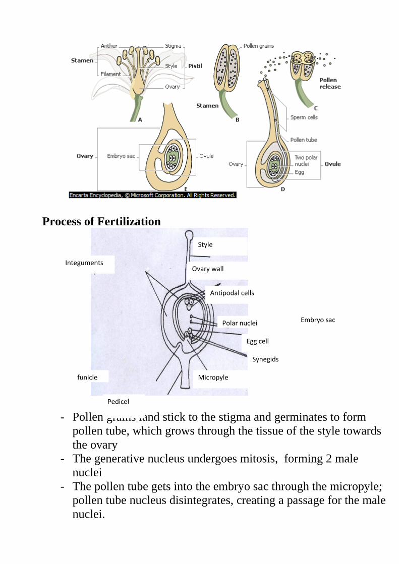
Process of Fertilization
- Pollen grains land stick to the stigma and germinates to form
pollen tube, which grows through the tissue of the style towards
the ovary
- The generative nucleus undergoes mitosis, forming 2 male
nuclei
- The pollen tube gets into the embryo sac through the micropyle;
pollen tube nucleus disintegrates, creating a passage for the male
nuclei.
Embryo sac
Pedicel
Synegids
Egg cell
Polar nuclei
Antipodal cells
Ovary wall
Style
Micropyle funicle
Integuments
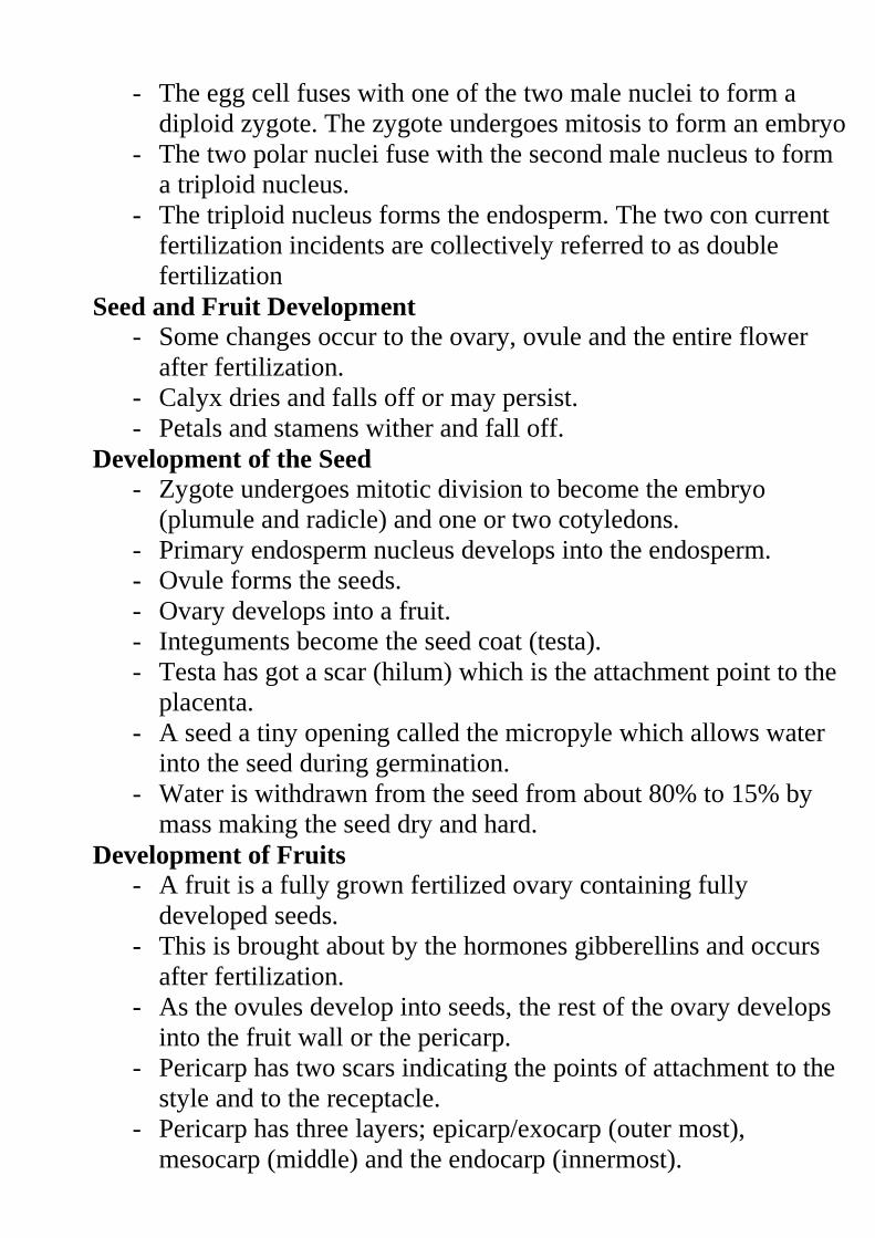
- The egg cell fuses with one of the two male nuclei to form a
diploid zygote. The zygote undergoes mitosis to form an embryo
- The two polar nuclei fuse with the second male nucleus to form
a triploid nucleus.
- The triploid nucleus forms the endosperm. The two con current
fertilization incidents are collectively referred to as double
fertilization
Seed and Fruit Development
- Some changes occur to the ovary, ovule and the entire flower
after fertilization.
- Calyx dries and falls off or may persist.
- Petals and stamens wither and fall off.
Development of the Seed
- Zygote undergoes mitotic division to become the embryo
(plumule and radicle) and one or two cotyledons.
- Primary endosperm nucleus develops into the endosperm.
- Ovule forms the seeds.
- Ovary develops into a fruit.
- Integuments become the seed coat (testa).
- Testa has got a scar (hilum) which is the attachment point to the
placenta.
- A seed a tiny opening called the micropyle which allows water
into the seed during germination.
- Water is withdrawn from the seed from about 80% to 15% by
mass making the seed dry and hard.
Development of Fruits
- A fruit is a fully grown fertilized ovary containing fully
developed seeds.
- This is brought about by the hormones gibberellins and occurs
after fertilization.
- As the ovules develop into seeds, the rest of the ovary develops
into the fruit wall or the pericarp.
- Pericarp has two scars indicating the points of attachment to the
style and to the receptacle.
- Pericarp has three layers; epicarp/exocarp (outer most),
mesocarp (middle) and the endocarp (innermost).
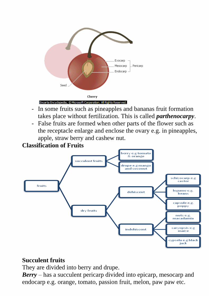
- In some fruits such as pineapples and bananas fruit formation
takes place without fertilization. This is called parthenocarpy.
- False fruits are formed when other parts of the flower such as
the receptacle enlarge and enclose the ovary e.g. in pineapples,
apple, straw berry and cashew nut.
Classification of Fruits
Succulent fruits
They are divided into berry and drupe.
Berry – has a succulent pericarp divided into epicarp, mesocarp and
endocarp e.g. orange, tomato, passion fruit, melon, paw paw etc.
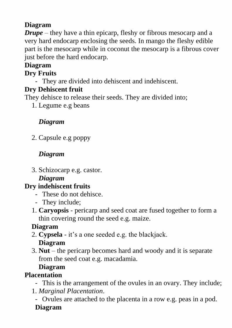
Diagram
Drupe – they have a thin epicarp, fleshy or fibrous mesocarp and a
very hard endocarp enclosing the seeds. In mango the fleshy edible
part is the mesocarp while in coconut the mesocarp is a fibrous cover
just before the hard endocarp.
Diagram
Dry Fruits - They are divided into dehiscent and indehiscent.
Dry Dehiscent fruit They dehisce to release their seeds. They are divided into;
1. Legume e.g beans
Diagram
2. Capsule e.g poppy
Diagram
3. Schizocarp e.g. castor.
Diagram
Dry indehiscent fruits
- These do not dehisce.
- They include;
1. Caryopsis - pericarp and seed coat are fused together to form a
thin covering round the seed e.g. maize.
Diagram
2. Cypsela - it’s a one seeded e.g. the blackjack.
Diagram
3. Nut – the pericarp becomes hard and woody and it is separate
from the seed coat e.g. macadamia.
Diagram
Placentation
- This is the arrangement of the ovules in an ovary. They include;
1. Marginal Placentation.
- Ovules are attached to the placenta in a row e.g. peas in a pod.
Diagram
2. Basal placentation
- Placenta is formed at the base of the ovary. Ovules are attached
to it sunflower and sweet pepper.
Diagram
3. Axile Placentation
- The edges of the carpels fuse together to form a central placenta
in the axile.
- Ovules are arranged on the placenta.
- The ovary is divided into a number of loculi by the walls of the
carpel e.g. in orange
Diagram
4. Parietal Placentation
- Edges of the carpels fuse together and dividing walls disappear
leaving a loculus.
- Placentas from each carpel appear as a ridge on the ovary wall
and have numerous ovules on them e.g. in paw paw.
Diagram
5. Free central placentation
- Edges of carpels fuse together and the dividing walls disappear
leaving one loculus.
- Placenta appears at the center and have numerous ovules on it
e.g. in primrose
Diagram
Adaptations of Fruits to Various Agents of Dispersal
a) Water dispersal
- Such seeds and fruits enclose air in them to lower their
density for buoyancy;
- They are fibrous/ spongy to lower the density for
buoyancy;
- Have impermeable seed coat or epicarp to prevent water
from entering during flotation so as to avoid rotting;
- The seeds can remain viable while in water and only
germinate while on a suitable medium;
b) Wind dispersal
- They are light; and small; to be easily carried by wind
currents due to lower density;
- Have developed extension (Parachute like structures and
Wing like structures) which create a larger surface area;
so as to be kept afloat in wind currents e.g. sonchus and
jacaranda
- In some a Perforated capsule is usually loosely attached
to a long stalk which is swayed away by wind scattering
seeds;
c) Animal dispersal
- Brightly colored to attract animals
- Fleshy to attract animals; e.g. mangoes, passion fruits,
oranges, tomatoes etc.
- aromatic /scented to attract animals;
- The seed coats are hard and resistant to digestive
enzymes; the seeds are therefore dropped away in
feaces/droppings e.g. passion fruit and tomatoes.
- Some have hook like structures to attach on animals fur
e.g. blackjack
d) Self dispersal
- They have weak lines (sutures) on the fruit wall (pod),
along which they burst open to release seeds, which get
scattered away from the parent plant e.g. in legumes such
as peas and beans.
SEXUAL REPRODUCTION IN ANIMALS
This involves gamete fusion.
The male produces the male gamete (sperms) and the female
produces the female gamete (ovum/ova).
The gametes are produced in special organs called gonads i.e. the
testes and ovaries.
The sperm fuses with the ovum to form a zygote through a
process called fertilisation the gametes are haploid and the zygote
is diploid.
Fertilisation may be internal or external.
External Fertilisation in Amphibians
The female lays eggs and the male sheds sperms on them (to
fertilise them). This is only possible in water.
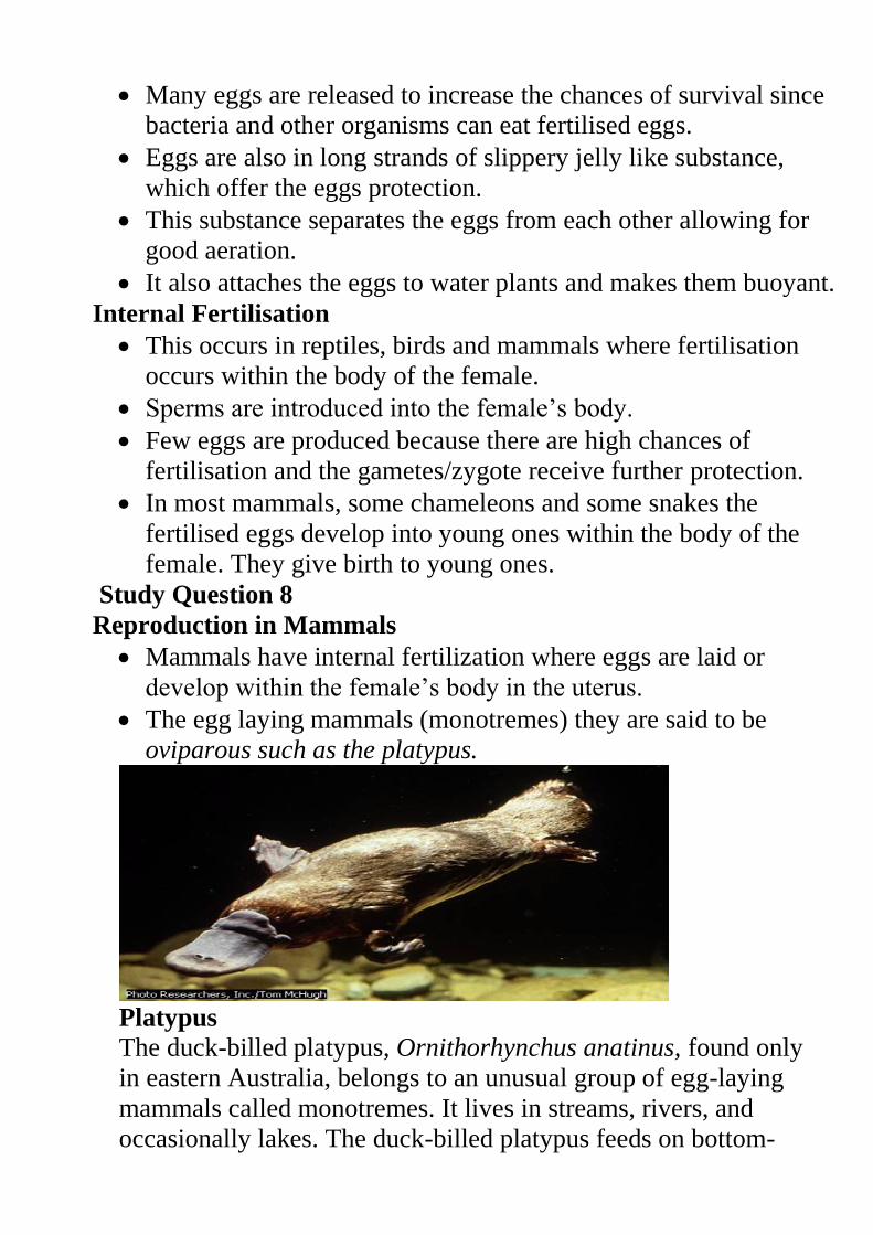
Many eggs are released to increase the chances of survival since
bacteria and other organisms can eat fertilised eggs.
Eggs are also in long strands of slippery jelly like substance,
which offer the eggs protection.
This substance separates the eggs from each other allowing for
good aeration.
It also attaches the eggs to water plants and makes them buoyant.
Internal Fertilisation
This occurs in reptiles, birds and mammals where fertilisation
occurs within the body of the female.
Sperms are introduced into the female’s body.
Few eggs are produced because there are high chances of
fertilisation and the gametes/zygote receive further protection.
In most mammals, some chameleons and some snakes the
fertilised eggs develop into young ones within the body of the
female. They give birth to young ones.
Study Question 8
Reproduction in Mammals
Mammals have internal fertilization where eggs are laid or
develop within the female’s body in the uterus.
The egg laying mammals (monotremes) they are said to be
oviparous such as the platypus.
Platypus
The duck-billed platypus, Ornithorhynchus anatinus, found only
in eastern Australia, belongs to an unusual group of egg-laying
mammals called monotremes. It lives in streams, rivers, and
occasionally lakes. The duck-billed platypus feeds on bottom-
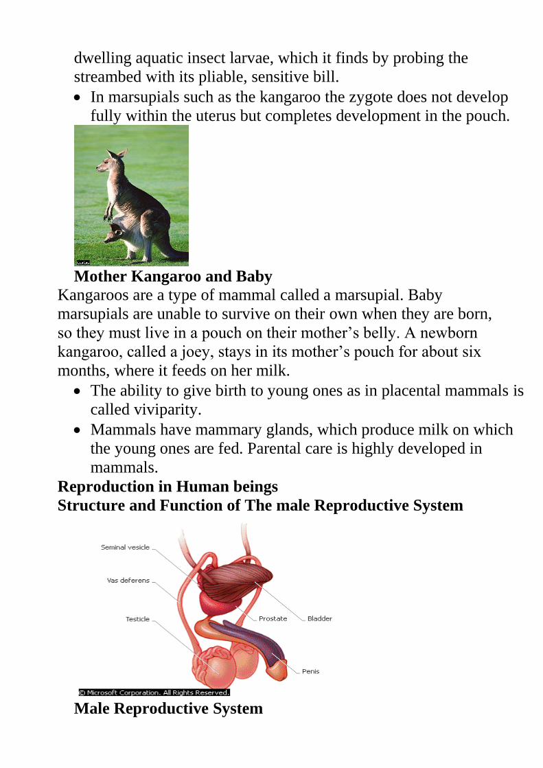
dwelling aquatic insect larvae, which it finds by probing the
streambed with its pliable, sensitive bill.
In marsupials such as the kangaroo the zygote does not develop
fully within the uterus but completes development in the pouch.
Mother Kangaroo and Baby
Kangaroos are a type of mammal called a marsupial. Baby
marsupials are unable to survive on their own when they are born,
so they must live in a pouch on their mother’s belly. A newborn
kangaroo, called a joey, stays in its mother’s pouch for about six
months, where it feeds on her milk.
The ability to give birth to young ones as in placental mammals is
called viviparity.
Mammals have mammary glands, which produce milk on which
the young ones are fed. Parental care is highly developed in
mammals.
Reproduction in Human beings
Structure and Function of The male Reproductive System
Male Reproductive System
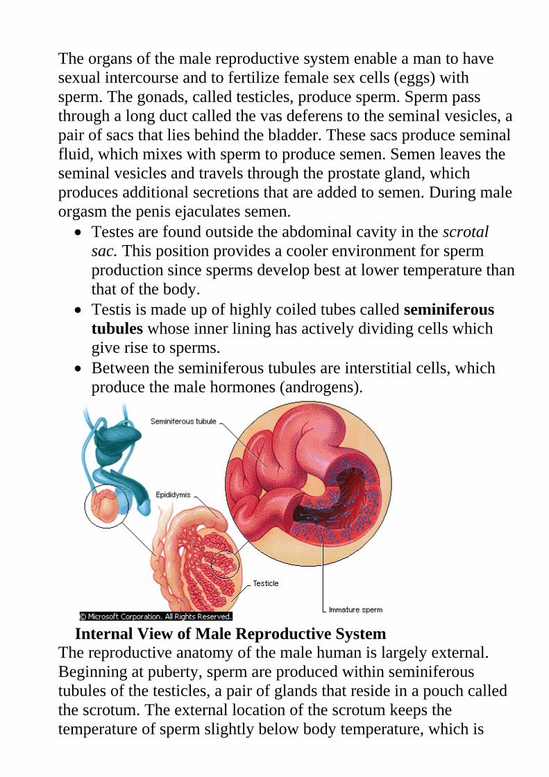
The organs of the male reproductive system enable a man to have
sexual intercourse and to fertilize female sex cells (eggs) with
sperm. The gonads, called testicles, produce sperm. Sperm pass
through a long duct called the vas deferens to the seminal vesicles, a
pair of sacs that lies behind the bladder. These sacs produce seminal
fluid, which mixes with sperm to produce semen. Semen leaves the
seminal vesicles and travels through the prostate gland, which
produces additional secretions that are added to semen. During male
orgasm the penis ejaculates semen.
Testes are found outside the abdominal cavity in the scrotal
sac. This position provides a cooler environment for sperm
production since sperms develop best at lower temperature than
that of the body.
Testis is made up of highly coiled tubes called seminiferous
tubules whose inner lining has actively dividing cells which
give rise to sperms.
Between the seminiferous tubules are interstitial cells, which
produce the male hormones (androgens).
Internal View of Male Reproductive System
The reproductive anatomy of the male human is largely external.
Beginning at puberty, sperm are produced within seminiferous
tubules of the testicles, a pair of glands that reside in a pouch called
the scrotum. The external location of the scrotum keeps the
temperature of sperm slightly below body temperature, which is
necessary for their healthy development and survival. From each
testicle, sperm migrate to a long, coiled tube known as the
epididymis, where they are stored for one to three weeks until they
mature. Also located outside the body is the penis, the erectile organ
responsible for the excretion of urine and the transfer of sperm to
the vagina of the female. Just before ejaculation during sexual
arousal, mature sperm travel from the epididymis, a coiled tube
behind each testicle, through a long duct called the vas deferens.
Sperm leave the body in semen, a fluid produced by the seminal
vesicles.
Seminiferous tubules unite to form the epididymis, which is
about 6m long and highly coiled. It stores the sperms.
It’s connected to the sperm duct/vas deferens. Sperm duct
connects the epididymis to the urethra, which is the ejaculatory
duct.
Seminal vesicles provide an alkaline fluid, which contains
nutrients for the sperms.
Prostate gland secretes an alkaline substance to neutralise the
vaginal fluids. It also activates the sperms.
Cowper’s glands secrete an alkaline fluid that neutralizes the
acidity along the urethra.
All these fluids combine with the spermatozoa to form the
semen.
Since the urethra serves both passage of urine and semen it is
said to be urino-genital in function.
The penis is erectile and made of spongy tissue, muscle and
blood vessels.
Once erect, the penis is able to penetrate the vagina in order to
deposit sperms into the female’s reproductive tract.
Study question 9 and Practical.
Structure and Function of The Female Reproductive System.
Diagram
The internal sex organs of the female consist of the vagina,
uterus, fallopian tubes (or oviducts), and ovaries.
The vagina is a flexible tube-shaped organ that is the passageway
between the uterus and the opening in the vulva. Because during
birth the baby travels from the uterus through the vagina, the
vagina is also known as the birth canal.
The woman's menstrual flow comes out of the uterus and through
the vagina.
When a man and a woman engage in vaginal intercourse, the
penis is inserted into the vagina.
The cervix is located at the bottom of the uterus and includes the
opening between the vagina and the uterus. It secretes a plug of
mucus, which prevents entry of pathogens into the uterus during
pregnancy.
The uterus is a muscular organ that has an inner lining
(endometrium) richly supplied with blood vessels and glands.
During pregnancy, the uterus holds and nourishes the developing
foetus.
Although the uterus is normally about the size of a fist, during
pregnancy it is capable of stretching to accommodate a fully
developed foetus, which is typically about 50 cm (about 20 in)
long and weighs about 3.5 kg (about 7.5 lbs).
The uterine muscles also produce the strong contractions of
labour.
At the top of the uterus are the pair of fallopian tubes (oviduct)
that lead to the ovaries.
The two ovaries produce eggs, or ova (the female sex cells that
can become fertilized), and female sex hormones, primarily
oestrogen and progesterone.
The fallopian tubes have finger like projections at the ends near
the ovaries that sweep the egg into the fallopian tube after it is
released from the ovaries.
Movement of ovum is also aided by the smooth muscles of the
oviduct.
If sperm are present in the fallopian tube, fertilization
(conception) may occur and the fertilized egg will be swept into
the uterus by cilia (hair like projections inside the fallopian tube).
Practical
The Human Sperm
Are formed in the seminiferous tubules of testes by meiosis.
Final products of meiosis enter the sertoli cells where they are
nourished and undergo maturation.
Mature sperms leave for epididymis where they are stored.
A mature sperm has an ovoid head, short neck, middle piece and
a tail.
Diagram
Head has a large nucleus carrying the genetic material, which is
haploid (n).
At the tip of the head there is the acrosome containing lytic
enzymes. These enzymes digest the wall of ova.
The short neck contains centrioles.
Middle piece has a large number of mitochondria, which provide
with the energy required for propulsion of the sperm to reach the
ova.
The tail propels the sperm forward by its side-to-side lashing
action.
Formation of The Ova
In females egg formation begins in the ovary of the foetus before
birth unlike in males where production of sperms starts at
puberty.
At birth there are about 70,000 potential egg cells in the ovaries
of a baby girl.
A layer of ovary cells called primary follicles, which provide
them with nourishment, encloses them.
Only about 500 of them develop into ova during puberty. During
puberty the primary follicles grow to become Graafian follicle.
At ovulation, the Graafian follicle bursts open to release a mature
ovum surrounded by a layer of cells.
Diagram
A mature ovum is spherical in shape with a diameter of about 0.2
mm.
It has a large haploid nucleus surrounded by a nuclear membrane.
Nucleus is within the cytoplasm enclosed by the plasma
membrane. Vitelline membrane surrounds the plasma
membrane.
Study Question 11
Fertilisation
Process where the nucleus of a male gamete fuses with the
nucleus of a female gamete to form a zygote.
This takes place in the upper part of he oviduct after copulation.
Sperms are drawn up by suction through the cervix into the
uterus. They swim up to the oviduct using their tails.
Very many sperms are released but only one is required to
fertilise the ovum.
The ovum releases chemical substances, which are neutralised by
those released by the acrosome.
When the ovum comes into contact with the egg the acrosome
bursts releasing lytic enzymes, which dissolve the egg
membranes.
The acrosome turns inside out forming a filament, which is used
to penetrate the eggs.
Diagrams
The Vitelline membrane undergoes a change, which stops any
other sperm from entering the ovum.
Once inside the cytoplasm the head bursts to release the male
nucleus, which then fuses with the female nucleus to form a
diploid zygote.
After ovulation the ovum can remain viable for 8-24 hours before
it dies.
The sperm can remain viable for 2-3 days in the female
reproductive tract.
Study Question 12
Implantation
This is the attachment of the blastocyst to the walls of the uterus
by the villi.
After fertilisation, the zygote undergoes various mitotic divisions
as it moves down the oviduct. Its movement is aided by cilia in
the oviduct and by the contractions of the smooth muscles lining
the oviduct.
By the time it reaches the uterus it has formed a hollow structure
of cells called blastocyst.
Movement of the zygote from the oviduct to the time it is
implanted takes about 7 days.
Diagrams
Sometime the zygote may fail to move down to the uterus and
gets implanted into the walls of the oviduct. This condition is
referred to as ectopic pregnancy.
Formation of Placenta
During implantation the blastocyst differentiates into three layers,
chorion, amnion and allantois.
Diagram
Chorion is the outermost and it has finger like projections called
chorionic villi. These villi grow into the endometrium. During
the early stages of embryo development, villi form the sites for
material exchange between the embryo and maternal blood
vessels.
Amnion surrounds the embryo forming an amniotic cavity.
Amniotic cavity contains the amniotic fluid, which suspends the
foetus providing it with support. It also acts as a shock absorber
hence protecting it against mechanical injury.
The chorionic villi, allantois and the endometrium form the
placenta.
The embryo is attached to the placenta by a tube called the
umbilical cord.
When the placenta is fully formed, the embryo becomes the
foetus at about three months of pregnancy.
The Role of The Placenta
This is a temporary organ found only in placental mammals. It is
the only organ in animals composed of cells derived from two
different organisms; the foetus and the mother.
It facilitates the transfer of nutrients and metabolic waste products
between the mother and the foetus. It selectively allows some
materials to pass through and not others.
Refer to the table below
Drugs, alcohol and some chemicals from cigarette smoke pass
through the placenta. Pregnant mothers should therefore not take
alcohol or smoke excessively.
There is no direct connection between the foetal blood system and
that of the mother.
If the two systems were directly connected, the delicate blood
vessels of the foetus would burst due the higher pressure in the
maternal circulatory system.
Exchange of materials occurs across the sinus in the uterine wall
and the capillary system of foetus across intercellular space by
diffusion.
Diagram
Study question 13
During pregnancy, placenta takes over the role of producing
hormones oestrogen and progesterone.
Major functions of oestrogen and progesterone during pregnancy
Oestrogen Progesterone.
i.) Growth of mammary
glands
ii.) Inhibits FSH release.
iii.) Inhibits prolactin release.
iv.) Prevent infection in
uterus
v.) Increase size of the
uterine muscle cells.
vi.) Increase ATP and
creatine phosphate
formation.
vii.) Increases sensitivity of
myometrium to oxytocin.
i.) Growth of mammary
glands.
ii.) Inhibits FSH release
iii.) Inhibits prolactin release.
iv.) Inhibits contraction of
myometrium.
What is allowed to pass
through the placenta
What is not allowed to pass
through the placenta
From the mother to the
foetus.
i.) Oxygen
ii.) Vitamins
iii.) Mineral salts
iv.) Hormones
v.) Water
vi.) Antibodies and antigens.
vii.) Glucose, amino acids,
fatty acids and glycerol.
From the foetus to the
mother
i.) Carbon (iv) oxide.
i.) All blood cells.
ii.) Plasma proteins.
iii.) Most bacteria.
ii.) Nitrogenous wastes.
Gestation Period
This is the period between conception and birth. This varies in
different animals.
E.g. mice 22 days
Rabbits, 30 days
Man, 9 months
Elephants, 18 month
When the human embryo is two weeks old, allantois,
chorion and amnion have already formed. Embryo then
differentiates into tissues and organs.
By the end of the third month, the heart and blood vessels
are fully developed. Spinal cord and the head region, which
includes the eyes and the nose, are also well developed.
Limbs show early signs of development.
By the end of 6 months the alveoli and nose are well
developed. Foetal movement can as well be felt.
By the end of the nine months, the foetus head is directly
above the cervix.
By now all the organs and systems are fully developed.
If birth occurs before completion of 6 months, this is called
miscarriage and the baby cannot survive.
If the foetal development is interfered with either
physically or chemically such that the foetus is released,
this is called abortion.
If birth occurs after 7 months but before term, this is called
premature birth. Such babies are raised in incubators and
they do survive.
Pregnant mothers must have a balanced diet. Calcium,
proteins, phosphates and iron should be abundant in her
diet.
Calcium and phosphorous are needed for bone formation
while iron is for haemoglobin formation.
Pregnant mother should visit antenatal clinic.
Birth/Parturition
Maternal posterior pituitary gland releases hormone
oxytocin. Progesterone level goes down. Oxytocin
stimulates contraction of the myometrium.
Oxytocin is released in waves during labour. This provides
the force required to expel the foetus from the uterus.
The cervix dilates, the amnion and chorion rupture
releasing the amniotic fluid.
The uterus starts contacting from the top downwards
pushing the foetus downwards head first through the
widened cervix and the birth canal.
After birth, the umbilical cord is ligatured/cut to separate
the baby from the placenta. Placenta is expelled later after
birth.
Then newborn baby takes in the first breath, lungs expand
and become functional. The respiratory role of the placenta
is taken over by the lungs.
Diagrams
Caesarean delivery
This is the surgical incision of the abdominal and uterine
walls for delivery to be achieved. This is done where there
are complications ns such that the foetus cannot pass
through the birth canal.
Parental care
The newborn baby is given food and protection. Placental
mammals feed their young ones on milk. Milk is produced
by the mammary glands under the influence of lactogenic
hormones e.g. prolactin.
Mother’s milk is the best as it contains all the nutrients
needed for the growth and development of the body.
For the first 3 days, colostrum is produced which contain
antibodies, which provide natural defence to the foetus
against diseases.
Milk is deficient of iron. The baby relies on iron stored in
its liver during gestation.
Milk let down is an example of a reflex action.
The prevailing environment as shown below influences it
either positively or negatively.
Milk production in various environments
Positive Environment Negative Environment
Sucking at the breast,
smell of the baby or
crying of the baby trigger
milk let down.
Hypothalamus relays
impulses to pituitary
gland which releases
hormone oxytocin
Oxytocin reaches the
breasts and causes
alveoli to contract
forcing milk into the
ducts.
Milk let down may be
inhibited or blocked if
the breastfeeding mother
experiences
embarrassment, fatigue
or anxiety.
Ducts conduct milk into
the reservoirs behind the
areola
Baby sucks the milk
from this reservoir.
Assignment
Child labour
Role of Hormones in Human Reproduction
Secondary sexual characteristics
These are physiological, structural and mental changes
associated with masculinity and femininity. They are controlled
by oestrogen in females and androgens in males. They occur at
puberty.
Secondary sexual characteristics in males
Hypothalamus stimulates pituitary gland to release
gonadotrophic hormones i.e. FSH and LH.
FSH stimulates sperm synthesis.
LH is also known as Interstitial Cell Stimulating Hormone
(ICSH) and it stimulates interstitial cells to release
Androgens mostly Testosterone. It stimulates the onset of
secondary sexual characteristics mostly at the age of 14.
These include;
i.) Deepening of voice
ii.) Growth of hair in pubic parts and armpit region
iii.) Appearance of beards
iv.) Body becomes masculine
v.) Testes enlarge and begin to produce sperms
Secondary sexual characteristics in females
In females they start at early age 10-12 years. They include,
i.) Development of mammary glands
ii.) Growth of hair in pubic parts and armpit region
iii.) Enlargement of the pelvic girdle and widening of the
hips
iv.) Body becomes feminine.
v.) Ovaries mature and start releasing eggs under the
influence of FSH and LH hence ovulation and menses.
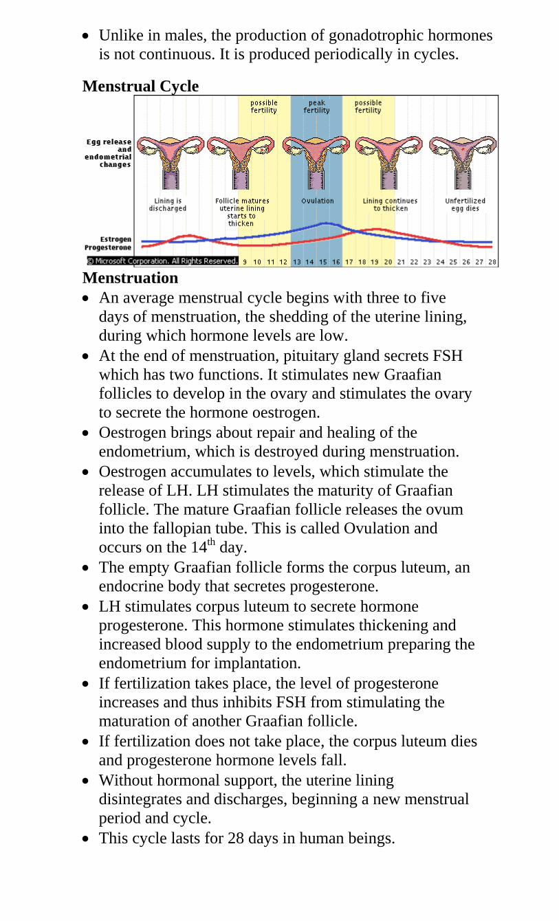
Unlike in males, the production of gonadotrophic hormones
is not continuous. It is produced periodically in cycles.
Menstrual Cycle
Menstruation
An average menstrual cycle begins with three to five
days of menstruation, the shedding of the uterine lining,
during which hormone levels are low.
At the end of menstruation, pituitary gland secrets FSH
which has two functions. It stimulates new Graafian
follicles to develop in the ovary and stimulates the ovary
to secrete the hormone oestrogen.
Oestrogen brings about repair and healing of the
endometrium, which is destroyed during menstruation.
Oestrogen accumulates to levels, which stimulate the
release of LH. LH stimulates the maturity of Graafian
follicle. The mature Graafian follicle releases the ovum
into the fallopian tube. This is called Ovulation and
occurs on the 14th day.
The empty Graafian follicle forms the corpus luteum, an
endocrine body that secretes progesterone.
LH stimulates corpus luteum to secrete hormone
progesterone. This hormone stimulates thickening and
increased blood supply to the endometrium preparing the
endometrium for implantation.
If fertilization takes place, the level of progesterone
increases and thus inhibits FSH from stimulating the
maturation of another Graafian follicle.
If fertilization does not take place, the corpus luteum dies
and progesterone hormone levels fall.
Without hormonal support, the uterine lining
disintegrates and discharges, beginning a new menstrual
period and cycle.
This cycle lasts for 28 days in human beings.
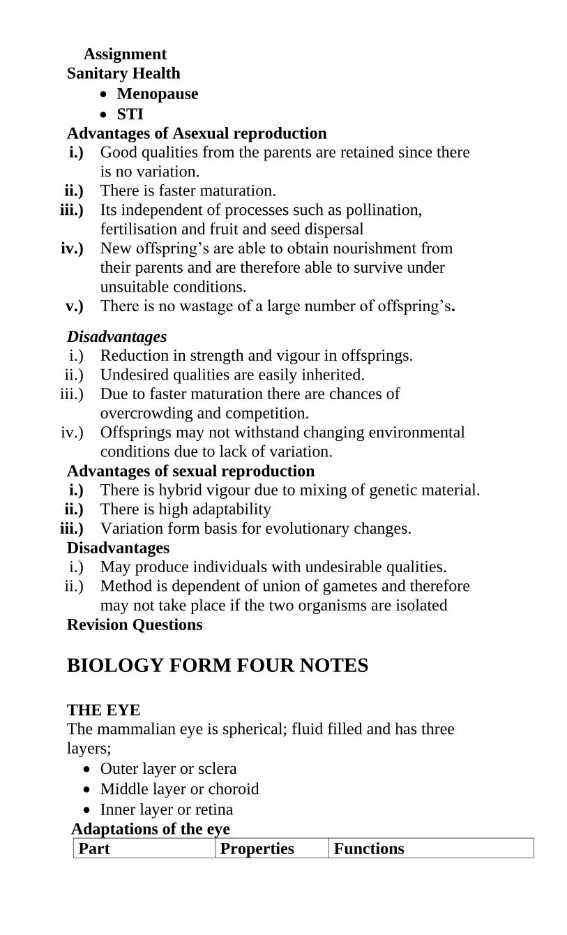
Assignment
Sanitary Health
Menopause
STI
Advantages of Asexual reproduction
i.) Good qualities from the parents are retained since there
is no variation.
ii.) There is faster maturation.
iii.) Its independent of processes such as pollination,
fertilisation and fruit and seed dispersal
iv.) New offspring’s are able to obtain nourishment from
their parents and are therefore able to survive under
unsuitable conditions.
v.) There is no wastage of a large number of offspring’s.
Disadvantages
i.) Reduction in strength and vigour in offsprings.
ii.) Undesired qualities are easily inherited.
iii.) Due to faster maturation there are chances of
overcrowding and competition.
iv.) Offsprings may not withstand changing environmental
conditions due to lack of variation.
Advantages of sexual reproduction
i.) There is hybrid vigour due to mixing of genetic material.
ii.) There is high adaptability
iii.) Variation form basis for evolutionary changes.
Disadvantages
i.) May produce individuals with undesirable qualities.
ii.) Method is dependent of union of gametes and therefore
may not take place if the two organisms are isolated
Revision Questions
BIOLOGY FORM FOUR NOTES
THE EYE
The mammalian eye is spherical; fluid filled and has three
layers;
Outer layer or sclera
Middle layer or choroid
Inner layer or retina
Adaptations of the eye
Part Properties Functions
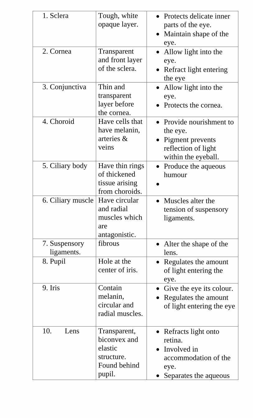
1. Sclera Tough, white
opaque layer. Protects delicate inner
parts of the eye.
Maintain shape of the
eye.
2. Cornea Transparent
and front layer
of the sclera.
Allow light into the
eye.
Refract light entering
the eye
3. Conjunctiva Thin and
transparent
layer before
the cornea.
Allow light into the
eye.
Protects the cornea.
4. Choroid Have cells that
have melanin,
arteries &
veins
Provide nourishment to
the eye.
Pigment prevents
reflection of light
within the eyeball.
5. Ciliary body Have thin rings
of thickened
tissue arising
from choroids.
Produce the aqueous
humour
6. Ciliary muscle Have circular
and radial
muscles which
are
antagonistic.
Muscles alter the
tension of suspensory
ligaments.
7. Suspensory
ligaments.
fibrous Alter the shape of the
lens.
8. Pupil Hole at the
center of iris. Regulates the amount
of light entering the
eye.
9. Iris Contain
melanin,
circular and
radial muscles.
Give the eye its colour.
Regulates the amount
of light entering the eye
10. Lens Transparent,
biconvex and
elastic
structure.
Found behind
pupil.
Refracts light onto
retina.
Involved in
accommodation of the
eye.
Separates the aqueous
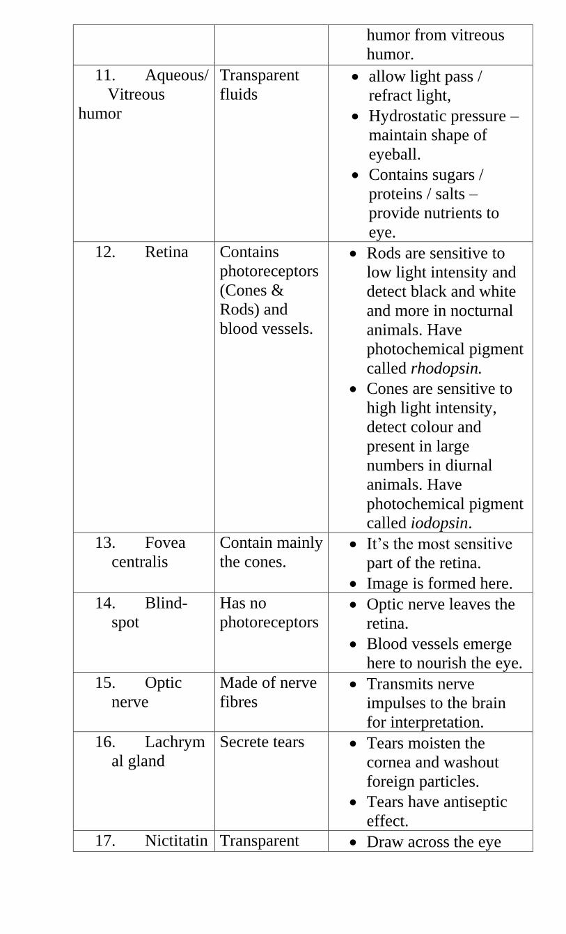
humor from vitreous
humor.
11. Aqueous/
Vitreous
humor
Transparent
fluids allow light pass /
refract light,
Hydrostatic pressure –
maintain shape of
eyeball.
Contains sugars /
proteins / salts –
provide nutrients to
eye.
12. Retina Contains
photoreceptors
(Cones &
Rods) and
blood vessels.
Rods are sensitive to
low light intensity and
detect black and white
and more in nocturnal
animals. Have
photochemical pigment
called rhodopsin.
Cones are sensitive to
high light intensity,
detect colour and
present in large
numbers in diurnal
animals. Have
photochemical pigment
called iodopsin.
13. Fovea
centralis
Contain mainly
the cones. It’s the most sensitive
part of the retina.
Image is formed here.
14. Blind-
spot
Has no
photoreceptors Optic nerve leaves the
retina.
Blood vessels emerge
here to nourish the eye.
15. Optic
nerve
Made of nerve
fibres Transmits nerve
impulses to the brain
for interpretation.
16. Lachrym
al gland
Secrete tears Tears moisten the
cornea and washout
foreign particles.
Tears have antiseptic
effect.
17. Nictitatin Transparent Draw across the eye
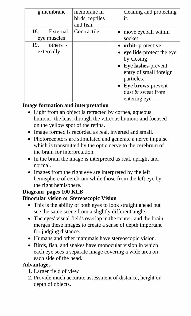
g membrane membrane in
birds, reptiles
and fish.
cleaning and protecting
it.
18. External
eye muscles
Contractile move eyeball within
socket
19. others -
externally-
orbit- protective
eye lids-protect the eye
by closing
Eye lashes-prevent
entry of small foreign
particles.
Eye brows-prevent
dust & sweat from
entering eye.
Image formation and interpretation
Light from an object is refracted by cornea, aqueous
humour, the lens, through the vitreous humour and focused
on the yellow spot of the retina.
Image formed is recorded as real, inverted and small.
Photoreceptors are stimulated and generate a nerve impulse
which is transmitted by the optic nerve to the cerebrum of
the brain for interpretation.
In the brain the image is interpreted as real, upright and
normal.
Images from the right eye are interpreted by the left
hemisphere of cerebrum while those from the left eye by
the right hemisphere.
Diagram pages 100 KLB
Binocular vision or Stereoscopic Vision
This is the ability of both eyes to look straight ahead but
see the same scene from a slightly different angle.
The eyes' visual fields overlap in the center, and the brain
merges these images to create a sense of depth important
for judging distance.
Humans and other mammals have stereoscopic vision.
Birds, fish, and snakes have monocular vision in which
each eye sees a separate image covering a wide area on
each side of the head.
Advantages
1. Larger field of view
2. Provide much accurate assessment of distance, height or
depth of objects.
3. Damage to one eye is compensated by the other
4. Cancels the effect of blind spot
Accommodation of The Eye
This is the refraction of light in order to fall on the fovea
centralis (yellow spot) for clear focus of image regardless
of the distance of the object.
Accommodation of a close object
Ciliary muscles contract thereby relaxing the tension on
suspensory ligaments.
Curvature of the lens increases.
The close objects are greatly refracted by the lens focusing
them on the retina.
Diagram
Accommodation of a distant object
Ciliary muscles relax increasing the tension on suspensory
ligaments.
The lens is stretched decreasing its curvature i.e. lens
become thinner.
Light rays from the object are less refracted and focused on
the retina.
Diagram
Control of Light Entering the Eye
1. Bright Light
Circular muscles of iris contract and the radial muscles
relax.
Diameter of pupil decrease and less light enters.
This protects retina from damage by too much light.
Diagram
2. Dim Light.
Circular muscles relax and radial muscles contract.
Diameter of pupil increase and more light enters the eye.
This allows in enough light to stimulate photoreceptors on
the retina.
Diagram
Defects of the Eye
1. Short sightedness(Myopia)
This is the ability to view near objects clearly but distant
objects are blurred.
This is due to a long eyeball and image is formed before the
retina.
Correction
Wearing concave (Diverging lens)
Diagram
2. Long sightedness (Hypermetropia)
This is the ability to view distant objects clearly but near
objects are blurred.
This is due to a short eyeball and image is formed behind
the retina.
Correction
Wearing convex (converging lens)
Diagram
3. Astigmatism
The curvature of the cornea is uneven hence the image is
formed on different planes.
It’s corrected by wearing cylindrical lens.
4. Squintedness
Eyeballs face different directions due to defective muscles
which move the eye left and right.
Corrected surgically
5. Old sight (Presbyopia)
6. Cataracts
7. Colour blindness
THE EAR
Functions of the ear
1. Hearing
2. Maintaining body balance and posture
Diagram
Adaptations of the ear to its Functions
The ear is divided into:
1. Outer ear
Pinna.
- Pinna is funnel shaped to direct sound waves into
auditory canal;
- its large to offer large surface area for collection of
sound waves;
Auditory meatus/canal
- Tube that directs sound waves to ear drum.
- Lined with hairs to trap solid particles.
- Lined with wax secreting cells to trap dust.
- Wax also maintains flexibility of the eardrum.
2. Middle ear. Its air filled
Tympanic membrane/ear drum.
- It’s a thin tough membrane.
- It transforms sound waves into vibrations.
Ossicles
- They are three bones; malleus, incus and stapes.
- They receive vibrations from tympanic membrane and
amplify them then transmit them to the oval window.
Eustachian tube
- Connects middle ear and pharynx.
- Equalizes pressure between the middle ear and outer ear
preventing distortion of the ear drum.
Oval window
- Picks vibrations from the ossicles and transmit them to
inner ear.
Round window
- Bring back vibrations from the inner into the middle ear.
3. Inner ear. Its fluid filled
Cochlea
- It’s responsible for hearing
- It’s filled with endolymph and perilymph
- Highly coiled to occupy a small space ; and increase
surface area ; for accommodation of many sensory cells
- Vibrations from oval window are transmitted to
perilymph which vibrates
- Sensory cells are stimulated by these vibrations to
generate nerve impulses which are transmitted to the
brain for interpretation.
Diagram
Semi circular canals
- Has semi circular canals which are at right angles to each
other
- Each canal has a swelling called ampulla at one end
containing sensory cells.
- Contains endolymph
- Movement of endolymph in canal helps to detect
changes in position of the body and maintain body
balance posture in relation to head movement.
Diagram
Vestibule
- Has utriculus and sacculus that have sensory cells.
- It maintains body balance posture in relation to gravity.
NB/ Auditory nerve – transmits nerve impulses to the brain
for interpretations.
Deffects of the Ear
Deafness
Vertigo
Tinnitus
GENETICS
This is the study of inheritance and variation.
Terms used in genetics
1. Inheritance; transmission of characteristics from the
parents to the offsprings
2. Variation; possession of characteristics different from those
of the parents and other offsprings.
3. DNA; De-oxyribonucleic acid
4. RNA; Ribonucleic acid
5. Monohybrid inheritance; inheritance of one characteristic
controlled by one pair of hereditary factors e.g. Tallness
6. Dihybrid inheritance; inheritance of two characteristics at
the same time e.g. colour and Texture/shape in the garden
pea plant
7. Dominance; ability of a trait to only express itself
8. Recessiveness; a trait that only expresses itself when in
homozygous state.
9. Heterozygosity; presence of two dissimilar members of an
allele e.g. Rr, Tt etc.
10. Homozygosity; presence of two similar of an allele e.g.
TT, RR, tt, rr etc.
11. Allele; one pair of genes which occupy corresponding
loci/positions in homologous chromosomes
12. Phenotype- the physical appearance of an individual
or organism. It’s influenced by the genotype and
environment.
13. Genotype- the genetic constitution of an organism. Its
purely genetical.
14. F1 generation (first filial generation) - are the
offsprings that represent the first generation of organisms
or individuals under study.
15. F2 generation (second filial generation) - these are
offsprings obtained after self crossing the F1 gen.
16. Incomplete dominance/ co-dominance- a condition
where no allele is dominant over the other. The phenotype
of the offspring is intermediate between that of the parents.
17. Multiple allelism- are characteristics determined by
more than two variant forms of a single gene e.g.
inheritance of the Blood groups in man (ABO)
18. Test cross / back cross- it’s a crossing involving a
homozygous recessive to determine the genotype of an
organism.
19. Mutation- these are spontaneous changes in the
individual’s genetic makeup.
Concepts of Variation
Variation refers to observable differences among living
organisms.
Types of variation
1. Discontinuous variation – in this type of variation, there
are distinct and definite groups of individuals with no
intermediate forms. E.g.
sex either male or female,
blood groups- one can only belong to one of the
four blood groups A,B,AB,O
ability to role the tongue
Presence of long hair in the nose and on the ear
pinna.
Presence of a free or attached ear lobe.
All these traits are controlled by one or two major genes. These
traits are not influenced by the environment
2. Continuous variation – this variation has a wide range of
differences for the same characteristic from one extreme
to the other e.g.
Height
Skin colour/pigmentation
Weight
Length of internodes
Number of leaves, fruits on a tree etc
Finger prints
When these traits are plotted on a graph a normal distribution
curve is obtained.
A normal distribution curve of heights
This type of variation is brought about by the interaction of both
the genetic environmental factors.
E.g. a plant with genes for tallness may fail to grow tall due to
climate and poor soils.
Practical Activity 1
Tongue rolling
Finger prints
Height
Causes of variation
1. Gamete formation – during gamete formation two
processes contribute to variation. These are Independent assortment- during metaphase I of
meiotic division, homologous chromosomes come together in
pairs and segregate into daughter cells independently of each
other. This independent assortment produces a variety of
different gametes. The total number of combinations is given by
2n where ‘n’ is the haploid number of chromosomes. In man n =
23 hence 2n = 223 which is equal to 8,388,608.
Crossing over during the prophase I of meiotic
cell division. I.e. when homologous chromosomes break and
rejoin at certain points called chiasmata.
2. Fertilization – during fertilization parental genes ca
come together in different combinations. Therefore desirable
Number of
individuals
and undesirable qualities of parents can be combined in the
offsprings.
3. Mutation- these are spontaneous changes in the
genetic makeup of an organism. Mutation brings about changes
in the living organisms.
The Chromosome
Each chromosome is made up of two parallel strands called
chromatids. Each pair of chromatids is connected at a point by
the centromere. Chromosomes contain the hereditary material
called the genes. All cells including the sperms and ova have
chromosomes. Chromosomes are present in the nucleus and are
only visible under the microscope during cell division – mitosis
and meiosis. There is a definite number of chromosomes in each
cell for every species of animal or plant.
E.g. in man somatic cells (body cells) have 46 chromosomes
while the sex cells/gametes have 23 chromosomes. During
fertilization fusion of the sperm- 23 chromosomes and the ova -
23 chromosomes restores the 46 chromosomes to form a diploid
zygote.
Chromosomal numbers in different organisms
Organism Number of chromosomes
Somatic cells –
2n
Gametes - n
Sheep (Ovis auries) 56 28
Cow (Bos Taurus) 60 30
Fruit-fly (Drosophila
melanogaster)
8 4
Maize (Zea mays) 20 10
Wheat(Triticum vulgare) 14 7
Tobacco (Nicotiana
tabacum)
12 6
Man ( Homo sapiens) 46 23
Practical Activity 2
Chromosomal behaviour during mitosis
Chromosomal behaviour in meiosis
GENES AND DNA
Genes occupy specific positions on the chromosomes called the
gene loci( gene locus)
The gene is a chemical in nature. The genes are in the form of a
nucleic acid molecule called De-oxyribonucleic acid (DNA). In
1953 two Biologists Francis Crick and James Watson worked
out the structure of the DNA. DNA was found to be composed
of three different components;
1. A five carbon sugar-pentose
2. phosphate molecule
3. nitrogenous base
There are four types of the nitrogenous base;
Adenine – N
Guanine – G
Thymine- G
Cytosine – C
A combination the pentose sugar, a phosphate molecule and a
nitrogenous base forms a nucleotide.
I.e.
DNA structure contains several nucleotides fused together to
form long chains called DNA strands. Two parallel strands twist
on one another forming a double helix structure. Adenine always
combines with Thymine and Cytosine with Guanine.
Diagram
Role of DNA
Stores genetic information in a coded form
Enables transfer of genetic information unchanged to
daughter cells through replication
Translates the genetic information into the
characteristics of an organism through protein
synthesis
DNA REPLICATION
During cell division both daughter cells arising from
mitotic division have the same genetic constitution as
the parent cell. DNA in the parent cell must therefore
duplicate accurately before the cell divides. The
process through which a DNA molecule forms an
exact Replica is called DNA replication.
The two strands forming the double helix separates
like a zipper. Each parallel strand becomes a template
that specifies the base sequence of a new
complimentary strand. Through the action of
replicating enzymes, free nucleotides take up positions
along the template strands.
The specificity of the base pairing ensures that only
complimentary bases link together with those on the
template strands. I.e. G-C and A-T.
Covalent bonds are formed between the nucleotides
resulting in the formation of a new DNA strand.
The template and the new DNA strand the undergo
coiling to form a double helix. In this way, two
identical DNA molecules are formed from the original
single molecule.
Each of the new DNA molecules gets incorporated
into one of the two nuclei formed just prior to the
separation of the daughter cells.
Diagram
Role of the DNA in protein synthesis
The sequence of bases along the DNA strand acts as the
alphabet and determines the sequence of amino acids when they
join to form a polypeptide chain. Protein synthesis takes place in
ribosome’s found in the cytoplasm. Since the DNA molecules
are confined in the nucleus, there has to be a way of
communicating the DNA information to the ribosome’s where
actual protein synthesis occurs.
The cell therefore has a special molecule called the Ribonucleic
acid (RNA). Its role is to carry genetic information from the
DNA to the site of protein synthesis in the cytoplasm. It’s
referred to as messenger RNA (mRNA). RNA is formed from
the DNA strands.
During formation of the mRNA a section of the DNA strands
acts as the template strand. The double helix of the DNA unzips
and free nucleotides align themselves opposite the template. The
base sequence of the template strand is copied onto a new
strand.
In RNA, Thymine is replaced by another base called Uracil (U)
The transfer of DNA sequence on the mRNA strand is referred
to as Transcription.
Diagram
After its formation the mRNA leaves the nucleus with
instructions from the DNA about the kind of protein to be
synthesised by the cell. This information is in the form of base
triplets known as Codons which code for a particular amino acid
of a protein molecule e.g.
AAA-phenylalanine
TTT-lysine
CAA- valine
CTA- aspartic acid
Differences between DNA and RNA
DNA RNA
1. Has De-ox ribose sugar Has ribose sugar
2. Double stranded Single stranded
3. Confined in the nucleus Found in nucleus and
cytoplasm
4. Have organic bases as
cytosine, guanine, adenine
and thymine.
Has organic bases as cytosine,
guanine, adenine and uracil
THE FIRST LAW OF HEREDITY
MENDEL’S EXPERIMENTS
An Austrian monk known as Mendel is considered to be the
father of genetics. He carried out various breeding experiments
and observed variations in different characteristics of the garden
pea. The characteristics include:
Height of the stems- tall or dwarf
Texture of the seed coat- smooth or wrinkled
Colour of the seeds- yellow or green
Texture of the ponds
Colour of the flowers- white or purple
Position of the flower- axial or terminal
He selected a group of dwarf plants and self pollinated them by
dusting mature pollen grains onto the stigmas of the same plant.
He collected the resulting seeds and planted them. He noted that
these seeds grew into dwarf plants only. He repeated the
experiment for several generations and obtained the same
results.
In another experiment, Mendel selected tall plants and self-
pollinated them. He planted the resulting seeds and observed
that they grew into a mixture of tall and dwarf plants. He took
seeds from the tall offsprings only and repeated the experiment
for many generations until he obtained only tall plants.
This way he was able to obtain a pure line of tall garden peas
and a pure line of dwarf garden peas.
He then cross-pollinated pure bred tall garden pea s with the
pure bred dwarf variety. He planted the resulting seeds and he
observed that all the offsprings were tall plants.
Diagram
He further crossed two of these tall offsprings and planted the
resulting seeds. Mendel observed that this second generation
consisted of a mixture of tall and dwarf plants. After counting
these plants he noted that the ratio of tall to dwarf plants was
approximately 3:1 respectively. He observed that this ratio was
always obtained when crosses were made between the non-pure
breeds of tall plants.
Diagrams
Mendel concluded that the traits of an organism are determined
by hereditary factors which occur in pairs. Only one of pair of
such factors can be represented in a single gamete. This later
became Mendel’s First Law, The Law of Segregation
At this time Mendel had no idea of genes and so he called them
factors. He postulated that these factors are found on the
chromosomes and are passed from the parents to the offsprings
via gametes.
Reasons behind Mendel’s success
1. He used favourable materials i.e. the garden pea plant
which is normally self pollinated. This made it easy for him
to employ cross pollination at will.
2. the pea plant he used had several contrasting traits
3. His study was focused on particular traits while those
before him had been attempting to determine wholesome
heredity of each organism.
4. He kept accurate data on all his experiments and fro the
analysis of this data he was able to formulate definite
hypothesis.
Mendel chose the garden pea plant because of the following
reasons
Plant had many contrasting traits e.g. flower colours,
seed coat texture, length of the stems etc.
Plant is normally self pollinated but cross pollination
can be employed t will.
Plant matures relatively fast
Plant produces many seeds that can be planted to
produce many offsprings
Monohybrid Inheritance
This is the inheritance of one trait like height in the garden pea
plant that is controlled by a single pair of hereditary factors
(genes) contributed by both parents. Genes occur in pairs on
chromosomes and such gene pairs are known as alleles.
The genetic constitution of an organism is called the Genotype
while the physical appearance is known as the Phenotype.
The genotype of an organism is represented using paired letter
symbols. Capital letters represents the dominant gene while
small letters represent the recessive gene.
Genetic Cross
Components of a genetic cross
Parental phenotypes
The parental genotype –the crossing X should be
shown here.
The gametes and should be circled.
The fusion process or fertilization.
The filial generation genotypes
NB. The conventional symbol for male is ♂ and that of female is
♀
Example 1
During gamete formation in the dwarf plat, each gene in the pair
segregates into different gametes. When the female and male
fuse during fertilization, the offspring produced contain the
same number of genes as in each parent. The inheritance of
dwarf ness in the pea plant can be illustrated diagrammatically
by the following genetic cross
Diagram
Example 2
Similarly the pair of genes in the pure breed tall plants will
segregate into different gametes during gametogenesis. When
self fertilised the resulting seeds will have half the number of
genes from each parent i.e.
Diagram
Example 3
When the purebred tall plant is crossed with dwarf plants, the
resulting seeds grow into tall plants only. These offsprings
represent the first generation (F1 gen)
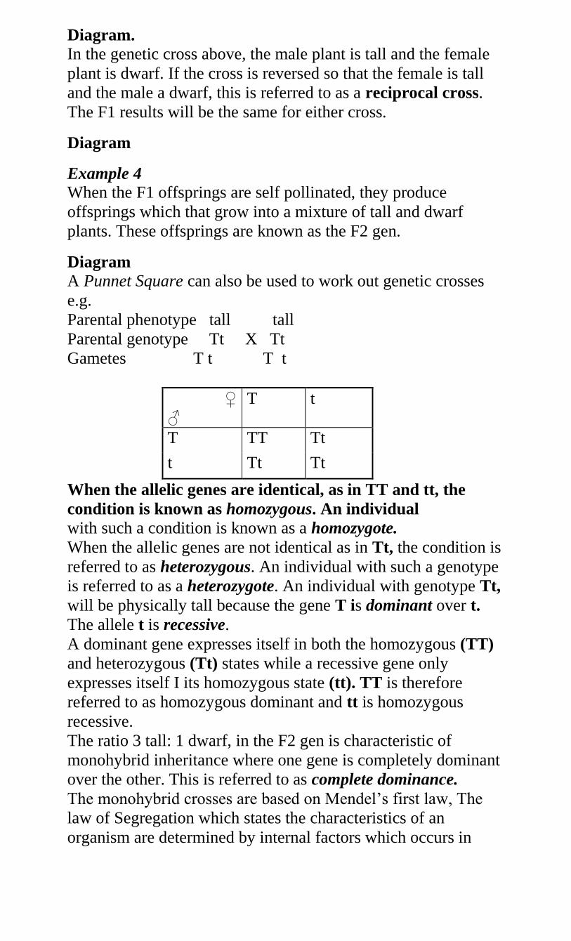
Diagram.
In the genetic cross above, the male plant is tall and the female
plant is dwarf. If the cross is reversed so that the female is tall
and the male a dwarf, this is referred to as a reciprocal cross.
The F1 results will be the same for either cross.
Diagram
Example 4
When the F1 offsprings are self pollinated, they produce
offsprings which that grow into a mixture of tall and dwarf
plants. These offsprings are known as the F2 gen.
Diagram
A Punnet Square can also be used to work out genetic crosses
e.g.
Parental phenotype tall tall
Parental genotype Tt X Tt
Gametes T t T t
♀
♂
T t
T TT Tt
t Tt Tt
When the allelic genes are identical, as in TT and tt, the
condition is known as homozygous. An individual
with such a condition is known as a homozygote.
When the allelic genes are not identical as in Tt, the condition is
referred to as heterozygous. An individual with such a genotype
is referred to as a heterozygote. An individual with genotype Tt,
will be physically tall because the gene T is dominant over t.
The allele t is recessive.
A dominant gene expresses itself in both the homozygous (TT)
and heterozygous (Tt) states while a recessive gene only
expresses itself I its homozygous state (tt). TT is therefore
referred to as homozygous dominant and tt is homozygous
recessive.
The ratio 3 tall: 1 dwarf, in the F2 gen is characteristic of
monohybrid inheritance where one gene is completely dominant
over the other. This is referred to as complete dominance.
The monohybrid crosses are based on Mendel’s first law, The
law of Segregation which states the characteristics of an
organism are determined by internal factors which occurs in
pairs. Only one of a pair of such factors can be represented in a
single gamete.
Diagram - the process of segregation
Ratios and Probability
The 3:1 ratio in monohybrid inheritance can be represented in
the form of probability. When a large number of heterozygous
garden pea plants are selfed, the probability of getting tall plants
is ¾ or 75% and that of dwarf will be ¼ or 25%.
Diagram
NB. The inheritance of characteristics involves probability. The
chance that a particular gamete will fuse with another is a
random occurrence, in genetics this done by showing all
possible fusions.
Practical Activity 3 and 4
Tossing a coin
To demonstrate random fusion of gametes in
monohybrid inheritance.
Similar monohybrid inheritance results as those of Mendel have
been obtained by using the fruit fly (Drosophila melanogaster)
the insect has many observable characteristics that are
contrasting such as,
Wing length – long wing dominant over vestigial wing
Eye colour – red eyes dominant over white eyes
Size of the abdomen – broad abdomen dominant over
narrow abdomen
Body colour – grey body colour dominant over black
body colour.
Assignment
Using appropriate letters work out the following crosses with
respect to the fruit fly
Cross between a purebred long winged and a vestigial
winged
Cross between two long winged heterozygotes
Cross between a red eyed heterozygote and a white
eyed fruit fly.
The fruit fly is suitable for genetic study because of the
following reasons.
1. The female lays very many eggs hence increasing the
sample size for study.
2. Have many observable characteristics that are distinct
and contrasting.
3. It is easily bred in the laboratory with minimum
requirements.
4. It has a short generation time 10-14 days. Therefore
many generations can be studied in a short period of
time.
5. Offsprings can be crossed with their parents at will
(backcrossing)
6. Flies are safe to handle because they do not transmit
any known human diseases.
Study Question 10
Practical Activity 5
Breeding fruit flies.
Incomplete Dominance (Co-dominance)
In Mendel’s experiments with garden pea p[plants, the genes
determining the various traits were clearly dominant or
recessive. However in some species, alleles determining several
contrasting traits do not have a clear cut dominant-recessive
relationship. This implies that neither of the alleles is completely
dominant over the other.
Heterozygous individuals are phenotypically different from
either of the parents. Mostly the phenotype of the heterozygous
offspring is intermediate between that of the parents. This
phenomenon is called Incomplete Dominance. Examples of
incomplete dominance.
1. Inheritance of flower colour in the 4 o’clock plant
(Mirabilis Jalapa). If a true breeding plant producing red
flowers is crossed with a true breeding plant producing
white flowers, all the F1 offsprings will have pink flowers.
When the F1 plants are self pollinated, they yield red
flowered, pink flowered and white flowered offspring at a
ratio of 1:2:1 respectively.
Diagrams
2. Incomplete dominance in short horn cattle. Mating red
and white shorthorn cattle yields Roan light Red) calves
due to presence of both red and white hairs. A mating
between two roan coloured shorthorns yields a mixture of
red, roan and white coloured calves at a ratio of 1:2:1
respectively.
Diagrams
Study Question 12
Inheritance of the Blood groups (Multiple allelism)
In all the kinds of inheritance discussed so far, each phenotypic
characteristic is determined by 2 variant forms of a single gene
located at a specific locus on the homologous chromosome.
However some characteristics are determined by more than two
variant forms of a single gene. This phenomenon is referred to
as multiple allelism and the genes involved are called multiple
alleles. E.g. in the ABO blood groups in humans, there are three
genes involved and they are responsible for the presence of
antigen types on the red blood cells.
These are gene A responsible for the presence of antigen A,
gene B for antigen B and gene O responsible for absence of
antigens on the red blood cells.
Genes A and B have equal degree of dominance i.e. are co-
dominant. They both express themselves when present together
as in the blood group AB.
Genes A and B are dominant over gene O. Gene O is recessive
and only expresses itself in the homozygous condition. The
genotypes for the four blood groups in the ABO system are
therefore,
Blood
group
(Phenotype)
Genotype Antigens
A AA, AO A
B BB,BO B
AB AB A and B
O OO O- Zero
i.e.
AA or AO-Blood group A
BB or BO – blood group B
AB – blood group AB
OO – blood group O
A marriage between a man of blood group A and a woman of
blood group B will produce children of all the four blood groups
if both parents are heterozygous.
Diagram
Marriage between a man of genotype AA (blood group A) and
Woman of genotype BB (blood group B) results in all the
offsprings having blood AB.
Diagram.
Assignment
Work out the following crosses
Both parents with blood group O
Heterozygous blood group A and blood group O
Study Question 13
Inheritance of the Rhesus factor
In man the possession of Rhesus antigens makes one Rh+ and
this is dominant over Rh–ve. If blood from a Rhesus positive
person is transfused into a rhesus negative person, this induces
antibodies against the Rhesus factor of the donor. This causes
agglutination of red blood cells of the recipient.
If a Rh-ve woman is married to a Rhe+ve, when she becomes
pregnant, the child will be Rh+ve. Rhesus antigens cross the
placenta into the mother’s blood stream. This stimulates the
mother’s immune system to produce Rhesus antibodies. When
these antibodies get into the foetal circulation, an antigen-
antibody reaction takes place and the red blood cells of the
foetus are destroyed (Haemolysed).
During the second pregnancy, the amount of Rhesus antibodies
are more and cause a lot of damage to the foetus’s red blood
cells resulting to death. This is called Haemolytic Disease of the
Newborn or Erythroblastosis foetalis.
Determining Unknown Genotypes
This can be done in two ways.
1. Carrying out a Test Cross
A test cross is a cross between an individual of unknown
genotype with an individual of a recessive genotype. A test cross
where an offspring is crossed with one of its parents is called a
Back Cross.
In garden pea plants the gene that determines red flowers is
dominant over that which determines white flowers. A plant
with red flowers may either be homozygous (RR) or
heterozygous (Rr) for this characteristic. To establish its correct
genotype it is crossed with a homozygous recessive plant i.e. a
white flowered one (rr)
If all their offsprings bear red flowers then this indicates that the
red flowered plant is homozygous or it’s from a pure line.
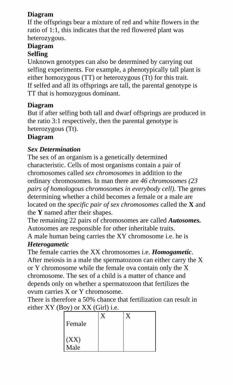
Diagram
If the offsprings bear a mixture of red and white flowers in the
ratio of 1:1, this indicates that the red flowered plant was
heterozygous.
Diagram
Selfing
Unknown genotypes can also be determined by carrying out
selfing experiments. For example, a phenotypically tall plant is
either homozygous (TT) or heterozygous (Tt) for this trait.
If selfed and all its offsprings are tall, the parental genotype is
TT that is homozygous dominant.
Diagram
But if after selfing both tall and dwarf offsprings are produced in
the ratio 3:1 respectively, then the parental genotype is
heterozygous (Tt).
Diagram
Sex Determination
The sex of an organism is a genetically determined
characteristic. Cells of most organisms contain a pair of
chromosomes called sex chromosomes in addition to the
ordinary chromosomes. In man there are 46 chromosomes (23
pairs of homologous chromosomes in everybody cell). The genes
determining whether a child becomes a female or a male are
located on the specific pair of sex chromosomes called the X and
the Y named after their shapes.
The remaining 22 pairs of chromosomes are called Autosomes.
Autosomes are responsible for other inheritable traits.
A male human being carries the XY chromosome i.e. he is
Heterogametic The female carries the XX chromosomes i.e. Homogametic.
After meiosis in a male the spermatozoon can either carry the X
or Y chromosome while the female ova contain only the X
chromosome. The sex of a child is a matter of chance and
depends only on whether a spermatozoon that fertilizes the
ovum carries X or Y chromosome.
There is therefore a 50% chance that fertilization can result in
either XY (Boy) or XX (Girl) i.e.
Female
(XX)
Male
X X
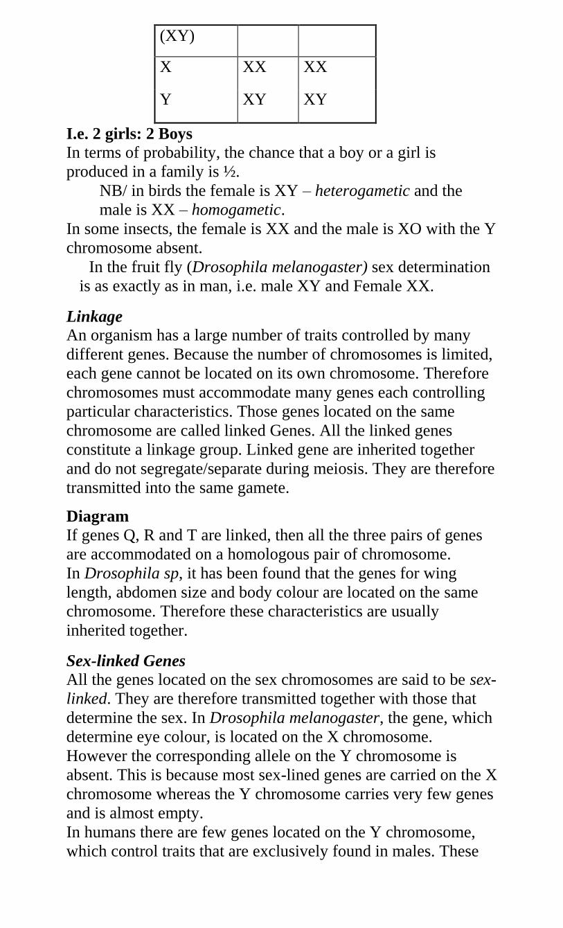
(XY)
X XX XX
Y XY XY
I.e. 2 girls: 2 Boys
In terms of probability, the chance that a boy or a girl is
produced in a family is ½.
NB/ in birds the female is XY – heterogametic and the
male is XX – homogametic.
In some insects, the female is XX and the male is XO with the Y
chromosome absent.
In the fruit fly (Drosophila melanogaster) sex determination
is as exactly as in man, i.e. male XY and Female XX.
Linkage
An organism has a large number of traits controlled by many
different genes. Because the number of chromosomes is limited,
each gene cannot be located on its own chromosome. Therefore
chromosomes must accommodate many genes each controlling
particular characteristics. Those genes located on the same
chromosome are called linked Genes. All the linked genes
constitute a linkage group. Linked gene are inherited together
and do not segregate/separate during meiosis. They are therefore
transmitted into the same gamete.
Diagram
If genes Q, R and T are linked, then all the three pairs of genes
are accommodated on a homologous pair of chromosome.
In Drosophila sp, it has been found that the genes for wing
length, abdomen size and body colour are located on the same
chromosome. Therefore these characteristics are usually
inherited together.
Sex-linked Genes
All the genes located on the sex chromosomes are said to be sex-
linked. They are therefore transmitted together with those that
determine the sex. In Drosophila melanogaster, the gene, which
determine eye colour, is located on the X chromosome.
However the corresponding allele on the Y chromosome is
absent. This is because most sex-lined genes are carried on the X
chromosome whereas the Y chromosome carries very few genes
and is almost empty.
In humans there are few genes located on the Y chromosome,
which control traits that are exclusively found in males. These
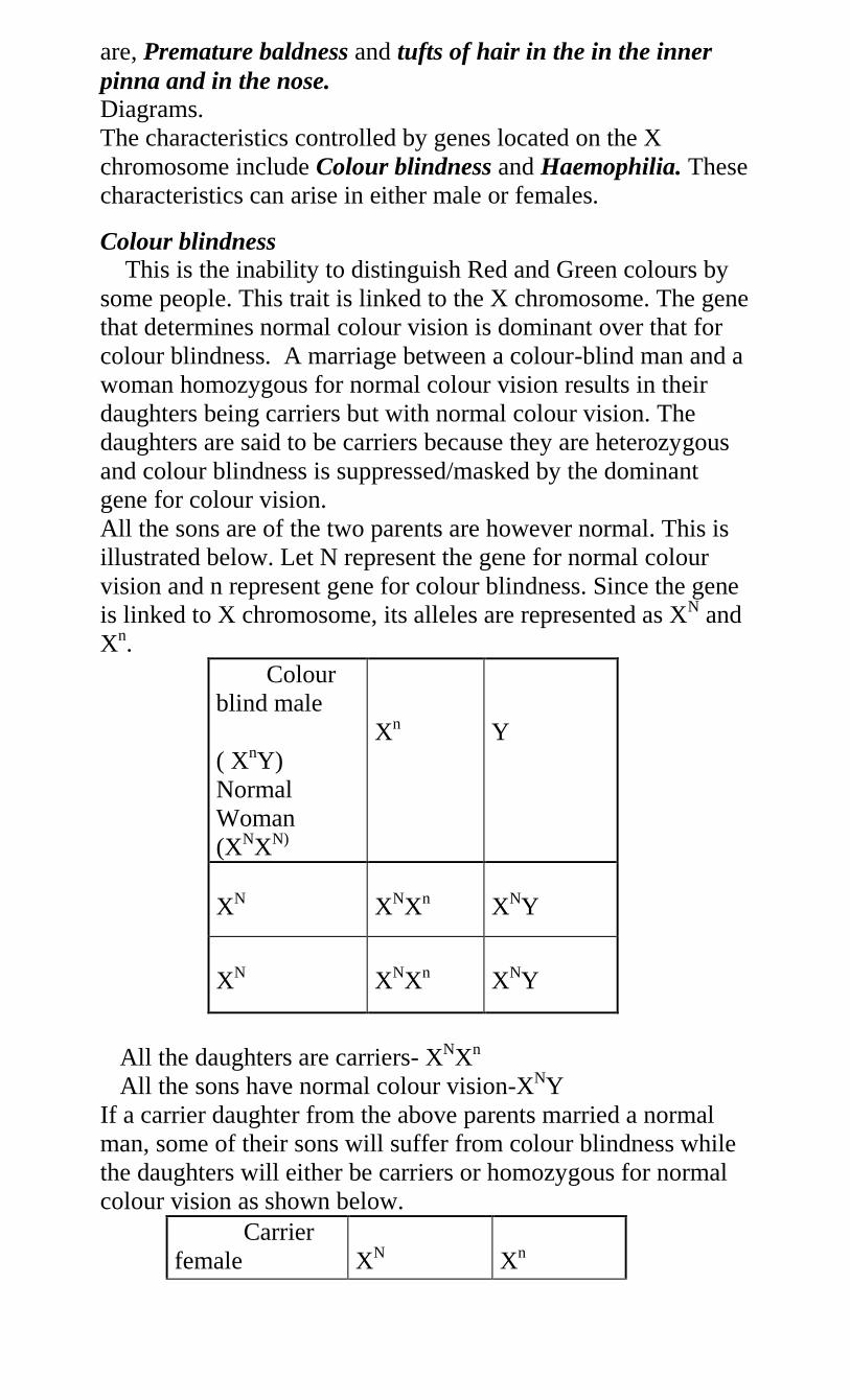
are, Premature baldness and tufts of hair in the in the inner
pinna and in the nose. Diagrams.
The characteristics controlled by genes located on the X
chromosome include Colour blindness and Haemophilia. These
characteristics can arise in either male or females.
Colour blindness
This is the inability to distinguish Red and Green colours by
some people. This trait is linked to the X chromosome. The gene
that determines normal colour vision is dominant over that for
colour blindness. A marriage between a colour-blind man and a
woman homozygous for normal colour vision results in their
daughters being carriers but with normal colour vision. The
daughters are said to be carriers because they are heterozygous
and colour blindness is suppressed/masked by the dominant
gene for colour vision.
All the sons are of the two parents are however normal. This is
illustrated below. Let N represent the gene for normal colour
vision and n represent gene for colour blindness. Since the gene
is linked to X chromosome, its alleles are represented as XN and
Xn.
Colour
blind male
( XnY)
Normal
Woman
(XNXN)
Xn
Y
XN
XNXn
XNY
XN
XNXn
XNY
All the daughters are carriers- XNXn
All the sons have normal colour vision-XNY
If a carrier daughter from the above parents married a normal
man, some of their sons will suffer from colour blindness while
the daughters will either be carriers or homozygous for normal
colour vision as shown below.
Carrier
female
XN
Xn
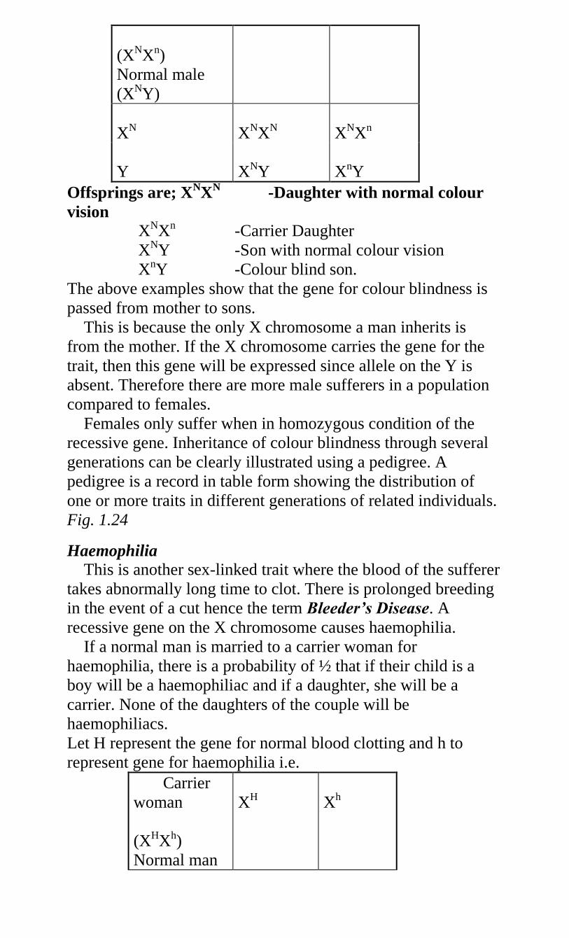
(XNXn)
Normal male
(XNY)
XN
XNXN
XNXn
Y
XNY
XnY
Offsprings are; XNX
N -Daughter with normal colour
vision
XNXn -Carrier Daughter
XNY -Son with normal colour vision
XnY -Colour blind son.
The above examples show that the gene for colour blindness is
passed from mother to sons.
This is because the only X chromosome a man inherits is
from the mother. If the X chromosome carries the gene for the
trait, then this gene will be expressed since allele on the Y is
absent. Therefore there are more male sufferers in a population
compared to females.
Females only suffer when in homozygous condition of the
recessive gene. Inheritance of colour blindness through several
generations can be clearly illustrated using a pedigree. A
pedigree is a record in table form showing the distribution of
one or more traits in different generations of related individuals.
Fig. 1.24
Haemophilia
This is another sex-linked trait where the blood of the sufferer
takes abnormally long time to clot. There is prolonged breeding
in the event of a cut hence the term Bleeder’s Disease. A
recessive gene on the X chromosome causes haemophilia.
If a normal man is married to a carrier woman for
haemophilia, there is a probability of ½ that if their child is a
boy will be a haemophiliac and if a daughter, she will be a
carrier. None of the daughters of the couple will be
haemophiliacs.
Let H represent the gene for normal blood clotting and h to
represent gene for haemophilia i.e.
Carrier
woman
(XHXh)
Normal man
XH
Xh
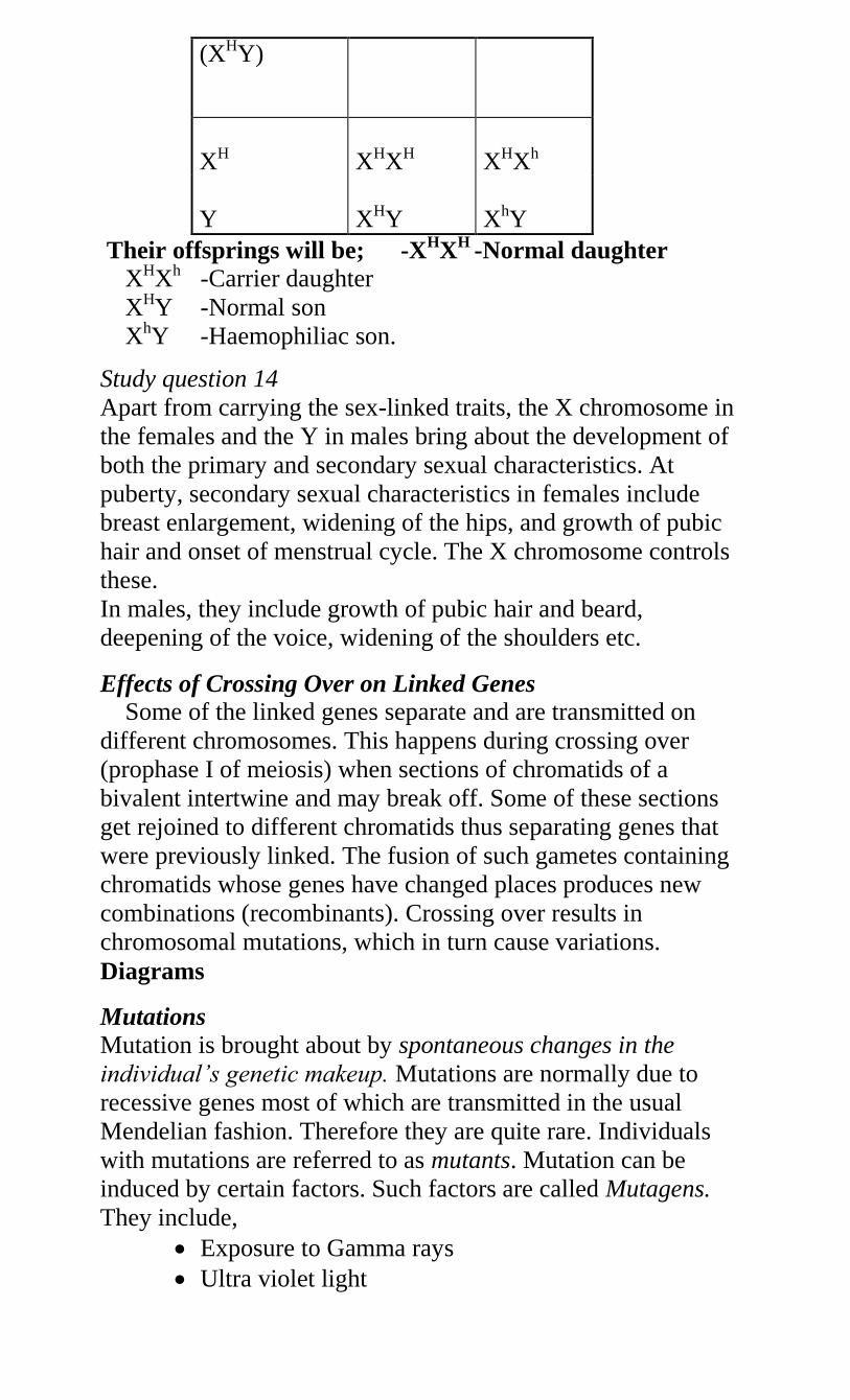
(XHY)
XH
XHXH
XHXh
Y
XHY
XhY
Their offsprings will be; -XHX
H -Normal daughter
XHXh -Carrier daughter
XHY -Normal son
XhY -Haemophiliac son.
Study question 14
Apart from carrying the sex-linked traits, the X chromosome in
the females and the Y in males bring about the development of
both the primary and secondary sexual characteristics. At
puberty, secondary sexual characteristics in females include
breast enlargement, widening of the hips, and growth of pubic
hair and onset of menstrual cycle. The X chromosome controls
these.
In males, they include growth of pubic hair and beard,
deepening of the voice, widening of the shoulders etc.
Effects of Crossing Over on Linked Genes
Some of the linked genes separate and are transmitted on
different chromosomes. This happens during crossing over
(prophase I of meiosis) when sections of chromatids of a
bivalent intertwine and may break off. Some of these sections
get rejoined to different chromatids thus separating genes that
were previously linked. The fusion of such gametes containing
chromatids whose genes have changed places produces new
combinations (recombinants). Crossing over results in
chromosomal mutations, which in turn cause variations.
Diagrams
Mutations
Mutation is brought about by spontaneous changes in the
individual’s genetic makeup. Mutations are normally due to
recessive genes most of which are transmitted in the usual
Mendelian fashion. Therefore they are quite rare. Individuals
with mutations are referred to as mutants. Mutation can be
induced by certain factors. Such factors are called Mutagens.
They include,
Exposure to Gamma rays
Ultra violet light
Colchicine
Mustard gas
NB: Mutations occurring in gametes are more important than
those in somatic cells. Mutational changes are the basis of
discontinuous variation in population.
Types of Mutations
1. Chromosomal mutation
2. Gene mutation
Chromosomal Mutations
This involves the change in the structure or the number of
chromosomes. During crossing over in meiosis homologous
chromosomes intertwine at points called chiasmata. These points
are later broken creating various opportunities for changes on
the chromatids. There are five types of chromosome mutations
(chromosome aberrations).
Deletion
Duplication
Inversion
Translocation
Non-disjunction
Deletion
This occurs when some sections of chromatids break off and fail
to recombine. They are therefore completely lost and the genetic
material they contain is said to be deleted out. Most deletions are
lethal since the offspring may lose genes responsible for the
synthesis of some vital protein molecules.
Diagrams
Duplication
In this case a section of chromatids replicates and adds an extra
length to itself. Duplication can produce serious effects
depending on the chromosome sections involved.
Diagrams
Inversion
In this case a chromatid breaks at two points. When rejoining,
the middle piece rotates and joins in an inverted position. This
reverses the gene sequence along the chromatid. This might
bring together genes whose combined effects are advantageous
or dis-advantageous.
Diagrams
Translocation
This occurs when a section of one chromatid breaks off and
becomes attached to another chromatid but of a non-homologous
pair.
Translocation therefore involves the movement of genes from
one non-homologous chromosome to another.
Diagrams
Non-disjunction
This leads to addition or loss of one or more whole
chromosomes. If it occurs at anaphase of the first meiotic
division, two homologous chromosomes fail to segregate and
they move into the same gamete cell. If it happens at anaphase
of the second meiotic division, sister chromatids fail to
segregate. This results in half the gametes containing two of the
same chromosome while the others have none.
Diagrams
Non-Disjunction causes the following
1. Downs’s Syndrome: this is where there is an extra somatic
chromosome number 21. such individuals have;
Slit eye appearance
Reduced resistance to infections
Mentally deficient
Thick tongue
Cardiac malfunctions
Short body with thick fingers
NB/ these conditions are common among children born of
mothers above 40 years old.
2. Klinefelter’s Syndrome: in this case individuals have an
extra sex chromosome. Such individuals have a total of 47
chromosomes in their cells i.e. XXY (male) and XXX
(female). This occurs as a result of non-disjunction during
spermatogenesis or oogenesis. The symptoms of
Klinefelter’s syndrome are
Infertility in males due to lack of sperm production
Under developed testes
Reduced facial hair in males
Very tall with signs of obesity
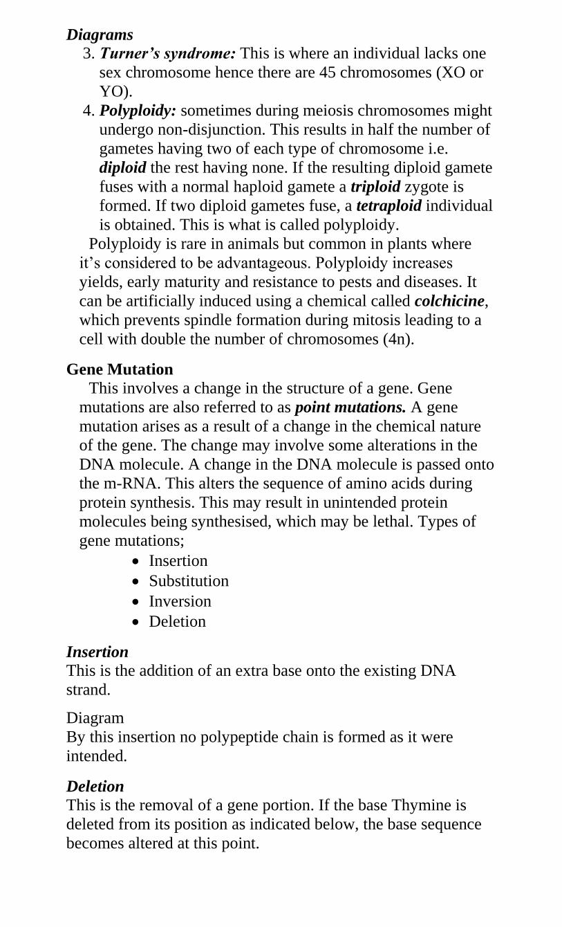
Diagrams
3. Turner’s syndrome: This is where an individual lacks one
sex chromosome hence there are 45 chromosomes (XO or
YO).
4. Polyploidy: sometimes during meiosis chromosomes might
undergo non-disjunction. This results in half the number of
gametes having two of each type of chromosome i.e.
diploid the rest having none. If the resulting diploid gamete
fuses with a normal haploid gamete a triploid zygote is
formed. If two diploid gametes fuse, a tetraploid individual
is obtained. This is what is called polyploidy.
Polyploidy is rare in animals but common in plants where
it’s considered to be advantageous. Polyploidy increases
yields, early maturity and resistance to pests and diseases. It
can be artificially induced using a chemical called colchicine,
which prevents spindle formation during mitosis leading to a
cell with double the number of chromosomes (4n).
Gene Mutation
This involves a change in the structure of a gene. Gene
mutations are also referred to as point mutations. A gene
mutation arises as a result of a change in the chemical nature
of the gene. The change may involve some alterations in the
DNA molecule. A change in the DNA molecule is passed onto
the m-RNA. This alters the sequence of amino acids during
protein synthesis. This may result in unintended protein
molecules being synthesised, which may be lethal. Types of
gene mutations;
Insertion
Substitution
Inversion
Deletion
Insertion
This is the addition of an extra base onto the existing DNA
strand.
Diagram
By this insertion no polypeptide chain is formed as it were
intended.
Deletion
This is the removal of a gene portion. If the base Thymine is
deleted from its position as indicated below, the base sequence
becomes altered at this point.
This results in the wrong proteins being synthesised.
Diagrams
Substitution
This is the replacement of a portion of the gene with a new
portion. If Adenine is substituted by Guanine on a DNA strand,
the base sequence is altered at this particular portion.
Diagrams
Inversion
If a portion of the DNA strand is rotated through 1800 that
portion is said to be inverted as shown below. This alters the
base sequence at this point.
Diagrams
Study Questions
Disorders Due to Gene Mutations
Such disorders include albinism, sickle cell anaemia,
haemophilia, colour blindness and chondrodystrophic dwarfism.
1. Albinism
This a condition where the synthesis of skin pigment called
melanin fails. The victim has a light skin, white hair and pink
eyes. Such a person is referred to as an Albino. Melanin is
derived from two amino acids – Phenylalanine and Tyrosine.
Melanin is synthesised through a series of reactions controlled
by a specific gene.
Gene ‘A’ is responsible for presence of melanin and ‘a’ is
responsible for its absence. Gene ‘aa’ in homozygous state
blocks in one or two places in the synthesis of melanin hence
no melanin is formed. This occurs as a result of one enzyme
(Tyronase) failing to be formed in the presence of the
recessive gene.
A person with genotype AA has normal skin pigmentation.
One with genotype Aa is a carrier and has normal skin
pigmentation. In a family an albino can be born under three
conditions only.
If both parents are albinos
If one of the parents is an albino and the other a carrier
If both parents are carriers
Assignment
Work out crosses in each case.
What is the probability of getting an albino child in
each case?
2. Sickle Cell Anaemia
This is a gene mutation as a result of substitution. Normal
haemoglobin Hb A consists of two polypeptide chains. In the
sickle cell condition, one amino acid called glutamic acid is
substituted by another amino acid called valine in each of the
two-polypeptide chains of the haemoglobin molecule. The
resulting haemoglobin is known as Haemoglobin S – Hb S and
is different from the Hb A in several ways.
Comparison between Hb A and Hb S
Normal haemoglobin (Hb
A)
Defective Haemoglobin (Hb
S)
1. A position in each
polypeptide chain is occupied
by glutamic acid
The same position is occupied
by valine in each polypeptide
chain
2. Does not easily crystallise
in low oxygen
concentration
Easily crystallises in low
oxygen concentration
3. The haemoglobin is
efficient in oxygen loading
and transportation
Not efficient in oxygen
loading and transportation
4. The red blood cells are
biconcave in shape
Red blood cells are sickle
shaped (crescent shape)
Sickle cell anaemia is therefore the condition where the
victim is homozygous for the defective gene that directs the
synthesis of haemoglobin S. Most of the victims’ red blood
cells are sickle shaped and the person frequently experiences
oxygen shortage to the body tissues. Such a person cannot
carry out strenuous physical exercises.
Many sickle cell victims die young due to insufficient oxygen
supply to body tissues.
In the heterozygous condition, less than half the number of
the red blood cells is sickle shaped. The rest are normal and
efficient in oxygen transport. This is referred to as sickle cell
trait. An individual with the sickle cell trait experiences a mild
case of anaemia but leads a normal life.
Inheritance of Sickle Cell Anaemia
If a man with sickle cell trait marries a normal woman, the
probability that any of the offspring will carry the sickle cell
trait is ½.
If both parents are carriers the probability of getting an
offspring with sickle cell anaemia is ¼.
3. Haemophilia: This condition where the blood takes
abnormally long time to clot. A haemophilic gene that
prevents the production of the clotting factors causes the
condition.
4. Colour blindness: There are different forms of colour
blindness. The most common one is the red-green colour
blindness. In this case an individual is unable to distinguish
between red and green colours.
NB. Most disadvantageous genes are recessive. Very few are
dominant e.g. the gene for chondrodystrophic dwarfism
Study Question 16
Effect of Environment on Heredity
The genotype and the environment influence the development of
an individual. In animals genetically identical individuals reared
under different environments will appear different than those
reared under very different conditions. Consider identical twins.
Practical Applications of Genetics
o Plants and animal breeding
o Blood transfusion
o Genetic counselling
o Genetic engineering
1. Plants and Animal Breeding
Man chooses those plants and animals with the desirable
qualities. This is referred to as artificial selection. Inbreeding or
crossbreeding does this. Inbreeding however increases the
chances of undesirable genes whereas crossbreeding increases
heterozygosity with the offspring’s having better performance
than both parents. This is referred to as hybrid vigour e.g. a
cross between Boran and Hereford.
Polyploidy has also been used in planting. The original wheat
had a diploid number of 14 chromosomes but the commercial
wheat has either 28 or 42 (tetraploid-4n or hexaploid-6n).
Examples of characteristics, which have been selected in
agriculture.
1. Resistance to diseases e.g. cassava resistant to cassava
mosaic, coffee variety resistant to CBD.
2. Early maturity in animals and plants.
3. Adaptations to various conditions e.g. rainfall, temperature
etc.
4. Ease of harvesting e.g. in coffee and bananas where dwarf
varieties have been developed
5. Increased productive season e.g. in chicken
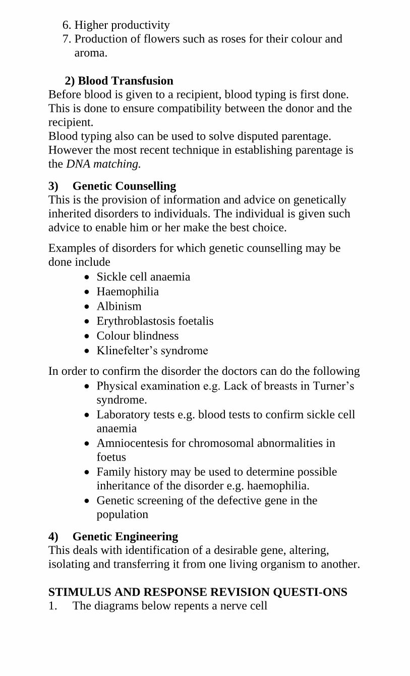
6. Higher productivity
7. Production of flowers such as roses for their colour and
aroma.
2) Blood Transfusion
Before blood is given to a recipient, blood typing is first done.
This is done to ensure compatibility between the donor and the
recipient.
Blood typing also can be used to solve disputed parentage.
However the most recent technique in establishing parentage is
the DNA matching.
3) Genetic Counselling
This is the provision of information and advice on genetically
inherited disorders to individuals. The individual is given such
advice to enable him or her make the best choice.
Examples of disorders for which genetic counselling may be
done include
Sickle cell anaemia
Haemophilia
Albinism
Erythroblastosis foetalis
Colour blindness
Klinefelter’s syndrome
In order to confirm the disorder the doctors can do the following
Physical examination e.g. Lack of breasts in Turner’s
syndrome.
Laboratory tests e.g. blood tests to confirm sickle cell
anaemia
Amniocentesis for chromosomal abnormalities in
foetus
Family history may be used to determine possible
inheritance of the disorder e.g. haemophilia.
Genetic screening of the defective gene in the
population
4) Genetic Engineering
This deals with identification of a desirable gene, altering,
isolating and transferring it from one living organism to another.
STIMULUS AND RESPONSE REVISION QUESTI-ONS 1. The diagrams below repents a nerve cell
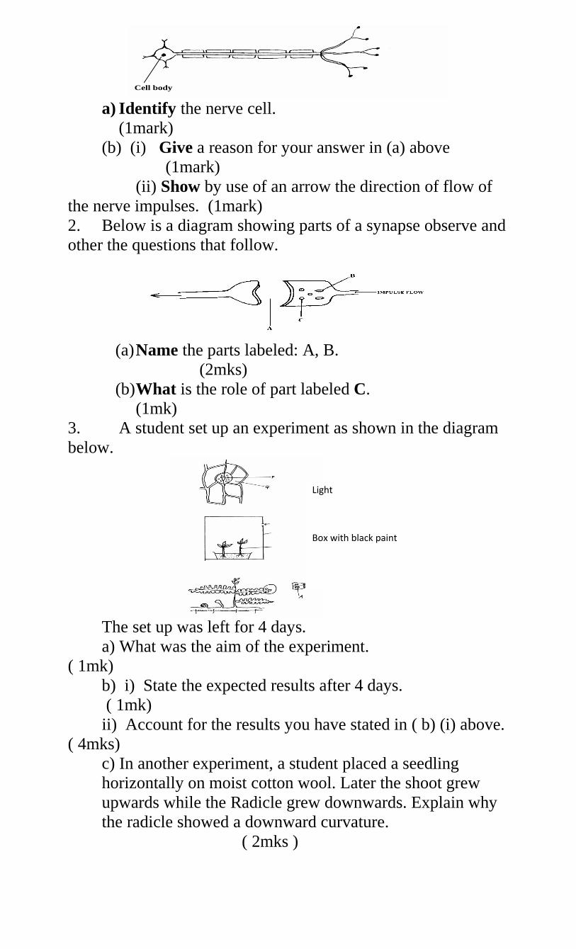
a) Identify the nerve cell.
(1mark)
(b) (i) Give a reason for your answer in (a) above
(1mark)
(ii) Show by use of an arrow the direction of flow of
the nerve impulses. (1mark)
2. Below is a diagram showing parts of a synapse observe and
other the questions that follow.
(a) Name the parts labeled: A, B.
(2mks)
(b) What is the role of part labeled C.
(1mk)
3. A student set up an experiment as shown in the diagram
below.
The set up was left for 4 days.
a) What was the aim of the experiment.
( 1mk)
b) i) State the expected results after 4 days.
( 1mk)
ii) Account for the results you have stated in ( b) (i) above.
( 4mks)
c) In another experiment, a student placed a seedling
horizontally on moist cotton wool. Later the shoot grew
upwards while the Radicle grew downwards. Explain why
the radicle showed a downward curvature.
( 2mks )
Cell body
Light
Box with black paint
Seedlings
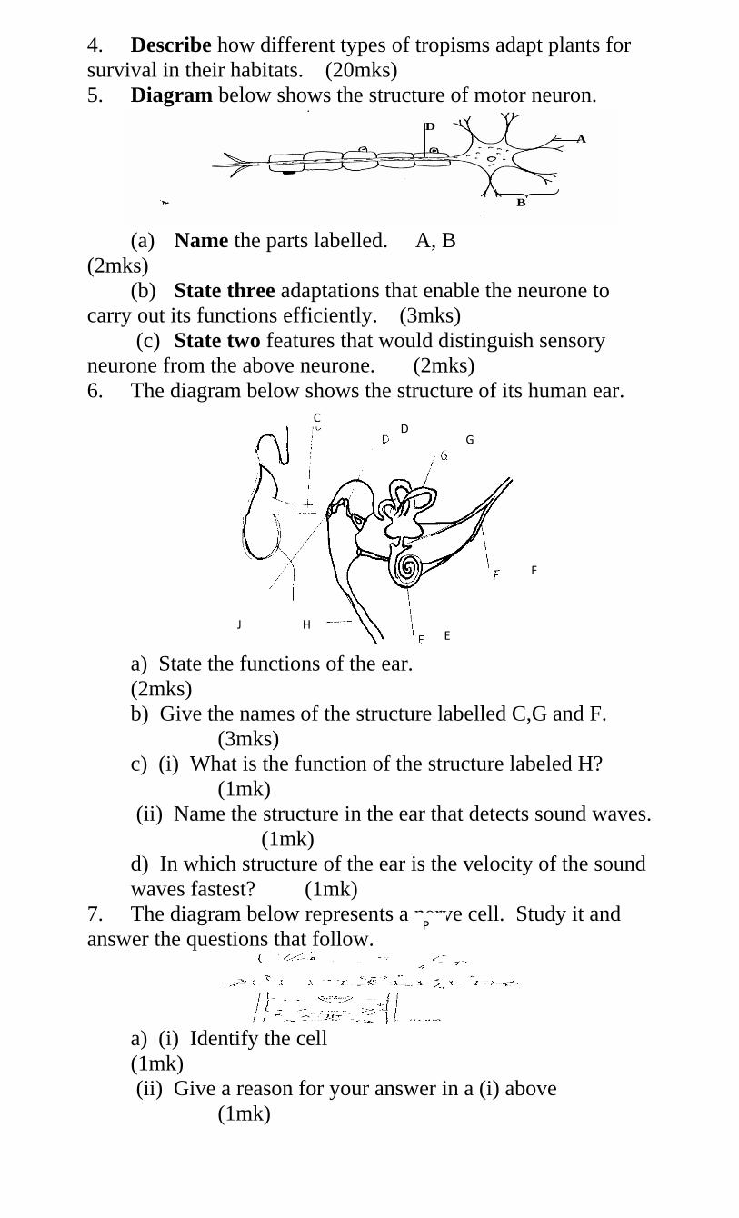
4. Describe how different types of tropisms adapt plants for
survival in their habitats. (20mks)
5. Diagram below shows the structure of motor neuron.
(a) Name the parts labelled. A, B
(2mks)
(b) State three adaptations that enable the neurone to
carry out its functions efficiently. (3mks)
(c) State two features that would distinguish sensory
neurone from the above neurone. (2mks)
6. The diagram below shows the structure of its human ear.
a) State the functions of the ear.
(2mks)
b) Give the names of the structure labelled C,G and F.
(3mks)
c) (i) What is the function of the structure labeled H?
(1mk)
(ii) Name the structure in the ear that detects sound waves.
(1mk)
d) In which structure of the ear is the velocity of the sound
waves fastest? (1mk)
7. The diagram below represents a nerve cell. Study it and
answer the questions that follow.
a) (i) Identify the cell
(1mk)
(ii) Give a reason for your answer in a (i) above
(1mk)
D
A
B
J H E
F
G D
C
P
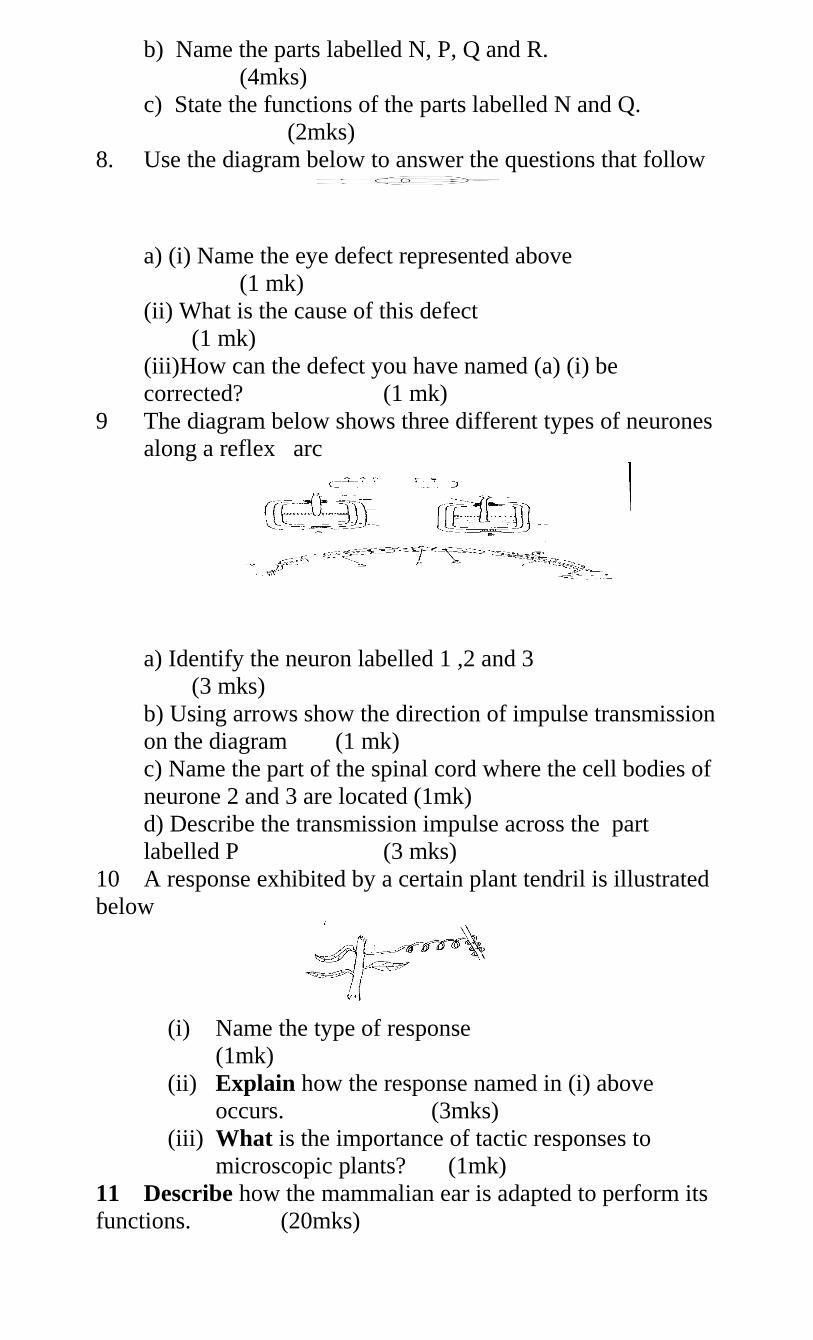
b) Name the parts labelled N, P, Q and R.
(4mks)
c) State the functions of the parts labelled N and Q.
(2mks)
8. Use the diagram below to answer the questions that follow
a) (i) Name the eye defect represented above
(1 mk)
(ii) What is the cause of this defect
(1 mk)
(iii)How can the defect you have named (a) (i) be
corrected? (1 mk)
9 The diagram below shows three different types of neurones
along a reflex arc
a) Identify the neuron labelled 1 ,2 and 3
(3 mks)
b) Using arrows show the direction of impulse transmission
on the diagram (1 mk)
c) Name the part of the spinal cord where the cell bodies of
neurone 2 and 3 are located (1mk)
d) Describe the transmission impulse across the part
labelled P (3 mks)
10 A response exhibited by a certain plant tendril is illustrated
below
(i) Name the type of response
(1mk)
(ii) Explain how the response named in (i) above
occurs. (3mks)
(iii) What is the importance of tactic responses to
microscopic plants? (1mk)
11 Describe how the mammalian ear is adapted to perform its
functions. (20mks)
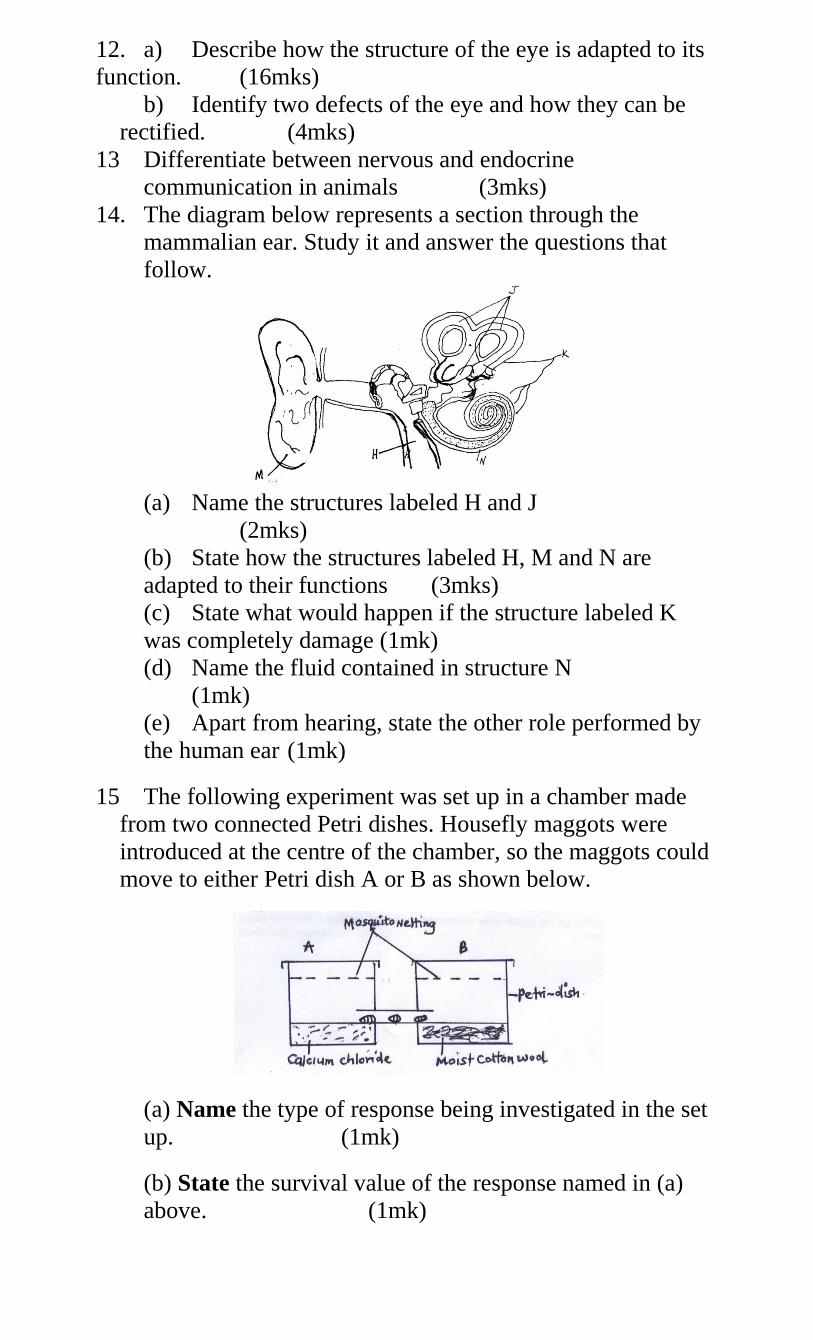
12. a) Describe how the structure of the eye is adapted to its
function. (16mks)
b) Identify two defects of the eye and how they can be
rectified. (4mks)
13 Differentiate between nervous and endocrine
communication in animals (3mks)
14. The diagram below represents a section through the
mammalian ear. Study it and answer the questions that
follow.
(a) Name the structures labeled H and J
(2mks)
(b) State how the structures labeled H, M and N are
adapted to their functions (3mks)
(c) State what would happen if the structure labeled K
was completely damage (1mk)
(d) Name the fluid contained in structure N
(1mk)
(e) Apart from hearing, state the other role performed by
the human ear (1mk)
15 The following experiment was set up in a chamber made
from two connected Petri dishes. Housefly maggots were
introduced at the centre of the chamber, so the maggots could
move to either Petri dish A or B as shown below.
(a) Name the type of response being investigated in the set
up. (1mk)
(b) State the survival value of the response named in (a)
above. (1mk)
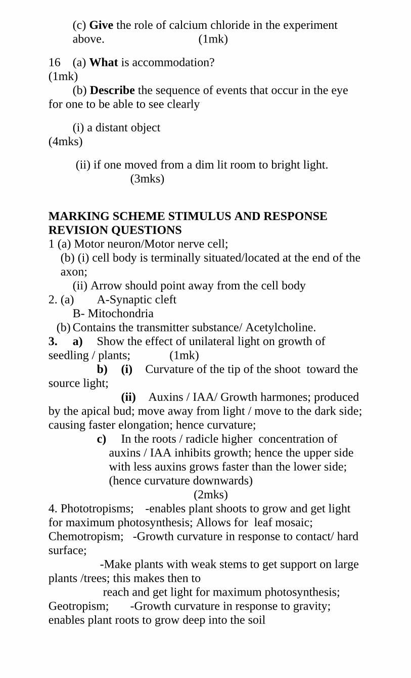
(c) Give the role of calcium chloride in the experiment
above. (1mk)
16 (a) What is accommodation?
(1mk)
(b) Describe the sequence of events that occur in the eye
for one to be able to see clearly
(i) a distant object
(4mks)
(ii) if one moved from a dim lit room to bright light.
(3mks)
MARKING SCHEME STIMULUS AND RESPONSE
REVISION QUESTIONS
1 (a) Motor neuron/Motor nerve cell;
(b) (i) cell body is terminally situated/located at the end of the
axon;
(ii) Arrow should point away from the cell body
2. (a) A-Synaptic cleft
B- Mitochondria
(b) Contains the transmitter substance/ Acetylcholine.
3. a) Show the effect of unilateral light on growth of
seedling / plants; (1mk)
b) (i) Curvature of the tip of the shoot toward the
source light;
(ii) Auxins / IAA/ Growth harmones; produced
by the apical bud; move away from light / move to the dark side;
causing faster elongation; hence curvature;
c) In the roots / radicle higher concentration of
auxins / IAA inhibits growth; hence the upper side
with less auxins grows faster than the lower side;
(hence curvature downwards)
(2mks)
4. Phototropisms; -enables plant shoots to grow and get light
for maximum photosynthesis; Allows for leaf mosaic;
Chemotropism; -Growth curvature in response to contact/ hard
surface;
-Make plants with weak stems to get support on large
plants /trees; this makes then to
reach and get light for maximum photosynthesis;
Geotropism; -Growth curvature in response to gravity;
enables plant roots to grow deep into the soil
to Maximum support/ anchorage;
Hydrotropism; - Growth curvature in response to moisture /
water;
- Enable plant roots to grow and find water in
the soil; water is then used as a raw material
During photolysis stage;
Chemotropism; -Growth curvature in response to chemical
concentration gradient;
- Enables pollen tubes to grow down the style
and into the ovary for fertilization to occur in
plants flowers;
Thermo tropism; -Growth curvature in response to temperature
changes;
-Enables some plants to grow to where they can acquire
optimum temperature for effective plant processes; e.g.
(Sunflower orientates towards the directions of the sun.
5 (a)
A- Dendrites
B-Cell body
(b) - Has long axon to conduct impulses from
CNs to effectors / muscles / glands;
- Axon enclosed with myelin sheath with
nodes of ranvies to enhance speed of
impulse conduction.
- Has dendrites which receive impulses from
adjacent neurones.
(c)
S.N M.N
- Cell body has no dendrites -Cell body has dendrites
- Cell body at a point
along nerve fibre;
-Cell body located at the top
of nerve fiber;
6 a) - Hearing / detection of sound;
Body balance / posture;
(2marks.)
b) (i) C- Ear canal / External auditory meetas;
(ii) G- Semi – Circular canals;
(iii) F- Auditory nerve;
(3marks)
c) (i) Equalise air pressure between middle ear
and outer ear / prevent damage of ear drum;
(1mark)
(ii) Cochlea/ E;
(1mark)
d) - Ear ossicles / D;
(1mark)
7. a) i) Sensory neurons / afferent neurone
ii) Cell body located off the axon
b) N- Axon
P- Cell body
Q- Myelin Sheath
R- Schwann cell
c) N- Transmission of impulse
Q- Insulation / speed up impulse transmission.
8 a) Long sightedness / Hypermetropia ;
1 mk)
b) Short eye ball;
Weak lens (any one) ;1 mks
c) Wearing of convex lens / conveying lenses; 1 mk
rj if a(i) is wrong
9 a) 1- Sensory neurone / Afferent neurone
2- Relay neurone / intermediate neurone
3- Motor neurone/ efferent neurone
3 mks
b) Check on the diagram arrows show points towards neurone
3 from 2 and 1 : 1 mk
c) Grey matter
1 mks
d) Impulse reaching the dendrites end of relay / Neurone 2
causes the synaptic vesicles, releases acetylcholine /
transmitter substances; into the synaptic cleft;the
acetylcholine / transmitter chemical diffuses across the
cleft; and causes the depolarization of the motor neuron/
neurone
(i) Name the type of response (1mk)
10 Thigmotropism/Haptotropism; (ii) Explain how the response named in (i)
above occurs. (3mks)
Contact with support; causes migration of auxins to the
outside; causing faster growth on the side away from
contact surface; (Causing dendrils to curl around the
support);
NB (a) (ii) is tied to (i)
(iii) What is the importance of tactic responses to
microscopic plants? (1mk)
Escape injurious stimuli/seek favourable
habitat
Rej. Seek mate and obtain food
11. Describe how the mammalian ear is adapted to perform its
functions. (20mks)
The pinna is flap made of skin and cartilage; for
collection and concentration of sound waves;
Auditory canal/meatus is a tube lined with hairs
which trap solid particles like dust; It has wax
secreting cells; that secrete wax for trapping solids
and insects entering the ear;
The eardrum/tympanic membrane is thin with double
layer of epidermis; It vibrates translating sound
waves into sound vibrations; Sound vibrations are
transmitted to ear ossicles;
Ear ossicles are malleus, incus and stapes; they
amplify and transmit vibrations to the oral window;
The oval window is a thin membrane which transmits
sound vibrations to the fluid of the inner ear;
perilymph and Endolymph;
Eustachian tube connects middle ear with pharynx
equalizing air pressure in the ear with atmospheric
pressure; to prevent distortion of the eardrum;
The cochlea is highly coiled tube with system of
canals (and sensory cells) to occupy a small
space/increase the surface area for accommodating
many sensory cells to detect sounds vibrations; and
generate impulses transmitted to the brain;
Auditory nerve transmits nerve impulses to the brain
for interpretation;
Semicircular canals are tubular cavities containing
fluid;
The canals are arranged at right angles to each other
in the three planes of spaces; to detect changes in
position of the body; the canals have ampulla:
utriculus and sacculus; to detect position of body in
relation to gravity;
Utriculus has otoliths attached to sensory cells which
generate impulses which are then transmitted to the
brain through the auditory nerve;
The perilymph and endolymph fluid in the inner ear
absorb mechanical shock/transmit sound
vibrations/protect delicate parts;
Total 25 max 20 marks
12 a) i) Conjunctiva transparent allow light to
enter eye
ii) Cornea transparent / curved allow light / refracts light
entering eye;
iii) Aqueous / vitreous humour – clear / allow light pass /
refract light, hydrostatic pressure –
Maintain shape of eyeball. Contains sugars / proteins /
salts – provide nutrients to eye.
iv) Iris – contractile – controls light intensity / amount of
light entering eye;
v) Ciliary body glandular – secretes humuor
vi) Ciliary muscle contractile – controls curvature of lens;
vii) Suspensory ligament – fibrous – holds lens in position
viii) Lens transparent / Biconvex – allow light go through
to retina / refract light / focus light.
ix) Retina – rods - rhodopsin – for dim light vision;
- Iodopsin – for bright light vision
x) Fovea centralis – high concentration of cones – for
accurate vision
xi) Choroid layer – blood vessels pigmented – for
nutrition
- reduce light reflection / absorb stray light
xii) Sclera – fibrous – protection / give eye shape;
xiii) Optical nerves – sensory neurone – transmit impulse
from retina to brain.
xiv) External eye muscle – contractile – move eyeball
within socket
xv) Blind spot – cone and rods absent – no image is
perceived
Correction
b) i) Short sightedness; biconcave / diverging
lens
ii) Long sightedness; converging lens / convex lens
iii) Astigmatism; use of cylindrical lens
iv) Squinting; surgery (any 2
identify correction 4mks)
13. Nervous communication Endocrine
communication
- Nerve impulse to evoke a response - Chemical
substance/ hormone to evoke
Response
- High speed of transmission- Low speed of
transmission;
- Rapid response - Response delayed
- Impulse transmitted through neurone - Hormones
transmitted in blood
- Responses specific and localized to one -
Responses affects several parts of the
Target organ body;
Note: Comparison should come out clearly to award.
14. a) H – Eustachian tube;
J – Semi-circular canals;
b) H – Tube open/ connection to the pharynx and to the
middle ear/ opens during swallowing/ yawning and
vomiting to equalize the air pressure in the middle ear
with the atmospheric air pressure;
M – (pinna) curved/ funnel shaped to receive or
collect and direct sound waves into the ear;
N – (cochlea) – long/highly coiled/ spiral in form to
increase surface area for sound Perception;
- Has sensory hairs/ cells which convert sound
vibrations to impulses/ generate impulses;
- Has endolymph to transmit vibrations;
Mark one for each structure.
Rej. If the adaptation is not tied to function.
c) Total deafness;
d) Endolymph;
e) Balance; acc body balance/ posture.
15 (a) Tactic response;
(b) Move away from a harsh environment/move to favorable
environment;
(c) To absorb any moisture from Petri dish A/OWTTE
16 .(a) A reflex mechanism/ability of the eye to adjust to bring
an image from near or far object into sharp focus on the
retina;
(b) Circular muscles of the iris contract; while the radial
muscles relax; Ciliary muscles relax; increasing tension on
suspensory ligaments; lens become thinner increasing the
focal length; image focused onto the retina;
(ii) Circular muscles of the iris contract; pupil
constrict/become smaller; and allows enough light for (sharp)
image to be focused onto the retina;
SUPPORT AND MOVEMENT IN PLANTS AND
ANIMALS
Support is the ability of organisms to bear their weight and
maintain their body forms. It involves holding body parts in
their position and allow for movement.
Movement is the displacement of parts of the body of an
organism e.g. growth movements in plants and limbs in
animals.
Locomotion is movement of the whole organisms.
Support and Movement in Plants
This can be at cell level e.g. gametes in bryophytes and
Pteridophytes or at organ level in tropic and nastic
responses.
Importance of Movement in Plants i. Enable plants to obtain resources such as sunlight, water
and nutrients due to tropic and nastic responses.
ii. Enhances fertilization in bryophytes and Pteridophytes
iii. Enhance fertilization in flowering plants by growth of
pollen tube towards the embryo sac.
iv. Helps plants to escape harmful stimuli such as high
temperature
Importance of Support in Plants i. Hold flowers in position for pollination to occur.
ii. Help plants to withstand forces of the environment such as
gravity and air currents.
iii. Fruits are held in appropriate position for dispersal to
occur.
iv. Increase the efficiency of photosynthesis as the leaves are
firm and arranged in mosaic pattern for maximum
absorption of light and carbon (iv) oxide.
Arrangement of Tissues in Plants
Diagrams
Parenchyma. The cells are spherical or elongated. They are
unspecialized cells forming the packing tissues. When
turgid, they help in providing support in herbaceous plants.
Collenchyma. It’s underneath the epidermis. They are
similar in appearance to parenchyma and they contain
living protoplasm. They have deposition of cellulose to
provide mechanical support. They are mainly found in
young leaves and stems.
Sclerenchyma. They appear as long fibres in stems. Cells
are dead and they have lignin. Mainly found in stems and
midrib of leaves. The walls are pitted to allow exchange of
substances between cells.
Xylem vessels and Tracheids. Xylem vessels are long tube
like structures with lignified walls used for transporting
water and mineral salts and also give plant mechanical
support. Tracheids are long cells with tapering ends whose
walls are lignified to give the plant mechanical support.
Both xylem vessels ant tracheids are made of dead cells
manly present in woody stems.
Tendrils and Climbing stems. Some herbaceous plants
support themselves by use of tendrils e.g. pumpkins, garden
peas etc. Others obtain support by twinning round other
hard objects such as stem of passion fruit, morning glory
etc.
Spines and Thorns. Some plants use spines and thorns to
attach to solid objects for support e.g. in rose.
Practical Activity 3
To Observe Wilting in Plants
Support and Movement in Animals
Animals have a firm and rigid framework for support called
the skeleton.
Importance of Movement in Animals
1. Enable searching of food, mate and shelter.
2. Move to avoid predators.
3. To colonize new areas
4. Move from areas with unfavourable conditions such as fire,
earthquakes, flood etc.
Types and Function of Skeletons
1. Hydrostatic skeleton
It is found in soft bodied animals such as the earthworm.
2. Exoskeleton
It is made of the external covering found in arthropods.
It’s made of waterproof cuticle which contains the protein
Chitin secreted by the epidermal cells.
Functions of the Exoskeleton
i. Reduces water loss
ii. Protection against microbial infections and mechanical
injury
iii. Support body tissues and organs.
iv. Provide point for attachment of muscles allowing
locomotion to take place.
v. Enhance flight in insects by means of wings which are the
flattened parts of the exoskeleton.
vi. Enhance walking in insects using jointed appendages.
NB/. 1. Exoskeleton has a disadvantage as it limits growth.
To overcome this limitation it is periodically shed through
moulting (ecdysis).
2. Insects that jump or hop have powerful hind limbs. The
femur of the hind limb has powerful antagonistic muscles.
Diagrams
3 Endoskeleton.
It is found in all vertebrates.
Muscles are external to the hard framework.
It is made of living tissues either cartilage or bone which
increase in size as the animal grows and therefore need not
to be shed as in exoskeleton.
Functions of the Endoskeleton
i. Supports the animal’s body
ii. Gives the body its shape
iii. Protects inner delicate organs such as the lungs, heart, liver
etc from mechanical injury e.g. ribs.
iv. Provide surface for muscle attachment facilitating
movement.
v. Production of blood cells i.e. the long and short bones
vi. Acts as a reservoir of calcium and phosphate ions in the
body
Locomotion in Finned Fish (Tilapia)
Diagrams
Practical Activity 5
Practical Activity 6
How a finned fish is adapted to locomotion in water 1. Streamlined body/ tapered anteriorly and posteriorly; to
minimize water resistance;
2. Inflexible head; to maintain forward thrust;
3. Overlapping scales facing posterior end; to bring about less
resistance; Overlapping of scales also prevents wetting of the
skin;
4. Slimy/oily substance to moisten scales; hence reduce
resistance between water and fish;
5. Swim bladder; air filled cavity which controls/ brings
buoyancy; and depth at which it swims;
6. Flexible backbone /series of vertebrae with Myotomes/
muscles blocks; which contract and relax alternately bringing
about thrust/force; which propels fish forwards;
7. Pectoral and pelvic fins (paired fins); which bring about
balancing effect; braking; and changing direction; they also
control pitching i.e. control upward and downward
movement;
8. Dorsal fin, caudal fin and anal fin (unpaired fins); to increase
vertical surface area; and therefore prevent rolling from side
to side; and yawing;
9. Tail fins/caudal fins that are long and flexible; for steering/
more force/ thrust;
10. Lateral line has sensory cells; which enables to perceive
vibrations; hence can locate objects so that it escapes /
changes direction;
Support and Movement in Mammals
Diagram of a human and rabbit skeleton
The skeleton is divided into:
Axial (skull, sternum, ribcage and vertebral column.)
Appendicular ( consists of girdles and the limbs attached to
them)
Axial Skeleton
1. Skull
Made up of many bones fused together to form the
cranium.
The bones are joined together forming immovable joins
called Sutures.
Cranium encloses and protects the brain, olfactory organs,
the eyes, middle and inner ear.
Facial skeleton has a fixed upper jaw called maxilla and a
movable lower jaw known as the mandible.
At the posterior end, there are two smooth rounded
projections called occipital condyles. These articulate with
the first bone of the vertebral column (atlas) forming a
hinge joint.
This joint permits nodding of the head.
2. Ribcage and sternum
Ribcage encloses the thoracic cavity protecting delicate
organs such as the lungs and heart.
Cage is made up of ribs that articulate with vertebral
column at the back and sternum to the front.
In birds, the sternum is modified to form the keel which
gives a large surface area for attachment of flight muscles.
Ribcage and sternum help during breathing because they
offer the surface for attachment of the intercostals muscles.
3. Vertebral column
Consists of bones called vertebrae that are separated from
each other by cartilage called inter-vertebral discs.
The discs absorb shock and reduce friction. It also makes
the vertebral column flexible.
There are five types of vertebrae in the vertebral column;
1. Cervical vertebrae
2. Thoracic vertebra
3. Lumbar vertebrae
4. Sacral vertebrae
5. Caudal vertebrae
All the vertebrae have a common basic plan.
Structure of a Vertebra
Each vertebra is made up of the following parts.
i.) Centrum (body). It supports the weight of the vertebra and
the weight of the entire vertebral column..
ii.) Neural arch. It encloses the neural canal.
iii.) Neural spine. Provides surface for muscle and ligament
attachment.
iv.) Neural canal. It protects the spinal cord which passes
through it.
v.) Transverse processes. Provides surface for muscle and
ligament attachment.
vi.) Zygapophysis (facets). These are smooth patches for
articulation with the other vertebrae. (The one in front and
the other one behind). The front facets are called Pre-
Zygapophysis while the back pair facets are called Post-
Zygapophysis
Diagram
1) Cervical vertebrae
a) Atlas (First cervical vertebra)
Distinctive features.
i.) No Centrum
ii.) Broad and flat transverse processes.
iii.) Has vertebraterial canal in each transverse process for
vertebral arteries to pass through.
iv.) Front facets are large and grooved to articulate with
condyles of the skull to allow nodding on the head.
v.) Neural spine is very small.
Diagram
Functions
i.) Protect the spinal cord.
ii.) Provide surface for muscle attachment.
iii.) Allows head to nod.
b) Axis (second)
Distinctive features.
i.) Centrum prolonged to from the odontoid process.
ii.) Has vertebraterial canal in each transverse process for
vertebral arteries to pass.
iii.) Small wing like transverse processes.
iv.) Wide neural canal.
Functions
i.) Protects the spinal cord.
ii.) Allows the head to rotate. Odontoid process forms a peg
which fits into the neural canal of the atlas.
iii.) Provide surface for muscle attachment
Diagram
c) The other cervical vertebrae.
Distinctive features
i.) Short neural spine
ii.) Transverse process divided and broad.
iii.) Has vertebraterial canal in each transverse process for
vertebral arteries to pass through.
iv.) Wide centrum
Diagram
Functions
i.) Provide surface for attachment of neck muscle.
ii.) Protect the spinal cord.
iii.) Supports the weight of the head.
2) Thoracic vertebrae
Distinctive features
i.) Long neural spine pointing backwards.
ii.) Large centrum.
iii.) Short transverse processes.
iv.) Tubercular facets on each transverse for articulation
with tuberculum of the rib.
v.) Two pairs of capitular demi-facets for articulation with
capitulum of the rib.
Diagram
Functions
i.) Helps to form the rib cage.
ii.) Provides articulation for one end of each rib.
iii.) Protects the spinal cord.
iv.) Provides surface for muscle attachment.
3) Lumbar vertebrae
Distinctive features
i.) Large broad centrum to offer support.
ii.) Broad neural spine.
iii.) Broad and long transverse processes.
iv.) Have extra processes like metapophysis, anapophysis and
hypapophysis.
Functions
i.) Protects the spinal cord.
ii.) Provides surface for muscle attachment.
iii.) Protect and support the heavy organs in the abdominal
cavity.
iv.) Supports the heavy weight of the upper part of the body.
4) Sacral vertebrae
Distinctive features
1. All sacral vertebrae fused to form sacrum
2. Transverse processes of first sacral vertebra large and wing
like for articulation with pelvic girdle
3. Pairs of holes on the lower surface for the spinal nerves to
pass through.
4. Sacrum is broader on the front side and narrow towards the
tail.
Functions
1. Protects alimentary canal on dorsal side.
2. Provides attachment to hip girdle
3. Protects the spinal cord
4. Provides attachment for the muscles
Diagram
5. Caudal vertebrae
Distinctive features
1. Very small in size
2. No neural canal
Functions
1. Provides attachment for tail muscles
2. Helps in the movement of the tail
Diagram
Biology Form 1-2 Work Revision
1. A student set up materials in an experiment as shown
below.
(a) State the physiological process being
investigated.
(1mk)
(b) If the experiment set up was left over-night, state
observation in the set up A and B. (2mks)
(c) Account for the observations in each set up.
(3mks)
(d) If another experiment C was set such that nothing is placed
in the potato cup, state and explain the results that would have
been obtained. (2mks)
2. An experiment was carried out to investigate, haemolysis
of human cells. The red blood cells were placed in different
concentration of sodium chloride solution. The percentage
of haemolysed cells was determined. The results were
shown in the table below.
Salt conc.
(g/100cm3)
0.33 0.36 0.38 0.39 0.42 0.44 0.48
Red blood cells
haemolysed %
100 91 82 69 30 15 0
(a) (i) On the grid provided plot a graph of haemolysed red
blood cells against salt concentration. (6mks
(ii) At what concentration of salt solution was the proportion of
haemolysed cells equal to non-haemolysed cells? (1mk)
(iii) State the percentage of red blood cells haemolysed at salt
concentration of 0.45. (1mk)
(b) Account for the results obtained at:
Sugar
solution
m
Boiled
potato
Fresh
potato
Water
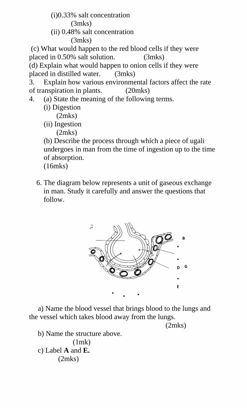
(i)0.33% salt concentration
(3mks)
(ii) 0.48% salt concentration
(3mks)
(c) What would happen to the red blood cells if they were
placed in 0.50% salt solution. (3mks)
(d) Explain what would happen to onion cells if they were
placed in distilled water. (3mks)
3. Explain how various environmental factors affect the rate
of transpiration in plants. (20mks)
4. (a) State the meaning of the following terms.
(i) Digestion
(2mks)
(ii) Ingestion
(2mks)
(b) Describe the process through which a piece of ugali
undergoes in man from the time of ingestion up to the time
of absorption.
(16mks)
6. The diagram below represents a unit of gaseous exchange
in man. Study it carefully and answer the questions that
follow.
a) Name the blood vessel that brings blood to the lungs and
the vessel which takes blood away from the lungs.
(2mks)
b) Name the structure above.
(1mk)
c) Label A and E.
(2mks)
B
A • •
• •
G H G D
• •
E
• • •
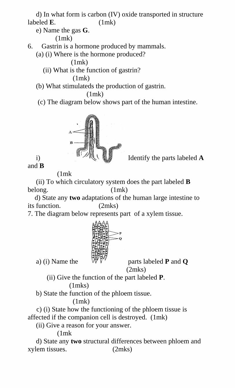
d) In what form is carbon (IV) oxide transported in structure
labeled E. (1mk)
e) Name the gas G.
(1mk)
6. Gastrin is a hormone produced by mammals.
(a) (i) Where is the hormone produced?
(1mk)
(ii) What is the function of gastrin?
(1mk)
(b) What stimulateds the production of gastrin.
(1mk)
(c) The diagram below shows part of the human intestine.
i) Identify the parts labeled A
and B
(1mk
(ii) To which circulatory system does the part labeled B
belong. (1mk)
d) State any two adaptations of the human large intestine to
its function. (2mks)
7. The diagram below represents part of a xylem tissue.
a) (i) Name the parts labeled P and Q
(2mks)
(ii) Give the function of the part labeled P.
(1mks)
b) State the function of the phloem tissue.
(1mk)
c) (i) State how the functioning of the phloem tissue is
affected if the companion cell is destroyed. (1mk)
(ii) Give a reason for your answer.
(1mk
d) State any two structural differences between phloem and
xylem tissues. (2mks)
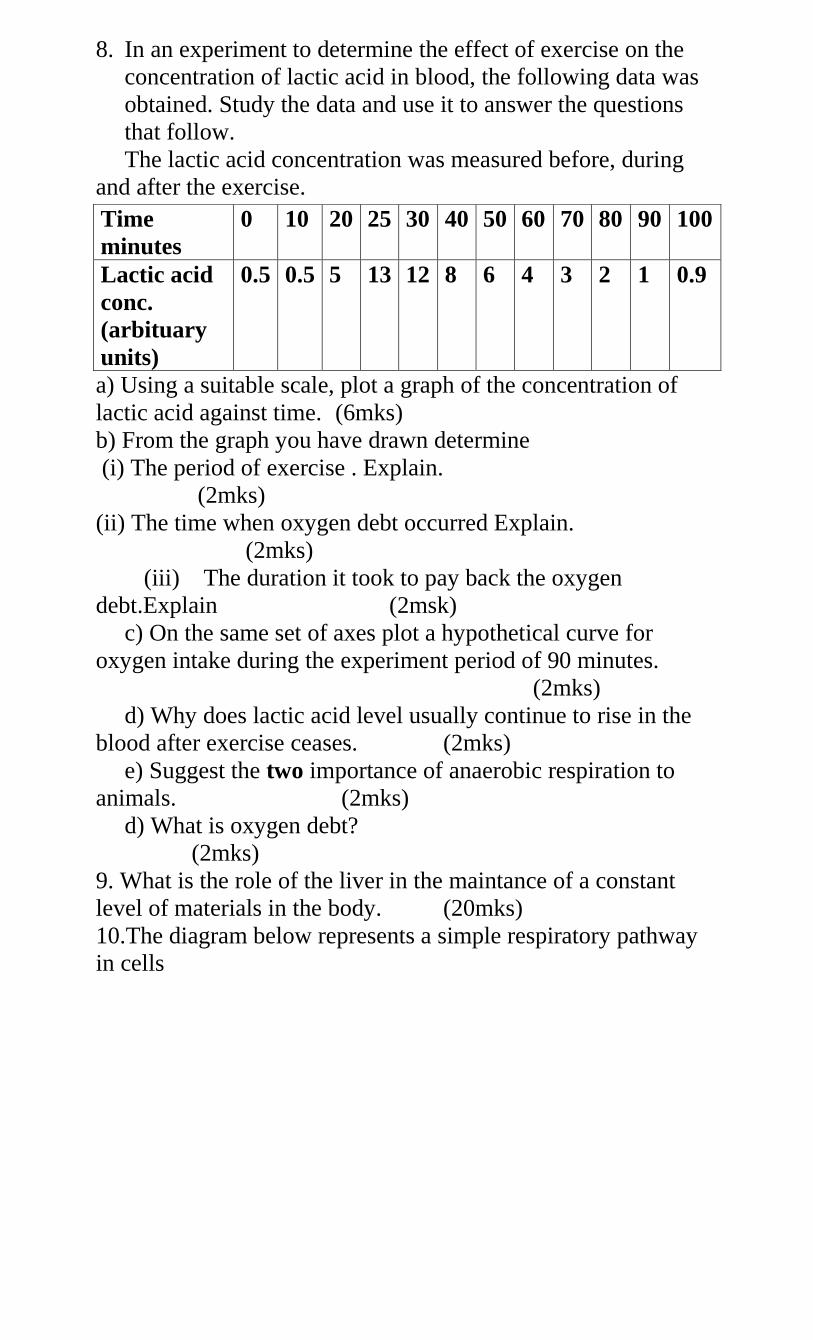
8. In an experiment to determine the effect of exercise on the
concentration of lactic acid in blood, the following data was
obtained. Study the data and use it to answer the questions
that follow.
The lactic acid concentration was measured before, during
and after the exercise.
a) Using a suitable scale, plot a graph of the concentration of
lactic acid against time. (6mks)
b) From the graph you have drawn determine
(i) The period of exercise . Explain.
(2mks)
(ii) The time when oxygen debt occurred Explain.
(2mks)
(iii) The duration it took to pay back the oxygen
debt.Explain (2msk)
c) On the same set of axes plot a hypothetical curve for
oxygen intake during the experiment period of 90 minutes.
(2mks)
d) Why does lactic acid level usually continue to rise in the
blood after exercise ceases. (2mks)
e) Suggest the two importance of anaerobic respiration to
animals. (2mks)
d) What is oxygen debt?
(2mks)
9. What is the role of the liver in the maintance of a constant
level of materials in the body. (20mks)
10.The diagram below represents a simple respiratory pathway
in cells
Time
minutes
0 10 20 25 30 40 50 60 70 80 90 100
Lactic acid
conc.
(arbituary
units)
0.5 0.5 5 13 12 8 6 4 3 2 1 0.9
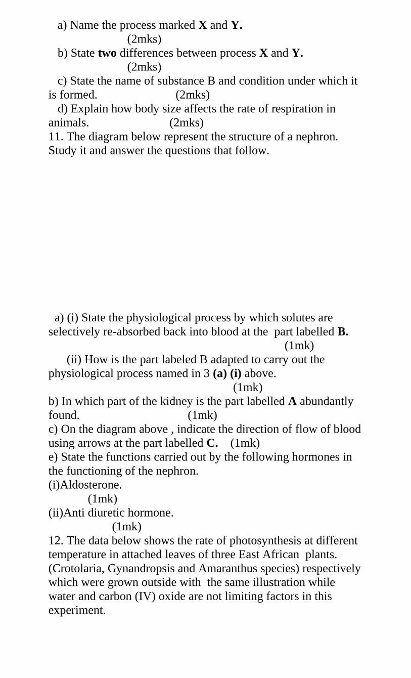
a) Name the process marked X and Y.
(2mks)
b) State two differences between process X and Y.
(2mks)
c) State the name of substance B and condition under which it
is formed. (2mks)
d) Explain how body size affects the rate of respiration in
animals. (2mks)
11. The diagram below represent the structure of a nephron.
Study it and answer the questions that follow.
a) (i) State the physiological process by which solutes are
selectively re-absorbed back into blood at the part labelled B.
(1mk)
(ii) How is the part labeled B adapted to carry out the
physiological process named in 3 (a) (i) above.
(1mk)
b) In which part of the kidney is the part labelled A abundantly
found. (1mk)
c) On the diagram above , indicate the direction of flow of blood
using arrows at the part labelled C. (1mk)
e) State the functions carried out by the following hormones in
the functioning of the nephron.
(i)Aldosterone.
(1mk)
(ii)Anti diuretic hormone.
(1mk)
12. The data below shows the rate of photosynthesis at different
temperature in attached leaves of three East African plants.
(Crotolaria, Gynandropsis and Amaranthus species) respectively
which were grown outside with the same illustration while
water and carbon (IV) oxide are not limiting factors in this
experiment.
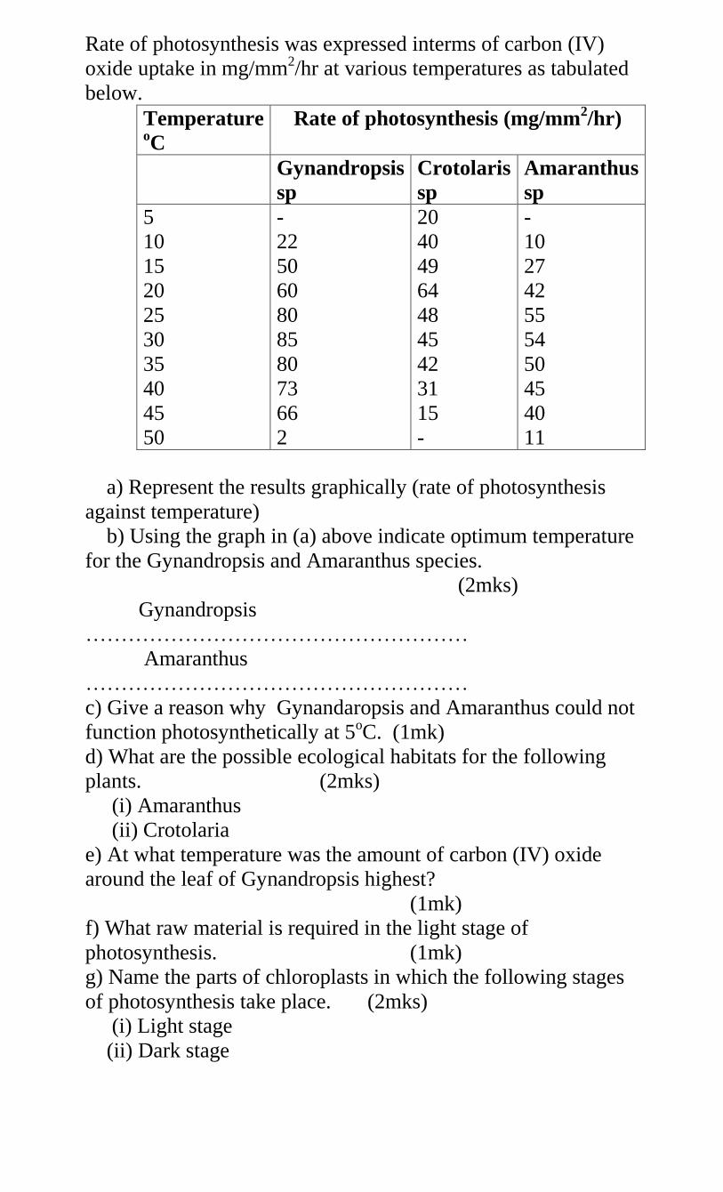
Rate of photosynthesis was expressed interms of carbon (IV)
oxide uptake in mg/mm2/hr at various temperatures as tabulated
below.
Temperature oC
Rate of photosynthesis (mg/mm2/hr)
Gynandropsis
sp
Crotolaris
sp
Amaranthus
sp
5
10
15
20
25
30
35
40
45
50
-
22
50
60
80
85
80
73
66
2
20
40
49
64
48
45
42
31
15
-
-
10
27
42
55
54
50
45
40
11
a) Represent the results graphically (rate of photosynthesis
against temperature)
b) Using the graph in (a) above indicate optimum temperature
for the Gynandropsis and Amaranthus species.
(2mks)
Gynandropsis
………………………………………………
Amaranthus
………………………………………………
c) Give a reason why Gynandaropsis and Amaranthus could not
function photosynthetically at 5oC. (1mk)
d) What are the possible ecological habitats for the following
plants. (2mks)
(i) Amaranthus
(ii) Crotolaria
e) At what temperature was the amount of carbon (IV) oxide
around the leaf of Gynandropsis highest?
(1mk)
f) What raw material is required in the light stage of
photosynthesis. (1mk)
g) Name the parts of chloroplasts in which the following stages
of photosynthesis take place. (2mks)
(i) Light stage
(ii) Dark stage
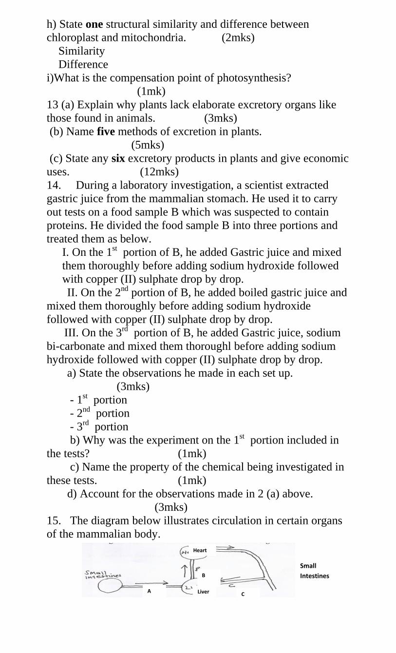
h) State one structural similarity and difference between
chloroplast and mitochondria. (2mks)
Similarity
Difference
i)What is the compensation point of photosynthesis?
(1mk)
13 (a) Explain why plants lack elaborate excretory organs like
those found in animals. (3mks)
(b) Name five methods of excretion in plants.
(5mks)
(c) State any six excretory products in plants and give economic
uses. (12mks)
14. During a laboratory investigation, a scientist extracted
gastric juice from the mammalian stomach. He used it to carry
out tests on a food sample B which was suspected to contain
proteins. He divided the food sample B into three portions and
treated them as below.
I. On the 1st portion of B, he added Gastric juice and mixed
them thoroughly before adding sodium hydroxide followed
with copper (II) sulphate drop by drop.
II. On the 2nd portion of B, he added boiled gastric juice and
mixed them thoroughly before adding sodium hydroxide
followed with copper (II) sulphate drop by drop.
III. On the 3rd portion of B, he added Gastric juice, sodium
bi-carbonate and mixed them thoroughl before adding sodium
hydroxide followed with copper (II) sulphate drop by drop.
a) State the observations he made in each set up.
(3mks)
- 1st portion
- 2nd portion
- 3rd portion
b) Why was the experiment on the 1st portion included in
the tests? (1mk)
c) Name the property of the chemical being investigated in
these tests. (1mk)
d) Account for the observations made in 2 (a) above.
(3mks)
15. The diagram below illustrates circulation in certain organs
of the mammalian body.
Heart
Small
Intestines B
C Liver A
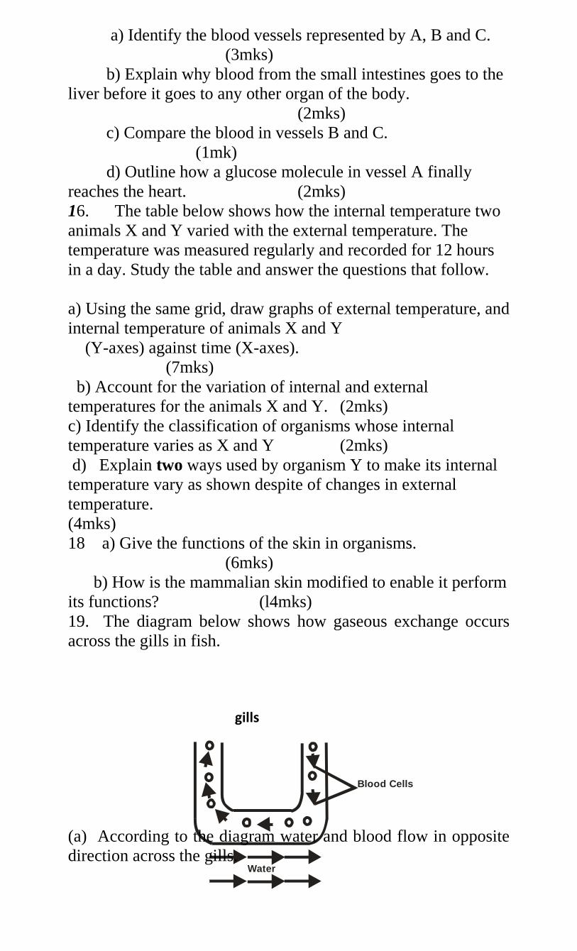
Blood Cells
Water
a) Identify the blood vessels represented by A, B and C.
(3mks)
b) Explain why blood from the small intestines goes to the
liver before it goes to any other organ of the body.
(2mks)
c) Compare the blood in vessels B and C.
(1mk)
d) Outline how a glucose molecule in vessel A finally
reaches the heart. (2mks)
16. The table below shows how the internal temperature two
animals X and Y varied with the external temperature. The
temperature was measured regularly and recorded for 12 hours
in a day. Study the table and answer the questions that follow.
a) Using the same grid, draw graphs of external temperature, and
internal temperature of animals X and Y
(Y-axes) against time (X-axes).
(7mks)
b) Account for the variation of internal and external
temperatures for the animals X and Y. (2mks)
c) Identify the classification of organisms whose internal
temperature varies as X and Y (2mks)
d) Explain two ways used by organism Y to make its internal
temperature vary as shown despite of changes in external
temperature.
(4mks)
18 a) Give the functions of the skin in organisms.
(6mks)
b) How is the mammalian skin modified to enable it perform
its functions? (l4mks)
19. The diagram below shows how gaseous exchange occurs
across the gills in fish.
(a) According to the diagram water and blood flow in opposite
direction across the gills.
gills
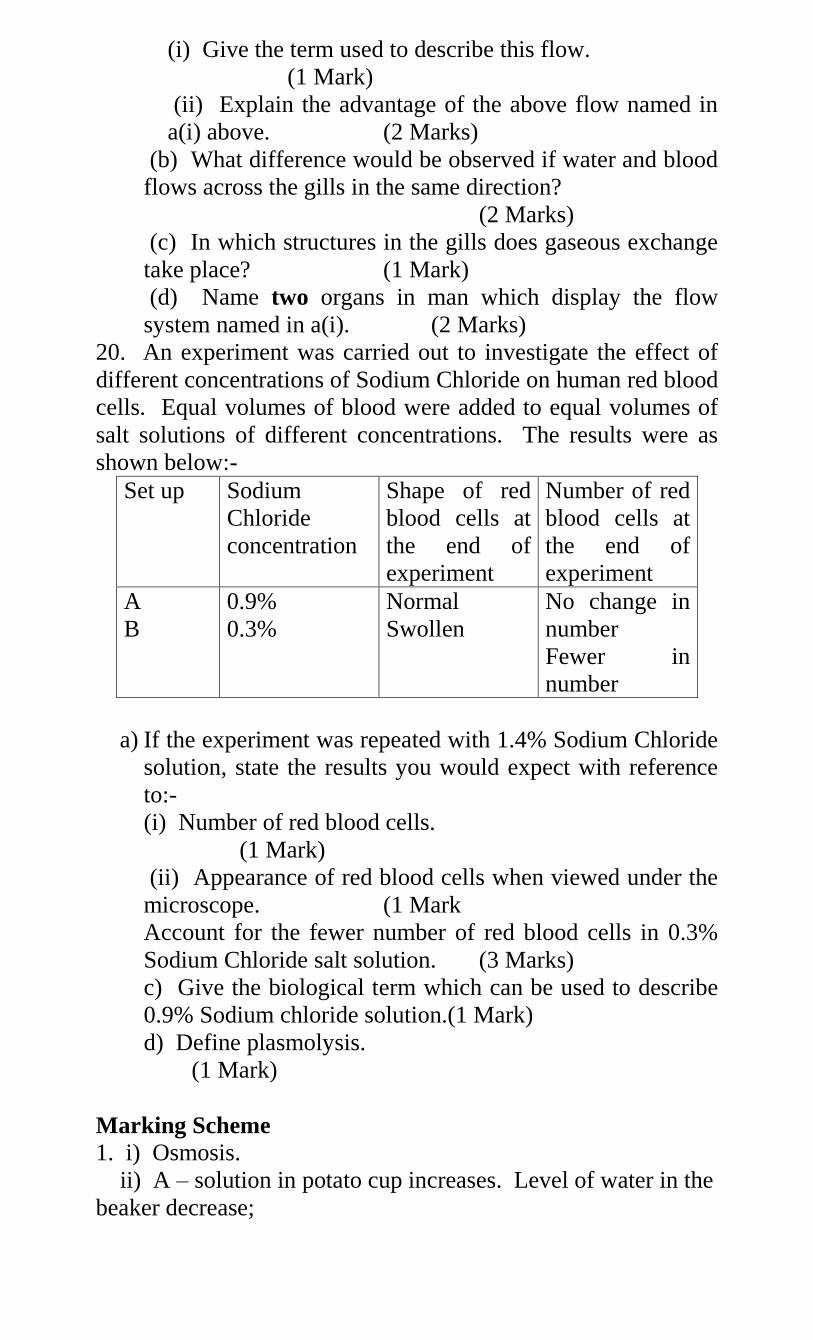
(i) Give the term used to describe this flow.
(1 Mark)
(ii) Explain the advantage of the above flow named in
a(i) above. (2 Marks)
(b) What difference would be observed if water and blood
flows across the gills in the same direction?
(2 Marks)
(c) In which structures in the gills does gaseous exchange
take place? (1 Mark)
(d) Name two organs in man which display the flow
system named in a(i). (2 Marks)
20. An experiment was carried out to investigate the effect of
different concentrations of Sodium Chloride on human red blood
cells. Equal volumes of blood were added to equal volumes of
salt solutions of different concentrations. The results were as
shown below:-
Set up Sodium
Chloride
concentration
Shape of red
blood cells at
the end of
experiment
Number of red
blood cells at
the end of
experiment
A
B
0.9%
0.3%
Normal
Swollen
No change in
number
Fewer in
number
a) If the experiment was repeated with 1.4% Sodium Chloride
solution, state the results you would expect with reference
to:-
(i) Number of red blood cells.
(1 Mark)
(ii) Appearance of red blood cells when viewed under the
microscope. (1 Mark
Account for the fewer number of red blood cells in 0.3%
Sodium Chloride salt solution. (3 Marks)
c) Give the biological term which can be used to describe
0.9% Sodium chloride solution.(1 Mark)
d) Define plasmolysis.
(1 Mark)
Marking Scheme
1. i) Osmosis.
ii) A – solution in potato cup increases. Level of water in the
beaker decrease;
B- Remain the same;
iii) A – Surrounding the cube is a region with high
concentration of water molecules while in the sugar
crystals, there are very few water molecules;
The sugar crystals exert on Osmosis pressure by Osmosis water
molecules move across the potato tissue, which acts as a
semi-permeable membrane. The level rises;
B- No change since boiling denatures the membrane
structure of potato cells;
iv) C- No water moves into the potato cup/remains the
same; since there is no concentration gradient;
2(a) i) Graph.
ii) 0.402 ± 0.01
iii) 11% ± 1%.
b) i) All cells have been haemolysed; cells contains one
hypertonic to salt solution; water enters cells by osmosis; cells
swell and eventually burst.
ii) No cells were haemolysed; cell contents were isotonic
to salt solutions (aments of water entering the cell was equal
to that leaving the cell); no net movement of water into cells;
c) The cells would become crenated; the cell contents
would be hypotonic to salt solutions; water would leave cells by
osmosis; membranes would shrink.
d) Contents of Onion epidermal cells would be hypertonic to
water; water would enter cells by Osmosis; cells would become
turgid;
3. - Temperature;- High temperature faster rate of
transpiration; high temperature increases the capacity of
atmosphere to hold water and moisture; also heat increase
internal temperature of the leaf hence water evaporation;
4 accept converse
- Atmospheric pressure; Low atmospheric pressure, high
rate of transpiration 2
- Humidity; Low humidity higher rate of transpiration;
low humidity increases the saturation defiant; hence water
moves form leaves to drier atmosphere; 4
- Wind; When it is windy the rate of transpiration is
higher; wind sweeps away vapour that has accumulated at the
surface of leaf; increasing saturation deficit; hence faster rate of
transpiration 5
- Light intensity;High light intensity faster rate of
transpiration high light intensity increase photosynthesis rate
hence stomata opens; 4
- Amount of water in soil; More water in the soil
increases the rate of transpiration; it wets the xylem (ensure
xylem is wet throughout); 3
Max 20
4. a) Define digestion and ingestion.
i) Digestion- It is break down of complex insoluble; √ food
substance into simple soluble food substance;√
ii) Ingestion- is introduction of food through the mouth
into the digestive system;
b) Describe the digestion of Ugali.
- Digestion of ugali begins in the mouth; √ ugali is
chewed by the teeth to increase large surface area √
for action of salivary amylase/ptyalin; √ The food
mixes with saliva produced by salivary glands;√
- Saliva contains mucus and enzyme ptyalin. Mucus
moistens, softens and lubricates the food;√ ptyalin
speeds up the conversion of starch to maltose; √ ugali
is made into bolus in the mouth;√
- The bolus moves along the oescophagus and
prestalsis/by contraction and relaxation of circular and
longitudinal muscles into the stomach;√
- The digestion continues until ugali become acidic
since the stomach does not contain
carbohydrase/carbohydrate digesting enzymes no
digestion of ugali takes place here.√
- Ugali now moves into duodenum by peristalsis in
form of acidic chime; √ where it mixes with the bile
from the liver and pancreatic juice from the
pancreases;√ bile being alkaline neutralizes the
stomach acid;√ and provides a suitable alkaline
medium for the enzymes to act on carbohydrates;√
- Pancreatic juice contains three enzymes out of the
which enzyme amylase speeds conversion of starch to
maltose;√
- When food reaches the ileum; it mixes with intestinal
juice which contains several enzymes. Maltase –
speeds up conversion of maltose to glucose;√
- Lactose which speeds up conversion of lactose to
glucose√ and galactose; sucrase which speeds up
conversion of sucrose into fructose and glucose;
- Absorption – glucose, the end product of all
carbohydrates diffuses through the epithelium of villi
and capillary walls and enters into blood stream and
is carried to the liver via hepatic portal veins;√
- Assimilation- in the liver excess glucose is converted
into glycogen and stored;
- - The rest of the glucose is carried by the blood
tissues where is oxidized during tissue respiration to
release energy;√ (21 max 18
mks Total 20mks)
5. (a) - Pulmonary artery
- Pulmonary vein
(b) Alveolus
(c) A- cavity of alveolus
E – Red blood cell
(d) Hydrogen carbonate ions;
Carbamino haemoglobin;
6. (a) (i) Walls of stomach;
(ii) Stimulates the secretion / production of gastric juice;
√
(b) Presence of food in the stomach;
(c) A – Blood capillaries; B – Lacteal;
(d) - Produces plenty of mucus to lubricate
coarse/indigestible material during peristalsis;
- Wide human accommodates /store indigestible food
- Elongate to increase surface are for absorption of
water.
- has muscles to facilitate peristalsis when they contract;
7. (a) (i) P – Tracheids Q – pits
(ii) P- water conducting elements of xylem
(b) Function of phloem – translocation/ transport of organic
substances from the leaves to the of the plant;
(c) (i) Translocation of food will not occur acc. Slow
translocation
(ii) Reason – it contains a lot of mitochondria which
provide energy for translocation;
(d)
Phloem Xylem
1) Made of living
cells
2) Have
companion
cells
3) Have
- made of dead cells
- lack companion cells
- lack cytoplasmic
strands
- have lignin deposits;
(any 2x1=2mks)
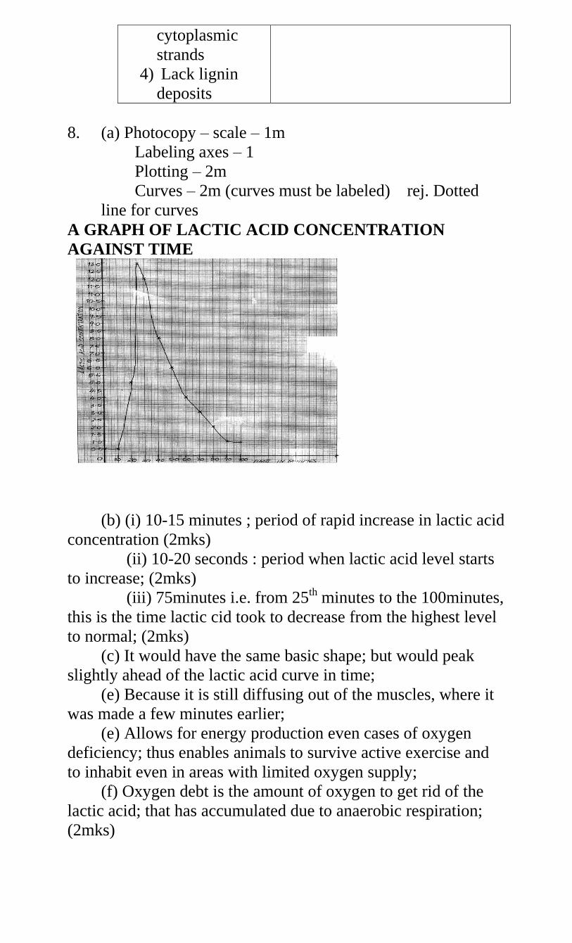
cytoplasmic
strands
4) Lack lignin
deposits
8. (a) Photocopy – scale – 1m
Labeling axes – 1
Plotting – 2m
Curves – 2m (curves must be labeled) rej. Dotted
line for curves
A GRAPH OF LACTIC ACID CONCENTRATION
AGAINST TIME
(b) (i) 10-15 minutes ; period of rapid increase in lactic acid
concentration (2mks)
(ii) 10-20 seconds : period when lactic acid level starts
to increase; (2mks)
(iii) 75minutes i.e. from 25th minutes to the 100minutes,
this is the time lactic cid took to decrease from the highest level
to normal; (2mks)
(c) It would have the same basic shape; but would peak
slightly ahead of the lactic acid curve in time;
(e) Because it is still diffusing out of the muscles, where it
was made a few minutes earlier;
(e) Allows for energy production even cases of oxygen
deficiency; thus enables animals to survive active exercise and
to inhabit even in areas with limited oxygen supply;
(f) Oxygen debt is the amount of oxygen to get rid of the
lactic acid; that has accumulated due to anaerobic respiration;
(2mks)
9. Regulation of blood sugar level; under the influence of
insulin; and glucagons (hormones). When there is excess sugar;
the hormone insulin stimulate/causes liver cell to convert it to
glycogen; some converted to fats/lipids for storage;
• When the blood sugar level is below normal: the hormone
glucagon causes liver cells to convert glycogen to glucose;
• Regulation of amino acids; excess amino acids; are
deaminated; by the liver (cells) leading to formation of
urea; which is transported by the blood to the kidney; for
elimination;
• Production of heat: the liver is involved in the
thermoregulation due to many metabolic, activities; taking
place in the liver cells a lot of heat is generated which is
distributed to the entire/whole body;
• Detoxication of toxic substances; (such as drugs and
hydroxide peroxide)
• Elimination of haemoglobin; and formation of bile;
breakdown worn out red blood cells; the bile salts (sodium
tyrochocolate and sodium glycocholate; in the bile
eEMULSIFYfats (in the duodenum)
• Storage of blood in its veins; thus regulating the volume
of blood circulating in the body
• Elimination of sex hormones after they have performed
their function/work; storage of vitamin AD and B12 some
mineral salts; thus regulating their levels in the blood
TOTAL 23 MAX 20
10. (a) X - glycolysis Y – Kreb’s cycle
(b)
Process X Process Y
- occurs in cytoplasm
- independent of oxygen
- produces less energy
- raw material is glucose
- End products are energy,
CO2, lactic acid or ethanol
- occurs in mitochondria
- Is oxygen dependent
- produces more energy
- Raw material is pyruvate
- End products are energy,
CO2 and water
(c) lactic acid; under anaerobic conditions
(d) small body size leads to alarge surface area to volume
ratio; hence more loss of heat to the environment; leading to
increased rate of respiration to replace the lost heat;
11. (a) (i)Active transport/diffusion
Tied (ii) Numerous Mitochondria in its wall to generate
energy/microvilli/coiling increase surface area/thin epithelium
for quick diffusion.
(b) Cortex
(c) on the diagram
(d) plasma proteins; Blood cells; accept specific examples e.g.
albumins, red blood cells;
(e) (i) Regulate re-absorption of Sodium salts;
(ii) Regulate re-absorption of water
12. (a) Allocation of marks on graph
(b) Gynandropsis – opt To = 30oC
Amaranthus – opt. to = 25oC
(c)At 5oC, the enzymes that catalyse the process of
photosynthesis are inactivated.
(d) Amaranthus – Terrestrial; Crotolaria – terrestrial;
(e) 50oC;
(f) water;
(g)(i) Granum; (ii) Stroma
(h) Similarity: Both have double membrane; 1mk – Both
have fluid filled matrix;
Difference : inner membrane of mitochondrion is folded to
form cristae while inner membrane of chloroplast is smooth;
- chloroplast is biconcave shaped while
mitochondria is oval/sausage shaped (any 1x1=1mk)
(i) Point at which the rate of photosynthesis equals to the
rate of respiration.
13. (a) – plants wastes accumulate slowly;
- plants produce less toxic wastes;
- some excretory products are recycled by plants e.g.
CO2, SO2)
- plant tissues are tolerant to toxic wastes;
- plant wastes are stored in temporary structures
which fall off e.g. leaves (any 3x1=3mks)
(b) – Diffusion;
- Transpiration;
- Exudation;
- Deposition of wastes/ leaf fall/ flower fall/ storage in
bark;
- Recycling;
- Guttation ; (any 5x1=5mks)
(c)
Excretory
products
Uses
1. Caffeine;
2. Popain;
3. Tannin;
4. Nicotine;
5. Latex;
6. Quinine;
7. Atropine;
8. Morphine;
9. Colchine;
10. Cocai
ne;
11. Cann
abis;
12. Khat/
miraa;
Body stimulant;
Meat tenderizer;
Leather tanning;
Stimulant; insecticide;
Manufacture of tyre/rubber
products;
Anti-malarial drugs;
Increase heart beat; dilate
eye pupil;
Cancer treatment;
Used in genetics to induce
polypoidy;
Anesthesia/painkiller/stimul
ant;
Pain killer;
Stimulant; (Any
6x2=12mks)
14. (a) 1st portion. - Blue; colour was observed
2nd portion – Purple; colour was observed
3rd portion – Purple; colour was observed
(b) A control experiment;
(c) Proteins are highly sensitive to temperature and pH
changes; (award if either temp of pH is stated singly)
(d) 1st portion – Enzyme pepsin broke down proteins into
peptones;
2nd portion – Enzyme pepsin works in acidic medium; (not
in basic medium)
15. (a) A – Hepartic portal vein; B – Hepartic vein;
C – Hepartic artery;
(b) – So that any toxic substances absorbed together with
food nutrients from the ileum be detoxified;
- So that food substances e.g. glucose, amino acids can be
regulated. Only the required quantity of glucose is left in
circulation as excess is either stored as glycogen, fat and excess
may be respired.
Excess amino acids are deaminated;
(c) B – Deoxygenated C - Oxygenated
(d) From the small intestines, it is transported to the liver
through Hepartic portal vein; (It is then transported to the heart
through the hepartic vein;
16. (a) graph
(b) X – Lacks internal mechanisms to regulate its internal
temperature
Y – Has internal means to regulate its internal
temperature. hence able to maintain it within narrow range
(c) X – Poikilotherm Y – Endotherm
(d) – Blood vessels vasodilate when temperature is higher
than norm to allow for heat loss from blood through radiation,
evaporation, etc; when temperature is lower, blood vessels
constrict to prevent loss of heat from blood through
radiation, evaporation etc;
- When temperature is higher, lies flat to allow for heat loss
from the body since insulation layer of air is removed; when
temperature is lower, hair strands erect to hold air which
insulates the body against heat loss through radiation,
evaporation etc.
17. (a) Higher temperature; increases the kinetic energy; of
water molecules which makes water turn into vapour on the
leaf surfaces faster and hence increase rate of transpiration
Higher light intensity; influences maximum opening of
stomata which increases the surface area; over which
transpiration occurs maximumly
Wind; carries away moisture around the plant and create a
higher saturation deficit; which then increases the rate of
water loss/transpiration in plants.
Higher relative humidity; reduces saturation deficit; which
causes lowering of water loss/transpiration in plants.
Higher amount of water in the soil; makes the plant to
absorb excess water which increases the need for the plant
to get rid of it through transpiration faster;
If the leaf is broader and has numerous larger open stomata;
the surface area over which water loss occurs is increased;
causing increase in the rate of transpiration.
(award max. 12mks)
18. (a) – It protects the underlying tissues against mechanical
injury, UV-light rays and entry of pathogens; (Rej. germs)
As an excretory organs, it enables organisms to eliminate
excess water, ions and traces of urea;
As a sensory organ, it enables the organisms to be aware
of deviations in pressure, touch and temperature from the
external environment;
It is a thermoregulator such that it enables the body to
lose excess heat to lower its temperature back to norm or
may enable the organisms to store it s heat if the
temperature is lower and hence raise it back to the norm;
It takes part in osmoregulation by enabling the body fluids
to get rid of excess water or excess ions;
It takes part in the regulation of the pH of body fluids by
enabling the body to get rid of either Hydrogen ions or bi-
carbonate ions;
(b) – Presence of the cornified layer; which tough and has
keratin to enable it protect the underlying tissues from
mechanical injury; It alsos has sebum; which is antiseptic and
enables it to prtect the ody against entry of pathogesn.
Presence of melanin; enables it to protect the underlying
tissues against damage by t UV-light radiations.
It has sweat glands with secretory cells; which absorb
excess water, excess ions and traces fo urea from blood and
secrete them into the sweat duct;
Has the sweat pores; which open son the skin surface to
allow for elimination of sweat containing excess water,
excess ions and traces of urea;
It has nerve endings; which enables it to detect any
deviations in temperature, pressure and contact/touch;
It has hair follicles; which stand erect when temperature is
lower than normal to reduce heat loss from the body or lie
flat to enable to body lose excess heat and lower
temperature back to the norm when the internal temperature
is higher;
Has blood vessels; which vasodilate when temperature is
higher than norm to enable the organisms lose than the
norm to reduce heat loss from the body;
19. a) i) Counter current system;
ii) Maintain a diffusion gradient so that there is maximum
uptake of oxygen; continue diffusing into blood and Carbon (iv)
oxide into water;
b) Parallel flow lower diffusion gradient; so that less oxygen
diffuse into blood/low rate of gaseous exchange
c) Gill filaments
d) Placenta
Kidney
20 a) i) Remain the same
ii) Crenated
b) The solution is hypotonic to red blood cells hence the cells
grains water; by osmosis; swelling until they burst.
c) Isotonic solution
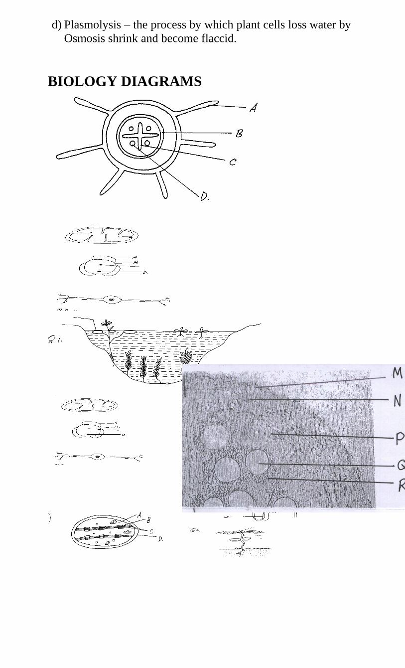
d) Plasmolysis – the process by which plant cells loss water by
Osmosis shrink and become flaccid.
BIOLOGY DIAGRAMS
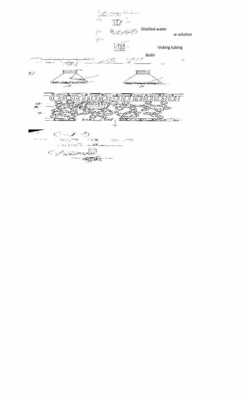
Starch and glucose solution
Boiling tube
Visking tubing
Distilled water
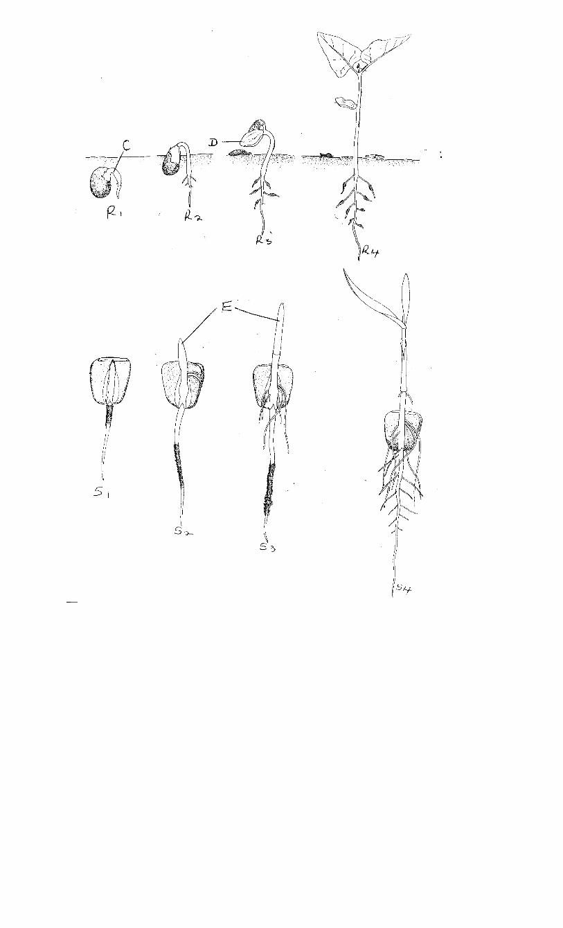
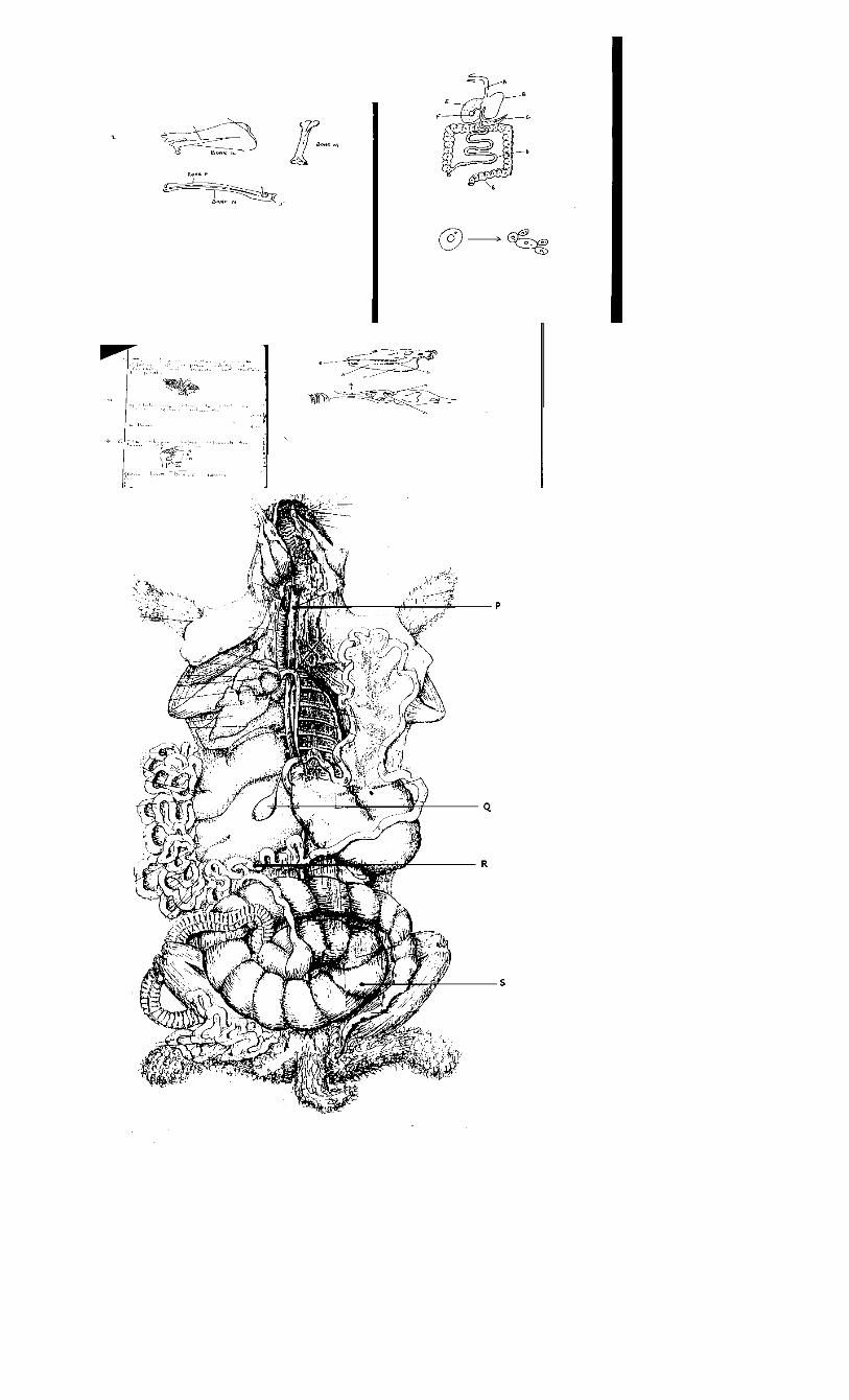
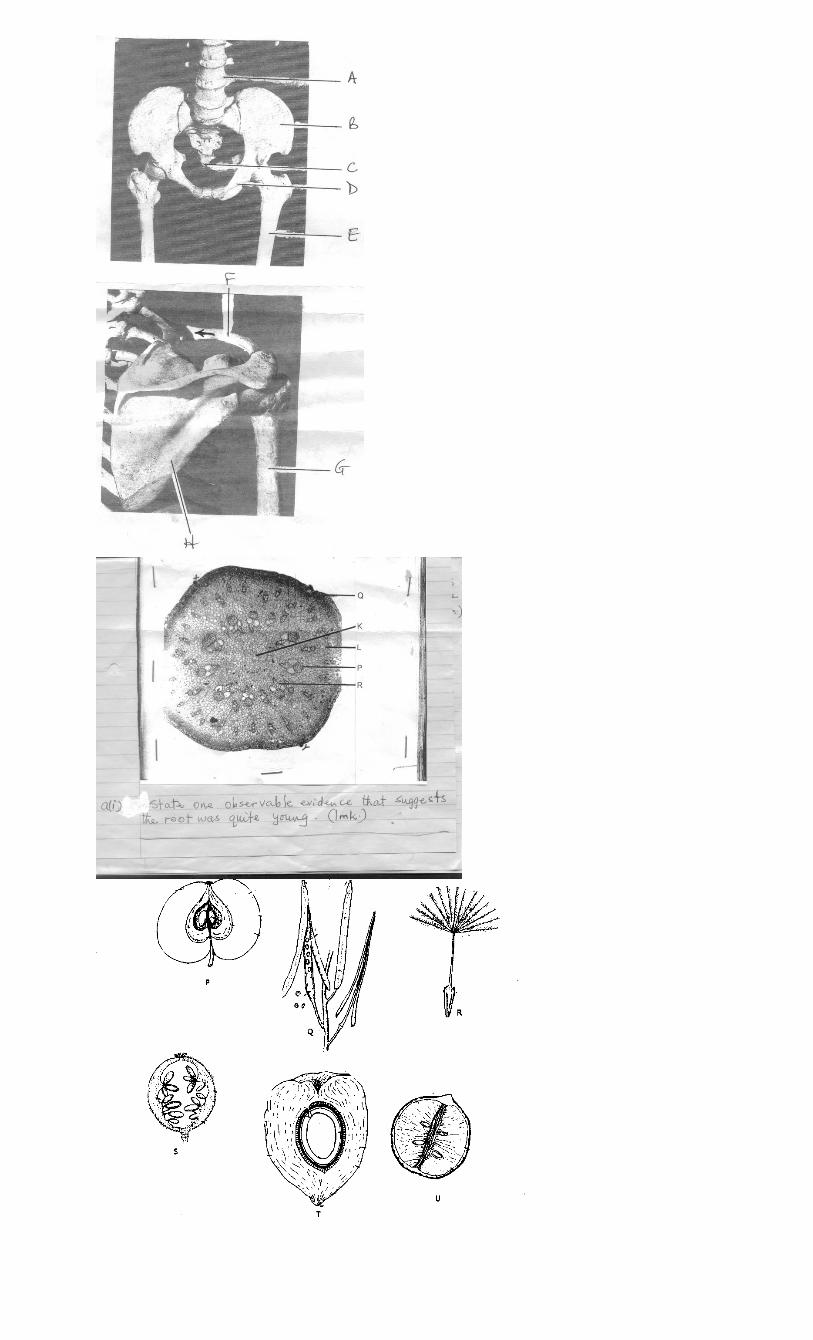
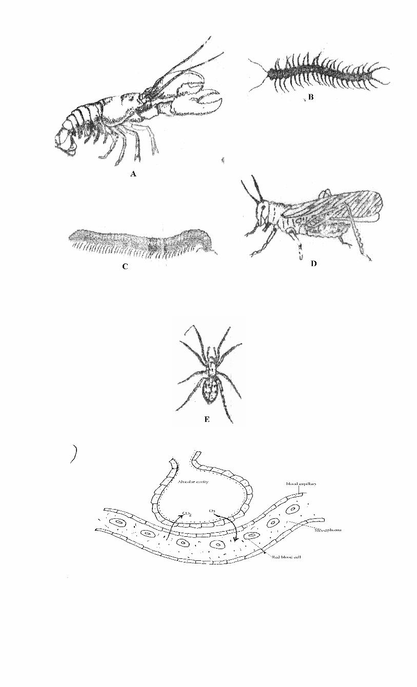
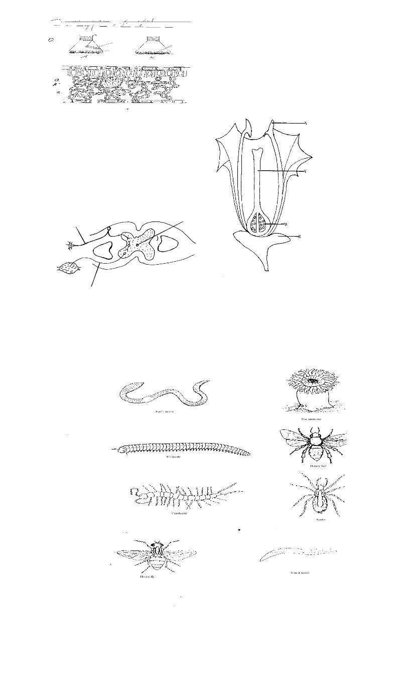
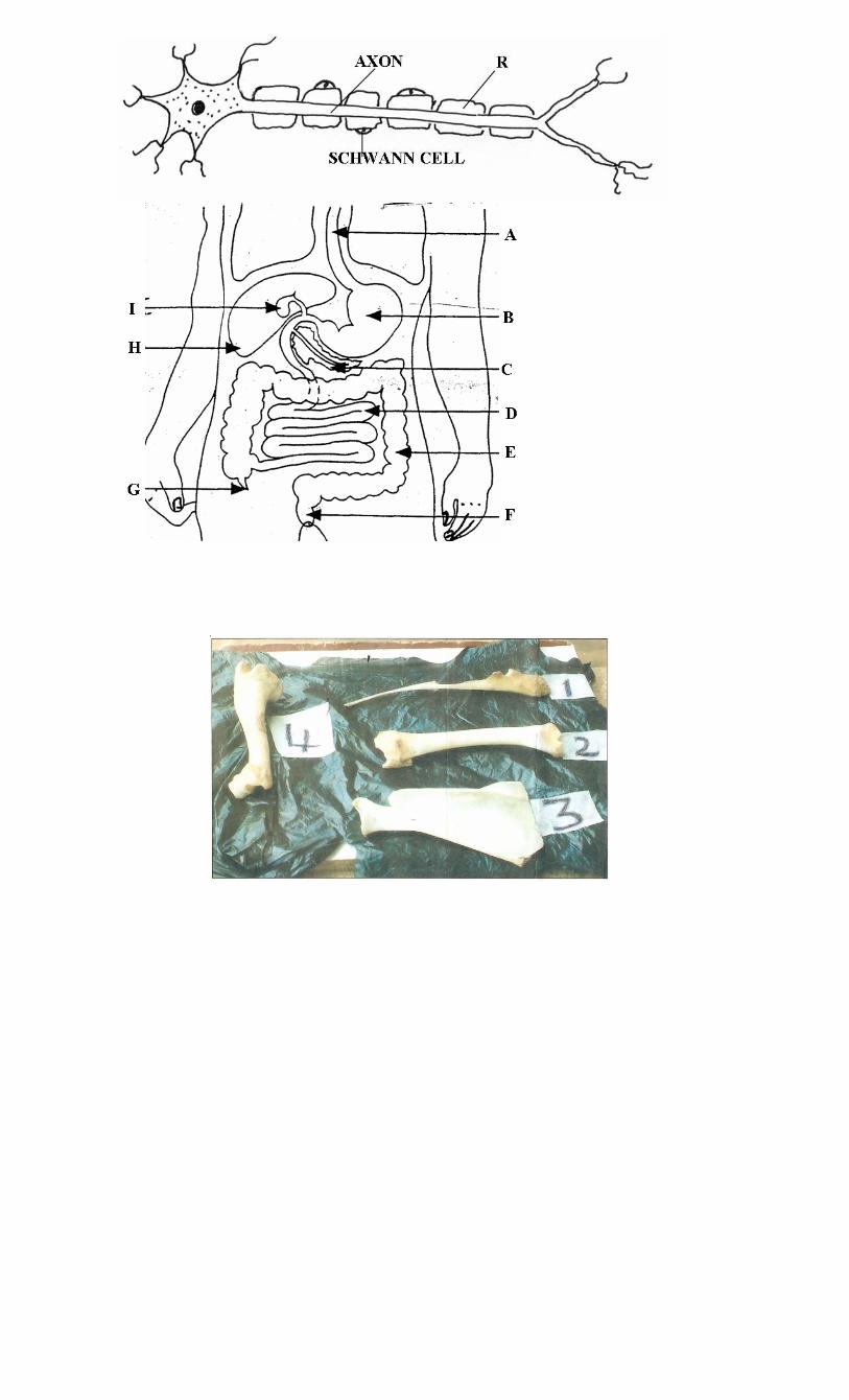
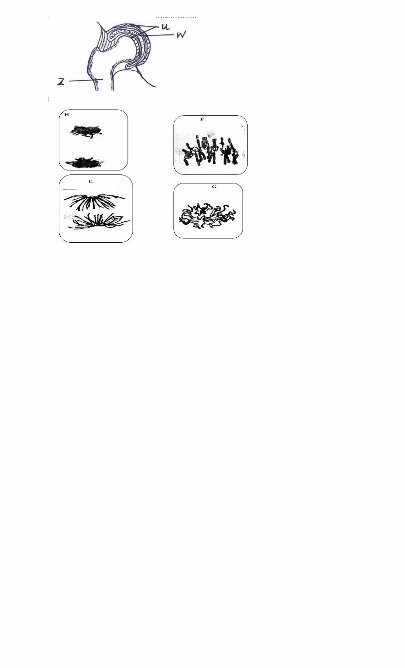
TOPICAL QUESTIONS FOR BIOLOGY
FORM I TOPICS
1. a) Define biology
-the study of life/living things
b) List the branches of biology
- Zoology (study of animals)
- Botany (study of plants)
- Microbiology (study of microorganisms)
c) Explain the importance of biology
- helps to solve environmental problems
- Helps to learn scientific skills
- For entry into other professions/careers
- To apply knowledge to everyday life situations
- To classify organisms into their right groups
- understanding living organisms
d) State the characteristics of living organisms
- feeding/nutrition
- Growth and development
- respiration (to produce energy)
- sensitivity/irritation/response
- excretion (getting rid of metabolic waste material)
- movement/locomotion
- reproduction
e) State the main differences between plants and animals
Animals
Plants
Specialized excretory organs
No specialized excretory
organs
Respond to stimulus quickly
Slow respond to stimulus
All body parts grow
equally(intercalary)
Grow at shoot tip and root
tip only
Move around to look for
food
Stationery
Heterotrophic
Autotrophic
Cells have no cell walls Cells have cell wall made of
cellulose
No chlorophyll Contain chlorophyll
Give parental care to young Plants don’t care for their
young
2. a) i) What is a hand lens?
- Convex lens mounted on a frame and used to magnify
small objects for viewing.
ii) How is a hand lens used?
- place the lens a short distance from the eye
- Bring the object to be viewed near the lens until an
enlarged and clear image can be seen.
ii) When is a hand lens used?
- For reasonably sized objects such as insect wing, leg,
flower parts.
- Cannot be used for small objects such as cells, stomata.
iv) Explain how to calculate drawing magnification
- drawing magnification equals to length of drawing divided by
length of object or image length divided by actual length i.e.
length of drawing or image length
Length of object actual length
b) i) what is classification?
- Orderly arrangement of living organisms into various groups
according to their similarities
ii) List the external features used to classify plants
- rhizoids(e.g. mosses)
- frond (e.g. ferns)
- roots e.g. taproot, fibrous roots, modified roots
- flowers
- leaves
- buds
- seeds
iii) List the external features used to classify animals
- horns e.g. cattle, goat, sheep, deer, gazelle etc
- hooves e.g. cattle, sheep, donkey
- mammary glands e.g. cattle, dog, sheep, cat
- hair e.g. human, cat
- Shell e.g. snail, Tortoise
- spines e.g. hedge hog, porcupine
c) Give the reasons why classification is important
- Placing/grouping living organisms into correct groups called
taxa
- Identification
- arrange information about living organisms into orderly and
sequential manner i.e. it is easy to study organisms in groups
-helps in understanding evolutionary relationships
- monitoring disappearance and appearance of organisms i.e.
predict characteristics of organisms
d) i) Name the taxonomic units of classification in
descending order
- Kingdom (largest unit)
- Phylum (animals)/division (plants)
- Class
- Order
- Family
- Genus
- Species(smallest unit)
ii) What is a species?
- all organisms which can interbreed and give rise to fertile
(viable) offspring
iii) Name the major kingdoms used in classification
-monera
-protoctista/protista
-fungi
-plantae
-animalia
e) i) Define the term binomial nomenclature
- a scientific system of naming organisms using the
generic/genus and specific/species names
- e.g. for humans, Homo sapiens
ii) State the principles followed during binomial
nomenclature
- the first (generic) name should begin with a capital letter
while the rest are small letters
- the two names are printed in italics and if handwritten
should be underlined each separately
iii) Give the advantages of using binomial nomenclature
- no confusion about which organism is referred to
- names are internationally accepted regardless of language
- shows evolutionary relationship hence easy to understand
- useful in naming many species unlike use of common
names
iv) Name the types of classification
- traditional (using common names)
- scientific(using binomial nomenclature)
3. a) i) Define the term cell - it is the basic unit of organization of an organism i.e. the
basic functional and structural unit of an organism.
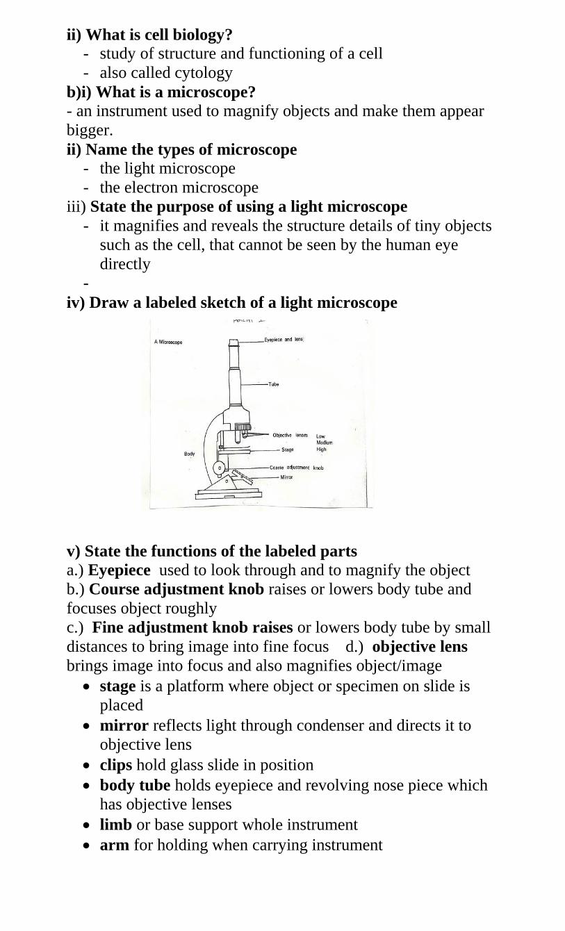
ii) What is cell biology?
- study of structure and functioning of a cell
- also called cytology
b)i) What is a microscope?
- an instrument used to magnify objects and make them appear
bigger.
ii) Name the types of microscope
- the light microscope
- the electron microscope
iii) State the purpose of using a light microscope
- it magnifies and reveals the structure details of tiny objects
such as the cell, that cannot be seen by the human eye
directly
-
iv) Draw a labeled sketch of a light microscope
v) State the functions of the labeled parts
a.) Eyepiece used to look through and to magnify the object
b.) Course adjustment knob raises or lowers body tube and
focuses object roughly
c.) Fine adjustment knob raises or lowers body tube by small
distances to bring image into fine focus d.) objective lens
brings image into focus and also magnifies object/image
stage is a platform where object or specimen on slide is
placed
mirror reflects light through condenser and directs it to
objective lens
clips hold glass slide in position
body tube holds eyepiece and revolving nose piece which
has objective lenses
limb or base support whole instrument
arm for holding when carrying instrument
revolving nose piece holds objective lens in place enabling
change from one objective lens to another
e) i) Explain the procedure followed when using a
microscope
- put the microscope on the bench with the stage facing away
from you (viewer)
- turn the lower power objective to click in line with the
eyepiece
- Ensure that the diaphragm/iris is fully open
- Adjust the mirror until the stage is illuminated with enough
light
- Place the slide containing the specimen on the stage for
magnification
- Draw the image and indicate magnification of the drawing.
ii) State the precautions that are necessary when handling a
microscope
always use two hands when carrying it
never place a microscope too close to the edge of the
bench or table
do not touch the mirror and lens with wet or dirty hands
clean dirty lenses using a special lens cleaning cloth
clean other parts using a soft cloth or tissue paper
low power objective must click into position before and
after use.
Do not wet any part of the microscope
Clean and store well after use
d) i) What is magnification?
- The power of making an image larger
ii) Give the formula used to calculate magnification in a light
microscope
eyepiece lens magnification x objective lens magnification
iii) Give the reasons for each of the following steps when
preparing a cross-section of a stem or leaf for examination
under the microscope
cutting very thin sections
thin sections allow light to pass through making it easy to
observe the tissue
Using a sharp razor blade during the cutting
sharp blade does not damage, deform, destroy or distort the
surface of cell or tissue
it makes thin sections
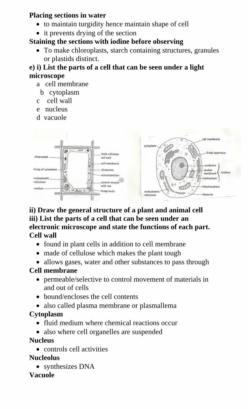
Placing sections in water
to maintain turgidity hence maintain shape of cell
it prevents drying of the section
Staining the sections with iodine before observing
To make chloroplasts, starch containing structures, granules
or plastids distinct.
e) i) List the parts of a cell that can be seen under a light
microscope
a cell membrane
b cytoplasm
c cell wall
e nucleus
d vacuole
ii) Draw the general structure of a plant and animal cell
iii) List the parts of a cell that can be seen under an
electronic microscope and state the functions of each part.
Cell wall
found in plant cells in addition to cell membrane
made of cellulose which makes the plant tough
allows gases, water and other substances to pass through
Cell membrane
permeable/selective to control movement of materials in
and out of cells
bound/encloses the cell contents
also called plasma membrane or plasmallema
Cytoplasm
fluid medium where chemical reactions occur
also where cell organelles are suspended
Nucleus
controls cell activities
Nucleolus
synthesizes DNA
Vacuole
sacs filled with fluid called cell sap
large in plants but small in animals
act as reservoirs for food and harmful wastes which would
otherwise interfere with the metabolism in cytoplasm
Lysosomes
store hydrolytic enzymes
destroy worn out cell organelles, cells, pathogens
digestion of food in unicellular organisms
autolysis
Golgi apparatus
processing/packaging of synthesized materials
transporting/secretion of packaged materials/cell materials
e.g. glycoproteins and mucus
production of lysosomes
Ribosomes
where protein synthesis takes place
Mitochondrion
synthesis of ATP/energy
Chloroplasts
where photosynthesis takes place
Endoplasmic reticulum
transport of cell secretions
can be rough or smooth
iv) State the functions of cell sap
stores chemical substances, sugar, salts
maintains shape of the cell/provides mechanical strength
plays a role in osmoregulation by creating an osmotic
gradient that brings about movement of water
e) Compare plant and animal cells
plant cells have chloroplasts lacking in animals
animal cells have many small vacuoles while plant cells
have a large central vacuole
plant cell have cellulose cell walls lacking in animal cells
cytoplasm in plant cell is in the periphery but in animal cell
it is centrally placed
plants store starch, oil and protein while animals store gats
and glycogen
animal cells have centrioles which plant cells do not have
f) Explain the meaning of each of the following
i) Cell
- Basic unit of organization in an organism
- Specialized animal cells include sperm, ovum muscle
Specialized plant cells include epidermal, guard cell and
palisade cell
ii) Tissue
these are cells of a particular type grouped together to
perform a certain function
animal tissues include epithelium, blood, nerves, muscle,
skeletal and connective tissues
plant tissues include epidermal, photosynthetic, vascular,
strengthening tissues
iii) Organ
tissues combine together to form organs
an organ is a complex structure with a particular function
animal examples include heart, liver, kidney, lungs, brain,
blood vessels, muscles, skeleton
Plant organs include leaves, roots, flowers, and stem.
iv) Organ system
organs are grouped together to form systems also called
organ systems
animal systems include excretory, digestive, respiratory,
nervous, circulatory, endocrine(hormones/glands), skeletal
systems
plant systems include transport system
g) i) Name the structures which are present in plant cells but
absent in animal cells
- Chloroplast
- Cell wall
ii) Name the structures which are present in animal cells but
absent in plant cells
Lysosomes
Centrioles
Pinocytic vesicles
h) Explain how to estimate cell size
i) Materials
cell sizes are measured in units known as micrometers (my)
required is a transparent ruler marked in millimeters
1mµ = 1 mm
1000
ii) Procedure
- Click to low power
place transparent ruler with its millimeter marks on the
stage
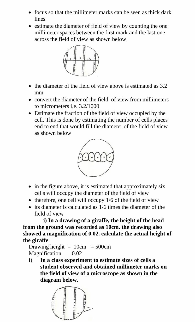
focus so that the millimeter marks can be seen as thick dark
lines
estimate the diameter of field of view by counting the one
millimeter spaces between the first mark and the last one
across the field of view as shown below
the diameter of the field of view above is estimated as 3.2
mm
convert the diameter of the field of view from millimeters
to micrometers i.e. 3.2/1000
Estimate the fraction of the field of view occupied by the
cell. This is done by estimating the number of cells places
end to end that would fill the diameter of the field of view
as shown below
in the figure above, it is estimated that approximately six
cells will occupy the diameter of the field of view
therefore, one cell will occupy 1/6 of the field of view
its diameter is calculated as 1/6 times the diameter of the
field of view
i) In a drawing of a giraffe, the height of the head
from the ground was recorded as 10cm. the drawing also
showed a magnification of 0.02. calculate the actual height of
the giraffe
Drawing height = 10cm = 500cm
Magnification 0.02
i) In a class experiment to estimate sizes of cells a
student observed and obtained millimeter marks on
the field of view of a microscope as shown in the
diagram below.
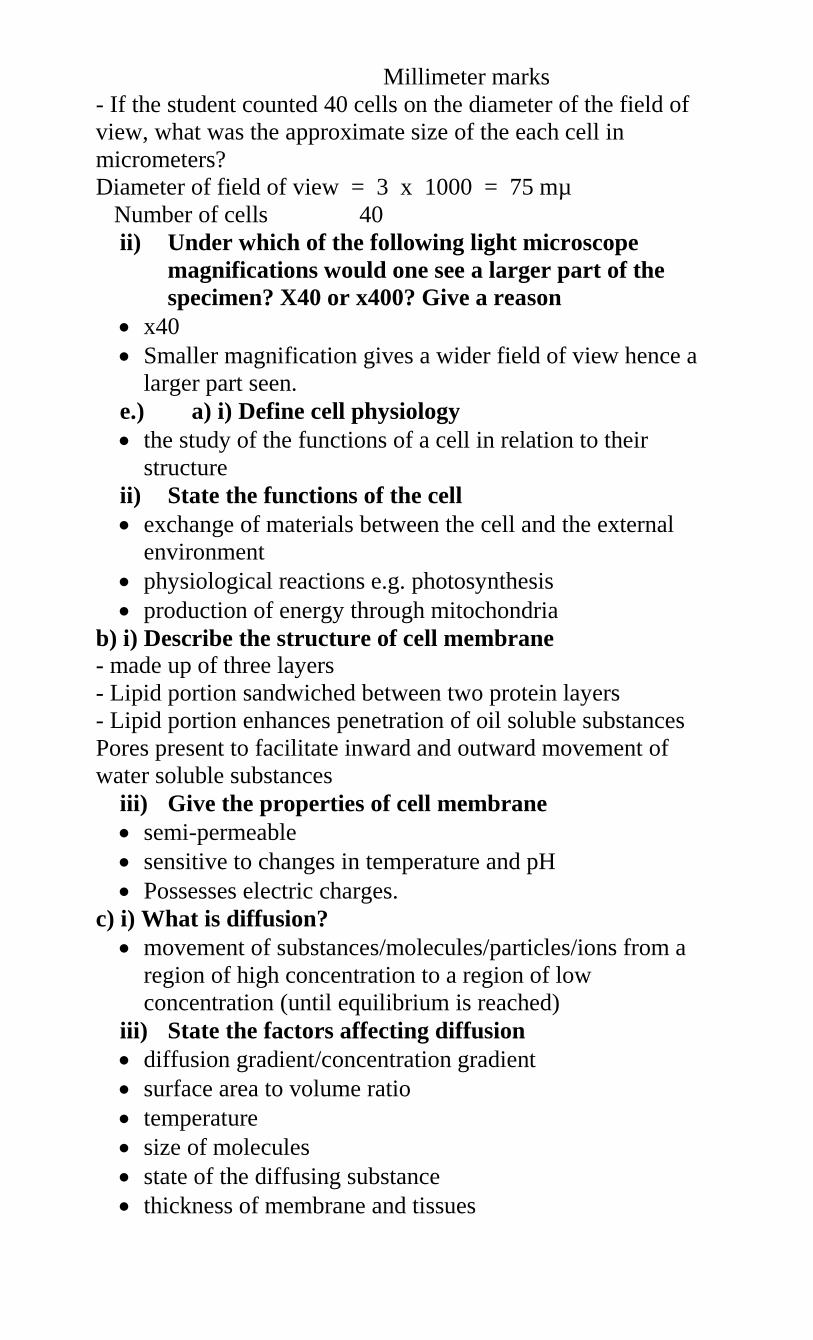
Millimeter marks
- If the student counted 40 cells on the diameter of the field of
view, what was the approximate size of the each cell in
micrometers?
Diameter of field of view = 3 x 1000 = 75 mµ
Number of cells 40
ii) Under which of the following light microscope
magnifications would one see a larger part of the
specimen? X40 or x400? Give a reason
x40
Smaller magnification gives a wider field of view hence a
larger part seen.
e.) a) i) Define cell physiology
the study of the functions of a cell in relation to their
structure
ii) State the functions of the cell
exchange of materials between the cell and the external
environment
physiological reactions e.g. photosynthesis
production of energy through mitochondria
b) i) Describe the structure of cell membrane
- made up of three layers
- Lipid portion sandwiched between two protein layers
- Lipid portion enhances penetration of oil soluble substances
Pores present to facilitate inward and outward movement of
water soluble substances
iii) Give the properties of cell membrane
semi-permeable
sensitive to changes in temperature and pH
Possesses electric charges.
c) i) What is diffusion?
movement of substances/molecules/particles/ions from a
region of high concentration to a region of low
concentration (until equilibrium is reached)
iii) State the factors affecting diffusion
diffusion gradient/concentration gradient
surface area to volume ratio
temperature
size of molecules
state of the diffusing substance
thickness of membrane and tissues
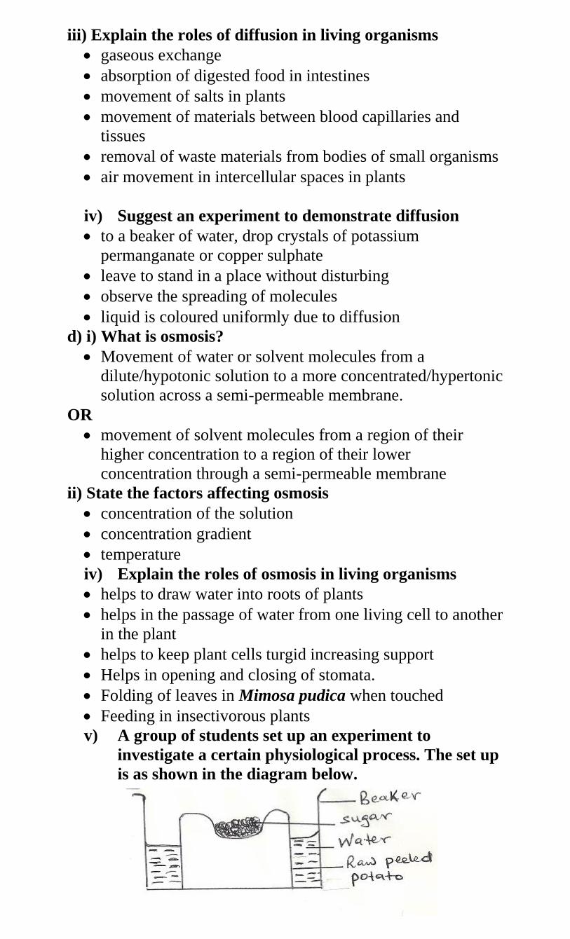
iii) Explain the roles of diffusion in living organisms
gaseous exchange
absorption of digested food in intestines
movement of salts in plants
movement of materials between blood capillaries and
tissues
removal of waste materials from bodies of small organisms
air movement in intercellular spaces in plants
iv) Suggest an experiment to demonstrate diffusion
to a beaker of water, drop crystals of potassium
permanganate or copper sulphate
leave to stand in a place without disturbing
observe the spreading of molecules
liquid is coloured uniformly due to diffusion
d) i) What is osmosis?
Movement of water or solvent molecules from a
dilute/hypotonic solution to a more concentrated/hypertonic
solution across a semi-permeable membrane.
OR
movement of solvent molecules from a region of their
higher concentration to a region of their lower
concentration through a semi-permeable membrane
ii) State the factors affecting osmosis
concentration of the solution
concentration gradient
temperature
iv) Explain the roles of osmosis in living organisms
helps to draw water into roots of plants
helps in the passage of water from one living cell to another
in the plant
helps to keep plant cells turgid increasing support
Helps in opening and closing of stomata.
Folding of leaves in Mimosa pudica when touched
Feeding in insectivorous plants
v) A group of students set up an experiment to
investigate a certain physiological process. The set up
is as shown in the diagram below.
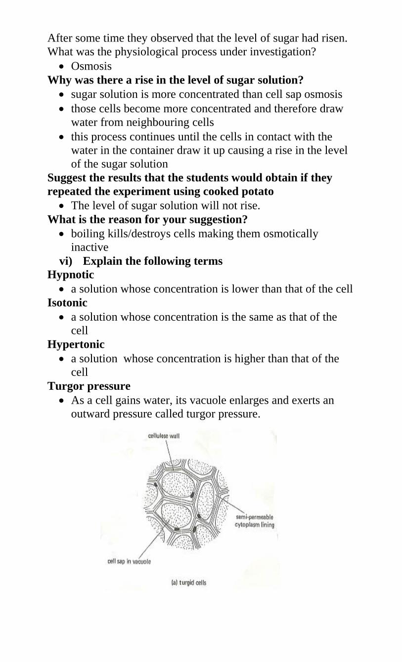
After some time they observed that the level of sugar had risen.
What was the physiological process under investigation?
Osmosis
Why was there a rise in the level of sugar solution?
sugar solution is more concentrated than cell sap osmosis
those cells become more concentrated and therefore draw
water from neighbouring cells
this process continues until the cells in contact with the
water in the container draw it up causing a rise in the level
of the sugar solution
Suggest the results that the students would obtain if they
repeated the experiment using cooked potato
The level of sugar solution will not rise.
What is the reason for your suggestion?
boiling kills/destroys cells making them osmotically
inactive
vi) Explain the following terms
Hypnotic
a solution whose concentration is lower than that of the cell
Isotonic
a solution whose concentration is the same as that of the
cell
Hypertonic
a solution whose concentration is higher than that of the
cell
Turgor pressure
As a cell gains water, its vacuole enlarges and exerts an
outward pressure called turgor pressure.
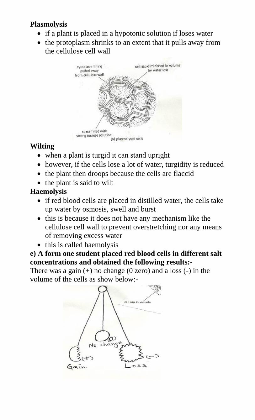
Plasmolysis
if a plant is placed in a hypotonic solution if loses water
the protoplasm shrinks to an extent that it pulls away from
the cellulose cell wall
Wilting
when a plant is turgid it can stand upright
however, if the cells lose a lot of water, turgidity is reduced
the plant then droops because the cells are flaccid
the plant is said to wilt
Haemolysis
if red blood cells are placed in distilled water, the cells take
up water by osmosis, swell and burst
this is because it does not have any mechanism like the
cellulose cell wall to prevent overstretching nor any means
of removing excess water
this is called haemolysis
e) A form one student placed red blood cells in different salt
concentrations and obtained the following results:-
There was a gain (+) no change (0 zero) and a loss (-) in the
volume of the cells as show below:-
Briefly explain the results of the experiment
in the first solution , red blood cell absorbed water by
osmosis, swell and burst (haemolysis) hence the solution is
hypotonic
in the second solution, there was no change in size or
structure as it was isotonic hence no osmotic gradient
in the third solution the red blood cell lost water to shrink
hence became crenated as the solution was hypotonic to
the cell cytoplasm.
f) i) What is active transport?
movement of molecules and ions against a concentration
gradient
the substances move from a lower to a higher concentration
gradient by use of energy
ii) State the factors affecting active transport
oxygen concentration
temperature
change in pH
glucose concentration
enzyme inhibitors
iii) Why is oxygen important in the process of active
transport?
- Oxygen is required for respiration, which produces energy
necessary for the process to occur.
2.0 ) the factors that affect the rates of the following process
in living organisms.
a.) DIFFUSION.
- Diffusion gradient which refers to the difference in
concentration of molecules between the region of high
concentration and the region of low concentration. Increasing
the concentration gradient causes an increase in rate of diffusion
and vice versa.
-Surface area to volume ratio .is the ratio of total surface area
exposed by an organism compared to its body volume. Small
sized living organisms have a large surface area to volume ratio.
The larger the surface area to volume ratio ,the high the rate of
diffusion and vice versa. Small organisms like amoeba and
paramecium can hence rely on diffusion for transport of
substances into and within its body and removal of waste
products
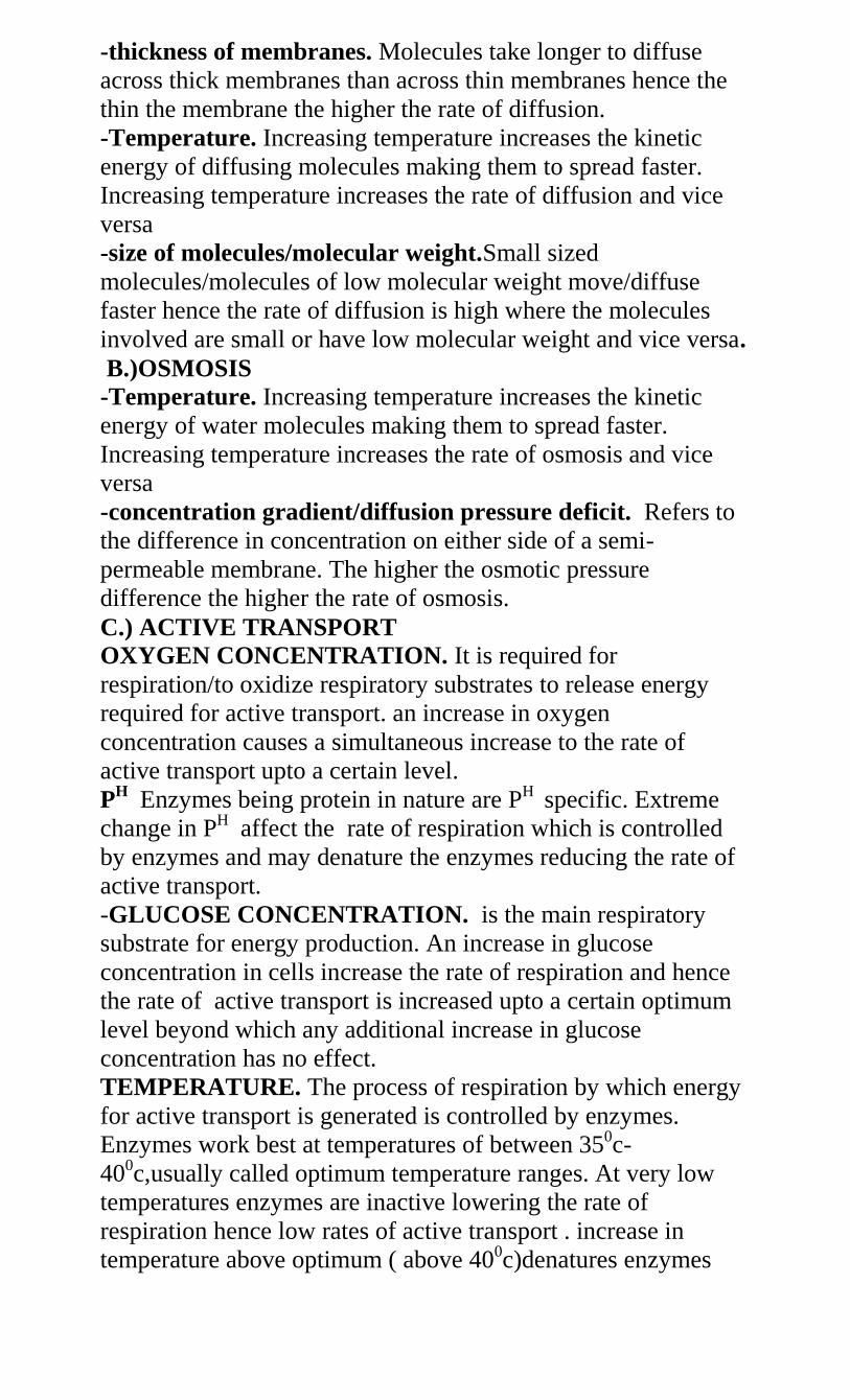
-thickness of membranes. Molecules take longer to diffuse
across thick membranes than across thin membranes hence the
thin the membrane the higher the rate of diffusion.
-Temperature. Increasing temperature increases the kinetic
energy of diffusing molecules making them to spread faster.
Increasing temperature increases the rate of diffusion and vice
versa
-size of molecules/molecular weight.Small sized
molecules/molecules of low molecular weight move/diffuse
faster hence the rate of diffusion is high where the molecules
involved are small or have low molecular weight and vice versa.
B.)OSMOSIS
-Temperature. Increasing temperature increases the kinetic
energy of water molecules making them to spread faster.
Increasing temperature increases the rate of osmosis and vice
versa
-concentration gradient/diffusion pressure deficit. Refers to
the difference in concentration on either side of a semi-
permeable membrane. The higher the osmotic pressure
difference the higher the rate of osmosis.
C.) ACTIVE TRANSPORT
OXYGEN CONCENTRATION. It is required for
respiration/to oxidize respiratory substrates to release energy
required for active transport. an increase in oxygen
concentration causes a simultaneous increase to the rate of
active transport upto a certain level.
PH Enzymes being protein in nature are PH specific. Extreme
change in PH affect the rate of respiration which is controlled
by enzymes and may denature the enzymes reducing the rate of
active transport.
-GLUCOSE CONCENTRATION. is the main respiratory
substrate for energy production. An increase in glucose
concentration in cells increase the rate of respiration and hence
the rate of active transport is increased upto a certain optimum
level beyond which any additional increase in glucose
concentration has no effect.
TEMPERATURE. The process of respiration by which energy
for active transport is generated is controlled by enzymes.
Enzymes work best at temperatures of between 350c-
400c,usually called optimum temperature ranges. At very low
temperatures enzymes are inactive lowering the rate of
respiration hence low rates of active transport . increase in
temperature above optimum ( above 400c)denatures enzymes
slowing down respiration and ac tive transport until it finally
stops.
ENZYMES INHIBITORS. They are substances which slow
down (by competing with the enzyme for the active sites in the
substrate) or stop ( by blocking the active sites of the enzyme)
the activity/funtioning of enzymes .this slows down or stops
respiration and so is active transport.
CONCENTRATION OF CARRIER MOLECULES IN THE
CELL MEMBRANE. They are substances that bind to the ions
being transported actively and carrying them across the
membrane . increase in concentration of carrier molecules
increases the rate of active transport upto a certain level and vice
versa.
4.) Explain briefly the role of osmosis in living tissues.
In plants:
Osmosis facilitates the absorption of water from the soil by plant
roots, water is required for the process of photosynthesis.
Turgidity of cells contributes to support in herbaceous plants
and helps plant to maintain shape.
Helps in closing and opening of stomata regulating the process
of gaseous exchange and transpiration.
It facilitates feeding in insectivorous plants like venus fly trap.
In animals:
Enables reabsorption of water from the kidney tubules back to
blood stream facilitating the process of osmoregulation.
It enables organisms in fresh water bodies like amoeba to absorb
water.
it is applied in food preservation.
5.) explain what happens when plant and animal cells are put in
hypotonic and hypertonic solutions.
a. i)plant cells in hypotonic solution.
The concentration of the plant cell sap is hypertonic to the
solution/water medium. the cell draws in water by osmosis
through the cell wall, cell membrane into the cell cytoplasm.
Water enters the cell vacuole by osmosis; it enlarges and exerts
an outward pressure on the cell wall called turgor pressure.
Increased turgor pressure pushes the cell cytoplasm against the
cell wall until the cell wall cannot stretch any further. The cell
becomes firm or rigid and is said to be turgid. As the cell wall is
being stretched outwards, it develops a resistant inwards
pressure that is equal and opposite to the turgor pressure and this
is called wall pressure.
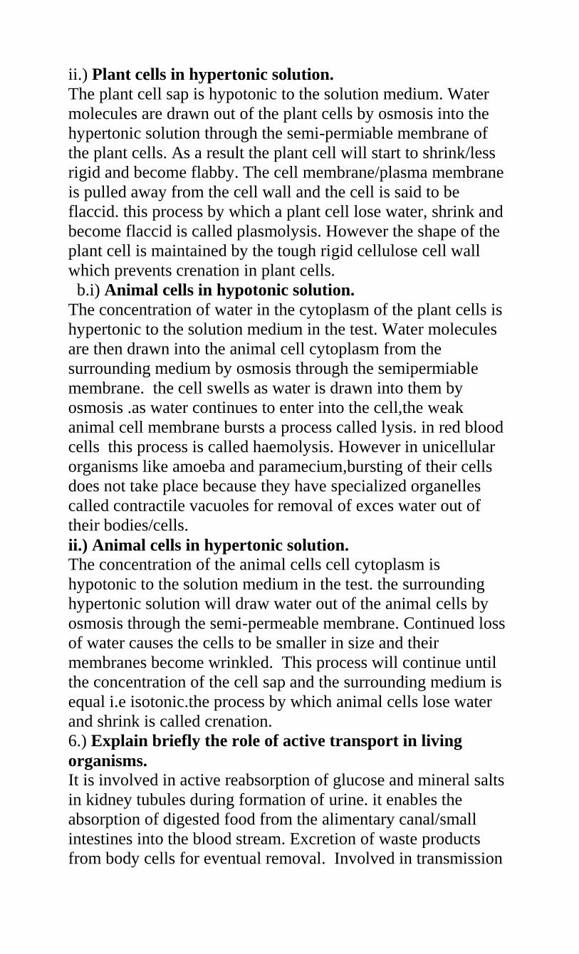
ii.) Plant cells in hypertonic solution.
The plant cell sap is hypotonic to the solution medium. Water
molecules are drawn out of the plant cells by osmosis into the
hypertonic solution through the semi-permiable membrane of
the plant cells. As a result the plant cell will start to shrink/less
rigid and become flabby. The cell membrane/plasma membrane
is pulled away from the cell wall and the cell is said to be
flaccid. this process by which a plant cell lose water, shrink and
become flaccid is called plasmolysis. However the shape of the
plant cell is maintained by the tough rigid cellulose cell wall
which prevents crenation in plant cells.
b.i) Animal cells in hypotonic solution.
The concentration of water in the cytoplasm of the plant cells is
hypertonic to the solution medium in the test. Water molecules
are then drawn into the animal cell cytoplasm from the
surrounding medium by osmosis through the semipermiable
membrane. the cell swells as water is drawn into them by
osmosis .as water continues to enter into the cell,the weak
animal cell membrane bursts a process called lysis. in red blood
cells this process is called haemolysis. However in unicellular
organisms like amoeba and paramecium,bursting of their cells
does not take place because they have specialized organelles
called contractile vacuoles for removal of exces water out of
their bodies/cells.
ii.) Animal cells in hypertonic solution.
The concentration of the animal cells cell cytoplasm is
hypotonic to the solution medium in the test. the surrounding
hypertonic solution will draw water out of the animal cells by
osmosis through the semi-permeable membrane. Continued loss
of water causes the cells to be smaller in size and their
membranes become wrinkled. This process will continue until
the concentration of the cell sap and the surrounding medium is
equal i.e isotonic.the process by which animal cells lose water
and shrink is called crenation.
6.) Explain briefly the role of active transport in living
organisms.
It is involved in active reabsorption of glucose and mineral salts
in kidney tubules during formation of urine. it enables the
absorption of digested food from the alimentary canal/small
intestines into the blood stream. Excretion of waste products
from body cells for eventual removal. Involved in transmission
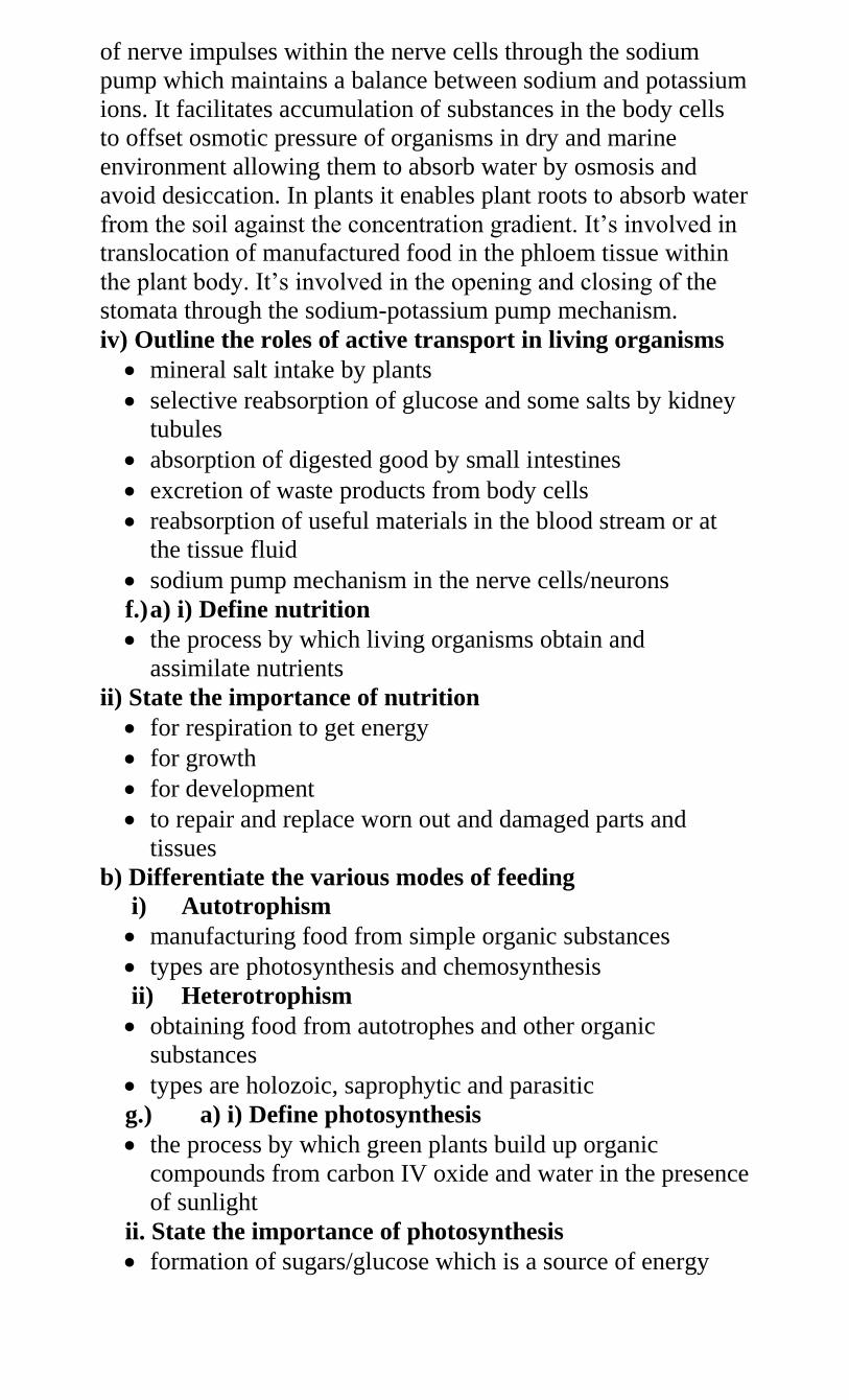
of nerve impulses within the nerve cells through the sodium
pump which maintains a balance between sodium and potassium
ions. It facilitates accumulation of substances in the body cells
to offset osmotic pressure of organisms in dry and marine
environment allowing them to absorb water by osmosis and
avoid desiccation. In plants it enables plant roots to absorb water
from the soil against the concentration gradient. It’s involved in
translocation of manufactured food in the phloem tissue within
the plant body. It’s involved in the opening and closing of the
stomata through the sodium-potassium pump mechanism.
iv) Outline the roles of active transport in living organisms
mineral salt intake by plants
selective reabsorption of glucose and some salts by kidney
tubules
absorption of digested good by small intestines
excretion of waste products from body cells
reabsorption of useful materials in the blood stream or at
the tissue fluid
sodium pump mechanism in the nerve cells/neurons
f.) a) i) Define nutrition
the process by which living organisms obtain and
assimilate nutrients
ii) State the importance of nutrition
for respiration to get energy
for growth
for development
to repair and replace worn out and damaged parts and
tissues
b) Differentiate the various modes of feeding
i) Autotrophism
manufacturing food from simple organic substances
types are photosynthesis and chemosynthesis
ii) Heterotrophism
obtaining food from autotrophes and other organic
substances
types are holozoic, saprophytic and parasitic
g.) a) i) Define photosynthesis
the process by which green plants build up organic
compounds from carbon IV oxide and water in the presence
of sunlight
ii. State the importance of photosynthesis
formation of sugars/glucose which is a source of energy
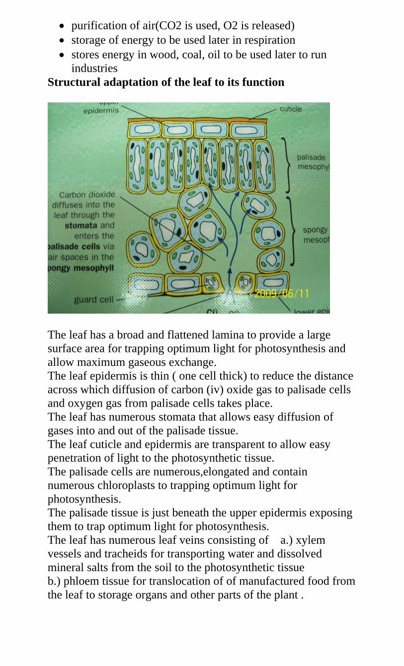
purification of air(CO2 is used, O2 is released)
storage of energy to be used later in respiration
stores energy in wood, coal, oil to be used later to run
industries
Structural adaptation of the leaf to its function
The leaf has a broad and flattened lamina to provide a large
surface area for trapping optimum light for photosynthesis and
allow maximum gaseous exchange.
The leaf epidermis is thin ( one cell thick) to reduce the distance
across which diffusion of carbon (iv) oxide gas to palisade cells
and oxygen gas from palisade cells takes place.
The leaf has numerous stomata that allows easy diffusion of
gases into and out of the palisade tissue.
The leaf cuticle and epidermis are transparent to allow easy
penetration of light to the photosynthetic tissue.
The palisade cells are numerous,elongated and contain
numerous chloroplasts to trapping optimum light for
photosynthesis.
The palisade tissue is just beneath the upper epidermis exposing
them to trap optimum light for photosynthesis.
The leaf has numerous leaf veins consisting of a.) xylem
vessels and tracheids for transporting water and dissolved
mineral salts from the soil to the photosynthetic tissue
b.) phloem tissue for translocation of of manufactured food from
the leaf to storage organs and other parts of the plant .
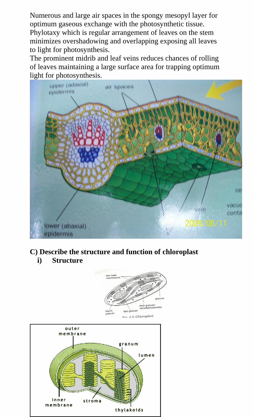
Numerous and large air spaces in the spongy mesopyl layer for
optimum gaseous exchange with the photosynthetic tissue.
Phylotaxy which is regular arrangement of leaves on the stem
minimizes overshadowing and overlapping exposing all leaves
to light for photosynthesis.
The prominent midrib and leaf veins reduces chances of rolling
of leaves maintaining a large surface area for trapping optimum
light for photosynthesis.
C) Describe the structure and function of chloroplast
i) Structure
ii) Function
structure in which photosynthesis takes place
iii) Adaptations
has numerous/many grana to provide large surface area for
packing many chlorophyll pigments
have numerous chlorophyll pigments which trap
sunlight/light for photosynthesis
has stroma/third matrix which contain certain enzymes that
catalyze photosynthetic reactions
d) i) Give a word equation for photosynthesis
Carbon (iv)Oxide + water sunlight sugar + oxygen
Chlorophyll
NB 6CO2 +6H2O C6H1206 + 6O2
.) Describe briefly the process of photosynthesis in plants.
The process of photosynthesis takes place in green plants
allowing them to make their own food. The process is controlled
by enzymes and involves a series of reactions that take place in
chloroplasts. The raw materials required are water and carbon
(iv) oxide.the process takes place in two consecutive stages i.e
Light reaction stage
It’s also called the light dependent stage as it requires light
energy . the reactions take place in the granna of the chloroplast.
light energy from the sun is trapped by chlorophyll in the
chloroplast and converted into chemical energy. This energy
splits water molecules into hydrogen ions and oxygen atoms a
process is called photolysis. The oxygen atoms are released as
aby product or used up in the process of respiration. The
hydrogen ions formed are used in the dark stage of
photosynthesis.
Water hydrogen ions + oxygen atoms
2H2O 4H+ + o2 g
Some of the light enrgy is used to combine a molecule called
adenosine diphosphate(ADP) with a phosphate group to form
the rich energy molecules called adenosine Tri-Phosphate(ATP)
ADP + P ATP
Dark reaction stage
It’s also called the light independent stage of photosynthesis
since light is not required because it can take place both in
presence and absence of light. the reactions are controlled by
enzymes. the hydrogen atoms released in the light stage are
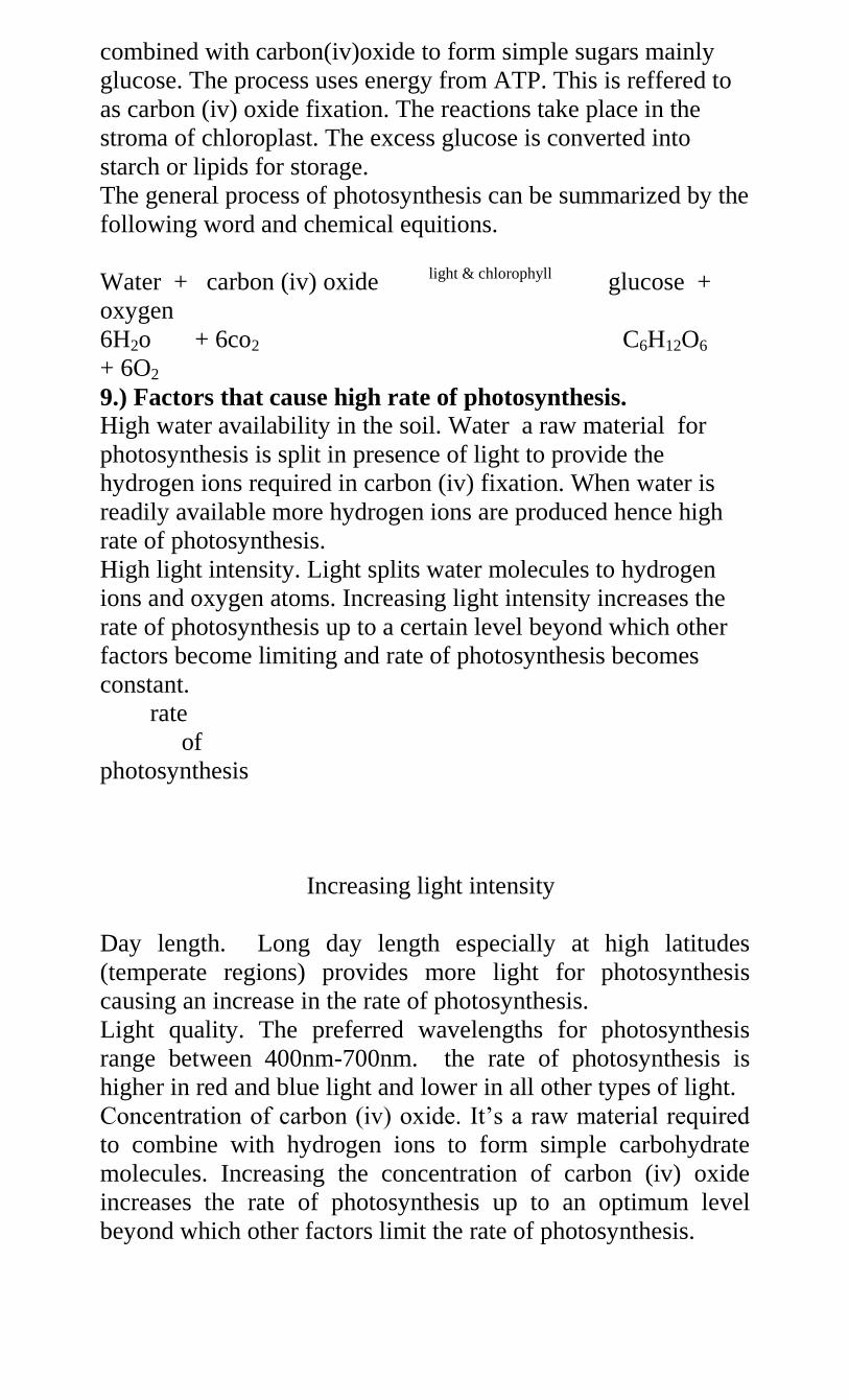
combined with carbon(iv)oxide to form simple sugars mainly
glucose. The process uses energy from ATP. This is reffered to
as carbon (iv) oxide fixation. The reactions take place in the
stroma of chloroplast. The excess glucose is converted into
starch or lipids for storage.
The general process of photosynthesis can be summarized by the
following word and chemical equitions.
Water + carbon (iv) oxide light & chlorophyll glucose +
oxygen
6H2o + 6co2 C6H12O6
+ 6O2
9.) Factors that cause high rate of photosynthesis.
High water availability in the soil. Water a raw material for
photosynthesis is split in presence of light to provide the
hydrogen ions required in carbon (iv) fixation. When water is
readily available more hydrogen ions are produced hence high
rate of photosynthesis.
High light intensity. Light splits water molecules to hydrogen
ions and oxygen atoms. Increasing light intensity increases the
rate of photosynthesis up to a certain level beyond which other
factors become limiting and rate of photosynthesis becomes
constant.
rate
of
photosynthesis
Increasing light intensity
Day length. Long day length especially at high latitudes
(temperate regions) provides more light for photosynthesis
causing an increase in the rate of photosynthesis.
Light quality. The preferred wavelengths for photosynthesis
range between 400nm-700nm. the rate of photosynthesis is
higher in red and blue light and lower in all other types of light.
Concentration of carbon (iv) oxide. It’s a raw material required
to combine with hydrogen ions to form simple carbohydrate
molecules. Increasing the concentration of carbon (iv) oxide
increases the rate of photosynthesis up to an optimum level
beyond which other factors limit the rate of photosynthesis.
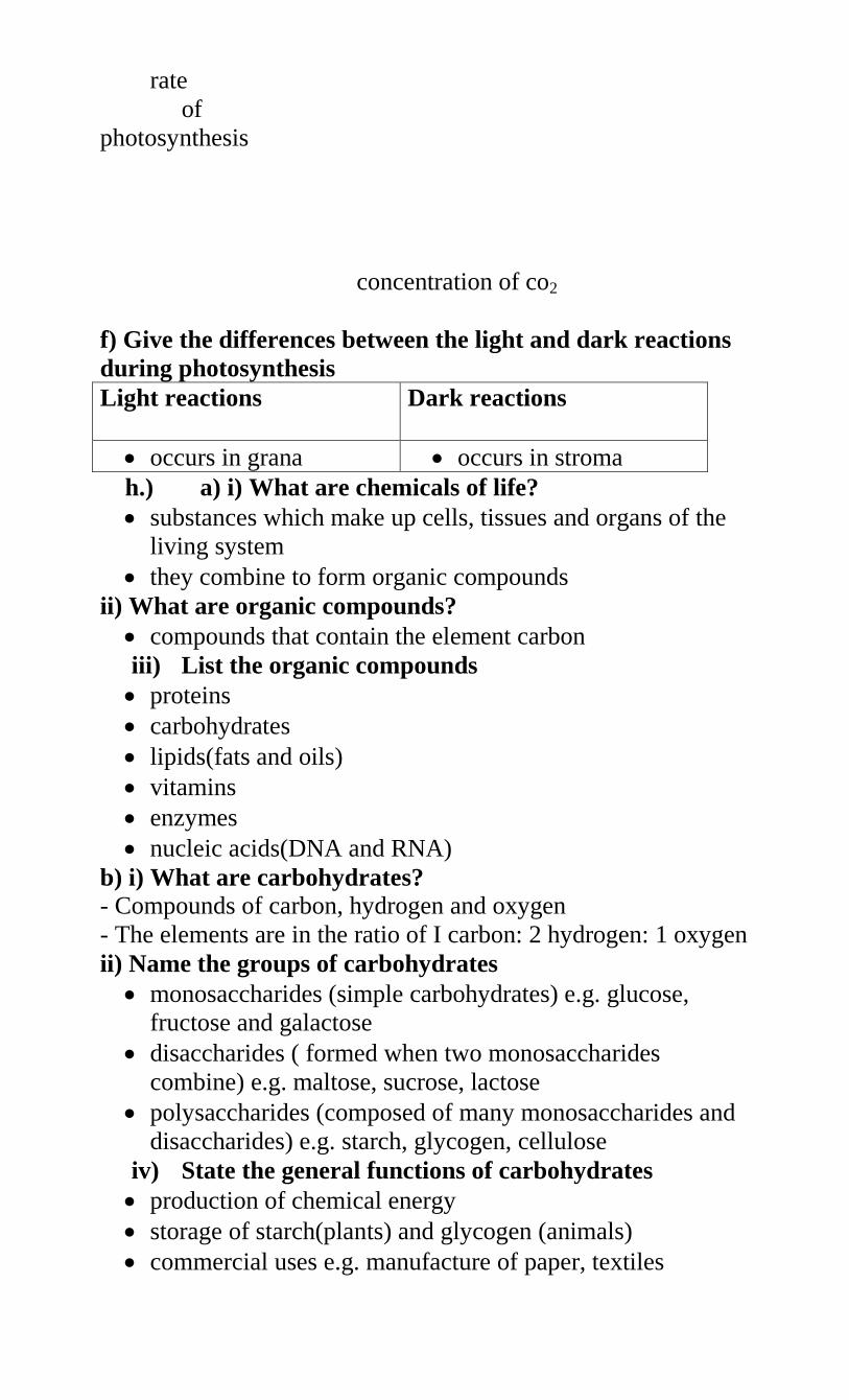
rate
of
photosynthesis
concentration of co2
f) Give the differences between the light and dark reactions
during photosynthesis
Light reactions Dark reactions
occurs in grana occurs in stroma
h.) a) i) What are chemicals of life?
substances which make up cells, tissues and organs of the
living system
they combine to form organic compounds
ii) What are organic compounds?
compounds that contain the element carbon
iii) List the organic compounds
proteins
carbohydrates
lipids(fats and oils)
vitamins
enzymes
nucleic acids(DNA and RNA)
b) i) What are carbohydrates?
- Compounds of carbon, hydrogen and oxygen
- The elements are in the ratio of I carbon: 2 hydrogen: 1 oxygen
ii) Name the groups of carbohydrates
monosaccharides (simple carbohydrates) e.g. glucose,
fructose and galactose
disaccharides ( formed when two monosaccharides
combine) e.g. maltose, sucrose, lactose
polysaccharides (composed of many monosaccharides and
disaccharides) e.g. starch, glycogen, cellulose
iv) State the general functions of carbohydrates
production of chemical energy
storage of starch(plants) and glycogen (animals)
commercial uses e.g. manufacture of paper, textiles
c) i) what are proteins?
compounds of carbon, hydrogen and oxygen and in
addition nitrogen, and sometimes sulphur and or
phosphorus
building blocks are called amino acids
ii) Name the types of amino acids
essential amino acids which must be supplied in food since
they body cannot synthesize them
Non-essential amino acids which body can synthesize.
iv) State the classes of proteins
first class proteins which supply all the essential amino
acids
second class proteins which lack at least one amino acid
v) Give the functions of proteins
structural compounds e.g. muscles, hair, hooves, and
feathers
as enzymes e.g. pepsin, trypsin
hormones e.g. insulin and glucagons
antibodies
part of haemoglobin molecule
actin and myosin in muscles
collagen in bones and cartilage
pigments in rods and cones for coordination
components of blood i.e. plasma proteins
d ) i) What are lipids
- Fats and oils
- They contain carbon, hydrogen and oxygen
- However, they contain a higher proportion of carbon and
hydrogen but less oxygen that in carbohydrates
ii) Name the types of lipids
oils(liquid under room temperature)
fats (solid under room temperature)
iii) What are the building blocks of lipids?
fatty acids and glycerol
v) State the functions of lipids
production of energy
source of metabolic water
structural compound
e) i) What are enzymes?
a chemical compound, protein in nature, which acts as a
biological catalyst
ii) State the properties of enzymes
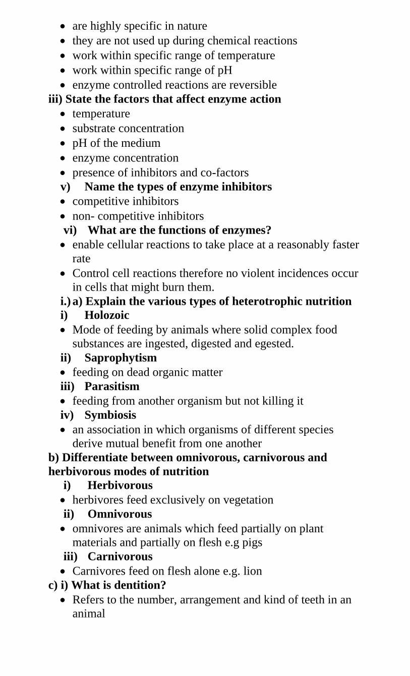
are highly specific in nature
they are not used up during chemical reactions
work within specific range of temperature
work within specific range of pH
enzyme controlled reactions are reversible
iii) State the factors that affect enzyme action
temperature
substrate concentration
pH of the medium
enzyme concentration
presence of inhibitors and co-factors
v) Name the types of enzyme inhibitors
competitive inhibitors
non- competitive inhibitors
vi) What are the functions of enzymes?
enable cellular reactions to take place at a reasonably faster
rate
Control cell reactions therefore no violent incidences occur
in cells that might burn them.
i.) a) Explain the various types of heterotrophic nutrition
i) Holozoic
Mode of feeding by animals where solid complex food
substances are ingested, digested and egested.
ii) Saprophytism
feeding on dead organic matter
iii) Parasitism
feeding from another organism but not killing it
iv) Symbiosis
an association in which organisms of different species
derive mutual benefit from one another
b) Differentiate between omnivorous, carnivorous and
herbivorous modes of nutrition
i) Herbivorous
herbivores feed exclusively on vegetation
ii) Omnivorous
omnivores are animals which feed partially on plant
materials and partially on flesh e.g pigs
iii) Carnivorous
Carnivores feed on flesh alone e.g. lion
c) i) What is dentition?
Refers to the number, arrangement and kind of teeth in an
animal
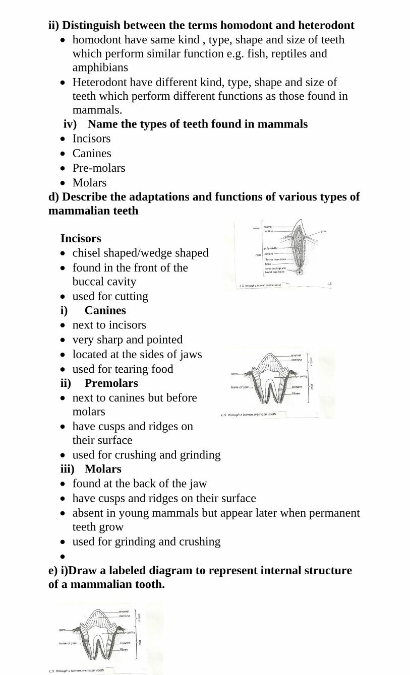
ii) Distinguish between the terms homodont and heterodont
homodont have same kind , type, shape and size of teeth
which perform similar function e.g. fish, reptiles and
amphibians
Heterodont have different kind, type, shape and size of
teeth which perform different functions as those found in
mammals.
iv) Name the types of teeth found in mammals
Incisors
Canines
Pre-molars
Molars
d) Describe the adaptations and functions of various types of
mammalian teeth
Incisors
chisel shaped/wedge shaped
found in the front of the
buccal cavity
used for cutting
i) Canines
next to incisors
very sharp and pointed
located at the sides of jaws
used for tearing food
ii) Premolars
next to canines but before
molars
have cusps and ridges on
their surface
used for crushing and grinding
iii) Molars
found at the back of the jaw
have cusps and ridges on their surface
absent in young mammals but appear later when permanent
teeth grow
used for grinding and crushing
e) i)Draw a labeled diagram to represent internal structure
of a mammalian tooth.
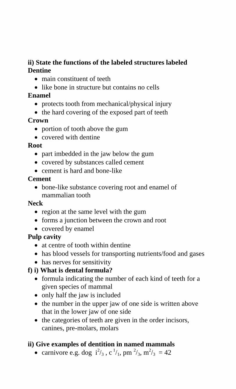
ii) State the functions of the labeled structures labeled
Dentine
main constituent of teeth
like bone in structure but contains no cells
Enamel
protects tooth from mechanical/physical injury
the hard covering of the exposed part of teeth
Crown
portion of tooth above the gum
covered with dentine
Root
part imbedded in the jaw below the gum
covered by substances called cement
cement is hard and bone-like
Cement
bone-like substance covering root and enamel of
mammalian tooth
Neck
region at the same level with the gum
forms a junction between the crown and root
covered by enamel
Pulp cavity
at centre of tooth within dentine
has blood vessels for transporting nutrients/food and gases
has nerves for sensitivity
f) i) What is dental formula?
formula indicating the number of each kind of teeth for a
given species of mammal
only half the jaw is included
the number in the upper jaw of one side is written above
that in the lower jaw of one side
the categories of teeth are given in the order incisors,
canines, pre-molars, molars
ii) Give examples of dentition in named mammals
carnivore e.g. dog i2/3 , c 1/1, pm 2/3, m
2/3 = 42
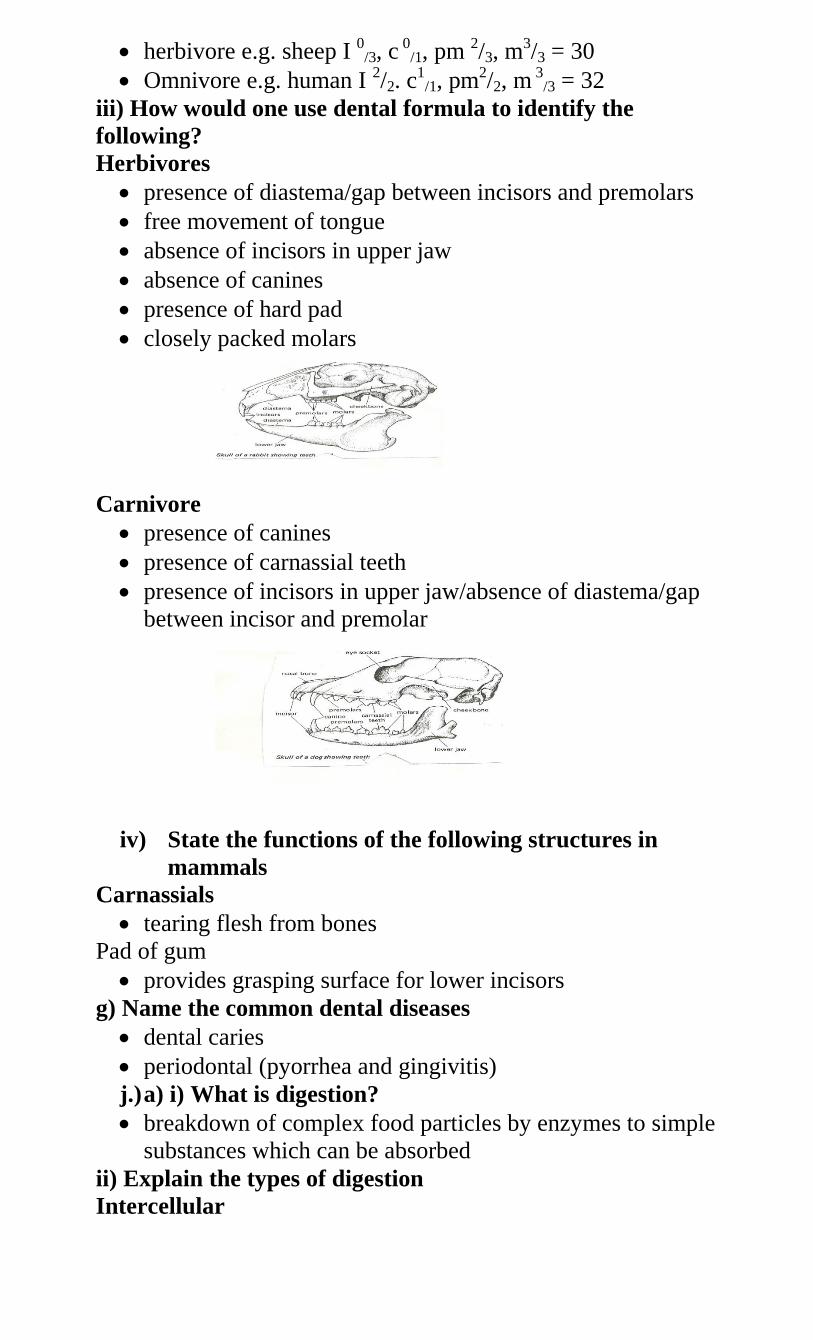
herbivore e.g. sheep I 0/3, c 0
/1, pm 2/3, m3/3 = 30
Omnivore e.g. human I 2/2. c1
/1, pm2/2, m 3
/3 = 32
iii) How would one use dental formula to identify the
following?
Herbivores
presence of diastema/gap between incisors and premolars
free movement of tongue
absence of incisors in upper jaw
absence of canines
presence of hard pad
closely packed molars
Carnivore
presence of canines
presence of carnassial teeth
presence of incisors in upper jaw/absence of diastema/gap
between incisor and premolar
iv) State the functions of the following structures in
mammals
Carnassials
tearing flesh from bones
Pad of gum
provides grasping surface for lower incisors
g) Name the common dental diseases
dental caries
periodontal (pyorrhea and gingivitis)
j.) a) i) What is digestion?
breakdown of complex food particles by enzymes to simple
substances which can be absorbed
ii) Explain the types of digestion
Intercellular
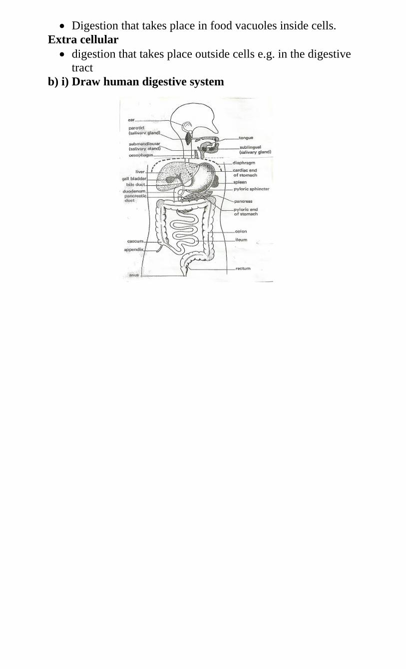
Digestion that takes place in food vacuoles inside cells.
Extra cellular
digestion that takes place outside cells e.g. in the digestive
tract
b) i) Draw human digestive system
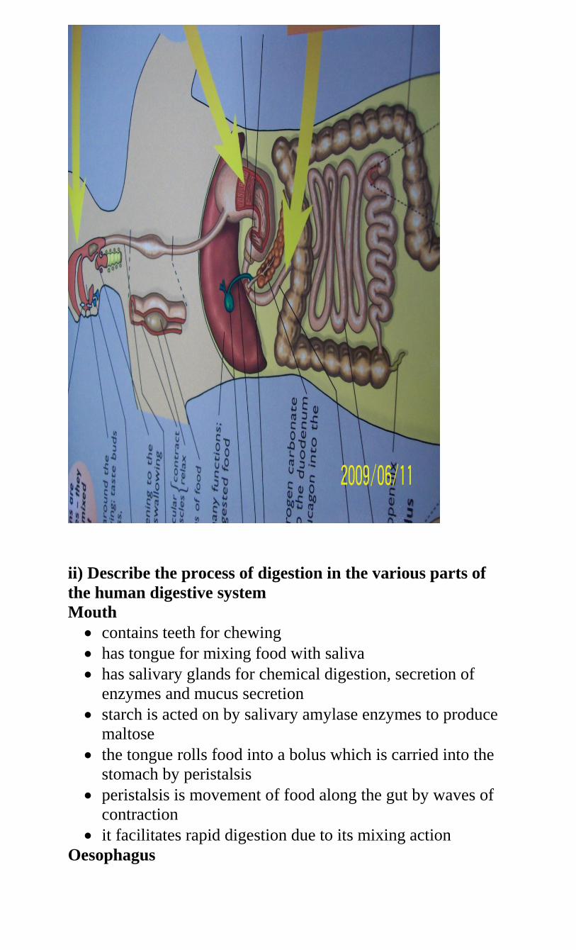
ii) Describe the process of digestion in the various parts of
the human digestive system
Mouth
contains teeth for chewing
has tongue for mixing food with saliva
has salivary glands for chemical digestion, secretion of
enzymes and mucus secretion
starch is acted on by salivary amylase enzymes to produce
maltose
the tongue rolls food into a bolus which is carried into the
stomach by peristalsis
peristalsis is movement of food along the gut by waves of
contraction
it facilitates rapid digestion due to its mixing action
Oesophagus
also called gullet
forms a passage for food by peristalsis
connects the mouth to the stomach
Stomach
has gastric glands which secrete gastric juices
these juices contain hydrochloric acid(HCL), mucus, and
the enzymes pepsin, rennin and lipase
HCL produces an acidic medium for enzyme action
Proteins are acted upon by pepsin to produce peptides
Caseinogen is acted upon by rennin to produce casein
Fats are acted upon by lipase to produce fatty acids and
glycerol
Mucus lubricates the stomach and prevents autolysis of
stomach (mucus protects stomach)
Duodenum
the first u-shaped part of the small intestine
food in the stomach is now in a semi-liquid form called
chime
chime leaves the stomach by peristalsis into the duodenum
there, the liver produces bile pigments, bile salts and
sodium hydrogen carbonate
the stomach is usually alkaline to neutralize chime which is
acidic
bile salts emulsify fats
bile comes from the gall bladder through the bile duct
sodium hydrogen carbonate provides the correct
pH/alkaline
pancreatic juices are released by pancreas into the
duodenum
the juices contain trypsin, chemotrypsin, amylase, lipase
and protease
proteins are acted upon by trypsin to form polypeptides and
amino acids
starch is broken down to maltose by amylase
Ileum
- produces intestinal juices
- Intestinal juice contains maltase, sucrase, lactase, erepsin,
lipase, and several other peptidases
- Maltose is broken down to glucose and galactose by lactase
- Sucrose is acted upon by sucrase to glucose
Polypeptides are broken down into amino acids by erepsin
- Mucus secretion is to protect the ileum wall from
digestion/autolysis
Colon
- Commonly called the large intestine
- Wider than the ileum
- has several mucus-producing cells
Highly folded for water absorption
- Also prepares food for egestion
- egestion is the process by which the insoluble parts of food are
discharged from the body in form of faeces.
Rectum
- Muscular and enlarged
- it produces mucus
- used for storage and removal of faeces
Anus
- found at the exterior end of the rectum
The rectum opens into the anus
- The anus has anal sphincter to control egestion
- Anus is used for egestion of faeces
c) Explain how mammalian intestines are adapted to
perform their function
- The mammalian intestines are relatively long and coiled. This
allows food enough time and increases surface area for digestion
and absorption of products of digestion
- The intestinal lumen (inner wall) has projections called villi to
increase surface area for absorption
- The villi have projections called micro-villi which lead to
further increase of surface area for absorption
- The walls have glands which secrete enzymes for digestion e.g.
maltase, sucrase, lactase, peptidase and enterokinase.
- Goblet cells (mucus secreting cells or glands) produce mucus
which protects the intestinal wall from being digested and
reduces friction.
- Intestines have openings of ducts which allow bile, a
pancreatic juice into the lumen
- The intestines have circular and longitudinal muscles whose
contraction and relaxation (peristalsis) leads to mixing of food
with enzymes (juices) helps push food along the gut.
- The intestines are well supplied with blood vessels that supply
oxygen and remove digested food.
- Intestines have lacteal vessels for transport of lipids (fats and
oils)
- Intestines have thin epithelium to facilitate fast/rapid
absorption/diffusion
d) What is the function of hydrochloric acid in digestion?
- kills bacteria
- activates trypsinogen to trypsin which digests proteins to
peptones and peptones to soluble amino acids
- provides acidic medium for gastric enzymes
e) i) What is assimilation?
- The process by which digested food is taken up by cells and
used in the body for various purposes.
ii) State the uses of digested food in the bodies of animals
- Protection
- Repair
- Growth
- Energy production
f) Name the types of food substances in the food that do not
undergo digestion in human digestive system
mineral salts
water
roughage
vitamins
k.) Explain the importance of the following food
substances in human nutrition
Vitamins
are organic chemical compounds essential for a healthy
body
are obtained from fresh fruits and vegetables
some are synthesized in the body e.g. vitamin K
they are destroyed by overcooking food
they protect the body against diseases, play regulatory
mechanisms in the body and act as co-enzymes
insufficient amounts lead to deficiency diseases e.g. rickets,
scurvy, beriberi
a) Mineral salts
are important in organic compounds containing elements
which are essential for normal body metabolism
those required in large quantities are called macro-nutrients
while those required in small quantities are called micro-
nutrients or trace elements
They are used in bone and teeth formation. In osmotic
balance and neurotransmission
insufficient amounts lead to anaemia, rickets, goiter
Excess amounts lead to high blood pressure, and dental
disorders.
b) Roughage
composed of cellulose and plant fibers
digested by cellulose contained by gut microorganisms
provides grip essential for peristalsis
lack of roughage leads to slow movement of food leading
to constipation
roughage adds bulk to food for peristalsis to take place
c) Water
used in transport in the body, universal solvent, hydrolysis
insufficient leads to dehydration
l.) Explain the factors that determine energy requirements
in humans
a) Basal Metabolic Rate (BMR)
this is the energy required when the body is completely at
rest
used to carry out breathing, heartbeat, circulation of blood
and other basic reactions
also used in maintaining body temperature at constant
all movements or physical work e.g. walking, eating
required more energy.
b) Occupation
means activity occurring everyday
everyday activity determines energy requirement
People doing heavy work like digging require more energy
than office workers.
c) Age
children carry out many activities and also have more cell
division than adults
their BMR is therefore higher than for adults
as they grow older, they become less active and their
energy requirements decrease
d) Body size
small bodied people have a large surface area to volume
ratio
their bodies lose more heat energy to the surrounding
they therefore require more energy-giving foods
this is the opposite for big bodied people
e) Sex
most males are more muscular than females
they also do heavier work than females hence require more
energy
females do lighter work hence require less energy
f) Climate
in warm climate the body requires less energy
in low temperatures the body requires more energy to
maintain body temperature
m.) Explain various tests carried out on food
Test Procedure Observation Conclusion
Starch - add iodine
solution
- colour
changes to
blue
black/dark
blue
Present
Reducing
sugar
Benedicts
solution
heat/boil/warm
in hot water
bath
- colour
changes to
Green to
yellow to
orange to
brown to red
Present
Non-
reducing
sugar
Dilute HCL,
NaHCO3,
heat/boil,
warm in hot
water bath
- colour
changes to
Green to
yellow to
orange to
brown to red
Present
Proteins 1% CuSO4,
5% NaOH
-- colour
changes to
purple/violet
Present
Ascorbic acid
(Vitamin C)
DCPIP drop
wise
DCPIP
decolorized
Present
Fats/oils
(lipids)
- rub on filter
paper
- ethanol
- translucent
mark
- white
emulsions
present
FORM TWO TOPICS
1. a) i) Define transport
movement of substances from one part of the body to
another
ii) Explain the necessity of transport in plants and animals
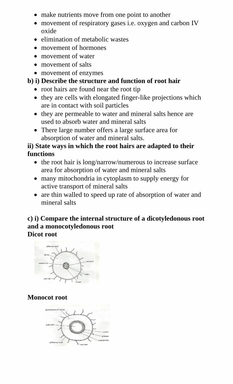
make nutrients move from one point to another
movement of respiratory gases i.e. oxygen and carbon IV
oxide
elimination of metabolic wastes
movement of hormones
movement of water
movement of salts
movement of enzymes
b) i) Describe the structure and function of root hair
root hairs are found near the root tip
they are cells with elongated finger-like projections which
are in contact with soil particles
they are permeable to water and mineral salts hence are
used to absorb water and mineral salts
There large number offers a large surface area for
absorption of water and mineral salts.
ii) State ways in which the root hairs are adapted to their
functions
the root hair is long/narrow/numerous to increase surface
area for absorption of water and mineral salts
many mitochondria in cytoplasm to supply energy for
active transport of mineral salts
are thin walled to speed up rate of absorption of water and
mineral salts
c) i) Compare the internal structure of a dicotyledonous root
and a monocotyledonous root
Dicot root
Monocot root
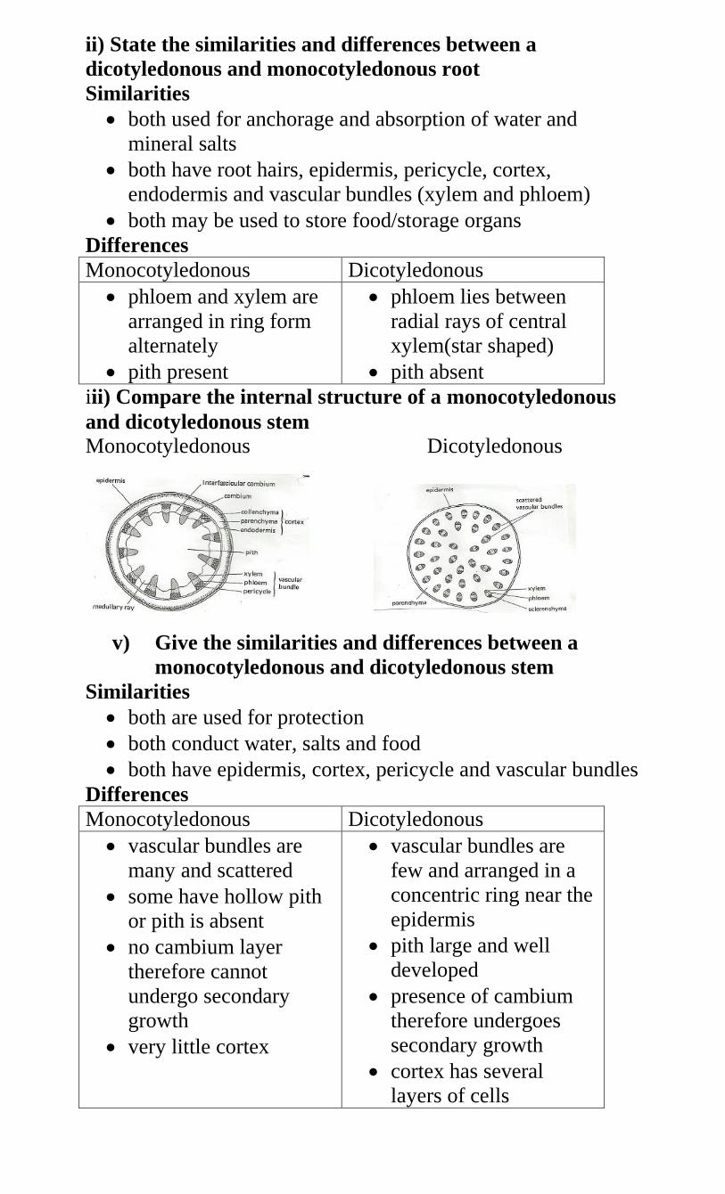
ii) State the similarities and differences between a
dicotyledonous and monocotyledonous root
Similarities
both used for anchorage and absorption of water and
mineral salts
both have root hairs, epidermis, pericycle, cortex,
endodermis and vascular bundles (xylem and phloem)
both may be used to store food/storage organs
Differences
Monocotyledonous Dicotyledonous
phloem and xylem are
arranged in ring form
alternately
pith present
phloem lies between
radial rays of central
xylem(star shaped)
pith absent
iii) Compare the internal structure of a monocotyledonous
and dicotyledonous stem
Monocotyledonous Dicotyledonous
v) Give the similarities and differences between a
monocotyledonous and dicotyledonous stem
Similarities
both are used for protection
both conduct water, salts and food
both have epidermis, cortex, pericycle and vascular bundles
Differences
Monocotyledonous Dicotyledonous
vascular bundles are
many and scattered
some have hollow pith
or pith is absent
no cambium layer
therefore cannot
undergo secondary
growth
very little cortex
vascular bundles are
few and arranged in a
concentric ring near the
epidermis
pith large and well
developed
presence of cambium
therefore undergoes
secondary growth
cortex has several
layers of cells
v) State the differences between the internal structure of
a root and a stem.
Root Stem
has root hairs
no cuticle
xylem and phloem
arranged alternately
in xylem, the small
vessels are towards the
outside
cortex is the widest
tissue
no root hairs
cuticle present
xylem and phloem
arranged on the same
radii
in xylem, the smallest
vessels are towards the
inside
pith is the widest tissue
c) i) Name the transport structures of a flowering plant
xylem vessels and tracheids transport water and mineral
salts from the soil
Phloem vessels translocate manufactured food from leaves
to other parts of the body.
ii) State the ways in which xylem vessels are adapted to their
function
lignified/thickened to prevent collapsing
narrow to facilitate capillary
no cross walls for continuous flow/column of water
have bordered pits for lateral movement of water
d) i) Why do flowering plants need water?
photosynthesis
transport
turgidity which helps in plant support
solvent i.e. medium for chemical reactions
cooling effect during transpiration
seed germination
ii) Describe the movement of water from the soil to the
leaves of a tall plant
Soil
water exists as a thin film in the soil, between soil particles
the concentration of cell sap of root hair is greater than that
of the surrounding solution in the soil, thus drawing the
water molecules across the cell wall and cell membrane
into the root hair by osmosis
water drawn into the root hair cell dilutes the cell sap
making it less concentrated than that in the adjacent cortex
cells of the root
due to osmotic gradient water moves from the root hair
cells into the cortex by osmosis, from cell to cell by
osmosis, across the endodermis by active transport into
xylem vessels of the root that conduct water into xylem
vessels of the stem into xylem vessels of the leaves
Stem
Once in the stem water moves up the plant aided by the
narrowness of the xylem vessels (capillary), root pressure,
attraction of water molecules to each other (cohesion).
Attraction of water molecules to the walls (adhesion)
from the stem water enters the xylem of leaves
water moves in the xylem vessels of the stem in a
continuous (uninterrupted) water column up to the tree
leaves
Leaves
once in the leaves water moves into the mesophyll cells by
osmosis
as water vaporizes from the spongy mesophyll cells their
sap becomes more concentrated than the adjacent cells
as the result water flows into the cell from other
surrounding cells which in turn takes in water from xylem
vessels within the leaf veins
this creates a pull(suction force) called transpiration pull
that pulls a stream of water from xylem vessels in the stem
and roots
The transpiration pull maintains a continuous column of
water from the roots to the leaves.
iii) Name the process by which mineral salts enter into a
plant
active transport
diffusion
vi) Explain the forces that make water and mineral salts
move through a plant
mineral salts are taken up due to diffusion because of the
concentration gradient between the mineral ions in sap and
those in soil solution
active transport involves energy in form of ATP due to
respiration which forces mineral salts through a plant
against a concentration gradient
water moves by osmosis through a semi-permeable
membrane of root hairs and between cells of stem
in stem water moves by cohesion(attraction of water
molecules to each other)
it also moves by adhesion(attraction of water molecules to
walls)
capillarity is due to narrowness of xylem vessels
transpiration pull occurs when water vapour evaporates
from sub-stomatal chambers into the air
root pressure is a force that pushes water up the stem from
the roots and causes guttation /exudation
vii) Explain the uptake of mineral salts by plants
plants require mineral salts for metabolism and proper
functioning of their bodies
mineral salts are taken up from the soil into the root hairs in
form of solution by active transport which requires energy
active transport involves substances called carriers taken up
together with water and are then carried to the stems and
leaves
the main process involved in uptake and movement of
mineral salts is active transport
e) i) What is transpiration?
loss of water from plant to the atmosphere
ii) Name the sites through which transpiration takes place in
a plant
stomata (stomatal transpiration)
lenticels (lenticular transpiration)
cuticle(cuticular transpiration)
iii) State the importance of transpiration to plants
cooling the plant
transport of water
transport of mineral salts
excretion of excess water from plants
excess transpiration causes wilting
v) Explain the structural factors that affect the rate of
transpiration in plants
number of stomata i.e. the more the stomata the higher the
rate and vice versa
turgidity of the guard cells which control the opening and
closing of stomata when they are open transpiration rate is
high
size of leaves where the larger the surface area the higher
the rate of transpiration
leaf fall leads to lower rate of transpiration and also drying
of leaves reduces rate of transpiration
Thin cuticle reduces distance through which water
vaporizes hence increase transpiration rate. Absence of
cuticle also increase rate of transpiration
vi) explain the environmental factors that affect rate of
transpiration in plants
high temperature increases rate of transpiration and low
temperature reduces the rate
humidity when high increases rate and when low reduces
the rate
transpiration rate is higher in moving air (wind) than in still
air
high light intensity increases internal temperature hence
higher rate of evaporation leading to higher rate of
transpiration
availability of water in the soil leads to more absorption
hence more loss to the atmosphere
atmospheric pressure when high leads to more evaporation
and when low leads to low rate evaporation of water
vii) State the structural differences between xylem vessels
and sieve tubes
sieve tubes have cross wall while xylem vessels have none
xylem vessels are lignified while sieve tubes are not
Sieve tubes have cytoplasm elements while xylem vessels
have none.
viii) State the adaptations of plants which enable them to
reduce water loss
thick waxy cuticle
reduced leaf size/thorns/spines
shedding of leaves
Sunken stomata. Water vapour accumulates in the
depression of stomata lowering the water vapour
concentration gradient leading to lower rate of evaporation
rolling of leaves
ix) State the factors that cause increase in the rate of
transpiration from leaves
increased light intensity
low relative humidity
temperature
x) Explain how drooping of leaves on a hot sunny day is
advantageous to a plant
reduces surface area exposed to sun reducing cuticular
transpiration
f) Explain how aquatic and terrestrial plants are adapted
to deal with problems of transpiration
a. Mesophytes
they grow in soils with enough water
water loss is perfectly balanced by absorption of more from
the soil
no special adaptations
b. Xerophytes
they grow in dry conditions
root grow very deep to absorb water
succulent/fleshy leaves to store water
few stomata which are sunken
thickened waxy cuticle
leaves are hairy and often folding
some leaves are needle-like/spines or scales
leaf surfaces are reduced i.e. small leaves
all these adaptations are to reduce water loss
c. Hydrophytes
plants that grow in water
presence of sclereids
leaves are broad
leaves have many stomata on upper side only (none on the
lower surface)
some leaves float on water
absence or reduced leaf cuticle
large air spaces
some leaves are submerged
poorly developed or reduced vascular bundles
g) i) What is translocation
transfer of manufactured food substances to the parts where
they are required
ii) Name the tissue which is responsible for translocation of
manufactured food in flowering plants
phloem tissue
iii) Name the processes that bring about the translocation of
manufactured food
-active transport
Diffusion
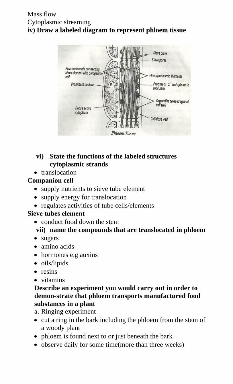
Mass flow
Cytoplasmic streaming
iv) Draw a labeled diagram to represent phloem tissue
vi) State the functions of the labeled structures
cytoplasmic strands
translocation
Companion cell
supply nutrients to sieve tube element
supply energy for translocation
regulates activities of tube cells/elements
Sieve tubes element
conduct food down the stem
vii) name the compounds that are translocated in phloem
sugars
amino acids
hormones e.g auxins
oils/lipids
resins
vitamins
Describe an experiment you would carry out in order to
demon-strate that phloem transports manufactured food
substances in a plant
a. Ringing experiment
cut a ring in the bark including the phloem from the stem of
a woody plant
phloem is found next to or just beneath the bark
observe daily for some time(more than three weeks)
a swelling of the bark appears above the ring
this is due to accumulation of food from leaves
the bark of a second similar plant is removed carefully
leaving the phloem intact
a swelling does not appear
ii) Use the radioactive tracers
plant is exposed to carbon containing radio-active carbon
C14
C14 is found in the end products of photosynthesis
It is finally detected in phloem
C14 is found to move in both directions
iii) Collecting exudate from stylets of aphids
aphids feed on certain plant phloem using their stylets
aphid mouthparts are dissected using a sharp razor
exudates from the mouthparts are collected and then
analyzed
sucrose is found to be a major component of the exudates
this proves that phloem translocates manufactured food
substances
h) Describe an experiment you would carry out to
demonstrate that xylem transports water
i. Either
cut a stem of a young plant or twig of a tree under water
or else uproot a young herbaceous plant and wash the soil
gently
put some water in a beaker and add a dye i.e. eosin or red
ink and place the cut stem or young plant in a beaker
leave for time e.g. between 20 minutes and one hour
cut a thin section of stem or leaf
mount it on a slide and examine under a microscope
observe and note the distribution of the dye or ink
the dye appears only in the xylem vessels
ii. OR
use radio-active tracers, C14 in form of carbon
ring a plant then put it in a container containing radio-
active phosphorous solution
The radio-active phosphorus is later detected in the leaves.
2. a) i)List the components of animal transport
systems
system of blood vessels in which materials are circulated
round the body
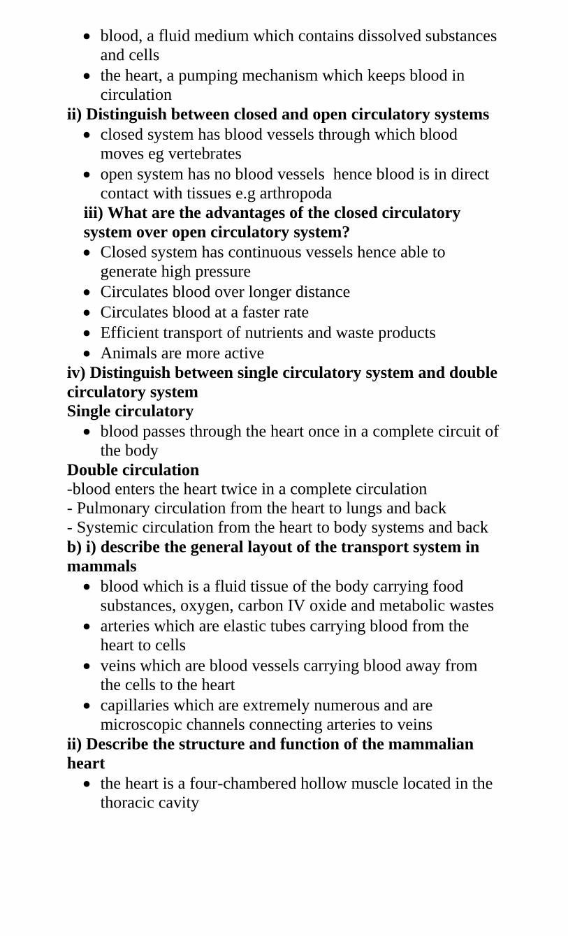
blood, a fluid medium which contains dissolved substances
and cells
the heart, a pumping mechanism which keeps blood in
circulation
ii) Distinguish between closed and open circulatory systems
closed system has blood vessels through which blood
moves eg vertebrates
open system has no blood vessels hence blood is in direct
contact with tissues e.g arthropoda
iii) What are the advantages of the closed circulatory
system over open circulatory system?
Closed system has continuous vessels hence able to
generate high pressure
Circulates blood over longer distance
Circulates blood at a faster rate
Efficient transport of nutrients and waste products
Animals are more active
iv) Distinguish between single circulatory system and double
circulatory system
Single circulatory
blood passes through the heart once in a complete circuit of
the body
Double circulation
-blood enters the heart twice in a complete circulation
- Pulmonary circulation from the heart to lungs and back
- Systemic circulation from the heart to body systems and back
b) i) describe the general layout of the transport system in
mammals
blood which is a fluid tissue of the body carrying food
substances, oxygen, carbon IV oxide and metabolic wastes
arteries which are elastic tubes carrying blood from the
heart to cells
veins which are blood vessels carrying blood away from
the cells to the heart
capillaries which are extremely numerous and are
microscopic channels connecting arteries to veins
ii) Describe the structure and function of the mammalian
heart
the heart is a four-chambered hollow muscle located in the
thoracic cavity
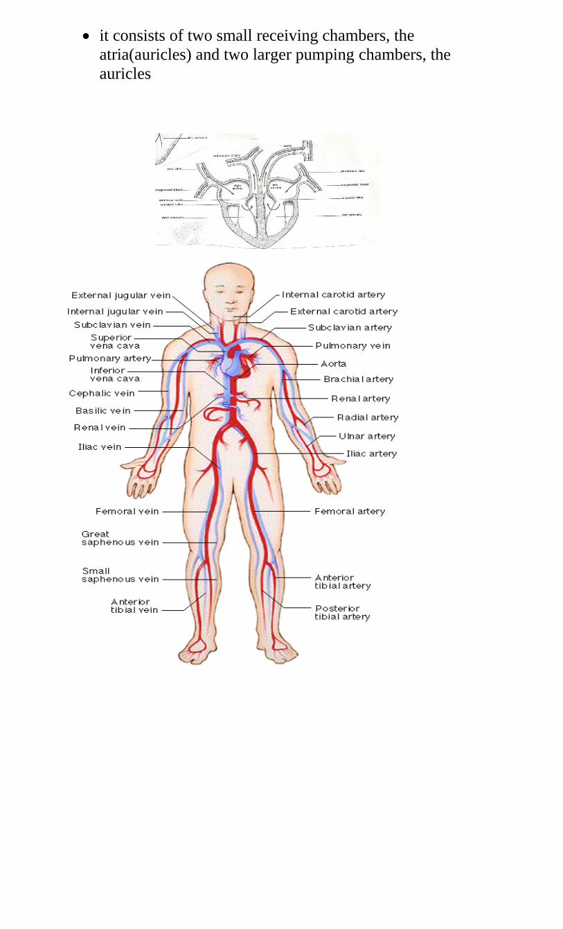
it consists of two small receiving chambers, the
atria(auricles) and two larger pumping chambers, the
auricles
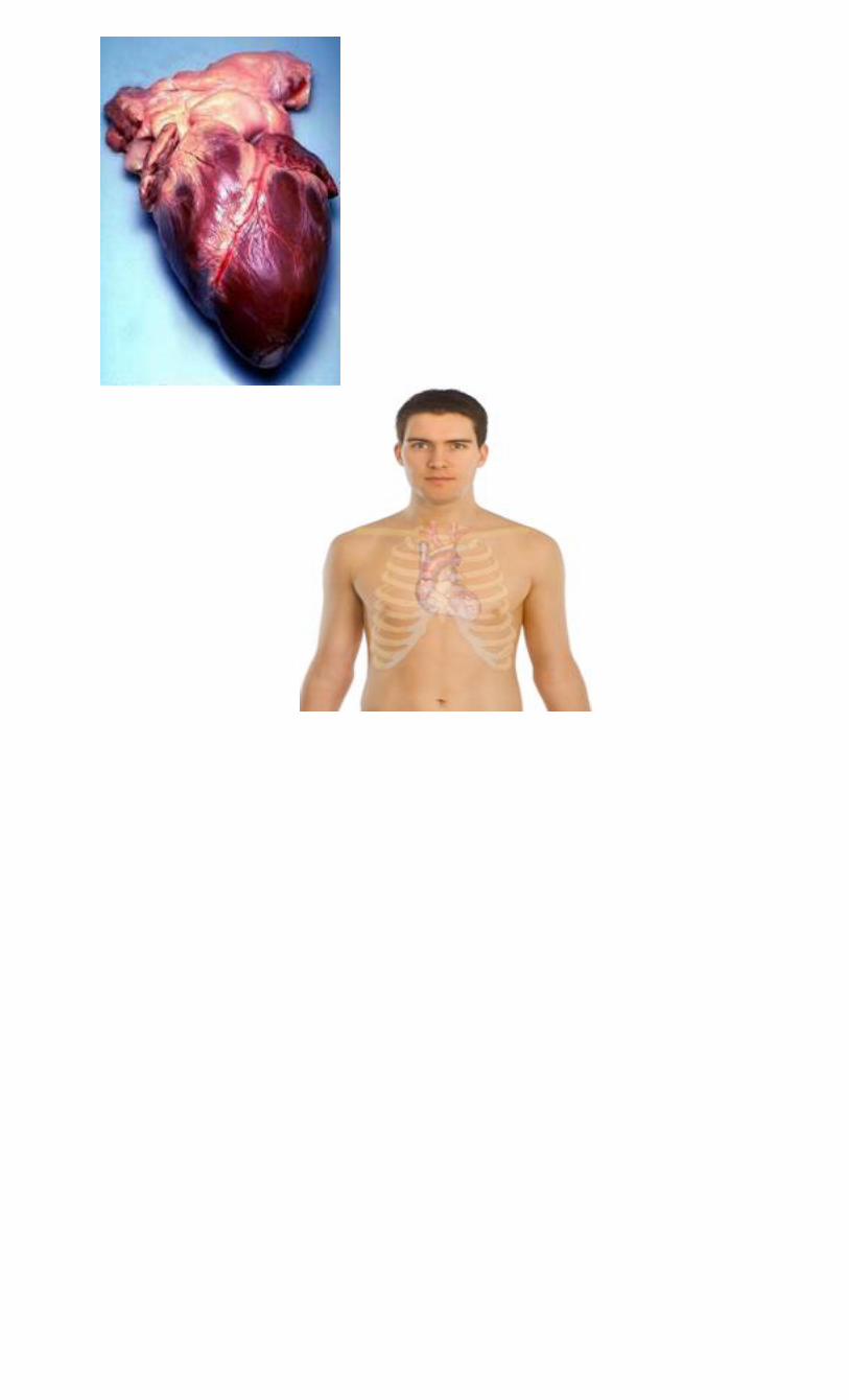
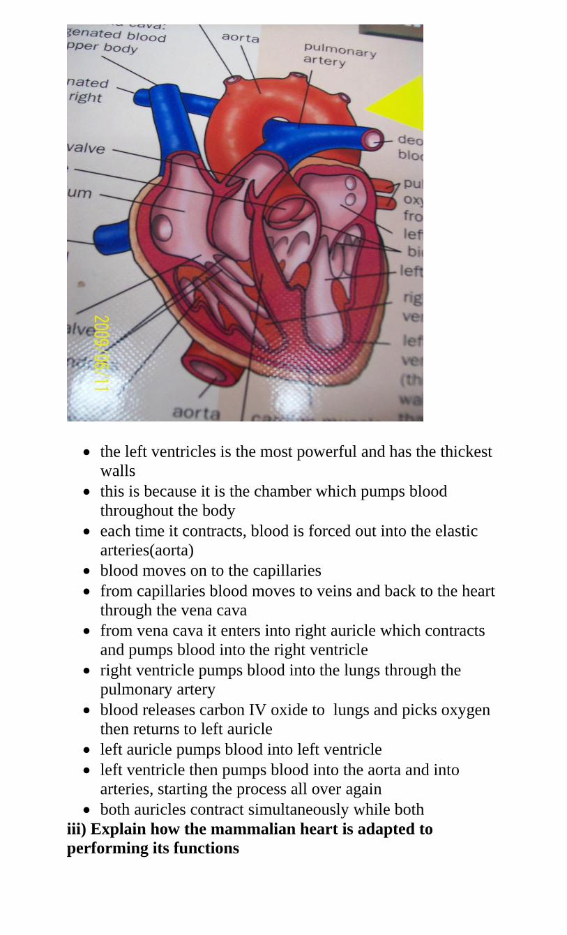
the left ventricles is the most powerful and has the thickest
walls
this is because it is the chamber which pumps blood
throughout the body
each time it contracts, blood is forced out into the elastic
arteries(aorta)
blood moves on to the capillaries
from capillaries blood moves to veins and back to the heart
through the vena cava
from vena cava it enters into right auricle which contracts
and pumps blood into the right ventricle
right ventricle pumps blood into the lungs through the
pulmonary artery
blood releases carbon IV oxide to lungs and picks oxygen
then returns to left auricle
left auricle pumps blood into left ventricle
left ventricle then pumps blood into the aorta and into
arteries, starting the process all over again
both auricles contract simultaneously while both
iii) Explain how the mammalian heart is adapted to
performing its functions
the heart is made of muscles that contract and relax
synchronously without requiring nervous stimulation
nerve supply however, determine contraction strength and
frequency
the heart is divided into four chambers
The right atrium is connected to the right auricle. It
receives blood from the whole body.
The blood is pumped from the left atrium to the right
ventricle
To avoid flow back into the right atrium, a valve is present
between the two chambers – the tricuspid valve
The right ventricle pumps blood to the lungs
This is facilitated by the presence of pulmonary artery
A valve is also present to avoid blood flowing back from
the pulmonary artery to the right ventricle
Blood from the lungs enters the heart through the
pulmonary vein into the left atrium.
When the left atrium contracts, blood flows into the left
ventricle
Blood will not flow back into the left atrium because of the
presence of bicuspid valve(mitral)
The left ventricle is connected with the aorta and when it
contracts, blood flows into the aorta for distribution into the
whole body
The heart muscle surrounding the left ventricle is thicker
than that surrounding the right ventricle to be able to
generate enough pressure to push blood to the whole body
A pace-maker is present in the heart muscle to initiate and
synchronise contractions.
For the heart muscle to be well nourished and be provided
with enough oxygen and carbon IV oxide removal, it is
supplied with blood by the coronary arteries and drained by
the coronary veins
iv) Explain why blood leaving the lungs may not be fully
oxygenated
under ventilation of the lungs
blockage of alveoli (air sacs)
high cardiac frequency i.e. high rate of pumping of blood in
the heart
e) Describe the structure and functions of the blood vessels
i. Arteries
carry away blood from the heart
carry oxygenated blood except pulmonary artery which
takes blood from the heart to lungs for oxygen
have thick, muscular walls
are elastic
have narrow lumen
all these adaptations are required to withstand high pressure
caused by heartbeat
ii. Capillaries
link arterioles and venules to arteries and veins
small in diameter to increase pressure resistance for
materials to filter out
thin walled as they consist of a single layer of cells to allow
diffusion of substances e.g leucocytes to tissues
thin walled to allow presence of intercellular spaces
large number i.e. numerous to provide a large surface area
for exchange of materials
have sphincter muscles at the junction of the arterioles and
capillaries to control movement of blood into them
lie close to the body for easy exchange of materials
iii. Veins
carry blood back to the heart
all carry deoxygenated blood except pulmonary vein that
carries blood from the heart to lungs
have thinner walls than arteries
have valves to prevent backflow of blood
have wide lumen
g) i) State the ways in which the composition of blood in
the pulmonary arterioles differs from that in the
pulmonary venules
Pulmonary arterioles Pulmonary venules
deoxygenated
high carbon IV oxide
low oxygen
more nutrients
oxygenated
low carbon IV oxide
high oxygen
less nutrients
ii) Give the reasons why pressure of blood is greater in the
arterioles than I the veins of mammals
blood is pumped to the arteries by the heart at high pressure
blood pressure in veins is reduced by capillary resistance
arteries have narrow lumen which maintains high
pressure/veins have wide lumen which reduces pressure
arteries have more/thicker muscular walls which generate
pressure/veins have less/thinner muscular walls which
reduce pressure
iii) Name the common heart diseases in humans
thrombosis
antheroma
arteriosclerosis
varicose veins
cerebral vascular thrombosis
h) i) State the functions of mammalian blood
transport of substances
defense against diseases
clotting
temperature regulation
ii) Describe how mammalian blood components carry out
their functions
Plasma
transport dissolved food substances like glucose, amino
acids, fatty acids and glycerol from small intestines to liver
and other body tissues
transports hormones, enzymes from secretory glands to
tissues when required
transports carbon IV oxide to lungs and urea from tissues to
the kidneys
distributes heat
bathes the tissues allowing for exchange of materials
contains protein fibrinogen and pro-thrombin which take
part in blood clotting
Red blood cells (Erythrocytes)
transports oxygen from the lungs to body tissues in form of
haemoglobin
transport carbon IV oxide from body tissues to the lungs in
form of bicarbonates
White blood cells (leucocytes)
engulf foreign bodies
produce antibodies for defense against disease
produce antitoxins which neutralize bacterial toxins
Blood platelets (thrombocytes)
produce an enzyme called thrombokinase/thromboplastin
necessary for blood clotting
prevents loss of blood, water and mineral salts
iii) State the ways in which the red blood cells are adapted to
their functions
many per unit volume hence carry more oxygen and carbon
IV oxide
biconcave in shape to provide large surface area for
absorption of oxygen and carbon IV oxide
absence of nucleolus hence more haemoglobin to carry
sufficient oxygen and carbon IV oxide
alter shape to be able to pass through the narrow lumen of
capillaries to deliver or supply oxygen and carry away
carbon IV oxide
have haemoglobin with high affinity for uptake of oxygen
and carbon IV oxide
iv) State the structural differences between a red blood cell
and a white blood cell.
Red blood cells white blood cells
has haemoglobin - not pigmented
smaller size - larger size
lacks nucleus - nucleated
v) State the functional differences between a red blood cell
and a white blood cell
Red blood cell White blood cell
- Transports oxygen and carbon IV oxide - protects body
against harmful pathogens
vi) How does the heart increase blood flow to some parts
of the body during exercise
stronger contractions
faster contractions/heartbeat
Explain how oxygen and carbon Iv oxide are transported in
the blood
Oxygen
oxygen concentration is higher in lungs(alveoli) that in
blood
oxygen in the alveoli dissolves in the film of moisture and
diffuses through thin epithelial and capillary walls into
plasma and red blood cells
the oxygen combines with haemoglobin to form
oxyhaemoglobin
blood then becomes oxygenated
blood from lungs then travels to all body tissues where the
oxyhaemoglobin breaks down to form oxygen and
haemoglobin
haemoglobin is transported back to the lungs to collect
more oxygen while the oxygen in capillaries diffuses into
body cells for respiration
respiration produces carbon IV oxide
Carbon IV oxide
carbon IV oxide produced during respiration diffuses out of
cells into blood plasma and red blood cells due to
concentration gradient
carbon IV oxide and water form carbonic acid carbamino
compounds with haemoglobin
in the presence of carboxyl anhydrase enzyme, hydrogen
carbonate is carried in blood to the lungs
in the lungs the hydrogen carbonate dissociates to liberate
carbon IV oxide which diffuses into alveolar cavity due to
concentration gradient
from alveolar space carbon IV oxide is expelled during
expiration
Most carbon IV oxide is transported from tissues to lungs
within the red blood cells and not in the blood plasma. Give
the advantages of this mode of transport.
- PH of blood is not altered/homeostasis is maintained
- Within the red blood cell is an enzyme, carbonic anhyrase
which helps in fast loading(combining) and offloading of
carbon Iv oxide
i) i) what is blood clotting?
process in which blood components clump together to
prevent loss of blood from an injured/cut vessel
ii) Name a protein, vitamin, an enzyme and a mineral
element involved in blood clotting
Protein – fibrinogen/prothrombin
Vitamin - k/quinine
Enzyme – thrombokinase/thromboplatin/thrombin
Mineral element – calcium
iii) Describe the blood clotting process
enzyme thromboplastin produced in the platelets of
damaged tissues converts plasma protein prothrombin into
thrombin in the presence of calcium ions
thrombin converts another plasma protein fibrogen into
fibrin in the presence of vitamin K
fibrin is insoluble
fibrin forms fibres which form a meshwork that forms a
clot
prothrombin thromboplastin thrombin
calcium ion
Fibrinogen thrombin fibrin clot
vitamin K
iv) State the role of blood clotting on wounds
prevents blood/body fluids from being lost
conserves water and salts
prevents entry of microorganisms/pathogens
regulates body temperature
enables wound to heal faster
v) Explain why blood flowing in blood vessels does not
normally clot
- Presence of anticoagulant in blood
j) i. list the major types of human blood groups
O, with neither B nor A antigen
AB, with both A and B antigens
A, with type A antigen
B, with type B antigen
iii. explain the meaning of :
Universal donor
a person who can donate blood to any other blood group
without agglutination/clumping
this is usually blood group O
however this person cannot receive blood from other blood
groups except group O
Universal recipient
can receive blood from all blood groups without
agglutination
this is usually blood group AB
however, can only donate blood to group AB
iii) What is the difference between rhesus positive and
Rhesus negative blood samples?
rhesus positive blood has the Rhesus (Rh) antigen
rhesus negative lacks the Rhesus antigen
vi) What is blood transfusion?
- Introduction of blood from one person to another
v) Under what conditions would blood transfusion be
necessary in people?
during accidents
during surgery in hospitals
bleeding mothers when giving birth
vi) How can low blood volume be brought back to
normal?
transfusion
taking fluids
eating iron rich food/taking iron tablets
How may excessive bleeding result in death?
Anaemia/low blood volume/loss of iron/low red blood cells
count/low haemoglobin leading to low oxygen, loss of
nutrients and dehydration.
State the precautions that must be taken before blood
transfusion
blood must be disease free
sterilized equipment must be used
blood of the recipient and that of the donor must b
compatible to both ABO and rhesus factor
Fresh blood must be used.
j) i) What is immunity?
- Resistance to disease by organisms
ii) Distinguish between natural and acquired immunity
natural immunity is inherited/transmitted from parent to
offspring/inborn/innate
Acquired immunity is developed after suffering from a
disease or through vaccination.
iii) What are allergic reactions?
Excessive sensitivity and reaction of an individual to
certain substances in environment e.g. dust, pollen,
perfumes, smoke etc.
vi) How does an allergic reaction occur?
the substances act as antigens
an antigen-antibody reaction occurs on surface of cells
the cells release a substance called histamine
the histamine causes irritation, itching and may stimulate
nasal discharge
vii) State the role of vaccination against certain diseases
protect body against infectious diseases
prevent spread/transmission of certain diseases
diseases for which vaccination is given include
tuberculosis, poliomylitis,measles, whooping cough,
diphtheria
3. a) i) What is gaseous exchange?
The continous exchange of oxygen and carbon Iv oxide
between the organism and environment.
ii) Why is gaseous exchange important to organisms?
to supply oxygen necessary for energy production
to remove carbon IV oxide produced during respiration
To remove water vapour.
b) i) name the structure used for gaseous exchange by plants
stomatal pores/stomata
lenticels
cuticle
pneumatophores
ii) Briefly describe the structure of stomata
are minute pores found in leaf epidermis
each consists of a slit-like opening
Each is bordered by two large, bean-shaped guard cells.
Guard cells contain chloroplasts, unlike the other epidermal
cells which enable photosynthesis to occur
Inner walls of guard cells are thicker than the outer cells
iii) State the factors which affect stomatal opening
water which when low stomata close and when high
stomata keeps open
light as stomata open in bright light and close in darkness
temperature
iv) Name the theories suggesting the mechanism of opening
and closing of stomata
interconversion of starch and sugar
pH theory
mineral ion concentration
v) Describe the mechanism of opening and closing of stomata
stomata close at night and open during daytime
This comes about due to changes in turgidity as a result of
pH changes in guard cells.
In the dark carbon Iv oxide accumulates in the intercellular
spaces
This raises concentration of carbonic acid
The pH drops (pH lowered)
Enzymes convert sugar into starch in guard cells
Osmotic pressure in guard cells is lowered
Water moves out of guard cells by osmosis making cells
lose turgidity hence become flaccid
The stomata close
During day time there is photosynthesis hence the
production of sugar, carbon IV oxide concentration is
lowered, pH increases, guard cells become turgid causing
stomata to open.
During the day potassium ions concentrate in guard cells,
raising their osmotic pressure and causes then to open
In the night the concentration of potassium ions decreases
increasing osmotic pressure in guard cells therefore causes
stomata to open.
vii) What is the advantage of having stomata open
during daytime and having them closed at night?
opening in the daytime allows diffusion of carbon IV into
the leaf for photosynthesis to take place and allows
diffusion of oxygen out of the leaf
transpiration also takes place, thus cooling the leaf and
facilitating uptake of water and mineral slats
Closing in the night is to conserve water in the plant
especially when there is not enough water available in the
soil.
c) i) State the ways in which leaves of plants are adapted to
gaseous exchange
presence of stomata for faster gaseous exchange
intercellular spaces/air spaces in the leaf for
movement/circulation of air
film of moisture around the surface of cells for easy
diffusion
broad/flattened shape to increase surface area
thin lamina to reduce distance of diffusion
exposed to air for easy diffusion
ii) Describe how gaseous exchange takes place in terrestrial
plants
Gaseous exchange takes place in spongy mesophyll
During the day air diffuses into large air spaces of spongy
mesophyll through stomata
The carbon iv oxide in the air diffuses into the
photosynthesis oxygen is produced
Some of the oxygen diffuses out of the leaf through stomata
During the night air diffuses out of air spaces of spongy
mesophyll
The air dissolves into film of moisture
The oxygen in the air diffuses into cells and is used in
respiration during which carbon iv oxide is produced
The carbon iv oxide diffuses out of the leaf through stomata
due to diffusion/concentration gradient
At night carbon iv oxide accumulates in the leaf since
photosynthesis does not take place
Some gaseous exchange also takes place through cuticle
Gaseous exchange occurs through epidermis of young
leaves and stems
The cork cells at lenticels are loosely packed
Gaseous exchange takes place between cork and
atmosphere within the loosely packed cells
iii) State the ways in which floating leaves of aquatic
plants are adapted to gaseous exchange
stomata found only on upper dermis to allow efficient
gaseous exchange
presence to aerenchyma tissues/large air spaces to enable it
float/buoyancy/storage of air
absence of cuticle to enhance gaseous exchange
iv) How is aerenchyma tissue adapted to its function?
has large airspaces which store gases/for gaseous
exchange/buoyancy
v) Explain stomatal distribution in plants of different
habitats
land plants have their stomata mainly on the lower side to
reduce water loss but if on both sides then upper side has
very few
water plants, floaters, have stomata on upper side to
enhance water loss
in dry areas, plants have leaves with sunken stomata to
reduce water loss by transpiration
Plants in wet areas have stomata equally distributed on both
sides.
d) i) List the types of respiratory surfaces of animals
cell membrane in unicellular organisms e.g. amoeba
gills in fish
tracheal system
skin, buccal cavity and lungs in amphibians
lings in mammals
ii) State the characteristics of respiratory surfaces in animals
moist
thin walled/thin membrane/thin surface
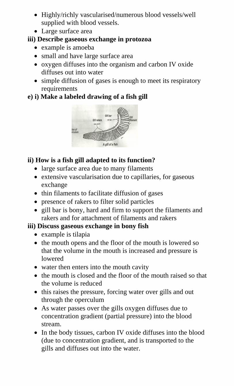
Highly/richly vascularised/numerous blood vessels/well
supplied with blood vessels.
Large surface area
iii) Describe gaseous exchange in protozoa
example is amoeba
small and have large surface area
oxygen diffuses into the organism and carbon IV oxide
diffuses out into water
simple diffusion of gases is enough to meet its respiratory
requirements
e) i) Make a labeled drawing of a fish gill
ii) How is a fish gill adapted to its function?
large surface area due to many filaments
extensive vascularisation due to capillaries, for gaseous
exchange
thin filaments to facilitate diffusion of gases
presence of rakers to filter solid particles
gill bar is bony, hard and firm to support the filaments and
rakers and for attachment of filaments and rakers
iii) Discuss gaseous exchange in bony fish
example is tilapia
the mouth opens and the floor of the mouth is lowered so
that the volume in the mouth is increased and pressure is
lowered
water then enters into the mouth cavity
the mouth is closed and the floor of the mouth raised so that
the volume is reduced
this raises the pressure, forcing water over gills and out
through the operculum
As water passes over the gills oxygen diffuses due to
concentration gradient (partial pressure) into the blood
stream.
In the body tissues, carbon IV oxide diffuses into the blood
(due to concentration gradient, and is transported to the
gills and diffuses out into the water.
iv) What is counter-flow system?
Where water in which the fish lives flows in opposite
direction across the gill.
vi) What is the advantage of counter-flow system?
maintains a diffusion gradient so that there is maximum
uptake of oxygen
oxygen continues diffusing into blood and carbon iv oxide
into water
f) i) Describe the mechanism of gaseous exchange in
terrestrial insects
example is cockroach
air in the atmosphere contains oxygen
air is drawn into the body of the insect through the spiracles
due to movement of abdominal muscles
these movements cause the opening of spiracles
air moves through the trachea to tracheoles
oxygen moves from the tracheoles into body cells by
diffusion due to concentration gradient
carbon iv oxide in the tissues diffuses into tracheoles due to
concentration gradient
From tracheoles carbon IV oxide moves into trachea and
out through the spiracles into the air.
ii) State how traceholes are adapted to gaseous exchange
thin walls of tracheoles
moist surface
large surface area due to numerous tracheoles
g) i) What is breathing?
Any process which speeds up the rate of gaseous exchange
between an animal and its surrounding.
ii) Name the structures in humans that are used in gaseous
exchange
nose
larynx
epiglottis
trachea
lungs
pleural membrane
pleural cavity
diaphragm muscles
iii) Describe the mechanism of gaseous exchange in a
mammal
Breathing in
external intercostals muscles contract while internal
intercostals muscles relax, raising the ribcage upwards and
outwards
muscles of the diaphragm contract hence it flattens
the volume of the thoracic cavity increases while pressure
decreases
higher air pressure in the atmosphere forces air into lungs
through the nose
Breathing out
external intercostals muscles relax while internal
intercostals muscles contract, moving the ribcage
downwards and inwards
muscles of the diaphragm relax hence the diaphragm
assumes dome shape
the volume of the thoracic cavity decreases while pressure
increases
the higher pressure forces air out of the lungs through the
nose
iv) Explain how mammalian lungs are adapted to gaseous
exchange
large number of alveoli that increase surface area
moist inner surface of alveoli for dissolving oxygen/gases
to facilitate exchange of gases through alveolar cavities and
blood
thin walls of alveoli to allow efficient/faster diffusion of
gases
rich capillary/blood supply on alveolar surface to transport
oxygen away from the lungs and carbon IV oxide to the
lungs
v) Name the features of alveoli that adapt them to their
function
have large surface area/spherical shaped
numerous/many to increase surface area
one cell thick
moist surface for air to diffuse
highly vascularised/numerous capillaries
vii) How is the trachea of a mammal suited to its
function?
has a ring of cartilage which keeps it open at all times
cilia that move mucus/particles to the top of the trachea i.e.
into larynx for removal
mucus to trap dust, solid particles and microorganisms
hollow for passage of air
viii) State the advantages of breathing through the nose
rather than through the mouth
nose has hairs to filter solid particles
it has mucus lining to trap dust particles
the nose has cells sensitive to smell for survival
it warms the air before it reaches the lungs
ix) Give the conditions under which the carbon iv oxide
level rises above normal in
mammalian blood
vigorous exercise
emotions/stress
disease infection
x) Explain the physiological changes that occur in the
body to lower the carbon iv oxide level back to
normal when it rises
heartbeat/cardiac frequency increases to pump blood faster
carbon iv oxide from the tissues and supply more oxygen
ventilation rate/rate and depth of breathing increases to take
more oxygen and remove carbon iv oxide from the lungs
arterioles to take in more oxygen and remove carbon iv
oxide from the lungs
arterioles dilate leading to faster flow of blood to and from
body tissues
h) i)Describe the factors which control the rate of breathing
in humans
breathing movements usually occur unconsciously
it is controlled by the medulla oblongata part of the brain
situated at the breathing centre
medullar oblongata is in the brain
respiratory centre transmits impulses to the diaphragm
through phrenic nerves
carbon iv oxide concentration in the blood determines the
breathing rate
if carbon iv oxide is less, the brain is triggered to decrease
breathing rate
cardiac frequency decreases and the arterioles constrict
therefore carbon iv oxide level is raised
this brings back to normal level of breathing and carbon iv
oxide level increases/is more the brain is triggered to
increase breathing rate
cardiac frequency is increased
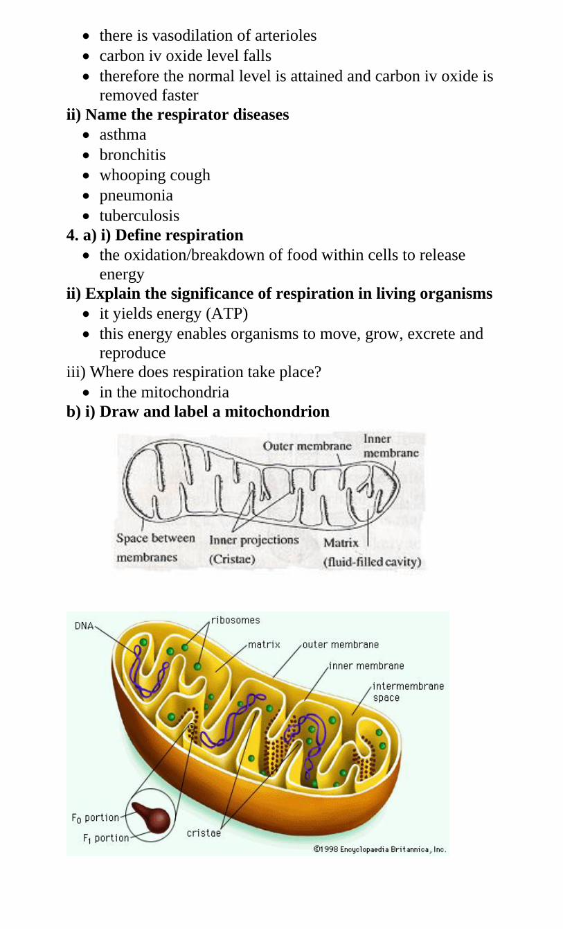
there is vasodilation of arterioles
carbon iv oxide level falls
therefore the normal level is attained and carbon iv oxide is
removed faster
ii) Name the respirator diseases
asthma
bronchitis
whooping cough
pneumonia
tuberculosis
4. a) i) Define respiration
the oxidation/breakdown of food within cells to release
energy
ii) Explain the significance of respiration in living organisms
it yields energy (ATP)
this energy enables organisms to move, grow, excrete and
reproduce
iii) Where does respiration take place?
in the mitochondria
b) i) Draw and label a mitochondrion
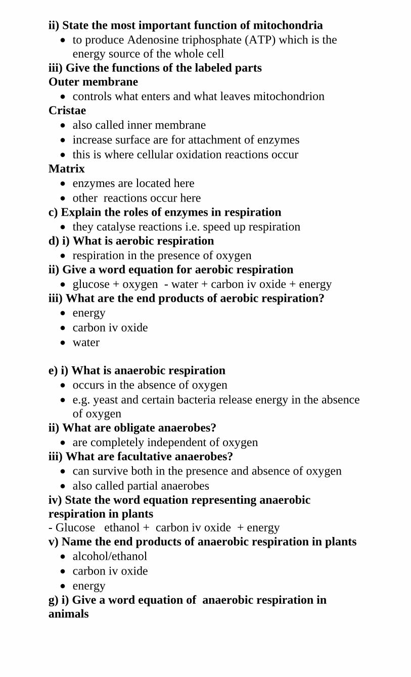
ii) State the most important function of mitochondria
to produce Adenosine triphosphate (ATP) which is the
energy source of the whole cell
iii) Give the functions of the labeled parts
Outer membrane
controls what enters and what leaves mitochondrion
Cristae
also called inner membrane
increase surface are for attachment of enzymes
this is where cellular oxidation reactions occur
Matrix
enzymes are located here
other reactions occur here
c) Explain the roles of enzymes in respiration
they catalyse reactions i.e. speed up respiration
d) i) What is aerobic respiration
respiration in the presence of oxygen
ii) Give a word equation for aerobic respiration
glucose + oxygen - water + carbon iv oxide + energy
iii) What are the end products of aerobic respiration?
energy
carbon iv oxide
water
e) i) What is anaerobic respiration
occurs in the absence of oxygen
e.g. yeast and certain bacteria release energy in the absence
of oxygen
ii) What are obligate anaerobes?
are completely independent of oxygen
iii) What are facultative anaerobes?
can survive both in the presence and absence of oxygen
also called partial anaerobes
iv) State the word equation representing anaerobic
respiration in plants
- Glucose ethanol + carbon iv oxide + energy
v) Name the end products of anaerobic respiration in plants
alcohol/ethanol
carbon iv oxide
energy
g) i) Give a word equation of anaerobic respiration in
animals
- Glucose lactic acid + energy
ii) Name the end products of respiration in animals when
there is insufficient oxygen supply
lactic acid
energy
iii) Why is there a high rate of lactic acid production during
exercise?
the demand for oxygen is more than supply leading to
anaerobic respiration
iv) Why does lactic acid level reduce after exercise?
lactic acid is oxidized to form carbon iv oxide and water
some is converted to glucose
some is converted into glycogen
v) State why accumulation of lactic acid during vigorous
exercise lead to an increase in heartbeat
lactic acid is poisonous to tissues and must be removed
to increase supply of oxygen to tissues
State the economic importance of anaerobic respiration
brewing of alcohol
biogas production
compost manure formation
silage formation
baking bread
production of dairy products
fermentation of milk
sewage treatment
Fermentation of tea in industries
What is oxygen debt?
amount of oxygen required to convert accumulated lactic
acid to water, carbon IV oxide and energy
h) i) What is respiratory quotient(RQ)?
ration of carbon IV oxide produced to oxygen consumed
RQ = volume of CO2 produced
Volume of oxygen consumed
ii) Why are respiratory quotient important
their calculation assists in identifying the kind of substrate
being used in respiration
iii) Name the respiratory substrates
carbohydrates
fats
proteins
iv) Why does anaerobic respiration of a given substrate yield
a smaller amount of energy than aerobic respiration?
- Some energy locked up in intermediate products like ethanol in
plants and lactic acid in animals
substrate is completely oxidized in aerobic respiration
iv) Explain the disadvantages of anaerobic respiration
Less energy produced in anaerobic respiration since food is
partially oxidized while in aerobic respiration food is
completely oxidized.
Some metabolic wastes accumulate in cells affecting
cellular functions
Ethanol produced in plants poisons the tissues while lactic
acid produced in animals causes muscle fatigue/muscle
cramp and may stop muscle contraction
Such intermediate wastes are not produced in aerobic
respiration
v) Mention the types of experiments carried out for
respiration
germinating seeds which yield energy in form of heat
animals produced heat when they respire
yeast cells respire to produce heat
5. a) i) Define the following terms
Excretion
the process by which organisms get rid of waste products
which result from chemical process which occur in living
cells
Secretion
the process by which organisms produce substances which
are useful to the body, by glands
Egestion
removal of indigestive materials from the body
Homeostasis
maintenance of constant internal environment
ii) Explain why excretion is necessary in plants and animals
-products of excretion are usually harmful while some are toxic
- if allowed to accumulate in the cells they would destroy tissues
and interfere with normal metabolism
- They are therefore removed through excretion
b) i) Describe how excretion takes place in green plants
carbon IV oxide, oxygen and water diffuse through the
stomata, lenticels and hydathodes
some toxic wastes are converted into non-toxic substances
these are deposited in certain tissues of the plant or stored
in aging structures
resins and tannins are exuded through the bark of stem or
lost during leaf fall
ii) Why do plants lack complex excretory structures like
those of animals?
plants have lower rates of metabolism
plants excrete non-poisonous products derived from
carbohydrate metabolism unlike animals which produce
toxic wastes derived from protein metabolism
plants re-use some of their wastes like nitrogenous wastes
used in protein synthesis
plants store waste products in roots, fruits and leaves
ii) State the excretory products of plants and some of their
uses to humans
caffeine from tea and coffee is used in medicine and as a
stimulant which is harmful to humans
quinine used for treating malaria
cocaine derived from leaves of cocoa plant used as a
stimulant by addicts or as a local anesthesia, also causes
damage to the brain, may cause addiction if not well used
and is an illegal drug
Tannins derived from barks of acacia (wattle bark) trees
are used to make ink and tanning (softening) of leather.
Nicotine got from leaves of tobacco plant stimulates the
central nervous, may cause addiction if much is used or
consumed. It is used to make cigarettes, cigars and is
poisonous. It is a precursor of lung cancer
Cannabis sative(bhang) is used to make drugs
Gum derived from glues is used for sticking substances
and making certain jellies
Rubber, a product of latex, got from rubber plant is sued to
make tyres and synthetic fibres
Morphine from opium poppy plant is a narcotic and illegal
drug as it causes addiction
Khat and miraa are used as stimulants
Colchicines used in inducing polyploidy, cancer therapy,
treatment of gouts in small quantities
Papain used as meat tenderizer
c) i) Describe excretion in unicellular organisms
-examples are amoeba and paramecium
-They have to remove waste products such as carbon IV oxide
and nitrogenous substances e.g urea and ammonia
- These diffuse from the body surface into the surrounding water
- Diffusion is due to large surface area
ii) List excretory organs and products of mammals
kidney excretes urea, water and salts
skin excretes water, slats and urea
lungs excrete carbon IV oxide and water
liver excretes bile salts
d)i) Draw and label a mammalian skin
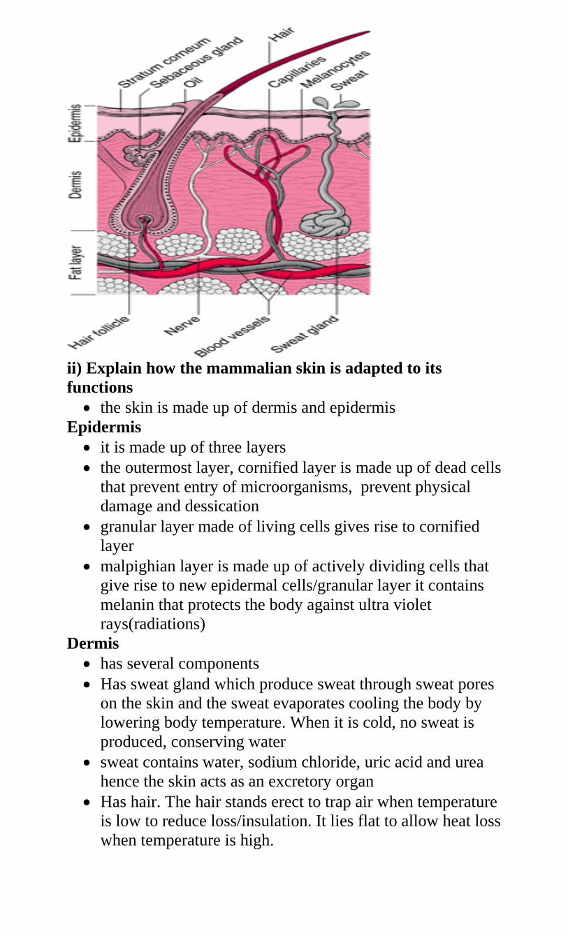
ii) Explain how the mammalian skin is adapted to its
functions
the skin is made up of dermis and epidermis
Epidermis
it is made up of three layers
the outermost layer, cornified layer is made up of dead cells
that prevent entry of microorganisms, prevent physical
damage and dessication
granular layer made of living cells gives rise to cornified
layer
malpighian layer is made up of actively dividing cells that
give rise to new epidermal cells/granular layer it contains
melanin that protects the body against ultra violet
rays(radiations)
Dermis
has several components
Has sweat gland which produce sweat through sweat pores
on the skin and the sweat evaporates cooling the body by
lowering body temperature. When it is cold, no sweat is
produced, conserving water
sweat contains water, sodium chloride, uric acid and urea
hence the skin acts as an excretory organ
Has hair. The hair stands erect to trap air when temperature
is low to reduce loss/insulation. It lies flat to allow heat loss
when temperature is high.
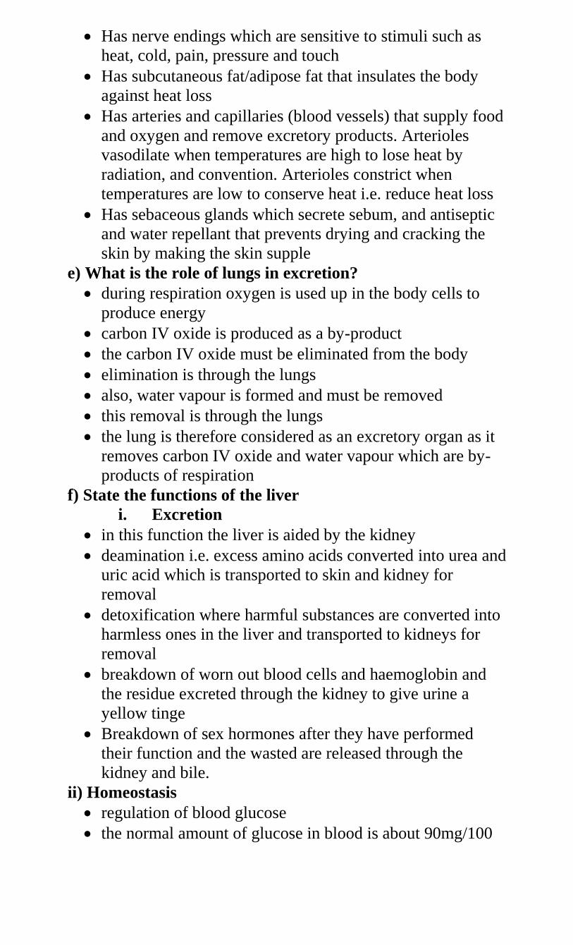
Has nerve endings which are sensitive to stimuli such as
heat, cold, pain, pressure and touch
Has subcutaneous fat/adipose fat that insulates the body
against heat loss
Has arteries and capillaries (blood vessels) that supply food
and oxygen and remove excretory products. Arterioles
vasodilate when temperatures are high to lose heat by
radiation, and convention. Arterioles constrict when
temperatures are low to conserve heat i.e. reduce heat loss
Has sebaceous glands which secrete sebum, and antiseptic
and water repellant that prevents drying and cracking the
skin by making the skin supple
e) What is the role of lungs in excretion?
during respiration oxygen is used up in the body cells to
produce energy
carbon IV oxide is produced as a by-product
the carbon IV oxide must be eliminated from the body
elimination is through the lungs
also, water vapour is formed and must be removed
this removal is through the lungs
the lung is therefore considered as an excretory organ as it
removes carbon IV oxide and water vapour which are by-
products of respiration
f) State the functions of the liver
i. Excretion
in this function the liver is aided by the kidney
deamination i.e. excess amino acids converted into urea and
uric acid which is transported to skin and kidney for
removal
detoxification where harmful substances are converted into
harmless ones in the liver and transported to kidneys for
removal
breakdown of worn out blood cells and haemoglobin and
the residue excreted through the kidney to give urine a
yellow tinge
Breakdown of sex hormones after they have performed
their function and the wasted are released through the
kidney and bile.
ii) Homeostasis
regulation of blood glucose
the normal amount of glucose in blood is about 90mg/100
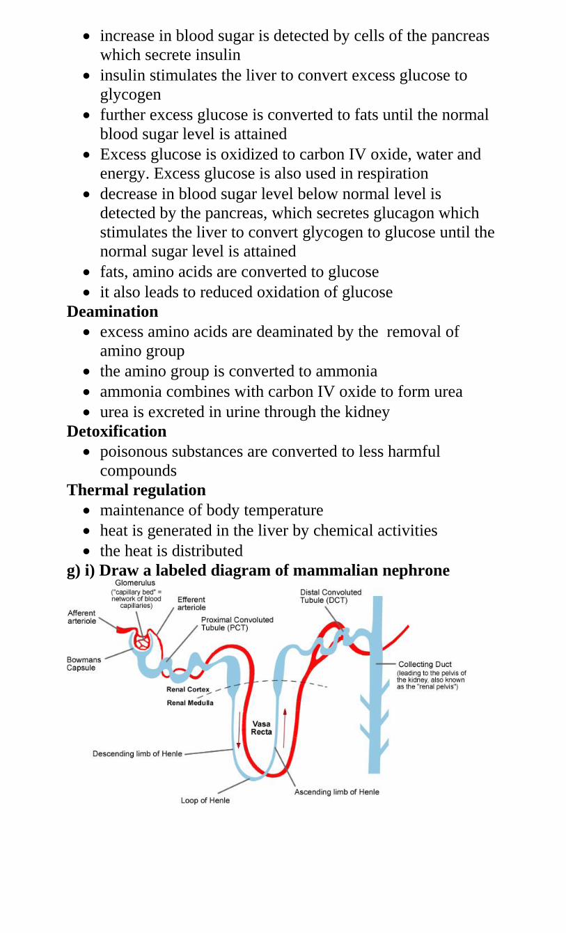
increase in blood sugar is detected by cells of the pancreas
which secrete insulin
insulin stimulates the liver to convert excess glucose to
glycogen
further excess glucose is converted to fats until the normal
blood sugar level is attained
Excess glucose is oxidized to carbon IV oxide, water and
energy. Excess glucose is also used in respiration
decrease in blood sugar level below normal level is
detected by the pancreas, which secretes glucagon which
stimulates the liver to convert glycogen to glucose until the
normal sugar level is attained
fats, amino acids are converted to glucose
it also leads to reduced oxidation of glucose
Deamination
excess amino acids are deaminated by the removal of
amino group
the amino group is converted to ammonia
ammonia combines with carbon IV oxide to form urea
urea is excreted in urine through the kidney
Detoxification
poisonous substances are converted to less harmful
compounds
Thermal regulation
maintenance of body temperature
heat is generated in the liver by chemical activities
the heat is distributed
g) i) Draw a labeled diagram of mammalian nephrone
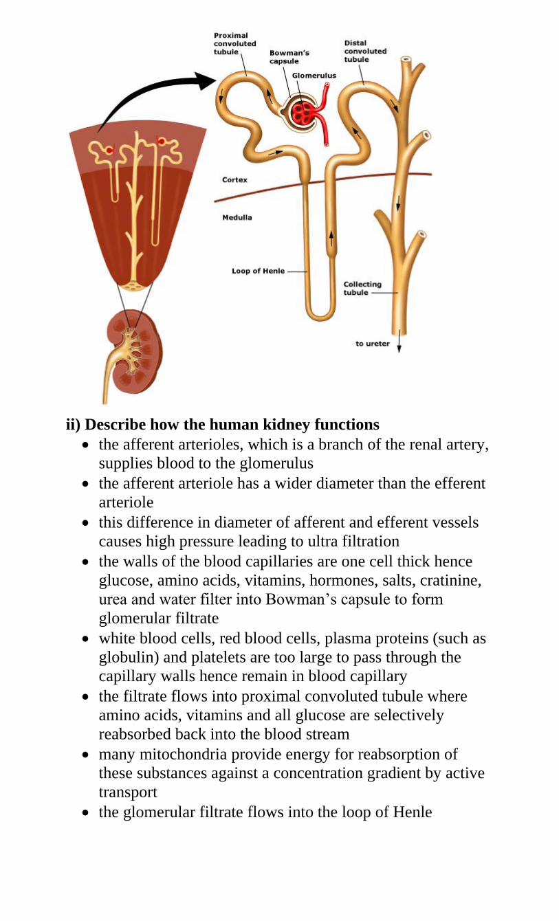
ii) Describe how the human kidney functions
the afferent arterioles, which is a branch of the renal artery,
supplies blood to the glomerulus
the afferent arteriole has a wider diameter than the efferent
arteriole
this difference in diameter of afferent and efferent vessels
causes high pressure leading to ultra filtration
the walls of the blood capillaries are one cell thick hence
glucose, amino acids, vitamins, hormones, salts, cratinine,
urea and water filter into Bowman’s capsule to form
glomerular filtrate
white blood cells, red blood cells, plasma proteins (such as
globulin) and platelets are too large to pass through the
capillary walls hence remain in blood capillary
the filtrate flows into proximal convoluted tubule where
amino acids, vitamins and all glucose are selectively
reabsorbed back into the blood stream
many mitochondria provide energy for reabsorption of
these substances against a concentration gradient by active
transport
the glomerular filtrate flows into the loop of Henle
water in the descending loop moves by osmosis into the
blood capillaries
sodium chloride is actively pumped from the ascending arm
of the loop of Henle into the blood capillaries
the glomerular filatrate flows into the distal convoluted
tubule
water and salts are reabsorbed from distal convoluted
tubule into blood capillaries
the glomerular filtrate flows into collecting tubule (duct)
from where more water is reabsorbed into blood stream
antiduretic hormone influences the amount of water
reabsorbed depending on osmotic pressure of blood
the glomerular filtrate from collecting duct, now referred to
as urine, is emptied into pelvis and ureter into bladder and
out of body through urethra
urine consists of excess water, slats and nitrogenous wastes
iii) State the adaptations of proximal convoluted tubule to its
function
folded to increase surface area for absorption
thin epithelium to reduce distance of diffusion
micro-villi on inner lining to increase surface area for
absorption
folded to reduce speed of flow for efficient absorption
numerous mitochondira to provide energy for reabsorption
dense capillary network to transport reabsorbed products
iv) Name the common kidney diseases
nephritis
kidney stones(renal calculi
cystitis
oedema
kidney failure
6. a) i) Why is homeostatic control necessary?
this provides a constant internal environment so that the
cells of the body have the optimum (best) condition for
their survival
ii) What is internal environment?
immediate surrounding of body cells
refers to tissue fluid within an organism
b) i) Why is constant body temperature maintained by
mammals?
most enzymes in the body function within a narrow range
of temperature
high temperature denatures enzymes
low temperature inactivates and inhibits enzymes
ii) Explain the advantage gained by possessing a constant
body temperature
animals remain active despite fluctuations in environmental
temperature
higher chances of survival in various environments ie they
colonize various environments
chemical processes in their body continues at an optimum
rate
iii) How do mammals regulate body temperature?
the body temperature of a mammal is kept constant
to maintain this temperature the mammal must be able to
balance its heat loss against the heat gain
body temperature is controlled by the hypothalamus, a
specialized part of the brain
changes in the temperature within the body and the
surrounding are detected by the hypothalamus
it transmits impulses to the skin and the blood stream in
response to temperature changes
hypothalamus acts as a thermostat for the body
a mammal loses heat by breathing out, urine, feaces, skin
by radiation and by evaporation of sweat.
A mammal generates heat by the activity of its muscles, by
general metabolism in respiration, or chemical activities
In hot conditions the hypothalamus stimulates responses
that increase heat loss from the body hence lowering the
body temperature
Such responses include sweating, vasodilation, keeping its
hair flat on the surface of skin and reduction of metabolic
rate
In cold conditions the hypothalamus stimulates responses
that generate heat gain in the body and reduce heat loss to
the environment
Such responses include shivering, vasoconstriction, raising
its hair to trap a layer of air around the skin because still air
is a good insulator of heat and by generation of heat by
increasing metabolic rate.
iv) Why does body temperature of a healthy person rise up
to 37oC on a hot humid day?
sweat evaporation is reduced hence cooling is less therefore
more heat is retained in the body causing temperature to
rise
v) Name the structures in the human body that detect
external temperature changes
temperature receptors (end bulb corpuscles e.g. bulb of
Krause (warmth) and organ of Ruffinni (cold)
heat (thermal) receptors
vi) State the advantages that organisms with small surface
area to volume ratio experience over those with larger
heat loss slow hence their body temperature can increase to
intolerable levels
Heat gain from surrounding slower hence may remain
inactive for a long time.
Need specialized and complex transport system and also
gaseous exchange system
Explain why individuals with smaller sizes require more
energy per unit body weight than those with larger sizes.
surface area to volume ratio is higher in smaller individuals
than larger ones, therefore smaller heat is lost faster by
smaller ones than larger ones
they therefore require more energy per unit body weight to
maintain body temperature
c) i) What is the meaning of osmoregulation?
mechanism which regulates osmotic pressure of internal
environment of an organism
the regulation/maintenance of salt/solute-water balance of
an internal environment
ii) State the importance of osmoregulation
- Maintenance of constant level of water and slats (osmotic
pressure) for optimum/suitable conditions for metabolism
suitable for cellular functions
iii) State the ways by which desert mammals conserve water
fewer glomeruli
longer loop of Henle
excretion of dry feaces or concentrated urine
hump for fat to be metabolized to give metabolic water for
use
nocturnal, burrowing, aestivate or hibernate
sweat glands few or absent
more ADH (vasopressin)
iv) Explain why some desert animals excrete uric acid rather
than water
uric acid is less toxic than ammonia, hence elimination of
uric acid requires less water than ammonia therefore more
water conserved
uric acid being less toxic is safer to excrete where there is
less water/desert
v) Explain why eating a meal with too much salt leads to
production of a small volume of concentrated urine
the concentration of salts in the blood rises leading to
production of more ADH hence higher rate of water
reabsorption by kidney tubules
vi) Explain how marine fish regulate their osmotic pressure
swallow plenty of sea water to increase amount of water in
the body
have chloride excretory cells in their gills to remove excess
salts
eliminate nitrogenous wastes in form of trimethalamine
oxide which requires little water for elimination
few/small glomeruli thus slow filtration rate in the kidneys
retain nitrogenous wastes in form of urea to raise osmotic
pressure of body fluids
d) i) What is the biological significance of maintaining a
relatively constant sugar level in a human body?
body cells are surrounded by tissue fluids that are
isotonic/same osmotic pressure as cytoplasm
if sugar level is high/hypertonic, cell will lose water by
osmosis to the surrounding, thus increasing the
concentration of the contents
this changes the physiology of the cell
if the blood sugar is lower than the normal, the cytoplasm
gains water by osmosis, diluting the cell contents, thus
altering the physiology of the cell
ii) Discuss the role of the following hormones in blood sugar
control
Insulin
insulin is produced when there is increase in blood sugar
concentration
it converts glucose to glycogen which is in the liver or
muscle thus lowering sugar level
Glucagon
when glucose level decreases glucagon is produced, which
causes the breakdown of glycogen to glucose thus raising
blood sugar level
e) Explain the part played by antidiuretic hormone in
homeostasis
Produced when there is less water (high osmotic pressure
above normal level of salt concentration) in the blood.
It acts on kidney tubules (nephron) thus increasing water
reabsorption from tubules to the blood stream, thus
restoring osmotic pressure
When there is more water(lower osmotic pressure) or
decreased salt concentration in blood, little or no ADH is
produced, less water reabsorbed hence water loss in urine
(more dilute urine) hence raising the osmotic pressure in
body fluids/blood
f) What is the role of blood clotting in homeostasis?
when a blood vessel is cut, there is exposure of blood
platelets to the air
this triggers fibrinogen to be converted to fibrin
the fibrin forms a clot that prevents body fluids e.g. blood
from being lost
therefore the clot conserves water and salts in the body
g) Describe the role of the following hormones in
homeostasis
i. Aldosterone
concerned with regulation of ionic balance
secreted by the cortex of adrenal glands
it increases sodium ion uptake by the gut and promotes the
reabsorption of sodium ions (and therefore water) in the
kidneys)
this is accompanied by elimination of potassium ions
this raises the overall level of sodium and lowers the
overall level of potassium in the blood
as sodium ions are absorbed in the blood, chlorine ions
follow so as to neutralize the effect of sodium ions
the production of aldosterone is regulated by the
concentration of sodium ions which has an inhibiting
effect, and a fall in sodium ions has a stimulating effect on
the adrenal cortex
the flow of aldosterone is stimulated by the adreno-cortic-
tropic hormone (ACTH) produced in the anterior of the
pituitary gland
however, the main method of control is dependent on the
fact that adrenal cortex itself is somehow sensitive to the
relative concentration of potassium and sodium in the blood
ii. Adrenaline
produced by adrenal glands
in high concentrations, it increases hydrolysis of glycogen
and increases blood sugar
it is usually released in emergency cases to increase
glucose level for respiration
this releases energy for the emergency
h) i) Distinuish between diabetes mellitus and diabetes
insipidus
diabetes mellitus is a condition resulting from insufficient
production of insulin causing hyperglucaemia and presence
of glucose in urine
diabetes insipidus is a condition whereby less or no
antidiuretic hormone is secreted hence a high volume of
water is passed out in urine in a condition called diuresis
ii) How can high blood sugar level in a person be controlled?
administer insulin
iii) Why does glucose not normally appear in urine even
though it is filtered in the mammalian Bowman’s capsule?
glucose molecules are actively reabsorbed in the proximal
convoluted tubules
iv) When is glycogen which is stored in the liver converted
into glucose and released into the blood?
after activity/when blood sugar (glucose) falls below
normal
when glucagon
stimulates the liver/when glucagon is produced
after strenuous/vigorous activity
during starvation
v) How would one find out from a sample of urine whether a
person is suffering from diabetes mellitus?
test or react urine in Benedict’s solution
positive result i.e. orange or red precipitate
Positive result is an indication of diabetes mellitus.
FORM III TOPICS
1. a) i) What is meant by the term binomial
nomenclature?
scientific system of naming organisms using the
generic(genus) and specific (species) names
ii) State briefly the general principles of classification of
living organisms
scientific names must be in Latin or should be latinised
family names are formed by adding the suffix “idea” to the
stem of the genus e.g. the genus Rana become Ranaidea
generic names should be a single unique name
b) State the main characteristics of the five kingdoms of
organisms
i. Monera
e.g. bacteria
unicellular (single celled)
prokaryotic (genetic material not surrounded by membrane)
cell wall without cellulose
lack most organelles
small in size (microscopic)
ii. Protista(protoctista)
single celled(unicellular)
eukaryotic (most cell organelles present)
when cell walls are present have no cellulose
e.g. protozoa and algae
usually microscopic
iii. Fungi
have hyphae (which form mycelia)
absence of chlorophyll
have rhizoids (lack roots, leaves, stem)
have spore forming structures (sporangia)
e.g. mucor, rhizopus
iv. Plantae
most are green/contain chlorophyll
autotrophic/feed by photosynthesis
cells have cellulose cell walls
respond slowly to stimuli (tropism)
lack locomotion (are stationary)
indefinite growth (at meristems)
lack specialized excretory structures
v. Animalia
cells do not have cell walls
most carry out locomotion
heterotrophic
fast response to stimuli (tactic)
have specialized excretory structures
c) Describe the economic importance of:
i. Fungi
some cause decay to our food
some cause diseases to humans and animals e.g. ringworms
may be used as food e.g. mushrooms, yeast
some are used in production of antibiotics e.g. penicillin,
chloromycin, streptomycin
yeast is used in brewing industry, baking and source of
vitamin B
many cause diseases to our crops e.g. late blight
important in recycling nutrients in soil since they cause
decay of organic matter
mycorrhizal association in forest development may help in
water intake/absorption
help in nitrogen fixation
Bacteria
are useful in the manufacture of antibiotics
silage formation,
fermentation of cheese, butter, milk yoghurt
curing of tea, tobacco and retting flax
formation of vitamin B12 and K
enzymes such as amylase and invertase
hormones such as insulin
vinegar, acetic acid, lactic acid, citric acid
in septic tanks and modern sewage works make use of
bacteria
biogas production
saprophytic bacteria are used in compost decomposition or
cause decay
symbiotic bacteria are used in compost decomposition or
cause decay
symbiotic bacteria in herbivores/ruminants help in
digestion
some diseases in animals/humans and plants are caused by
bacteria
many bacteria cause food spoilage/decay
nitrifying and nitrogen fixing bacteria increase soil
fertility/make nitrates available
denitrifying bacteria reduce soil fertility/convert nitrates
into nitrogen/reduce nitrates
d) State the main characteristics of the following division of
kingdom plantae
i. Bryophyte
e.g. mosses and liverworts
presence of rhizoids
lack of vascular tissues (lack phloem and xylem)
body parts not differentiated into root, stem, leaves
capsule or seta
gametophyte generation dominant.
ii. Pteridophyta
e.g. ferns
has true roots, stems and leaves
fond with sori on under-surface
vascular tissues present
sporophyte generation is dominant
iii. Spermatophyte
photosynthetic
well differentiated into roots, stems and leaves
well developed vascular system
seed bearing plants
e) Name sub-divisions of spermatophyte and state the
characteristics of each class
i. Gymnospermae (cornifers)
naked seeds (exposed
are all woody trees
reproduce by means of cones
show xerophytic characteristics
xylem have tracheids but lack vessels
phloem lacks companion cells
single fertilization
pollen lands directly on ovules
ii. Angiospermae (flowering plants)
reproduce by flowers
seeds enclosed (in fruits)
flowers bisexual hence double fertilization
herbaceous
pollen grains land on stigma of pistil
xylem contains vessels
phloem contains companion cells
ovules contained in ovary
iii. Name the classes and state characteristics of
angiospermae
Dicotyledonae
two seed leaves
network venation of leaves
regularly arranged vascular bundles
tap root system
broad leaves
secondary growth occurs
Monocotyledonae
one seed leaf
parallel venation of leaves
irregularly arranged vascular bundles
fibrous root system
narrow leaves
sheath like leaf stalk (petiole)
no secondary growth
iv) State the importance of plants
balancing carbon IV oxide and oxygen in the atmosphere
during photosynthesis and respiration
influence water cycle
reduce soil erosion by bind soil particles together
useful products e.g. food, medicine, timber, paper and
clothing
habitat ( e.g. forests and grassland) for animals which may
also be tourist attraction
earn money from sales of products
aesthetic value/beauty e.g. flowers, shade/shelter, live
fences, windbreaks
Some are harmful e.g. poisons, weeds, injurious (stinging
nettles, thorns), water hyacinth.
f) i) Give the general characteristics of phylum arthropoda
jointed appendages
presence of exoskeleton
triploblastic and coelomate
segmented body
bilateral symmetry (similar halves)
ii. State the characteristics of the following classes of
arthropoda
Diplopoda
the millipedes
two pairs of legs per segment
many segments
terrestrial habitat
body cylindrical and long
herbivorous
one pair of antennae
Chilopoda
the centipedes
one pair of legs per segment
many segments
terrestrial habitat
body long and ventro-dorsally flattened
carnivorous
last pair of legs pointing backwards with poison claws
called maxillipedes
one pair of antennae
Insecta
three body parts i.e. head thorax, abdomen
six legs/three pairs of legs
a pair of compound eyes
presence of wings
a pair of antennae
Crustacean
two body parts
segmented body
have pincers (modified legs) to catch prey
have hard exoskeleton
a pair of compound eyes
Arachnida
body divided into two parts( abdomen and cephalothorax)
simple eyes
eight legs (four pairs of legs)
iii) State the economic importance of insects
Beneficial effects
food supply
important in food chains
pollinators
biological control of pests and other organisms
aesthetic value
contribute to decomposition e.g. litter feeders like beetles
Harmful effects
pests
vectors
dirt and disease carriers
injurious e.g. stings and bites
g) i) State the general characteristics of chordate
notochord
dorsal slits (pharyngeal cleft during development)
bilateral symmetry
triploblastic (three layer body-ectoderm, mesoderm and
endoderm)
clear cut head formation
multilayered epidermis
post anal tail
closed circulatory system
segmented muscle blocks(myotomes)
single pair of gonads
Give the characteristics of the following classes of chordate
Pisces
presence of fins for locomotion
two chambered heart
presence of overlapping scales
presence of gills or operculum for gaseous exchange
presence of lateral line for protection
streamlined body
poikilothermic (body temperature varies with that of
environment)
Amphibian
partially live in fresh water and partially on land
poikilothermic
pentadactylous with two pairs of limbs
webbed feet for locomotion in water
body streamlined
heart is three chambered
moist skin for gaseous exchange
Reptilia
scales on body
poikilothermic
homodont teeth except tortoise and turtle
all have limbs except snakes
skin is dry
oviparous (lay eggs)
no pinna (external ear)
three chambered heart 9crocodile has four chambers)
skin not glandular
no mammary glands
Aves
the birds
homoeothermic (constant body temperature)
four chambered heart
streamlined body for locomotion in air
skin dry and covered by feathers
scales on legs
hollow bones
oviparous (lay eggs)
mouths modified into beaks
Mammalian
hair on the body
homoeothermic
viviparous (give birth to live young) except a few
have mammary glands
glandular skin e.g. sweat glands, sebaceous glands
four chambered heart
pinna (external ear)
two pairs of pentadactyl limbs
presence of diaphragm
have salivary glands
i) i) What is a dichotomous key?
A biological device (tool) which enables one to identify an
organism by progressively opting between two alternative
observable characteristics
ii. State the necessity of using a dichotomous key
used to identify organisms quickly and accurately
by following the statements in the key we are able to
identify each organism on the basis of a characteristic
which is not to be found in other specimens
iii. List the rules followed in constructing a
dichotomous key
use observable characteristics only
start with major characteristics, placing organisms into two
groups at each stage
use a single characteristics at a time
use contrasting characteristics at each stage e.g 1(a) short,
1(b) tall
avoid repeating the same characteristics
iv) Describe the procedure of using a dichotomous key.
Make a list of major features of the characteristics to be
identified
look at the features of similarities
look at the features of differences between the organisms
we can then be able to identify the organisms by
distinguishing one from another
the key uses a method of elimination by following
statements that are correct only for the organism
iv You are provided with a specimen kale leaf. Use the
dichotomous key below to identify the taxonomic group to
which the specimen belongs. Show the steps (number and
letter) in the key that you followed to arrive at the identify of
the specimen
1 a) leaf broad go to 2
b) leaf narrow Araicaria
2 a) leaf parallel veined Cynodon
b) leaf net-veined go to 3
3 a) leaf with one lobe (simple) go to 4
b) leaf with many lobes (compound) Grevellea
4 a) leaf fleshy Kalanchoa
b) leaf not fleshy go to 5
5 a) leaf petiole modified to form sheath go to 6
b) leaf petiole not modified to form sheath Brassica
6 a) leaf purple Tradescantia
b) leaf green Commelina
steps – 1a, 2b, 3a, 4b, 5b
Identify – Brassica
v) You have been provided with four animals labeled K
(mature adult housefly), L (mature adult grasshopper,
M(maize flour beetle) and N(worker termite) use the
dichotomous key below to identify the specimens. Write
down in the correct order, the steps (number and letter) in
the key that you followed to arrive at your answer.
Dichotomous key
1 a) animal with wings go to 2
b) animal without wings go to 7
2 a) with two pairs of wings go to 3
b) with one pair of wings Diptera
3 a) with membranous wings go to 4
b) hind pair of membranous wings go to 6
4 a) with long abdomen Odontata
b) medium sized abdomen go to 5
5 a) wings with coloured scales Lepidoptera
b) wings without scales hymenoptera
6 a) forewings hard and shell-like coleopteran
b) forewings hard but not shell-like Orthoptera
7 a) body horizontally flattened Isoptera
b) body laterally flattened Symphonoptera
Identify the orders o the various specimens as per the table
below
Specimen Order Steps followed
K-housefly
L- grasshopper
M- beetle
M-termite
Diptera
Orthoptera
Coleoptera
Isoptera
1a,2b
1a, 2a, 3b, 6b
1a, 2a, 3b, 6a
1b, 7a
2 a) Define the following ecological terms
i. Ecology
study of the interrelationships between organisms and their
environment
ii. Environment
surrounding of the organism i.e. biotic or a biotic factors
iii. Habitat
A specific locality (home) of a living organism with a set of
factors (conditions) in which an organism lives.
iv. Ecological niche
Role of an organism in its habitat e.g. feeding relationship
v. Population
Number (group) of organisms of a species occupying a
given habitat
vi. Community
Refers to different species of (plants and animals)
organisms in a given habitat (area) co-existing or
interacting (living) with each other and the environment in
which they live
vii. Ecosystem
A community of organisms interacting with one another
and the environment in which they live
viii. Biosphere
The earth and its atmosphere where living organisms are
found
ix. Autecology
Study of a single (individual) species of plants or animals
within a community, ecosystem, habitat or environment.
x. Synecology
Study of natural communities (plants and animals) or
populations interacting within an ecosystem.
xi. Carrying capacity
maximum number of organisms an area can support
without being depleted
xii. Biome
geographical area with particular climatic conditions and
flora and fauna
it constitutes many ecosystems
xiii. Biomass
dry weight (mass) of a living organism in a given area
units of measurement are kg/m2/year
b) i) What are abiotic factors?
non-living components of the ecosystem
ii) Explain how abiotic factors affect living organisms
Wind
this influences rate of water evaporation from organisms
therefore it affects distribution of organisms e.g. wind
increases rate of transpiration and evaporation of water
from the soil
wind is an agent of soil erosion, may break and uproot trees
may aid in the formation of sand dunes which can form
habitats for some desert plants
wind disperses fruits, seeds, spores
wind forms waves in lakes and oceans which enhances
aeration of water which replenishes oxygen concentration
necessary for life
wind is an agent of pollination
Temperature
influences rate of enzyme action in photosynthesis and
other metabolic reactions in plants and animals
organisms function within a narrow range of temperature
it affects distribution of organisms
changes in temperature affect rate of photosynthesis and
biochemical reactions e.g. metabolism and enzyme reaction
temperature increases rate of transpiration
Light
needed by green plants and photosynthetic bacteria which
are primary producers
animals depend on plants directly or indirectly for food
main source of light is the sun
light is necessary for synthesis of vitamin D in certain
animals
some plants need light for flowering
seeds like lettuce need light for germination
Humidity
amount of water vapour held by the air
affects the rate at which water is lost from organisms body
by evaporation and stomatal transpiration
when humidity is low the rate of transpiration increases
humidity influences distribution of organisms
PH
each plant requires a specific PH in which to grow (acidic,
neutral or alkalinic)
pH affects enzyme reaction in metabolism
Salinity
some ions are needed for plant and animal nutrition
osmoregulation implants and animals is affected by salinity
Topography
altitude affects light, atmospheric pressure and light
Slope influences surface runoff, wind erosion, etc.
mountains affect distribution of organisms which differs in
leeward side and windward side
mountains affect distribution of organisms which differ on
lowlands and on highlands
mountains also form physical barriers to migration of
organism and may cause isolation of species
background may offer camouflage to some organisms
hence protection from enemies
Rainfall (water) or precipitation
amount and distribution of rainfall affect vegetation type
this consequently affects distribution of animals e.g. polar
region water frozen hence only well adapted organisms
survive
fewer organisms found in deserts where rainfall is less
Water is required for seed germination, raw material for
photosynthesis, solvent for mineral salts. Provides turgidity
for plant support, medium for transport, disperses fruits,
seeds and spores
Pressure
the weight atmosphere exerts upon the earth
varies with altitude 9the higher the altitude the less the
pressure
this variation implies change in density which directly
means less oxygen for respiration and less carbon iv oxide
for photosynthesis and this affects distribution of organisms
Mineral salts (trace elements)
these affect distribution of plants in the soil
plants thrive best where elements are available
Plants living in soil deficient in a particular element must
have special methods of obtaining it.
They harbor nitrogen fixing bacteria and others have
carnivorous habit
Plant distribution influences animal distribution
c) i) What are biotic factors?
refers to living organisms in an area
biotic environment of an organism constitutes all organisms
around it, which it relates or interacts with in various ways
ii) Give examples of biotic factors affecting ecosystems
feeding relationships
predation
competition
diseases and pests
human activities
d) Discuss how the various biotic factors affect living
organisms
i. Competition
organisms compete with one another for food, light, water,
mates and shelter
organisms must live together for competition for available
resources
those which cannot cope either structurally or behaviorally
will migrate or die
those remaining, due to better adaptations will increase in
population
competition between members of the same species is called
intra-specific competition e.g. for mates
Competition between members of different species is inter
specific competition e.g. for food and space.
ii. Predation
this is predator-prey relationship
predator feeds on prey hence both control the other’s
population
Distribution of predator and prey is important as predator
cannot survive without prey
It there is no predator the prey will increase in population
beyond carrying capacity hence die due to environment
depletion
iii. Parasitism
an association where an organism lives in or on another
living organism obtaining food(and other benefits) from it,
causing harm to it (without necessary killing it)
parasites may kill host
they deprive host of food
make host weak by introducing diseases
make reproductive ability of host low hence host becomes
susceptible to predation
iv. Diseases and parasites
make organisms weak and susceptible to predation
kill organisms and reduce their population
v. Symbiotic
and association of organisms of different species where
both benefit from the association i.e. there is mutual benefit
vi. Human activities
these are human factors which have an influence on the
biosphere
examples are road construction, industrialization,
deforestation, agriculture, pollution, poaching, fishing
conservation, population control
affect ecosystem and balance of nature
Saprophytism
saprophytes are organisms which obtain organic matter in
solution from dead and decaying tissues of plants and
animals
they include saprophytic bacteria and fungi
they make available carbon, nitrogen and other elements
form dead to living organisms
they are useful in recycling nutrients in nature
e)i) What is nitrogen cycle?
The process by which nitrogen in the air is made available
plants and animals and eventually returns to the air.
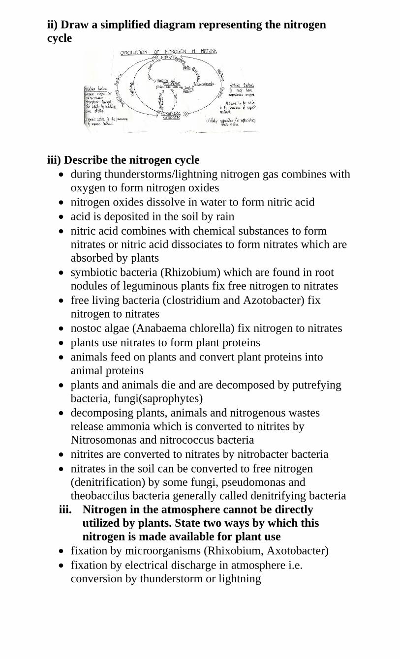
ii) Draw a simplified diagram representing the nitrogen
cycle
iii) Describe the nitrogen cycle
during thunderstorms/lightning nitrogen gas combines with
oxygen to form nitrogen oxides
nitrogen oxides dissolve in water to form nitric acid
acid is deposited in the soil by rain
nitric acid combines with chemical substances to form
nitrates or nitric acid dissociates to form nitrates which are
absorbed by plants
symbiotic bacteria (Rhizobium) which are found in root
nodules of leguminous plants fix free nitrogen to nitrates
free living bacteria (clostridium and Azotobacter) fix
nitrogen to nitrates
nostoc algae (Anabaema chlorella) fix nitrogen to nitrates
plants use nitrates to form plant proteins
animals feed on plants and convert plant proteins into
animal proteins
plants and animals die and are decomposed by putrefying
bacteria, fungi(saprophytes)
decomposing plants, animals and nitrogenous wastes
release ammonia which is converted to nitrites by
Nitrosomonas and nitrococcus bacteria
nitrites are converted to nitrates by nitrobacter bacteria
nitrates in the soil can be converted to free nitrogen
(denitrification) by some fungi, pseudomonas and
theobaccilus bacteria generally called denitrifying bacteria
iii. Nitrogen in the atmosphere cannot be directly
utilized by plants. State two ways by which this
nitrogen is made available for plant use
fixation by microorganisms (Rhixobium, Axotobacter)
fixation by electrical discharge in atmosphere i.e.
conversion by thunderstorm or lightning
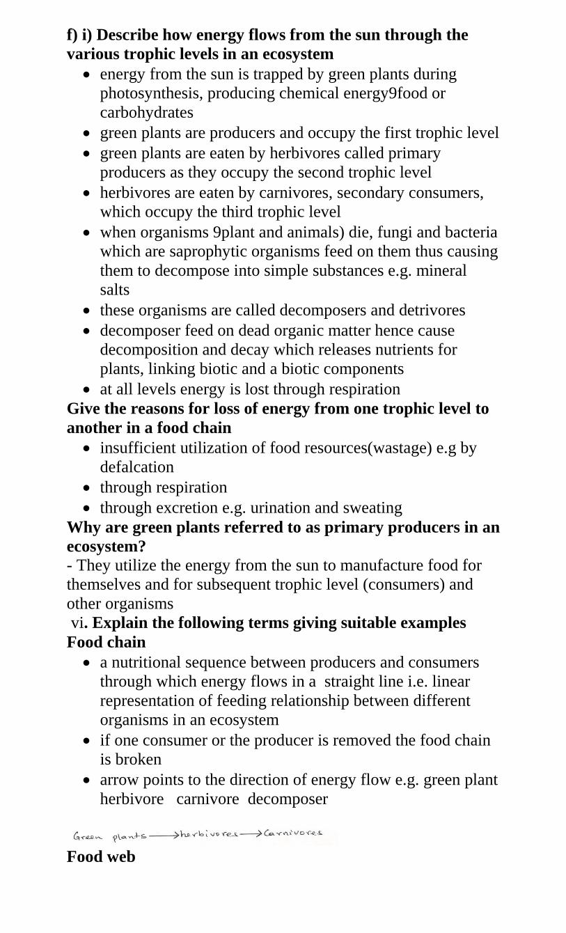
f) i) Describe how energy flows from the sun through the
various trophic levels in an ecosystem
energy from the sun is trapped by green plants during
photosynthesis, producing chemical energy9food or
carbohydrates
green plants are producers and occupy the first trophic level
green plants are eaten by herbivores called primary
producers as they occupy the second trophic level
herbivores are eaten by carnivores, secondary consumers,
which occupy the third trophic level
when organisms 9plant and animals) die, fungi and bacteria
which are saprophytic organisms feed on them thus causing
them to decompose into simple substances e.g. mineral
salts
these organisms are called decomposers and detrivores
decomposer feed on dead organic matter hence cause
decomposition and decay which releases nutrients for
plants, linking biotic and a biotic components
at all levels energy is lost through respiration
Give the reasons for loss of energy from one trophic level to
another in a food chain
insufficient utilization of food resources(wastage) e.g by
defalcation
through respiration
through excretion e.g. urination and sweating
Why are green plants referred to as primary producers in an
ecosystem?
- They utilize the energy from the sun to manufacture food for
themselves and for subsequent trophic level (consumers) and
other organisms
vi. Explain the following terms giving suitable examples
Food chain
a nutritional sequence between producers and consumers
through which energy flows in a straight line i.e. linear
representation of feeding relationship between different
organisms in an ecosystem
if one consumer or the producer is removed the food chain
is broken
arrow points to the direction of energy flow e.g. green plant
herbivore carnivore decomposer
Food web
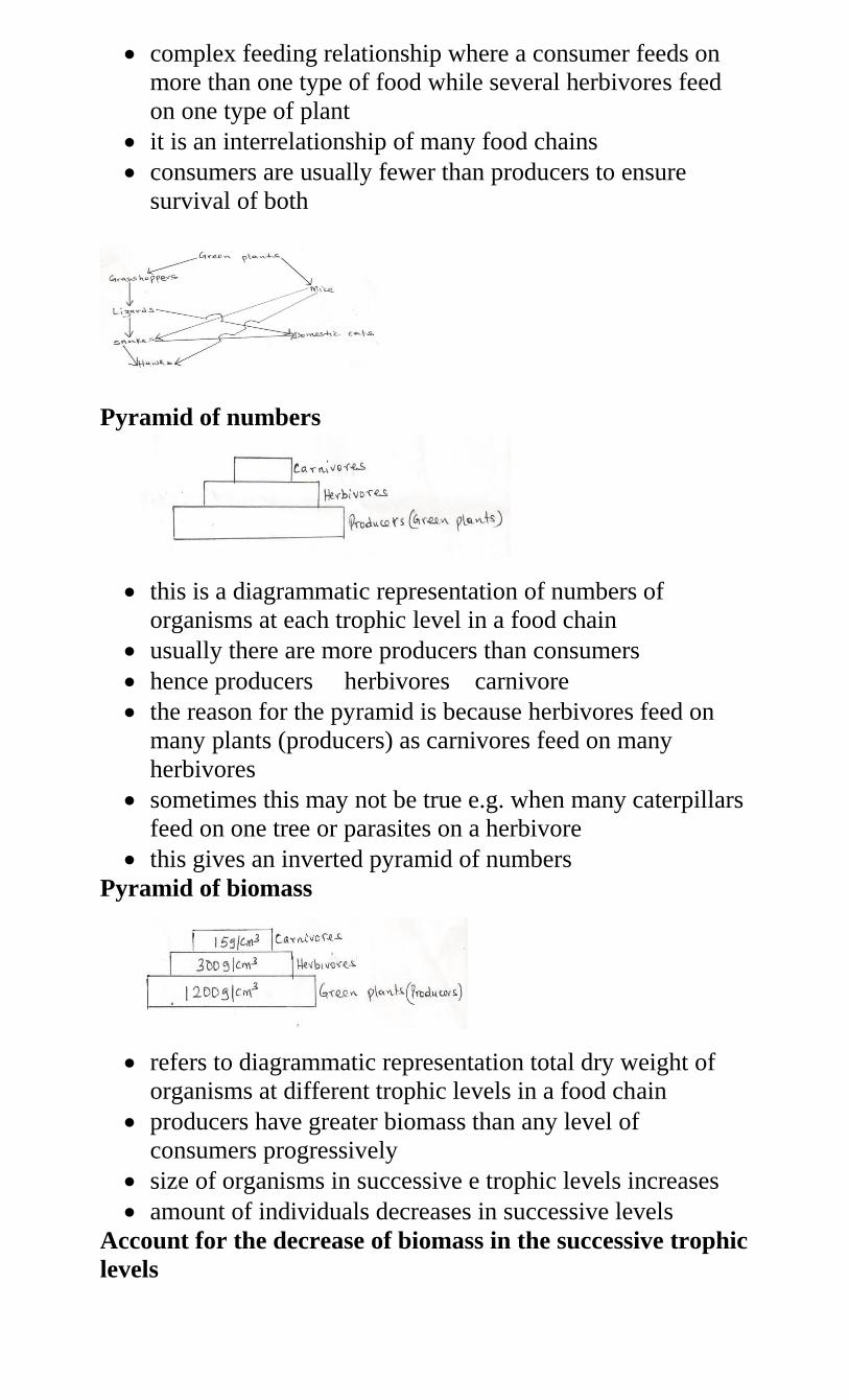
complex feeding relationship where a consumer feeds on
more than one type of food while several herbivores feed
on one type of plant
it is an interrelationship of many food chains
consumers are usually fewer than producers to ensure
survival of both
Pyramid of numbers
this is a diagrammatic representation of numbers of
organisms at each trophic level in a food chain
usually there are more producers than consumers
hence producers herbivores carnivore
the reason for the pyramid is because herbivores feed on
many plants (producers) as carnivores feed on many
herbivores
sometimes this may not be true e.g. when many caterpillars
feed on one tree or parasites on a herbivore
this gives an inverted pyramid of numbers
Pyramid of biomass
refers to diagrammatic representation total dry weight of
organisms at different trophic levels in a food chain
producers have greater biomass than any level of
consumers progressively
size of organisms in successive e trophic levels increases
amount of individuals decreases in successive levels
Account for the decrease of biomass in the successive trophic
levels
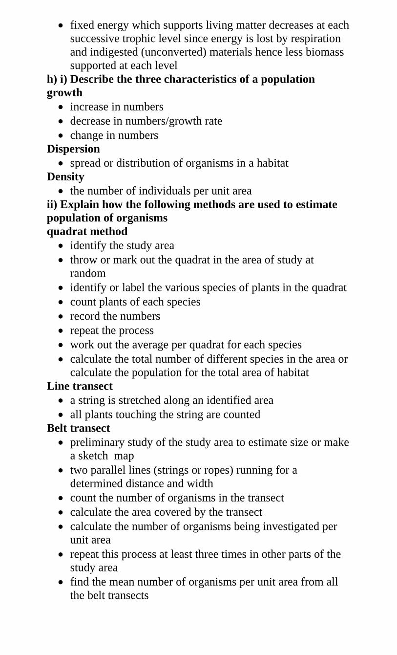
fixed energy which supports living matter decreases at each
successive trophic level since energy is lost by respiration
and indigested (unconverted) materials hence less biomass
supported at each level
h) i) Describe the three characteristics of a population
growth
increase in numbers
decrease in numbers/growth rate
change in numbers
Dispersion
spread or distribution of organisms in a habitat
Density
the number of individuals per unit area
ii) Explain how the following methods are used to estimate
population of organisms
quadrat method
identify the study area
throw or mark out the quadrat in the area of study at
random
identify or label the various species of plants in the quadrat
count plants of each species
record the numbers
repeat the process
work out the average per quadrat for each species
calculate the total number of different species in the area or
calculate the population for the total area of habitat
Line transect
a string is stretched along an identified area
all plants touching the string are counted
Belt transect
preliminary study of the study area to estimate size or make
a sketch map
two parallel lines (strings or ropes) running for a
determined distance and width
count the number of organisms in the transect
calculate the area covered by the transect
calculate the number of organisms being investigated per
unit area
repeat this process at least three times in other parts of the
study area
find the mean number of organisms per unit area from all
the belt transects
from this figure calculate the total population of the desired
organisms in the study area.
Capture-recapture method
e.g. grasshoppers or fish
capture the grasshoppers
count and mark using permanent ink
record
release and allow time
recapture and count the marked and unmarked
total population is equal to the number of marked and
unmarked grasshoppers in the second sample multiplied by
the number of marked grasshoppers in the first sample
divided by number of grasshoppers marked in the second
sample that were recaptured
2. a) Describe the adaptations of plants to various
habitats
i. Xerophytes
grow in areas with scarcity of water
roots grow deeply and extensively (widely spread) to
ensure access to water
thick succulent stems, roots and leaves for water storage
photosynthetic stems take place of leaves which would lose
a lot of water
Leaves are needle-like (reduced to spines), scaly, have
sunken stomata. Some have curled (rolled) leaves. Some
have thick waxy cuticle, reduced number of stomata to
reduce water loss by transpiration
some shed leaves during dry season to reduce water loss
presence of thorns for protection
short life cycle to ensure survival
reversed stomatal rhythm
ii. Hyrophytes
grow in places with plenty of water(waterlogged)
aerenchyma a tissue (airspaces) and large intercellular
spaces and long fibrous roots for buoyancy (floating in
water)
poorly developed support tissues (sclerenchyma) because
water provides the necessary support
upper epidermis of leaves have more stomata than lower
epidermis for gaseous exchange or for increased rate of
transpiration
poorly developed conducting tissues (xylem and phloem)
because plants obtain water by diffusion
iii. Mesophytes
grow in well watered soils (common plants)
no special adaptations, but depending on particular habitat,
may have some adaptations
in forests they grow fast, tall to capture light. Have
climbers while some are adapted to carry out
photosynthesis in low light intensities (those that form
undergrowth)
in places with adequate water they form broad leaves, thin
cuticle and many stomata on both leaf surfaces
in direr regions they possess more stomata on the lower leaf
surface and are deep rooted
some are shallow rooted and develop buttress and prop
roots for support
some have waxy or glossy surface to reflect sun rays and
drip off rain water
iv. Halophytes
plants that grow in very salty soil where the salt
concentration is higher than that in the plant
have root cells which concentrate a lot of salts in them and
enable then to take in water by osmosis
succulent roots to store water
have pneumatophores (breathing roots) to take in oxygen
some have buttress roots for support
secrete excess salt by use of salt glands
have large airspaces in leaves and stems for buoyancy and
to store air
capable of photosynthesis at low light intensities
e.g. mangrove
b) i) What is pollution?
any process which leads to adverse or harmful changes in
the environment
ii) Explain the various human activities that have caused
pollution
Causes and effects of air pollution
sulphur iv oxide, hydrogen sulphide, chlorine, oxides of
nitrogen produced by industries, sewage, decomposing
organic matter and fumes affect gaseous exchange, makes
acid rain and damage plant leaves
aerosols, herbicides, insecticides (agrochemicals), paint
spays, acaricides and CFC’s sprayed to control diseases,
pests and weeds affect respiratory organs of animals. The
chemicals are residual and persistent (not easily broken
down) and bring depletion of the ozone layer
smoke and fumes produced in areas with heavy industries,
motor vehicles, fires which burn fuel, oil, wood and coal
cause carbon ii oxide, poisoning affect respiratory systems
and affect visibility
particles in smoke and fumes settle on leaves and stop
photosynthesis
carbon iv oxide causes green house effect which causes
temperature inversion as a result of heating the lower layers
of atmosphere
sound and noise produced incessantly by machines,
aeroplanes and heavy vehicles affect hearing in animals
dust from cement factories, quarries, dust roads settles on
leaves limiting photosynthesis
removal of vegetation interferes with carbon cycle
radio-active emissions from nuclear reactors, mines and
bombs cause cancer, mutations and death.
Control of air pollution
use of lead free petrol in motor vehicles, air craft,
aeroplanes and petroleum engines
uses of smokeless fuels and electricity
filtration, dissolution and use of chemicals to remove
harmful gases
factories should be erected far away from residential areas
use of tall chimneys
reduce volume or intensity of sound e.g. by use of ear
muffs
concords should fly at higher altitudes and aeroplanes to
fly high up
State the causes, effects and methods of controlling and
prop roots for support water pollution
Causes and effects
agrochemicals e.g. fertilizers cause eutrophication leading
to increase in animal population
Silting makes water surfaces shallow and silt clogs stomata
and gills of fish reducing rates of photosynthesis and
gaseous exchange. It also leads to reduction of algae which
causes reduction of consumers i.e. animal population
industrial and domestic wastes contain toxic materials
which kill producers and other organism while oily
substances in wastes may clog gills of fish and may change
pH of water oxygen solubility is also reduced by oily
surfaces
Untreated sewage and effluents where decomposition or
organic matter in sewage reduces oxygen supply and
sewage provides food for bacteria increasing their
population and demand for oxygen thus depriving fish of
oxygen.
Human feaces causes eutrophication, carbon IV oxide
produced by decomposition of faecal matter changes pH of
water interferes with photosynthesis and may clog fish gills
or block light penetration which interferes with producers
thereby decreasing productivity.
Dumping of chemicals from industries with toxic pollutants
which kill organisms
Spillage of oil and chemicals block oxygen and kill
organisms
Discharge of water from industries into water body where
high temperatures reduce amount of oxygen in the water
causing organism to suffocate and die
Untreated sewage may lead to outbreak of epidemics
Control of water pollution
-pollution caused by domestic effluents may be controlled by
treating domestic waste, using biotechnology, banning the use of
phosphate-based detergents, using plastic pipes instead of those
made from lead, recycling gabbage, using biodegradable
detergents.
Pollution caused by industrial waste may be controlled by
treating/cooling industrial waste, carrying out environmental
impact assessment before establishing industries
Oil spillage may be controlled by cleaning spilled oil
biotechnology and penalizing the industry individual or
companies which cause oil spills/water pollution
Pollution caused by agrochemicals may be controlled by using
mechanical control of weeds, biological control of weeds and
pests, biodegradable organic fertilizer herbicides, insecticides
pesticides, organic farming educate farmers on the use of correct
amount of agrochemicals
silting may be controlled by appropriate farming practices,
contour farming, reafforestation, building gabions and
terracing
iv) State the causes /effects and control methods of soil
pollution
Causes and effects
Air pollutants e.g. sulphur IV oxide fumes from sulphuric
acid with rain water. The acid rain alters soil pH therefore
affecting plants that cannot tolerate acidic soil
most aerosols sprayed to control pests and diseases
precipitate in the soil and are taken up by plants which
make its concentration many times higher, increasing the
toxicity in the plants which absorb them
petroleum products due to spillage by oil tankers making it
impossible for plant roots to obtain oxygen in oil saturated
soils, therefore plants are killed
agrochemicals and inorganic fertilizers contain heavy
metals that are not used up by plants and eventually soil
microorganisms cannot inhabit the soils
organic matter slows down, life ceases and soil becomes
exhausted
community, household wastes and industrial wastes
disposal is a major problem in big towns and cities.
commodities packaged in metal tins, rubber, plastic
containers, scrap metal, glass bottles, different types of
paper are nuisance to the environment, rendering it useless
for agricultural purposes
Control of soil pollution
use of organic farming techniques
biological control of pests, diseases, parasites
recycling of non-degradable containers or burying them
safely after use
controlled burning of garbage
treatment of human and industrial waste for safe disposal
avoid spilling chemicals and oil when used
v) Define biological control give suitable examples
using a living organism to regulate, control or reduce the
population of another organism e.g beetles to feed on water
hyacinth, fish to feed on mosquito larvae.
vi) What is eutrophication?
enrichment of water bodies with nutrients due to discharge
of sewage leading to rapid growth of surface plants
viii) What are the effects of eutrophication?
enrichment of water bodies with nutrients due to discharge
of sewage leading to rapid growth of surface plants
vii) What are the effects of eutrophication?
- The plants block light from reaching plants underneath hence
no photosynthesis
The plants die and decompose leading to lack of oxygen hence
animals also die
c) Describe the symptoms, mode of transmission and control
of cholera, typhoid malaria and amoebic dysentery in
humans
Disease Causative
agent
Transmissio
n
Symptom
s
Control
Cholera Vibrio
cholerae(bac
terium)
Contaminate
d food or
water
Spread by
flies form
faeces
Intestinal
pain
Diarrhea
Vomiting
Dehydrati
on
Proper
hygiene e.g.
boiling
drinking
water
Vaccination
Typhoid Salmonella
typhi
(bacterium)
contami
nated
food or
water
spread
by flies
from
faeces
fever
rashe
s
diarr
hea
+
bloo
d
from
bowe
ls
proper
sanitati
on
vaccina
tion
Malaria Plasmodium
(protozoa)
- bite by
infected
female
anopheles
mosquito
- fever
Joint
pains
Vomiting
Headache
Anaemia
killing
mosqui
to
killing
the
mosqui
to
larvae
drainin
g
stagnan
t water
clearin
g
bushes
treatme
nt
sleep
under
mosqui
to net
Amoebi
c
dysenter
y
(amoebi
asis)
Entamoeba
hystolytica
(bacterium)
-
contaminated
food or water
due to
improper
disposal of
faeces
-
intestinal
pain
Diarrhea
Vomiting
Dehydrati
on
sanitati
on
person
al
hygien
e
cook
food
well
treatme
nt
using
drugs
d) Discuss Ascaris lumbricoides under the following sub-
headings
i. Mode of transmission
through ingestion of contaminated food
live in intestines
ii. Effects of parasite on the host
inflammation of lungs
pneumonia
produce toxic substances
intestinal obstruction
iii. Adaptations
thick cuticle which protects it against digestion
lays many eggs to ensure survival
mouthparts for sucking partly digested food
lack of elaborate alimentary canal
tolerant to low oxygen concentration
two hosts to ensure survival
eggs have protective cover to ensure survival in adverse
environments
iv. Control and prevention
proper sanitation
wash hand after defaecation and before eating
e) Discuss schistosoma under the following sub-headings
i. Mode of transmission
through contaminated water in swamps, etc
ii. Effects on host
bleeding in lungs
blood stained urine
unthriftiness
iii. Adaptations
has two hosts to increase chances of survival
eggs have a hook like structure which raptures the walls of
intestine or bladder
lay large number of eggs to ensure survival
larvae have a sucker for attachment on human skin which it
digests
larva has a tail which it swims with in search of host in
water
prolonged association between male and female to ensure
that fertilization takes place
adults can tolerate low oxygen concentration (in the animal
tissues)
adult worm secretes chemicals against antibodies
larvae and eggs (have glands that) secrete lytic enzymes to
soften the tissues that ease penetration
larvae are encysted so as to survive adverse conditions
iv) Control and prevention
proper use of toilet facilities
boiling water before use
avoid bathing/washing in infected water
Use of molluscicides (chemicals that kill snails/biological
control/clearing water weeds on which snails feed.
Drainage of stagnant water
3. Wearing gum/rubber boots
a) i) What is reproduction?
process by which living organisms give rise to new
members of their own species which resemble the parents
ii) Why is reproduction important?
for continuity of species/ to ensure survival of species
maintaining life of species
replace dead individuals
iii) Name the types of reproduction

sexual which involves fusion of male and female gametes
asexual in which no gametes are involved but parts of a
mature organism develops into new individuals
b) i) What is cell division?
process by which cells are formed from pre-existing cells
ii) What are chromosomes?
Threadlike structures found in nucleus of a cell.
The units called genes
Genes are factors that cause inheritance or determine
characteristics of offspring
c) i) What is mitosis?
A type of cell division that occurs during growth leading to
increase in number of cells
all cells maintain the same chromosome constitution i.e. the
diploid state
ii) Describe the five stages of mitosis
Interphase
replication of organelles
duplication of DNA
production of energy (ATP) for cell division
Prophase
stage of dehydration
chromosomes shorten and thicken
chromosome replicates into two chromatids
chromatids joined at centromere
formation of spindle fibers
Metaphase
chromosomes move to equator (early metaphase)
chromosomes line up at the equator
homologous chromosomes do not associate
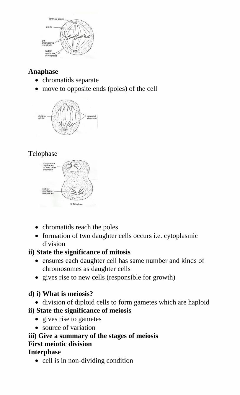
Anaphase
chromatids separate
move to opposite ends (poles) of the cell
Telophase
chromatids reach the poles
formation of two daughter cells occurs i.e. cytoplasmic
division
ii) State the significance of mitosis
ensures each daughter cell has same number and kinds of
chromosomes as daughter cells
gives rise to new cells (responsible for growth)
d) i) What is meiosis?
division of diploid cells to form gametes which are haploid
ii) State the significance of meiosis
gives rise to gametes
source of variation
iii) Give a summary of the stages of meiosis
First meiotic division
Interphase
cell is in non-dividing condition
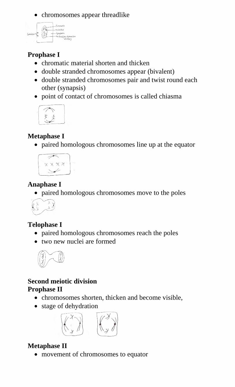
chromosomes appear threadlike
Prophase I
chromatic material shorten and thicken
double stranded chromosomes appear (bivalent)
double stranded chromosomes pair and twist round each
other (synapsis)
point of contact of chromosomes is called chiasma
Metaphase I
paired homologous chromosomes line up at the equator
Anaphase I
paired homologous chromosomes move to the poles
Telophase I
paired homologous chromosomes reach the poles
two new nuclei are formed
Second meiotic division
Prophase II
chromosomes shorten, thicken and become visible,
stage of dehydration
Metaphase II
movement of chromosomes to equator
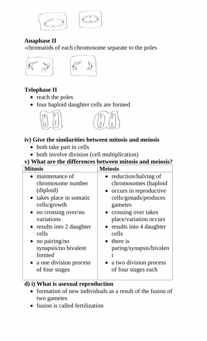
Anaphase II
-chromatids of each chromosome separate to the poles
Telophase II
reach the poles
four haploid daughter cells are formed
iv) Give the similarities between mitosis and meiosis
both take part in cells
both involve division (cell multiplication)
v) What are the differences between mitosis and meiosis?
Mitosis Meiosis
maintenance of
chromosome number
(diploid)
takes place in somatic
cells/growth
no crossing over/no
variations
results into 2 daughter
cells
no pairing/no
synapsis/no bivalent
formed
a one division process
of four stages
reduction/halving of
chromosomes (haploid
occurs in reproductive
cells/gonads/produces
gametes
crossing over takes
place/variation occurs
results into 4 daughter
cells
there is
paring/synapsis/bivalen
t
a two division process
of four stages each
d) i) What is asexual reproduction
formation of new individuals as a result of the fusion of
two gametes
fusion is called fertilization
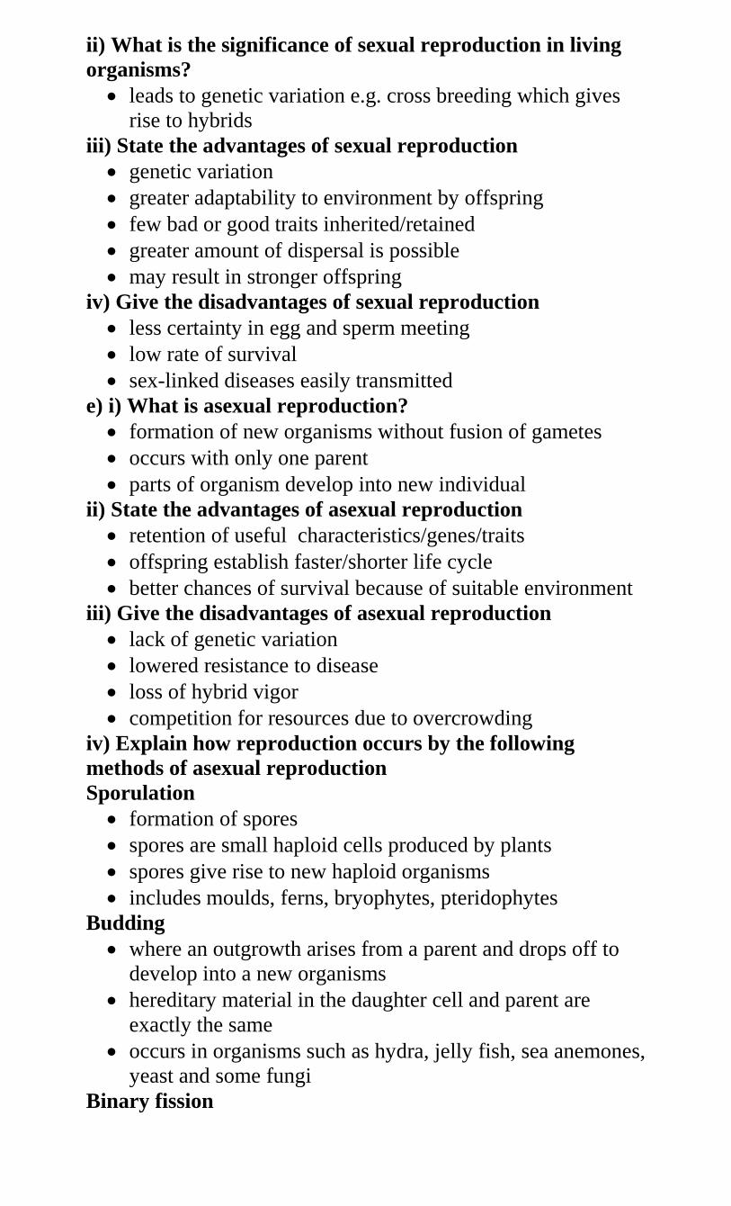
ii) What is the significance of sexual reproduction in living
organisms?
leads to genetic variation e.g. cross breeding which gives
rise to hybrids
iii) State the advantages of sexual reproduction
genetic variation
greater adaptability to environment by offspring
few bad or good traits inherited/retained
greater amount of dispersal is possible
may result in stronger offspring
iv) Give the disadvantages of sexual reproduction
less certainty in egg and sperm meeting
low rate of survival
sex-linked diseases easily transmitted
e) i) What is asexual reproduction?
formation of new organisms without fusion of gametes
occurs with only one parent
parts of organism develop into new individual
ii) State the advantages of asexual reproduction
retention of useful characteristics/genes/traits
offspring establish faster/shorter life cycle
better chances of survival because of suitable environment
iii) Give the disadvantages of asexual reproduction
lack of genetic variation
lowered resistance to disease
loss of hybrid vigor
competition for resources due to overcrowding
iv) Explain how reproduction occurs by the following
methods of asexual reproduction
Sporulation
formation of spores
spores are small haploid cells produced by plants
spores give rise to new haploid organisms
includes moulds, ferns, bryophytes, pteridophytes
Budding
where an outgrowth arises from a parent and drops off to
develop into a new organisms
hereditary material in the daughter cell and parent are
exactly the same
occurs in organisms such as hydra, jelly fish, sea anemones,
yeast and some fungi
Binary fission
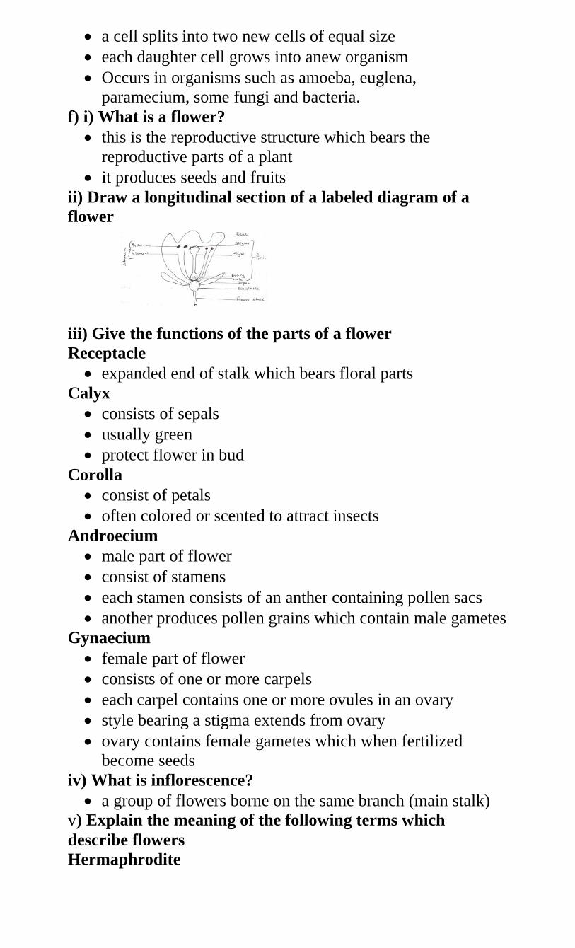
a cell splits into two new cells of equal size
each daughter cell grows into anew organism
Occurs in organisms such as amoeba, euglena,
paramecium, some fungi and bacteria.
f) i) What is a flower?
this is the reproductive structure which bears the
reproductive parts of a plant
it produces seeds and fruits
ii) Draw a longitudinal section of a labeled diagram of a
flower
iii) Give the functions of the parts of a flower
Receptacle
expanded end of stalk which bears floral parts
Calyx
consists of sepals
usually green
protect flower in bud
Corolla
consist of petals
often colored or scented to attract insects
Androecium
male part of flower
consist of stamens
each stamen consists of an anther containing pollen sacs
another produces pollen grains which contain male gametes
Gynaecium
female part of flower
consists of one or more carpels
each carpel contains one or more ovules in an ovary
style bearing a stigma extends from ovary
ovary contains female gametes which when fertilized
become seeds
iv) What is inflorescence?
a group of flowers borne on the same branch (main stalk)
v) Explain the meaning of the following terms which
describe flowers
Hermaphrodite
one with both stamen and carpel
most flowers are hermaphrodite/bisexual
Unisexual
have only one of carpel or stamen i.e. either male or female
Carpelate
also called pistilate
contains only carpels hence a female flower
Staminate
also called male flower
contains only stamens
Dioecious plants
have pistilate and staminate flowers on different plants e.g.
pawpaw
Monoecius plants
have pistilate and staminate on one plant
however, pistilate and staminate occur at different plants
e.g. maize
Complete flower
Has all four parts i.e. Calyx, corolla, androecium and
gynoecium
Incomplete flower
does not have all four parts
at least one is missing
vi) Explain the meaning of the following types of ovary
Superior
ovary occurs above other floral parts on the receptacle
Inferior (epigynous)
other floral parts arise above ovary on the receptacle
g) i) What is pollination?
transfer of pollen grains from anther of a stamen to stigma
of a flower
ii) Explain the types of pollination
self pollination takes place when mature pollen grains of a
flower fall on the stigma of the same flower
cross pollination takes place when pollen grains of a flower
fall on the stigma of another flower of the same species
iii) State the advantages of pollination
healthy offspring
leads to variation
greater chances of dispersal
iv) List the agents of pollination
wind
water
insects
v) How are flowers adapted to wind and insect pollination?
Insect pollinated flowers (entomophilus)
are scented to attract insects
have stick stigma for pollen grains to stick on
are brightly coloured to attract insects
presence of nectar to attract insects
have nectar guides to guide insects to the nectarines
have nectarines to secrete nectar
stigma/ anthers located inside the flower/tubal/funnel
shaped corolla to increase chances of contact by insects
sticky/spiny/spiky pollen grains which stick on the body of
insects and on stigma
large/conspicuous flowers easily seen by/attract insects
anthers firmly attached to the filament for insects to brush
against them
landing platform to ensure contact with anthers and stigma
mimicry to attract (male) insects
Wind pollinated flower (anemophilus)
anthers/stigma hang outside the flower to increase chances
of pollination
the style/filament is long to expose stigma/anthers
stigma is hairy/feathery/branched to increase surface area
over which pollen grains land/to trap pollen grains
pollen grains are smooth/dry/light/small to be easily carried
by wind
large amount of pollen grains to increase chances of
pollination
anthers loosely attached to filaments to enable them to
sway to release pollen grains
pollen grains may have structures which contain air to
increase buoyancy
flowers have long stalks holding them out in the wind
vi) State the ways in which plants prevent self-pollination
protandry(anthers/stamens mature first)
protagyny (pistils mature first)
monoecism (where male and female parts are on same plant
but different parts)
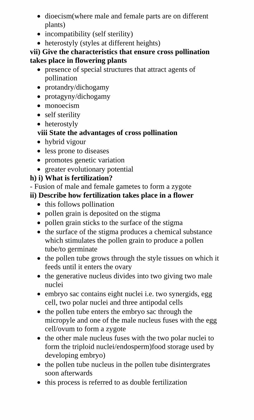
dioecism(where male and female parts are on different
plants)
incompatibility (self sterility)
heterostyly (styles at different heights)
vii) Give the characteristics that ensure cross pollination
takes place in flowering plants
presence of special structures that attract agents of
pollination
protandry/dichogamy
protagyny/dichogamy
monoecism
self sterility
heterostyly
viii State the advantages of cross pollination
hybrid vigour
less prone to diseases
promotes genetic variation
greater evolutionary potential
h) i) What is fertilization?
- Fusion of male and female gametes to form a zygote
ii) Describe how fertilization takes place in a flower
this follows pollination
pollen grain is deposited on the stigma
pollen grain sticks to the surface of the stigma
the surface of the stigma produces a chemical substance
which stimulates the pollen grain to produce a pollen
tube/to germinate
the pollen tube grows through the style tissues on which it
feeds until it enters the ovary
the generative nucleus divides into two giving two male
nuclei
embryo sac contains eight nuclei i.e. two synergids, egg
cell, two polar nuclei and three antipodal cells
the pollen tube enters the embryo sac through the
micropyle and one of the male nucleus fuses with the egg
cell/ovum to form a zygote
the other male nucleus fuses with the two polar nuclei to
form the triploid nuclei/endosperm)food storage used by
developing embryo)
the pollen tube nucleus in the pollen tube disintergrates
soon afterwards
this process is referred to as double fertilization
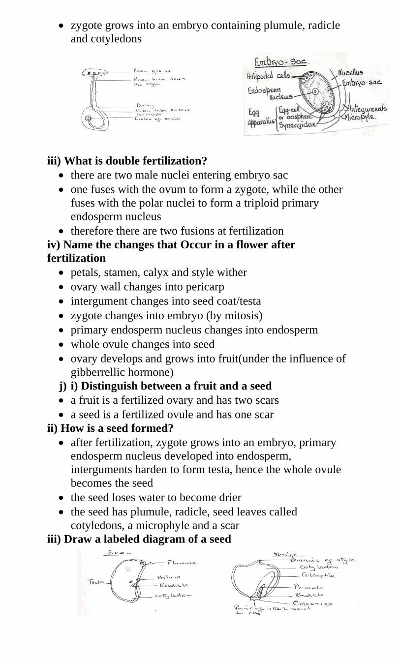
zygote grows into an embryo containing plumule, radicle
and cotyledons
iii) What is double fertilization?
there are two male nuclei entering embryo sac
one fuses with the ovum to form a zygote, while the other
fuses with the polar nuclei to form a triploid primary
endosperm nucleus
therefore there are two fusions at fertilization
iv) Name the changes that Occur in a flower after
fertilization
petals, stamen, calyx and style wither
ovary wall changes into pericarp
intergument changes into seed coat/testa
zygote changes into embryo (by mitosis)
primary endosperm nucleus changes into endosperm
whole ovule changes into seed
ovary develops and grows into fruit(under the influence of
gibberrellic hormone)
j) i) Distinguish between a fruit and a seed
a fruit is a fertilized ovary and has two scars
a seed is a fertilized ovule and has one scar
ii) How is a seed formed?
after fertilization, zygote grows into an embryo, primary
endosperm nucleus developed into endosperm,
interguments harden to form testa, hence the whole ovule
becomes the seed
the seed loses water to become drier
the seed has plumule, radicle, seed leaves called
cotyledons, a microphyle and a scar
iii) Draw a labeled diagram of a seed
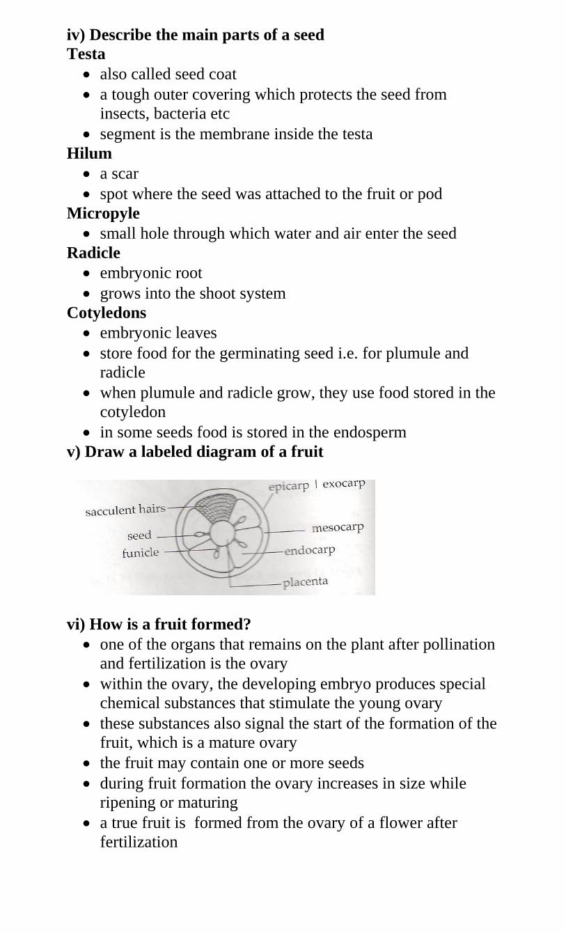
iv) Describe the main parts of a seed
Testa
also called seed coat
a tough outer covering which protects the seed from
insects, bacteria etc
segment is the membrane inside the testa
Hilum
a scar
spot where the seed was attached to the fruit or pod
Micropyle
small hole through which water and air enter the seed
Radicle
embryonic root
grows into the shoot system
Cotyledons
embryonic leaves
store food for the germinating seed i.e. for plumule and
radicle
when plumule and radicle grow, they use food stored in the
cotyledon
in some seeds food is stored in the endosperm
v) Draw a labeled diagram of a fruit
vi) How is a fruit formed?
one of the organs that remains on the plant after pollination
and fertilization is the ovary
within the ovary, the developing embryo produces special
chemical substances that stimulate the young ovary
these substances also signal the start of the formation of the
fruit, which is a mature ovary
the fruit may contain one or more seeds
during fruit formation the ovary increases in size while
ripening or maturing
a true fruit is formed from the ovary of a flower after
fertilization
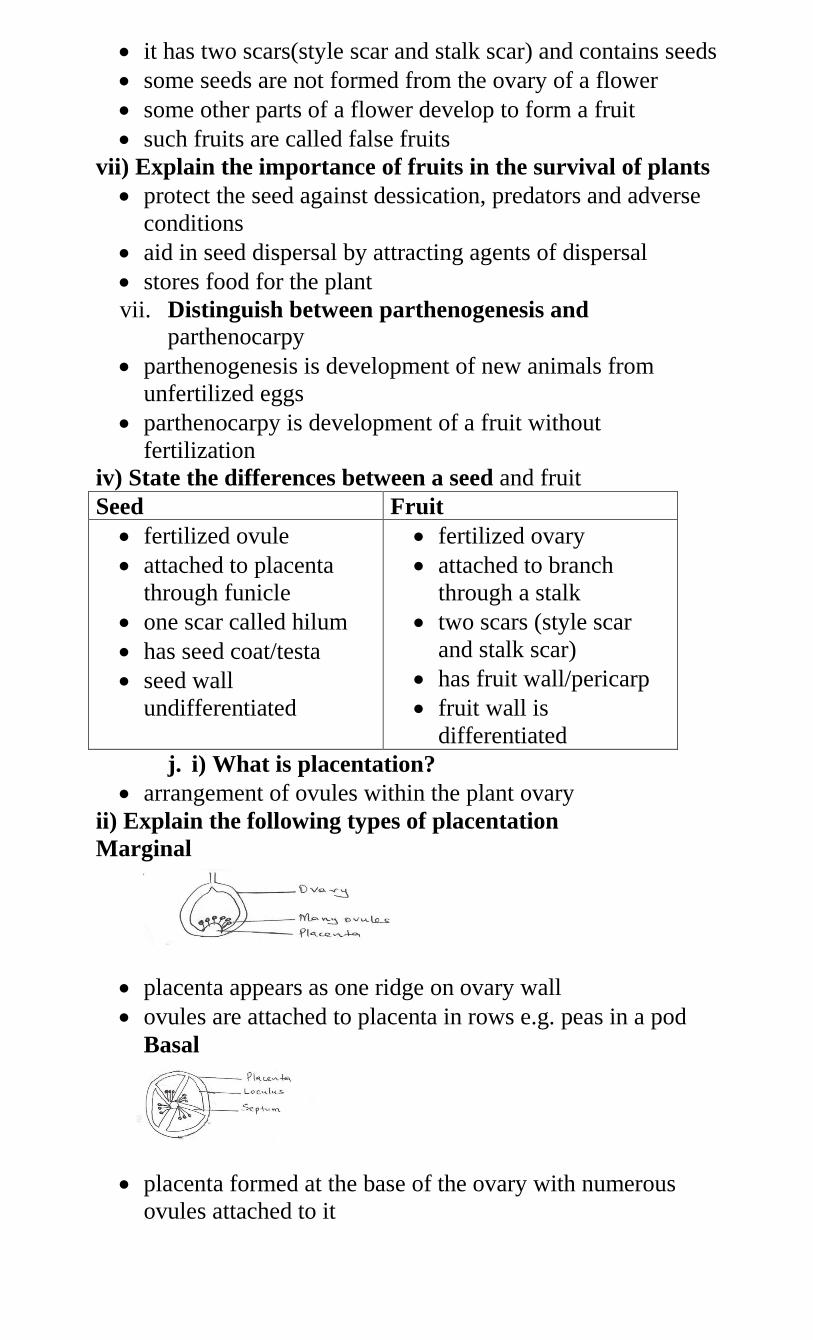
it has two scars(style scar and stalk scar) and contains seeds
some seeds are not formed from the ovary of a flower
some other parts of a flower develop to form a fruit
such fruits are called false fruits
vii) Explain the importance of fruits in the survival of plants
protect the seed against dessication, predators and adverse
conditions
aid in seed dispersal by attracting agents of dispersal
stores food for the plant
vii. Distinguish between parthenogenesis and parthenocarpy
parthenogenesis is development of new animals from
unfertilized eggs
parthenocarpy is development of a fruit without
fertilization
iv) State the differences between a seed and fruit
Seed Fruit
fertilized ovule
attached to placenta
through funicle
one scar called hilum
has seed coat/testa
seed wall
undifferentiated
fertilized ovary
attached to branch
through a stalk
two scars (style scar
and stalk scar)
has fruit wall/pericarp
fruit wall is
differentiated
j. i) What is placentation?
arrangement of ovules within the plant ovary
ii) Explain the following types of placentation
Marginal
placenta appears as one ridge on ovary wall
ovules are attached to placenta in rows e.g. peas in a pod
Basal
placenta formed at the base of the ovary with numerous
ovules attached to it
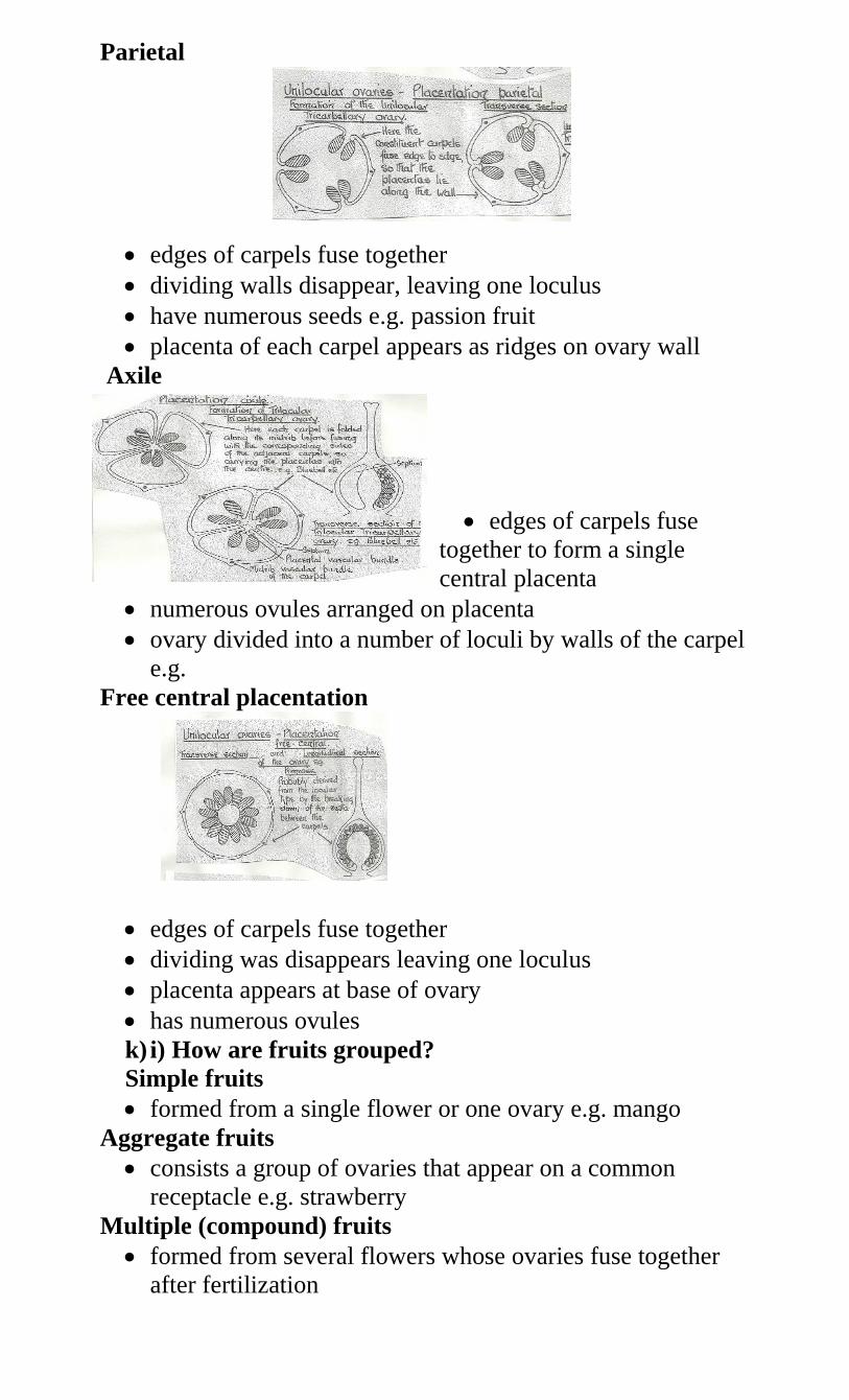
Parietal
edges of carpels fuse together
dividing walls disappear, leaving one loculus
have numerous seeds e.g. passion fruit
placenta of each carpel appears as ridges on ovary wall
Axile
edges of carpels fuse
together to form a single
central placenta
numerous ovules arranged on placenta
ovary divided into a number of loculi by walls of the carpel
e.g.
Free central placentation
edges of carpels fuse together
dividing was disappears leaving one loculus
placenta appears at base of ovary
has numerous ovules
k) i) How are fruits grouped?
Simple fruits
formed from a single flower or one ovary e.g. mango
Aggregate fruits
consists a group of ovaries that appear on a common
receptacle e.g. strawberry
Multiple (compound) fruits
formed from several flowers whose ovaries fuse together
after fertilization
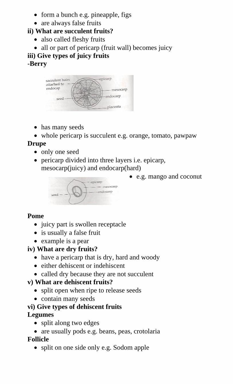
form a bunch e.g. pineapple, figs
are always false fruits
ii) What are succulent fruits?
also called fleshy fruits
all or part of pericarp (fruit wall) becomes juicy
iii) Give types of juicy fruits
-Berry
has many seeds
whole pericarp is succulent e.g. orange, tomato, pawpaw
Drupe
only one seed
pericarp divided into three layers i.e. epicarp,
mesocarp(juicy) and endocarp(hard)
e.g. mango and coconut
Pome
juicy part is swollen receptacle
is usually a false fruit
example is a pear
iv) What are dry fruits?
have a pericarp that is dry, hard and woody
either dehiscent or indehiscent
called dry because they are not succulent
v) What are dehiscent fruits?
split open when ripe to release seeds
contain many seeds
vi) Give types of dehiscent fruits
Legumes
split along two edges
are usually pods e.g. beans, peas, crotolaria
Follicle
split on one side only e.g. Sodom apple
Capsule
has several lines of weakness/sutures
open in many places e.g. castor oil, cotton
vii) What are indehiscent fruits?
non-splitting fruits
usually one seeded only
ix) Give main types of indehiscent fruits
Nut
pericarp woody, hard and thick e.g. cashew
Achene
has thin, tough pericarp e.g. sunflower
l) i) What is seed and fruit dispersal?
spreading of seeds and fruits away from parents so as to
settle where conditions are suitable for their germination
ii) Why is dispersal of seeds and fruits necessary?
prevent overcrowding
reduces competition for space, nutrients and light
colonization of new areas is made possible
to increase chances of survival
to prevent inbreeding
to avoid extinction due to over competition for the
necessities
iii) Explain how seeds and fruits are adapted to various
methods of dispersal
Adaptations for wind dispersal
they have wings, feathers or hair-like structures to increase
surface area for wind to carry them easily/buoyancy
seeds/fruits are loosely attached on the stalks so that they
can easily be released and carried away by wing
seeds/fruits are generally light and small sized to be easily
carried by wind
some seeds/fruits have parachute-like structures to be easily
carried b wind
some have censor mechanism where seeds and fruits are
borne on long stalks that are loosely attached which allows
swaying so that movements of capsule by wind releases the
seeds
Water dispersal seeds
seed/ mesocarp has air spaces thus light/buoyant to float
hence carried by water
they have waterproof cover and tough pericarp protects
seeds from getting soaked
fibrous and spongy mesocarp to easily float
Animal dispersal seeds
presence of hooks for attachment to animals thus carried to
other parts
fruits are brightly coloured, succulent and scented to attract
animals
seed coats are hard and resistant to digestive enzymes
hence seeds are dropped away from mother plant
large in size or borne on clusters to be easily seen
Self dispersal/explosive
self opening seeds
they have lines of weakness called sutures for violent
opening thus scattering seeds away from parent plant
5. a) i) Distinguish between external and internal
fertilization in animals
in external fertilization fusion of the male and female
gametes takes place outside the body of the female e.g.
amphibians and fish
in internal fertilization union of gametes occurs inside the
body of the female
ii) State the advantages and disadvantages of external
fertilization
Advantages
large numbers produced therefore many offspring per
breeding season
female does not suffer gestation stress
mother does not need to care for the young except in a few
species
the surviving individuals are highly selected for better
survival
Disadvantages
many predators surround the eggs before and after
fertilization
fewer chances of fertilization/a lot of gametes wasted
embryo development at mercy of environment
large numbers of female gametes are required therefore
female gets much exhausted
iii) State the advantages and disadvantages of internal
fertilization
Disadvantages
number of gametes fewer hence less number of offspring
less adapted for sudden change of environment after birth
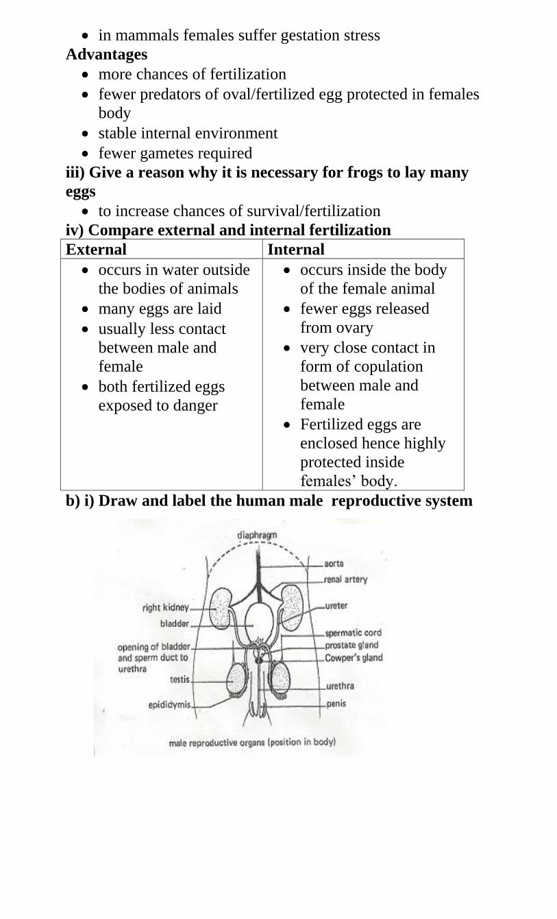
in mammals females suffer gestation stress
Advantages
more chances of fertilization
fewer predators of oval/fertilized egg protected in females
body
stable internal environment
fewer gametes required
iii) Give a reason why it is necessary for frogs to lay many
eggs
to increase chances of survival/fertilization
iv) Compare external and internal fertilization
External Internal
occurs in water outside
the bodies of animals
many eggs are laid
usually less contact
between male and
female
both fertilized eggs
exposed to danger
occurs inside the body
of the female animal
fewer eggs released
from ovary
very close contact in
form of copulation
between male and
female
Fertilized eggs are
enclosed hence highly
protected inside
females’ body.
b) i) Draw and label the human male reproductive system
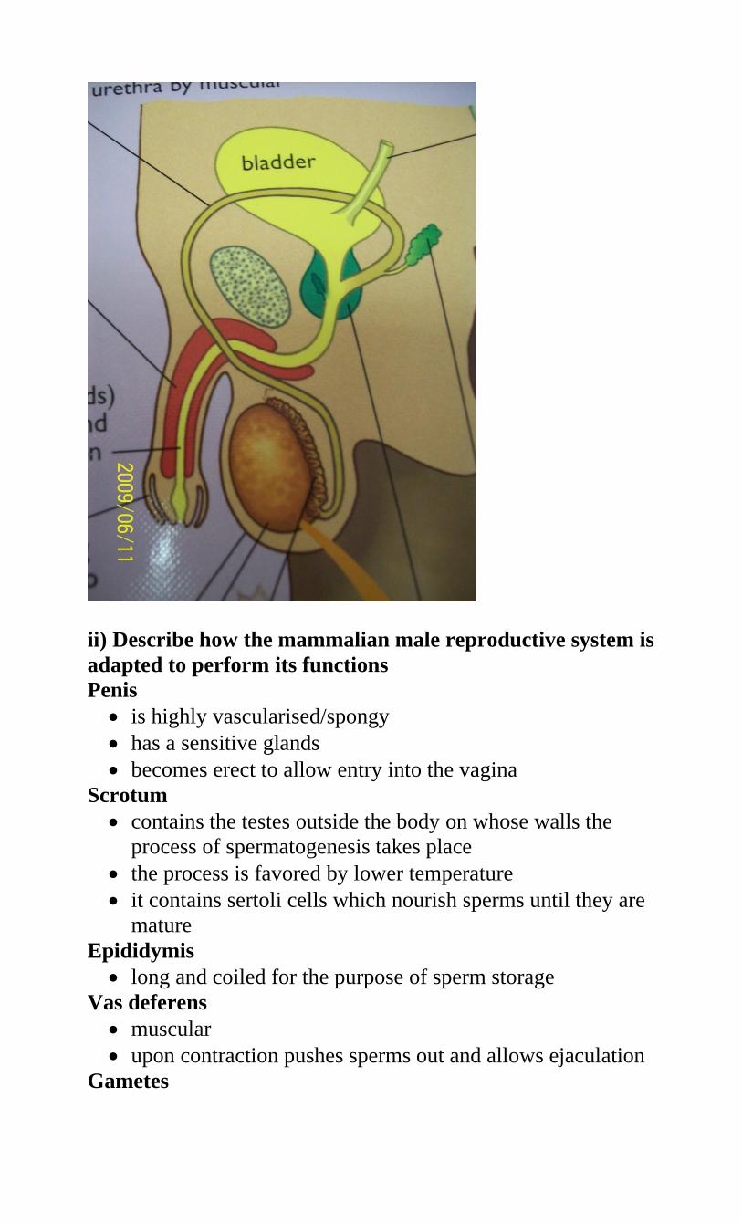
ii) Describe how the mammalian male reproductive system is
adapted to perform its functions
Penis
is highly vascularised/spongy
has a sensitive glands
becomes erect to allow entry into the vagina
Scrotum
contains the testes outside the body on whose walls the
process of spermatogenesis takes place
the process is favored by lower temperature
it contains sertoli cells which nourish sperms until they are
mature
Epididymis
long and coiled for the purpose of sperm storage
Vas deferens
muscular
upon contraction pushes sperms out and allows ejaculation
Gametes
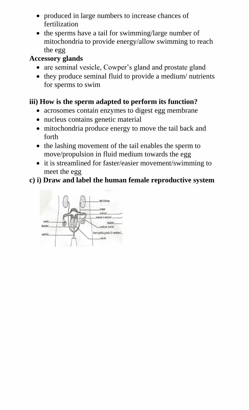
produced in large numbers to increase chances of
fertilization
the sperms have a tail for swimming/large number of
mitochondria to provide energy/allow swimming to reach
the egg
Accessory glands
are seminal vesicle, Cowper’s gland and prostate gland
they produce seminal fluid to provide a medium/ nutrients
for sperms to swim
iii) How is the sperm adapted to perform its function?
acrosomes contain enzymes to digest egg membrane
nucleus contains genetic material
mitochondria produce energy to move the tail back and
forth
the lashing movement of the tail enables the sperm to
move/propulsion in fluid medium towards the egg
it is streamlined for faster/easier movement/swimming to
meet the egg
c) i) Draw and label the human female reproductive system
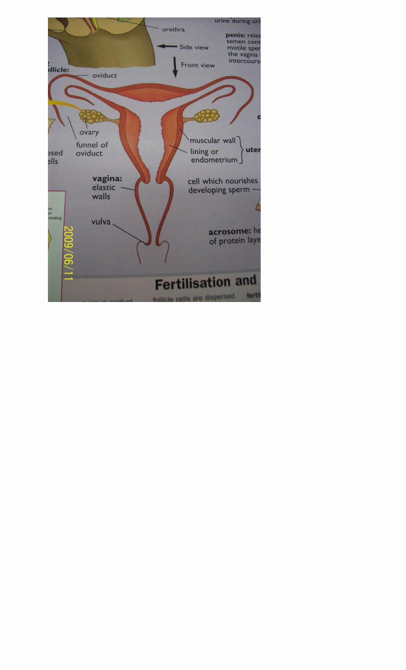
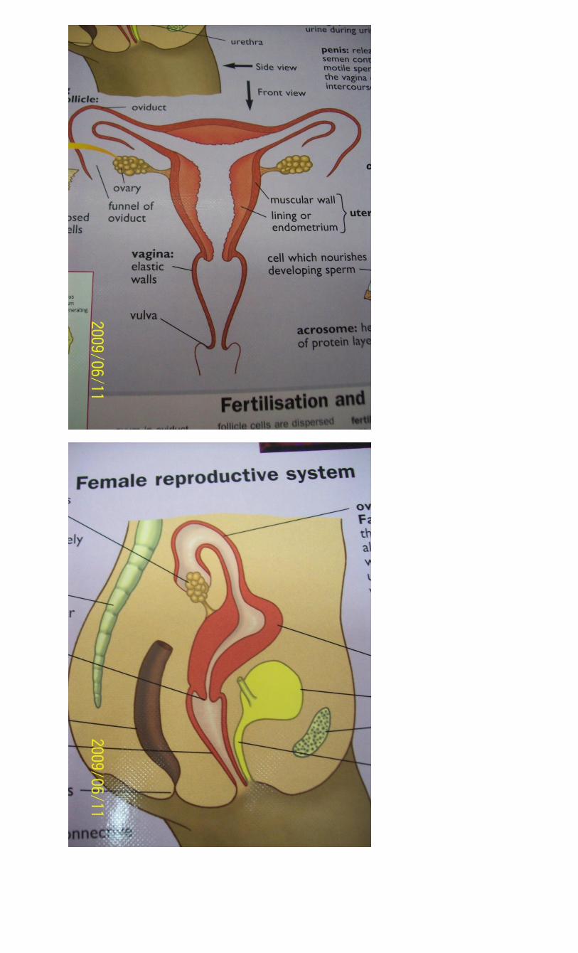
ii) Describe how the various structures of the human female
reproductive system are adapted to their function
Ovaries
have several graafian follicles that develop and burst open
to release/produce mature ova
secretes sex hormones)oestrogen) which initiate/control
development of secondary sexual characteristics
produce hormones oestrogen and progesterone which
prepare the uterus for implantation and subsequent
nourishment of the embryo
Oviducts (fallopian tube)
are thin narrow and tubular to increase flowing speed of
semen containing sperms
are funnel shaped on the end next to ovary which enables
them to receive the ovum
their lining contains cilia which propel the ovum towards
the uterus
has peristaltic muscles that enable movement of
zygote/ovum to the uterus for implantation
is fairly long to increase surface area for fertilization
Uterus
is muscular for protection of developing embryo
has elastic wall that allows growth and development of
foetus/embryo
has a highly vascularised endometrium that provides
nutrients/gaseous exchange to developing embryo
Cervix
has valves that close the lower end of the uterus to ensure
continued pregnancy during gestation period
is capable of dilating
has narrow entrance/neck-like entrance to uterus that
enables quick swimming of sperms to uterus
has suction mechanism that draws up/pulls sperms into
uterus
has a “W” shape that fits well with the glands of the penis
to ensure sperms are deposited at the right point
Vagina
is elastic and muscular to enable good accommodation or
penetration of the penis thus proper deposition of sperms
and for easy parturition
allows menstrual flow
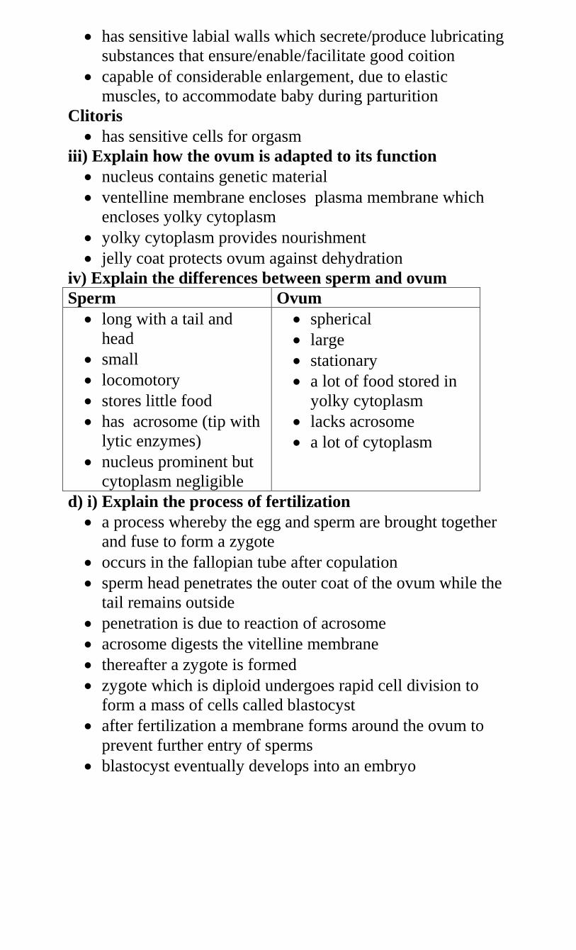
has sensitive labial walls which secrete/produce lubricating
substances that ensure/enable/facilitate good coition
capable of considerable enlargement, due to elastic
muscles, to accommodate baby during parturition
Clitoris
has sensitive cells for orgasm
iii) Explain how the ovum is adapted to its function
nucleus contains genetic material
ventelline membrane encloses plasma membrane which
encloses yolky cytoplasm
yolky cytoplasm provides nourishment
jelly coat protects ovum against dehydration
iv) Explain the differences between sperm and ovum
Sperm Ovum
long with a tail and
head
small
locomotory
stores little food
has acrosome (tip with
lytic enzymes)
nucleus prominent but
cytoplasm negligible
spherical
large
stationary
a lot of food stored in
yolky cytoplasm
lacks acrosome
a lot of cytoplasm
d) i) Explain the process of fertilization
a process whereby the egg and sperm are brought together
and fuse to form a zygote
occurs in the fallopian tube after copulation
sperm head penetrates the outer coat of the ovum while the
tail remains outside
penetration is due to reaction of acrosome
acrosome digests the vitelline membrane
thereafter a zygote is formed
zygote which is diploid undergoes rapid cell division to
form a mass of cells called blastocyst
after fertilization a membrane forms around the ovum to
prevent further entry of sperms
blastocyst eventually develops into an embryo
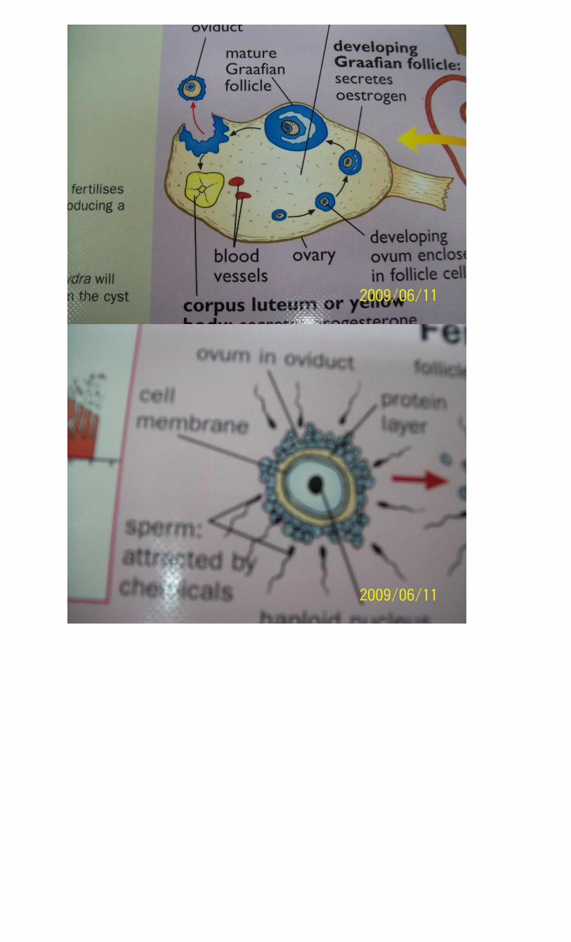
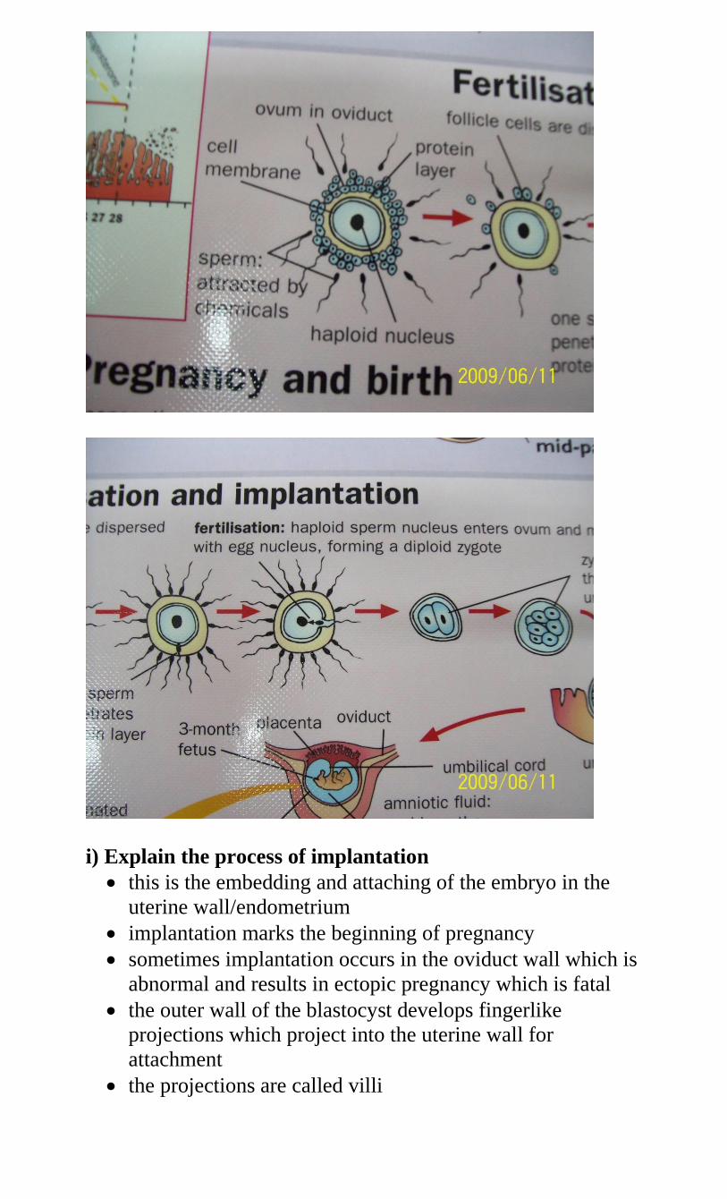
i) Explain the process of implantation
this is the embedding and attaching of the embryo in the
uterine wall/endometrium
implantation marks the beginning of pregnancy
sometimes implantation occurs in the oviduct wall which is
abnormal and results in ectopic pregnancy which is fatal
the outer wall of the blastocyst develops fingerlike
projections which project into the uterine wall for
attachment
the projections are called villi
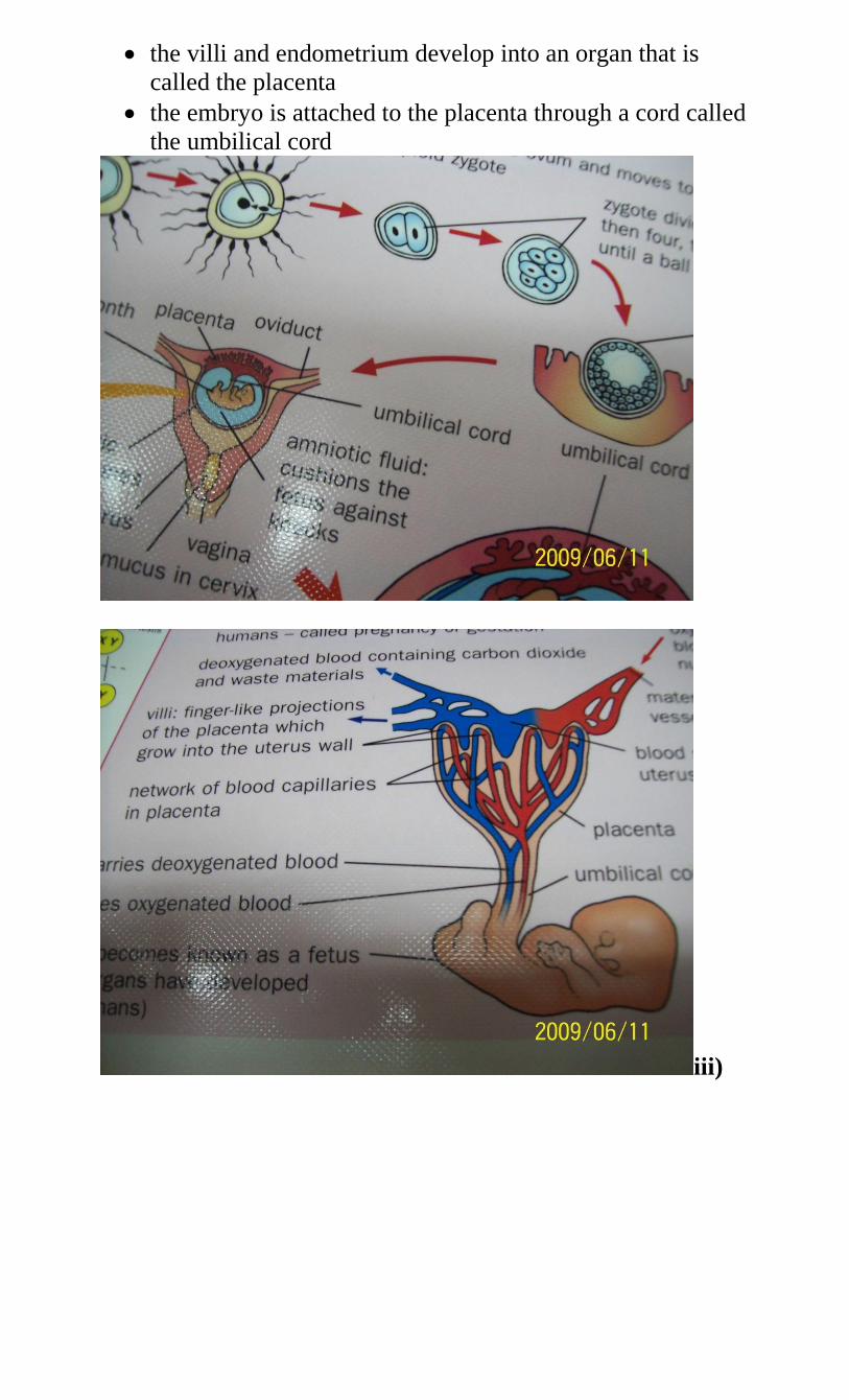
the villi and endometrium develop into an organ that is
called the placenta
the embryo is attached to the placenta through a cord called
the umbilical cord
iii)
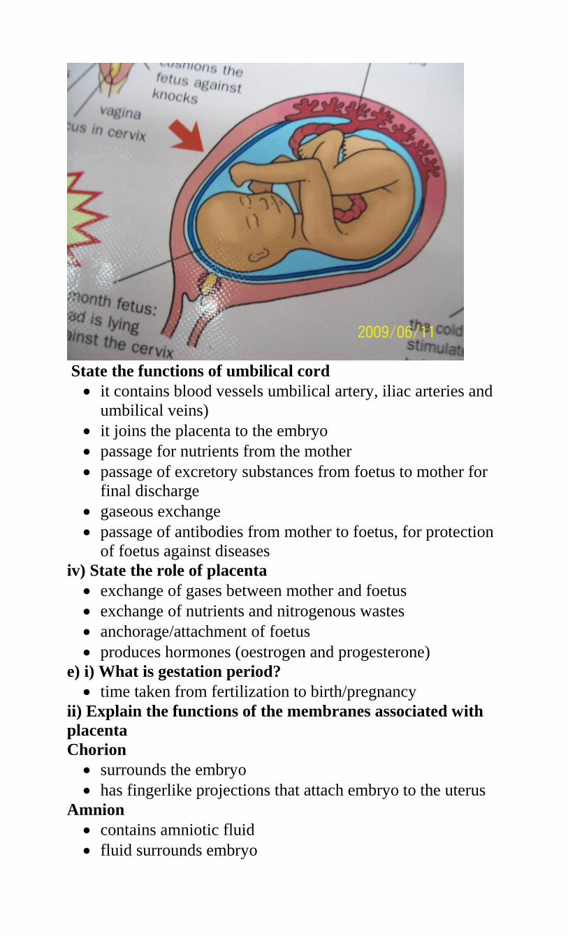
State the functions of umbilical cord
it contains blood vessels umbilical artery, iliac arteries and
umbilical veins)
it joins the placenta to the embryo
passage for nutrients from the mother
passage of excretory substances from foetus to mother for
final discharge
gaseous exchange
passage of antibodies from mother to foetus, for protection
of foetus against diseases
iv) State the role of placenta
exchange of gases between mother and foetus
exchange of nutrients and nitrogenous wastes
anchorage/attachment of foetus
produces hormones (oestrogen and progesterone)
e) i) What is gestation period?
time taken from fertilization to birth/pregnancy
ii) Explain the functions of the membranes associated with
placenta
Chorion
surrounds the embryo
has fingerlike projections that attach embryo to the uterus
Amnion
contains amniotic fluid
fluid surrounds embryo
protects embryo from mechanical injury by acting as shock
absorbers
fluid also protects embryo from dehydration
distributes pressure equally over embryo
Yolk sac
surround the yolk
produces blood cells for embryo until its own liver is able
to perform the task
Allantois
present only for a short time
removes and store waste material
it eventually becomes the umbilical cord
iii) Explain the events that take place to facilitate parturition
near birth the placenta produces less progesterone
oxytocin hormone is produced by posterior lobe of pituitary
gland
because progesterone level has decreased the uterus
becomes sensitive to oxytocin
oxytocin causes the contraction of the uterus (myometrium)
these contractions are called labour pains
just before parturition the head turns downwards
the contractions eventually push the baby through the
vagina
amnion breaks and amniotic fluid is released
oxytocin dilates the cervix
foetus is expelled through cervix with head coming out first
finally the whole infant comes out
the umbilical cord is cut and the placenta is expelled as
afterbirth
iv) State the reasons why later in pregnancy the ovary will b
e removed without disturbing the pregnancy
corpus luteum in the ovary secretes progesterone which
maintains pregnancy and development of foetus after
conception
after four months pregnancy is maintained by progesterone
from the placenta
f) i) What are secondary sexual characteristics
- Characteristics (physiological and anatomical) that start
developing at puberty due to the influence of male and female
hormones
ii) State the main secondary changes in Boys
deepening of the voice
growth of hair on face, pubic part, chest, legs
penis and testes become bigger
muscular development
sperm production begins at puberty and may continue
throughout life
Girls
growth of hair on pubic part and armpits
widening/enlargement of hips
development of breasts
menstrual cycle starts as ovaries mature
body acquires extra fat
iii) Describe the role of hormones in secondary sexual
characteristics inBoys
Follicle stimulation hormone (FSH)
from pituitary
stimulates production of androgens(male hormones) mainly
testosterone by testis
Testosterone
secondary sexual characteristics
Girls
FHS
from pituitary
development of follicles
stimulates oestrogen production by ovary
LH
from pituitary
ovulation
stimulates release of progesterone by ovary
Oestrogen
stimulates release of LH
secondary sexual characteristics
Progesterone
also from placenta
sustains pregnancy as it inhibits prolactin and oxytocin
during pregnancy
Prolactin
milk formation
Oxytocin
parturition
milk ejection
g) i) What is menstruation?
- vaginal discharge due to disintegration of endometrium
ii) Describe the role of hormones in the human menstrual
cycle
it is controlled by sex hormones which are responsible for
the onset of secondary sexual characteristics and also
control of the menstrual cycle
the onset is signaled by discharge of blood/menses 14 days
following the start of menstruation
anterior lobe of pituitary gland secretes follicle stimulating
hormone(FSH)
Follicle stimulating hormone causes graafian follicle to
develop in the ovary. It also stimulates tissues of the
ovary/wall (theca) to secrete oestrogen
oestrogen causes repair/healing of uterine wall
oestrogen stimulates anterior lobe of pituitary to produce
luteinising hormonge (LH)
LH causes ovulation. It also causes graafian follicle to
change into corpus luteum. LH stimulates corpus luteum to
secrete progesterone
Progesterone causes proliferation/thickening of uterine wall
Oesterogen/progesterone inhibits the production of FSH by
anterior lobe of pituitary, thus no more follicles develop
and oestrogen production reduces
In the next two weeks progesterone level rises and inhibits
production of LH from anterior lobe of pituitary
The corpus lutetium stops secreting progesterone and
menstruation occurs when the level of progesterone drops
Anterior lobe of pituitary starts secreting FSH again.
iii) What is menopause?
end of ovulation in women
occurs after age of 45 years
does not occur in males
h) Explain the symptoms, methods of transmitting and
prevention (control) of the following sexually transmitted
diseases
i) Gonorrhea
- caused by a bacterium called Neisseria gonorrhea
- transmitted through sexual intercourse,
- infects urethra and vaginal tract (epithelia)
- Symptoms include pain, discharge of mucus and bad smell
(females)
- Effects include sterility, heart diseases, blindness of foetus and
arthritis
- Treatment by antibiotics
- Control and prevention by proper sexual conduct
ii) Herpes
notably Herpes simplex and H. genitalis
caused by virus which attacks genitalia
symptoms are painful sores in genitalia, skin lesions
transmitted in saliva, sexual intercourse and injection by
drug addicts
no treatment
iii) Syphilis
caused by bacterium called Treponema palladium
symptoms are painless wounds in genitalia
attack genitalia, nervous system, lips
treated by antibiotics
iv) Trichomoniasis
caused by plasmodium called Trichomonas
attacks reproductive tract
symptoms are itching and discharge of pus from the
genitals
treated by antibiotics
v) Hepatitis
- Viral disease
Affects the liver
Transmitted through sexual intercourse
No known treatment
vi) Candidiasis
caused by fungus called Candida albicans
transmitted through sexual intercourse
symptoms include itching urethra, and vaginal discharge
(odourless)
controlled by personal hygiene, early treatment and
responsible sexual behavior
vii) HIV/AIDS
caused by HIV virus
transmitted by sexual intercourse, blood transfusion,
sharing piercing instruments from infected mother to
foetus, infant and baby
symptoms include fever, swollen lymph nodes, night
sweating, cough, weight loss, fatigue, loss of appetite,
diarrhea, headache, a opportunistic infections and tumors
Control by responsible sexual behaviour, education,
screening blood for transfusion and using sterile piercing
instruments.
6. a) Define the terms
i) Growth
an irreversible change in size of a cell, organ or whole
organism
growth is due to synthesis of protoplasm or extracellular
substances
ii) Development
refers to a series of changes which an organism goes
through in its lie cycle
during development both qualitative and quantitative
changes take place(involves differentiation)
iii) Differentiation
refers to changes in which the cells of the body undergo
and become specialised to perform specific functions
b) i) Differentiate growth in plants and animals
Plants Animals
confined to shoots/root
tips (apical)
have definite growth
regions(meristems)
often
indefinite/continuous
growth results in
branching
affected by light,
auxins, hormones and
temperature
occurs all over the
body(intercalary)
different parts grow at
different
rates(allometric)
maximum growth on
maturity(definite)
no branching
affected by nutrients
ii) List the processes involved in growth
assimilation
cell enlargement
cell division(by mitosis)
iv) List the parameters used to measure growth
height/length
dry weight
number of individuals
volume
leaf area of plant
iv) Name the patterns of growth in organisms
- allometric and isometric
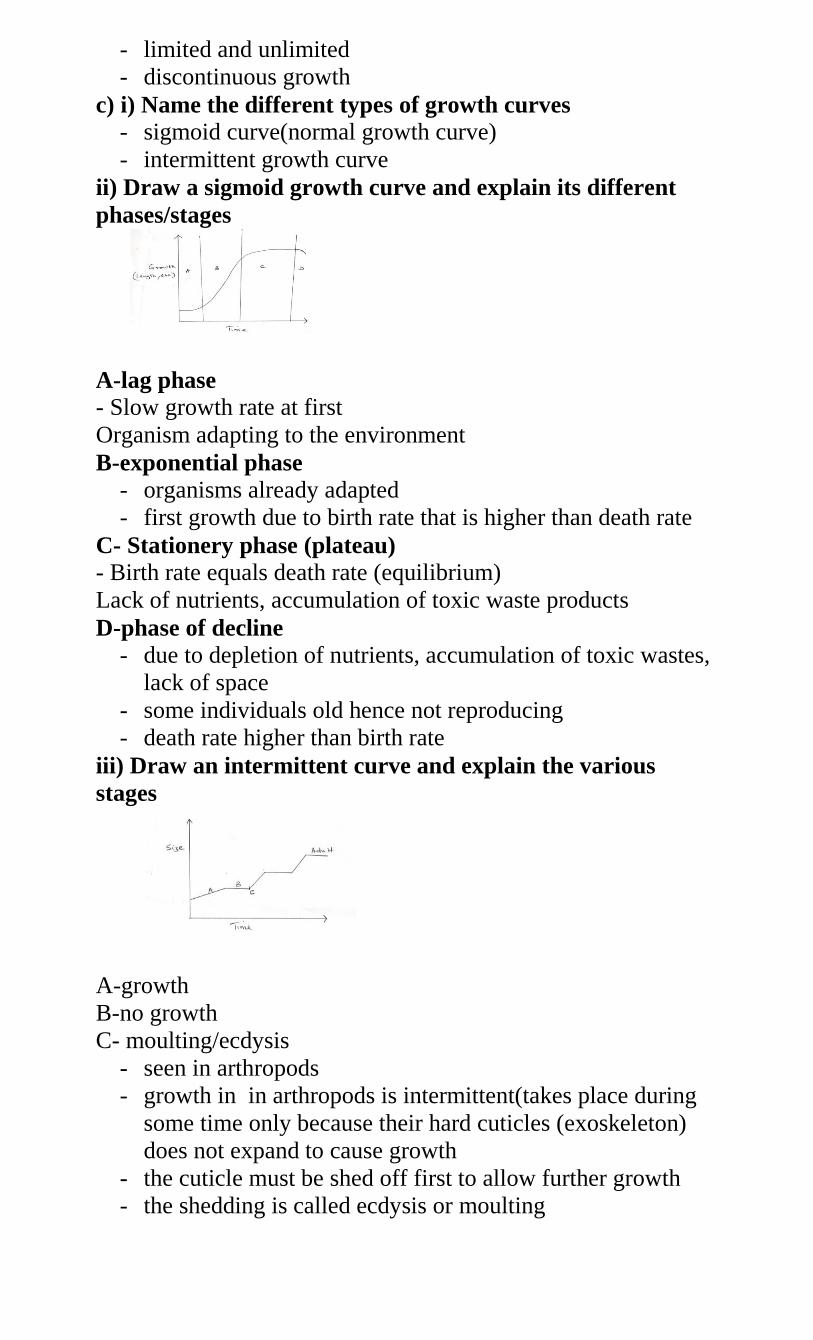
- limited and unlimited
- discontinuous growth
c) i) Name the different types of growth curves
- sigmoid curve(normal growth curve)
- intermittent growth curve
ii) Draw a sigmoid growth curve and explain its different
phases/stages
A-lag phase
- Slow growth rate at first
Organism adapting to the environment
B-exponential phase
- organisms already adapted
- first growth due to birth rate that is higher than death rate
C- Stationery phase (plateau)
- Birth rate equals death rate (equilibrium)
Lack of nutrients, accumulation of toxic waste products
D-phase of decline
- due to depletion of nutrients, accumulation of toxic wastes,
lack of space
- some individuals old hence not reproducing
- death rate higher than birth rate
iii) Draw an intermittent curve and explain the various
stages
A-growth
B-no growth
C- moulting/ecdysis
- seen in arthropods
- growth in in arthropods is intermittent(takes place during
some time only because their hard cuticles (exoskeleton)
does not expand to cause growth
- the cuticle must be shed off first to allow further growth
- the shedding is called ecdysis or moulting
- when moulting has taken place animal grows but growth
stops when the exoskeleton hardens again
d) i) What is seed dormancy?
- A state where a viable seed is incapable of germinating
when all conditions are favourable.
ii) State the biological importance of seed dormancy
- gives embryo time to reach maturity
- gives time for dispersal
- allows plant to survive adverse conditions
iii) State the factors which cause seed dormancy
Internal factors
- presence of abscisic acid/ABA/ presence of germination
inhibitors
- embryo not fully developed
- absence of hormones/enzymes/inactivity of
hormones/enzymes/gibberellins/cytokinins
- impermeability of seed coat
External factors
- unsuitable temperature
- absence of light
- lack of oxygen
- lack of oxygen
- lack of water
iv) Give the conditions necessary to break seed dormancy
- scarification/scratching to make seed coat impermeable
- vernalisation/cold treatment in some seeds like wheat
- burning/nicking/expose to heat e.g. wattle seeds
- destruction of germination inhibitors
e) i) What is seed germination?
- process by which a seed develops in a seedling
ii) What is viability
- ability of a seed to germinate
iii) Discuss the various conditions necessary for the
germination of seeds
Water
- medium for enzymatic activity
- hydrolysis of food into simpler substances
- medium of transport
- softens the seed
- acts as a solvent
Air
- in form of oxygen
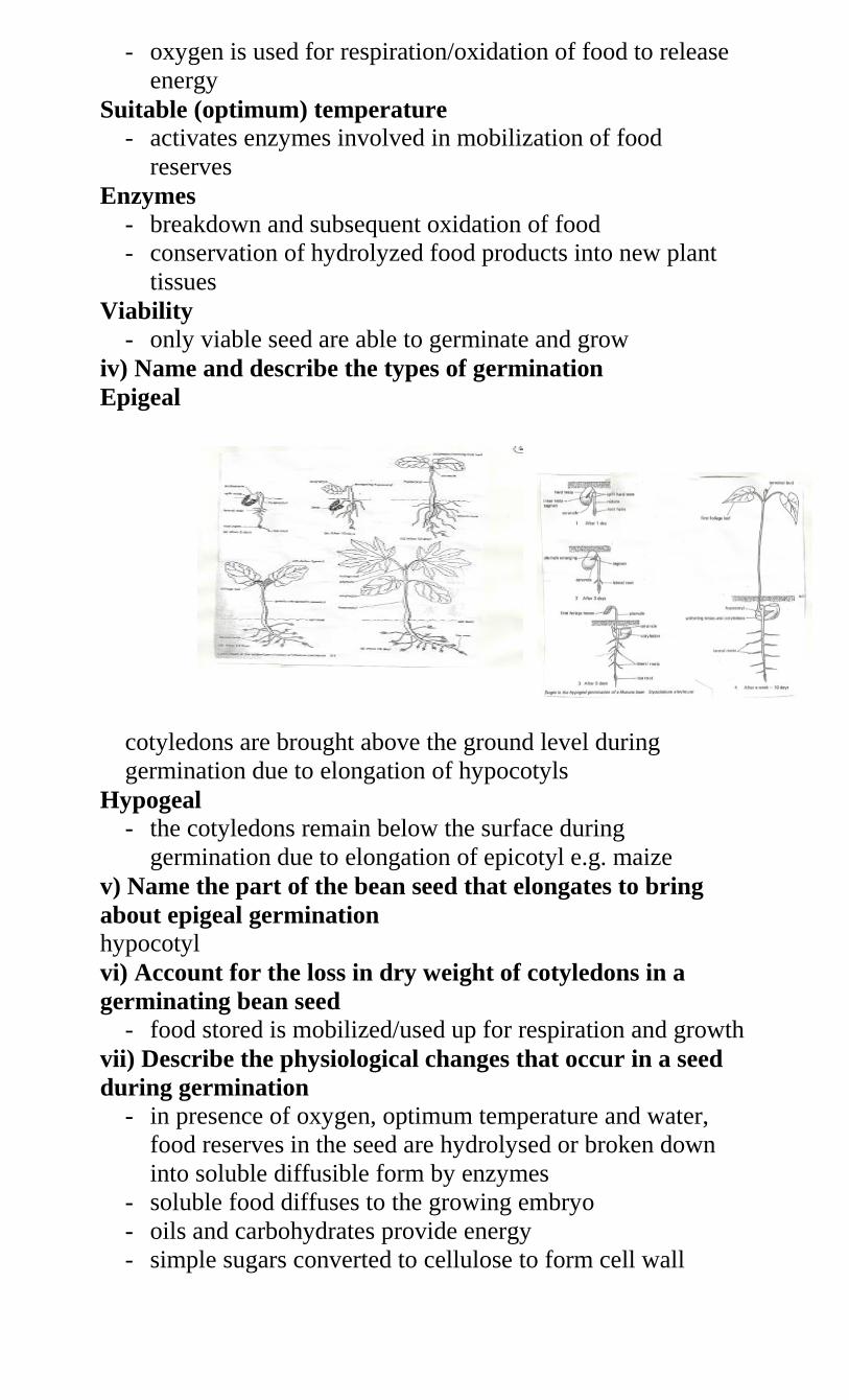
- oxygen is used for respiration/oxidation of food to release
energy
Suitable (optimum) temperature
- activates enzymes involved in mobilization of food
reserves
Enzymes
- breakdown and subsequent oxidation of food
- conservation of hydrolyzed food products into new plant
tissues
Viability
- only viable seed are able to germinate and grow
iv) Name and describe the types of germination
Epigeal
cotyledons are brought above the ground level during
germination due to elongation of hypocotyls
Hypogeal
- the cotyledons remain below the surface during
germination due to elongation of epicotyl e.g. maize
v) Name the part of the bean seed that elongates to bring
about epigeal germination
hypocotyl
vi) Account for the loss in dry weight of cotyledons in a
germinating bean seed
- food stored is mobilized/used up for respiration and growth
vii) Describe the physiological changes that occur in a seed
during germination
- in presence of oxygen, optimum temperature and water,
food reserves in the seed are hydrolysed or broken down
into soluble diffusible form by enzymes
- soluble food diffuses to the growing embryo
- oils and carbohydrates provide energy
- simple sugars converted to cellulose to form cell wall
- amino acids make protoplasm
- seed develops plumule and radicle hence germinates
viii) Explain the biological significance of cotyledons being
brought above the ground in epigeal germination
- cotyledons have inadequate food
- they are brought above the ground to acquire chloroplasts
to carry out photosynthesis before the formation of foliage
leaves to supplement food supply required for growth
during germination
f) i) Distinguish between primary and secondary growth
Primary growth
- occurs at the apical (shoot and tip) apices regions where
meristematic cells occur
- causes plant elongation since cells divide by mitosis
Secondary growth
-occurs at the cambium meristems
Increases width (girth) of the stem
ii) What are meristems
- dividing cells
- meristem means they are dividing
iii) State the characteristics of meristematic cells
- dense cytoplasm
- thin cell walls
- absence of vacuoles/cell sap
iv) State the location and function of the following
meristematic tissues
Apical meristem
- located at tips of roots and shoots
- increase length of stem and roots/primary growth
Intercalary meristem
- found at bases of internodes
- responsible for elongation of internodes and increase in leaf
sheath in grasses
Lateral meristems
- found near the periphery of stem and root
- responsible for secondary growth/growth in girth of stem
and root/lateral growth
- called cambium and constitute vascular and cork cambium
v) Describe primary growth
- occurs at tips of shoots and roots in the meristematic tissues
of apical meristem
- at the apex there is a zone of cell division/mitosis
- cells elongate at elongation zone
- the elongated cells differentiate at the region of
differentiation resulting in increase in size
- in the stems meristems give rise to leaf premodia which
envelop the apex to form a bud
- the bud protects the delicate inner cells
- in roots the meristem is protected by root cap
- after cells differentiate the form permanent tissues
vi) Describe secondary growth in plants
- also called secondary thickening
- only occurs in dicotyledonous plants that have cambium
- monocotyledonous plants do not undergo secondary growth
because they lack intervascular cambium
- cambium cells divide to produce more cells on either side
of the cambium
- cells produced to the inside become secondary cambium
- cells produced to the outside become secondary phloem
- division of cambium cells occurs yearly producing new
rings of secondary phloem and secondary xylem each year
- intervascular cambium(cambium between vascular
bundles) divide to form secondary parenchyma, thereby
increasing growth of medullary rays
- much more xylem is formed than phloem, thus pushing
phloem and cambium ring outward
- the rate of secondary growth is depended on seasons(rains)
resulting in annual rings
- cork cambium is located beneath epidermis ad is
responsible for secondary thickening of the bark of
perennial plants
- cork cambium divides to form new cork(bark) tissues to
accommodate increased growth on outside and secondary
cortex on the inside
- Cork cells (cells of the bark) are loosely parked at some
points to form lenticels for gaseous exchange.
vii) State the significance of secondary growth
- increase girth or circumference of trees
- annual rings which show seasonal growth can be used to
tell the age of trees
g) i) Describe one method which can be used to measure the
average growth rate of a single leaf of a plant
EITHER
- chose/identify a young leaf(just unfolded)
- use the same leaf throughout
- measure (total) length of (whole) leaf
- record
- repeat at regular intervals until no more change
occurs/constant length
- average rate of growth is equal to total increase in length
divided by the period taken to achieve full length
Average rate of growth = total increase in length
period taken to achieve full length
OR
- choose/identify a young leaf(just unfolded)
- use the same leaf throughout
- trace the outline on a graph paper and work out the area
- record
- repeat at regular intervals until regular area
- average rate of growth equals to total increase in area
divided by the period of time taken to achieve full area
Average rate of growth = total increase in area
period of time taken to achieve final area
i) Describe how the growth of a root can be determined
Materials
- fine thread, marking ink, germinating bean seedlings,
blotting paper, ruler marked in millimeters, pins, cork, a
boiling tube and moist cotton wool
Procedure
- dry seedlings using blotting paper
- place inside against the ruler marked in mm
- dip the fine thread in waterproof ink
- mark the radicle at equal intervals
- pin the seedling to the cork
- suspend the seedling into the boiling tube containing moist
cotton wool
- allow the seedling to grow for two days/some
time observe the intervals with the marks
- record your observations the widest intervals are found in
the region just behind the tip indicating/showing region of
greatest growth
iii) A boy hammered a nail in the bark of a tree at a height of
1.5metres above the ground.
Four years later, the nail was found at the same height
although the tree had grown 3 meters taller. Explain the
above observation
The nail was hammered at a point where vertical growth had
stopped/further growth was confined to increase in
width/diameter.
Vertical growth is confined to tips/apex/vertical apical meristem
h) i) Describe the role of hormones in growth and
development of plants
indole acetic acid/IAA/ auxins
Cell division/increase in cell division
Tropic responses
Cell elongation/increases in ell elongation
Development of abscision layer
Growth of ovaries into fruits/parthenocarpy/initiates flowers
Inhibits growth of lateral buds/produces apical dominance
Stimulates adventitious/lateral roots
Gibberellins (Gibberellic acid/GA3
Promote cell elongation/rapid cell division/increase in length of
the internodes
Promote fruit formation without fertilization/parthenocarpy
Reduces root growth
Breaks seed dormancy/promotes germination
Cytokinnins (Kinnins/Kinnetin/Zeatin)
- breaks dormancy
- promotes flowering
- promotes cell division
- stabilizes protein and chlorophyll
- promotes root formation on a shoot
- low concentration encourages leaf senses
- normal concentration increases cell enlargement in leaves
- stimulates lateral bud development
Ethylene (ethynelC2H4)
- accelerates ripening in fruits
- encourages fruit fall/leaf fall
- induces thickening in stern/inhibits stem elongation
- promotes flowering (in pineapples)
- promotes germination in certain seeds
Abscisic acid (ABA) abscisin hormone/dormin)
- causes bud dormancy
- encourages fruit/leaf fall
- high concentration causes closing of stomata
- causes seed dormancy
- inhibits cell elongation
Traumatin
- heals wounds by callous formation
Florigen
- promotes flowering
ii) State the applications of plant hormones in agriculture
- induce root growth in stem cuttings
- selective weed killers
- encourage sprouting of lateral buds
- breaking seed dormancy
- induce parthenocarpy
- accelerate ripening of fruits
- promote flowering
- cause dormancy
iii) Explain apical dominance
- a phenomenon whereby production of auxins by a growing
apical bud of a shoot inhibits growth of lateral buds
- this inhibition is due to high concentration of auxins
(indoleacetic acid/IAA) in apical bud
- removal of terminal/apical bud causes development and
sprouting of several buds which later develop into branches
- applied in pruning coffee, tea and hedges
- this leads to more yield
iv) Describe the role of hormones in the growth and
development of animal
somatotrophin (growth hormones)
- from anterior pituitary
- promotes cell division
- overproduction causes gigantism
- underproduction causes dwarfism
Thyroxine
- promotes growth and metamorphosis
- underproduction leads to a child becoming a cretin
(mentally retarted)
Androgens
- in males
- growth of male reproductive organs
Oestrogen
- in females
- growth of female reproductive organs
Ecdysone
- in arthropods
- moulting (ecdysis)
t) i) What is metamorphosis?
- change in form during which there are changes in structure
and function in body of organism
- prepares organism for life in a different habitat
ii) Explain complete metamorphosis
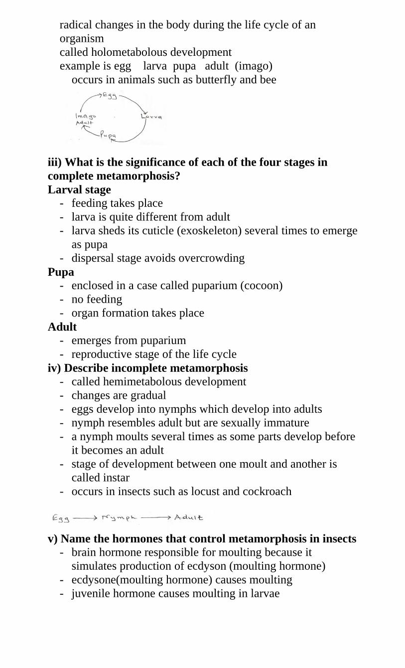
radical changes in the body during the life cycle of an
organism
called holometabolous development
example is egg larva pupa adult (imago)
occurs in animals such as butterfly and bee
iii) What is the significance of each of the four stages in
complete metamorphosis?
Larval stage
- feeding takes place
- larva is quite different from adult
- larva sheds its cuticle (exoskeleton) several times to emerge
as pupa
- dispersal stage avoids overcrowding
Pupa
- enclosed in a case called puparium (cocoon)
- no feeding
- organ formation takes place
Adult
- emerges from puparium
- reproductive stage of the life cycle
iv) Describe incomplete metamorphosis
- called hemimetabolous development
- changes are gradual
- eggs develop into nymphs which develop into adults
- nymph resembles adult but are sexually immature
- a nymph moults several times as some parts develop before
it becomes an adult
- stage of development between one moult and another is
called instar
- occurs in insects such as locust and cockroach
v) Name the hormones that control metamorphosis in insects
- brain hormone responsible for moulting because it
simulates production of ecdyson (moulting hormone)
- ecdysone(moulting hormone) causes moulting
- juvenile hormone causes moulting in larvae
vi) State the advantages of metamorphosis in the life of
insects
- the adult and larvae exploit different niches
- do not compete for food
- pupa cam survive adverse pupa can survive adverse
conditions eg-feeding stage
- dispersal prevents overcrowding
FORM IV TOPICS
1. a) i) Define the term genetics
the study of heredity(inheritance) and variation or study of
mechanisms by which characteristics are passed from
parents to offspring
iii) List some characteristics which are inherited
size
height/length
colour/type
shape
yield
iii) State the importance of genetics
helps to explain differences between organisms of the same
species
helps to explain the transmission of characters from
generation to generation
improvement in livestock
improvement in crops
can be used to treat some difficult diseases
b) i) Explain the meaning of the following terms
Heredity
the resemblance among individuals related by descent
transmission of traits from parents to offspring
Trait
also called character
A character of the organism e.g. type of ear, colour of eyes,
height, yield etc.
Gene
unit of inheritance
it is the heredity factor which transmits traits from parents
to offspring
genes are located at fixed points on chromosomes
each point is called a locus (loci)
Allele
genes can exist in a series of alternative forms at a
particular locus
allele refers to alternative forms of genes controlling a
particular characteristic
Chromosomes
threadlike structures found in nuclei of all plants and
animals
they carry genes which are hereditary materials
they consist of substances called DNA and proteins called
histosones
DNA
deoxyribonucleic acid
substances that make up chromosomes
double helix(strand) molecule that contains genes
DNA consists of nucleotides
A nucleotide consists of an inorganic phosphate, ribose
sugar and a base
There are four bases in a DNA molecule i.e. Adenine(A),
guanine(G), thymine(T) and cystosine (C)
Ribose sugar has four bases attached to it i.e. adenine,
cystosine, guanine and thymine
Adenine pairs with thymine while guanine pairs with
cystosine
Nucleotide initiates and controls protein synthesis
ii) List the types of chromosomes
somatic (body) chromosomes also called autosomes
sex chromosomes (related to reproduction)
c) i) What is variation?
sequence of differences occurring among individuals of the
same species
ii) State the causes of variation in organisms
random assortment of genes during meiosis
crossing over
fertilization
doubling of chromosome numbers(mutation)
environmental conditions
iii) Name the types of variation
Continuous variation (differences not clear cut) e.g. height,
length, weight, skin colour, intelligence etc. They are
quantitative and show intermediates
discontinuous variation(differences are clear cut) e.g.
ability to roll tongue, ABO blood grouping system, RH
factor, patterns of fingerprints, and ability to taste PTC.
They are qualitative and have no intermediates
iv) Explain the following terms
Acquired characteristics
they are as a result of adaptations due to the environment
and are not inherited
Inherited characteristics
are passed down to offspring during sexual reproduction
Genotype
genetic constitution of an individual/genetic makeup
Phenotype
characteristics of an individual observed or discernible by
other means i.e. observable character
Dominant gene (character)
expressed in the phenotype when homozygous or
heterozygous
Recessive gene
only expressed in homozygous state
Homozygous
when two alleles are identical e.g. LL,ll
Heterozygous
when two alleles are different at a particular locus e.g. Ll
F1 and F2
F1 means first filial generation i.e. the first generation
produced when two varieties can be crossed
F2 means second generation i.e. product of offspring or
from F1 generation
d) i) Explain Mendels first law of inheritance
also called law of segregation
it states that genes are responsible for the development of
individual characters
these characters are transmitted individually without any
alterations
Only one character from a contrasting pair can be carried in
a gamete, hence only one character can be inherited.
ii) Give an example of this law
In an experiment, Drosophila (fruit fly) with long wings
were crossed with those having short wings. Assume letter
L denotes gene for wing size. The gene for long wings is
dominant to that for short wings
the genes for dominant are LL and for recessive ll.
State the expected results for the first cross
iii) What is monohybrid inheritance?
when inheritance of one character is studied one at a time
e.g. wing size only
the F2 generation (when selfed) always gives a phenotypic
ratio of 3:1 and a genotypic ratio of 1:2:1 in a complete
dominance
v) What is complete dominance?
refers to where only one dominant character is expressed
while the other character which is recessive is not
expressed in the heterozygous state e.g. the case of wing
size above
e) i) What is meant by co dominance?
When genes produce independent effects when
heterozygous/none of the genes is dominant over the
other/where two or more alleles does not show complete
dominance/recessiveness due to the failure of any allele to
be dominate in a heterozygous condition.
ii) Give an example of co dominance
In a certain plant species, some individual plants may have only
white, red or pink flowers. In an experiment a plant with white
flowers was crossed with a parent with red flowers. Show results
of F1 generation. Use letter R for red gene and W for white
gene.
If the plants form F1 were selfed, work out the phenotype
ratio for the F2 generation
Phenotypic ratio 1red:2pink:1white
Genotypic ratio 1:2:1
f) i) What is a test cross?
- A cross between an individual showing a character for a
dominant gene(that is homozygous or heterozygous) with a
homozygous recessive individual
OR
a cross between individual(organism) of unknown genotype
with a homozygous recessive individual
ii) State the importance of a test cross in genetics
helps in determining the genetic constitution/genotype of
an organism
iii) What are multiple alleles?
a set of more than two alleles that may determine a
character
example is blood group which can be determined by any
two of three alleles i.e. A,B and O
iv) Explain the inheritance of ABO blood groups
in humans blood groups are determined by three alleles i.e.
A,B and O
it is only possible to have two genes at a time
genes A and B are co-dominant while gene O is recessive
to genes A and B
Give a worked example using parents with heterozygous
blood groups AO and BO
vi) Explain the inheritance of Rhesus factor (Rh) in
human beings
in humans blood is either Rh positive or Rh negative
people who have Rh antigen are Rh(+ve) while those
without Rh antigen in their blood are Rh(-ve)
Rh(+ve) is due to a dominant gene while the recessive gene
causes lack of Rh factor. When a person who is
homozygous dominant marries a person who is
homozygous recessive the result is as shown below
Let the gene for dominant Rh factor be R while gene for
recessive be r
vii) How is sex determined in human beings?
there are two sex chromosomes in humans, x and y
males are xy and females are xx
in females all ova have x chromosome
in males 50% of sperms contain x chromosomes while 50%
of sperms contain y chromosome
when a sperm containing x chromosome fuses with an
ovum this results into a girl
when a sperm containing y chromosome fuses with an
ovum the result is a boy
an example is given below
g) i) What does the term linkage mean?
- These are genes which occur together on a chromosome and
are passed to offspring without being separated
ii) Define the term sex-linked genes
genes carried in the sex chromosome that are transmitted
along with genes that determine sex
iii) What is meant by the term sex linkage?
genes are located on the sex chromosome
they are transmitted along with those that determine sex
iv) Name the sex-linked traits in humans
colour blindness
haemophilia
Hairy ears. pinna, nose
Baldness
Duchene muscular dystrophy (DMD) muscular wasting
v) Give an example of a sex linked trait in humans on:
Y Chromosome
tuft of hair sprouting from pinna/baldness
X Chromosome
colour blindness/haemophilia
vi) In humans red-green colour blindness is caused by a
recessive gene C, which is sex-linked. A normal man married
to a carrier woman transmits the trait to his children. Show
the possible genotypes of the children.
Let C represent the gene for normal colour vision (dominant)
Let c represent the gene for colour blindness
Parental phenotype Norman man x carrier woman
viii) State the importance of sex linkage
possible to determine sex of day old chicks
ix) Haemophilia is due to a recessive gene. The gene is
sex-linked and located on the x chromosome. The
figure below shows sworn offspring from
phenotypically normal parents
What are the parental genotypes?
XY and XhX
Work out the genotypes of the offspring
h) i) What is mutation?
sudden change in the structure of DNA at a particular
locus/chromosome/gen
ii) Describe how mutations arise
mutations arise due to alterations in normal number of
chromosomes
change in a portion of a chromosome affecting one or more
genes
by chromosomal aberration e.g.
dleltion/duplication/substitution/inversion/translocation/cro
ssing over
caused by mutagenic agents e.g. radiation (x-rays, ultra
violet light, gamma rays) and chemicals e.g. mustard
gas/colchicines
iii) State the factors that may cause mutation
these are chemicals and radiations
Radiations Effects
X-rays gene/chromosome alteration
Ultra violet rays structural distortion of DNA
Chemicals Effect
- colchicines prevents spindle formation
Cyclamate chromosome aberrations
Mustard gas chromosomes aberrations
Nitrous acid adenine in DNA is deaminated so
behaves like guanine
Acridone orange addition and removal of bases of
DNA
Formaldehyde
iv) State the characteristics of mutations
arise suddenly
are unpredictable
random
generally rare
may breed true
some are desirable while others are lethal
v) Explain chromosomal mutation
- Change in nature, structure or number of chromosomes
vi) Explain how the following types of chromosomal
mutations occur
Duplication
a section of a chromosome is repeated/replicates
therefore genes are repeated
Inversion
occurs when chromatids break at 2 places and when
rejoining the middle piece rotates and joins in an inverted
position
Deletion
portion of a chromosome is left out after it breaks off
alters number and sequence of genes
Translocation
occurs when a section of a chromatid breaks off and
becomes attached to another chromatid of another
chromosome
Non-disjuntion
Failure of homologous chromosomes/sister chromatids to
separate/segregate during meiosis
Polyploidy
where number of chromosomes double or triple
beneficial in plants due to the following
increased yields/hybrid vigour//heterosis
resistance to pests
early maturity
resistance to drought
resistance to diseases
vii) What are gene mutations?
an alteration in the structure of a gene
vii) Explain how the following occur during gene
mutation
Deletion
some bases/nucleotides of a gene are removed
Inversion
the order of some bases/nucleotides of a gene is reversed
Insertion
addition of a base between two existing bases
Substitution
a portion of a gene is replaced by a new portion
viii) Name the disorders in humans caused by gene
mutation
albinism
sickle cell anaemia
achondroplasia/chondordystorphic dwarfism
haemophilia
colour blindness
phenylketonuria
I. State the practical applications of genetics
i. Breeding programmes (research)
high yielding/hybrid vigour/heterosis
resistance to diseases
resistance to drought/salinity
early maturing
ii. Genetic engineering
genetic manipulation to produce desired characteristics
iii. Law
- legal questions of paternity knowledge of blood groups or
blood transfusion
iv) Genetic counseling
aimed at reducing harmful traits e.g. albinism, congenital
idiots, colour blindness e.t.c
v) Others
- Pre-sex determination
Understanding human evolution and origin of other species.
2. a) i) Explain the meaning of evolution
a gradual change in living organisms from simple life
forms to more complex forms over a long period of time.
ii) Differentiate organic evolution from chemical evolution as
theories of origin of life
organic evolution refers to the emergence of present forms
of organisms gradually from pre-existing forms (some of
which no longer exist)
chemical evolution explains the origin of life as having
occurred when simple chemical compounds reacted to form
the simplest life forms
iii) What is special creation?
maintains that the whole universe and all living organisms
came into being due to the act of a supernatural being
b) Discuss the various kinds of evidence for evolution
i) Fossils
fossils are remains of organisms preserved in naturally
occurring materials for many years
they give evidence of types of plants/animals that existed at
certain geological age/long ago/millions of years ago
gives evidence of morphological/anatomical/structural
changes that have taken place over a long period of time
e.g. human skull, leg of horse
ii) Comparative anatomy
gives evidence of relationship among organisms/gives
evidence of a common ancestry of a group of organisms
organisms have similar structures/organs performing the
same function e.g. digestive system/ urinary
system/nervous system/vestigial structures and vertebrate
heart
Divergence where the basic structural form is modified to
serve different functions e.g. vertebrate forelimb/beak
structure in birds/birds feet/parts of a flower. These are
called homologous structures
homologous structures have a common embryonic origin
but are modified to perform different functions e.g. the
pentadactyl limb
adaptive radiation is a situation where organism have a
homologous structure with common embryonic origin
which is modified to perform different functions to adapt
organisms to different ecological niches/habitats e.g. beaks
of Darwinian finches(birds)
Convergence is where different structures are modified to
perform a similar function e.g. wings of birds and
insects/eyes of humans and octopuses. These are called
analogous structures
Vestigial structures are greatly reduced in size and have
ceased to function e.g. human appendix/caecium/coccix in
humans, wings of kiwi (flightless bird), presence of hind
limb pad in python, halters in insects, human hair
nictitating membrane in human eye, human ear muscle,
pelvic girdle in whale and third digit of wing of bird.
iii) Comparative embryology
some embryos of different animals appear very similar thus
showing relationship and possibility of a common ancestry
e.g. different classes of vertebrates larvae of annelida and
mollusca are similar (tocophere)
iv) Comparative serology/physiology
these show biochemical and immunological comparisons of
blood groups/components to show immunological
similarities of tissues therefore showing relatedness of
different organisms
e.g. antigen antibody reactions, human blood groups/Rh
factor reveal some phylogenic relationship among
organisms/common ancestry
v) Geographical distribution
organisms differ in various geographical regions
present continents are thought to have been a large land
mass joined together/pangea/Eurasia/Gondwanaland
present continents drifted apart from one land
mass/continental drift
as a result of continental drift isolation of organisms
occurred bring about different patterns of evolution
organisms in each continent evolved along different lines
hence emergence of new species/divergence/convergence
Examples
marsupials in Australia
illama, jaguar, panther in S. America
lion, camel in Africa
tiger in Asia
vi) Cell biology (cytology)
structures and functioning of cells are similar
occurrence of organelles e.g. mitochondria in all cells/both
plant and animal cells
these point at a common ancestry
c) i) State the evolutionary characteristics that adopt human
beings to the environment
- Brain
- Eyes
- Upright posture/bipedal locomotion
- prehensible arm/hand
- Speech
ii) State the ways in which Homo sapiens differs from Homo
habilis
standing upright/erect posture
intellectual capacity/higher thinking capacity/bigger
brain/higher brain capacity
communication through language/speech
d) i) Explain Larmarck’s theory of evolution
- Inheritance of acquired characteristics/environment induces
production of a favorable trait which is then inherited
ii) Explain why Lamarck’s theory of evolution is not
accepted by biologists today
- evidence does not support Lamarck’s theory
- acquired characteristics are not inherited/inherited
characteristics are found in reproductive cells only
iii) Explain Darwin’s theory of evolution
- inheritance of genetically acquired characteristics
- a character happens to appear spontaneously which gives
advantage to an organism therefore adapted then inherited
through natural selection
e) i) What is natural selection?
- Organisms with certain characteristics are favoured by the
environment
Such organisms tend to survive and produce viable offspring
Others not favored are eliminated from subsequent generations
ii) With examples, explain how natural selection takes place
- organism with certain characteristics are favored by their
environment
- such organisms tend to survive and produce viable
offspring
- others not favored are eliminated from subsequent
generations
- as the environmental conditions change the survival value
of a character may alter with time so that characteristics
which were favored may no longer have advantage and
other characters may then become favorable
- if a favorable character is inherited, then offspring produce
generations which are better adapted to survive in a
population
- more offspring are produced than can survive which results
in struggle for survival
- the fittest survive
iii) State the advantages of natural selection to organisms
- assist to eliminate disadvantageous
characteristics/perpetuates advantageous characteristics
- allows better adapted organisms to survive adverse changes
in the environment/less adapted organisms are eliminated
iv) State the ways in which sexual reproduction is important
in the evolution of plants and animals
- brings about useful variations/desirable characters
- variations make offspring better adapted for survival/more
resistant to diseases
- may lead to origin of new species
v) Explain the significance of mutation in evolution
- Mutation bring about variation which can be inherited
- Some of these variations are advantageous to the organism
- Others are disadvantageous
- The advantageous variations favour the organism to compete
better in the struggle for survival
- This results into a more adapted organism to its environment or
new species/varieties
- Those with disadvantageous characters will be discriminated
against therefore eliminated from the population/death/perish
vi) Plain why it is only mutations in genes of gametes that
influence evolution
- gametes form the new offspring
vii) How would you prove that evolution is still taking place?
- resistance of organism to antibiotics, pesticides and drugs
- new varieties of bacteria are resistant to certain antibiotics
such as penicillin
- houseflies and mosquitoes are resistant to DDT
vii) Explain why some bacteria develop resistance to a
drug after they have bee subjected to it for some time
- bacteria mutates/develops a new strain/chemical
composition is altered hence is able to produce
enzymes/chemicals which degrade the drug rendering it
non-susceptible to the drug
- the new strain is favoured by selection pressure/ natural
selection
f) How has industrial melaninism i.e. peppered moth
contributed towards the mechanism of evolution
- This is an example of natural selection
- The peppered moth exists in two distinct forms, the speckled
white form (normal form) and a melanic form (the black/dark)
- They usually rest on leaves and barks of trees that offer
camouflage for protection
- Originally the “speckled white” form predominated the
unpolluted area of England
- This colouration offered protection against predatory birds
- Due to industrial pollution tree barks have blackened with soot
- The white form underwent mutation
- A black variety/mutant emerged suddenly by mutation
- It had selective advantage over the white forms that were
predated upon in the industrial areas
- The speckled white form is abundant in areas without
soot/smoke
3. a) i) Define irritability, stimulus and response irritability
-also called sensitivity
- Responsiveness to change in environment
Stimulus
A change in the environment of organism which causes change
in organism’s activity
Response
- change in activity of an organism caused by a stimulus
ii) State importance of irritability to living organisms
- Adjusting to environmental conditions.
Sensitive/defect/responding
iii) List the examples of external stimuli to organisms
- air/oxygen (aero)
- light(photo)
- osmotic pressure (osmo)
- current (Rheo)
- chemical concentration (chemo)
- \water/moisture (hydro)
- Touch/contact (hapto/thigmo)
- Gravity/soil (geo)
- Temperature (thermo)
b) i) What are tactic responses?
- response in which whole organism or its motile parts move
e.g. gamete
ii) What causes tactic responses?
- caused by unidirectional stimulus
- usually doesn’t involve growth
- response is either positive or negative
- named according to source of stimulus
- e.g phototaxis, aerotaxis, chemotaxis
iii) State the importance of tactic response to:
Members of kingdom protista
- move towards favorable environment/move away from
unfavorable environment
- move towards their prey/food
Microscopic plants
- escape injurious stimuli/seek favorable habitats
iv) Name the type of response exhibited by:
Euglena when they swim towards the source of light
- phototaxis
- sperms when they swim towards the ovum
- chemotaxis
v) State the advantages of tactic responses to organisms
- to avoid unfavorable environment/injurious stimuli
- escape from predators
- to seek favorable environment
- to seek for food/prey
c) i) Define the term tropism
- growth movement of plants in response to external
unilateral/unidirectional stimuli
ii) Explain the various types of tropism in plants
Phototropism
- growth movements of plant shoots in response to unilateral
sources of light
- the tip of the shoots produce auxins down the shoot
- light causes auxins to migrate to outer side/darker side
causing growth on the side away from light hence growth
curvature towards source of light roots are negatively
phototrophic
Geotropism
- response of roots/parts of a plant to the direction of force of
gravity
- auxins grow towards the direction of force of gravity
causing positive geotropism in roots while shoot grows
away from force of gravity (negatively geotrophic)
Thimotropism/Haptotropism
- growth response of plant when in contact with an object
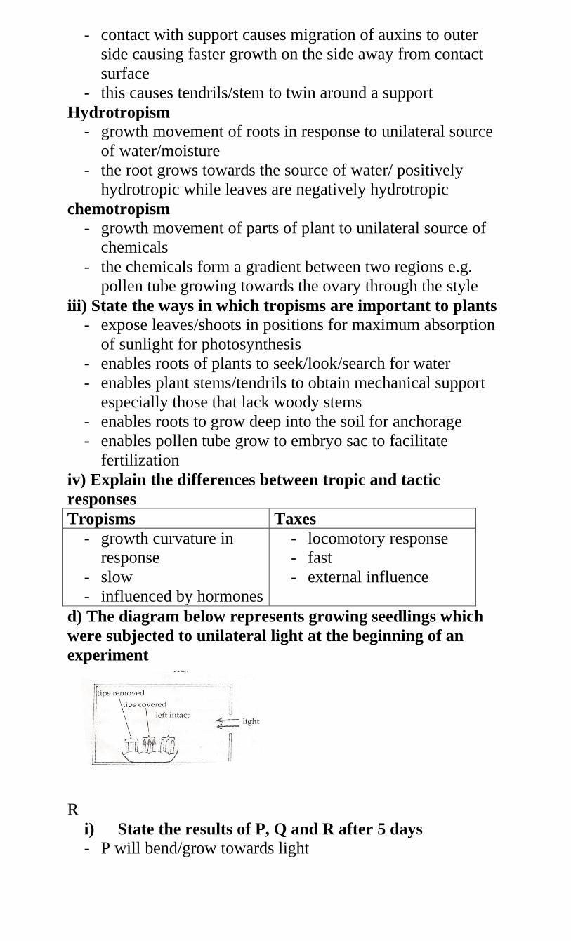
- contact with support causes migration of auxins to outer
side causing faster growth on the side away from contact
surface
- this causes tendrils/stem to twin around a support
Hydrotropism
- growth movement of roots in response to unilateral source
of water/moisture
- the root grows towards the source of water/ positively
hydrotropic while leaves are negatively hydrotropic
chemotropism
- growth movement of parts of plant to unilateral source of
chemicals
- the chemicals form a gradient between two regions e.g.
pollen tube growing towards the ovary through the style
iii) State the ways in which tropisms are important to plants
- expose leaves/shoots in positions for maximum absorption
of sunlight for photosynthesis
- enables roots of plants to seek/look/search for water
- enables plant stems/tendrils to obtain mechanical support
especially those that lack woody stems
- enables roots to grow deep into the soil for anchorage
- enables pollen tube grow to embryo sac to facilitate
fertilization
iv) Explain the differences between tropic and tactic
responses
Tropisms Taxes
- growth curvature in
response
- slow
- influenced by hormones
- locomotory response
- fast
- external influence
d) The diagram below represents growing seedlings which
were subjected to unilateral light at the beginning of an
experiment
R
i) State the results of P, Q and R after 5 days
- P will bend/grow towards light
- Q will remain straight/have little or no growth
- R will remain/grow straight/grow upwards
ii) Account for your results in (i) above
P- Growth substance/growth hormone/IAA/auxin are produced
by the stem tip
- they move (downwards and get distributed) to the side
away from light where they cause rapid/more growth/cell
division/elongation that results in bending
Q- Source of auxin has been removed
R- The auxins cannot be affected by light because the tip has
been covered
iii) If the tin foil were removed from the tip of seedling R,
what results would be observed after two days
- it will bend/grow towards light
iv) State the expected results after 3 day is if the box
were removed
- all seedlings will grow straight/upwards
e) In an experiment to investigate a certain aspect of plant
response, a seedling was placed horizontally as shown in
diagram I below. After seven days the appearance of the
seedling was as shown in diagram 2
Account for the curvature of the shoot and root after the
seven days
i) Shoot
- auxins accumulate on the lower side of the seedling due to
gravity
- high concentration of auxins in shoot stimulates faster
growth causing more elongation on the lower side than the
upper side hence curvature occurs upwards
ii) Root
- the high concentration of auxins inhibits growth hence the
upper side with less auxins grows faster than the lower side
therefore the curvature occurs downwards
f) What is etiolation?
- phenomenon exhibited by plants when grown in darkness
- such plants are pale yellow due to absence of chlorophyll,
have small leaves, long stems/hypocotyle and slender stems
- plants exhibit etiolation to reach light/obtain light
- this is a survival response
3. a) i) What is coordination in animals
4. - The linking together of all physiological activities that
occur in the body so that they take place at the right time
and in the correct place
ii) Name the main systems for coordination in animals
- Nervous system/sensory system
- Endocrine (hormonal system)
iii) List the components of the mammalian sensory system
- Central nervous system (CNS), brain & spinal cord
- Peripheral nervous system (PNS) cranial and spinal nerves
- Sense organs
- Autonomic nervous system (ANS) nerve fibers and ganglia
iv).Explain the terms receptors, conductors and effectors
- Receptors are structures that detect stimuli i.e. sense organs
- Conductors transmit impulses from receptors to effectors e.g.
neurons
- Effectors are the responding parts e.g. muscles, glands
v) What are the functions of the central nervous system?
- provides a fast means of communication between receptors and
effectors
- coordinates the activities of the body
vi) State the differences between somatic and autonomic
systems of peripheral nervous system
- Somatic is concerned with controlling the conscious or
voluntary actions of the body i.e. skin, bones, joints and skeletal
muscles
- the autonomic (automatic) nervous system controls involuntary
actions of internal organs, digestive system, blood vessels,
cardiac muscles and glandular products.
b) i) What is a neurone?
the basic unit of the nervous system
also called nerve cell
conducts impulses
include monitor sensory and relay neurons
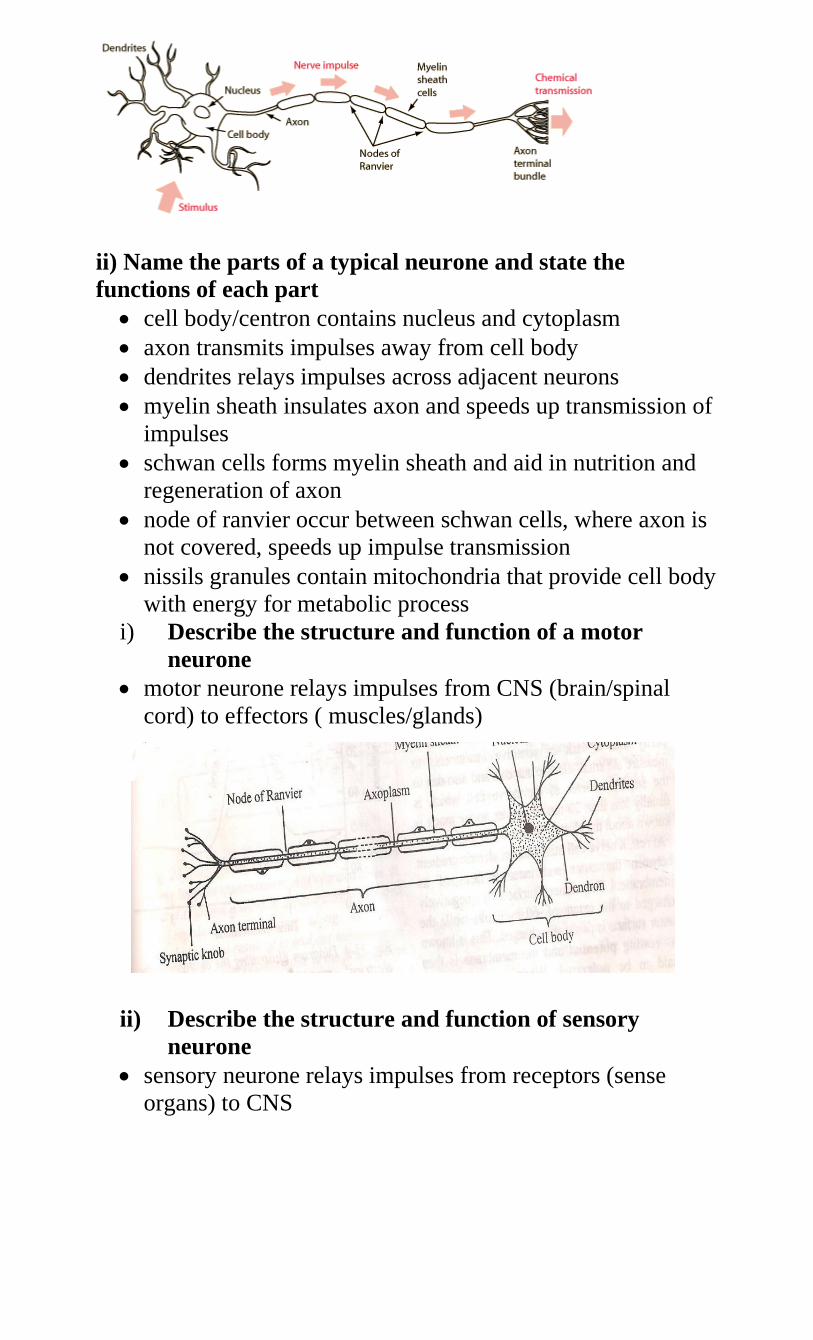
ii) Name the parts of a typical neurone and state the
functions of each part
cell body/centron contains nucleus and cytoplasm
axon transmits impulses away from cell body
dendrites relays impulses across adjacent neurons
myelin sheath insulates axon and speeds up transmission of
impulses
schwan cells forms myelin sheath and aid in nutrition and
regeneration of axon
node of ranvier occur between schwan cells, where axon is
not covered, speeds up impulse transmission
nissils granules contain mitochondria that provide cell body
with energy for metabolic process
i) Describe the structure and function of a motor
neurone
motor neurone relays impulses from CNS (brain/spinal
cord) to effectors ( muscles/glands)
ii) Describe the structure and function of sensory
neurone
sensory neurone relays impulses from receptors (sense
organs) to CNS
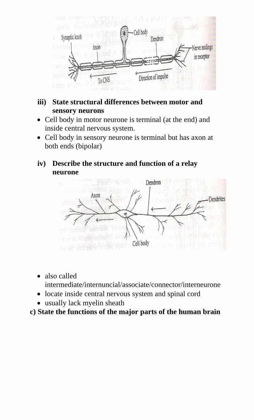
iii) State structural differences between motor and
sensory neurons
Cell body in motor neurone is terminal (at the end) and
inside central nervous system.
Cell body in sensory neurone is terminal but has axon at
both ends (bipolar)
iv) Describe the structure and function of a relay
neurone
also called
intermediate/internuncial/associate/connector/interneurone
locate inside central nervous system and spinal cord
usually lack myelin sheath
c) State the functions of the major parts of the human brain
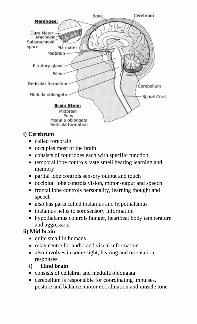
i) Cerebrum
called forebrain
occupies most of the brain
consists of four lobes each with specific function
temporal lobe controls taste smell hearing learning and
memory
partial lobe controls sensory output and touch
occipital lobe controls vision, motor output and speech
frontal lobe controls personality, learning thought and
speech
also has parts called thalamus and hypothalamus
thalamus helps to sort sensory information
hypothalamus controls hunger, heartbeat body temperature
and aggression
ii) Mid brain
quite small in humans
relay centre for audio and visual information
also involves in some sight, hearing and orientation
responses
i) Hind brain
consists of cellebral and medulla oblongata
cerebellum is responsible for coordinating impulses,
posture and balance, motor coordination and muscle tone
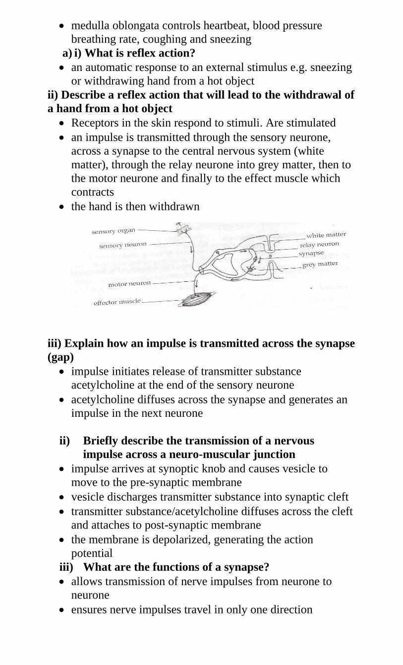
medulla oblongata controls heartbeat, blood pressure
breathing rate, coughing and sneezing
a) i) What is reflex action?
an automatic response to an external stimulus e.g. sneezing
or withdrawing hand from a hot object
ii) Describe a reflex action that will lead to the withdrawal of
a hand from a hot object
Receptors in the skin respond to stimuli. Are stimulated
an impulse is transmitted through the sensory neurone,
across a synapse to the central nervous system (white
matter), through the relay neurone into grey matter, then to
the motor neurone and finally to the effect muscle which
contracts
the hand is then withdrawn
iii) Explain how an impulse is transmitted across the synapse
(gap)
impulse initiates release of transmitter substance
acetylcholine at the end of the sensory neurone
acetylcholine diffuses across the synapse and generates an
impulse in the next neurone
ii) Briefly describe the transmission of a nervous
impulse across a neuro-muscular junction
impulse arrives at synoptic knob and causes vesicle to
move to the pre-synaptic membrane
vesicle discharges transmitter substance into synaptic cleft
transmitter substance/acetylcholine diffuses across the cleft
and attaches to post-synaptic membrane
the membrane is depolarized, generating the action
potential
iii) What are the functions of a synapse?
allows transmission of nerve impulses from neurone to
neurone
ensures nerve impulses travel in only one direction
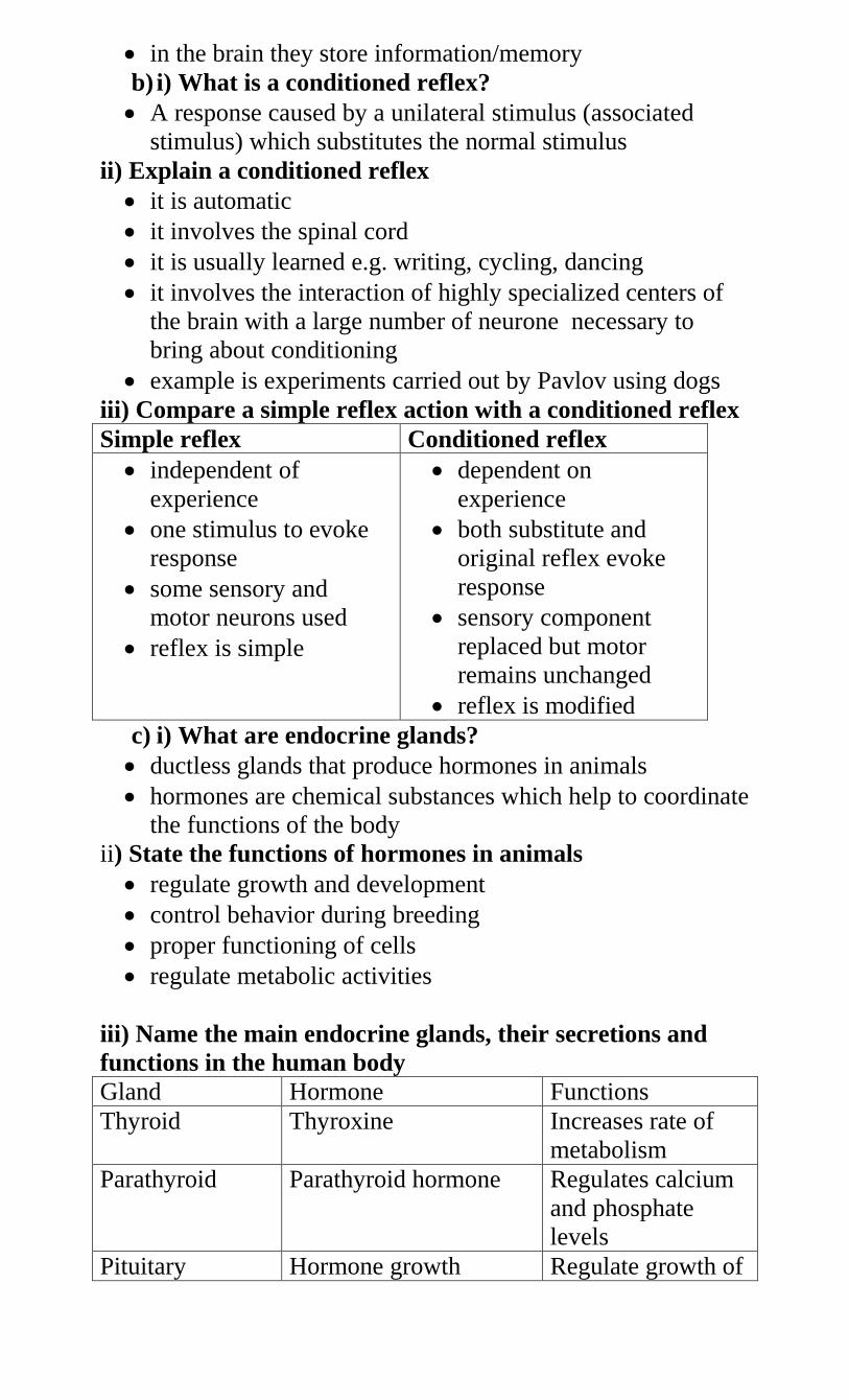
in the brain they store information/memory
b) i) What is a conditioned reflex?
A response caused by a unilateral stimulus (associated
stimulus) which substitutes the normal stimulus
ii) Explain a conditioned reflex
it is automatic
it involves the spinal cord
it is usually learned e.g. writing, cycling, dancing
it involves the interaction of highly specialized centers of
the brain with a large number of neurone necessary to
bring about conditioning
example is experiments carried out by Pavlov using dogs
iii) Compare a simple reflex action with a conditioned reflex
Simple reflex Conditioned reflex
independent of
experience
one stimulus to evoke
response
some sensory and
motor neurons used
reflex is simple
dependent on
experience
both substitute and
original reflex evoke
response
sensory component
replaced but motor
remains unchanged
reflex is modified
c) i) What are endocrine glands?
ductless glands that produce hormones in animals
hormones are chemical substances which help to coordinate
the functions of the body
ii) State the functions of hormones in animals
regulate growth and development
control behavior during breeding
proper functioning of cells
regulate metabolic activities
iii) Name the main endocrine glands, their secretions and
functions in the human body
Gland Hormone Functions
Thyroid Thyroxine Increases rate of
metabolism
Parathyroid Parathyroid hormone Regulates calcium
and phosphate
levels
Pituitary Hormone growth Regulate growth of
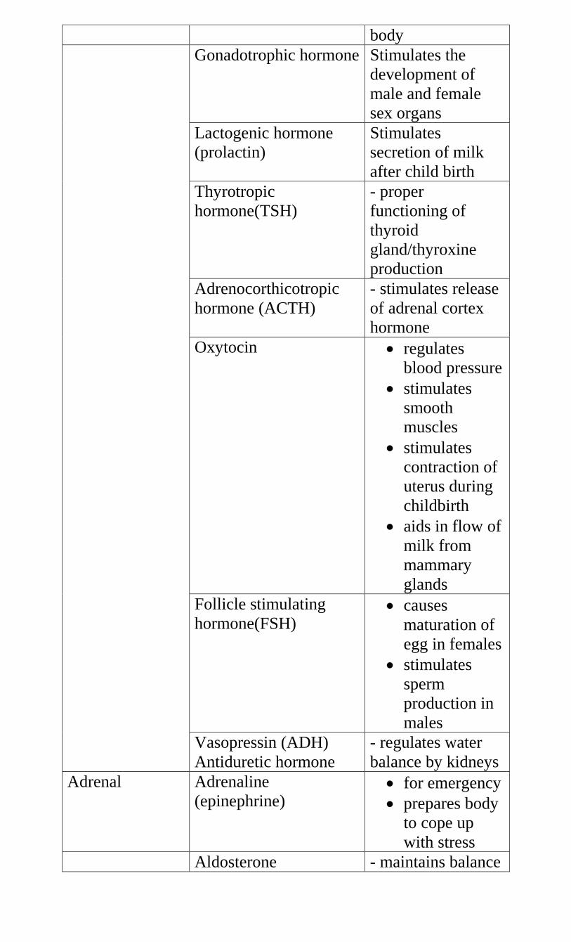
body
Gonadotrophic hormone Stimulates the
development of
male and female
sex organs
Lactogenic hormone
(prolactin)
Stimulates
secretion of milk
after child birth
Thyrotropic
hormone(TSH)
- proper
functioning of
thyroid
gland/thyroxine
production
Adrenocorthicotropic
hormone (ACTH)
- stimulates release
of adrenal cortex
hormone
Oxytocin regulates
blood pressure
stimulates
smooth
muscles
stimulates
contraction of
uterus during
childbirth
aids in flow of
milk from
mammary
glands
Follicle stimulating
hormone(FSH) causes
maturation of
egg in females
stimulates
sperm
production in
males
Vasopressin (ADH)
Antiduretic hormone
- regulates water
balance by kidneys
Adrenal Adrenaline
(epinephrine) for emergency
prepares body
to cope up
with stress
Aldosterone - maintains balance
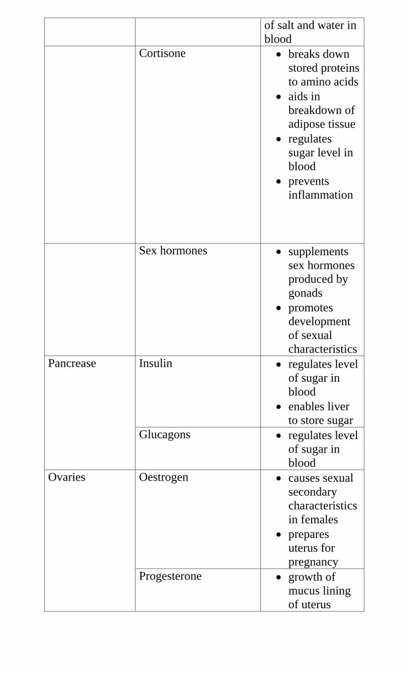
of salt and water in
blood
Cortisone breaks down
stored proteins
to amino acids
aids in
breakdown of
adipose tissue
regulates
sugar level in
blood
prevents
inflammation
Sex hormones supplements
sex hormones
produced by
gonads
promotes
development
of sexual
characteristics
Pancrease Insulin regulates level
of sugar in
blood
enables liver
to store sugar
Glucagons regulates level
of sugar in
blood
Ovaries Oestrogen causes sexual
secondary
characteristics
in females
prepares
uterus for
pregnancy
Progesterone growth of
mucus lining
of uterus
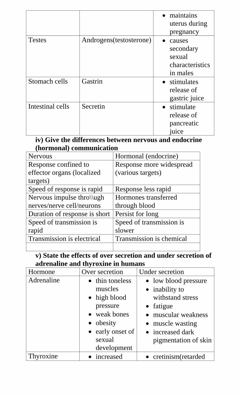
maintains
uterus during
pregnancy
Testes Androgens(testosterone) causes
secondary
sexual
characteristics
in males
Stomach cells Gastrin stimulates
release of
gastric juice
Intestinal cells Secretin stimulate
release of
pancreatic
juice
iv) Give the differences between nervous and endocrine
(hormonal) communication
Nervous Hormonal (endocrine)
Response confined to
effector organs (localized
targets)
Response more widespread
(various targets)
Speed of response is rapid Response less rapid
Nervous impulse thro\\ugh
nerves/nerve cell/neurons
Hormones transferred
through blood
Duration of response is short Persist for long
Speed of transmission is
rapid
Speed of transmission is
slower
Transmission is electrical Transmission is chemical
v) State the effects of over secretion and under secretion of
adrenaline and thyroxine in humans
Hormone Over secretion Under secretion
Adrenaline thin toneless
muscles
high blood
pressure
weak bones
obesity
early onset of
sexual
development
low blood pressure
inability to
withstand stress
fatigue
muscular weakness
muscle wasting
increased dark
pigmentation of skin
Thyroxine increased cretinism(retarded
metabolism
increased
heartbeat
physical
restlessness
mental
restlessness
protruding
eyeballs
enlarged
thyroid gland
growth and low
mental development
lowered metabolism
low ventilation rate
of lungs
low body
temperature
lowered mental
activity
coarse hair
puffy eyes
enlarged thyroid
gland
g) i) Define the following terms
Drug
a substance that causes a change in body function
Drug abuse
indiscriminate use of drugs without minding their side
effects
misuse or wrong use of drugs
ii) State the types of drugs, examples and side effects
Sedatives
also called depressant
a drug that decreases the action of the central nervous
system
reduce anxiety, and tension, induce sleep and act as muscle
relaxants
when abused they cause withdrawal effects such as anxiety,
delirium and death
includes barbiturates, other sedatives, tranquilizers and
alcohol
Pain-killers
suppress centers of pain in the brain
Hallucinogens
given to people with hallucination or mentally ill patients to
calm then down
when abused they lead to a feeling of confusion, agitation,
depression and violent behavior that can lead to murder or
suicide
examples include valium, LSD, bhang, narcotics and
cannabis
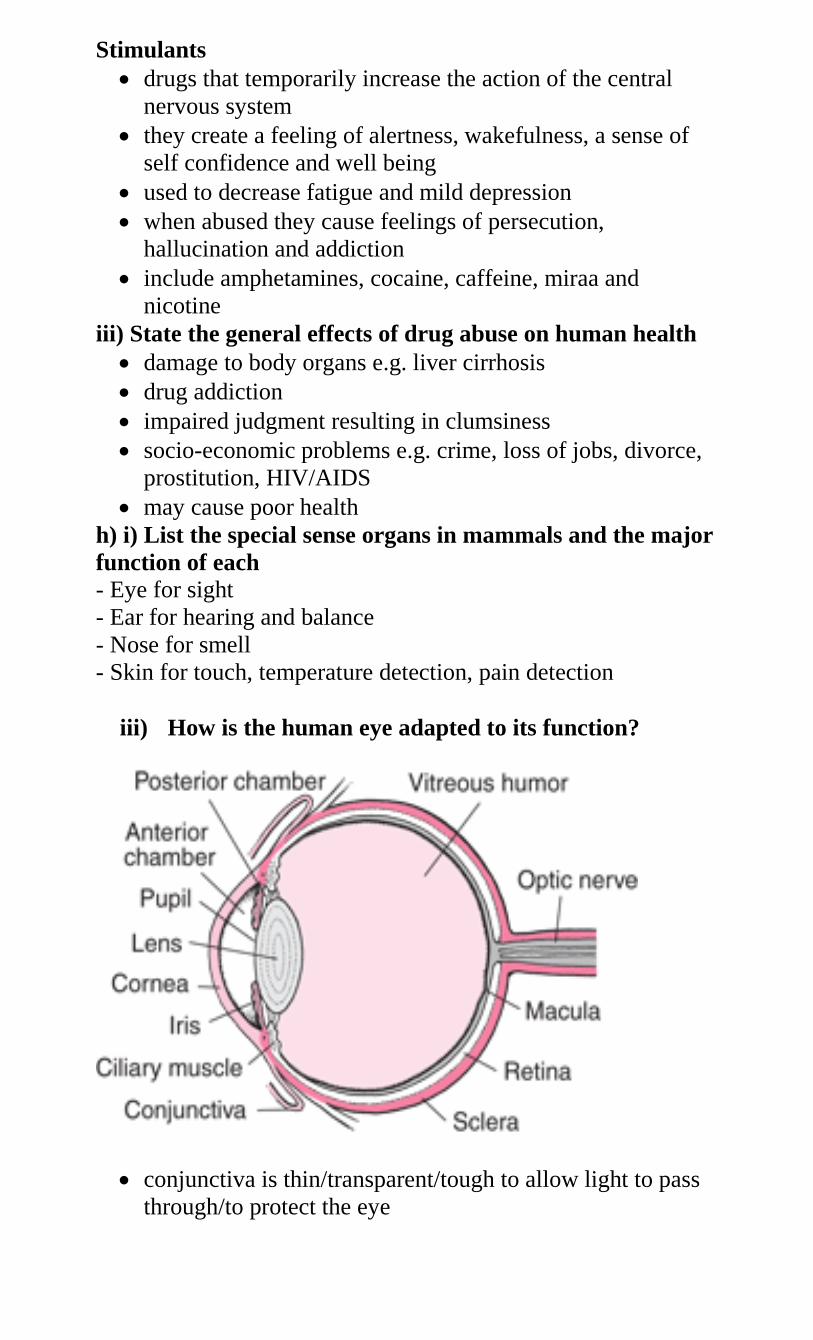
Stimulants
drugs that temporarily increase the action of the central
nervous system
they create a feeling of alertness, wakefulness, a sense of
self confidence and well being
used to decrease fatigue and mild depression
when abused they cause feelings of persecution,
hallucination and addiction
include amphetamines, cocaine, caffeine, miraa and
nicotine
iii) State the general effects of drug abuse on human health
damage to body organs e.g. liver cirrhosis
drug addiction
impaired judgment resulting in clumsiness
socio-economic problems e.g. crime, loss of jobs, divorce,
prostitution, HIV/AIDS
may cause poor health
h) i) List the special sense organs in mammals and the major
function of each
- Eye for sight
- Ear for hearing and balance
- Nose for smell
- Skin for touch, temperature detection, pain detection
iii) How is the human eye adapted to its function?
conjunctiva is thin/transparent/tough to allow light to pass
through/to protect the eye
Sclerotic layer is made up of (collagen) fibers/fibrous. It
maintains shape of the eyeball/protects the eye
cornea is transparent/curved thus refracts light rays/allows
light to pass through
Choroid is a layer of tissue with black pigment/dark
pigment. Prevents internal reflection of light in the
eye/contains blood vessels that supply
oxygen/nutrients/remove (metabolic) wastes from the eye
retina has cones/rods for bright colour vision/low light
vision
yellow spot has a high concentration of cones for accurate
vision/visual acuity
Blind spot has no cones and rods. Place where optic nerve
leaves/enters the eye
optic nerve has (sensory) nerve fibers for transmission of
impulses to the brain (for interpretation)
Lens is biconvex/made up of elastic material/transparent.
Adjust focus on far or near objects allow light to pass
through/for refraction of light rays
ciliary body is made up of muscle fibers/glandular which
contract/relax to change shape
suspensory ligaments are inelastic to hold lens in
position/attach it to ciliary body
iris(is the coloured part of the eye it) has radial and circular
muscles which control size of pupil
pupil is the small hole at the centre of iris through which
light passes into the eye
aqueous humor is a fluid through which oxygen/nutrients
pass to the cornea/lens/maintains shape of the
eyeball/refracts light rays
vitreous humor is a fluid which maintains shape of
eye/refracts light rays
iii) What is accommodation of the eye?
ability of the eye to adjust to bring an image from a near or
far object into sharp focus on the retina
iv) Explain how an eye viewing a near object adjusts to
viewing a far object
ciliary muscles relax
suspensory ligaments become taut/tight
lens decreases curvature/becomes thinner
radial muscles relax
circular muscles contract
size of pupil decreases to reduce amount of light
v) What changes occur in the eye if it changes from
observing an object at a distance to one at a closer range?
- ciliary muscles contract
- Tension in suspensory ligaments reduces/relax/ slackens
- Lens bulges/thickens/increases curvature
- Radial muscles contract
- Circular muscles relax
- Size of pupil becomes large to allow in more light.
viii) State the changes which would take place in the eye if
a person in a dark room had lights switched on
circular muscles contract and radial muscles relax
pupil becomes small to allow less light into the eye
ix) Explain how the eye forms an image
the mammalian eye works like a camera
light rays enter the cornea pass through the pupil, aqueous
humor, lens and vireous humor
light rays are refracted by the aqueous and humor and
lenses
finally light falls on the retina to form an image
the image is real and inverted and smaller than object, back
to front/reversed
Retina forms a fine image when light rays reach it.
x) Name the defects of the eye and state how they can be
corrected
Short sight (Myopia)
eye cannot focus on far objects
image is formed in front of the retina because light rays
converge in front of retina
the lens is too thick, curve and eyeball too long
corrected by wearing concave/biconcave/lenses
these lenses diverge light rays onto retina
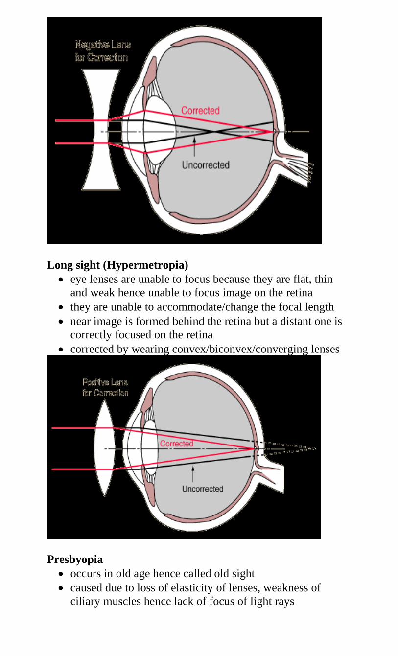
Long sight (Hypermetropia)
eye lenses are unable to focus because they are flat, thin
and weak hence unable to focus image on the retina
they are unable to accommodate/change the focal length
near image is formed behind the retina but a distant one is
correctly focused on the retina
corrected by wearing convex/biconvex/converging lenses
Presbyopia
occurs in old age hence called old sight
caused due to loss of elasticity of lenses, weakness of
ciliary muscles hence lack of focus of light rays
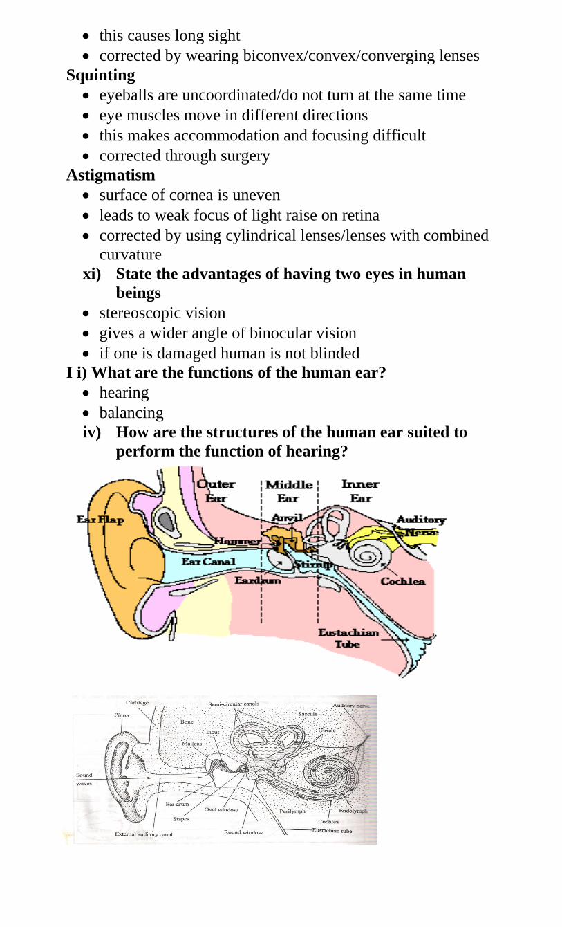
this causes long sight
corrected by wearing biconvex/convex/converging lenses
Squinting
eyeballs are uncoordinated/do not turn at the same time
eye muscles move in different directions
this makes accommodation and focusing difficult
corrected through surgery
Astigmatism
surface of cornea is uneven
leads to weak focus of light raise on retina
corrected by using cylindrical lenses/lenses with combined
curvature
xi) State the advantages of having two eyes in human
beings
stereoscopic vision
gives a wider angle of binocular vision
if one is damaged human is not blinded
I i) What are the functions of the human ear?
hearing
balancing
iv) How are the structures of the human ear suited to
perform the function of hearing?
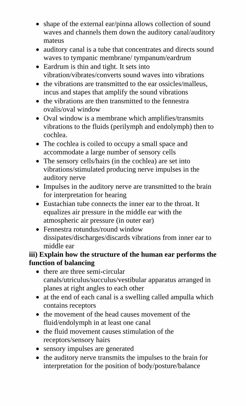
shape of the external ear/pinna allows collection of sound
waves and channels them down the auditory canal/auditory
mateus
auditory canal is a tube that concentrates and directs sound
waves to tympanic membrane/ tympanum/eardrum
Eardrum is thin and tight. It sets into
vibration/vibrates/converts sound waves into vibrations
the vibrations are transmitted to the ear ossicles/malleus,
incus and stapes that amplify the sound vibrations
the vibrations are then transmitted to the fennestra
ovalis/oval window
Oval window is a membrane which amplifies/transmits
vibrations to the fluids (perilymph and endolymph) then to
cochlea.
The cochlea is coiled to occupy a small space and
accommodate a large number of sensory cells
The sensory cells/hairs (in the cochlea) are set into
vibrations/stimulated producing nerve impulses in the
auditory nerve
Impulses in the auditory nerve are transmitted to the brain
for interpretation for hearing
Eustachian tube connects the inner ear to the throat. It
equalizes air pressure in the middle ear with the
atmospheric air pressure (in outer ear)
Fennestra rotundus/round window
dissipates/discharges/discards vibrations from inner ear to
middle ear
iii) Explain how the structure of the human ear performs the
function of balancing
there are three semi-circular
canals/utriculus/succulus/vestibular apparatus arranged in
planes at right angles to each other
at the end of each canal is a swelling called ampulla which
contains receptors
the movement of the head causes movement of the
fluid/endolymph in at least one canal
the fluid movement causes stimulation of the
receptors/sensory hairs
sensory impulses are generated
the auditory nerve transmits the impulses to the brain for
interpretation for the position of body/posture/balance
iv) State what would happen if the auditory nerve was
completely damaged
deafness
loss of body balance
impulse not transmitted to the brain
7. a) i) What is support?
to support is to carry part of the weight/mass of an
organism
ii) What is locomotion?
progressive change in the position of an organism
iii) State the importance of support systems in living
organisms
they provide a framework for the body of organisms and
help to determine their shape
provide land animals with means for support to their
weights against gravity
organs are attached to the skeleton for support and stability
to avoid entanglement and crushing each other
they protect very important and delicate organs whether
inside or outside the body e.g. eyes, heart
in large plants the rigid trunks of trees support the greater
mass of leaves and fruits
iv) State the importance of locomotion in animals
in search of food
search for mates
escaping predators
b) i) Name the tissues in higher plants that provide
mechanical support
sclerenchyma
collenchyma (not lignified)
xylem/tracheids and vessels
ii) State the importance of support in plants
exposing the surface area of leaf to sunlight for
photosynthesis
ensure flowers are exposed to pollination agents
expose fruits and seeds to agents of dispersal
to resist breakages due to their own weight and that of other
organisms
for proper transport and translocation of materials
iii) Name the types of plant stems
herbaceous e.g. shrubs
woody e.g. trees
weak stems in creepers, twining plants and plants bearing
tendrils
iv) Name the tissues in plants that are strengthened with
lignin
sclerenchyma
xylem vessels/tracheids/xylem
v) What makes young herbaceous plants remain upright?
turgidity
presence of collenchyma
vi) State the ways by which plants compensate for lack of
ability to move from one place to another
ability to pollinate
response to nastic and tropic movement
ability to exploit localized nutrients
ability to disperse seed or fruit propagation
c) i) Explain the ways in which erect posture is maintained in
a weak herbaceous stem - This is the function of turgidity and presence of collencyma
Cells take in water and become turgid
ii) Explain how support in plants is achieved
Turgor pressure due to absorption of water keeps cells firm
hence hold herbaceous plants upright
collenchyma and clerenchyma tissues are closely packed in
stem and roots to provide support
inelastic cuticle on epidermis is covered by a waxy layer
hence keeping shape of plant and setting inward pressure
against turgid cells and this causes a force to hold plant
upright
xylem vessels and tracheids are lignified to provide support
to stems, roots and leaves
climbing plants obtain mechanical support from other
plants and objects
they have climbing structures like tendrils which hold on to
other objects
d) i) Give the reasons why support is necessary in animals
for attachment of muscles
For attachment of other body organs
to protect delicate body organs
to maintain body shape/form
to enable movement/locomotion
ii) Why is movement necessary in animals?
enables animals to search for food
enables animals to search for shelter
enables animals to escape predators/harmful conditions
enables animals to search for water
enables animals to search for mates
enables animals to search for breeding sites
e) i) Name the organ used for support by animals
- Skeleton
ii) Name the different types of skeletons in animals, giving an
example of an animal for each type of skeleton named
exoskeleton e.g. arthropoda (crab, insect)
endoskeleton e.g. chordata (cat, fish)
iii) State the difference between exoskeleton and
endoskeleton
endoskeleton is a rigid framework covered by body tissues
of an animal
exoskeleton is a rigid framework found on the surface of an
animal
iv) State the advantages of having an exoskeleton
supports/protects delicate inner parts
water proof/prevents drying up of body
provided surface for muscle attachment
v) Explain the importance of having an endoskeleton
support the body
give body its shape
protect delicate organs e.g. skull, brain, ribs
used in locomotion e.g. bones serve as levers
red blood cells are formed in bone marrow
minerals are stored in bones e.g. calcium and phosphorus
f) i) Explain how a fish is adapted to living in water
streamlined body for easy movement in water
swim bladder controls depth of swimming
fins for movement, balance, direction and stability
gills for gaseous exchange in water
presence of lateral line to sense vibrations
scales provide protection
colour which offers camouflage against predators
ii) Explain how a finned fish is adapted to locomotion in
water
streamlined body to reduce resistance/friction )to swim
smoothly)
the vertebral column consists of a series of vertebrae held
together loosely so that it is flexible
myotomes/muscles associated with vertebral column
produce movement
the sideways and backwards thrust of the tail and body
against water results in resistance of water pushing the fish
sideways and forwards in a direction opposed to thrust
heat not flexible so as to maintain forward thrust
presence of fins help in propulsion/balance/paired fins
(pectoral and pelvic) for controlling pitch and slow down
movement/unpaired fins (dorsal, ventral, anal) for yawing
and rolling (caudal) for swimming/propulsion and
steering/change of direction
presence of swim bladder to make fish buoyant
scales tip towards the back to provide smooth surface
body covered with mucus to reduce friction
flattened surface for easy floating
g) i) Name the main parts of the vertebral column giving the
types of bones found in each part
Axial skeleton
forms the main axis of the body
formed by the skull, sternum, ribs and vertebrae
Appendicular skeleton
composed of limbs and girdles
the forelimbs are connected to the trunk by the pectoral
girdles (shoulder bones)
hind limbs are connected to the pelvic girdle (hips)
bones are scapular, clavicle, humerus, ulna, femur, tibia,
fibula, metacarpals, carpals, tarsals, metatarsals, phalanges,
ilium, ischium and pubis
ii) What are the vertebrae?
bones of the vertebral column
iii) State the functions of the vertebral column
gives flexibility
absorbs shock
protects spinal cord
supports weight of body
provide surface for muscle attachment
between the vertebrae are soft discs which offer cushioning
called interverterbral discs
iv) State the general characteristics of vertebrae
have solid structure called centrum to support weight of
body
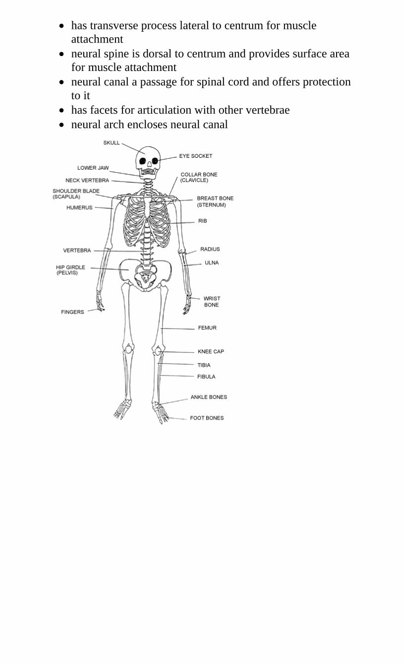
has transverse process lateral to centrum for muscle
attachment
neural spine is dorsal to centrum and provides surface area
for muscle attachment
neural canal a passage for spinal cord and offers protection
to it
has facets for articulation with other vertebrae
neural arch encloses neural canal
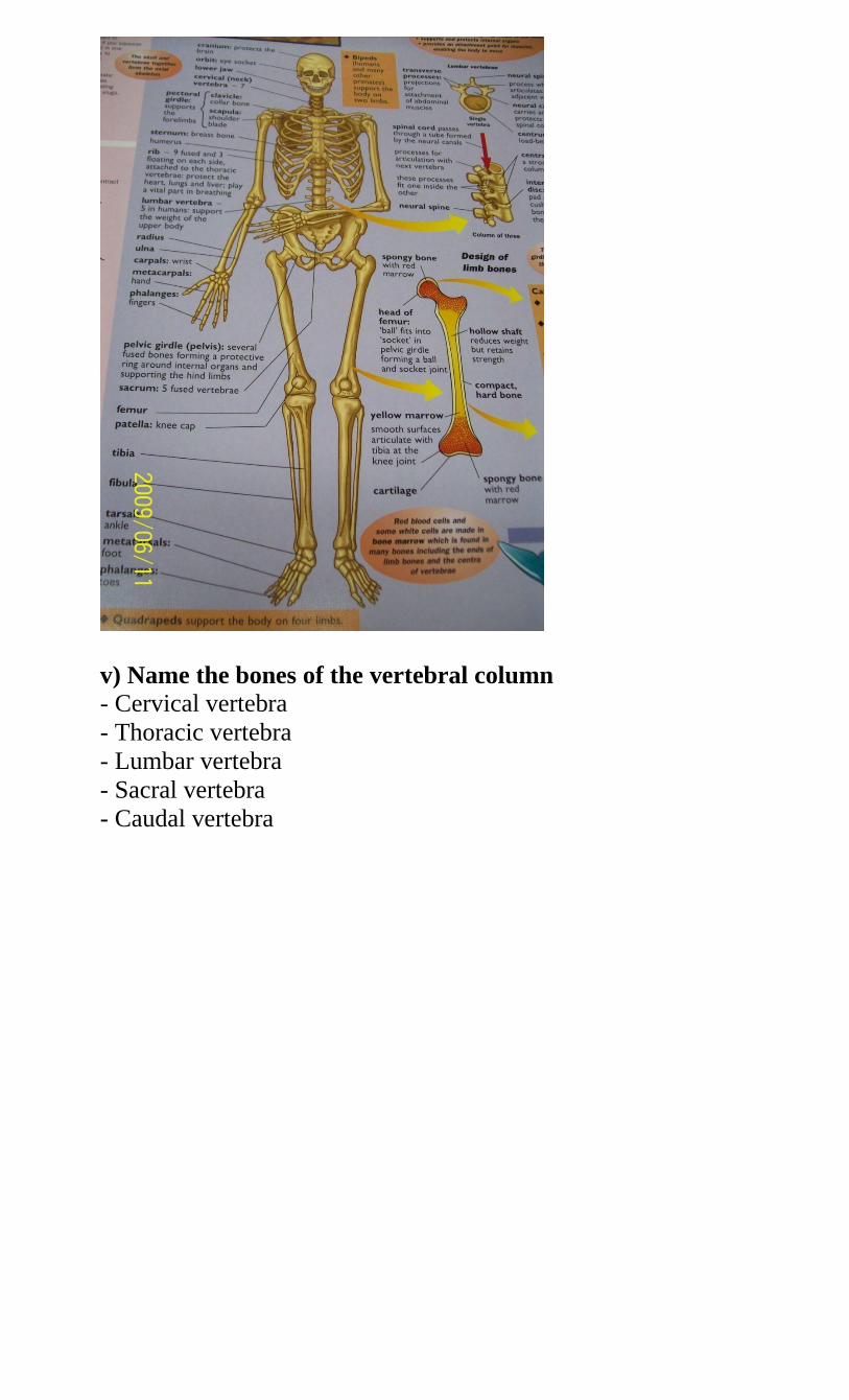
v) Name the bones of the vertebral column
- Cervical vertebra
- Thoracic vertebra
- Lumbar vertebra
- Sacral vertebra
- Caudal vertebra
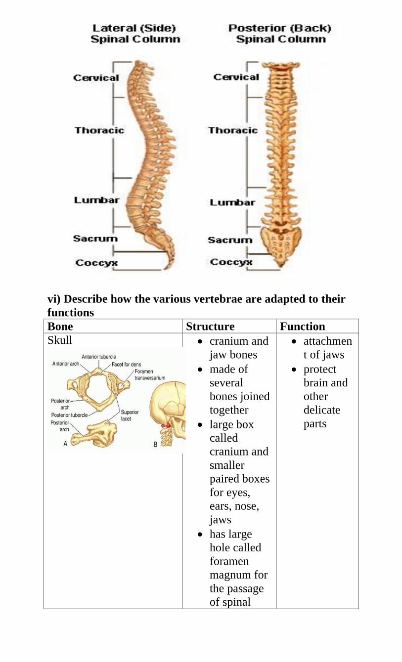
vi) Describe how the various vertebrae are adapted to their
functions
Bone Structure Function
Skull
cranium and
jaw bones
made of
several
bones joined
together
large box
called
cranium and
smaller
paired boxes
for eyes,
ears, nose,
jaws
has large
hole called
foramen
magnum for
the passage
of spinal
attachmen
t of jaws
protect
brain and
other
delicate
parts
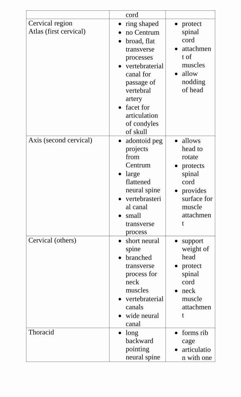
cord
Cervical region
Atlas (first cervical)
ring shaped
no Centrum
broad, flat
transverse
processes
vertebraterial
canal for
passage of
vertebral
artery
facet for
articulation
of condyles
of skull
protect
spinal
cord
attachmen
t of
muscles
allow
nodding
of head
Axis (second cervical)
adontoid peg
projects
from
Centrum
large
flattened
neural spine
vertebrasteri
al canal
small
transverse
process
allows
head to
rotate
protects
spinal
cord
provides
surface for
muscle
attachmen
t
Cervical (others)
short neural
spine
branched
transverse
process for
neck
muscles
vertebraterial
canals
wide neural
canal
support
weight of
head
protect
spinal
cord
neck
muscle
attachmen
t
Thoracid
long
backward
pointing
neural spine
forms rib
cage
articulatio
n with one
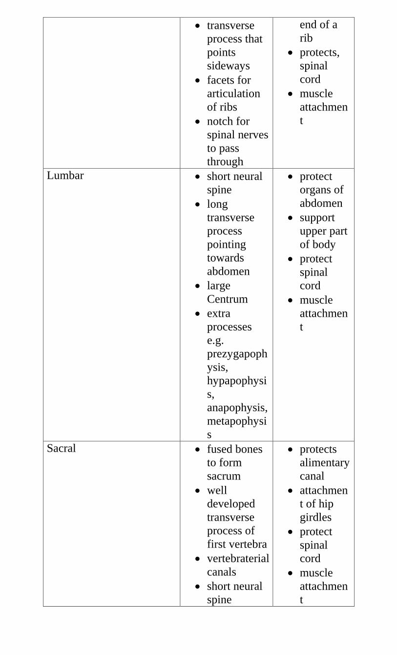
transverse
process that
points
sideways
facets for
articulation
of ribs
notch for
spinal nerves
to pass
through
end of a
rib
protects,
spinal
cord
muscle
attachmen
t
Lumbar
short neural
spine
long
transverse
process
pointing
towards
abdomen
large
Centrum
extra
processes
e.g.
prezygapoph
ysis,
hypapophysi
s,
anapophysis,
metapophysi
s
protect
organs of
abdomen
support
upper part
of body
protect
spinal
cord
muscle
attachmen
t
Sacral
fused bones
to form
sacrum
well
developed
transverse
process of
first vertebra
vertebraterial
canals
short neural
spine
protects
alimentary
canal
attachmen
t of hip
girdles
protect
spinal
cord
muscle
attachmen
t
Rib
long
flattened
attached to
sternum
from front
protect
internal
organs
muscle
attachmen
t
vii) Describe the bones that form the appendicular skeleton
Bone Structure Function
Pectoral girdle scapular
(shoulder bone)
Broad i.e.
Flattened blade
glenoid cavity to
articulate with
humerus
metacromion/acro
mion for muscle
attachment
hard to provide
support
socket with
cartilage/smooth
surface to reduce
friction
Support
Muscle
attachment
Articulates
with humerus
Humerus
long shaft for
muscle attachment
round head to
articulate with
glenoid cavity
trochlea for
articulation with
ulna
olecranon fosa to
prevent arm
bending the other
way
moveme
nt
muscle
attachme
nt
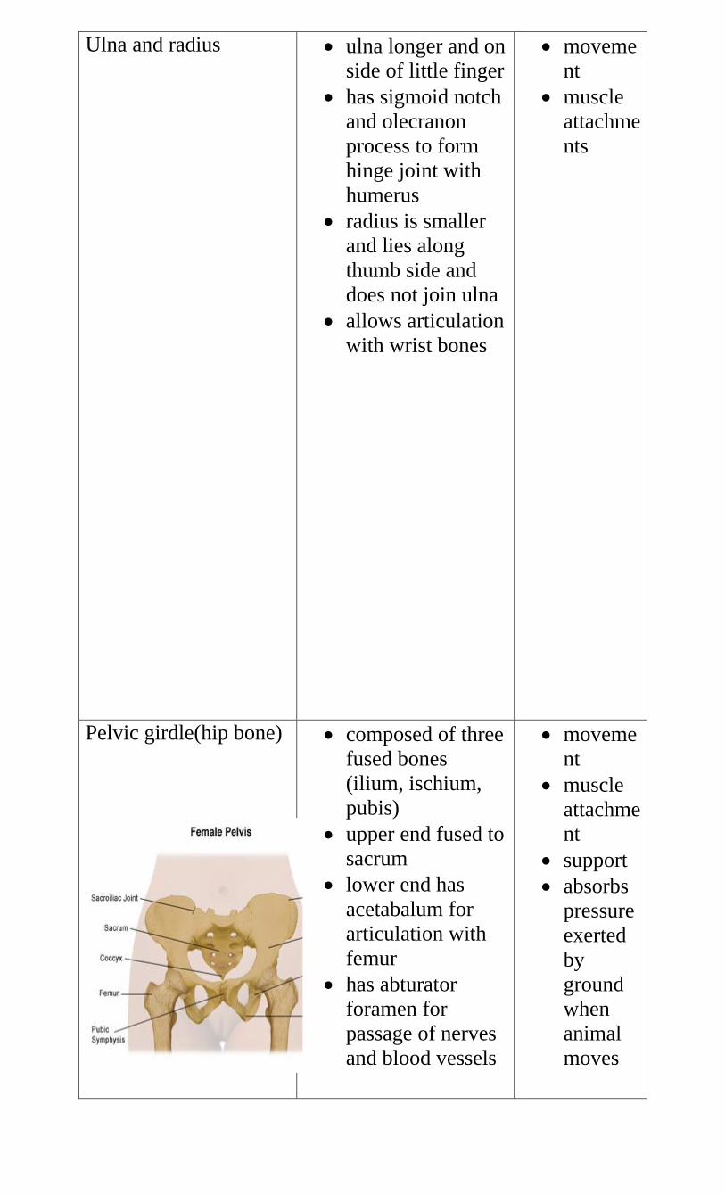
Ulna and radius
ulna longer and on
side of little finger
has sigmoid notch
and olecranon
process to form
hinge joint with
humerus
radius is smaller
and lies along
thumb side and
does not join ulna
allows articulation
with wrist bones
moveme
nt
muscle
attachme
nts
Pelvic girdle(hip bone)
composed of three
fused bones
(ilium, ischium,
pubis)
upper end fused to
sacrum
lower end has
acetabalum for
articulation with
femur
has abturator
foramen for
passage of nerves
and blood vessels
moveme
nt
muscle
attachme
nt
support
absorbs
pressure
exerted
by
ground
when
animal
moves
Femur
rounded head to fit
in acetabulum of
pelvis
projections called
trochanter for
attachment of
thigh muscles
condyles at lower
end for
articulation with
tibi
patella that covers
knee and prevents
leg from bending
backwards
moveme
nt
muscle
attachme
nt
Tibia and fibula
tibia is longer than
fibula
tibia is outer bone
and fibula is inner
bone
tibia lies on side
of large toe
fibula is fused to
tibia (on outer
side)
moveme
nt
muscle
attachme
nt
8. a) What is a joint?
the point where bones meet
ii) State the functions of joints
provide a point of articulation between bones
iii) Name the main types of joints
immovable joints e.g. skull, pelvic girdles and sacrum
slightly movable joints e.g. between vertebrae
Freely movable joints e.g. knee, elbow
iv) Give the features of movable joints
ends of bones covered with articular cartilage
ends bound by capsules of ligaments
have joint cavity filled with lubricating fluid called
synovial fluid secreted by synovial membrane
they are called synovial joints
b) Describe the synovial joints
iv) Ball and socket
allow movement in all planes /directions i.e. 360o
rounded end of bone fits into a rounded cavity in another
bone
e.g. shoulder joint and hip joint
v) Hinge joint
convex surface of one bone fits into the concave surface of
another bone
this allows movement in only one plane/direction 180o
e.g. elbow joint and knee joint
vi) Pivot joint
allows rotation e.g. where atlas pivots on olecranon process
of axis
c) i) What is synovial fluid?
lubricating fluid produced by synovial membrane at
movable joints
ii) State the functions of synovial fluid
absorbs shock
reduces friction/gives lubrication
nourishment
distributes pressure
d) Explain the following terms
v) Ligament
connective tissue joining one bone to another
vi) Cartilage
supporting soft tissue found at joints
they cushion the bones and absorb shock
vii) Tendon
tissue that connects muscle to bones
9. Muscles
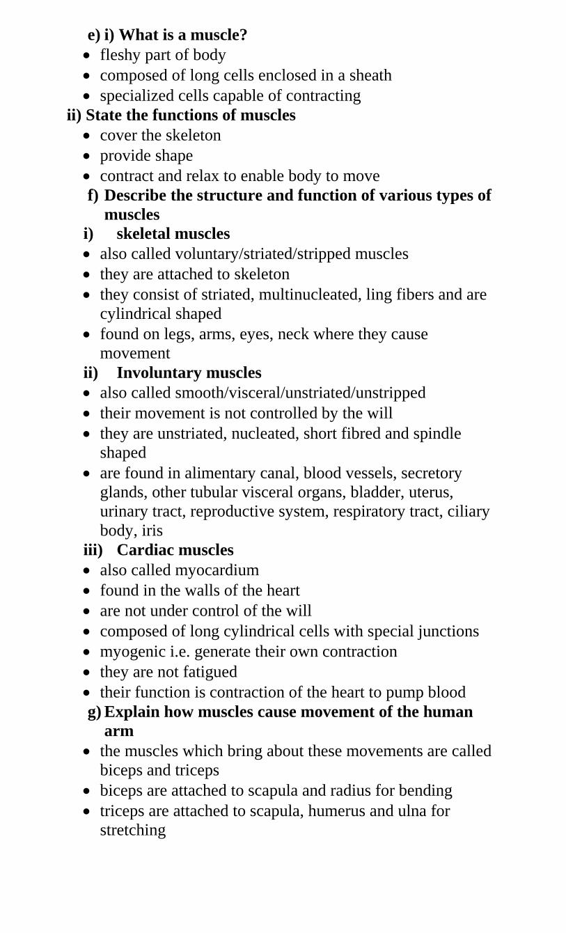
e) i) What is a muscle?
fleshy part of body
composed of long cells enclosed in a sheath
specialized cells capable of contracting
ii) State the functions of muscles
cover the skeleton
provide shape
contract and relax to enable body to move
f) Describe the structure and function of various types of
muscles
i) skeletal muscles
also called voluntary/striated/stripped muscles
they are attached to skeleton
they consist of striated, multinucleated, ling fibers and are
cylindrical shaped
found on legs, arms, eyes, neck where they cause
movement
ii) Involuntary muscles
also called smooth/visceral/unstriated/unstripped
their movement is not controlled by the will
they are unstriated, nucleated, short fibred and spindle
shaped
are found in alimentary canal, blood vessels, secretory
glands, other tubular visceral organs, bladder, uterus,
urinary tract, reproductive system, respiratory tract, ciliary
body, iris
iii) Cardiac muscles
also called myocardium
found in the walls of the heart
are not under control of the will
composed of long cylindrical cells with special junctions
myogenic i.e. generate their own contraction
they are not fatigued
their function is contraction of the heart to pump blood
g) Explain how muscles cause movement of the human
arm
the muscles which bring about these movements are called
biceps and triceps
biceps are attached to scapula and radius for bending
triceps are attached to scapula, humerus and ulna for
stretching
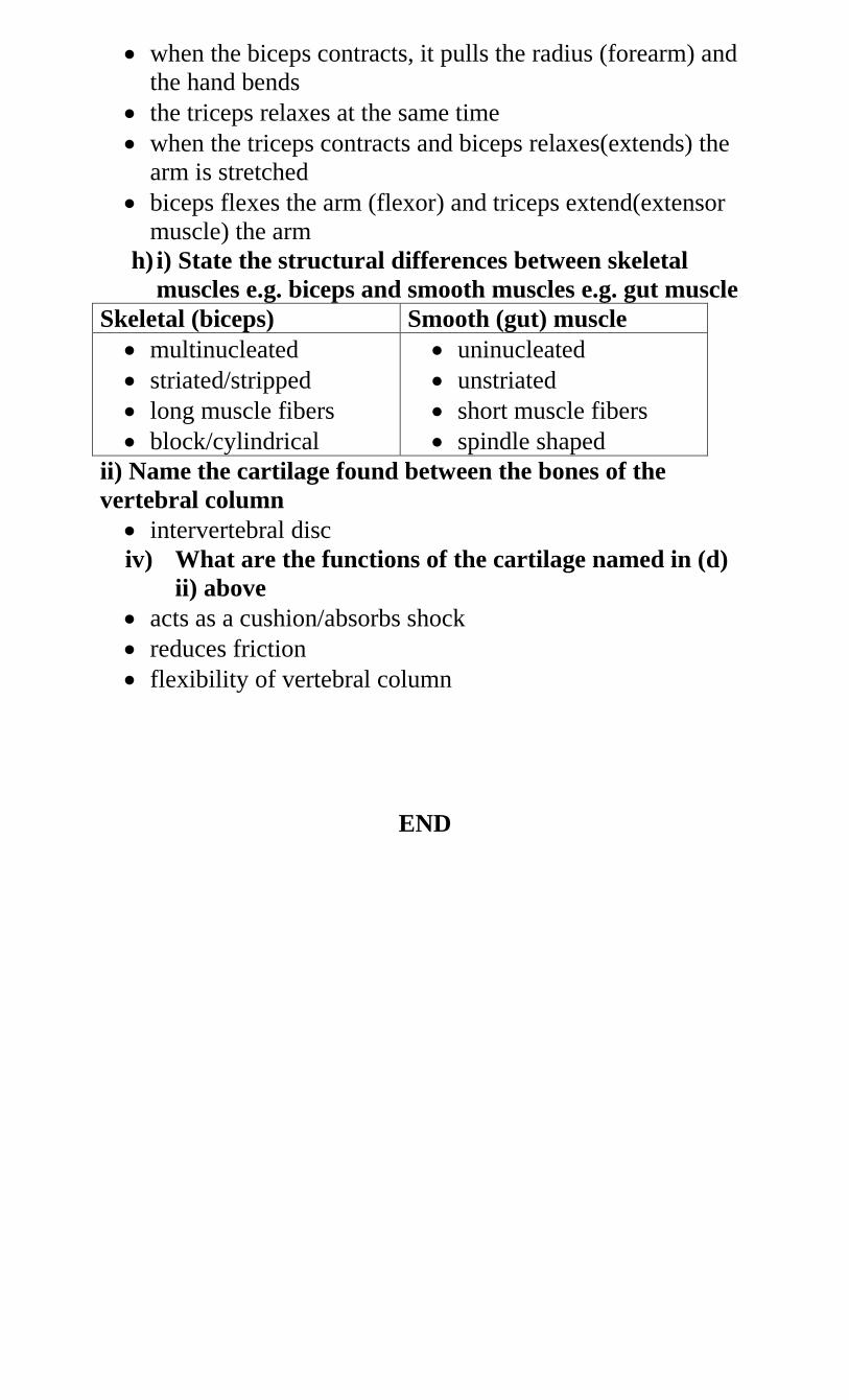
when the biceps contracts, it pulls the radius (forearm) and
the hand bends
the triceps relaxes at the same time
when the triceps contracts and biceps relaxes(extends) the
arm is stretched
biceps flexes the arm (flexor) and triceps extend(extensor
muscle) the arm
h) i) State the structural differences between skeletal
muscles e.g. biceps and smooth muscles e.g. gut muscle
Skeletal (biceps) Smooth (gut) muscle
multinucleated
striated/stripped
long muscle fibers
block/cylindrical
uninucleated
unstriated
short muscle fibers
spindle shaped
ii) Name the cartilage found between the bones of the
vertebral column
intervertebral disc
iv) What are the functions of the cartilage named in (d)
ii) above
acts as a cushion/absorbs shock
reduces friction
flexibility of vertebral column
END
Related Documents