Biologically-Inspired Microfabricated Force and Position Mechano-Sensors P. Dario, C. Laschi, S. Micera, F. Vecchi, M. Zecca, A. Menciassi, B. Mazzolai, M.C. Carrozza Scuola Superiore Sant’Anna – ARTS & MiTech Labs Piazza Martiri della Libertà, 33 56127 Pisa, Italy dario (cecilia, silvestro, fabrizio, zecca, arianna, barbara, chiara)@mail-arts.sssup.it ABSTRACT The aim of this paper is to discuss an ideal design procedure for biologically-inspired mechano- sensors. The main steps of this procedure are the following: (1) analysis of force and position sensors in animals and humans; (2) analysis of technologies available for MEMS (Micro Electro Mechanical Systems) and; (3) design and implementation of biologically-inspired sensors in innovative mechatronic and biomechatronic systems (e.g., antropomorphic robots, prostheses, and neuroprostheses). According to this sequence, the first part of the paper is dedicated to the presentation of some features of force and motion sensors in animals and humans. Then, technologies for fabricating miniaturized force and motion sensors (and some examples of such sensors) are briefly presented. Finally, some applications of biologically-inspired systems developed by the authors to sense force and position in anthropomorphic robots and in prosthetics are illustrated and discussed. 1 - INTRODUCTION The main reason which determined the failure of early approaches to machine intelligence was probably the assumption that intelligence is essentially abstract thinking, and that therefore human intelligence could be reproduced by developing powerful computers capable of high speed calculations and logic (sequential) reasoning [7]. As the animal world vividly demonstrates, the reality is that intelligence evolves and is built primarily on the availability and processing of sensory information. Recent research on intelligent machines and systems assumes that sensors are key components for obtaining adaptive and intelligent behavior. This assumption holds not only for complex, sometimes human-like machines, such as the robot, but virtually for every modern (“mechatronic”) products (e.g., camcorders, washing machines, car subsystems, etc.) [13, 24], to which embedded computers and many different sensors provide the capability to react to variable working conditions [11]. In fact, whereas in the past the need for sensors was widely recognized, but their practical use in machines was difficult because of the lack of processing capabilities, today embedded computing makes sensors not only useful but usable, and better sensors strongly needed. An interesting and important question is: which properties a “better” sensor should possess? Obviously a general answer is not possible, but it is certainly true that, along with many engineering properties very important for practical application, such as measuring range, sensitivity, linearity, 1

Welcome message from author
This document is posted to help you gain knowledge. Please leave a comment to let me know what you think about it! Share it to your friends and learn new things together.
Transcript

Biologically-Inspired Microfabricated Force and Position Mechano-Sensors
P. Dario, C. Laschi, S. Micera, F. Vecchi, M. Zecca,
A. Menciassi, B. Mazzolai, M.C. Carrozza
Scuola Superiore Sant’Anna – ARTS & MiTech Labs
Piazza Martiri della Libertà, 33 56127 Pisa, Italy
dario (cecilia, silvestro, fabrizio, zecca, arianna, barbara, chiara)@mail-arts.sssup.it
ABSTRACT
The aim of this paper is to discuss an ideal design procedure for biologically-inspired mechano-sensors. The main steps of this procedure are the following: (1) analysis of force and position sensors in animals and humans; (2) analysis of technologies available for MEMS (Micro Electro Mechanical Systems) and; (3) design and implementation of biologically-inspired sensors in innovative mechatronic and biomechatronic systems (e.g., antropomorphic robots, prostheses, and neuroprostheses).
According to this sequence, the first part of the paper is dedicated to the presentation of some features of force and motion sensors in animals and humans. Then, technologies for fabricating miniaturized force and motion sensors (and some examples of such sensors) are briefly presented. Finally, some applications of biologically-inspired systems developed by the authors to sense force and position in anthropomorphic robots and in prosthetics are illustrated and discussed.
1 - INTRODUCTION
The main reason which determined the failure of early approaches to machine intelligence was probably the assumption that intelligence is essentially abstract thinking, and that therefore human intelligence could be reproduced by developing powerful computers capable of high speed calculations and logic (sequential) reasoning [7]. As the animal world vividly demonstrates, the reality is that intelligence evolves and is built primarily on the availability and processing of sensory information. Recent research on intelligent machines and systems assumes that sensors are key components for obtaining adaptive and intelligent behavior. This assumption holds not only for complex, sometimes human-like machines, such as the robot, but virtually for every modern (“mechatronic”) products (e.g., camcorders, washing machines, car subsystems, etc.) [13, 24], to which embedded computers and many different sensors provide the capability to react to variable working conditions [11]. In fact, whereas in the past the need for sensors was widely recognized, but their practical use in machines was difficult because of the lack of processing capabilities, today embedded computing makes sensors not only useful but usable, and better sensors strongly needed.
An interesting and important question is: which properties a “better” sensor should possess? Obviously a general answer is not possible, but it is certainly true that, along with many engineering properties very important for practical application, such as measuring range, sensitivity, linearity,
1

accuracy, repeatability, robustness, and so forth, some properties inspired by the observation of biological sensors and their use by plants and animals might also be very useful. Recent developments of microfabrication technologies for MEMS (Micro Electro Mechanical Systems) [26] and for “BioMEMS” (hybrid artificial-biological microstructures) [21] make it possible to design and fabricate a range of new miniaturized sensors made out of different (inorganic and organic) materials and with sensing and processing capabilities previously impossible to obtain. This opens up new perspectives to research on bionic sensors, that is on sensors which aim either at imitating or at substituting biological sensors for application in robotics, prosthetics and, in general, in current and future mechatronic products and biomechatronic systems [17].
In this paper we start from the analysis of some representative biological sensors, focusing on force and motion sensors in animals and humans. Then, we describe microfabrication technologies for MEMS and BioMEMS, and examples of microfabricated force and motion sensors inspired by animal sensors. Finally, we discuss some applications of these sensors in anthropomorphic robotics, and in prosthetics, with reference to research projects carried on in the authors’ laboratory.
2 - SENSORS OF FORCE AND MOTION IN ANIMALS AND HUMANS
In this section we outline the main features of typical sensors of force and motion which can be found in some animal species. In particular we consider invertebrates, such as arthropods [57] and cephalopods [3]; and vertebrates [1], such as fish, amphibians, birds and nocturnal mammalians. Special attention is devoted to the analysis of touch and position sensors in humans [39, 56], which represent a very sophisticated and thus inspiring model for mechano-reception in artificial machines.
2.1 - INVERTEBRATES
2.1.1 – Phylum Arthropoda
Arthropods (like cockroaches, crabs and spiders) sense force primarily using strain gages, which differ each other for dimension: campaniform sensillum of insects is 20 microns diameter; lyriform slit sense organ in spider has a major axis of 175 microns and a minor axis of 75 microns; force-sensitive organs of crabs are only 5 microns long. However all these sensors are linked to the central nervous system in similar ways. Each is attached to the dendrite of the receptor cell. When a force deforms the exoskeleton around a sensillum, the tip of the dendrite deforms with it and the nerve cell generates bursts of action potentials, which are conducted to the central nervous system via the axon of the neuron. The arthropod cuticle and the human bone have an elastic module on the order of 10 billion newtons per square meter. Such a stiff structure indicates high performances: they are as sensitive to displacement as the receptors in the human ear to sound.
The campaniform sensillum responds best to forces that compress the organ perpendicular to its long axis, while the spider and the crustacean sensors are 3D.
Arthropods have also a sort of goniometer, consisting of hair-like mechanoreceptors which extend from the exoskeleton; each hair plate contains a number of sensilla that sense the position of the joint. Bending the sensillum stretches and deforms the dendrite of the receptor cell generating bursts of action potentials. Some arthropods have also a gyroscope, called statocyte, a sensory vesicle lined with ciliated mechanoreceptor cells that make contact with statoliths, which can be sand grains, calcareous concretions, or some other relatively dense material. Statocyte functions in the perception of the position of the body in space.
2.1.2 – Class Cephalopoda
Cephalopods (i.e. octopus, sepia, squid, nautilus) have tactile cells which are present in the epithelium of tentacles and in the suckers. They give information about structural characteristics of 2

surfaces, but not about forms, maybe because these organs have no structure or division that can act as measuring unit (like the distance between thumb and index in primates). Cephalopods have a statocyte, which works almost in the same way as the one of arthropods.
2.2 - VERTEBRATES
2.2.1 - Fish and amphibians
Fish and amphibians have a force and pressure sensory system, (called lateral line system) that is a set of external receptors based on hair cells that detect motion in the surrounding water. The stereocilia of a hair cell are arranged in order of increasing length from one side of the cell to the other. When the cilia bend toward the tallest cilium, a hair cell depolarizes; whereas, when they bend in the opposite direction, the cell hyperpolarizes. At rest, about 15% of the channels in a hair cell are open, producing a resting potential of about – 60mV. Hair cells do not produce action potentials. Instead, they form chemical synapses onto afferent neurons. The amount of transmitter released onto the afferent neurons determines their frequency of discharge.
2.2.2 - Birds and nocturnal mammalians
While birds have a lot of tactile sensing sites on their tongue and on the beak, nocturnally active mammals (e.g., cats, rats) use their vibrissae (which are lengthened tactile hairs on the snouts) to tactually sense objects in the vicinity of their body, extending and enriching the adaptive function of the sense of touch.
2.2.3 - Humans
Humans skin is an active sensory organ that is both highly sensitive and resistant. The glabrous skin has about 17,000 tactile units composed of five major types of receptors: free receptors, Meissner Corpuscles, Merkel's disks, Pacinian corpuscles, and Ruffini endings. These receptors are found at all depths below the skin surface (see Figure 1) and can be described as follows [56]:
• Meissner’s corpuscles are the 43% of the tactile units in the hand. The average size of these corpuscles for an adult is about 80 µm x 30 µm, with the long axis perpendicular to the surface of the skin. They are velocity detectors or touch receptors, since they move with the ridged skin of the fingers and the palm and provide the best reception of movement across the skin;
• Pacinian corpuscles are from 1 to 4 mm in length and from 0.5 to 1 mm in diameter. There are about 2000 Pacinian corpuscles distributed all around the body, with one third of them localized in the digits. They are acceleration detectors and provide vibration reception. They cannot detect steady pressure, but they are responsible for the threshold detection of light touch;
• Ruffini’s endings are composed of a fusiform structure with a definite capsule in the subcutaneous tissue of the pulp of the human finger. They are the 19% of the tactile units in the hand, and they are detectors of intensity and pressure. They are also responsible of detecting shear on the skin;
• Merkel’s disks are 25% of the tactile units in the hand. They are composed of disk-like nerve ending and a specialized receptor cell, and they are about 70–90 nm in diameter. They provide excellent detection of intensity; however they provide tactile and vibration information;
• Free receptors are the most important cutaneous receptors, as they permeate the entire thickness of the dermis. They have diameters ranging from 0.5 to 2.5 µm, and generally they form thermoreceptors and nociceptors (pain receptors).
3

Figure 1: Schematic representation of the human skin and of its sensors.
The above mentioned cutaneous receptors could also be electrophysiologically defined by classifying their responses to different stimuli. We can distinguish three types of receptors: thermoreceptors, nociceptors, and mechanoreceptors. The first ones could be divided into two groups, one that is preferentially receptive to cold, the other to warmth. Both groups have the same resting discharge at normal temperature, and thus cannot be distinguished except by observing their responses to warm and cold stimuli.
Nociceptors respond to stimuli that activate both the thermoreceptors and the mechanoreceptors, taking over all other receptors once the stimuli exceed the threshold for that receptor. In the human skin we can find purely mechanosensitive, purely thermosensitive and thermomechanosensitive (polymodal) nociceptors.
ADAPTATION TO CONSTANT PRESSURE STIMULI
Slow Moderately Rapid Very Rapid
Hairless skin
Merkel's disk Meissner corpuscle Pacinian corpuscle
Hairy Skin
Tactile disk, Ruffini ending
Hair-follicle receptor Pacinian corpuscle
Intensity detector
Velocity detector
Acceleration
detector
CLASSIFICATION BY ADEQUATE STIMULI
Table 1: Classification of cutaneous mechanoreceptors according to rate of adaptation and adequate stimulus [56].
4

Mechanoreceptors detect pressure, touch, vibration, and tactile sensation. They are divided into three main classes: slowly adapting (SA), rapidly or fast adapting (RA or FA) and very rapidly adapting (VRA). Each adaptation class can be further divided into two types, namely, type I and type II, according to their receptive field: small with sharp borders for the type I, large with obscure borders for the type II. Table 1 shows the classification of mechanoreceptors according to their rate of adaptation.
Other important mechanoreceptors of the human body are the proprioceptors. They are sensory receptors that respond to stimuli arising within the body. Proprioception provides information on the orientation of our limbs with respect to one another. More generally, proprioception is the perception of the body's movement and its position in space, whether still or in motion. Proprioceptors generate the sense of position, the sense of movement, and the sense of force. The first let us know the position of our limbs without visual feedback; the second enable us to perceive the speed of retraction as well as controlled extension of our limbs; the latter is the ability to know how much force to use to push, pull, or lift.
The cutaneous receptors previously discussed could also be used as proprioceptors, since position and movement can be perceived from the deformation of the skin.
Joint receptors detect position, velocity, and acceleration occurring at the joint capsule. This is possible because whenever a joint is moved, the joint capsules are either compressed or stretched. Physiologically, the rate of change of impulse frequency yields the angular speed, and the magnitude of the impulse frequency yields the position of a joint.
Musculotendinous receptors are divided into tendon receptors and muscle receptors. Golgi tendon organs are located between the muscle and its tendon. Their function is to inhibit muscle contraction when the muscle’s associated muscle is stretched. Golgi tendon organs are sensitive detectors of tension in distinct, localized regions of their host muscle. Probably they are important contributors to fine motor control. On the other hand, muscle spindles are located throughout the muscle between parallel individual muscle fibers. They detect the stretch of their adjacent muscle fibers. Their functional substructure provides constant monitoring and regulation of sensory-motor functions that enable appropriate body movement.
3 - TECHNOLOGIES FOR MICROFABRICATING FORCE AND MOTION ARTIFICIAL SENSORS
Technologies for biologically-inspired sensors aim essentially to merge sensing and processing capabilities in order to obtain a smart, adapting, intelligent device. Nowadays, the degree of development of technologies for microsensors is quite dissimilar. However, microsensors that measure physical parameters are common on the market, as they exlpoit mature, state of the art technology. Technologies popular for microsensor fabrication are: (1) silicon bulk micromachining and (2) silicon surface micromachining [42].
In bulk micromachining, mechanical devices are fabricated by selectively removing material from a silicon wafer which is used as substrate. Membranes, cavities, masses and bridges are the basic mechanical structures etched (isotropically or anisotropically) by this technology. Most pressure and acceleration sensors rely on this technology that is usually fully compatible with microelectronic circuit fabrication.
Surface micromachining allows the fabrication of thin structures by the deposition and selective etching of thin layers of appropriate materials on a silicon wafer which serves mainly as support. Selectively etched layers are called sacrificial layers; they permit the fabrication of free-standing membranes, beams or mobile parts separated from the substrate by very thin air gaps. Surface micromachining is compatible, in some cases, with IC and even with bulk micromachining.
5

Therefore, the main advantage of silicon technologies is the possibility to combine sensing and processing capabilities in a reliable way and often at low cost. In fact, many industrial IC lines based on CMOS technology can be used for, or can be converted into, lines for sensors fabrication.
Other techniques, derived from precision machining or from hybrid fabrication technologies, are also being developed for sensor applications, although they are not yet suitable for large scale production. For example LIGA, micro-electro discharge machining and micromolding techniques are used to fabricate of high aspect-ratio microstructures made out of polymers, ceramics, metals, etc. Machining materials different from silicon is an attractive opportunity and opens new scenarios for microsensors. However, the main obstacle to this is that hybrid and precision machining technologies are not compatible with IC fabrication, although some significant steps have been made towards this goal (e.g. the introduction of the so called SLIGA technique [46]).
Many types of sensors can be fabricated using electrically conducting or semiconducting polymers as sensing layers. Some conductive polymer composites have mechanical properties close to those of biological tissues. There are two types of conducting polymers:
1. polymers that are intrinsically conducting or can be made so by doping;
2. composites that contain an electrically insulating polymer matrix loaded with a conductive filler (conductive rubbers) [28].
In general, by applying force or pressure to a conductive polymer composite, the filler concentration locally changes and the resistivity changes too. By exploiting this effect (and related side effects), force and pressure sensors can be fabricated, with the advantage to be easily integrated in biomimetic devices. In fact, conductive polymers and electro-active polymers can be used also as actuators for biological applications and substitutions (e.g. artificial muscles).
In particular, conducting polymer can be used as actuating systems by exploiting electro-mechano-chemical phenomena which occur when ionic species are exchanged with the surrounding medium. These systems are promising candidates for pseudo-muscular actuators because they exhibit large active stresses and low drive voltages.
Polymeric gels are biphasic systems composed of a solid phase or elastic matrix permeated by a fluid, generally water, and a number of different types have been developed and studied. These systems exhibit plastic contraction with changes in temperature, pH, magnetic or electrical field, and have a vast number of applications, for example soft actuators in the biomedical field or for controlled drug release. Gel actuators are characterised by large strains (50% or more) and lower forces than conducting polymers, and several prototype actuating systems have been realised for biomedical use (for example an artificial sphincter).
If we consider biological tissues, they are composed of a cellular component in a natural gel matrix, both of which are permeated with water. Bi-phasic materials which posses both rheological characteristics of both solids and fluids are common in nature. In this respect, biological tissue and hydrogels are very similar, and the two can be represented by analogus mathematical models [18].
A very promising technology, called Shape Deposition Manufacturing (SDM), has been proposed recently. SDM is not a technique to fabricate specifically sensors and precision structures, but it is a “manufacturing philosophy” which allows to integrate sensors and actuators in many complex structures with a strong biomimetic approach [2]. The basic SDM cycle consists of alternate deposition and shaping (often, machining) of layers of part material and sacrificial support material [35].
This cycle of material deposition and removal results in three key features:
1. building parts in incremental layers allows complete access to the internal geometry of any mechanisms;
6

2. this access allows to embed actuators, sensors and other pre-fabricated functional components inside the structure;
3. by changing the materials used in the deposition process, it is possible to spatially vary the material properties of the mechanism itself.
A completely different approach is attempting to engineer biological, cell-based, tissues in vitro to restore, maintain, or improve tissue functions (including sensing function). Instead of fabricating artificial devices for sensing and actuating substitution, “tissue engineering” [40] aims at growing tissues in a physiological environment. First of all, the development of functional tissues requires to fabricate a bioresorbable 3D scaffold where seeding cells in static culture. Then cells proliferate and differentiate in a dynamic environment (e.g., spinner). Growth of mature tissue happens in a physiological environment (bioreactor), until surgical transplantation. All these steps are quite critical but, from an engineering point of view, the fabrication of an effective scaffold is probably the most interesting problem. Photolithography is often used to pattern biomolecules on glass substrates which mediate cell adhesion. However, other scaffold materials are being investigated for tissue engineering, including ceramics, polyimides, polyphosphazenes and biological polymers such as collagen.
Although necessarily condensed, this paragraph illustrates a trend in biomimetic sensor technology, that is the evolution from technologies suitable for the development of discrete sensors to technologies suitable for obtaining “composite” structures (including tissues) where different functions (structural sensing, actuation, signal processing) are integrated.
4 - EXAMPLES OF FORCE AND MOTION SENSORS 4.1 – FORCE/PRESSURE/TACTILE SENSORS
Measuring force essentially means measuring the displacement or strain induced by force in an instrumented deformable structure (e.g., membrane, cantilever) [20].
Due to their simple construction and wide applicability, mechanical sensors play the most important part in the field of MEMS (Micro Electro Mechanical Systems) and MST (Micro System Technology). Pressure microsensors were the first to be developed and used by industry. Miniaturized pressure sensors must be inexpensive and have a high resolution, accuracy, linearity and stability. Pressure sensors are largely used also as force sensors: by considering the area where load is applied it is possible to shift between force and pressure measurements. A few examples which represent well the state of the art in the field of microfabricated contact sensors are presented in the following sections.
4.1.1 - Piezoresistive pressure sensor
Pressure is most often measured via a thin membrane which deflects when pressure is applied. Either the deflection of the membrane or its change in resonance frequency is measured, both of these values are proportional to the pressure applied. These mechanical changes are transformed into electric signals. Pressure sensors usually employ capacitive or piezoresistive measuring principles.
Figure 2 shows the design of a typical piezoresistive pressure sensor [30]. The piezoresistors are integrated in the membrane, and change their resistance proportionally to the applied pressure. The resistance change indicates how far the membrane is deflected and the deflection is proportional to the pressure.
7

Figure 2: Schematic of a piezoresistive pressure sensor [30].
4.1.2 - Capacitive pressure sensors
Capacitive sensors make use of the change of capacitance between two metal plates. The membrane deflects when pressure is applied, which causes the distance between the two electrodes to be changed. The capacitance change is measured and the pressure value can be calculated from the amount of membrane deflection. Figure 3a shows a silicon-based capacitive pressure sensor with integrated CMOS components including sensor, transformer, amplifier and temperature compensator [44]. The sensor chip has a dimension of 8.4 mm x 6.2 mm.
Another example is shown in Figure 3b [19]. The electrodes are made of a planar comb structure. Here, the applied force is exerted parallel to the sensor surface. The sensor element mainly consists of two parts: first, a movable elastic structure which transforms a force into a displacement, and second, a transformation unit consisting of the electrodes which transform the displacement into a measurable change of capacitance. By the separate measurement of the capacitance changes on both sides high linearity and sensitivity is obtained. Compared to piezoresistive sensors, capacitive sensors have no hysteresis, better long-term stability and higher sensitivity. However, the advantages of capacitive pressure sensors go along with more complex signal processing and higher production costs.
(a) [44] (b) [19]
Figure 3: Silicon-based capacitive pressure sensors
4.1.3 - Resonance sensor for measuring pressure
In both of the sensing principles introduced above, the sensor signal is generated by a deflecting membrane or a displaced mass. It is also possible to get a signal from a change of resonance frequency of the membrane caused by pressure. The main advantage of this measurement principles is that the transmission of the measured value in form of a frequency is practically noiseless and the signals can be digitally processed. An example of pressure resonance sensor is shown in Figure 4 [54].
8

Figure 4: A pressure resonance sensor [54].
The device consists of a silicon substrate, a diaphragm and three transducers equally spaced on the annular diaphragm. Each transducer consists of two resonators which oppose each other. If a pressure is applied to the diaphragm, the deformation causes the resonant frequencies of the resonators 1 and 2 to increase or decrease, respectively. The frequency difference between the two resonators serves as the output signal of the sensor. The sensor has the following dimensions and performances: diaphragm diameter, 1.2 mm and thickness, 3 µm; resonator length, 100 µm and thickness, 0.5 µm; maximum diaphragm lift, 0.7 µm; pressure range, up to 1000 Pa; accuracy, 0.01 Pa and non-linearity, 0.1%.
4.1.4 - Mach-Zehnder interferometer
Many physical quantities can be measured by optical sensors, making use of the change of light which is sent through fiber optical cables. A Mach-Zehnder interferometer can be used as pressure sensor (see Figure 5) [22].
Figure 5: Schematic of a Mach-Zender interferometer [22]
Laser light is brought into the device by a fiber optical cable. The light is split and channeled via two waveguides to a photodiode. One of the light branches crosses a microstructured membrane which can be exposed and serves as a reference signal. When the sensor membrane is actuated by pressure, the waveguide deforms and changes the properties of the light beam. The modulated light beam has a different propagation speed than the reference light beam, resulting in a phase shift which is detected by the integrated photodiodes. A sensor prototype with four membranes produced an output signal of 14 µV/mbar. The entire chip size was 0.3 mm x 5 mm and the size of the individual membranes was 200 µm x 200 µm.
9

4.1.5 Array of tactile sensors
The key aspects of the neurophysiology of touch consist in the representation and coding of spatial and temporal patterns of mechanical stimuli, as perceived by various mechanoreceptor population.
One example of sensor which tries to imitate the human skin was presented in [12] (see Figure 6). The sensor consists of three main layers. The lower or "dermal" layer is a relatively thick layer (110 µm) of PVDF (PolyVinyliDene Fluoride). This corresponds to the dermis of the skin, and it measures pressure applied at the surface of the sensor. This layer is bonded to a printed circuit board patterned with an array of electrodes, which allows for the detection of location as well as magnitude of sensation. The second layer is made of conductive rubber, which is used to enhance the recording of applied force by adding an additional output as a result of applied force. The top or "epidermal" layer is a thin film of PVDF (about 60 µm), and is used to measure small variation in pressure. This layer can be used to sense slippage, texture, or lighter tactile contacts. The final element of this tactile sensor is a layer of resistive paint between the rubber and the epidermal layer. This layer can be electrically heated so that when the sensor contacts an object, heat is drained off and this can be measured by the pyroelectric effect of the upper PVDF layer.
Figure 6: Sensor incorporating PVDF [12]. Figure 7: Layout of the kapton film used in [12].
This sensor alone, however, is a poor replication of the human tactile system, as it can sample data only on one point at time, thus making impossible the accurate sensing and reconstruction of local indentation profiles (fine-forms) that is one of the key aspects in which the sense of touch is superior to vision. In order to overcome this problem, thus making a better replication of the human tactile sense, in recent years a variety of technological solutions have been proposed to obtain high resolution bidimensional maps of contact displacements of forces. Reviews on this subject are in [48,49].
An example of a sensor array which attempts to emulate the piezoelectric texture of the biological skin by using an array of synthetic polymer (PVDF) elements is presented in [8, 48,49]. In this array 42 sensing elements are assembled in seven small hexagonal zones (see Figure 7), each containing 6 polymeric strips. These strips have been cut along appropriate axis, in such a way that each strip presents a piezoelectric response to one particular direction of the stress field.
Another example of tactile sensors is the “KIST Tactile Sensor” developed in the authors’ laboratory using Force Sensing Resistor technology (see Section 5.2.4) [41].
A further example of array sensor is a capacitive tactile sensor. In this case, polymide is used, which is highly flexible and has excellent electrical, mechanical and chemical properties. The sensor consists of a polymide base to which a 4 mm2 inner electrode is deposited. Over this there is an outer electrode separated from the inner electrode by a 25 µm thick air gap. When a force is applied, the capacitance changes allowing the force and location of the force to be determined. The entire signal processing circuit could also be integrated on the substrate.
10

4.2 - POSITION AND SPEED MICROSENSORS
Position and speed microsensors are essential for many practical applications, especially for use in automobiles, robots and medical instruments. In particular, position and speed control is also a major concern in artificial hands, where it is important to measure finger joint position, and in biomedical microdevices, such as microendoscopes, wher it is important to determine the exact position of the tip and of instrumented end-effector at any time of examination.
4.2.1 Magnetic sensor to measure angular displacement
A classical sensor used in robotics in order to accurately control the movements of arms, hands and legs or other components having rotating joints, consists of a rotor which has a row of teeth on its bottom [20].
The rotor faces a stator which contains several Hall sensors and electronic circuits. A permanent magnet is located under the Hall sensors, producing a magnetic field. When the rotor moves, the teeth passing by the Hall sensor change the magnetic field. This change is picked up by the Hall sensors and they produce voltage signals.
The developed prototype of the sensor matrix was produced on a GaAs substrate having a 1 µm thick silicon dioxide layer. The prototype is about 4 mm long and can measure rotational angles with an accuracy of 0.028 degrees at temperatures between –10°C and +80°C.
4.2.2 Inductive position sensors
Inductive position sensors combine the advantages of silicon integration and inductive sensing principle. Using coil-on-chip technology, the bulky coil windings of the related resolvers and LVDT's are reduced to the size of a silicon chip. In the same package, an application specific integrated circuit (ASIC) provides signal conditioning and a robust interface, suitable for motion control and industrial control applications [43]. An example of inductive position sensor is illustrated in Figure 8.
Figure 8: Inductive position sensor [43].
4.2.3 Vibratory Microgyroscope
Gyroscopes are widely used to detect orientation in space. Standard technology has a number of drawbacks, since it allows to fabricate gyroscopes that are expensive and too bulky (volume, mass). Furthermore they have high power consumption and limited lifetime. MEMS technologies could improve such limitations and provide for a low cost solution with the capability to merge sensing and processing functions in a synergetic way.
An interesting example of microfabricated gyroscope is the one developed at the University of California at Los Angeles, which is illustrated in Figure 9. This silicon micromachined vibratory microgyroscope depends on the Coriolis force to induce energy transfer between oscillating modes in order to detect rotation. The advantages of this gyroscope are significant, since it is compact and
11

inexpensive. Furthermore it has low power consumption, long lifetime, negligible turn-on time and large dynamic range.
Figure 9: The gyroscope developed at UCLA [34].
4.2.4 Implantable joint angle transducer (IJAT)
Joint angle transducers for biological applications are particular important for implantation and prosthetics. A permanent magnet can be implanted in one bone of an articulating joint and an array of Hall-effect sensors can be implanted in the opposing bone [38]. A typical sensor array consists of three Hall-effect sensors arranged in an equilateral triangle (see Figure 10). As the joint is moved, the relative position and orientation between the sensors and the magnet changes, producing sensor voltage changes related to the joint movement. The implantable transducer allows measurement of wrist position in two degrees of freedom, flexion/extension and radial/ulnar deviation. The IJAT is sufficiently small in size to allow implantation in the wrist joint, does not restrict the joint movement, and measures up to 135° of wrist motion.
Figure 10: Xerographic picture of an IJAT [38].
5 – APPLICATIONS IN HUMANOID ROBOTICS, PROSTHETICS, AND NEUROPROSTHETICS
5.1 – “ANIMALOID” AND “HUMANOID” ROBOTICS
Robotics has always wandered between the need of developing useful machines with high mechanical dexterity and “smart” behavior deriving from their mechatronics design, and the dream of mimicking shape and functions of animals (“Animaloid Robotics”) and even humans
12

(“Humanoid Robots”). Humanoid robotics aims at developing biologically-inspired components, such as sensors, actuators, and behavioral schemes, as well as biologically-inspired robots.
Artificial creatures have been developed at different levels of the evolutionary scale [45]: from insects [6] to fishes, reptiles [32] and mammalians [25, 52], up to humans [5, 29, 36].
Focussing on sensory systems, some attempts have been made to apply the working principles of animal sensors to detect contact, position and strain [15, 33]. Interesting examples of biologically-inspired sensors are whiskers, inspired by the vibrissae of some mammals and/or by insect antennae. These sensors are realized through thin conductive sticks or wires, passing through a hole in a conductive plate, on which they close a circuit when bent by the contact with an external body [50].
Often animal sensors are replicated in their functionality, not necessarily in their working principle. For example, the proprioceptive sensors needed to control robotic limbs (just like animal limbs) can be realized by various means, such as potentiometers or Hall-effect sensors or optical encoders, based on principles very different from biological proprioceptors. However, these artificial sensors provide essentially the same information for the same purpose of motion control in biological systems. Examples of artificial sensors used in animaloid and in humanoid robots are described in Table 2.
Robot class Force sensors Motion sensors Insect-like robots
Whiskers Load cells (force on feet) Bump sensors
Potentiometers (joint position) Mercury tilt sensors
Pet robots Whiskers Microswitches Pressure sensors with balloons Touch sensors Piezo-electric contact sensors Tactile arrays
Acceleration sensors
Humanoids Contact sensors Tactile arrays Force/torque sensors
Encoders Slip sensors Vibration sensors
Table 2: Different force and position sensors used in “animaloid” robots.
A research goal for developing animaloids and humanoids is to obtain a physical platform to test theoretical models on low level sensory-motor behavior and even on “high level” brain functions. For example, insect-like robots have been developed for two main purposes: 1) to study and replicate six-legged locomotion and 2) replicate reactive behavior. Especially for the latter purpose, simple and fast sensors play a very important role. For example, artificial whiskers replicate the functionality of antennae in detecting contact; simple load cells detect contact forces (for examples at the feet) while other types of contact sensors can be used to detect contact with the robot body. Potentiometers are often used to measure joint position, while little more sophisticated mercury sensors have been used for tilt detection [38].
The class of pet robots considered here includes a quite wide (and fast growing) range, such as artificial dogs (see Figure 11) [32], cats [52], rats, seals[52], and many more [6]. Robots in this category are characterized by relatively high capabilities of interaction with the environment, very much related to the perception of external stimuli and to their interpretation. Sensory systems in this case include, together with whiskers and touch sensors, more sophisticated tactile arrays and acceleration sensors for joint motion detection.
13

Figure 11: The AIBO Robot [32].
The most ambitious goal of biologically-inspired robotics is certainly the development of humanoid robots. This goal is attracting increasing research efforts. Force sensors for humanoids include contact sensors, tactile arrays, and force/torque sensors, mostly used in manipulation. Motion information relevant in humanoids includes joint angle detection, obtained mostly through encoders; the measurement of acceleration and direction, usually obtained by means of gyroscopes; and slip and vibration detection.
In the author’s laboratory, a multifunctional sensorized fingertip has been developed for integration in a hand designed as a “cybernetic” prosthesis as well as for use in humanoids [15]. The integrated fingertip consists of a phalanx equipped with a tactile array, a thermal sensor and a dynamic sensor, with processing electronic circuitry located inside the supporting case. Fingertip design and implementation is presented in more detail in Section 5.2.4, in relation to its application to human prostheses and neuroprostheses.
Next Section discusses how microsensors can be used for applications which can be seen as an interface (either physical or conceptual) between the biological nervous system and its artificial counterpart in prosthetics and humanoids.
5.2 – PROSTHETICS AND NEUROPROSTHETICS
5.2.1 - Sensorized glove
An important application of microsensors, bioengineering and robotics methods and technologies is the development of solutions for persons with spinal cord lesions. Functional Electrical Stimulation (FES) may represent a valuable tool in order to restore some simple limb movements in these patients. An implantable neuroprosthesis (i.e., a system which uses FES to restore motor or sensory functions) has been developed in the authors’ laboratory for hand motor function restoration (see Figure 12). The FES system comprises a sensorized glove, which is used to implement closed-loop control of the stimulation parameters during grasp.
The glove is sensorized with 20 Hall-effect position sensors and 7 piezoresistive force sensors, which provide information on the rotation of 20 joints of the patient’s hand, as well as on contact forces generated during grasp tasks [23, 55].
14

Figure 12: Scheme of the GRIP final demonstrator [27].
5.2.2 - Nerve regeneration and interfacing
Peripheral nerve regeneration and interfacing with microsystems is a very promising field of application of tissue engineering. In fact, tactile and position information extracted from the electroneurographic (ENG) signals recorded by means of implantable microelectrodes can be processed and used in order to control neuroprostheses [53].
The first examples of microelectrodes for nerve recording and stimulation is the silicon probe developed by Najafi et al. [47], for high-amplitude multichannel monitoring of neural activity in the cortex (see Figure 13).
Figure 13: Silicon probe for neural recording [47] .
Silicon micromachined structures, in which cultured neurons can be implanted and grown, have been described in [4]. The silicon structure is composed of an array of microchannels (24 and 48 µm wide, and 7 µm deep), in which the neurons are guided and grown.
Smart microchips for culture, stimulation, and recording of neural cells arrays have also been developed [31]. A typical microchip includes a 4 x 4 array of indium-tin oxide electrodes, passivated outside of measurement areas by polymeric layers.
A class of implantable, regeneration type Neural Interfaces (NIs) for mammalian peripheral nerve recording and stimulation has been developed by the authors’ in collaboration with European
15
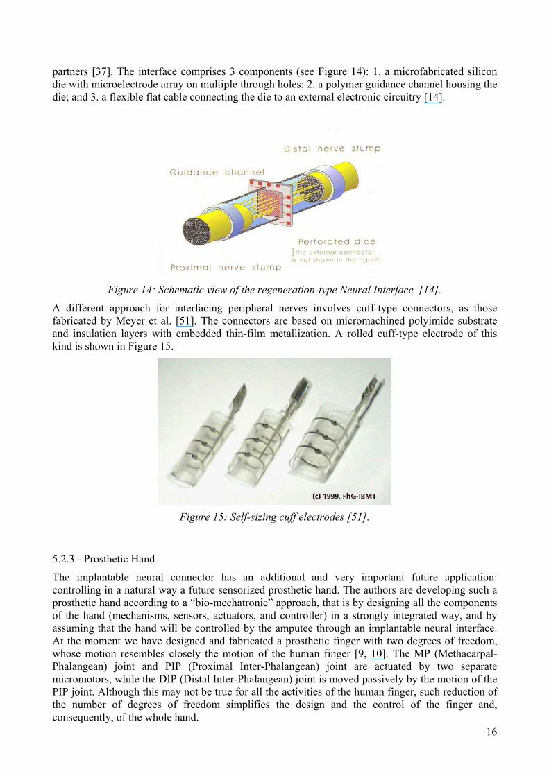
partners [37]. The interface comprises 3 components (see Figure 14): 1. a microfabricated silicon die with microelectrode array on multiple through holes; 2. a polymer guidance channel housing the die; and 3. a flexible flat cable connecting the die to an external electronic circuitry [14].
Figure 14: Schematic view of the regeneration-type Neural Interface [14].
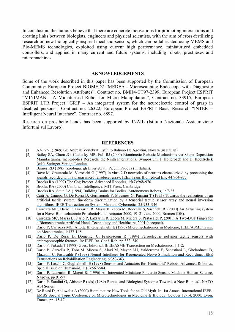
A different approach for interfacing peripheral nerves involves cuff-type connectors, as those fabricated by Meyer et al. [51]. The connectors are based on micromachined polyimide substrate and insulation layers with embedded thin-film metallization. A rolled cuff-type electrode of this kind is shown in Figure 15.
Figure 15: Self-sizing cuff electrodes [51].
5.2.3 - Prosthetic Hand
16
The implantable neural connector has an additional and very important future application: controlling in a natural way a future sensorized prosthetic hand. The authors are developing such a prosthetic hand according to a “bio-mechatronic” approach, that is by designing all the components of the hand (mechanisms, sensors, actuators, and controller) in a strongly integrated way, and by assuming that the hand will be controlled by the amputee through an implantable neural interface. At the moment we have designed and fabricated a prosthetic finger with two degrees of freedom, whose motion resembles closely the motion of the human finger [9, 10]. The MP (Methacarpal-Phalangean) joint and PIP (Proximal Inter-Phalangean) joint are actuated by two separate micromotors, while the DIP (Distal Inter-Phalangean) joint is moved passively by the motion of the PIP joint. Although this may not be true for all the activities of the human finger, such reduction of the number of degrees of freedom simplifies the design and the control of the finger and, consequently, of the whole hand.

The finger incorporates a complex and rich sensory system in order to be properly controlled. Joint sensors based on Hall-effect and skin-like fingertip sensors are located in each finger. The main characteristics of the skin-like tactile sensor are illustrated in the following paragraph.
5.2.4 - Skin-like tactile sensor
The fingertip skin-like tactile sensor developed in the author’s lab [12, 16, 41] integrates three tactile modalities: pressure sensing (like Ruffini endings or Merkel's disks), thermal flux sensing (like thermoreceptors) and slip sensing (like Pacinian Corpuscle).
The pressure is sensed by a fovea-like array of 64 tactile piezoresistive units based on FSR (or Force Sensing Resistors) technology, designed by the authors and manufactured by Interlink Inc., Camarillo, CA. The maximum resolution of the fovea-like structure (at the center of the sensor) is set by the current limit for FSR technology, that is about 1 mm. The minimum spatial resolution at periphery is about 5 mm. This disposition increases the size of the tactile sensing area, for the same number of sensing sites.
The thermal flux sensor behaves like the thermoreceptors in the human hand. However, the artificial sensors do not discriminate between cold and warmth, but they just detect the amount of heat flowing from the fingertip towards the touched object.
Lastly, the dynamic sensor is a small piezoelectric ceramic bimorph element, mounted on the fingertip frame and connected electrically to the preprocessing electronics. The sensor detects the vibrations caused by the movement of the fingertip along a surface (see Figure 16).
Figure 16: Layout of the tactile sensor [16, 41].
6 – CONCLUSIONS In this paper we have presented the main characteristics of force and motion sensors in animals and humans. The analysis of these characteristics is an important source of inspiration for the design of artificial sensors of force and motion applicable to robotics and prosthetics. Recent progresses in microfabrication technologies make it possible to convert biological inspiration into guidelines for the design of real sensors and sensory systems. These biologically-inspired mechano-sensors could become not only a mere demonstration of technology capabilities or a scientific “virtuosity”, but tools usable in engineering practice. In areas like robotics, intelligent machines, and prosthetics, the increasing computing performance of embedded microcontrollers and the continuos miniaturization of the system (“micromechatronics”) are key factors for the potential use of more and more microsensors in general, and of biologically-inspired sensors in particular.
17
In conclusion, the authors believe that there are concrete motivations for promoting interactions and creating links between biologists, engineers and physical scientists, with the aim of cross-fertilizing research on new biologically-inspired mechano-sensors, which can be fabricated using MEMS and Bio-MEMS technologies, exploited using current high performance, miniaturized embedded controllers, and applied in many current and future systems, including robots, prostheses and micromachines.
AKNOWLEDGEMENTS Some of the work described in this paper has been supported by the Commission of European Community: European Project BIOMED2 “MEDEA - Microscanning Endoscope with Diagnostic and Enhanced Resolution Attributes”, Contract no. BMH4-CT97-2399; European Project ESPRIT “MINIMAN - A Miniaturised Robot for Micro Manipulation”, Contract no. 33915, European ESPRIT LTR Project “GRIP – An integrated system for the neuroelectric control of grasp in disabled persons”, Contract no. 26322; European Project ESPRIT Basic Research “INTER – Intelligent Neural Interface”, Contract no. 8897.
Research on prosthetic hands has been supported by INAIL (Istituto Nazionale Assicurazione Infortuni sul Lavoro).
REFERENCES [1] AA. VV. (1969) Gli Animali Vertebrati. Istituto Italiano De Agostini, Novara (in Italian). [2] Bailey SA, Cham JG, Cutkosky MR, Full RJ (2000) Biomimetic Robotic Mechanisms via Shape Deposition
Manufacturing. In: Robotics Research: the Ninth International Symposium, J. Hollerbach and D. Koditschek (eds), Springer-Verlag, London.
[3] Barnes RD (1985) Zoologia: gli Invertebrati. Piccin, Padova (in Italian). [4] Bove M, Grattarola M, Verreschi G (1997) In vitro 2-D networks of neurons characterized by processing the
signals recorded with a planar microtransducer array. IEEE Trans Biomedical Eng 44:964-977 [5] Brooks RA (1997) The Cog Project. Advanced Robotics, 15(7):968-970 [6] Brooks RA (2000) Cambrian Intelligence. MIT Press, Cambridge. [7] Brooks RA, Stein LA (1994) Building Brains for Bodies, Autonomous Robots, 1: 7-25. [8] Caiti A, Canepa G, De Rossi D, Germagnoli F, Maganes G, Parisini T (1995) Towards the realization of an
artificial tactile system: fine-form discrimination by a tensorial tactile sensor array and neural inversion algorithms. IEEE Transaction on System, Man and Cybernetics 25:933–946
[9] Carrozza MC, Dario P, Lazzarini R, Massa B, Zecca M, Roccella S, Sacchetti R, (2000) An Actuating system for a Novel Biomechatronic ProstheticHand. Actuator 2000, 19–21 June 2000, Bremen (DE).
[10] Carrozza MC, Massa B, Dario P, Lazzarini R, Zecca M, Micera S, Pastacaldi P, (2001) A Two-DOF Finger for a Biomechatronic Artificial Hand. Technology and Healthcare, 2001 (accepted).
[11] Dario P, Carrozza MC, Allotta B, Guglielmelli E (1996) Micromechatronics in Medicine, IEEE/ASME Trans. on Mechatronics, 1:137-148.
[12] Dario P, De Rossi D, Domenici C, Francesconi R (1994) Ferroelectric polymer tactile sensors with anthropomorphic features. In: IEEE Int. Conf. Rob, pp 332–340.
[13] Dario P, Fukuda T (1998) Guest Editorial, IEEE/ASME Transaction on Mechatronics, 3:1-2. [14] Dario P, Garzella P, Toro M, Micera S, Alavi M, Meyer J-U, Valderrama E, Sebastiani L, Ghelarducci B,
Mazzoni C, Pastacaldi P (1998) Neural Interfaces for Regenerated Nerve Stimulation and Recording. IEEE Transactions on Rehabilitation Engineering, 6:353-363.
[15] Dario P, Laschi C, Guglielmelli E (1998) Sensors and Actuators for ‘Humanoid’ Robots. Advanced Robotics, Special Issue on Humanoid, 11(6):567-584.
[16] Dario P, Lazzarini R, Magni R, (1996) An Integrated Miniature Fingertip Sensor. Machine Human Science, Nagoya, pp 91-97
[17] Dario P, Sandini G, Abisher P (eds) (1989) Robots and Biological Systems: Towards a New Bionics?, NATO ASI Series.
[18] De Rossi D, Ahluwalia A (2000) Biomimetics: New Tools for an Old Myth. In: 1st Annual International IEEE-EMBS Special Topic Conference on Microtechnologies in Medicine & Biology, October 12-14, 2000, Lyon, France, pp. 15-17.
18

[19] Despont M et al. (1992) A comparative study of bearing designs and operational environments for harmonic side-drive micromotors. In: proceedings of MEMS 92, pp 171–176
[20] Fatikow S, Rembold U (1997) Microsystem Technology. Springer-Verlag, Germany [21] Ferrari M (1999) Editorial, Journal of Biomedical Microdevices 1:97-98 [22] Fisher K (1991) Mikromechanik und Mikroelektronik vereint mit Optik. Technische Rundschau 106–108 (in
German). [23] Freschi C, Vecchi F, Micera S, Sabatini AM, Dario P (2000) Force Control during Grasp using FES
Techniques: Preliminary Results. 5th Annual Conference of the International Functional Electrical Stimulation Society (IFESS 2000), 17-24 june 2000, Aalborg (DK).
[24] Fujimasa I (1996) Micromachines, A New Era in Mechanical Engineering, Oxford University Press, Oxford. [25] Fujita M (1999) AIBO: Towards the Era of Digital Creatures. In: International Symposium on Robotics
Research, Snowbird, UH, October 9-12, pp. 257–262. [26] Fukuda T, Menz W (1998) Handbook of sensors and actuators. Micro Mechanical Systems, Elsevier Science,
Amsterdam [27] GRIP Esprit LTR Project #26322 “An integrated system for the neuroelectric control of grasp in disabled
persons”. [28] Harsanyi G (1995) Polymer films in sensor applications. Technomic Publishing Co., Basel [29] Hashimoto S et al. (2000) Humanoid robots in Waseda University – Hadaly-2 and Wabian. In: First IEEE-RAS
International Conference on Humanoid Robots – Humanoids 2000, Cambridge, MA, September 7-8. [30] Heuberger A.(1991) Mikromechanick: Mikrofertigung mit Methoden der Halbeitertechnologie. Springer-
Verlag, Berlin (in German). [31] Heuschkel MO, Guerin L, Buisson B, Bertrand D, Renaud P (1998) Buried microchannels in photopolymer for
delivering of solutions to neurons in a network. Sensors and Actuators B48:356-361 [32] Hirose H (1993) Biologically Inspired Robots (Snake-like Locomotor and Manipulator). Oxford University
Press. [33] Howe RD, Cutkosky MR (1992) Touch Sensing for Manipulation and Recognition. In: Kathib O, Craig J,
Lozano-Pérez T (eds) The robotics review 2, pp.55-112, MIT Press, Cambridge [34] http://csmt.jpl.nasa.gov/csmtpages/Technologies/mgyro/mgyro.html [35] http://www-cdr.stanford.edu [36] Inoue H (2000) HRP: Humanoid Robotics Project of MITI. In: First IEEE-RAS International Conference on
Humanoid Robots – Humanoids 2000, Cambridge, MA, September 7-8. [37] INTER Project promoted by the European Commission (“Intelligent Neural Interface” Esprit Basic Research
Project # 8897). [38] Johnson MW, Peckham PH, Bhadra N, Kilgore KL, Gazdik MM, Keith MW, Strojnik P (1999) Implantable
transducer for two-degree-of-freedom joint angle sensing”. IEEE Trans. Biomed. Eng. 7(3):349-359. [39] Kandel ER, Schwartz JH, Jessel TM (1991) Principles of Neural Science, 3nd edition. Elsevier Science. [40] Langer R., Vacanti J (1993). Tissue Engineering. Science 260: 920-926 [41] Lazzarini R, Magni R, Dario P (1995) A Tactile Array Sensor Layered in an Artificial Skin. In: Proceedings of
the 1995 IEEE/RSJ International Conference on Intelligent Robots and Systems, Iros ’95, 3:114-119. [42] Madou M (1997) Fundamentals of Microfabrication. CRC Press, Boca Raton, New York [43] Mannion P(1999) Integration and Inductive Sensing Combine to Improve Automotive/Industrial Sensing.
Electronic Design [44] Mehlhorn T at al. (1992) CMOS-compatible Capacitive Silicon Pressure Sensors. Micro System Technologies
92:277–285. [45] Menzel P, D’Aluisio F, Mann CC (2000) RoboSapiens. MIT Press, Cambridge [46] Muller RS, Howe RT, Senturia SD, Smith RL, White RM (1991) Microsensors, IEEE Press, New York [47] Najafi K, Wise KD, Mochizuki T (1985) A High-Yield IC- Compatible Multichannel Recording Array. IEEE
Trans. Electronic Devices ED-32:1206-1211 [48] Nicholls HR, Lee MH (1989) A survey of robot tactile sensing technology. Int J. Robotics Research 8:3–30 [49] Nicholls HR, Lee MH (1999) Tactile sensing for mechatronics –– a state of the art survey. Mechatronics 9:1–
32. [50] Russel RA (1990) Robot Tactile Sensing. Prentice Hall Ltd, Australia [51] Schuettler M, Stiegllitz T, Meyer J-U (1999) A Multipolar Precision Hybrid Cuff Electrode for FES on Large
Peripheral Nerves. 21st Annual International Conference of the IEEE Engineering in Medicine and Biology Society, October 13-16, Atlanta, USA, 1999.
[52] Shibata T, Tanie K (1999) Creation of Subjective Value through Physical Interaction between Human and Machine. In: 4th International Symposium on Artificial Life and Robotics, January 19-22, Oita, Japan
[53] Sinkjaer T, Haugland M, Struijk J, Riso RR (1999) Long-term cuff electrode recordings from peripheral nerves in animals and humans. In U. Windhorst and H.Johansson (eds), Modern Techinques in Neuroscience Research. NY: Springer-Verlag.
[54] Tilmans H, Bouwstra S (1993) A novel design of a highly sensitive low differential-pressure sensor using built-in resonant strain gauges. J. Micromech Microeng 3:198–202.
19
[55] Vecchi F, Freschi C, Micera S, Sabatini AM, Dario P (2000) Experimental evaluation of two commercial force sensors for applications in biomechanics and motor control. 5th Annual Conference of the International Functional Electrical Stimulation Sociaty (IFESS 2000), June 17-24, 2000, Aalborg (DK).
[56] Webster JG(1998) Tactile Sensors for Robotics and Medicine. John Wiley & Sons, New York. [57] Zill SN, Seyfarth E (1996) Exoskeletal Sensors for Walking. Scientific American 70–74
20
Related Documents