International Journal of Medicine and Pharmaceutical Sciences (IJMPS) ISSN 2250-0049 Vol. 3, Issue 4, Oct 2013, 89-100 © TJPRC Pvt. Ltd. BIOLOGICAL SYNTHESIS OF SILVER NANOPARTICLES USING RAPHANUS SATIVUS VAR. LONGIPINNATUS LEAF EXTRACT AND EVALUATION OF THEIR ANTIOXIDANT AND ANTIBACTERIAL ACTIVITY RAMA KOYYATI, VEERABABU NAGATI, RAMCHANDER MERUGU & PRATAPRUDRA MANTHURPADIGYA Department of Biochemistry, Osmania University, Hyderabad, Andhra Pradesh, India ABSTRACT Nanocrystalline silver particles have found tremendous applications in the field of diagnostics and therapeutics. In recent years, plant mediated biological synthesis of nanoparticles is gaining importance due to its eco-friendliness. In this study, the synthesis and characterization of silver nanoparticles was carried out by using Raphanus sativus var. longipinnatus leaf extract as reducing agent and evaluation of antibacterial and antioxidant activity. The synthesized silver nanoparticles were characterized with UV–Vis spectrophotometry (UV-Vis), Fourier transform infrared spectroscopy (FTIR), Scanning electron microscopy-Energy dispersive spectroscopy (SEM- EDX), Transmission electron microscopy (TEM) and X-Ray diffraction spectroscopy (XRD). The antibacterial activity of silver nanoparticles and in-vitro antioxidant activity have been evaluated. These synthesized silver nanoparticles were found to have significant antibacterial activity and antioxidant capacity, thus can be used as potential radical scavenger against deleterious damages caused by the free radicals. KEYWORDS: Raphanus sativus var. longipinnatus Leaf Extract, Reducing Agent, Antibacterial Activity and In-Vitro Antioxidant Activity, Potential Radical Scavenger, Free Radicals INTRODUCTION Nanosized silver particles have found tremendous applications in the field of high sensitivity biomolecular detection and diagnostics, antimicrobials, antioxidants and therapeutics, catalysis [1] and micro-electronics. Nanoparticles can be synthesized by various approaches like chemical and photochemical reactions in reverse micelles, microwave assisted, thermal decomposition, electrochemical, sonochemical process [2] and also by biological methods. Plant mediated biological synthesis of nanoparticles is gaining importance due to its simplicity, cost effective and ecofriendliness [3, 4]. Silver nanoparticles were proven to be most efficient as they possess good anti-microbial [5,6,7,8], anti- inflammatory [9,10,11], anti-plasmodial [12,13] anti-cancer [14] and anti-oxidant activities [15,16,17] etc. Bioreduction of silver ions to yield metal nanoparticles using plants such as Cajanus cajan [18], Cuminum cyminum [19], Pisonia grandis [20], Allium cepa [21], Partheniun hysterophorus [22], Ocimum basillicum [23],Murraya koenigii [24], Glycyrrhiza glabra [25], Coriandrum sativum [26] and Ocimum sanctum [27] have been reported. Nanoparticles attach to the cell surface of bacteria causes structural changes and damage disturbing the vital cell functions and finally leading to cell death [28]. Antioxidants are the substances which act as free radical scavengers by preventing and repairing damages caused by reactive oxygen species and can also enhance the immune defense and degenerative diseases [29]. Radish (Raphanus sativus var. longipinnatus) is a common vegetable crop in Asia. The plant produces a white and large, cylindrical, long, fleshy root which usually weighs up to 2-3kg.The leaves usually are medium, green and lobed

Welcome message from author
This document is posted to help you gain knowledge. Please leave a comment to let me know what you think about it! Share it to your friends and learn new things together.
Transcript
International Journal of Medicine and
Pharmaceutical Sciences (IJMPS)
ISSN 2250-0049
Vol. 3, Issue 4, Oct 2013, 89-100
© TJPRC Pvt. Ltd.
BIOLOGICAL SYNTHESIS OF SILVER NANOPARTICLES USING RAPHANUS
SATIVUS VAR. LONGIPINNATUS LEAF EXTRACT AND EVALUATION OF
THEIR ANTIOXIDANT AND ANTIBACTERIAL ACTIVITY
RAMA KOYYATI, VEERABABU NAGATI, RAMCHANDER MERUGU &
PRATAPRUDRA MANTHURPADIGYA
Department of Biochemistry, Osmania University, Hyderabad, Andhra Pradesh, India
ABSTRACT
Nanocrystalline silver particles have found tremendous applications in the field of diagnostics and therapeutics. In
recent years, plant mediated biological synthesis of nanoparticles is gaining importance due to its eco-friendliness. In this
study, the synthesis and characterization of silver nanoparticles was carried out by using Raphanus sativus var.
longipinnatus leaf extract as reducing agent and evaluation of antibacterial and antioxidant activity. The synthesized silver
nanoparticles were characterized with UV–Vis spectrophotometry (UV-Vis), Fourier transform infrared spectroscopy
(FTIR), Scanning electron microscopy-Energy dispersive spectroscopy (SEM- EDX), Transmission electron microscopy
(TEM) and X-Ray diffraction spectroscopy (XRD). The antibacterial activity of silver nanoparticles and in-vitro
antioxidant activity have been evaluated. These synthesized silver nanoparticles were found to have significant
antibacterial activity and antioxidant capacity, thus can be used as potential radical scavenger against deleterious damages
caused by the free radicals.
KEYWORDS: Raphanus sativus var. longipinnatus Leaf Extract, Reducing Agent, Antibacterial Activity and In-Vitro
Antioxidant Activity, Potential Radical Scavenger, Free Radicals
INTRODUCTION
Nanosized silver particles have found tremendous applications in the field of high sensitivity biomolecular
detection and diagnostics, antimicrobials, antioxidants and therapeutics, catalysis [1] and micro-electronics. Nanoparticles
can be synthesized by various approaches like chemical and photochemical reactions in reverse micelles, microwave
assisted, thermal decomposition, electrochemical, sonochemical process [2] and also by biological methods. Plant mediated
biological synthesis of nanoparticles is gaining importance due to its simplicity, cost effective and ecofriendliness [3, 4].
Silver nanoparticles were proven to be most efficient as they possess good anti-microbial [5,6,7,8], anti-
inflammatory [9,10,11], anti-plasmodial [12,13] anti-cancer [14] and anti-oxidant activities [15,16,17] etc. Bioreduction of
silver ions to yield metal nanoparticles using plants such as Cajanus cajan [18], Cuminum cyminum [19], Pisonia grandis
[20], Allium cepa [21], Partheniun hysterophorus [22], Ocimum basillicum [23],Murraya koenigii [24], Glycyrrhiza
glabra [25], Coriandrum sativum [26] and Ocimum sanctum [27] have been reported. Nanoparticles attach to the cell
surface of bacteria causes structural changes and damage disturbing the vital cell functions and finally leading to cell death
[28]. Antioxidants are the substances which act as free radical scavengers by preventing and repairing damages caused by
reactive oxygen species and can also enhance the immune defense and degenerative diseases [29].
Radish (Raphanus sativus var. longipinnatus) is a common vegetable crop in Asia. The plant produces a white
and large, cylindrical, long, fleshy root which usually weighs up to 2-3kg.The leaves usually are medium, green and lobed
90 Rama Koyyati, Veerababu Nagati, Ramchander Merugu & Prataprudra Manthurpadigya
and have rough texture and has large amounts of vitamin B and C as well as pectin, phytin, manganese, iron and copper.
Leaves are used to treat dysentery, asthma, cough, diarrhea and malnutrition [30]. It contains ferulic acid, gentisic acid,
raphanusin, erucic acid, sinapate, raphanin and sulforaphen. The seeds are carminative, diuretic and laxative. Roots have
been used for treating syphilis, haemorrhoids, gonorrhea, cancer [31] and urinary complaints.
In this investigation, green synthesis of silver nanoparticles were carried out using aqueous leaf extract of
Raphanus sativus var. longipinnatus and characterized using UV-visible spectra, Fourier transform infrared spectra,
Scanning and transmission electron microscopy, Energy X-ray diffraction spectra and XRD. The antibacterial activities
have been investigated against Gram negative and Gram positive bacteria and evaluated in-vitro antioxidant properties.
Results shown that RsAgNPs were effective bactericidal and antioxidants. To our knowledge green synthesis of silver
nanoparticles by this plant leaf extract has not been reported so far.
MATERIALS AND METHODS
Collection of Raphanus sativus var. longipinnatus
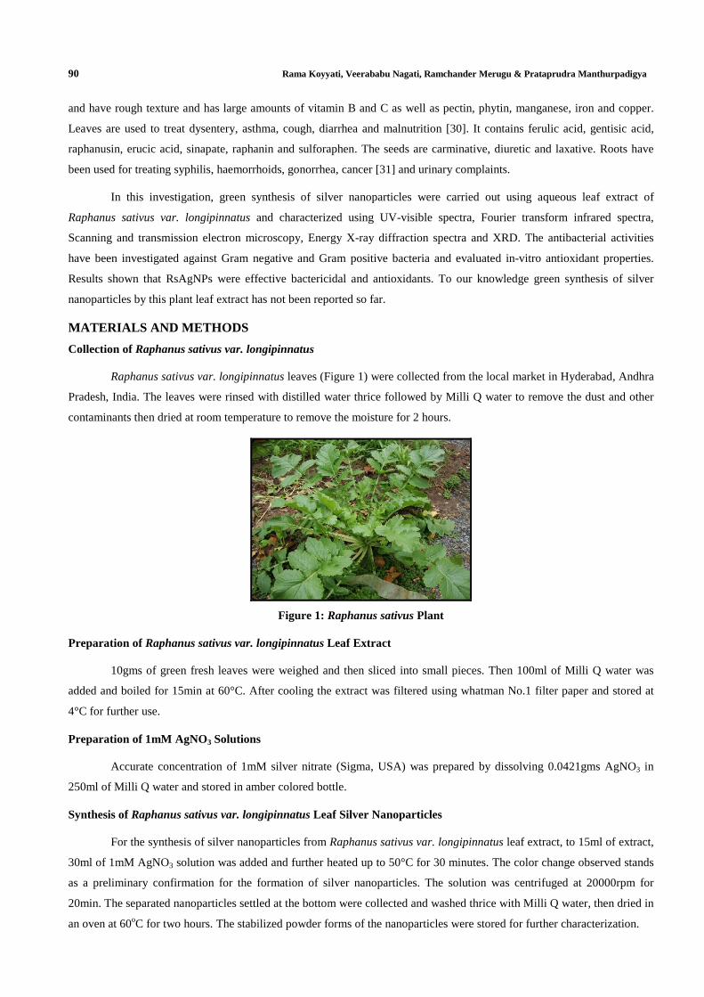
Raphanus sativus var. longipinnatus leaves (Figure 1) were collected from the local market in Hyderabad, Andhra
Pradesh, India. The leaves were rinsed with distilled water thrice followed by Milli Q water to remove the dust and other
contaminants then dried at room temperature to remove the moisture for 2 hours.
Figure 1: Raphanus sativus Plant
Preparation of Raphanus sativus var. longipinnatus Leaf Extract
10gms of green fresh leaves were weighed and then sliced into small pieces. Then 100ml of Milli Q water was
added and boiled for 15min at 60°C. After cooling the extract was filtered using whatman No.1 filter paper and stored at
4°C for further use.
Preparation of 1mM AgNO3 Solutions
Accurate concentration of 1mM silver nitrate (Sigma, USA) was prepared by dissolving 0.0421gms AgNO3 in
250ml of Milli Q water and stored in amber colored bottle.
Synthesis of Raphanus sativus var. longipinnatus Leaf Silver Nanoparticles
For the synthesis of silver nanoparticles from Raphanus sativus var. longipinnatus leaf extract, to 15ml of extract,
30ml of 1mM AgNO3 solution was added and further heated up to 50°C for 30 minutes. The color change observed stands
as a preliminary confirmation for the formation of silver nanoparticles. The solution was centrifuged at 20000rpm for
20min. The separated nanoparticles settled at the bottom were collected and washed thrice with Milli Q water, then dried in
an oven at 60oC for two hours. The stabilized powder forms of the nanoparticles were stored for further characterization.
Biological Synthesis of Silver Nanoparticles Using Raphanus sativus Var. longipinnatus 91 Leaf Extract and Evaluation of their Antioxidant and Antibacterial Activity
Characterization of Raphanus sativus var. longipinnatus Silver Nanoparticles (RsAgNPs)
An ELICO SL-159 UV-Vis spectrophotometer was used for the spectrometric analysis to confirm silver
nanoparticles formation. The leaf extract was used as reference blank. The purified suspension was oven dried and the
powder was subjected to FTIR spectroscopy analysis (Paragon 500, Perkin Elmer-RX1 spectrophotometer) in the diffuse
reflectance mode at a resolution of 4cm−1
in KBr pellet. Further the size and shape of synthesized AgNPs was characterized
by Scanning electron microscope (SEM) in Zeiss 700 Scanning electron microscope and Transmission electron microscope
(TEM) in Philips model CM 200 instrument operated at an accelerating voltage at 200 kV and the confirmation of the
presence of elemental silver signal was characterized by energy-dispersive X-ray microanalysis spectroscopy (EDX;
Sigma) and X-Ray diffractometer operated at a voltage of 40 kV and a current of 30 mA with Cu Kα radiation.
Antibacterial Activity Using Disc Diffusion Method
The antimicrobial activity of synthesized silver nanoparticles was determined using disc diffusion method. Luria
Bertani media was prepared and poured into sterilized petriplates and then plates were spreaded with of Pseudomonas
putida, Klebsilla pneumonia, Staphylococcus aureus and Bacillus subtilis separately. Then sterile discs were kept and the
samples were added to the disc and the plates were incubated at 37°C overnight. Then zone of inhibition was measured.
Antioxidant Activity of Raphanus sativus var. longipinnatus Silver Nanoparticles (RsAgNPs)
Determination of Total Antioxidant Activity
The Total antioxidant activity of the silver nanoparticles was assessed by the phosphomolybdenum reduction
assay [32, 33]. The assay is based on the reduction of Mo (VI)–Mo (V) by the RsAgNPs and subsequent formation of a
green phosphate/Mo(V) complex at acid pH. To various concentrations (20, 40, 60, 80, 100,120 & 140 μg/mL) of
RsAgNPs diluted in methanol was combined with 3ml of reagent solution (0.6M sulfuric acid, 28mM sodium phosphate
and 4mM ammonium molybdate). Then the tubes were incubated at 95°C for 90 min. Then the absorbance of the green
phosphomolybdenum complex was measured at 695 nm using a UV-visible spectrophotometer against blank after cooling
to room temperature. Methanol in the place of RsAgNPs is used as the blank. For reference, L-ascorbic acid was used as a
control and prepared by dissolving 1mg of L-ascorbic acid in 1ml methanol. The following equation was used for
calculating Total antioxidant activity expressed as gram equivalents
Estimation of Radical Scavenging Activity (RSA) Using DPPH Assay
The radical scavenging activity of silver nanoparticles was estimated using the method of DPPH assay[34].A
solution of DPPH (2,2-diphenyl-1-picrylhydrazyl) 5mg in 100ml methanol was prepared and 3.0 ml of this solution was
mixed with various concentrations (20, 40, 60, 80 & 100 μg/mL) of synthesized RsAgNPs . The reaction mixture was
shaken vigorously and left in the dark at room temperature for 15 min. The absorbance was measured at 517 nm with
ascorbic acid as standard. The following equation was used for calculating percentage inhibition:
DPPH % inhibition = [(Abs control – Abs sample)]/ (Abs control)] x 100
Abs control is the absorbance of DPPH radical + methanol; Abs sample is the absorbance of DPPH radical +
synthesized RsAgNPs solution/standard.
Estimation of Hydroxyl Radical Scavenging Activity
Hydroxyl radical scavenging activity of the RsAgNPs was carried out by the method of Inbathamizh L [35].
Various concentrations of RsAgNPs was added with 1.0mL of EDTA solution (0.13 g of ferrous ammonium sulphate and

92 Rama Koyyati, Veerababu Nagati, Ramchander Merugu & Prataprudra Manthurpadigya
0.26 g of EDTA were dissolved in 100mL of water) and mixed with 1.0mL of DMSO (0.85%) in 0.1M phosphate buffer
(pH 7.4) to initiate the reaction followed by the addition of 0.5mL of 0.22% ascorbic acid. The reaction mixture was kept
in a water bath at 90°C for 15 min and the reaction was terminated by adding 1.0 mL of ice-cold 1 7.5% trichloroacetic
acid. Further 3.0mL of Nash reagent (75 g of ammonium acetate, 3.0mL of glacial acetic acid and 2.0mL of acetyl acetone
in 1.0 L of water) was added to all the test tubes and incubated for 15 min for color development. Reaction mixture without
ascorbic acid served as control. Absorbance was observed at 412 nm. The ability to scavenge hydroxyl radical was
calculated by the following equation:
Hydroxyl Radical scavenging activity (%) = [(Abs control – Abs sample)]/ (Abs control)] x 100
Estimation of Hydrogen Peroxide Scavenging Activity
Hydrogen peroxide scavenging activity of Raphanus sativus var. longipinnatus silver nanoparticles was estimated
by replacement titration [36]. Aliquot of 1.0ml of 0.1mmole of H2O2 and 1.0ml of various concentrations (400μg/ml,
800μg/ml and 1200μg/ml) of Raphanus sativus var. longipinnatus silver nanoparticles were mixed, followed by 2 drops of
3% ammonium molybdate, 7.0ml of 1.8 M KI and 10ml of 2M of H2SO4. The mixed solution was titrated with 5.09 mM of
NaS2O3 until yellow color disappeared. The percentage of scavenging of hydrogen peroxide was calculated as:
% Inhibition = [(V0 - V1) / V0] * 100
Where V0 was volume of NaS2O3 solution used to titrate the control sample in the presence of hydrogen peroxide
(without Raphanus sativus var. longipinnatus silver nanoparticles /Ascorbic acid), V1 was the volume of NaS2O3 solution
used in the presence of the Raphanus sativus var. longipinnatus silver nanoparticles/Ascorbic acid.
RESULTS AND DISCUSSIONS
The synthesis of silver nanoparticles is new technique in modern biotechnology and is evolving as an important
branch of nanotechnology. This study deals with the synthesis and characterization of silver nanoparticles using leaf extract
of Raphanus sativus var. longipinnatus (Figure 2A). Green synthesized silver nanoparticles were reddish brown in color.
The color of the extract was changed from light yellowish to reddish brown after addition of AgNO3 and on incubation for
5-10 min at 60ºC.The coloration was due to the excitation of the surface Plasmon vibration in the silver nanoparticles.
Change in color after the reduction of silver ions to silver nanoparticles is shown in (Figure 2B). The reduction rate and
formation of nanoparticles can be increased further by increase in incubation time.
Figure 2: A. Plant Extract and Silver Nitrate Solution, B. Synthesis of Silver Nanoparticles
UV-Vis Spectrophotometer
The UV-Vis spectroscopy was the preliminary technique for the characterization of the silver nanoparticles. The

Biological Synthesis of Silver Nanoparticles Using Raphanus sativus Var. longipinnatus 93 Leaf Extract and Evaluation of their Antioxidant and Antibacterial Activity
UV-Vis absorption was analyzed after centrifuging and redispensing the particles in deionized water, the maximum smooth
and broad absorption peak was seen at 470nm.(Figure 3).
Figure 3: UV-Vis Spectra of Silver Nanoparticles Obtained at Different Time Intervals
FTIR Analysis of Silver Nanoparticles
The FTIR spectrum indicates various functional groups present at different positions. FTIR spectroscopy study
has confirmed that the carbonyl group of amino acid residues and peptides binds to RsAgNPs. The peaks in the region
3293 to 2917 were assigned to O-H stretching of alcohol and phenol compounds and aldehyde –C-H- stretching of alkanes.
The peaks in the region 1584 corresponds to aromatic C=C with nitro group bending vibration, 1398 to 1087 corresponds
to N-H group of primary and secondary amides and –C-N- stretching vibration of amines and –C-O- stretching of alcohols,
ethers, carboxylic acids and anhydrides and peaks between 841 and 751 were assigned to alkyl halides (Figure 4). FTIR
analysis reveals the dual function of biological molecules possibly responsible for the reduction and stabilization of silver
nanoparticles in the aqueous medium.
Figure 4: FTIR Spectrum of Silver Nanoparticles
XRD Analysis
XRD analysis of Raphanus sativus var. longipinnatus silver nanoparticles which showed diffraction peaks at
38.160, 44.20
0, 64.5
0 and 77.42
0, indexing the planes 111, 200, 220 and 311of the cubic face-centered silver(Figure 5). The
lattice constant calculated from this pattern was a = 4.086Å and the data obtained was matched with the database of Joint
Committee on Powder Diffraction Standards (JCPDS) file No. 04-0783. Average grain size of the silver nanoparticles

94 Rama Koyyati, Veerababu Nagati, Ramchander Merugu & Prataprudra Manthurpadigya
formed in the bioreduction process was determined using Scherer’s formula, ( d = 0.9×λ/ β *cos θ) and was estimated as
22nm.
Figure 5: XRD Analysis of Silver Nanoparticles
SEM-EDX Analysis
The morphology of the synthesized silver nanoparticles using Raphanus sativus var. longipinnatus leaf extract,
the sample was spherical in shape and an average size of 22nm (Figure 6A). The EDS spectra shown that the sample (silver
nanoparticle) contains 40.83% silver (Figure 6B and Table 1.)
Figure 6: A. SEM Analysis, B. EDS Spectra of Synthesized Silver Nanoparticles
Table 1: The Composition of Silver Nanoparticles Synthesized from Raphanus sativus Leaf Extract
Element Weight% Atomic%
C K -12.76 -70.83
O K 17.79 74.13
Na K 3.08 8.93
S K 4.52 9.41
Cl K 8.01 15.06
K K 8.52 14.52
Ca K 4.78 7.95
Ag L 66.06 40.83
Total 100.00
TEM Analysis
The silver nanoparticles synthesized by the help of Raphanus sativus var. longipinnatus leaf extract when scanned
using TEM from which we conclude that the average mean size of silver nanoparticles was in between 5-22nm and seems

Biological Synthesis of Silver Nanoparticles Using Raphanus sativus Var. longipinnatus 95 Leaf Extract and Evaluation of their Antioxidant and Antibacterial Activity
to be spherical in morphology as shown in (Figure 7). Thus the transmission electron microscopy gave a detailed
descriptive image of the silver nanoparticles synthesized with their structural details and their size.
Figure 7: TEM Analysis and Particle Size Distribution
Antibacterial Activity by Disc Diffusion Technique
Antibacterial activity of synthesized silver nanoparticles against Gram negative (Pseudomonas putida and
Klebsiella pneumonia) and Gram positive (Staphylococcus aureus and Bacillus subtilis) bacteria was revealed and zone of
inhibition was measured (Figure 8 and Table 2). The results indicated that silver nanoparticles synthesized from Raphanus
sativus var. longipinnatus leaf extract showed effective antibacterial activity both in Gram negative and Gram positive
bacteria which is compared with ampicillin.
Figure 8: Antibacterial Activity of Silver Nanoparticles
Table 2: Zone of Inhibition (mm)
Name of the Organism
Zone of Inhibition in mm
Ampicillin
(5µl)
1mM AgNO3
(5µl)
Rs Leaf Extract
(5µl)
RsAgNPs
(5 µl)
Bacillus subtilis 13 10 Not determined 9
Staphylococcus aureus 16 11 Not determined 10
Klebsiella pnuemoniae 12 9 Not determined 9
Pseudomonas putida 11 9 Not determined 8
Antioxidant Activity of Raphanus sativus var. longipinnatus Silver Nanoparticles (RsAgNPs)
Total Antioxidant Activity
Total antioxidant capacity of Raphanus sativus var. longipinnatus silver nanoparticles is expressed as the number
of equivalents of ascorbic acid. The phosphomolybdenum method is quantitative, since the antioxidant activity is expressed

96 Rama Koyyati, Veerababu Nagati, Ramchander Merugu & Prataprudra Manthurpadigya
as the number of equivalents of ascorbic acid. The antioxidant activity of the extract is in the increasing trend with the
increasing concentration of the ascorbic acid and AgNPs.
At a concentration of 30µg/ml and 50µg/ml both RsAgNPs and Ascorbic acid showed similar antioxidant activity
[Figure 9].
Figure 9: Total Antioxidant Activity by Phosphomolybdenum Assay
DPPH Radical Scavenging Assay
The radical-scavenging activity of silver nanoparticles synthesized from leaf extract of Raphanus sativus var.
longipinnatus w as estimated by comparing the percentage inhibition of formation of DPPH radicals with that of Ascorbic
acid. The silver nanoparticles showed moderate antioxidant activity when compared with Ascorbic acid. Radical
scavenging activity of silver nanoparticles increased with increasing the concentration [Figure 10].
The IC 50 value was 99µg/ml for silver nanoparticles and 42µg//ml for Ascorbic acid. These results suggest that
at concentration above 140µg/ml, the synthesized silver nanoparticles may serve as potent antioxidants.
Figure 10: DPPH Radical Scavenging Activity
Estimation of Hydroxyl Radical Scavenging Activity
The scavenging capacity of the silver nanoparticles from leaf extract of Raphanus sativus var. longipinnatus was
shown in [Figure 11]. At a concentration of 100mg/ml, the silver nanoparticles showed 69.51% (IC50 -45µg/ml) hydroxyl
radical scavenging activity with the standard Ascorbic acid activity being 85.58% (IC50-26µg/ml). The radical scavenging
capacity of the sample might be attributed to phenolic compounds in the sample.

Biological Synthesis of Silver Nanoparticles Using Raphanus sativus Var. longipinnatus 97 Leaf Extract and Evaluation of their Antioxidant and Antibacterial Activity
Figure 11: Hydroxyl Radical Scavenging Activity
Estimation of Hydrogen Peroxide Scavenging Activity
silver nanoparticles showed moderate inhibition against peroxyl radical which was less in comparison with
Ascorbic acid. These results showed that silver nanoparticles synthesized from leaf extract of Raphanus sativus var.
longipinnats highly potent in neutralizing hydrogen peroxide radicals. Most of the hydrogen peroxide w as scavenged by
the RsAgNPs. IC 50 values for silver nanoparticles were 1120μg/mL, respectively whereas that of Ascorbic acid was 980
μg/ml [Figure 12]. H2O2 itself is not very reactive, but it can sometimes be toxic to cell because it may give rise to hydroxyl
radical in the cells. The results showed that silver nanoparticles have less H2O2 scavenging activity than Ascorbic acid.
Figure 12: Hydrogen Peroxide Scavenging Activity
CONCLUSIONS
In this study, silver nanoparticles which were synthesized from Raphanus sativus var. longipinnatus leaf extract
showed antibacterial activity and antioxidant activity. Thus it is proven from this study that the silver nanoparticles
synthesized from Raphanus sativus var. longipinnatus leaf extract seem to be promising and effective antibacterial agent
against bacterial strains and potent antioxidant. This biological chemistry approach towards the synthesis of silver
nanoparticles is highly essential effort being addressed in nanomedicine because of its varied advantages. Plant extract
being very eco friendly and cost effective can be used for the large scale synthesis of silver nanoparticles in
nanotechnology processing industries.
ACKNOWLEDGEMENTS
The authors acknowledge Department of Physics and chemistry, Osmania University, Hyderabad for providing
support in carrying out SEM and XRD analysis.

98 Rama Koyyati, Veerababu Nagati, Ramchander Merugu & Prataprudra Manthurpadigya
REFERENCES
1. Tada, H.; Teranishi, K.; Inubushi, Y.-i.; Ito, S. Ag nanocluster loading effect on TiO2 photocatalytic reduction of
bis(2-dipyridyl)disulfide to 2-mercaptopyridine by H2O. Langmuir, 2000, 16 (7), pp 3304–3309
2. Maribel GG, Jean D, Stephan G. Synthesis of silver nanoparticles by chemical reduction method and their
antibacterial activity. International Journal of Chemical and Biological Engineering 2:3 2009.104-111.
3. Yogeswari R , Sikha B, Akshya Kumar O and Nayak PL. Green synthesis of silver nanoparticles using Ocimum
sanctum (Tulashi) and study of their antibacterial and antifungal activities. Journal of Microbiology and
Antimicrobials Vol. 4(6), pp. 103-109, November 2012
4. Farooqui MA, Chauhan PS, Krishnamoorthy P, Shai J. Extraction of silver nano-particles from the leaf extracts of
Clerodendrum inerme. Digest Journal of Nanomaterials and Biostructures. 2010; 5: 43-49.
5. Karunakar Rao K, Manisha RD, Ramchander M, Prashanthi Y, Pratap Rudra MP. Microwave assisted green
synthesis of silver nanoparticles using Stigmaphyllon littorale leaves, their characterization and anti-microbial
activity. International Journal of Nanomaterials and Biostructures 2013; 3(1): 13-16.
6. Jayesh P. R, Arup Kumar C, Siddhartha P. D, Suparna M. . Strain specificity in antimicrobial activity of silver and
copper nanoparticles. Acta Biomaterialia,Volume 4, Issue 3, May 2008, Pages 707–716.
7. Jun Sung Kim, Eunye Kuk, Kyeong Nam Yu, Jong-Ho Kim, Sung Jin Park, et al. Antimicrobial effects of silver
nanoparticles. Nanomedicine: Nanotechnology, Biology and Medicine,Volume 3, Issue 1, March 2007, Pages 95–
101
8. Anima N and Saravanan M. Biosynthesis of silver nanoparticles fromStaphylococcus aureus and its antimicrobial
activity against MRSA and MRSE. Nanomedicine: Nanotechnology, Biology and Medicine, Volume 5, Issue 4,
December 2009, Pages 452–456
9. Nadworny PL, Wang JF, Tredget EE, Burrell RE. Anti-inflammatory activity of nanocrystalline silver in a porcine
contact dermatitis model. Nanomedicine,volume 4(2008):241–251.
10. Tsai CY, Shiau AL, Chen SY, Chen YH, Cheng PC, Chang MY, et al. Amelio-ration of collagen-induced arthritis
in rats by nanogold.Arthritis Rheum 56(2007):544–554
11. Chaloupka K, Malam Y, and Seifalian A. M. 'Nanosilver as a new generation of nanoproduct in biomedical
applications', Trends in Biotechnology,28 (11)(2010), 580-88
12. Ponarulselvam S, Panneerselvam C, Murugan K, Aarthi N, Kalimuthu K, Thangamani S. Synthesis of silver
nanoparticles using leaves of Catharanthus roseus Linn. G. Don and their antiplasmodial activities. Asian Pac J
Trop Biomed 2012; 2(7): 574-580.
13. Panneerselvam C, Ponarulselvam S and Murugan K. Potential Anti-plasmodial Activity of Synthesized Silver
Nanoparticle using Andrographis paniculataNees (Acanthaceae). Arch. Appl. Sci. Res., 2011, 3 (6):208-217
14. Saraniya deviJ, valentin bhimba B, krupa R3. Invitro anticancer activity of silver nanoparticles synthesized using
the extract of Gelidiella sp. Int j pharm pharm sci, vol 4, suppl 4, 710-715.
Biological Synthesis of Silver Nanoparticles Using Raphanus sativus Var. longipinnatus 99 Leaf Extract and Evaluation of their Antioxidant and Antibacterial Activity
15. Edhaya naveena B and Prakash S. Biological synthesis of gold nanoparticles using marine algae Gracilaria
corticata and its application as a potent antimicrobial and antioxidant agent. Asian j pharm clin res, vol 6, issue 2,
2013, 179-182
16. Joyita B, Narendhirakannan R, T. Biosynthesis of silver nanoparticles from Syzygium cumini (l.) seed extract and
evaluation of their in vitro antioxidant activities. Digest Journal of Nanomaterials and Biostructures Vol. 6, No 3,
July – September 2011, 961 – 968
17. Amit Kumar M, Abhishek K and Uttam Chand B. Free Radical Scavenging and Antioxidant Activity of Silver
Nanoparticles Synthesized from Flower Extract of Rhododendron dauricum. Nano Biomed. Eng. 2012, 4(3),118-
124
18. Veera babu N, Rama K, Manisha RD, Jahnavi A, Karunakar Rao K, Pratap Rudra MP. Green Synthesis and
characterization of Silver nanoparticles from Cajanus cajan leaf extract and its antibacterial activity. International
Journal of Nanomaterials and Biostructures 2012; 2(3) 39-43.
19. Karunakar Rao K, Manisha RD, Jahnavi A, Rama K, Veerababu N, Ramchander M, et al. Biofabrication of silver
nanoparticles using Cuminum cyminumthrough microwave irradiation, International Journal of Nanomaterials and
Biostructures 2012; 2(4) 65-69
20. Jannathul Firdhouse M, Lalitha P, Sripathi SK. Novel synthesis of silver nanoparticles using leaf ethanol extract
of Pisonia grandis (R. Br). Der Pharma Chemica, 2012, 4 (6):2320-2326
21. Benjamin.G, Bharathwaj.S. Biological Synthesis of Silver Nanoparticles from Allium Cepa (Onion) &Estimating
Its Antibacterial Activity. International Conference on Bioscience, Biochemistry andBioinformatics,IPCBEE vol.5
(2011)
22. Ashok kumar D. Rapid and green synthesis of silver nanoparticles using the leaf extracts of, Partheniun
hysterophorus : A novel biological approach. International research journal of pharmacy, 2012,3(2):169-173.
23. Sivaranjani K and Meenakshisundaram. Biological synthesis of silver nanoparticles using Ocimum basillicum leaf
extract and their antimicrobial activity.International research journal of pharmacy,2013,4(1):225-229
24. Laura C, Singaravelu V, Manjusri M, Amar Kumar M. Biosynthesis of silver nanoparticles using murraya
koenigii (curry leaf): An investigation on the effect of broth concentration in reduction mechanism and particle
size. Adv. Mat. Lett. 2011, 2(6), 429-434.
25. Dinesh S, Karthikeyan S and Arumugam P. Biosynthesis of silver nanoparticles from Glycyrrhiza glabra root
extract. Archives of Applied Science Research, 2012, 4 (1):178-187.
26. Sathyavathi R, Balamurali Krishna M, Venugopal Rao S, Saritha R, and Narayana Rao D. Biosynthesis of Silver
Nanoparticles Using Coriandrum Sativum Leaf Extract and Their Application in Nonlinear Optics. Advanced
Science Letters.Vol. 3, 138–143, 2010.
27. Garima Singh, Riju Bhavesh , Kunal Kasariya , Ashish Ranjan Sharma ,Rajendra Pal Singh. Biosynthesis of silver
nanoparticles using Ocimum sanctum (Tulsi) leaf extract and screening its antimicrobial activity. J Nanopart Res
(2011) 13:2981–2988
28. Li W R, Xie X B, Shi Q S, Zeng H Y Ou Yang Y S, Chen Y B. Antibacterial activity and mechanism of silver
nanoparticles on Escherichia coli. Appl Microbiol. Biotechnol 2010; 85: 1115-1122.
100 Rama Koyyati, Veerababu Nagati, Ramchander Merugu & Prataprudra Manthurpadigya
29. Ingaravelu G, Arockiamary J S, Ganesh Kumar V, Govindaraju K. A novel extracellular synthesis of mondisperse
gold nanoparticles using marine alga, sargassum wightii greville. Colloids surf b 2007; 57: 97–101.
30. Thomas S.C.Li. Vegetables and Fruits:Nutritional and therapeutic values.39.
31. Eng-Chong P, Guck-Eng S, Gek-Lan C and Ln-Feng K.Synergisticeffect of ethylene inhibitors and putreseine of
Chinese radish( Raphanus sativus L. var. longipinnatus Bailey) in vitro.Plant cell reports(1996) 15; 685-690 .
32. Velavan S, Arivoli P and Mahadevan K. Biological reduction of silver nanoparticles using Cassia auriculata
flower extract and evaluation of their in vitro antioxidant activities. Nanoscience and Nanotechnology: An
International Journal 2012; 2(4): 30-35.
33. P. Preito, M. Pineda, M. Aguilar, Anal. Biochem. 269, 337 (1999)
34. Padmanabhan P, Jangle S.N. Evaluation of DPPH Radical Scavenging Activity and Reducing Power of Four
Selected Medicinal Plants and Their Combinations. IJPSDR April-June, 2012, Vol 4, Issue 2 (143-146).
35. Inbathamizh L, Mekalai Ponnu T, Jancy Mary E. In vitro evaluation of antioxidant and anticancer potential of
Morinda pubescens synthesized silver nanoparticles. Journal of pharmacy research 6 (2013) 32-38.
36. Klein SM, Cohen G, Cederbaum AI. Productionof formaldehydeduring metabolism of dimethyl sulfoxide by
hydroxyl radicalgenerating system.Biochemistry. 1991; 20:6006-6012.
Related Documents