Synthesis, characterization and biological activity of colloidal silver nanoparticles stabilized by gemini anionic surfactants Nabel A. Negm, Salah M. Tawfik, Ali A. Abd-Elaal * Petrochemicals Department, Egyptian Petroleum Research Institute, Nasr City, Cairo, Egypt 1. Introduction Many techniques of synthesizing silver nanoparticles (AgNPs) have been investigated. Some of them are chemical reduction [1], electrochemical [2], photochemical reduction [3], micro emulsion [4,5], g-ray irradiation [6–9], UV-irradiation [10], microwave [11] and ultrasonic [12]. Silver nano-particles have attracted consider- able attention because of their potential applications in various fields such as oxidative catalysis [13], nano electronics (single- electron transistors, electrical connects) [14], conductive coatings [15], and biosensors [16,17]. Several applications utilizing silver nanoparticles as an antimicrobial agent have been in use for a number of years. There is also continuing research being performed toward the improve- ment of current applications as well as the development of new nano-silver-containing products. This section briefly mentions a wide variety of silver nanoparticle applications in both the medical and consumer products industries. Selected research case studies are discussed in detail, followed by a discussion of potential future applications. It has been known that silver ions exhibit strong inhibitory effects toward a broad spectrum of bacterial strains [18]. Additionally, in the case of silver nanoparticle (AgNP) usage as an antibacterial agent, the bacterial resistance has not been observed up to the present moment. This fact is supposedly caused by a difference in the mechanism of the antibacterial actions of the diverse forms of silver [19]. The fact of the non-existing bacteria resistance against the destructive effects of the silver NPs, that are observed already at low concentrations (units of milligrams per liter), lead to a recent rapid development in the field of synthesis of the silver NPs conveying the antibacterial activity. The performed research had proven that antibacterial activity of the silver NPs is dependent not only on their size [20–22] but also on their shape. Increasing the hydrophobic chain length increases the adsorption tendency of the biocide molecules at the various surfaces (water or microorganism’s membranes). Hence, the potent action of the molecules is increased due to their high population at the cellular membrane. On contrarily, the higher CMC, maximum surface excess and lower area at the interface indicates the high adsorption tendency of the molecules. Consequently, the accumulation and coverage at the cellular membrane is increased [23–26]. The objective of this study was to synthesis of AgNPs using tri sodium citrate as reducing agent in the presence of the synthesized surfactants. The synthesized anionic surfactants act as stabilizing agent for the synthesized of AgNPs. We investigate the application of the synthesized gemini anionic surfactants and their nanos- tructures as antimicrobial compounds against Gram positive and Gram negative bacteria as well as fungi. Journal of Industrial and Engineering Chemistry xxx (2014) xxx–xxx * Corresponding author. Tel.: +20 1226324796. E-mail address: [email protected] (A.A. Abd-Elaal). A R T I C L E I N F O Article history: Received 4 February 2014 Received in revised form 12 May 2014 Accepted 14 May 2014 Available online xxx Keywords: Silver nanoparticles Anionic surfactants Transmission electron microscope Antimicrobial activity Dynamic light scattering (DLS) A B S T R A C T Here in, the new gemini anionic surfactants with different alkyl chain length were synthesized (C8A, C12A, C16A and C18A). The chemical structure of the synthesized surfactants was confirmed using infra- red spectroscopy (IR) and proton nuclear magnetic resonance spectroscopy ( 1 H NMR). Silver nanoparticles colloidal solution of 20–25 nm diameters was prepared using trisodium citrate as reducing agent. The nanostructure of the synthesized surfactant with silver nanoparticles with diameters ranging from 6.3 to 23.2 nm was prepared and characterized using ultra violet spectrophotometer (UV), transmission electron microscope (TEM), dynamic light scattering (DLS) and energy dispersive X-ray (EDX). The results declare formation and stabilization of silver nanoparticle using synthesized anionic surfactants. Antimicrobial activity of the synthesized anionic surfactants and their nanostructure with silver nanoparticles were evaluated against pathogenic bacteria and fungi. The antimicrobial activity showed the enhancement in the antimicrobial activity of the synthesized gemini anionic surfactants in the nanostructures form. ß 2014 The Korean Society of Industrial and Engineering Chemistry. Published by Elsevier B.V. All rights reserved. G Model JIEC-2042; No. of Pages 7 Please cite this article in press as: N.A. Negm, et al., J. Ind. Eng. Chem. (2014), http://dx.doi.org/10.1016/j.jiec.2014.05.015 Contents lists available at ScienceDirect Journal of Industrial and Engineering Chemistry jou r n al h o mep ag e: w ww .elsevier .co m /loc ate/jiec http://dx.doi.org/10.1016/j.jiec.2014.05.015 1226-086X/ß 2014 The Korean Society of Industrial and Engineering Chemistry. Published by Elsevier B.V. All rights reserved.

Welcome message from author
This document is posted to help you gain knowledge. Please leave a comment to let me know what you think about it! Share it to your friends and learn new things together.
Transcript
Journal of Industrial and Engineering Chemistry xxx (2014) xxx–xxx
G Model
JIEC-2042; No. of Pages 7
Synthesis, characterization and biological activity of colloidal silvernanoparticles stabilized by gemini anionic surfactants
Nabel A. Negm, Salah M. Tawfik, Ali A. Abd-Elaal *
Petrochemicals Department, Egyptian Petroleum Research Institute, Nasr City, Cairo, Egypt
A R T I C L E I N F O
Article history:
Received 4 February 2014
Received in revised form 12 May 2014
Accepted 14 May 2014
Available online xxx
Keywords:
Silver nanoparticles
Anionic surfactants
Transmission electron microscope
Antimicrobial activity
Dynamic light scattering (DLS)
A B S T R A C T
Here in, the new gemini anionic surfactants with different alkyl chain length were synthesized (C8A,
C12A, C16A and C18A). The chemical structure of the synthesized surfactants was confirmed using infra-
red spectroscopy (IR) and proton nuclear magnetic resonance spectroscopy (1H NMR). Silver
nanoparticles colloidal solution of 20–25 nm diameters was prepared using trisodium citrate as
reducing agent. The nanostructure of the synthesized surfactant with silver nanoparticles with
diameters ranging from 6.3 to 23.2 nm was prepared and characterized using ultra violet
spectrophotometer (UV), transmission electron microscope (TEM), dynamic light scattering (DLS)
and energy dispersive X-ray (EDX). The results declare formation and stabilization of silver nanoparticle
using synthesized anionic surfactants. Antimicrobial activity of the synthesized anionic surfactants and
their nanostructure with silver nanoparticles were evaluated against pathogenic bacteria and fungi. The
antimicrobial activity showed the enhancement in the antimicrobial activity of the synthesized gemini
anionic surfactants in the nanostructures form.
� 2014 The Korean Society of Industrial and Engineering Chemistry. Published by Elsevier B.V. All rights
reserved.
Contents lists available at ScienceDirect
Journal of Industrial and Engineering Chemistry
jou r n al h o mep ag e: w ww .e lsev ier . co m / loc ate / j iec
1. Introduction
Many techniques of synthesizing silver nanoparticles (AgNPs)have been investigated. Some of them are chemical reduction [1],electrochemical [2], photochemical reduction [3], micro emulsion[4,5], g-ray irradiation [6–9], UV-irradiation [10], microwave [11]and ultrasonic [12]. Silver nano-particles have attracted consider-able attention because of their potential applications in variousfields such as oxidative catalysis [13], nano electronics (single-electron transistors, electrical connects) [14], conductive coatings[15], and biosensors [16,17].
Several applications utilizing silver nanoparticles as anantimicrobial agent have been in use for a number of years. Thereis also continuing research being performed toward the improve-ment of current applications as well as the development of newnano-silver-containing products. This section briefly mentions awide variety of silver nanoparticle applications in both the medicaland consumer products industries. Selected research case studiesare discussed in detail, followed by a discussion of potential futureapplications. It has been known that silver ions exhibit stronginhibitory effects toward a broad spectrum of bacterial strains [18].Additionally, in the case of silver nanoparticle (AgNP) usage as an
* Corresponding author. Tel.: +20 1226324796.
E-mail address: [email protected] (A.A. Abd-Elaal).
Please cite this article in press as: N.A. Negm, et al., J. Ind. Eng. Che
http://dx.doi.org/10.1016/j.jiec.2014.05.015
1226-086X/� 2014 The Korean Society of Industrial and Engineering Chemistry. Publis
antibacterial agent, the bacterial resistance has not been observedup to the present moment. This fact is supposedly caused by a
difference in the mechanism of the antibacterial actions of the
diverse forms of silver [19]. The fact of the non-existing bacteria
resistance against the destructive effects of the silver NPs, that are
observed already at low concentrations (units of milligrams per
liter), lead to a recent rapid development in the field of synthesis of
the silver NPs conveying the antibacterial activity. The performed
research had proven that antibacterial activity of the silver NPs is
dependent not only on their size [20–22] but also on their shape.
Increasing the hydrophobic chain length increases the adsorption
tendency of the biocide molecules at the various surfaces (water or
microorganism’s membranes). Hence, the potent action of the
molecules is increased due to their high population at the cellular
membrane. On contrarily, the higher CMC, maximum surface
excess and lower area at the interface indicates the high adsorption
tendency of the molecules. Consequently, the accumulation and
coverage at the cellular membrane is increased [23–26].The objective of this study was to synthesis of AgNPs using tri
sodium citrate as reducing agent in the presence of the synthesized
surfactants. The synthesized anionic surfactants act as stabilizing
agent for the synthesized of AgNPs. We investigate the application
of the synthesized gemini anionic surfactants and their nanos-
tructures as antimicrobial compounds against Gram positive and
Gram negative bacteria as well as fungi.
m. (2014), http://dx.doi.org/10.1016/j.jiec.2014.05.015
hed by Elsevier B.V. All rights reserved.
N.A. Negm et al. / Journal of Industrial and Engineering Chemistry xxx (2014) xxx–xxx2
G Model
JIEC-2042; No. of Pages 7
2. Materials and methods
2.1. Chemicals
Polyethylene glycol-400 and phosphoric acid were purchasedfrom El Goumhoria Trade Pharmaceuticals & Chemicals Company(Cairo, Egypt). Octyl, dodecyl, hexdecyl and octadecyl alcohol;sodium hydroxide and silver nitrate (AgNO3, 99%), were analyticalgrade chemicals were obtained from Aldrich chemical Company(Germany). All the reagents were analytical grade and used asreceived without further purification.
2.2. Synthesis
2.2.1. Synthesis of fatty alkyl phosphoric acid esters
Octyl, dodecyl, hexdecyl and octadecyl alcohols (0.5 mol.) wereesterified separately by phosphoric acid (0.5 mol.) in xylene as asolvent and 0.01% p-toluene sulphonic acid as a catalyst, until theazeotropic amount of water (0.5 mol., 9 mL) was removed. Thenthe solvent was removed using vacuum rotary evaporator. Thecatalyst was extracted from the reaction medium using petroleumether. Subsequent purification was done by means of vacuumdistillation to remove the excess and residual materials [27].
2.2.2. Synthesis of gemini anionic surfactants
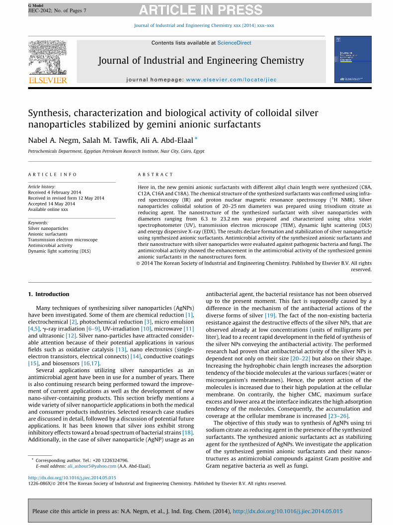
Alkyl ester of phosphoric acid (0.2 mol.) and polyethyleneglycol-400 (9 U of ethylene glycol per molecule) (0.1 mol.) wereesterified individually in xylene (250 mL) as the solvent underreflux conditions at 138 8C. The reaction was stopped aftercomplete removal of the water of the reaction (0.2 mol., 3.6 mL).Then, the reaction mixture was quenched using sodium hydroxide(0.2 mol.), and followed by removing all of the volatile materialfrom the resulting solution [28,29]. The gemini anionic surfactantsobtained were designated as C8A, C12A, C16A and C18A. Scheme 1shows the chemical structures of the synthesized compounds.
Scheme 1. Synthesis of gem
Please cite this article in press as: N.A. Negm, et al., J. Ind. Eng. Che
2.2.3. Synthesis of the silver nanoparticles (AgNPs)
Chemical reduction method was used to prepare the silvernanoparticles colloidal solution. All solutions of reacting materialswere prepared in distilled water. In a typical experiment 100 mL ofAgNO3 solution (1 � 10�3 M) was heated to boiling. To thissolution 10 ml of 1% tri-sodium citrate was added drop by drop.The solution was mixed vigorously and heated until the yellowcolor obtained. Then it was removed from the heating element andstirred until cooled to room temperature [30].
2.2.4. Synthesis of the nanostructure of gemini anionic surfactants
with AgNPs
The silver nanoparticles solution (20 mL) was mixed with 5 mLsolution of the synthesized gemini anionic surfactants (C8A, C12A,C16A and C18A) (0.01 mol) in distilled water. The mixture wasstirred continuously for 24 h till the color change. The resultingsolution was used for ultraviolet experiments, TEM image,dynamic light scattering and energy dispersive X-ray [31].
2.3. Instruments
The chemical structures of the synthesized surfactants werecharacterized as follows:
� FT-IR spectra using ATI Mattsonm Infinity seriesTM, Bench top961 controlled by Win FirstTM V2.01 software. (EgyptianPetroleum Research Institute).� 1H NMR was measured in DMSO-d6 by Spect Varian, GEMINI 200
(1H 200 MHz) (Micro-analytical Center, Cairo University).
Silver nanoparticles stabilized by anionic surfactants werecharacterized by:
� UV spectroscopy: The synthesized Ag nanoparticles were moni-tored periodically in a UV spectrophotometer (Shimadzu, UV-2550, Japan).
ini anionic surfactants.
m. (2014), http://dx.doi.org/10.1016/j.jiec.2014.05.015
N.A. Negm et al. / Journal of Industrial and Engineering Chemistry xxx (2014) xxx–xxx 3
G Model
JIEC-2042; No. of Pages 7
� Transmission electron microscope (TEM): A convenient way toproduce good TEM samples is to use copper grids. A copper gridpre-covered with a very thin amorphous carbon film. Toinvestigate the prepared AgNPs using TEM, small droplets ofthe liquid were placed on the carbon-coated grid. A photographicplate of the transmission electron microscopy employed on thepresent work to investigate the microstructure of the preparedsamples. Nanoparticle size was determined by using TEM model‘‘Type JEOL JEM-1230 operating at 120 kV attached to a CCDcamera’’ (Egyptian Petroleum Research Institute ‘‘EPRI’’)� Dynamic light scattering (DLS): The hydrodynamic diameter and
zeta potential of the same solution which used in TEM, UV–visand EDX, was characterized by dynamic light scattering (DLS)using a Malvern Zetasizer Nano (Malvern Instruments Ltd.,Worcestershire, UK). Each DLS measurement was run intriplicate using automated, optimal measurement time andlaser attenuation settings. The recorded correlation functionsand measured particles mobility’s were converted into sizedistributions and zeta potentials, respectively, using the MalvernDispersion Software (V5.10, http://www.zetasizer.com/).� Energy dispersive X-ray (EDX) spectroscopy: The energy-dispersive
X-ray (EDX) spectroscopy was recorded with an EDX detector(Oxford LINKISIS 300) equipped on a transmission electronmicroscope (TEM, Hitachi S-520) operated at 10 kV acceleratingvoltage.
2.4. Biological activity
The antimicrobial activity of synthesized gemini anionicsurfactants and their nanostructures with silver nanoparticleswere measured against a wide range of test-organisms comprising:(bacteria and fungi)
2.4.1. The media
The following media used in the antimicrobial activity ofsynthesized products, the bacterial species grow on nutrient agar,while fungi mold grow on Czapek’s dox agar.(a) Nutrient agar:Nutrient agar consists of Beef extract (3.0 g/l); peptone (5.0 g/l),sodium chloride (3.0 g/l) and agar (20.0 g/l), then, completes thevolume to 1 l, heated the mixture until the boiling and sterilizes themedia by autoclave. (b) Czapek’s Dox agar: Czapek’s Dox agarconsists of sucrose (20.0 g/l), sodium nitrate (2.0 g/l), magnesiumsulfate (0.5 g/l), potassium chloride (0.5 g/l), ferrous sulfate(0.01 g/l) and agar (20.0 g/l), then, complete the volume to 1 l,heated the mixture until the boiling, and sterilize the media byautoclave.
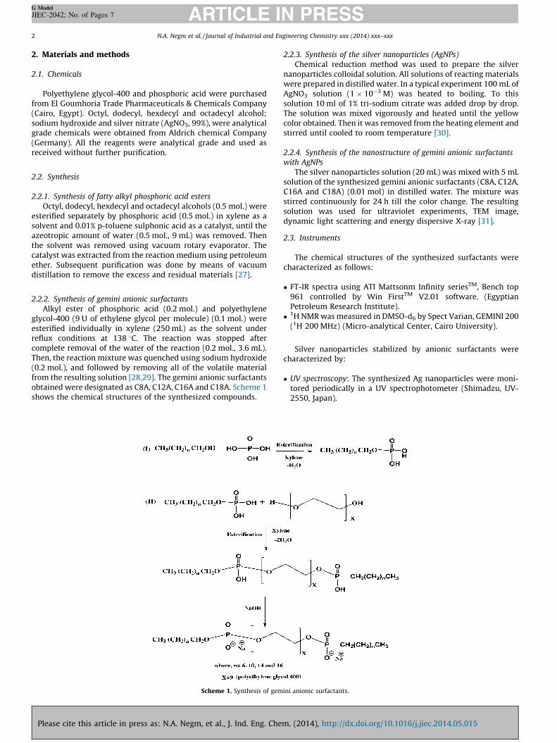
Fig. 1. FT-IR spectrum of the synthe
Please cite this article in press as: N.A. Negm, et al., J. Ind. Eng. Che
2.4.2. Microorganisms
The different species of tested organisms were obtained fromthe unit of Operation Development Center, Egyptian PetroleumResearch Institute.
The used microorganisms were Gram-positive bacteria (Bacillus
pumilus and Micrococcus luteus), Gram-negative bacteria (Pseudo-
monas aeuroginosa and Sarcina lutea) and Fungi (Candida albicans
and Penicillium chrysogenum).An assay is made to determine the ability of an antibiotic to kill
or inhibit the growth of living microorganisms, the techniquewhich used is: filter-paper disk-agar diffusion (Kirby-Bauer) are asthe following [32]:
1. Inoculate flask of melted agar medium with the organism to betested.
2. Pour this inoculated medium into a Petri dish.3. After the agar has solidified, a multilobed disk that impregnated
with different antibiotics laid on top of agar.4. The antibiotic in each lobe of disk diffuses into medium and if
the organism is sensitive to a particular antibiotic, no growthoccur in a large zone surrounding that lobe (clear zone).
5. The diameters of inhibition zones were measured after 24–48 hat 35–37 8C (for bacteria) and 3–4 days at 25–27 8C (for yeastand fungi) of incubation at 28 8C
6. Measure each clear zone and compare between them todetermine the antibiotic which is more inhibitor.
3. Results and discussion
3.1. Structure
The chemical structures of the synthesized anionic geminisurfactants (C8A, C12A, C16A and C18A) were confirmed using FT-IR and 1H NMR spectroscopy as follows:
3.1.1. FT-IR spectra
The chemical structure for the synthesized surfactant wascharacterized by FTIR spectroscopy in the range 4000–500 cm�1.The FTIR absorption spectra showed an absorption band at the2850 cm�1 region was due to the methyl asymmetric stretchingvibration. In addition, there was a strong band at 1460 cm�1,indicating the presence of multiple (CH2)n groups. The sharp bandat 2956 cm�1 was due to the stretching vibration of the symmetricmethylene group. The band at 1108 cm�1 indicating to theformation P–O–C– group, whereas the peak at 1277 cm�1 wasdue to C–O stretching absorption bands. FTIR spectra confirmedthe expected functional groups in the synthesized anionicsurfactant (Fig. 1).
sized anionic surfactant (C8A).
m. (2014), http://dx.doi.org/10.1016/j.jiec.2014.05.015
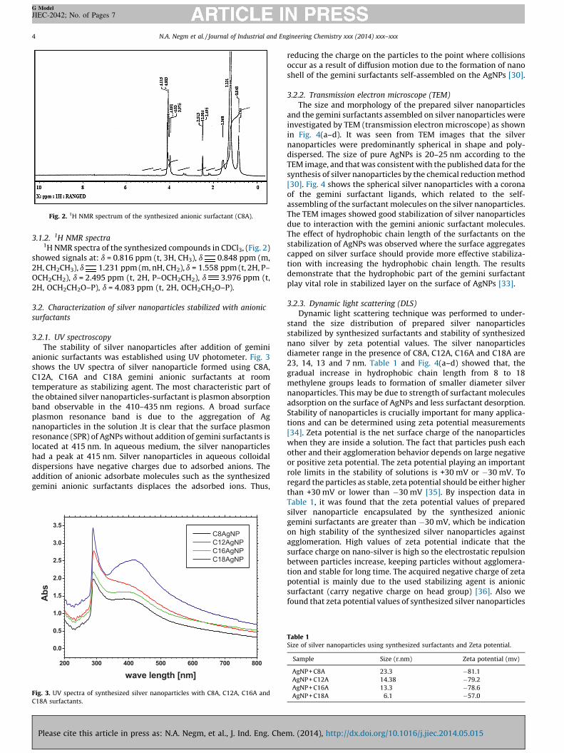
Fig. 2. 1H NMR spectrum of the synthesized anionic surfactant (C8A).
N.A. Negm et al. / Journal of Industrial and Engineering Chemistry xxx (2014) xxx–xxx4
G Model
JIEC-2042; No. of Pages 7
3.1.2. 1H NMR spectra1H NMR spectra of the synthesized compounds in CDCl3, (Fig. 2)
showed signals at: d = 0.816 ppm (t, 3H, CH3), d 0.848 ppm (m,2H, CH2CH3), d 1.231 ppm (m, nH, CH2), d = 1.558 ppm (t, 2H, P–OCH2CH2), d = 2.495 ppm (t, 2H, P–OCH2CH2), d 3.976 ppm (t,2H, OCH2CH2O–P), d = 4.083 ppm (t, 2H, OCH2CH2O–P).
3.2. Characterization of silver nanoparticles stabilized with anionic
surfactants
3.2.1. UV spectroscopy
The stability of silver nanoparticles after addition of geminianionic surfactants was established using UV photometer. Fig. 3shows the UV spectra of silver nanoparticle formed using C8A,C12A, C16A and C18A gemini anionic surfactants at roomtemperature as stabilizing agent. The most characteristic part ofthe obtained silver nanoparticles-surfactant is plasmon absorptionband observable in the 410–435 nm regions. A broad surfaceplasmon resonance band is due to the aggregation of Agnanoparticles in the solution .It is clear that the surface plasmonresonance (SPR) of AgNPs without addition of gemini surfactants islocated at 415 nm. In aqueous medium, the silver nanoparticleshad a peak at 415 nm. Silver nanoparticles in aqueous colloidaldispersions have negative charges due to adsorbed anions. Theaddition of anionic adsorbate molecules such as the synthesizedgemini anionic surfactants displaces the adsorbed ions. Thus,
200 30 0 40 0 50 0 60 0 70 0 80 0
0.0
0.5
1.0
1.5
2.0
2.5
3.0
3.5
Ab
s
wave l eng th [nm]
C8AgNP
C12 AgNP
C16 AgNP
C18 AgNP
Fig. 3. UV spectra of synthesized silver nanoparticles with C8A, C12A, C16A and
C18A surfactants.
Please cite this article in press as: N.A. Negm, et al., J. Ind. Eng. Che
reducing the charge on the particles to the point where collisionsoccur as a result of diffusion motion due to the formation of nanoshell of the gemini surfactants self-assembled on the AgNPs [30].
3.2.2. Transmission electron microscope (TEM)
The size and morphology of the prepared silver nanoparticlesand the gemini surfactants assembled on silver nanoparticles wereinvestigated by TEM (transmission electron microscope) as shownin Fig. 4(a–d). It was seen from TEM images that the silvernanoparticles were predominantly spherical in shape and poly-dispersed. The size of pure AgNPs is 20–25 nm according to theTEM image, and that was consistent with the published data for thesynthesis of silver nanoparticles by the chemical reduction method[30]. Fig. 4 shows the spherical silver nanoparticles with a coronaof the gemini surfactant ligands, which related to the self-assembling of the surfactant molecules on the silver nanoparticles.The TEM images showed good stabilization of silver nanoparticlesdue to interaction with the gemini anionic surfactant molecules.The effect of hydrophobic chain length of the surfactants on thestabilization of AgNPs was observed where the surface aggregatescapped on silver surface should provide more effective stabiliza-tion with increasing the hydrophobic chain length. The resultsdemonstrate that the hydrophobic part of the gemini surfactantplay vital role in stabilized layer on the surface of AgNPs [33].
3.2.3. Dynamic light scattering (DLS)
Dynamic light scattering technique was performed to under-stand the size distribution of prepared silver nanoparticlesstabilized by synthesized surfactants and stability of synthesizednano silver by zeta potential values. The silver nanoparticlesdiameter range in the presence of C8A, C12A, C16A and C18A are23, 14, 13 and 7 nm. Table 1 and Fig. 4(a–d) showed that, thegradual increase in hydrophobic chain length from 8 to 18methylene groups leads to formation of smaller diameter silvernanoparticles. This may be due to strength of surfactant moleculesadsorption on the surface of AgNPs and less surfactant desorption.Stability of nanoparticles is crucially important for many applica-tions and can be determined using zeta potential measurements[34]. Zeta potential is the net surface charge of the nanoparticleswhen they are inside a solution. The fact that particles push eachother and their agglomeration behavior depends on large negativeor positive zeta potential. The zeta potential playing an importantrole limits in the stability of solutions is +30 mV or �30 mV. Toregard the particles as stable, zeta potential should be either higherthan +30 mV or lower than �30 mV [35]. By inspection data inTable 1, it was found that the zeta potential values of preparedsilver nanoparticle encapsulated by the synthesized anionicgemini surfactants are greater than �30 mV, which be indicationon high stability of the synthesized silver nanoparticles againstagglomeration. High values of zeta potential indicate that thesurface charge on nano-silver is high so the electrostatic repulsionbetween particles increase, keeping particles without agglomera-tion and stable for long time. The acquired negative charge of zetapotential is mainly due to the used stabilizing agent is anionicsurfactant (carry negative charge on head group) [36]. Also wefound that zeta potential values of synthesized silver nanoparticles
Table 1Size of silver nanoparticles using synthesized surfactants and Zeta potential.
Sample Size (r.nm) Zeta potential (mv)
AgNP + C8A 23.3 �81.1
AgNP + C12A 14.38 �79.2
AgNP + C16A 13.3 �78.6
AgNP + C18A 6.1 �57.0
m. (2014), http://dx.doi.org/10.1016/j.jiec.2014.05.015
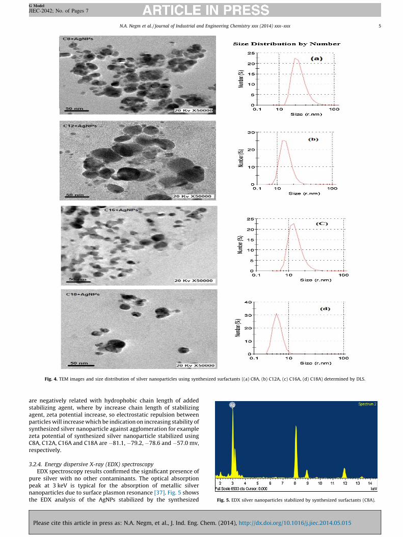
Fig. 4. TEM images and size distribution of silver nanoparticles using synthesized surfactants ((a) C8A, (b) C12A, (c) C16A, (d) C18A) determined by DLS.
Fig. 5. EDX silver nanoparticles stabilized by synthesized surfactants (C8A).
N.A. Negm et al. / Journal of Industrial and Engineering Chemistry xxx (2014) xxx–xxx 5
G Model
JIEC-2042; No. of Pages 7
are negatively related with hydrophobic chain length of addedstabilizing agent, where by increase chain length of stabilizingagent, zeta potential increase, so electrostatic repulsion betweenparticles will increase which be indication on increasing stability ofsynthesized silver nanoparticle against agglomeration for examplezeta potential of synthesized silver nanoparticle stabilized usingC8A, C12A, C16A and C18A are �81.1, �79.2, �78.6 and �57.0 mv,respectively.
3.2.4. Energy dispersive X-ray (EDX) spectroscopy
EDX spectroscopy results confirmed the significant presence ofpure silver with no other contaminants. The optical absorptionpeak at 3 keV is typical for the absorption of metallic silvernanoparticles due to surface plasmon resonance [37]. Fig. 5 showsthe EDX analysis of the AgNPs stabilized by the synthesized
Please cite this article in press as: N.A. Negm, et al., J. Ind. Eng. Chem. (2014), http://dx.doi.org/10.1016/j.jiec.2014.05.015
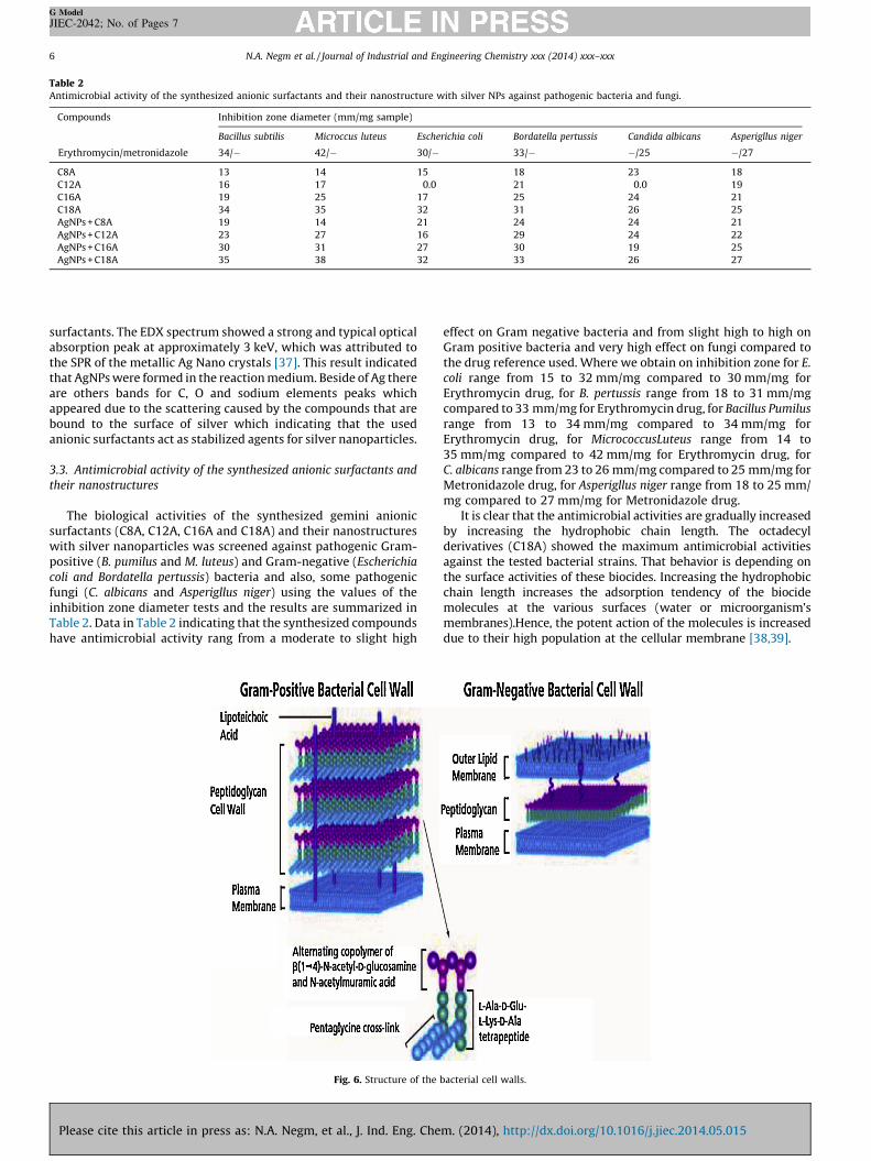
Table 2Antimicrobial activity of the synthesized anionic surfactants and their nanostructure with silver NPs against pathogenic bacteria and fungi.
Compounds Inhibition zone diameter (mm/mg sample)
Bacillus subtilis Microccus luteus Escherichia coli Bordatella pertussis Candida albicans Asperigllus niger
Erythromycin/metronidazole 34/� 42/� 30/� 33/� �/25 �/27
C8A 13 14 15 18 23 18
C12A 16 17 0.0 21 0.0 19
C16A 19 25 17 25 24 21
C18A 34 35 32 31 26 25
AgNPs + C8A 19 14 21 24 24 21
AgNPs + C12A 23 27 16 29 24 22
AgNPs + C16A 30 31 27 30 19 25
AgNPs + C18A 35 38 32 33 26 27
N.A. Negm et al. / Journal of Industrial and Engineering Chemistry xxx (2014) xxx–xxx6
G Model
JIEC-2042; No. of Pages 7
surfactants. The EDX spectrum showed a strong and typical opticalabsorption peak at approximately 3 keV, which was attributed tothe SPR of the metallic Ag Nano crystals [37]. This result indicatedthat AgNPs were formed in the reaction medium. Beside of Ag thereare others bands for C, O and sodium elements peaks whichappeared due to the scattering caused by the compounds that arebound to the surface of silver which indicating that the usedanionic surfactants act as stabilized agents for silver nanoparticles.
3.3. Antimicrobial activity of the synthesized anionic surfactants and
their nanostructures
The biological activities of the synthesized gemini anionicsurfactants (C8A, C12A, C16A and C18A) and their nanostructureswith silver nanoparticles was screened against pathogenic Gram-positive (B. pumilus and M. luteus) and Gram-negative (Escherichia
coli and Bordatella pertussis) bacteria and also, some pathogenicfungi (C. albicans and Asperigllus niger) using the values of theinhibition zone diameter tests and the results are summarized inTable 2. Data in Table 2 indicating that the synthesized compoundshave antimicrobial activity rang from a moderate to slight high
Fig. 6. Structure of the
Please cite this article in press as: N.A. Negm, et al., J. Ind. Eng. Che
effect on Gram negative bacteria and from slight high to high onGram positive bacteria and very high effect on fungi compared tothe drug reference used. Where we obtain on inhibition zone for E.
coli range from 15 to 32 mm/mg compared to 30 mm/mg forErythromycin drug, for B. pertussis range from 18 to 31 mm/mgcompared to 33 mm/mg for Erythromycin drug, for Bacillus Pumilus
range from 13 to 34 mm/mg compared to 34 mm/mg forErythromycin drug, for MicrococcusLuteus range from 14 to35 mm/mg compared to 42 mm/mg for Erythromycin drug, forC. albicans range from 23 to 26 mm/mg compared to 25 mm/mg forMetronidazole drug, for Asperigllus niger range from 18 to 25 mm/mg compared to 27 mm/mg for Metronidazole drug.
It is clear that the antimicrobial activities are gradually increasedby increasing the hydrophobic chain length. The octadecylderivatives (C18A) showed the maximum antimicrobial activitiesagainst the tested bacterial strains. That behavior is depending onthe surface activities of these biocides. Increasing the hydrophobicchain length increases the adsorption tendency of the biocidemolecules at the various surfaces (water or microorganism’smembranes).Hence, the potent action of the molecules is increaseddue to their high population at the cellular membrane [38,39].
bacterial cell walls.
m. (2014), http://dx.doi.org/10.1016/j.jiec.2014.05.015
N.A. Negm et al. / Journal of Industrial and Engineering Chemistry xxx (2014) xxx–xxx 7
G Model
JIEC-2042; No. of Pages 7
General observation for data in Table 2 indicates that the Gram-positive bacteria are more resistant to the tested compoundscompared with the Gram-negative bacteria. The data provided fromthe inhibition zone diameter are describing the general behavior ofthe tested biocides against the different bacterial genera.
The results of the antifungal activity obtained from thebiological study showed promising features of the tested biocidesagainst the most pathogenic fungal strain (C. albicans).
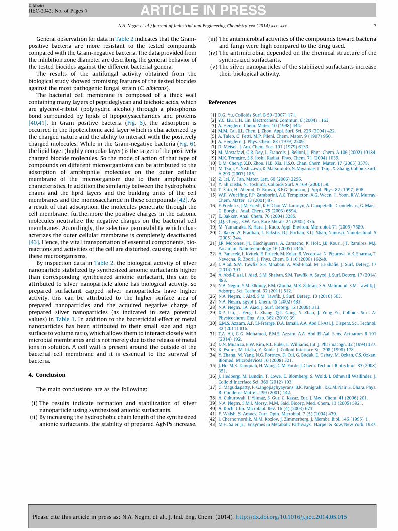
The bacterial cell membrane is composed of a thick wallcontaining many layers of peptideglycan and teichoic acids, whichare glycerol-ribitol (polyhydric alcohol) through a phosphorusbond surrounded by lipids of lipopolysaccharides and proteins[40,41]. In Gram positive bacteria (Fig. 6), the adsorption isoccurred in the lipoteichonic acid layer which is characterized bythe charged nature and the ability to interact with the positivelycharged molecules. While in the Gram-negative bacteria (Fig. 6),the lipid layer (highly nonpolar layer) is the target of the positivelycharged biocide molecules. So the mode of action of that type ofcompounds on different microorganisms can be attributed to theadsorption of amphiphile molecules on the outer cellularmembrane of the microorganism due to their amphipathiccharacteristics. In addition the similarity between the hydrophobicchains and the lipid layers and the building units of the cellmembranes and the monosaccharide in these compounds [42]. Asa result of that adsorption, the molecules penetrate through thecell membrane; furthermore the positive charges in the cationicmolecules neutralize the negative charges on the bacterial cellmembranes. Accordingly, the selective permeability which char-acterizes the outer cellular membrane is completely deactivated[43]. Hence, the vital transportation of essential components, bio-reactions and activities of the cell are disturbed, causing death forthese microorganisms.
By inspection data in Table 2, the biological activity of silvernanoparticle stabilized by synthesized anionic surfactants higherthan corresponding synthesized anionic surfactant, this can beattributed to silver nanoparticle alone has biological activity, soprepared surfactant capped silver nanoparticles have higheractivity, this can be attributed to the higher surface area ofprepared nanoparticles and the acquired negative charge ofprepared silver nanoparticles (as indicated in zeta potentialvalues) in Table 1. In addition to the bactericidal effect of metalnanoparticles has been attributed to their small size and highsurface to volume ratio, which allows them to interact closely withmicrobial membranes and is not merely due to the release of metalions in solution. A cell wall is present around the outside of thebacterial cell membrane and it is essential to the survival ofbacteria.
4. Conclusion
The main conclusions are as the following:
(i) The results indicate formation and stabilization of silvernanoparticle using synthesized anionic surfactants.
(ii) By increasing the hydrophobic chain length of the synthesizedanionic surfactants, the stability of prepared AgNPs increase.
Please cite this article in press as: N.A. Negm, et al., J. Ind. Eng. Che
(iii) The antimicrobial activities of the compounds toward bacteriaand fungi were high compared to the drug used.
(iv) The antimicrobial depended on the chemical structure of thesynthesized surfactants.
(v) The silver nanoparticles of the stabilized surfactants increasetheir biological activity.
References
[1] D.G. Yu, Colloids Surf. B 59 (2007) 171.[2] Y.C. Liu, L.H. Lin, Electrochem. Commun. 6 (2004) 1163.[3] A. Henglein, Chem. Mater. 10 (1998) 444.[4] M.M. Cai, J.L. Chen, J. Zhou, Appl. Surf. Sci. 226 (2004) 422.[5] A. Taleb, C. Petti, M.P. Pileni, Chem. Mater. 9 (1997) 950.[6] A. Henglein, J. Phys. Chem. 83 (1979) 2209.[7] D. Meisel, J. Am. Chem. Soc. 101 (1979) 6133.[8] M. Mostafavi, G.R. Dey, L. Francois, J. Belloni, J. Phys. Chem. A 106 (2002) 10184.[9] M.K. Temgire, S.S. Joshi, Radiat. Phys. Chem. 71 (2004) 1039.
[10] D.M. Cheng, X.D. Zhou, H.B. Xia, H.S.O. Chan, Chem. Mater. 17 (2005) 3578.[11] M. Tsuji, Y. Nishizawa, K. Matsumoto, N. Miyamae, T. Tsuji, X. Zhang, Colloids Surf.
A 293 (2007) 185.[12] Z. Lei, Y. Fan, Mater. Lett. 60 (2006) 2256.[13] Y. Shiraishi, N. Toshima, Colloids Surf. A 169 (2000) 59.[14] T. Sato, H. Ahemd, D. Brown, B.F.G. Johnson, J. Appl. Phys. 82 (1997) 696.[15] W.P. Wuelfing, F.P. Zamborini, A.C. Templeton, X.G. Ween, H. Yoon, R.W. Murray,
Chem. Mater. 13 (2001) 87.[16] F. Frederix, J.M. Friedt, K.H. Choi, W. Laureyn, A. Campetelli, D. ondelears, G. Maes,
G. Borghs, Anal. Chem. 75 (2003) 6894.[17] E. Bakker, Anal. Chem. 76 (2004) 3285.[18] J.Q. Cheng, S.W. Yao, Rare Metals 24 (2005) 376.[19] M. Yamanaka, K. Hara, J. Kudo, Appl. Environ. Microbiol. 71 (2005) 7589.[20] C. Baker, A. Pradhan, L. Pakstis, D.J. Pochan, S.I.J. Shah, Nanosci. Nanotechnol. 5
(2005) 244.[21] J.R. Morones, J.L. Elechiguerra, A. Camacho, K. Holt, J.B. Kouri, J.T. Ramirez, M.J.
Yacaman, Nanotechnology 16 (2005) 2346.[22] A. Panacek, L. Kvitek, R. Prucek, M. Kolar, R. Vecerova, N. Pizurova, V.K. Sharma, T.
Nevecna, R. Zboril, J. Phys. Chem. B 110 (2006) 16248.[23] I. Aiad, S.M. Tawfik, S.S. Mhaban, A. Abd-Elaal, M. El-Shafie, J. Surf. Deterg. 17
(2014) 391.[24] A. Abd-Elaal, I. Aiad, S.M. Shaban, S.M. Tawfik, A. Sayed, J. Surf. Deterg. 17 (2014)
483.[25] N.A. Negm, Y.M. Elkholy, F.M. Ghuiba, M.K. Zahran, S.A. Mahmoud, S.M. Tawfik, J.
Adsorpt. Sci. Technol. 32 (2011) 512.[26] N.A. Negm, I. Aiad, S.M. Tawfik, J. Surf. Deterg. 13 (2010) 503.[27] N.A. Negm, Egypt J. Chem. 45 (2002) 483.[28] N.A. Negm, I.A. Aiad, J. Surf. Deterg. 12 (2009) 313.[29] X.P. Liu, J. Feng, L. Zhang, Q.T. Gong, S. Zhao, J. Yong Yu, Colloids Surf. A:
Physicochem. Eng. Asp. 362 (2010) 39.[30] E.M.S. Azzam, A.F. El-Frarrge, D.A. Ismail, A.A. Abd El-Aal, J. Dispers. Sci. Technol.
32 (2011) 816.[31] T.A. Ali, G.G. Mohamed, E.M.S. Azzam, A.A. Abd El-Aal, Sens. Actuators B 191
(2014) 192.[32] D.N. Muanza, B.W. Kim, K.L. Euler, L. Williams, Int. J. Pharmacogn. 32 (1994) 337.[33] K. Esumi, M. Iitaka, Y. Koide, J. Colloid Interface Sci. 208 (1998) 178.[34] Y. Zhang, M. Yang, N.G. Portney, D. Cui, G. Budak, E. Ozbay, M. Ozkan, C.S. Ozkan,
Biomed. Microdevices 10 (2008) 321.[35] J. Ho, M.K. Danquah, H. Wang, G.M. Forde, J. Chem. Technol. Biotechnol. 83 (2008)
351.[36] J. Hedberg, M. Lundin, T. Lowe, E. Blomberg, S. Wold, I. Odnevall Wallinder, J.
Colloid Interface Sci. 369 (2012) 193.[37] G. Magudapatty, P. Gangopaghyayrans, B.K. Panigrahi, K.G.M. Nair, S. Dhara, Phys.
B: Condens. Matter. 299 (2001) 142.[38] A. Cukurovali, I. Yilmaz, S. Gur, C. Kazaz, Eur. J. Med. Chem. 41 (2006) 201.[39] N.A. Negm, S.M.I. Morsy, M.M. Said, Bioorg. Med. Chem. 13 (2005) 5921.[40] A. Koch, Clin. Microbiol. Rev. 16 (4) (2003) 673.[41] F. Walsh, S. Amyes, Curr. Opin. Microbiol. 7 (5) (2004) 439.[42] I. Chernomordik, M.M. Kozlov, J. Zimmerberg, J. Membr. Biol. 146 (1995) 1.[43] M.H. Saier Jr., Enzymes in Metabolic Pathways, Harper & Row, New York, 1987.
m. (2014), http://dx.doi.org/10.1016/j.jiec.2014.05.015
Related Documents