Chapter 12 BIOLOGICAL MEMBRANES All cells are surrounded by a plasma membrane, and eukaryotes (but not prokaryotes) have membrane- bounded organelles as well. The terms plasma membrane and cell wall, so often confused by students, refer to very different structures. The plasma membrane is as thin and fragile as a soap bubble, yet it forms an effective diffusion barrier. It consists of lipids and proteins. The cell wall, on the other hand, is strong and stiff and maintains the shape of the cell. Plants and bacte- ria have a cell wall that is made of tough polysacchar- ides such as cellulose or peptidoglycan, but humans do not. Human cells are kept in shape by the cytoskel- eton instead, and human tissues derive mechanical strength from the extracellular matrix. This chapter introduces the structure and properties of cellular membranes. MEMBRANES CONSIST OF LIPID AND PROTEIN Under the electron microscope, a biological membrane in cross-section looks like a railroad track, with a lightly stained layer sandwiched between two deeply stained layers. This structure, with a total diameter of 8 nm, is formed from two layers of lipids. The membrane lipids are amphiphilic or amphi- pathic. This means that hydrophilic and hydrophobic parts are combined in the same molecule. Phospholipids contain a phosphate group in their hydrophilic part, and glycolipids contain covalently attached carbohy- drate. Based on their chemical building blocks, three classes of membrane lipids can be distinguished: phos- phoglycerides, sphingolipids, and cholesterol. Membranes contain proteins as well as lipids. Lipids form the structural backbone of the membrane, and pro- teins are in charge of specific functions. These functions include enzymatic activities, regulated transport, ion permeability and excitability, contact with structural proteins, and transmission of physiological signals. Therefore the protein/lipid ratio is highest in membranes with high metabolic activity, such as the inner mitochon- drial membrane (Fig. 12.1). PHOSPHOGLYCERIDES ARE THE MOST ABUNDANT MEMBRANE LIPIDS Phosphoglycerides account for more than half of all lipids in most membranes (see Fig. 12.1). Their parent compound is phosphatidic acid, or phosphatidate. It looks similar to a triglyceride but with the third fatty acid of the triglyceride replaced by phosphate: H 2 C O H 2 C CH O O R 1 Triglyceride O C O R 3 O C R 2 C H 2 C O H 2 C CH O O R 1 Phosphatidate O C O O O – O – P R 2 C The major membrane phosphoglycerides have a second alcohol bound to the phosphate group in phosphatidic acid, and they are named as derivatives of phosphatidic acid (phosphatidyl-) (Fig. 12.2). 182

Welcome message from author
This document is posted to help you gain knowledge. Please leave a comment to let me know what you think about it! Share it to your friends and learn new things together.
Transcript

Chapter 12
BIOLOGICAL MEMBRANES
All cells are surrounded by a plasma membrane, andeukaryotes (but not prokaryotes) have membrane-bounded organelles as well.
The terms plasma membrane and cell wall, so oftenconfused by students, refer to very different structures.The plasma membrane is as thin and fragile as a soapbubble, yet it forms an effective diffusion barrier. Itconsists of lipids and proteins.
The cell wall, on the other hand, is strong and stiffand maintains the shape of the cell. Plants and bacte-ria have a cell wall that is made of tough polysacchar-ides such as cellulose or peptidoglycan, but humansdo not. Human cells are kept in shape by the cytoskel-eton instead, and human tissues derive mechanicalstrength from the extracellular matrix. This chapterintroduces the structure and properties of cellularmembranes.
MEMBRANES CONSIST OF LIPID AND PROTEIN
Under the electron microscope, a biological membranein cross-section looks like a railroad track, with alightly stained layer sandwiched between two deeplystained layers. This structure, with a total diameter of8 nm, is formed from two layers of lipids.
The membrane lipids are amphiphilic or amphi-pathic. This means that hydrophilic and hydrophobicparts are combined in the same molecule. Phospholipidscontain a phosphate group in their hydrophilic part,and glycolipids contain covalently attached carbohy-drate. Based on their chemical building blocks, threeclasses of membrane lipids can be distinguished: phos-phoglycerides, sphingolipids, and cholesterol.
Membranes contain proteins as well as lipids. Lipidsform the structural backbone of the membrane, and pro-teins are in charge of specific functions. These functionsinclude enzymatic activities, regulated transport, ionpermeability and excitability, contact with structuralproteins, and transmission of physiological signals.Therefore the protein/lipid ratio is highest in membraneswith high metabolic activity, such as the inner mitochon-drial membrane (Fig. 12.1).
PHOSPHOGLYCERIDES ARE THE MOSTABUNDANT MEMBRANE LIPIDS
Phosphoglycerides account for more than half of alllipids in most membranes (see Fig. 12.1). Their parentcompound is phosphatidic acid, or phosphatidate. Itlooks similar to a triglyceride but with the third fattyacid of the triglyceride replaced by phosphate:
H2C
O
H2C
CH
O
O
R1
Triglyceride
O
CO
R3
O
C
R2 C
H2C
O
H2C
CH
O
O
R1
Phosphatidate
O
C
O
O
O–
O–
P
R2 C
The major membrane phosphoglycerides have a secondalcohol bound to the phosphate group in phosphatidicacid, and they are named as derivatives of phosphatidicacid (phosphatidyl-) (Fig. 12.2).
182

The variable alcohol that is bound to the phosphateeither is charged or has a high hydrogen bonding poten-tial. Together with the negatively charged phosphate, itforms the hydrophilic head group of the molecule,whereas the fatty acids form two hydrophobic tails.
The fatty acid in position 1 usually is saturated, andthat in position 2 is unsaturated.
Two less common phosphoglycerides are shown inFigure 12.3. Cardiolipin (diphosphatidylglycerol) iscommon only in the inner mitochondrial membrane.
0
Nuclear membrane(liver)
25 50 75 100
Inner mitochondrialmembrane (liver)
Outer mitochondrialmembrane (liver)
Endoplasmicreticulum (liver)
Golgi apparatus(liver)
Plasma membrane(liver)
Plasma membrane(erythrocytes)
Plasma membrane(myelin)
Figure 12.1 Composition of
biological membranes. , Protein; ,
phosphatidyl choline; ,
phosphatidyl ethanolamine; ,
phosphatidyl serine; , phosphatidyl
inositol; , cardiolipin; ,
sphingomyelin; , glycolipids; ,
cholesterol; , others.
O–
H2C Phosphatidyl ethanolamine
Phosphatidyl serine
Phosphatidyl inositol
Phosphatidyl choline(‘‘lecithin’’)
C
O
O
C O CH
O CH2O
Hydrophobic tails Hydrophilic head groups
O–
CH3
CH3
O
O–
O OH OH
HO
OH
OH
COO–
+
NH3+
+NH3CH2CH2OPO
CH2CH2OPO
O–
O
CHCH2OPO
OPO
N CH3
Figure 12.2 Structures of the most common phosphoglycerides.
183Biological Membranes
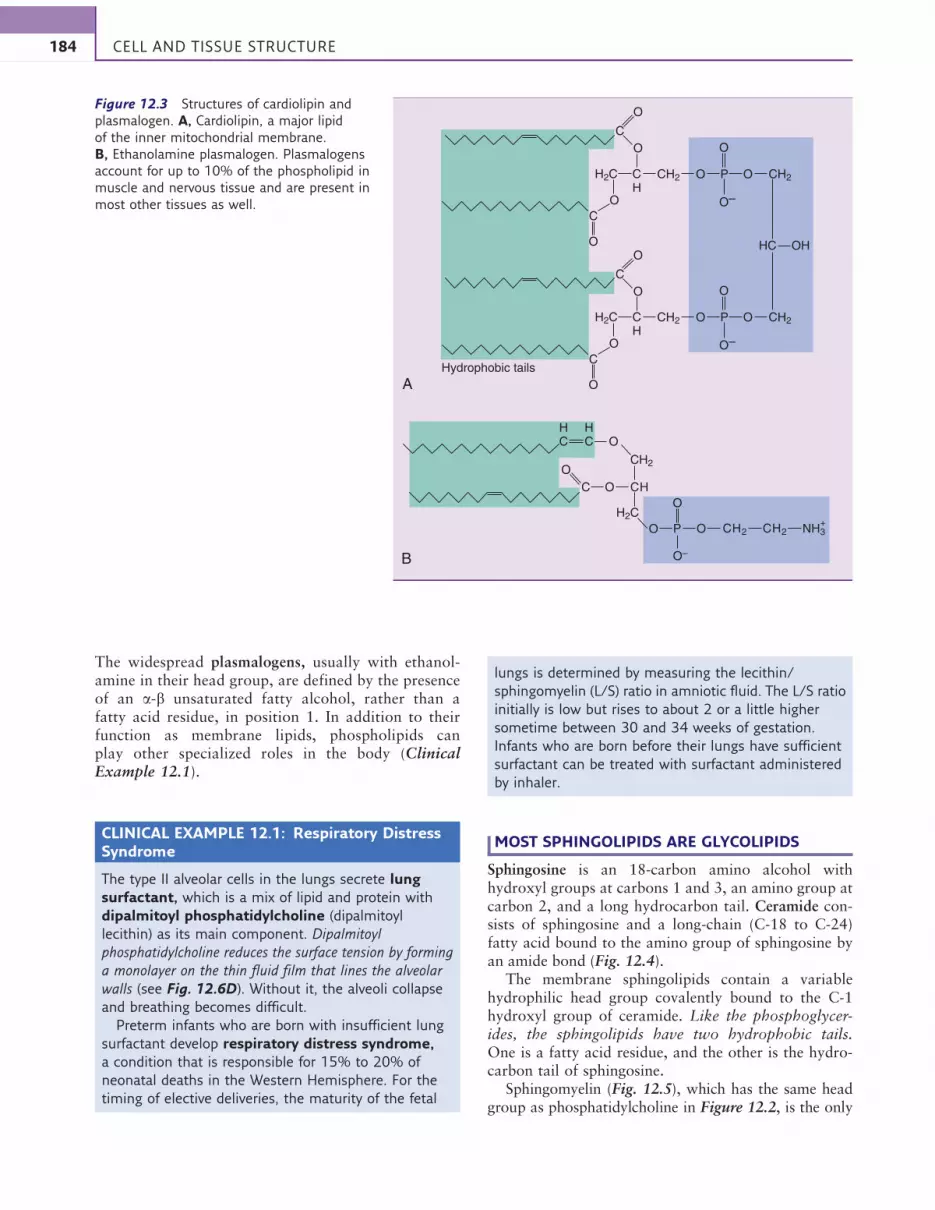
The widespread plasmalogens, usually with ethanol-amine in their head group, are defined by the presenceof an a-b unsaturated fatty alcohol, rather than afatty acid residue, in position 1. In addition to theirfunction as membrane lipids, phospholipids canplay other specialized roles in the body (ClinicalExample 12.1).
CLINICAL EXAMPLE 12.1: Respiratory DistressSyndrome
The type II alveolar cells in the lungs secrete lung
surfactant, which is a mix of lipid and protein with
dipalmitoyl phosphatidylcholine (dipalmitoyl
lecithin) as its main component. Dipalmitoyl
phosphatidylcholine reduces the surface tension by forming
a monolayer on the thin fluid film that lines the alveolar
walls (see Fig. 12.6D). Without it, the alveoli collapse
and breathing becomes difficult.
Preterm infants who are born with insufficient lung
surfactant develop respiratory distress syndrome,
a condition that is responsible for 15% to 20% of
neonatal deaths in the Western Hemisphere. For the
timing of elective deliveries, the maturity of the fetal
lungs is determined by measuring the lecithin/
sphingomyelin (L/S) ratio in amniotic fluid. The L/S ratio
initially is low but rises to about 2 or a little higher
sometime between 30 and 34 weeks of gestation.
Infants who are born before their lungs have sufficient
surfactant can be treated with surfactant administered
by inhaler.
MOST SPHINGOLIPIDS ARE GLYCOLIPIDS
Sphingosine is an 18-carbon amino alcohol withhydroxyl groups at carbons 1 and 3, an amino group atcarbon 2, and a long hydrocarbon tail. Ceramide con-sists of sphingosine and a long-chain (C-18 to C-24)fatty acid bound to the amino group of sphingosine byan amide bond (Fig. 12.4).
The membrane sphingolipids contain a variablehydrophilic head group covalently bound to the C-1hydroxyl group of ceramide. Like the phosphoglycer-ides, the sphingolipids have two hydrophobic tails.One is a fatty acid residue, and the other is the hydro-carbon tail of sphingosine.
Sphingomyelin (Fig. 12.5), which has the same headgroup as phosphatidylcholine in Figure 12.2, is the only
O–
O
+NH3CH2CH2OP
O
O
C CH
O
A
B
O
O
Hydrophobic tails
C
O
O
C
O
O
H2C
OHC
O
C
O–
O
CH2O
C
HC
HC
H2C CH
CH2 CH2O P O
O–
O
H2C CH
CH2 CH2O P O
OH
Figure 12.3 Structures of cardiolipin and
plasmalogen. A, Cardiolipin, a major lipid
of the inner mitochondrial membrane.
B, Ethanolamine plasmalogen. Plasmalogens
account for up to 10% of the phospholipid in
muscle and nervous tissue and are present in
most other tissues as well.
184 CELL AND TISSUE STRUCTURE

important phosphosphingolipid. All other sphingolipidsare glycolipids. The most complex glycosphingolipidsare the gangliosides. They contain between one and fourresidues of the acidic sugar derivativeN-acetylneuraminic
acid (NANA) in terminal positions of their oligosaccha-ride chain:
H
HN
O
H
HH
H
N-Acetylneuraminic acid(NANA)
OH
OH
COO–
OH
O
OH
CH CH OHCH2
H3C C
Glycosphingolipids are most abundant in the outer leaf-let of the plasma membrane, where their carbohydrateheads face the extracellular environment. Sphingomye-lin and galactocerebroside (the latter partly in a sulfatedform) are important constituents of myelin, and gang-liosides and galactocerebroside are most abundant inthe gray matter of the brain.
CHOLESTEROL IS THE MOST HYDROPHOBICMEMBRANE LIPID
Cholesterol is structurally more rigid than the othermembrane lipids, with a stiff steroid ring system insteadof wriggly hydrocarbon tails; and instead of a statelyhydrophilic head group, only a puny hydroxyl groupis present at one end of the molecule:
O
C
CH2
CH2
H2C
CH2
H2C
CH2
H2C
CH2
H2C
CH2
H2C
CH2
H2C
CH2
H2C
CH2
H2C
CH3
H2C
HO
CH2
CH
HC
H2C
C
HC
HO
H
CH2
H2C
CH2
H2C
CH2
H2C
CH2
H2C
CH2
H2C
H3C
Sphingosine
CH2
CH
NH
HC
H2C
C
HC
HO
H
CH2
H2C
CH2
H2C
CH2
H2C
CH2
H2C
CH2
H2C
H3C
Ceramide
+NH3
CH2 HO CH2
Figure 12.4 Structures of sphingosine and ceramide. The
fatty acid residues in ceramide often are very long (C-20 to
C-24). The hydroxyl group of ceramide that is substituted in
the sphingolipids is marked by an arrow.
O
O
O–CH3
CH3
CH2OH
OH
HOOH
Glucocerebroside(= glucosylceramide)
Sphingomyelin
HC
C
CH
HO
H
O
HC
C
CH
HO
H
O+H3C N O O CH2 O CH2PCH2 CH2
HC NH
C HC NH
C
Figure 12.5 Two types of
sphingolipid. Sphingomyelin is
a phosphosphingolipid, and
glucocerebroside is a
glycosphingolipid.
185Biological Membranes

H3C CH2
CH2 CH3
CH3
HO
CH
CH2 CHCH3
CH3
With this structure, cholesterol is by far the leastwater-soluble membrane lipid. Also, unlike the othermembrane lipids, cholesterol alone cannot formmembrane-like structures; it occurs only as a minorcomponent in membranes whose basic structure isformed by other lipids.
Cholesterol accounts for 10% or more of the totallipid only in the plasma membrane and the Golgi mem-brane. It is prominent only in animals. Plants have phy-tosterols instead, and most bacteria have no sterols atall. Therefore a vegan diet is cholesterol free.
MEMBRANE LIPIDS FORM A BILAYER
The hydrophilic head groups of the membrane lipidsinteract with water, whereas the hydrophobic tailsavoid water. Rather than dissolving in water as individ-ual molecules, the membrane lipids form noncovalentaggregates (Fig. 12.6).
Most polar lipids, including ordinary detergents,form globular micelles. Monolayers form only at aque-ous/nonaqueous interfaces (e.g., between water andair), whereas bilayers are surrounded by water on bothsides. All biological membranes contain a lipid bilayeras their structural backbone. The bilayer is heldtogether by hydrophobic interactions between thehydrocarbon tails of the membrane lipids.
The geometry of the lipid molecules determineswhether a bilayer or a globular micelle forms. A bilayeris formed only if the cross-sectional area of the headgroups matches that of the hydrophobic tails. Forexample, if one of the fatty acids is removed from phos-phatidylcholine (lecithin) by the enzyme phospholipaseA2, the hydrophobic portion becomes too thin. Theresulting lysolecithin no longer fits into a bilayer butforms micelles instead. Phospholipase A2 occurs insome snake venoms. It causes hemolysis by hydrolyzingphosphoglycerides in the red blood cell membrane.
THE LIPID BILAYER IS A TWO-DIMENSIONALFLUID
A lipid bilayer cannot exist as a flat sheet because itshydrophobic core would be exposed to the surroundingwater at the edges. Therefore pieces of lipid bilayer tendto close in on themselves to form vesicles. For the samereason, any tear or hole in the bilayer is energeticallyunfavorable and is liable to close spontaneously. As aresult, membranes are self-sealing.
A B C
D
Air
WaterAir
Water
Hydrophilichead groups
Hydrophobic core:3.5–4.0 nm across
E
Leaflets ofthe bilayer
Water
Figure 12.6 Behavior of polar lipids in water. A, A micelle is a small, spherical structure with a hydrophilic surface and a
hydrophobic core. B, A bilayer is the prototype of a biological membrane. As in the micelle, the hydrophilic head groups are on
the surface and the hydrophobic tails are buried in the center. C, A liposome is the prototype of a membrane-bounded vesicle.
It forms spontaneously from a lipid bilayer. D, A monolayer forms at the interface between water and air. E, A soap bubble
consists of two monolayers enclosing a thin water film.
186 CELL AND TISSUE STRUCTURE

Lipid bilayers are easily deformed even by slightforces. The hydrophobic tails of the lipids can merrilywriggle around, and each molecule is free to diffuse lat-erally in the plane of the bilayer. Lateral diffusion pro-ceeds at a speed of about 2 mm/s in artificial bilayers.
When a synthetic lipid bilayer that contains only onelipid is cooled, it “freezes” at a well-defined tempera-ture. Above the phase transition, the lipids movearound like people on a busy town square, but belowthe transition they are immobile like a platoon of sol-diers standing at attention.
Real membranes contain a mixture of many differentlipids along with proteins, and the phase transition isgradual. At ordinary body temperature, membranesbehave like a viscous liquid.
Long, saturated fatty acid chains in the membranelipids make the membrane more rigid because theyalign themselves in parallel, forming multiple van derWaals interactions. Unsaturated fatty acids destabilizethis orderly alignment because their cis double bondsintroduce kinks in the hydrocarbon chain (Fig. 12.7).
Therefore a high content of unsaturated fatty acid resi-dues makes the membrane more fluid.
Animals adjust their membrane fluidity by varyingthe fatty acid composition of their membrane lipids.For example, cold-water fish have more unsaturatedfatty acids in their membranes than do tropical fish.This maintains optimal membrane fluidity at frigidtemperatures, and it makes cold-water fish a valuabledietary source of polyunsaturated fatty acids.
Because of its stiff ring system, cholesterol tends tomake membranes more rigid. However, it also insertsitself between the fatty acid chains and prevents theircrystallization. In this respect, it acts like an impuritythat decreases the melting point of a chemical.
THE LIPID BILAYER IS A DIFFUSION BARRIER
To penetrate a lipid bilayer, a substance has to pass fromthe aqueous solution through the region of the hydro-philic head groups, then across the hydrophobic coreand out between the head groups on the opposite side.
A
B
CH2
CH2
H2C CH
C
C
Cis double bond
C
C
H C
Trans double bond
C H118°
121°
CH
CH
CH2NH3+
O
P
CH2
O
O–
O
CH2
H2C
CH2
H2C
CH2
H2C
CH2
H2C
CH2
H2C
H2C
CH2
H2C
O O
CH2
CH2
CH2 PNH3+
CH2
O
O
O–
O
CH2
H2C
CH2
H2C
CH2
H2C
CH2
H2C
CH2
H2C
H2C
CH2
H2C
CH2
CH3
H2C
O
CH2
COCO COCO
CH2
H2C
CH2
H2C
CH2
H2C
CH2
H2C
CH2
H2C
H2C
CH2
H2C
CH2
CH3
H2C
CH2
CH3
H2C
CH2
CH2
H2C
CH2
H2C
CH2
H2C
HC
H2C
CH3
O
H2C CH
CH2
H2C CH2
H2C CH2
HC CH2
Figure 12.7 Effect of a cis double bond on the
array of fatty acid chains in the hydrophobic core of
the lipid bilayer. A, The geometry of trans and cis
double bonds. There is no free rotation around the
bond, and all four substituents of the double-bonded
carbons are in the same plane. The double bonds
in natural fatty acids are always in cis configuration.
B, A phospholipid with an unsaturated fatty acid in
the lipid bilayer (right side).
187Biological Membranes
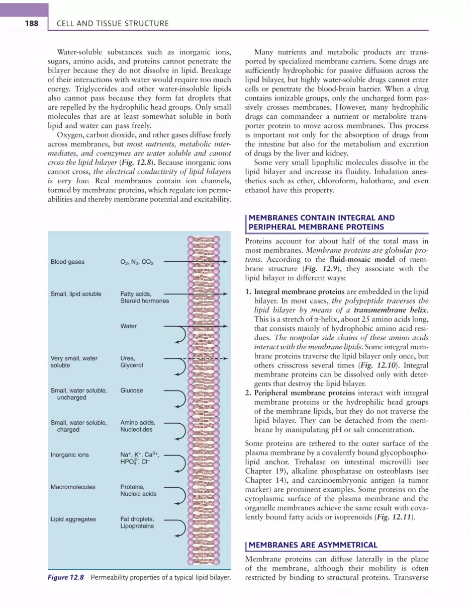
Water-soluble substances such as inorganic ions,sugars, amino acids, and proteins cannot penetrate thebilayer because they do not dissolve in lipid. Breakageof their interactions with water would require too muchenergy. Triglycerides and other water-insoluble lipidsalso cannot pass because they form fat droplets thatare repelled by the hydrophilic head groups. Only smallmolecules that are at least somewhat soluble in bothlipid and water can pass freely.
Oxygen, carbon dioxide, and other gases diffuse freelyacross membranes, but most nutrients, metabolic inter-mediates, and coenzymes are water soluble and cannotcross the lipid bilayer (Fig. 12.8). Because inorganic ionscannot cross, the electrical conductivity of lipid bilayersis very low. Real membranes contain ion channels,formed bymembrane proteins, which regulate ion perme-abilities and thereby membrane potential and excitability.
Many nutrients and metabolic products are trans-ported by specialized membrane carriers. Some drugs aresufficiently hydrophobic for passive diffusion across thelipid bilayer, but highly water-soluble drugs cannot entercells or penetrate the blood-brain barrier. When a drugcontains ionizable groups, only the uncharged form pas-sively crosses membranes. However, many hydrophilicdrugs can commandeer a nutrient or metabolite trans-porter protein to move across membranes. This processis important not only for the absorption of drugs fromthe intestine but also for the metabolism and excretionof drugs by the liver and kidney.
Some very small lipophilic molecules dissolve in thelipid bilayer and increase its fluidity. Inhalation anes-thetics such as ether, chloroform, halothane, and evenethanol have this property.
MEMBRANES CONTAIN INTEGRAL ANDPERIPHERAL MEMBRANE PROTEINS
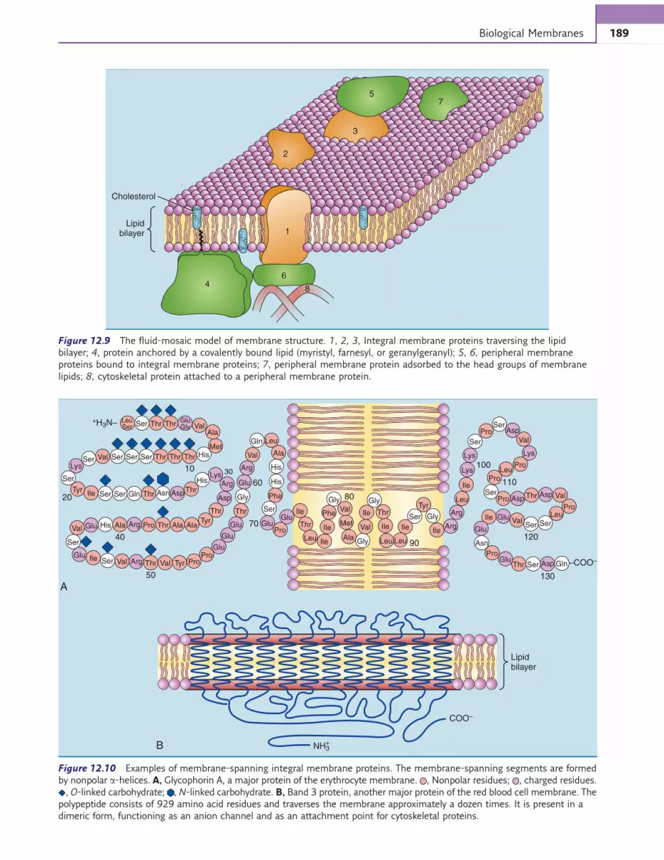
Proteins account for about half of the total mass inmost membranes. Membrane proteins are globular pro-teins. According to the fluid-mosaic model of mem-brane structure (Fig. 12.9), they associate with thelipid bilayer in different ways:
1. Integral membrane proteins are embedded in the lipidbilayer. In most cases, the polypeptide traverses thelipid bilayer by means of a transmembrane helix.This is a stretch of a-helix, about 25 amino acids long,that consists mainly of hydrophobic amino acid resi-dues. The nonpolar side chains of these amino acidsinteract with themembrane lipids. Some integralmem-brane proteins traverse the lipid bilayer only once, butothers crisscross several times (Fig. 12.10). Integralmembrane proteins can be dissolved only with deter-gents that destroy the lipid bilayer.
2. Peripheral membrane proteins interact with integralmembrane proteins or the hydrophilic head groupsof the membrane lipids, but they do not traverse thelipid bilayer. They can be detached from the mem-brane by manipulating pH or salt concentration.
Some proteins are tethered to the outer surface of theplasma membrane by a covalently bound glycophospho-lipid anchor. Trehalase on intestinal microvilli (seeChapter 19), alkaline phosphatase on osteoblasts (seeChapter 14), and carcinoembryonic antigen (a tumormarker) are prominent examples. Some proteins on thecytoplasmic surface of the plasma membrane and theorganelle membranes achieve the same result with cova-lently bound fatty acids or isoprenoids (Fig. 12.11).
MEMBRANES ARE ASYMMETRICAL
Membrane proteins can diffuse laterally in the planeof the membrane, although their mobility is oftenrestricted by binding to structural proteins. Transverse
Blood gases O2, N2, CO2
Small, lipid soluble Fatty acids,Steroid hormones
Very small, watersoluble
Water
Small, water soluble, uncharged
Urea,Glycerol
Small, water soluble,charged
Glucose
Inorganic ions
Amino acids,Nucleotides
Macromolecules
Na+, K+, Ca2+,HPO4 , Cl–2–
Lipid aggregates
Proteins,Nucleic acids
Fat droplets,Lipoproteins
Figure 12.8 Permeability properties of a typical lipid bilayer.
188 CELL AND TISSUE STRUCTURE
3
2
4
Lipidbilayer
Cholesterol
H3C
CH
2 CH
2
CH
3
CH
3C
H3
CH
3
CH
29
CH
OH
H3 C
CH
2CH
2
CH
3
CH
3C
H3
CH
3
CH
2 9C
H
OH
1
57
8
6
H3 C
CH
2CH
2
CH
3
CH
3C
H3
CH
3
CH
2 9C
H
OH
Figure 12.9 The fluid-mosaic model of membrane structure. 1, 2, 3, Integral membrane proteins traversing the lipid
bilayer; 4, protein anchored by a covalently bound lipid (myristyl, farnesyl, or geranylgeranyl); 5, 6, peripheral membrane
proteins bound to integral membrane proteins; 7, peripheral membrane protein adsorbed to the head groups of membrane
lipids; 8, cytoskeletal protein attached to a peripheral membrane protein.
A
B
Lipidbilayer
COO–
–COO–
NH3+
+H3N– SerLeuSer
GluGlyThr Thr Val
Val His
His
Gly
His
His
Gly
Gly
Gly
Thr
Met
Met
Ala
Ser ThrLys
Lys
Thr Thr
10
20
30
60
70
80
90
100
110
SerSerSer
TyrIle
Ser
Ser Ser Gln
Gln
GlnSer
SerSer Gly
Ser
Ser
120
Ser
Ser
Ser
Ser
Asn
ThrThr
Thr Thr
Thr
Thr
TyrAla
Ala
Phe
Phe
Ala
Val
Val
Ile
Ile
Ile IleIle
Ile
AlaThr
Thr Val Tyr ProPro
Pro
Pro
Pro
Pro
Tyr
Pro
Pro
Pro
Ile
Ile
Ile
Val
Val
Val
Thr
Leu
Leu
Leu
50
ProArg
Arg
Ala
40
Glu
Glu
Glu
Glu GluGlu
Glu
Glu
LysLys
Lys
Asp
Asp
Arg
Arg
Asp
Glu
Glu
Val
Val
Arg
Val
Leu
Leu LeuLeu
Ile
Ser
Glu
His
Asn Asp
Asp
130
Arg
Asp
Figure 12.10 Examples of membrane-spanning integral membrane proteins. The membrane-spanning segments are formed
by nonpolar a-helices. A, Glycophorin A, a major protein of the erythrocyte membrane. , Nonpolar residues; , charged residues.
, O-linked carbohydrate; , N-linked carbohydrate. B, Band 3 protein, another major protein of the red blood cell membrane. The
polypeptide consists of 929 amino acid residues and traverses the membrane approximately a dozen times. It is present in a
dimeric form, functioning as an anion channel and as an attachment point for cytoskeletal proteins.
189Biological Membranes

diffusion (“flip-flop”) of membrane proteins has neverbeen observed. In erythrocytes, for example, the asym-metrical orientation of the membrane proteins is main-tained throughout the 120-day lifespan of the cell.
The same is true for membrane lipids. To flip-flopfrom one leaflet of the bilayer to the other, the polarhead group of the lipid has to abandon its interactionswith water molecules and neighboring head groups todive across the hydrophobic core. Only cholesterolflip-flops spontaneously, but lipids with large hydro-philic head groups require the assistance of specializedproteins.
As a result, the lipid distribution in biological mem-branes is asymmetrical. Plasma membranes, for exam-ple, contain most of their phosphatidylethanolamine,phosphatidylserine, and phosphatidylinositol in thecytoplasmic leaflet and most of their glycolipids, phos-phatidylcholine, and sphingomyelin in the exoplasmicleaflet (Fig. 12.12).
In the plasma membrane, the carbohydrate por-tions of glycolipids and glycoproteins face the extra-cellular space (see Fig. 12.10, A). The carbohydrateportions of membrane glycoproteins and glycolipidsare constructed on their protein or lipid core byenzymes in the endoplasmic reticulum and Golgiapparatus. Being located in the lumen of these orga-nelles, the enzymes form the carbohydrates only onthe noncytoplasmic surface of the membrane. WhenGolgi-derived vesicles fuse with the plasma mem-brane, the carbohydrate is placed on the exoplasmicface (Fig. 12.13).
MEMBRANES ARE FRAGILE
All noncovalent structures are fragile. Biological mem-branes are especially vulnerable to agents that disrupthydrophobic interactions. Exposed membranes aredestroyed by detergents and nonpolar organic solvents.
C
Inositol
Man
Man
Gly
COO–
Protein
OMyristate
Plasmamembrane
Intracellular Extracellular
Ser
COO–
ProteinO
Palmitate
H3N+
Cys
OCH3
Protein
O
C
O
O
Farnesylresidue
NH3+
Cys
C
EA
Protein
NH3+
Glycerol
Man
GlcNH2
P
P
S
C
O
O
O
O C
NH
C
Figure 12.11 Attachment of proteins to the plasma membrane by covalently bound lipids. The structure of the glycosyl
phosphatidylinositol anchor shown on the right varies somewhat in different membrane proteins. EA, Ethanolamine; GlcNH2,
nonacetylated glucosamine; Man, D-mannose.
190 CELL AND TISSUE STRUCTURE

Phenol, ethanol, and cationic detergents act as disinfec-tants by disrupting the membranes of microorganisms.
Crystalline materials damage membranes mechani-cally. Crystals of hemoglobin S damage the erythrocytemembrane in sickle cell disease (see Chapter 9), crystalsof sodium urate damage the membranes of phagocyticcells in patients with gouty arthritis (see Chapter 28),and ice crystals damage the cells of frostbitten limbs(Clinical Example 12.2).
CLINICAL EXAMPLE 12.2: Cryopreservation
The preservation of cells and tissues in the frozen state
(cryopreservation) is difficult. Freezing and thawing do
not destroy proteins and nucleic acids, but they can
destroy cellular membranes. This is in part because of
osmotic stress and in part because the relentlessly
growing ice crystals pierce the membranes.
Quick freezing of dispersed cells or small tissue
samples in the presence of antifreeze avoids the
formation of large ice crystals. Sperm and embryos are
routinely preserved by quick freezing in 10% glycerol.
The cryopreservation of oocytes is more difficult,
although it is becoming routine in fertility clinics.
However, complete organs cannot be cryopreserved
because their large heat capacity makes quick freezing
impossible. The same applies to entire human bodies.
A patient with an incurable disease would be ill advised
to jump into liquid nitrogen in the hope that someone
will thaw him someday when a cure for his disease has
been found.
MEMBRANE PROTEINS CARRY SOLUTESACROSS THE LIPID BILAYER
In a few biological membranes, most notably the outermitochondrial membrane, membrane proteins formpores that allow the passage of all small, water-solublemolecules. Usually, however, passive diffusion is limitedto lipid-soluble molecules that are able to cross the lipidbilayer.
Channels are more selective than pores. They have agate with a binding site for a specific solute and are per-meable only for that solute. Inorganic ions are movedacross membranes through channels. These channelscan be regulated, for example, by a neurotransmitterthat binds to the channel (see Chapter 17) or by themembrane potential.
Transporters, also known as membrane carriers,work somewhat like channels but undergo conforma-tional changes during the transport cycle (Fig. 12.14).Carrier-mediated transport is called facilitated diffusionif it is passive and active transport if it requires meta-bolic energy (Table 12.1). Carrier-mediated transportis distinguished from simple diffusion by three impor-tant features:
1. Substrate specificity. Because the substrate must bindnoncovalently to the carrier, transport depends onthe proper fit between substrate and carrier. The glu-cose transporter in red blood cells, for example,transports D-glucose but not L-glucose, and it hasmarkedly reduced affinities for other hexoses suchas D-mannose and D-galactose.
2. Saturability. The rate of passive diffusion is directlyproportional to the concentration gradient, butcarrier-mediated transport is limited by the numberof carriers in the membrane (Fig. 12.15).
3. Specific inhibition and physiological regulation. Car-riers, like enzymes, can be inhibited. Glucose trans-port into erythrocytes, for example, is competitivelyinhibited by various glucose analogs. Membranetransport can also be a rate-limiting and regulatedstep in metabolic pathways. For example, the carrierthat brings glucose into muscle and adipose tissue(but not erythrocytes) is activated by insulin.
TRANSPORT AGAINST AN ELECTROCHEMICALGRADIENT REQUIRES METABOLIC ENERGY
Like chemical reactions,membrane transport is driven bythe free energy change△G [see Equation (5) in Chapter 4].However, the situation is less complex because there is no
SMPC
Outer leaflet%
of m
em
bra
ne p
hospholip
id
Inner leaflet
PS
PE
PI, PIP,PIP2, PA
30
20
10
10
20
30
Figure 12.12 Distribution of phospholipids in the outer and
inner leaflets of the erythrocyte membrane. PA, Phosphatidic
acid; PC, phosphatidylcholine; PE, phosphatidylethanolamine;
PI, phosphatidylinositol; PIP, phosphatidylinositol
4-phosphate; PIP2, phosphatidylinositol 4,5-bisphosphate;
PS, phosphatidylserine; SM, sphingomyelin.
191Biological Membranes

enthalpy change (△H ¼ 0), and the process is purelyentropy driven. For an uncharged molecule, the drivingforce △G for the transfer of a molecule from a compart-ment with the concentration c1 to a compartment withthe concentration c2 is given by the equation
∆G = R × T × ln =C2
C1
C2
C1
2.303 × R × T × logð1Þ
where R ¼ gas constant (1.987 # 10$3 kcal # mol$1 #K$1) and T ¼ absolute temperature. It now is possible
ER
Membrane Protein
Plasma membrane
Cytoplasm
Golgi (cis)
Golgi (trans)
Extracellularspace
Figure 12.13 Placement of a glycoprotein in the plasma membrane. Note that the luminal surface of the organelles
corresponds to the exoplasmic face of the plasma membrane. Glycolipids are synthesized the same way, with their carbohydrate
initially facing the lumen of the endoplasmic reticulum (ER) and Golgi apparatus.
Glucose dissociates
Glucose binds
CytoplasmExtracellularspace
Conformationalchange
Conformationalchange
Glucose
Figure 12.14 Facilitated
diffusion of glucose across the
erythrocyte membrane. There is
no external energy source, so
the net transport is down the
concentration gradient. All
steps in this cycle are
reversible. A net transport of
glucose into the cell takes place
only because glucose is
consumed in the cell, thereby
maintaining a concentration
gradient.
192 CELL AND TISSUE STRUCTURE
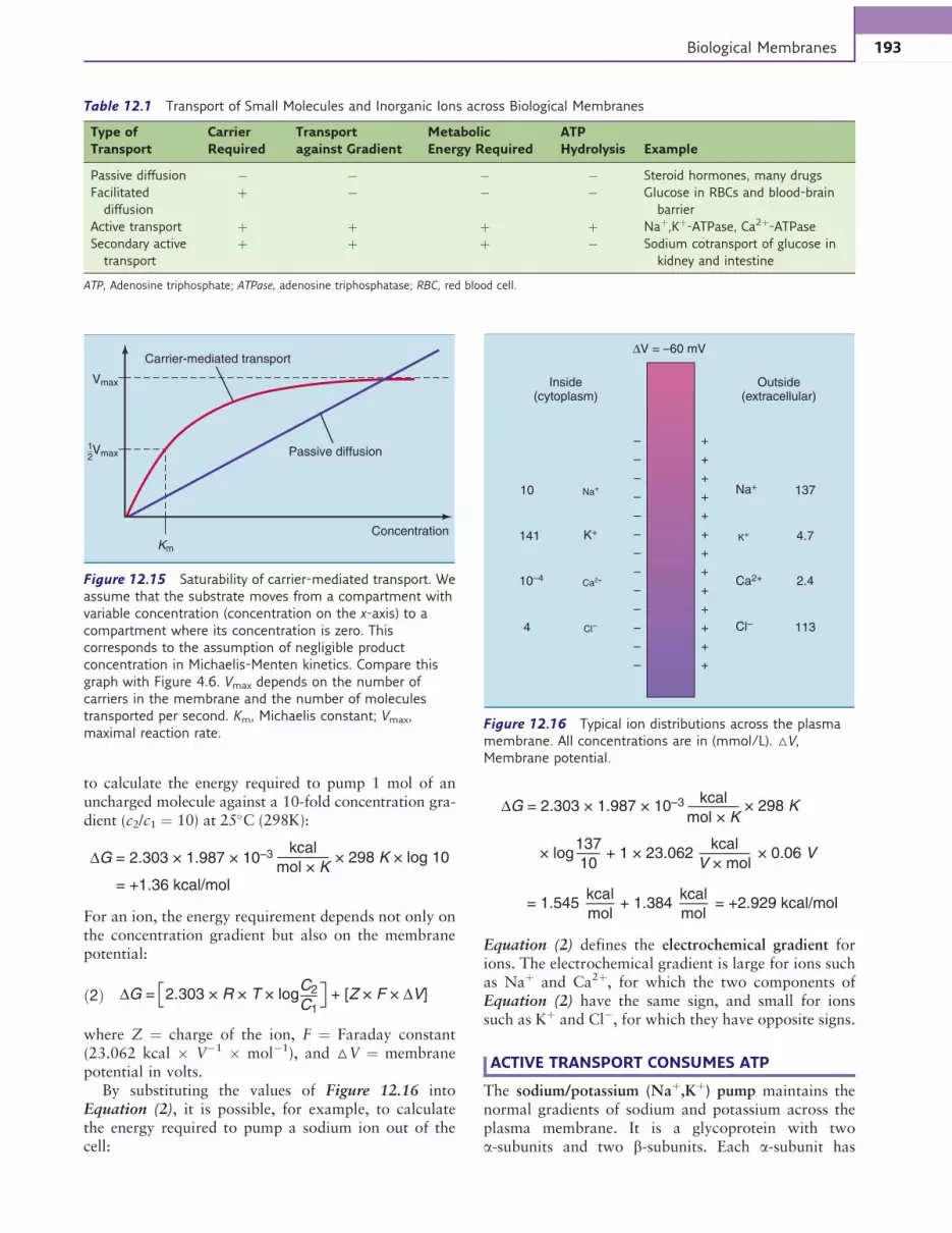
to calculate the energy required to pump 1 mol of anuncharged molecule against a 10-fold concentration gra-dient (c2/c1 ¼ 10) at 25%C (298K):
∆G = 2.303 × 1.987 × 10–3 kcal
mol × K× 298 K × log 10
= +1.36 kcal/mol
For an ion, the energy requirement depends not only onthe concentration gradient but also on the membranepotential:
∆G = 2.303 × R × T × logC2
C1+ [Z × F × ∆V]ð2Þ
where Z ¼ charge of the ion, F ¼ Faraday constant(23.062 kcal # V$1 # mol$1), and △V ¼ membranepotential in volts.
By substituting the values of Figure 12.16 intoEquation (2), it is possible, for example, to calculatethe energy required to pump a sodium ion out of thecell:
∆G = 2.303 × 1.987 × 10–3 kcal
mol × K× 298 K
× log137
10
kcal
V × mol
kcal
mol
kcal
mol
+ 1 × 23.062 × 0.06 V
= 1.545 + 1.384 = +2.929 kcal/mol
Equation (2) defines the electrochemical gradient forions. The electrochemical gradient is large for ions suchas Naþ and Ca2þ, for which the two components ofEquation (2) have the same sign, and small for ionssuch as Kþ and Cl$, for which they have opposite signs.
ACTIVE TRANSPORT CONSUMES ATP
The sodium/potassium (Naþ,Kþ) pump maintains thenormal gradients of sodium and potassium across theplasma membrane. It is a glycoprotein with twoa-subunits and two b-subunits. Each a-subunit has
Vmax
Km
Concentration
Carrier-mediated transport
Passive diffusionVmax12
Figure 12.15 Saturability of carrier-mediated transport. We
assume that the substrate moves from a compartment with
variable concentration (concentration on the x-axis) to a
compartment where its concentration is zero. This
corresponds to the assumption of negligible product
concentration in Michaelis-Menten kinetics. Compare this
graph with Figure 4.6. Vmax depends on the number of
carriers in the membrane and the number of molecules
transported per second. Km, Michaelis constant; Vmax,
maximal reaction rate.
Table 12.1 Transport of Small Molecules and Inorganic Ions across Biological Membranes
Type of
Transport
Carrier
Required
Transport
against Gradient
Metabolic
Energy Required
ATP
Hydrolysis Example
Passive diffusion $ $ $ $ Steroid hormones, many drugs
Facilitated
diffusion
þ $ $ $ Glucose in RBCs and blood-brain
barrier
Active transport þ þ þ þ Naþ,Kþ-ATPase, Ca2þ-ATPase
Secondary active
transport
þ þ þ $ Sodium cotransport of glucose in
kidney and intestine
ATP, Adenosine triphosphate; ATPase, adenosine triphosphatase; RBC, red blood cell.
Inside(cytoplasm)
Outside(extracellular)
∆V = –60 mV
+
+
+
+
+
+
+
+
+
+
+
+
+
–
–
–
–
–
–
–
–
–
–
–
–
–
Cl– 113Cl–4
Ca2+ Ca2+10–4 2.4
K+K+141 4.7
Na+ Na+10 137
Figure 12.16 Typical ion distributions across the plasma
membrane. All concentrations are in (mmol/L). △V,
Membrane potential.
193Biological Membranes
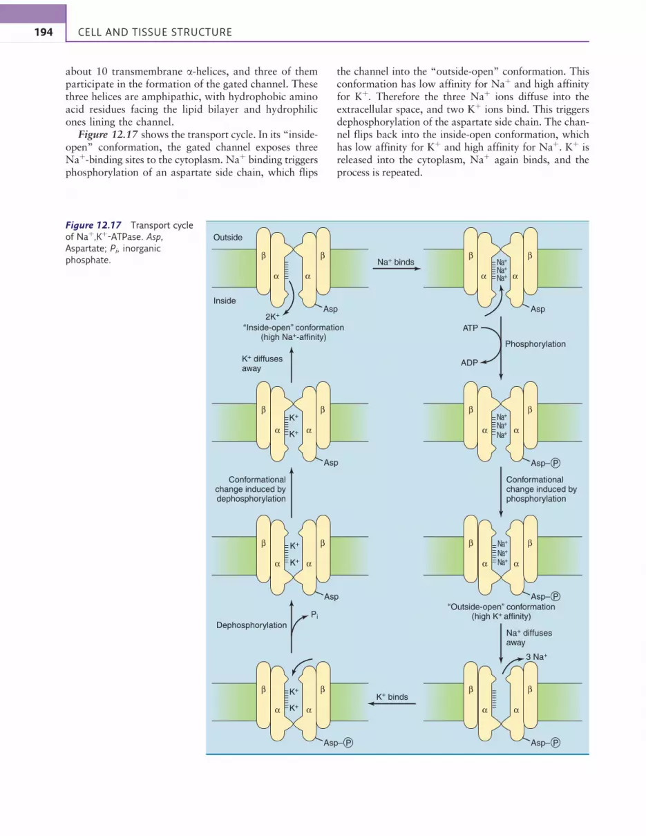
about 10 transmembrane a-helices, and three of themparticipate in the formation of the gated channel. Thesethree helices are amphipathic, with hydrophobic aminoacid residues facing the lipid bilayer and hydrophilicones lining the channel.
Figure 12.17 shows the transport cycle. In its “inside-open” conformation, the gated channel exposes threeNaþ-binding sites to the cytoplasm. Naþ binding triggersphosphorylation of an aspartate side chain, which flips
the channel into the “outside-open” conformation. Thisconformation has low affinity for Naþ and high affinityfor Kþ. Therefore the three Naþ ions diffuse into theextracellular space, and two Kþ ions bind. This triggersdephosphorylation of the aspartate side chain. The chan-nel flips back into the inside-open conformation, whichhas low affinity for Kþ and high affinity for Naþ. Kþ isreleased into the cytoplasm, Naþ again binds, and theprocess is repeated.
Outside
Na+ binds
‘‘Inside-open’’ conformation(high Na+-affinity)
‘‘Outside-open’’ conformation(high K+ affinity)
2K+
K+ diffusesaway
Conformationalchange induced bydephosphorylation
Conformationalchange induced byphosphorylation
AspInside
β β
α α
K+ binds
Phosphorylation
ATP
ADP
K+
K+
Asp
β β
α α
Asp
β β
α α
K+
K+
Asp
β β
α α
β β
α α
Na+
Na+
Na+
β β
α α
Dephosphorylation
Pi
Na+ diffusesaway
3 Na+
K+
K+
β β
α α
β β
α α
Asp– P Asp– P
Asp– P
Asp– P
Na+
Na+
Na+
Na+
Na+
Na+
Figure 12.17 Transport cycle
of Naþ,Kþ-ATPase. Asp,
Aspartate; Pi, inorganic
phosphate.
194 CELL AND TISSUE STRUCTURE

During each transport cycle, three Naþ ions aretransported out of the cell, two Kþ ions are transportedinto the cell, and one ATP molecule is consumed.Because of the net transport of an electrical charge, thistransport is called electrogenic.
Most cells spend at least 10% of their metabolicenergy for sodium/potassium pumping. In the brain thisproportion is as high as 70% because sodium move-ments into neurons during membrane depolarizationneed to be balanced by sodium pumping.
The calcium pump that accumulates calcium in thesarcoplasmic reticulum of muscle fibers uses the sametransport mechanism as the sodium/potassium pump.It constitutes almost 90% of the total membrane pro-tein in the sarcoplasmic reticulum of skeletal muscleand consumes close to 10% of the total metabolicenergy in resting muscle.
SODIUM COTRANSPORT BRINGS MOLECULESINTO THE CELL
The coupled transport of two substrates by the samecarrier is called cotransport. If, as in the case of thesodium/potassium pump, the two substrates are trans-ported in opposite directions, the mechanism is calledantiport. If they are transported in the same direction,it is called symport.
In sodium cotransport, the carrier transports a mole-cule or inorganic ion into the cell together with a sodiumion. Sodium moves down its steep electrochemical gradi-ent, and this drives the uphill transport of the cotran-sported substrate. This type of transport does nothydrolyze ATP but depends on the maintenance of thesodium gradient by the sodium/potassium pump. There-fore it is characterized as secondary active transport.
Sodium cotransport is used for the absorption of glu-cose and amino acids in the intestinal mucosa and theirreabsorption in the kidney tubules (Fig. 12.18). Kidneysand intestines often use the same sodium cotransporter,and many inherited transport defects are thereforeexpressed in both organs.
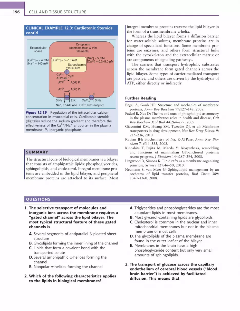
CLINICAL EXAMPLE 12.3: Cardiotonic Steroids
The contraction of the myocardium, like that of skeletal
muscle, is triggered by calcium. The higher the
intracellular calcium concentration, the greater is the force
of contraction.Myocardial cells regulate their intracellular
calcium stores by pumping calcium out of the cell in
exchange for sodium. Thus the extrusion of excess calcium
from the cell requires a sodium gradient (Fig. 12.19).
The sodium gradient depends on the sodium/
potassium pump. Steroidal glycosides from the plant
Digitalis purpurea L. inhibit the sodium/potassium
pump, weaken the sodium gradient, and thereby impair
the removal of calcium from the cell. The excess calcium
is pumped into the sarcoplasmic reticulum, which stores
it for release into the cytoplasm during contraction. This
increases the force of contraction (positive inotropic
effect). Digitalis glycosides are still used for treatment of
congestive heart failure, but they are very toxic because
they cause fatal cardiac arrhythmias at high doses.
Continued
Microvilli
Basolateral membrane
Apic
al m
em
bra
ne
2 K+
3 Na+
Na+,K+-ATPase
ATP
ADP + Pi
Glucose
(facilitateddiffusion)
Desmosome
Zonulaadherens
Tightjunction
Mucosalcell
Intestinallumen
Glucose + Na+
(Na+ – cotransport)
Figure 12.18 Absorption of glucose in the brush border of
the small intestine. The apical (luminal) membrane and the
basolateral (serosal) membrane of the epithelial cells are
physiologically different. The tight junctions between
adjacent cells prevent not only the diffusion of solutes around
the cells but also the lateral diffusion of membrane proteins.
Therefore different sets of carriers are present in the two
parts of the plasma membrane. Pi, Inorganic phosphate.
195Biological Membranes
CLINICAL EXAMPLE 12.3: Cardiotonic Steroids—cont’d
SUMMARY
The structural core of biological membranes is a bilayerthat consists of amphipathic lipids: phosphoglycerides,sphingolipids, and cholesterol. Integral membrane pro-teins are embedded in the lipid bilayer, and peripheralmembrane proteins are attached to its surface. Most
integral membrane proteins traverse the lipid bilayer inthe form of a transmembrane a-helix.
Whereas the lipid bilayer forms a diffusion barrierfor water-soluble solutes, membrane proteins are incharge of specialized functions. Some membrane pro-teins are enzymes, and others form structural linkswith the cytoskeleton and the extracellular matrix orare components of signaling pathways.
The carriers that transport hydrophilic substratesacross the membrane form gated channels across thelipid bilayer. Some types of carrier-mediated transportare passive, and others are driven by the hydrolysis ofATP, either directly or indirectly.
Further Reading
Engel A, Gaub HE: Structure and mechanics of membraneproteins, Annu Rev Biochem 77:127–148, 2008.
Fadeel B, Xue D: The ins and outs of phospholipid asymmetryin the plasma membrane: roles in health and disease, CritRev Biochem Mol Biol 44:264–277, 2009.
Giacomini KM, Huang SM, Tweedie DJ, et al: Membranetransporters in drug development, Nat Rev Drug Discov 9:215–236, 2010.
Kaplan JH: Biochemistry of Na, K-ATPase, Annu Rev Bio-chem 71:511–535, 2002.
Kinoshita T, Fujita M, Maeda Y: Biosynthesis, remodelingand functions of mammalian GPI-anchored proteins:recent progress, J Biochem 144:287–294, 2008.
Lingwood D, Simons K: Lipid rafts as a membrane-organizingprinciple, Science 327:46–50, 2010.
Neumann S, van Meer G: Sphingolipid management by anorchestra of lipid transfer proteins, Biol Chem 389:1349–1360, 2008.
Cytoplasm(contains thick & thin
filaments)
Sarcoplasmicreticulum
Na+, K+-ATPase
ADP, PiATP
ADP, PiATP
Ca2+, Na+-antiport
Ca2+
Ca2+3 Na+ 3 Na+2 K+
Ca2+-ATPase
Extracellularspace
[Ca2+] ≈ 2.4 mM[Na+] ≈ 140 mM
[Na+] ≈ 5 mM[Ca2+] = 0.2–5.0 µM[Ca2+] = 5 –10 mM
Figure 12.19 Regulation of the intracellular calcium
concentration in myocardial cells. Cardiotonic steroids
(digitalis) reduce the sodium gradient and therefore the
effectiveness of the Ca2þ/Naþ antiporter in the plasma
membrane. Pi, Inorganic phosphate.
QUESTIONS
1. The selective transport of molecules andinorganic ions across the membrane requires a“gated channel” across the lipid bilayer. Themost typical structural feature of these gatedchannels is
A. Several segments of antiparallel b-pleated sheetstructure
B. Glycolipids forming the inner lining of the channelC. Lipids that form a covalent bond with the
transported soluteD. Several amphipathic a-helices forming the
channelE. Nonpolar a-helices forming the channel
2. Which of the following characteristics appliesto the lipids in biological membranes?
A. Triglycerides and phosphoglycerides are the mostabundant lipids in most membranes.
B.Most glycerol-containing lipids are glycolipids.C. Cholesterol is common in the nuclear and inner
mitochondrial membranes but not in the plasmamembrane of most cells.
D. The glycolipids of the plasma membrane arefound in the outer leaflet of the bilayer.
E. Membranes in the brain have a highphosphoglyceride content but only very smallamounts of sphingolipids.
3. The transport of glucose across the capillaryendothelium of cerebral blood vessels (“blood-brain barrier”) is achieved by facilitateddiffusion. This means that
196 CELL AND TISSUE STRUCTURE

A. Specific inhibition of cerebral glucose uptake isnot possible
B. The cerebral glucose uptake is always directlyproportional to the concentration gradient forglucose across the endothelium
C. The inhibition of ATP synthesis in the endothelialcells will prevent glucose uptake into the brain
D.As long as glucose is only consumed but notproduced in the brain, the cerebrospinal fluidglucose concentration is always less than theblood glucose concentration
E. There is no upper limit to the amount of glucosethat can be taken up by the brain
4. Many properties of biological membranesdepend on the structure of the lipid bilayer.Typical features of lipid bilayers include
A. Impermeability for small inorganic ions such assodium and protons
B. Rapid exchange of phospholipids between thetwo leaflets of the bilayer
C. High electrical conductivityD. Lack of lateral mobility of membrane lipids at
normal body temperatureE. Permeability for proteins
197Biological Membranes
Chapter 13
THE CYTOSKELETON
As diffusion barriers, biological membranes form theboundary between the cell and its surroundings, andthey form compartments within eukaryotic cells. How-ever, they do not give the cell its shape. They do notprovide structural strength, resistance to mechanicalstress, or resilience to deformation. These propertiesrequire a network of cellular fibers known collectivelyas the cytoskeleton.
In addition to giving the cell its shape and mechanicalstrength, the cytoskeleton has two additional functions:intracellular transport and cell motility. Transport ofproteins and organelles down the axons of neurons,amoeboid movement of phagocytic cells, beating of ciliaand flagella, and muscle contraction all are specializedfunctions of the cytoskeleton.
THE ERYTHROCYTE MEMBRANE IS REINFORCEDBY A SPECTRIN NETWORK
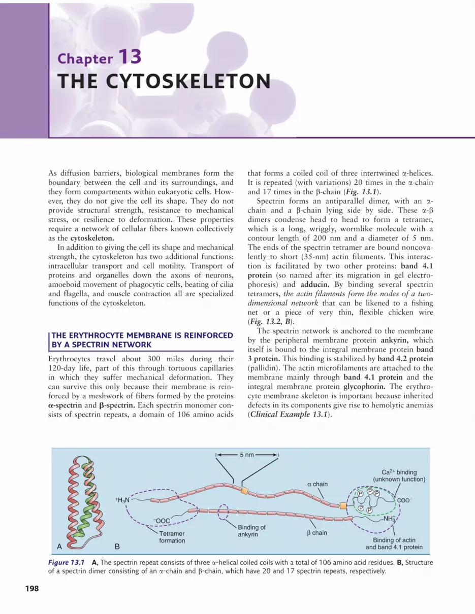
Erythrocytes travel about 300 miles during their120-day life, part of this through tortuous capillariesin which they suffer mechanical deformation. Theycan survive this only because their membrane is rein-forced by a meshwork of fibers formed by the proteinsa-spectrin and b-spectrin. Each spectrin monomer con-sists of spectrin repeats, a domain of 106 amino acids
that forms a coiled coil of three intertwined a-helices.It is repeated (with variations) 20 times in the a-chainand 17 times in the b-chain (Fig. 13.1).
Spectrin forms an antiparallel dimer, with an a-chain and a b-chain lying side by side. These a-bdimers condense head to head to form a tetramer,which is a long, wriggly, wormlike molecule with acontour length of 200 nm and a diameter of 5 nm.The ends of the spectrin tetramer are bound noncova-lently to short (35-nm) actin filaments. This interac-tion is facilitated by two other proteins: band 4.1protein (so named after its migration in gel electro-phoresis) and adducin. By binding several spectrintetramers, the actin filaments form the nodes of a two-dimensional network that can be likened to a fishingnet or a piece of very thin, flexible chicken wire(Fig. 13.2, B).
The spectrin network is anchored to the membraneby the peripheral membrane protein ankyrin, whichitself is bound to the integral membrane protein band3 protein. This binding is stabilized by band 4.2 protein(pallidin). The actin microfilaments are attached to themembrane mainly through band 4.1 protein and theintegral membrane protein glycophorin. The erythro-cyte membrane skeleton is important because inheriteddefects in its components give rise to hemolytic anemias(Clinical Example 13.1).
BA
P
5 nm
PP
P P
–OOC
Tetramerformation
β chain
Ca2+ binding(unknown function)
NH3+
Binding of actinand band 4.1 protein
α chain
+H3N
Binding ofankyrin
COO–
Figure 13.1 A, The spectrin repeat consists of three a-helical coiled coils with a total of 106 amino acid residues. B, Structureof a spectrin dimer consisting of an a-chain and b-chain, which have 20 and 17 spectrin repeats, respectively.
198

CLINICAL EXAMPLE 13.1: Spherocytosisand Elliptocytosis
Hereditary spherocytosis is defined by the
presence of erythrocytes that are spherical instead
of biconcave. Mild anemia can result because the
spherocytes are fragile and are easily trapped and
destroyed in the spleen.
Most patients with hereditary spherocytosis have
primary defects in ankyrin, b-spectrin, or band
3 protein. The amount of spectrin is always reduced
because any spectrin that is not tied into the membrane
skeleton falls prey to proteolytic enzymes during
erythrocyte maturation.
In hereditary elliptocytosis, the erythrocytes are
ellipsoidal rather than spherical. Mutations in the genes
for band 4.1 protein or a-spectrin are the most
common causes.
With a prevalence of 1 in 5000 each, spherocytosis
and elliptocytosis are the most common inherited
hemolytic anemias in many countries. Seventy-five
percent of cases are inherited as autosomal dominant
traits. Splenectomy cures the anemia in most patients.
KERATINS ARE THE MOST IMPORTANTSTRUCTURAL PROTEINS OF EPITHELIAL TISSUES
Epithelial cells receive most of their structural supportfrom keratin, which is one of several classes of interme-diate filaments. In addition to its role in living epithelia,the keratin cytoskeleton of dead cells forms hair, finger-nails, and the horny layer of the skin.
B
A
= Ankyrin
= Adducin
= Band 4.1 protein
= Band 4.2 protein
Glycophorin
Band 3 protein
Actinmicrofilament
Spectrin (tetramer)
Plasma membrane
Figure 13.2 Hypothetical model of the membrane skeleton in red blood cells. A, Transverse section. B, Tangential section.
199The Cytoskeleton

All keratins contain long stretches of a-helix inter-rupted by short nonhelical segments (Fig. 13.3, A). Thetwo different types are the acidic (type I) and the basic(type II) keratins. Each comes in about 15 differentvariants. They form heterodimers, with a type I polypep-tide forming a coiled coil with a type II polypeptide(Fig. 13.3, B). The a-helices of the two keratins make con-tact through hydrophobic amino acid side chains on oneedge of each helix. Typical keratin fibrils contain between12 and 24 of these heterodimers in a staggered array.
Different keratins are expressed in different celltypes. The basal layer of the epidermis forms K14 asthe major type I keratin and K5 as the major type IIkeratin. In the more mature cells of the spinous andgranular layers, keratins K10 and K1 are the majortype I and type II keratins, respectively (Fig. 13.4).
Single-layered epithelia express keratins 18, 19, and/or20 (type I) and keratins 7 and 8 (type II). Various otherkeratin pairs are expressed in the cells that form hairand nails.
Several intermediate filament proteins other than thekeratins are expressed in various cell types (Table 13.1).All of them are dynamic structures that are assembledand disassembled continuously.
The lamins are the only intermediate filament pro-teins that are found in the nucleus rather than thecytoplasm. They form a supporting fiber networkunder the nuclear envelope. During mitosis, the laminsbecome phosphorylated by the cell cycle–induced pro-tein kinase Cdk1. This leads to the disassembly of thefibers and the collapse of the nuclear envelope (seeChapter 18).
B
A
+H3N– –COO–
Nonhelical end domains2A helix
Nonhelical link sequencesNonhelical end domains
2B helix1B helix1A helix
+H3N–Type I keratin
Type II keratin
–COO–
+H3N– –COO–
Figure 13.3 Structure of keratin, the major intermediate filament protein of epithelial tissues. A, Domain structure of a single
polypeptide (type I keratin). The central, mostly a-helical part consists of approximately 310 amino acids. B, Parallel
heterodimer formed from a type I and a type II keratin polypeptide.
KeratinK5/K14
Basal layer(dividing cells)
Anchoring fibrils
(type VII collagen)
Fibroblast
Collagen (type I and III) and elastic fibers
Basal lamina(‘‘basementmembrane’’)
Spinous layer
Granular layer
Horny layer(keratin-filleddead cells)
Epidermis
Dermis
KeratinK1/K10
Spot desmosomes
Figure 13.4 Layers of human skin. The epidermal cells are
held together by numerous spot desmosomes. These spot
desmosomes are attachment points for the intracellular
keratin filaments.
Table 13.1 Major Types of Intermediate Filament Proteins*
Protein Tissue or Cell Type
Keratin Epithelial cells, hair, nails
Vimentin Embryonic tissues, mesenchymal
cells, most cultured cells
Desmin Myocardium, at Z disk in skeletal
muscle
Glial fibrillary acidic
protein
Astrocytes, Schwann cells
Peripherin Neurons of PNS
a-Internexin Neurons of CNS
Neurofilament proteins
(NF-L, NF-M, NF-H)
Neurons of CNS and PNS
Lamin Nucleus of all nucleated cells.
CNS, Central nervous system; PNS, peripheral nervous system.
*All of these proteins have the general structure depicted in Figure 13.3, for
keratin.
200 CELL AND TISSUE STRUCTURE

CLINICAL EXAMPLE 13.2: Skin BlisteringDiseases
A blister forms when the epidermis detaches from the
dermis. Therefore any condition that weakens the
boundary between dermis and epidermis leads to
abnormal blistering.
Epidermolysis bullosa (EB) is a group of dominantly
inherited skin blistering diseases in which even mild
mechanical stress damages the dermal-epidermal
junction. It comes in all degrees of severity, from mild
forms with occasional blistering to severe forms that are
fatal shortly after birth.
The classic forms of EB are caused by point
mutations in the genes of keratin K14 or keratin K5,
which are expressed in the basal cells of the epidermis.
Therefore shear forces easily destroy the basal cell layer
but leave the overlying cells intact.
Point mutations in the genes for K1 and K10,
the major keratins of the spinous and granular
cell layers, cause epidermolytic hyperkeratosis,
a dominantly inherited type of skin disease with
scaling, hyperkeratosis, and blistering.
CLINICAL EXAMPLE 13.3: Laminopathies
Mutations that affect the lamins of the nuclear
lamina, especially the predominant lamin A, cause
an astonishing spectrum of disease. Hutchinson-
Gilford progeria is an extremely rare syndrome of
premature aging, with an incidence of about 1 in
5 million live births. Although normal at birth,
patients present with failure to thrive at 1 or 2 years,
followed by signs of premature aging: hair loss,
osteoporosis, loss of subcutaneous fat, atherosclerosis.
Most patients die of myocardial infarction or stroke at
age 12 to 14 years. The usual mutation in this disease is
a point mutation that activates a cryptic splice site,
creating a messenger RNA (mRNA) that is missing 150
nucleotides and a lamin A protein that is missing 50
amino acids.
Different mutations in the lamin A gene cause
different diseases, including subtypes of limb girdle and
Emery-Dreifuss muscular dystrophies,
cardiomyopathies, lipodystrophies, skin disorders, and
peripheral neuropathy.
The mechanisms by which lamin mutations cause so
many seemingly unrelated syndromes is not known. The
lamins interact not only with each other and with
proteins of the inner nuclear membrane but also with
core histones and many other components of
chromatin. In addition to mechanical fragility of the
nucleus, deranged gene expression is a possible
mechanism.
ACTIN FILAMENTS ARE FORMED FROMGLOBULAR SUBUNITS
All cells contain microfilaments that are formed by thepolymerization of globular actin subunits. Collectively,the six isoforms of actin that occur in different tissuesare among the most abundant types of protein in thehuman body. In most cells, the microfilaments are con-centrated under the plasma membrane where they formthe gel-like cortex of the cytoplasm. When actin mono-mers polymerize into microfilaments, the cytoplasmturns into a gel; when they disassemble, the cytoplasmturns into a viscous liquid.
The loose subunits are called G-actin (G for globular).They have a molecular weight (MW) of 42,000 anda nucleotide binding site that is occupied by ATP orADP. These subunits can polymerize into a filament inwhich two strands are coiled gently around one another(Fig. 13.5). Microfilaments are dynamic structures thatcan be assembled and disassembled continuously.
The two ends of the actin filament are not equiva-lent. At the positive (þ) end, addition and dissociationof actin monomers are fast. At the opposite end, thenegative (!) end, both processes are slow. The boundnucleotide is also important. ATP-actin binds stronglyto other actin monomers and tends to add to the micro-filament, whereas ADP-actin binds weakly and tends tobreak away from the microfilament.
The large majority of free actin monomers in the cyto-plasm contain a bound ATP. This form adds to theþ endof the microfilament. In the microfilament, however, theATP is hydrolyzed. When the concentration of G-actin is
– end
+ end
ATP
ADP
Figure 13.5 Assembly and disassembly of an actin
microfilament. The filament grows at the þ end and is
disassembled at the ! end. , Actin monomer with bound
ADP; , actin monomer with bound ATP.
201The Cytoskeleton
high, the addition of new actin monomers to theþ end isfaster than the hydrolysis of the bound ATP. As a result,the last subunits at the þ end are in the ATP form,whereas the rest of the microfilament is in the ADP form.This filament tends to grow at theþ end and frizzle awayat the ! end.
Cells have a bloatedbureaucracy of proteins to regulatethe formation, growth, and dissolution ofmicrofilaments.Some initiate the formation of a newmicrofilament, someanchor the filaments to membranes or cytoskeletal struc-tures, and others bundle them into networks or parallelarrays (Table 13.2).
Many specialized cellular functions depend onmicrofilaments, including
1. Muscle contraction2. Amoeboid motility3. Phagocytosis4. Contraction of intestinal microvilli5. Formation of the cleavage furrow during mitosis6. Shape change of activated platelets7. Outgrowth of dendrites and axons in developing
neuroblasts
Actin-dependent processes are inhibited by cytochalasinB, a fungal metabolite that prevents actin polymerizationby capping the þ end of the growing microfilament.Phalloidin, another fungal toxin, prevents the depo-lymerization of actin filaments. These agents change
the shapes of many cells, inhibit cell motility, and pre-vent the outgrowth of axons from ganglia.
STRIATED MUSCLE CONTAINS THICKAND THIN FILAMENTS
Amoeboid motion, phagocytosis, and muscle contrac-tion all require the interaction of actin microfilamentswith the ATPase myosin. Various forms of myosin arepresent in most cells, but only the myosin of muscle(myosin II) forms stable fibers. These are the thick fila-ments, in contrast to the thin filaments that are formedfrom actin.
A skeletal muscle fiber has a diameter of 20 to 50 mmand a length of 1 to 40 mm. It is functionally dividedinto myofibrils that run lengthwise through the musclefiber (Fig. 13.6, A). Each myofibril is cylindrical inshape, about 0.6 mm in diameter, and surrounded bycisternae of the sarcoplasmic reticulum.
The myofibrils are organized into sarcomeres bytransverse partitions known as Z disks. Invaginationsof the plasma membrane form the transverse (T)tubules, which reach each sarcomere at the level ofthe Z disk. The T tubules are in close apposition tothe cisternae of the sarcoplasmic reticulum that envelopthe sides of the sarcomere.
The þ ends of the thin filaments (7-nm diameter) areattached to the Z disk, and their capped ! ends pro-trude toward the center of the sarcomere. The thickfilaments (16-nm diameter) are suspended in the centerof the sarcomere, overlapping with the thin filaments.The length of the filaments does not change during con-traction, but the thick and thin filaments slide alongeach other (see Fig. 13.6, B and C). This shortens thesarcomere by about 30%.
The thin filaments of skeletal muscle contain tropo-myosin and troponin in addition to actin. Tropomyosinis a long coiled coil of two a-helical polypeptides thatwinds along the microfilament near the groove betweenthe two actin strands. Troponin consists of the threeglobular subunits Tn-T (tropomyosin binding), Tn-I(inhibitory, actin binding), and Tn-C (calcium binding).This complex is spaced at regular intervals of 38.5 nmalong the thin filament, corresponding to the length ofthe tropomyosin dimer (Fig. 13.7). Troponin makesthe thin filament sensitive to calcium.
MYOSIN IS A TWO-HEADED MOLECULEWITH ATPASE ACTIVITY
The myosin of skeletal muscle contains one pair ofheavy chains (MW 230,000 each) and two pairs of lightchains (MW 16,000 and 20,000) (Fig. 13.8, A). Thecarboxyl terminal 60% of the two heavy chains formsan a-helical coiled coil with a length of 130 nm and adiameter of 2 nm. This coiled coil bundles the myosininto the thick filaments.
Table 13.2 Proteins That Regulate Actin Microfilaments
Protein Function
Thymosin Binds free actin monomers, making them
unavailable for polymerization
Profilin Delivers actin monomers to growing
microfilaments
ARP complex Nucleates microfilaments at the ! end
Formin Binds to the þ end of microfilaments,
promotes elongation
Tropomyosin Strengthens microfilaments, regulates their
length
Caldesmon Prevents myosin from binding to actin/
tropomyosinTroponin
Spectrin
Fodrin Link microfilaments into a gel
Filamin
a-Actinin
Fimbrin Link microfilaments into parallel bundles
Villin
Talin
Myosin-1
Catenin Link microfilaments to the plasma membrane
Vinculin
a-Actinin
Cap Z Caps and stabilizes theþ end ofmicrofilaments
Tropomodulin Caps and stabilizes the! end ofmicrofilaments
Gelsolin Cuts microfilaments
g
g
gg
202 CELL AND TISSUE STRUCTURE

C
B
A
D
A band
H zone
Z disk
Section in Fig. 13.6D
Z disk
I band I band
Myofibrils
Muscle fiber(20–50 µm diameter)
A band (thick filaments)
H zone (thick filaments only) I band (thin filaments only)
Z disk
Cisternae ofthe sarcoplasmicreticulum
Z disk
T tubules
I band
Sarcomere (2.3 µm)
Sarcomere (1.5 µm)
Figure 13.6 Structure of the skeletal muscle fiber. A, Section through a muscle fiber. The fiber has a diameter of
20 to 50 mm and is surrounded by the plasma membrane (sarcolemma). Its nuclei (N, up to 100 per fiber) are located
peripherally, and the mitochondria are interspersed between the myofibrils. More than 100 myofibrils (diameter
0.6–1.0 mm) run the length of the muscle fiber. B, Sarcomere structure of the myofibril in the relaxed state.
C, The sarcomere in the contracted state. D, Cross-section through the overlap zone of thick and thin filaments.
The filaments are neatly packed, with each thick filament surrounded by six thin filaments and each thin filament
surrounded by three thick filaments.
203The Cytoskeleton

Together with the light chains, the amino terminalends of the two heavy chains form two globular heads(see Fig. 13.8, A). The myosin heads hydrolyze ATPvery fast when they are in physical contact with actin,but ADP and inorganic phosphate remain tightly boundto the catalytic site and prevent the access of furtherATP molecules.
The thick filament consists of 300 to 400 myosinmolecules whose heads protrude in all directions (seeFig. 13.8, B). In the middle of the filament the mole-cules are bundled tail to tail; therefore, this central por-tion has no heads. A hinge region in the myosin tailfunctions as a joint, allowing the myosin heads to wagback and forth on the surface of the thick filament.
B
A
Actin Actin
TM
Actin
Tn-T
Tn-C
Tn-I
Relaxed state Contracting
Myosin
Myosin
TM
TM
Actin Actin
Myosin
Myosin
TM
TM
G
Figure 13.7 Thin filaments
of skeletal muscle. A,
Simplified model of thin
filament structure. The
troponin complex (Tn-C, Tn-I,
and Tn-T) binds to a specific
site on the dimeric
tropomyosin (TM) molecule. B,Position of tropomyosin (T) in
the relaxed state (low [Ca2þ])
and during contraction (high
[Ca2þ]). When tropomyosin
moves into the groove
between the actin monomers,
the myosin-binding sites on
actin become exposed.
B
A
–COO–
–COO–
Lightchains
ATP binding
Actin binding
Hinge
130 nm
Figure 13.8 Structure of myosin and the thick filaments. A, Structure of a single myosin molecule. B, Structure of the
thick filaments in skeletal muscle. The globular heads of myosin are on the surface of the filament. Its center consists only
of the fibrous tails and therefore is without globular heads. The packed tails have a diameter of 10.7 nm.
204 CELL AND TISSUE STRUCTURE
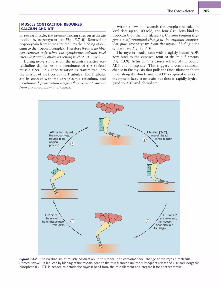
MUSCLE CONTRACTION REQUIRESCALCIUM AND ATP
In resting muscle, the myosin-binding sites on actin areblocked by tropomyosin (see Fig. 13.7, B). Removal oftropomyosin from these sites requires the binding of cal-cium to the troponin complex. Therefore the muscle fibercan contract only when the cytoplasmic calcium levelrises substantially above its resting level of 10!7 mol/L.
During nerve stimulation, the neurotransmitter ace-tylcholine depolarizes the membrane of the skeletalmuscle fiber. This depolarization is transmitted intothe interior of the fiber by the T tubules. The T tubulesare in contact with the sarcoplasmic reticulum, andmembrane depolarization triggers the release of calciumfrom the sarcoplasmic reticulum.
Within a few milliseconds the cytoplasmic calciumlevel rises up to 100-fold, and four Ca2þ ions bind totroponin C on the thin filaments. Calcium binding trig-gers a conformational change in the troponin complexthat pulls tropomyosin from the myosin-binding sitesof actin (see Fig. 13.7, B).
The myosin heads, each with a tightly bound ADP,now bind to the exposed actin of the thin filaments(Fig. 13.9). Actin binding causes release of the boundADP and phosphate. This triggers a conformationalchange in the myosin that pulls the thick filament about7 nm along the thin filament. ATP is required to detachthe myosin head from actin but then is rapidly hydro-lyzed to ADP and phosphate.
ADP
Pi
ADP
Elevated [Ca2+]: myosin head binds to actin
ATP is hydrolyzed,the myosin headreturns to itsoriginalposition
ADP and Pi
are released, the myosin head tilts to a 45° angle
ATP binds, the myosinhead dissociates from actin
1
Pi
AT
P
23
4
Figure 13.9 The mechanism of muscle contraction. In this model, the conformational change of the myosin molecule
(“power stroke”) is induced by binding of the myosin head to the thin filament and the subsequent release of ADP and inorganic
phosphate (Pi). ATP is needed to detach the myosin head from the thin filament and prepare it for another stroke.
205The Cytoskeleton

CLINICAL EXAMPLE 13.4: Rigor Mortis
Binding of the myosin heads to the thin filaments
requires calcium, and their dissociation from the thin
filaments requires ATP. In death, the cytoplasmic Ca2þ
concentration rises while ATP is depleted. Therefore the
myosin heads bind to the thin filaments but cannot
dissociate in the absence of ATP. The resulting stiffness
of the muscles is called rigor mortis.
THE CYTOSKELETON OF SKELETAL MUSCLEIS LINKED TO THE EXTRACELLULAR MATRIX
Dystrophin is a distant relative of spectrin that is foundunder the plasma membrane of skeletal, cardiac, and
smooth muscle. It is a large protein with 3685 aminoacids, containing an actin-binding domain, 24 spectrinrepeats, a calcium-binding domain, and a carboxyl-terminal domain for membrane attachment (Fig. 13.10).
Dystrophin constitutes only 0.002% of the total mus-cle protein, but it is essential for the structural integrityof the muscle fiber. It binds to a set of membrane proteinsknown as the dystroglycan complex. These membraneproteins bind to proteins of the basal lamina. They formthe link between the cytoskeleton and the extracellularmatrix. The connection between cytoskeleton and extra-cellular matrix is essential for the structural integrityof the muscle fiber, and inherited defects in any of itscomponents can cause degenerative muscle diseases (seeFig. 13.10, B, and Clinical Example 13.5).
+H3N–
–OOC–
–COO–
–NH3+
Basallamina
Laminin-2
Sarcolemma
Dystrobrevin
A
B
Syntrophin
Sarcospan
Sarcoglycan
β γ
α
α
βα β γ δ
Actinfilament
Caveolin-3
2
2
1
Dystroglycan
3
Biglycan
Dystrophin
Figure 13.10 Structure of dystrophin, the major component of the membrane skeleton in muscle fibers. Dystrophin is
thought to form an antiparallel dimer. A, Domain structure of dystrophin. , Actin-binding domain; , calcium-binding domain;
, membrane attachment; , spectrin repeat. B, Dystrophin-associated proteins in the sarcolemma. These proteins link the
cytoskeleton to the extracellular matrix. Disease associations: 1 Duchenne and Becker muscular dystrophies; 2 limb girdle
muscular dystrophy; 3 congenital muscular dystrophy.
206 CELL AND TISSUE STRUCTURE
MICROTUBULES CONSIST OF TUBULIN
Microtubules are thick hollow tubes with an outerdiameter of 24 nm, an inner diameter of 14 nm, and alength up to several micrometers. They are importantfor the maintenance of cell shape and for many kindsof intracellular transport. During mitosis, for example,microtubules serve as ropes to pull the chromosomesto opposite poles of the cell, and in neurons they areused as railroad tracks to ship vesicular organelles fromthe perikaryon to the nerve terminals.
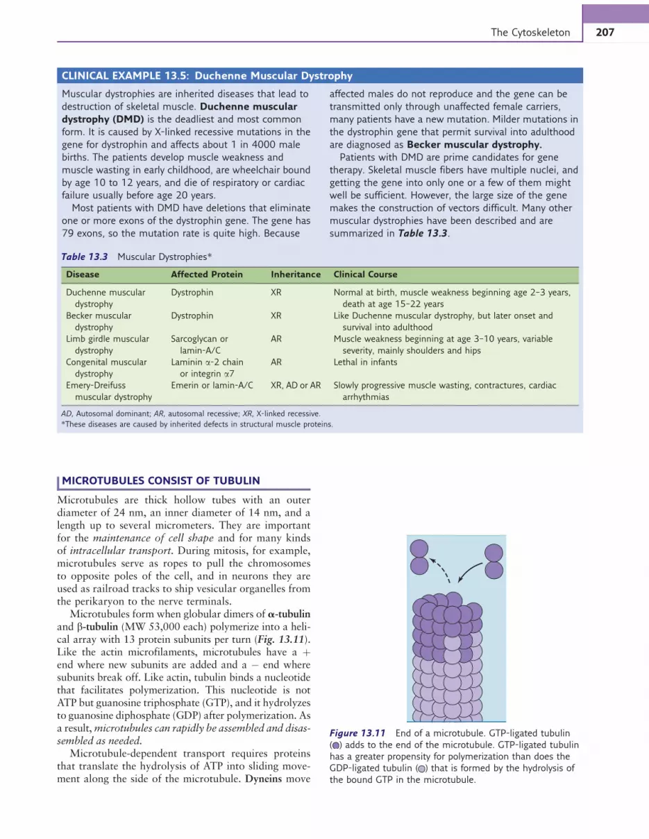
Microtubules form when globular dimers of a-tubulinand b-tubulin (MW 53,000 each) polymerize into a heli-cal array with 13 protein subunits per turn (Fig. 13.11).Like the actin microfilaments, microtubules have a þend where new subunits are added and a ! end wheresubunits break off. Like actin, tubulin binds a nucleotidethat facilitates polymerization. This nucleotide is notATP but guanosine triphosphate (GTP), and it hydrolyzesto guanosine diphosphate (GDP) after polymerization. Asa result,microtubules can rapidly be assembled and disas-sembled as needed.
Microtubule-dependent transport requires proteinsthat translate the hydrolysis of ATP into sliding move-ment along the side of the microtubule. Dyneins move
CLINICAL EXAMPLE 13.5: Duchenne Muscular Dystrophy
Muscular dystrophies are inherited diseases that lead to
destruction of skeletal muscle. Duchenne muscular
dystrophy (DMD) is the deadliest and most common
form. It is caused by X-linked recessive mutations in the
gene for dystrophin and affects about 1 in 4000 male
births. The patients develop muscle weakness and
muscle wasting in early childhood, are wheelchair bound
by age 10 to 12 years, and die of respiratory or cardiac
failure usually before age 20 years.
Most patients with DMD have deletions that eliminate
one or more exons of the dystrophin gene. The gene has
79 exons, so the mutation rate is quite high. Because
affected males do not reproduce and the gene can be
transmitted only through unaffected female carriers,
many patients have a new mutation. Milder mutations in
the dystrophin gene that permit survival into adulthood
are diagnosed as Becker muscular dystrophy.
Patients with DMD are prime candidates for gene
therapy. Skeletal muscle fibers have multiple nuclei, and
getting the gene into only one or a few of them might
well be sufficient. However, the large size of the gene
makes the construction of vectors difficult. Many other
muscular dystrophies have been described and are
summarized in Table 13.3.
Table 13.3 Muscular Dystrophies*
Disease Affected Protein Inheritance Clinical Course
Duchenne muscular
dystrophy
Dystrophin XR Normal at birth, muscle weakness beginning age 2–3 years,
death at age 15–22 years
Becker muscular
dystrophy
Dystrophin XR Like Duchenne muscular dystrophy, but later onset and
survival into adulthood
Limb girdle muscular
dystrophy
Sarcoglycan or
lamin-A/C
AR Muscle weakness beginning at age 3–10 years, variable
severity, mainly shoulders and hips
Congenital muscular
dystrophy
Laminin a-2 chain
or integrin a7
AR Lethal in infants
Emery-Dreifuss
muscular dystrophy
Emerin or lamin-A/C XR, AD or AR Slowly progressive muscle wasting, contractures, cardiac
arrhythmias
AD, Autosomal dominant; AR, autosomal recessive; XR, X-linked recessive.
*These diseases are caused by inherited defects in structural muscle proteins.
Figure 13.11 End of a microtubule. GTP-ligated tubulin
( ) adds to the end of the microtubule. GTP-ligated tubulin
has a greater propensity for polymerization than does the
GDP-ligated tubulin ( ) that is formed by the hydrolysis of
the bound GTP in the microtubule.
207The Cytoskeleton
organelles and proteins from the þ end to the – end ofthe microtubule, and kinesins move things in the oppo-site direction. In the axons of neurons, for example,where all microtubules have the same orientation, kine-sins move vesicles from the cell body toward the nerveterminals, and dyneins move things in the oppositedirection at a speed of up to 25 cm/day (3 mm/s).
Colchicine, the poison of autumn crocus, blocks thepolymerization of tubulin. It inhibits microtubule-dependent processes, including mitosis.
EUKARYOTIC CILIA AND FLAGELLA CONTAINA 9 þ 2 ARRAY OF MICROTUBULES
Cilia and flagella are hairlike cell appendages that arecapable of beating or swirling motion (Fig. 13.12). Cili-ated cells are found in the epithelia of the bronchial tree,upper respiratory tract, and fallopian tubes. The onlyflagellated cell in humans is the sperm cell. Cilia are about6 mm long, and the sperm flagellum is about 40 mm long.
The skin of cilia and flagella is an extension of theplasma membrane, and their skeleton consists of micro-tubules: two single microtubules in the center, and ninedouble microtubules in the periphery. The doublemicrotubules consist of a circular A fiber and a cres-cent-shaped B fiber (Fig. 13.13). Unlike the cytoplasmicmicrotubules that are assembled and dismantled asneeded, the microtubules of cilia and flagella are per-manent structures.
The A subfiber of the doublet microtubules extendstwo arms that are formed by the protein dynein. Theouter dynein arm has three globular heads, and the innerarm has either two or three. The dynein heads use theenergy of ATP hydrolysis to walk along the B subfiberof a neighboring doublet microtubule. Thus dynein playsthe same role in flagellar movement that myosin plays inmuscle contraction. Even the role of ATP is similar in thetwo systems. ATP is needed to dissociate the dyneinheads from the neighboring B subfiber, as it is neededto dissociate the myosin heads from the thin filament.
CLINICAL EXAMPLE 13.6: Immotile CiliaSyndrome
Defects in dynein or other microtubule-associated
proteins of cilia and flagella result in immotile cilia
syndrome, also known as Kartagener syndrome.
Patients with this rare recessively inherited disease
(incidence at birth: 1:20,000 to 1:60,000) suffer from
frequent infections of the bronchi and nasal sinuses.
The epithelium in these locations is covered by a mucus
blanket with a thickness of about 5 mm. Most inhaled
particles and microorganisms get caught on this glue
trap and are moved up the bronchi and the trachea by
coordinated ciliary beating. This “mucus elevator”
removes 30 to 40 g of mucus from the bronchial
system every day.
Male patients with this syndrome are infertile because
their sperm cells are paralyzed. The fertility of affected
females is reduced as well, presumably for lack of ciliary
movement in the fallopian tubes. Themost surprising (and
still unexplained) observation, however, is that 50% of all
patients with immotile cilia syndrome have complete situs
inversus (left-right inversion of the internal organs).
A
B
Fluid movedto the right
Cell surface
1
8
2
3
6
4
57
Fluid moved upward(cell moved downward)
1 23
Figure 13.12 Motile patterns of cilia and flagella. A, Cilium.
B, Flagellum. Sperm flagella beat 30 to 40 times per second.
Figure 13.13 Cross-section through a cilium or flagellum.
All eukaryotic (but not prokaryotic) cilia and flagella have this
general structure.
208 CELL AND TISSUE STRUCTURE

CELLS FORM SPECIALIZED JUNCTIONS WITHOTHER CELLS AND WITH THE EXTRACELLULARMATRIX
The cells of solid tissues form specialized sites of con-tact with neighboring cells and with structural proteinsof the extracellular matrix.
Anchoring junctions link the cytoskeleton eitherwith the cytoskeleton of a neighboring cell or with theextracellular matrix. All anchoring junctions contain atransmembrane protein, which is a protein of the cad-herin family in cell-cell junctions and an integrin incell-matrix junctions. The transmembrane protein con-nects to either microfilaments or intermediate filamentsthrough an adapter protein.
Table 13.4 lists the composition of the four kindsof anchoring junction. The zonula adherens (“beltdesmosome,” Fig. 13.14) is the most characteristicanchoring junction in single-layered epithelia. In intes-tinal mucosal cells, for example, it forms a belt thatencircles the cells. Ordinary desmosomes do not form abelt, but they form spot welds between the cells. Unlikethe zonula adherens, they are linked to intermediate fila-ments rather than actin filaments. In the epidermis, theyconnect the keratin filaments of neighboring cells.
The integrins of hemidesmosomes and focal adhe-sions link the cell to collagen, laminin, fibronectin,and other proteins of the extracellular matrix. Forexample, epidermal cells of the skin are glued to thebasal lamina by hemidesmosomes.
CLINICAL EXAMPLE 13.7: Pemphigus
Whereas some serious skin diseases are inherited (see
Clinical Example 13.2), others are caused by
autoimmunity. In the serious disease pemphigus,
antibodies are formed against a cadherin in the
epidermis. This leads to disruption of the desmosomes
that hold the epidermal cells together, resulting in
blistering and epidermal fragility.
Tight junctions form a continuous belt around thecells of single-layered epithelia. The intestinal epithe-lium, for example, has an apical surface to absorbnutrients from the lumen and a basolateral surface totransfer the nutrients from the cell to the extracellularfluid and the blood. These two surfaces have differentsets of membrane carriers, and those of the apical mem-brane must be prevented from mixing with those of thebasolateral membrane.
The barrier between the two surfaces is formed bythe tight junctions: a network of long strands formedby the integral membrane proteins claudin and occludin(Fig. 13.15). The tight junction forms a seal that pre-vents the diffusion of many water-soluble moleculesthrough the narrow clefts between the epithelial cells.Because the protein strands cut through the lipidbilayer, it also forms the boundary between the apicaland basolateral membrane by preventing the lateraldiffusion of membrane proteins and membrane lipids.
The tightness of tight junctions differs in differenttissues. For example, those in the intestine are 10,000times more permeable for small cations such as sodiumthan are those in the urinary bladder.
Gap junctions are clusters of small channels thatinterconnect the cytoplasm of neighboring cells. Eachhalf-channel is formed by six subunits of the transmem-brane protein connexin (Fig. 13.16). With a diameter of2 nm, gap junctions allow the passage of molecules upto a molecular weight of approximately 1200 D.Because they are permeable to inorganic ions, gap junc-tions can transmit membrane depolarization from cellto cell. Myocardial contraction, for example, dependson the electrical coupling of the cells by gap junctions.
Table 13.4 Four Types of Anchoring Junction
Adherens Junction Desmosome
Contact with Neighboring cell Neighboring cell
Transmembrane
protein
Cadherin Cadherin
Cytoskeletal
attachment
Microfilaments Intermediate
filaments
Intracellular
adapter protein
Catenin, vinculin,
plakoglobin
Desmoplakin,
plakoglobin
Focal Adhesion Hemidesmosome
Contact with Extracellular matrix Extracellular matrix
Transmembrane
protein
Integrin Integrin
Cytoskeletal
attachment
Microfilaments Intermediate
filaments
Intracellular
adapter proteins
Talin, vinculin,
filamin
Plectin
25 nm
Plasma membrane
Stress fibers
Figure 13.14 Belt desmosome (“adherens junction”). The
major adhesive membrane protein is E-cadherin ( ).
E-cadherin is bound to b-catenin or plakoglobin ( ) on the
cytoplasmic side of the membrane, and these are bound to
a-catenin ( ), which interacts with actin microfilaments
(“stress fibers”). Spot desmosomes have a similar molecular
architecture but are linked to intermediate filaments rather
than microfilaments.
209The Cytoskeleton

Gap junctions closewhen the cytoplasmic calcium levelrises. This happens when a cell dies. In this situation, thesurrounding cells have to sever their trade relations withthe dying neighbor to maintain their own ion gradientsand to prevent a unidirectional drain of their metabolites.
Many different connexins occur in human tissuesthat are encoded by separate genes. For example, muta-tions in the gene for connexin-26, which is expressedmainly in the inner ear, are the most common cause ofrecessively inherited deafness.
SUMMARY
Cytoskeletal fibers are formed either by the bundlingof fibrous proteins (keratin, myosin) or by the poly-merization of globular protein subunits (tubulin,actin). They participate in the maintenance of cellshape, cell motility, and intracellular transport.
Microfilaments consist of polymerized actin. Theydetermine the physical consistency of the cytoplasm,interact with proteins of the membrane skeleton such asspectrin and dystrophin, and form links with specializedcell-cell and cell-matrix adhesions. They are essential formost kinds of cell motility and are most prominent inmuscle fibers, where they form the thin filaments.
Intermediate filaments give structural support to thecell. The most important class are the keratins, whichguarantee the integrity of skin and other epithelia.
Microtubules are large hollow tubes of polymerizedtubulin. They participate in intracellular transportprocesses, and they form the skeleton of cilia andflagella.
Further Reading
Arin MJ: The molecular basis of human keratin disorders,Hum Genet 125:355–373, 2009.
Burridge K, Wennerberg K: Rho and rac take center stage,Cell 116:167–179, 2004.
Calderwood DA, Shattil SJ, Ginsberg MH: Integrins and actinfilaments: reciprocal regulation of cell adhesion and signal-ing, J Biol Chem 275:22607–22610, 2000.
Capell BC, Collins FS: Human laminopathies: nuclei gonegenetically awry, Nat Rev Genet 7:940–951, 2006.
Dalkilic I, Kunkel LM: Muscular dystrophies: genes to patho-genesis, Curr Opin Genet Dev 13:231–238, 2003.
Goldman YE: Wag the tail: structural dynamics of actomyo-sin, Cell 93:1–4, 1998.
Michele DE, Campbell KP: Dystrophin-glycoprotein complex:post-translational processing and dystroglycan function,J Biol Chem 278:15457–15460, 2003.
Perez-Moreno M, Jamora C, Fuchs E: Sticky business: orches-trating cellular signals at adherens junctions, Cell 112:535–548, 2003.
Pollard TD, Borisy GG: Cellular motility driven by assemblyand disassembly of actin filaments, Cell 112:453–465,2003.
Rankin J, Ellard S: The laminopathies: a clinical review, ClinGenet 70:261–274, 2006.
Reisler E, Egelman EH: Actin structure and function: what westill do not understand, J Biol Chem 282:36133–36137,2007.
Weis WI, Nelson WJ: Re-solving the cadherin-catenin-actinconundrum, J Biol Chem 281:35593–35597, 2006.
ExtracellularspacePlasma
membranes
Claudin,occludin
Figure 13.15 Tight junction. The junctional proteins
(claudin, occludin) form a tight seal that restricts the diffusion
of water-soluble molecules and ions through the narrow
clefts of extracellular space between the cells. The proteins
prevent the lateral diffusion of membrane proteins and
membrane lipids as well. Therefore the cell can maintain
different protein and lipid compositions on the two sides of
the tight junction.
ConnexinCentral
pore
Extracellularspace
Plasmamembranes
Connexin subunits
Central pore:2-nm diameter
Side view
Cross section
Figure 13.16 Gap junction. In the “open” state, the central
pore allows the passage of solutes with molecular weights up
to about 1200 D.
210 CELL AND TISSUE STRUCTURE
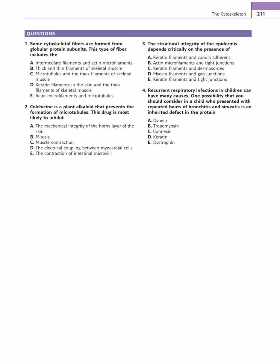
QUESTIONS
1. Some cytoskeletal fibers are formed fromglobular protein subunits. This type of fiberincludes the
A. Intermediate filaments and actin microfilamentsB. Thick and thin filaments of skeletal muscleC. Microtubules and the thick filaments of skeletal
muscleD.Keratin filaments in the skin and the thick
filaments of skeletal muscleE. Actin microfilaments and microtubules
2. Colchicine is a plant alkaloid that prevents theformation of microtubules. This drug is mostlikely to inhibit
A. The mechanical integrity of the horny layer of theskin
B.MitosisC. Muscle contractionD. The electrical coupling between myocardial cellsE. The contraction of intestinal microvilli
3. The structural integrity of the epidermisdepends critically on the presence of
A. Keratin filaments and zonula adherensB. Actin microfilaments and tight junctionsC. Keratin filaments and desmosomesD.Myosin filaments and gap junctionsE. Keratin filaments and tight junctions
4. Recurrent respiratory infections in children canhave many causes. One possibility that youshould consider in a child who presented withrepeated bouts of bronchitis and sinusitis is aninherited defect in the protein
A. DyneinB. TropomyosinC. ConnexinD.KeratinE. Dystrophin
211The Cytoskeleton
Related Documents