www.afm-journal.de FULL PAPER © 2011 WILEY-VCH Verlag GmbH & Co. KGaA, Weinheim 2624 www.MaterialsViews.com wileyonlinelibrary.com Adv. Funct. Mater. 2011, 21, 2624–2632 Dan Y. Lewitus, John Landers, Jonathan R. Branch, Karen L. Smith, Gerardo Callegari, Joachim Kohn,* and Alexander V. Neimark* 1. Introduction It is generally recognized that cortical neural prosthetic devices are limited to 12 months or less before their recording perform- ance deteriorates substantially. [1,2] This limitation lies with the fact that a sustained reactive response develops upon insertion of the probe. This response, known as gliosis, diminishes the long-term performance of devices. [1,3–5] Control of the brain cell response to the inserted device could lead to improvement of its long-term performance. A number of approaches have been considered, both in terms of biochem- istry and design. Examples include the addition of anti-inflammatory agents [5–9] or cell cycle-inhibiting drugs, [9,10] as well as surface modification of silicon substrates. [9,11–13] Nevertheless, these approaches are burdened by the large stiff constructs that will be present in the tissue throughout its lifetime. To circumvent this, an approach has recently emerged relying on two principals. First, these devices should be made of flexible materials. This will reduce the mechan- ical disparity between the device and the brain and possibly reduce development of the chronic glial response, [9,14–18] Second, devices smaller in size, comparable to the neuronal soma, could lead to a reduction in the chronic glial response through the restoration of neuronal and astroglial syn- apses. [17,19,20] Therefore, smaller and more flexible devices may reduce reactive responses and improve long-term performance, e.g., recording of neural signals. In this work, a new material comprised of carbon nanotubes (CNT) and the polysaccharide agarose is presented as a novel solution for the fabrication of neural probes which in turn may reduce the limitations stated above. CNT display unique char- acteristics of superior conductivity, tremendous stiffness and a high aspect ratio. As such, they have been extensively employed in novel materials [21–23] stemming from their ability to absorb strain and induce conductivity. In addition, it has been shown that macroscopic materials made out of CNT are in fact bio- compatible, [24,25] making their inclusion into materials destined for medical applications that much more promising. Indeed, this coincides with reports that the incorporation of carbon nanotubes maintains a material’s structural stability during cell growth. [26] This attribute is coupled with the fact that CNT can support neuron cell growth and differentiation, [24,27] a deci- sive factor for any device that hopes to induce electrical stim- ulation with neurons in vivo. The evolving interest in natural polymers destined for drug delivery and tissue engineering has led to the emergence of new hybrid materials. So far a popular method to fabricate CNT/polymer hybrids is through the tech- nique of wet spinning. [23,24,28–34] Wet spinning has been uti- lized in producing CNT/polymer composite fibers for the last 10 years. [21,23,30] Despite its inherent advantages, the scale up of the production of CNT fibers, specifically those intended for biomedical applications, presents some challenges. This Biohybrid Carbon Nanotube/Agarose Fibers for Neural Tissue Engineering A novel approach for producing carbon nanotube fibers (CNF) composed with the polysaccharide agarose is reported. Current attempts to make CNFs require the use of a polymer or precipitating agent in the coagulating bath that may have negative effects in biomedical applications. It is shown that, by taking advantage of the gelation properties of agarose, one can substitute the bath with distilled water or ethanol and, hence, reduce the complexity associ- ated with alternating the bath components or the use of organic solvents. It is also demonstrated that these CNF can be chemically functionalized to express biological moieties through available free hydroxyl groups in aga- rose. Agarose CNF are not only conductive and nontoxic; in addition, their functionalization is shown to facilitate cell attachment and response both in vitro and in vivo. Our findings suggest that agarose/CNT hybrid materials are excellent candidates for applications involving neural tissue engineering and biointerfacing with the nervous system. DOI: 10.1002/adfm.201002429 Dr. D. Y. Lewitus, J. R. Branch, Prof. J. Kohn The New Jersey Center for Biomaterials Rutgers, the State University of New Jersey 145 Bevier Rd., Piscataway, NJ, 08854, USA E-mail: [email protected] K. L. Smith Wadsworth Center New York State Department of Health 1 Government Center Albany NY, 12201, USA Dr. G. Callegari TRI/Princeton, Princeton, NJ 08542, USA J. Landers, Prof. A. V. Neimark Department of Chemical and Biochemical Engineering Rutgers, State University of New Jersey Piscataway, NJ, 08854, USA E-mail: [email protected]

Welcome message from author
This document is posted to help you gain knowledge. Please leave a comment to let me know what you think about it! Share it to your friends and learn new things together.
Transcript
www.afm-journal.de
FULL
PAPER
2624
www.MaterialsViews.com
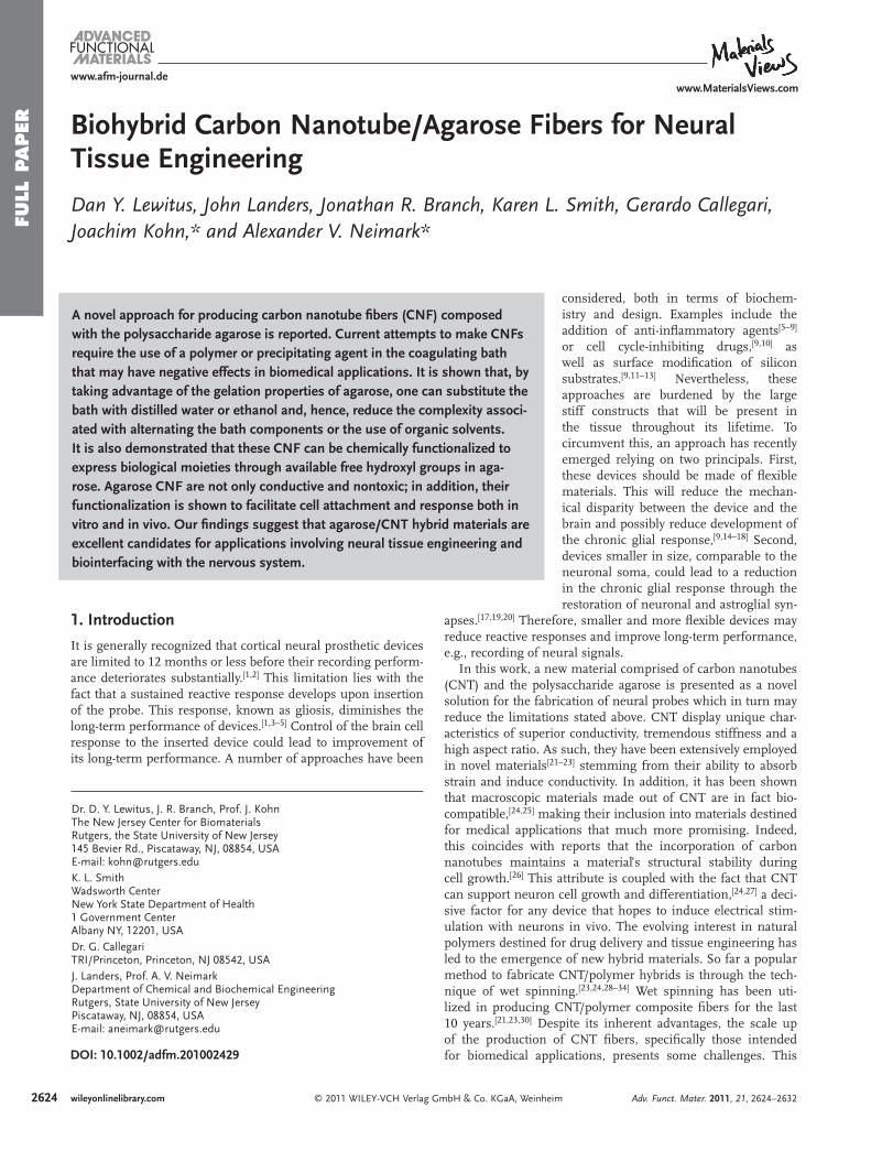
Biohybrid Carbon Nanotube/Agarose Fibers for Neural Tissue Engineering
Dan Y. Lewitus , John Landers , Jonathan R. Branch , Karen L. Smith , Gerardo Callegari , Joachim Kohn , * and Alexander V. Neimark *
A novel approach for producing carbon nanotube fi bers (CNF) composed with the polysaccharide agarose is reported. Current attempts to make CNFs require the use of a polymer or precipitating agent in the coagulating bath that may have negative effects in biomedical applications. It is shown that, by taking advantage of the gelation properties of agarose, one can substitute the bath with distilled water or ethanol and, hence, reduce the complexity associ-ated with alternating the bath components or the use of organic solvents. It is also demonstrated that these CNF can be chemically functionalized to express biological moieties through available free hydroxyl groups in aga-rose. Agarose CNF are not only conductive and nontoxic; in addition, their functionalization is shown to facilitate cell attachment and response both in vitro and in vivo. Our fi ndings suggest that agarose/CNT hybrid materials are excellent candidates for applications involving neural tissue engineering and biointerfacing with the nervous system.
1. Introduction
It is generally recognized that cortical neural prosthetic devices are limited to 12 months or less before their recording perform-ance deteriorates substantially. [ 1 , 2 ] This limitation lies with the fact that a sustained reactive response develops upon insertion of the probe. This response, known as gliosis, diminishes the long-term performance of devices. [ 1 , 3–5 ] Control of the brain cell response to the inserted device could lead to improvement of its long-term performance. A number of approaches have been
© 2011 WILEY-VCH Verlag GmbH & Co. KGaA, Weinheimwileyonlinelibrary.com
DOI: 10.1002/adfm.201002429
Dr. D. Y. Lewitus , J. R. Branch , Prof. J. Kohn The New Jersey Center for Biomaterials Rutgers, the State University of New Jersey 145 Bevier Rd., Piscataway, NJ, 08854, USA E-mail: [email protected] K. L. Smith Wadsworth Center New York State Department of Health 1 Government Center Albany NY, 12201, USA Dr. G. Callegari TRI/Princeton, Princeton, NJ 08542, USA J. Landers , Prof. A. V. Neimark Department of Chemical and Biochemical Engineering Rutgers, State University of New Jersey Piscataway, NJ, 08854, USA E-mail: [email protected]
considered, both in terms of biochem-istry and design. Examples include the addition of anti-infl ammatory agents [ 5–9 ] or cell cycle-inhibiting drugs, [ 9 , 10 ] as well as surface modifi cation of silicon substrates. [ 9 , 11–13 ] Nevertheless, these approaches are burdened by the large stiff constructs that will be present in the tissue throughout its lifetime. To circumvent this, an approach has recently emerged relying on two principals. First, these devices should be made of fl exible materials. This will reduce the mechan-ical disparity between the device and the brain and possibly reduce development of the chronic glial response, [ 9 , 14–18 ] Second, devices smaller in size, comparable to the neuronal soma, could lead to a reduction in the chronic glial response through the restoration of neuronal and astroglial syn-
apses. [ 17 , 19 , 20 ] Therefore, smaller and more fl exible devices may reduce reactive responses and improve long-term performance, e.g., recording of neural signals.
In this work, a new material comprised of carbon nanotubes (CNT) and the polysaccharide agarose is presented as a novel solution for the fabrication of neural probes which in turn may reduce the limitations stated above. CNT display unique char-acteristics of superior conductivity, tremendous stiffness and a high aspect ratio. As such, they have been extensively employed in novel materials [ 21–23 ] stemming from their ability to absorb strain and induce conductivity. In addition, it has been shown that macroscopic materials made out of CNT are in fact bio-compatible, [ 24 , 25 ] making their inclusion into materials destined for medical applications that much more promising. Indeed, this coincides with reports that the incorporation of carbon nanotubes maintains a material’s structural stability during cell growth. [ 26 ] This attribute is coupled with the fact that CNT can support neuron cell growth and differentiation, [ 24 , 27 ] a deci-sive factor for any device that hopes to induce electrical stim-ulation with neurons in vivo. The evolving interest in natural polymers destined for drug delivery and tissue engineering has led to the emergence of new hybrid materials. So far a popular method to fabricate CNT/polymer hybrids is through the tech-nique of wet spinning. [ 23 , 24 , 28–34 ] Wet spinning has been uti-lized in producing CNT/polymer composite fi bers for the last 10 years. [ 21 , 23 , 30 ] Despite its inherent advantages, the scale up of the production of CNT fi bers, specifi cally those intended for biomedical applications, presents some challenges. This
Adv. Funct. Mater. 2011, 21, 2624–2632
FULL P
APER
www.afm-journal.dewww.MaterialsViews.com
problem is apparent when a polymer, such as PVA, is utilized as either the bath component or the dispersant. The former leads to several shortcomings which make the process diffi -cult to scale commercially. The primary concern arises when the gel ribbon becomes suspended at the spinning position. Thus it is necessary to continually raise the tip of the spinning bath to prevent the ribbon from crashing into itself. However, when the polymer is absent as a component in the coagulation bath, several process variables are eliminated, which allows for a simpler system to study. Several authors have demonstrated this practicality by using the polymer as the dispersant. [ 28 , 30 , 31 , 34 ] This provides several technical advantages, including the fact that the spun ribbon can be reeled up onto a spool. Alternative methods have been proposed, which lead to a cleaner product and less expensive process. [ 30 ] The use of polymeric hydrogels has a certain advantage due in part to their ability to imitate the natural extra cellular matrix (ECM), thus promoting cell growth. [ 33 , 35 ] Lastly, deciphering the composition of the fi ber becomes easier as it is dependent only on the initial concen-trations of the dispersion; in contrast, the composition of the fi ber after spinning in a polymer bath will be dependent on the polymer concentration and adsorption kinetics.
In this process, CNT are dispersed with the aid of a surfactant or polymer by non-covalent means. The literature is scattered with examples of polymers which aid in this process. [ 32 , 34 ] Some of these materials are based on the use of natural polymers or nat-urally based dispersants that are known to be biocompatible, such as chitosan, hyaluronic acid, DNA, and chondroitin sulfate. [ 29 , 31 ] However, both chitosan and hyaluronic acid are biodegradable and are undesirable for long-term indwelling recording electrodes. One alternative which is absent from the current list of proposed polymers is the naturally occurring polysaccharide agarose.
Agarose is an algae derived linear polysaccharide hydrogel possessing a sub-micrometer pore structure. It is a poly (1 → 4)-3,6-anhydro- α -l-galactopyranosyl-(1 → 3)- β -d-galactopyranose) with thermoreversive properties. Although it is not cell-adherent, due to its benign and biocompatible nature, it is commonly used as a nonadhesive substrate for in vitro cell studies. [ 36 ] In addition, agarose has several distinct advantages over other natural polymers: i) its thermal dependant hydrogel properties allow it to be easily malleable into different shapes and forms without the use of additional reagents or organic solvents. ii) Unlike extracellular matrix based polymers, spe-cifi c proteins or DNA, it lacks native ligands and is thus inert to mammalian cells. [ 37 ] iii) Through available primary and secondary hydroxyl groups, agarose can be chemically modi-fi ed. This leads to a functionalization through grafting of pro-teins, peptides, and glycogens to the polysaccharide backbone, allowing it to be specifi cally tailored for various biorelevant applications. [ 38–40 ] iv) The addition of such molecules can alter not only its biocompatible properties, but its mechanical proper-ties as well. v) Its high surface to volume ratio and porosity [ 37 , 41 ] combined with its hydrophilic nature allows for a more effective penetration of cells during seeding while supporting delivery of nutrients and metabolites to these cells. [ 38 , 39 ] Carrying out such modifi cations will result in a substantial increase in cell attachment, continuous support of 3D neural cell cultures, the ability to orient cell migration, and specifi cally enhance neurite extension with the grafting of neuron conductive constituents
© 2011 WILEY-VCH Verlag GAdv. Funct. Mater. 2011, 21, 2624–2632
such as laminin or various oligopeptides. [ 38 , 39 ] vi) Unlike other biopolymers, it is non-biodegradable, and will, therefore, allow for long term performance and integration of the carbon nano-tubes and avoid disintegration of the fabricated structures. [ 37 ] vii) Agarose is a cheap and abundant polysaccharide, sourced from plants (algae) and can be grown in highly controlled envi-ronments. This is compared with the prohibitive cost associated with making fi bers with either DNA or hyaluronic acid. Due to these reasons, agarose has found use in the fi eld of neural engi-neering and nerve regeneration where it has been suggested as the primary support construct in nerve guide conduits. [ 37 , 42 ]
In this work, we aim to combine three elements that have not yet been adjoined, the ease of wet spinning as a fabrication tech-nique, with the reinforcing and conductance properties of CNTs, along with the gelation and functionalization potential of agarose, to create a continuous, electro- and neuro conductive biohybrid nanocomposite fi ber. The result is a fi ber that is stiff when dry yet fl exible when hydrated. We believe the impact of this work will provide a foundation for long-term neural recording devices as an alternative to silicone/metal based electrodes in the quest to evade gliosis and performance degradation.
2. Results and Discussion
2.1. Fiber Fabrication
Fibers were fabricated by two methods: wet spinning and molding the fi ber in a hollow tube. In the former, the liquid dispersion used to make the fi bers was injected into a rotating bath, with the rotation velocity larger than the injecting velocity. Upon entering the bath, the dispersion displays an axial dif-fusion which is inhibited by two factors. First, the stretching imposed by the rotating velocity fi eld and second, by the gela-tion of the agarose/CNT composite. By controlling the speed and the rheology of the injecting dispersion and the rotating solution, the width and morphology of the fi ber precursor can be controlled. Therefore, a greater rotation speed may result in better alignment of the single walled carbon nanotubes (SWNTs) encapsulated in the agarose gel matrix.
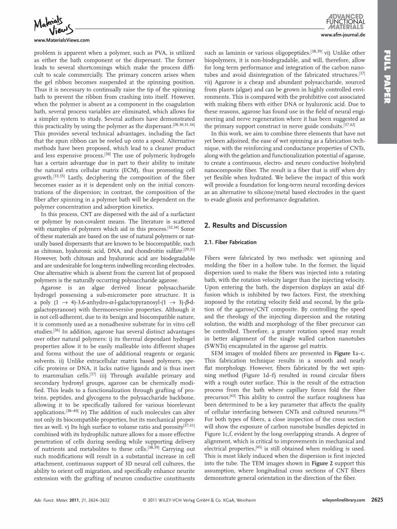
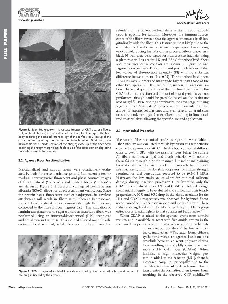
SEM images of molded fi bers are presented in Figure 1 a–c. This fabrication technique results in a smooth and nearly fl at morphology. However, fi bers fabricated by the wet spin-ning method (Figure 1 d–f) resulted in round circular fi bers with a rough outer surface. This is the result of the extraction process from the bath where capillary forces fold the fi ber precursor. [ 43 ] This ability to control the surface roughness has been determined to be a key parameter that affects the quality of cellular interfacing between CNTs and cultured neurons. [ 44 ] For both types of fi bers, a close inspection of the cross section will show the exposure of carbon nanotube bundles depicted in Figure 1 c,f, evident by the long overlapping strands. A degree of alignment, which is critical to improvements in mechanical and electrical properties, [ 45 ] is still obtained when molding is used. This is most likely induced when the dispersion is fi rst injected into the tube. The TEM images shown in Figure 2 support this assumption, where longitudinal cross sections of CNT fi bers demonstrate general orientation in the direction of the fi ber.
mbH & Co. KGaA, Weinheim 2625wileyonlinelibrary.com
FULL
PAPER
2626
www.afm-journal.dewww.MaterialsViews.com
Figure 1 . Scanning electron microscopy images of CNT agarose fi bers. Left, molded fi bers a) cross section of the fi ber, b) close up of the fi ber body depicting the smooth morphology of the surface, c) Close up of the cross section depicting the carbon nanotube bundles. Right, wet spun agarose fi bers: d) cross section of the fi ber, e) close up of the fi ber body depicting the rough morphology f) close up of the cross section depicting the carbon nanotube bundles.
2.2. Agarose Fiber Functionalization
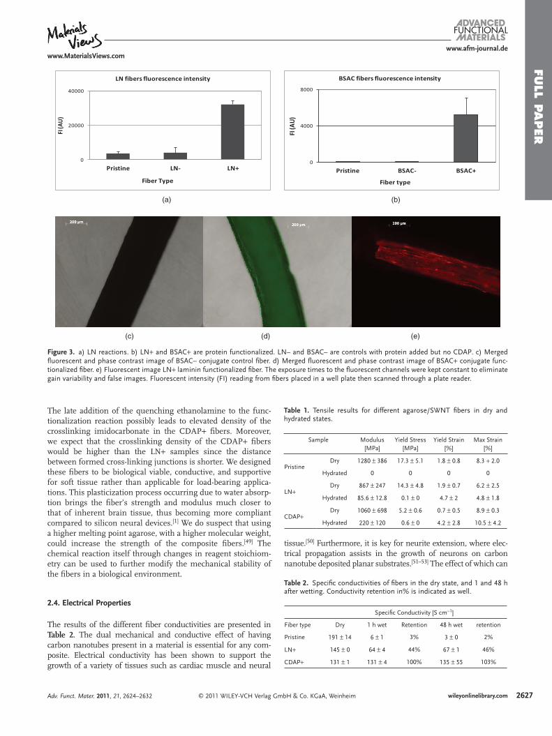
Functionalized and control fi bers were qualitatively evalu-ated by both fl uorescent microscopy and fl uorescent intensity reading. Representative fl uorescent and phase contrast images of functionalized (“protein” + ) and control fi bers (“protein”−) are shown in Figure 3 . Fluorescein conjugated bovine serum albumin (BSAC) allows for direct attachment verifi cation. Since the protein has a fl uorescent marker conjugated, its covalent attachment will result in fi bers with inherent fl uorescence. Indeed, functionalized fi bers demonstrate high fl uorescence, compared to the control fi ber (Figures 3 a,b). The validation of laminin attachment to the agarose carbon nanotube fi bers was performed using an immunohistochemical (IHC) technique and are shown in Figure 3 c. This method allowed not only vali-dation of the attachment, but also to some extent confi rmed the
© 2011 WILEY-VCH Verlag Gwileyonlinelibrary.com
Figure 2 . TEM images of molded fi bers demonstrating fi ber orientation molding indicated by the arrows.
retention of the protein conformation, as the primary antibody used is specifi c for laminin. Moreover, the immunofl uores-cence of the fi bers reveals that the agarose orientates itself lon-gitudinally with the fi ber. This feature is most likely due to the elongation of the dispersion when it experiences the rotating velocity fi eld during the fabrication process. Fibers placed in a black 96 well plate were tested for fl uorescence intensity using a plate reader. Results for LN and BSAC functionalized fi bers and their prospective controls are shown in Figure 3 d and Figure 3 e respectively. The control and pristine fi bers exhibited low values of fl uorescence intensity (FI) with no statistical difference between them (P > 0.05). The functionalized fi bers FI values were 2 orders of magnitude higher than those of the other two types (P < 0.05), indicating successful functionaliza-tion. The actual quantifi cation of the functionalized sites by the CDAP chemical reaction and amount of bound proteins was not performed, though could be possible based on the barbituric acid assay. [ 40 ] These fi ndings emphasize the advantage of using agarose. It is a “clean slate” for biochemcal manipulation. This allows for specifi c cellular cues and even several different cues to be covalently conjugated to the fi bers, resulting in functional-ized material thus allowing for specifi c use and application.
2.3. Mechanical Properties
The results of the mechanical tensile testing are shown in Table 1 . Fiber stability was evaluated through hydration at a temperature close to the agarose mp (50 ° C). The dry fi bers exhibited stiffness close to over 1 GPa, with the pristine fi bers being the stiffest. All fi bers exhibited a rigid and tough behavior, with none of them failing through a brittle manner, but rather maintaining their strength past the yield point until complete failure. Such intrinsic strength in the dry state surpasses the critical strength required for pial penetration, reported to be (0.3–1.3 MPa). Moreover, the low strain values allow for minimal collateral damage during insertion process. [ 46 ] Once hydrated, only the CDAP functionalized fi bers (LN + and CDAP + ) exhibited enough mechanical integrity to be evaluated and studied for their tensile properties). A 90% and 80% drop in the elastic modulus for the LN + and CDAP + respectively was observed for hydrated fi bers, accompanied with a decrease in yield and maximal strain. These reduced strength values in the kPa range bring the fi ber’s prop-erties closer (if still higher) to that of inherent brain tissue. [ 47 ]
When CDAP is added to the agarose, cyano-ester termini results, and is available to react with free amide groups in the reaction. Competing reaction exists, where either a carbamate
or an imidocarbonate can be formed from
mbH & Co. KGaA, We
in the direction of
the cyanate ester. [ 40 ] The latter forms either a cyclic bond within an agarose backbone or a crosslink between adjacent polymer chains, thus resulting in a slightly crosslinked and more stable CNT fi ber (CDAP + ). When laminin, a high molecular weight pro-tein is added to the reaction (LN + ), there is increased coupling, principally due to the available ε -amines of surface lysine. This in turn creates the formation of an isourea bond resulting in the observed CNF stability. [ 48 ]
inheim Adv. Funct. Mater. 2011, 21, 2624–2632
FULL P
APER
www.afm-journal.dewww.MaterialsViews.com
Figure 3 . a) LN reactions. b) LN + and BSAC + are protein functionalized. LN– and BSAC– are controls with protein added but no CDAP. c) Merged fl uorescent and phase contrast image of BSAC– conjugate control fi ber. d) Merged fl uorescent and phase contrast image of BSAC + conjugate func-tionalized fi ber. e) Fluorescent image LN + laminin functionalized fi ber. The exposure times to the fl uorescent channels were kept constant to eliminate gain variability and false images. Fluorescent intensity (FI) reading from fi bers placed in a well plate then scanned through a plate reader.
0
20000
40000
Pristine LN- LN+
FI (A
U)
Fiber Type
LN fibers fluorescence intensity
0
4000
8000
Pristine BSAC- BSAC+
FI (A
U)
Fiber type
BSAC fibers fluorescence intensity
(a) (b)
(c) (d) (e)
Table 1. Tensile results for different agarose/SWNT fi bers in dry and hydrated states.
Sample Modulus [MPa]
Yield Stress [MPa]
Yield Strain [%]
Max Strain [%]
PristineDry 1280 ± 386 17.3 ± 5.1 1.8 ± 0.8 8.3 + 2.0
Hydrated 0 0 0 0
LN + Dry 867 ± 247 14.3 ± 4.8 1.9 ± 0.7 6.2 ± 2.5
Hydrated 85.6 ± 12.8 0.1 ± 0 4.7 ± 2 4.8 ± 1.8
CDAP + Dry 1060 ± 698 5.2 ± 0.6 0.7 ± 0.5 8.9 ± 0.3
Hydrated 220 ± 120 0.6 ± 0 4.2 ± 2.8 10.5 ± 4.2
Table 2. Specifi c conductivities of fi bers in the dry state, and 1 and 48 h after wetting. Conductivity retention in% is indicated as well.
Specifi c Conductivity [S cm − 1 ]
Fiber type Dry 1 h wet Retention 48 h wet retention
Pristine 191 ± 14 6 ± 1 3% 3 ± 0 2%
LN + 145 ± 0 64 ± 4 44% 67 ± 1 46%
CDAP + 131 ± 1 131 ± 4 100% 135 ± 55 103%
The late addition of the quenching ethanolamine to the func-tionalization reaction possibly leads to elevated density of the crosslinking imidocarbonate in the CDAP + fi bers. Moreover, we expect that the crosslinking density of the CDAP + fi bers would be higher than the LN + samples since the distance between formed cross-linking junctions is shorter. We designed these fi bers to be biological viable, conductive, and supportive for soft tissue rather than applicable for load-bearing applica-tions. This plasticization process occurring due to water absorp-tion brings the fi ber’s strength and modulus much closer to that of inherent brain tissue, thus becoming more compliant compared to silicon neural devices. [ 1 ] We do suspect that using a higher melting point agarose, with a higher molecular weight, could increase the strength of the composite fi bers. [ 49 ] The chemical reaction itself through changes in reagent stoichiom-etry can be used to further modify the mechanical stability of the fi bers in a biological environment.
2.4. Electrical Properties
The results of the different fi ber conductivities are presented in Table 2 . The dual mechanical and conductive effect of having carbon nanotubes present in a material is essential for any com-posite. Electrical conductivity has been shown to support the growth of a variety of tissues such as cardiac muscle and neural
© 2011 WILEY-VCH Verlag GmAdv. Funct. Mater. 2011, 21, 2624–2632
tissue. [ 50 ] Furthermore, it is key for neurite extension, where elec-trical propagation assists in the growth of neurons on carbon nano tube deposited planar substrates. [ 51–53 ] The effect of which can
bH & Co. KGaA, Weinheim 2627wileyonlinelibrary.com
FULL
PAPER
2628
www.afm-journal.dewww.MaterialsViews.com
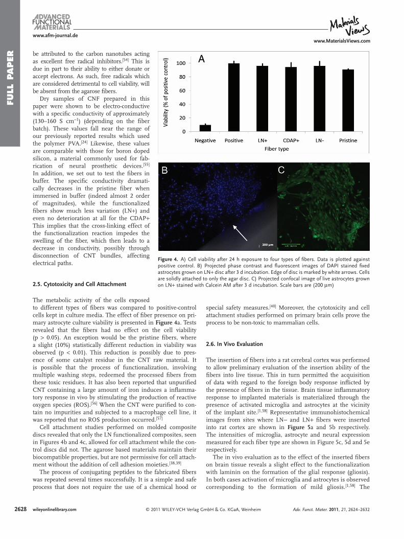
Figure 4 . A) Cell viability after 24 h exposure to four types of fi bers. Data is plotted against positive control. B) Projected phase contrast and fl uorescent images of DAPI stained fi xed astrocytes grown on LN + disc after 3 d incubation. Edge of disc is marked by white arrows. Cells are solidly attached to only the agar disc. C) Projected confocal image of live astrocytes grown on LN + stained with Calcein AM after 3 d incubation. Scale bars are (200 μ m)
be attributed to the carbon nanotubes acting as excellent free radical inhibitors. [ 54 ] This is due in part to their ability to either donate or accept electrons. As such, free radicals which are considered detrimental to cell viability, will be absent from the agarose fi bers.
Dry samples of CNF prepared in this paper were shown to be electro-conductive with a specifi c conductivity of approximately (130–160 S cm − 1 ) (depending on the fi ber batch). These values fall near the range of our previously reported results which used the poly mer PVA. [ 24 ] Likewise, these values are comparable with those for boron doped silicon, a material commonly used for fab-rication of neural prosthetic devices. [ 55 ] In addition, we set out to test the fi bers in buffer. The specifi c conductivity dramati-cally decreases in the pristine fi ber when immersed in buffer (indeed almost 2 order of magnitudes), while the functionalized fi bers show much less variation (LN + ) and even no deterioration at all for the CDAP + This implies that the cross-linking effect of the functionalization reaction impedes the swelling of the fi ber, which then leads to a decrease in conductivity, possibly through disconnection of CNT bundles, affecting electrical paths.
2.5. Cytotoxicity and Cell Attachment
The metabolic activity of the cells exposed to different types of fi bers was compared to positive-control cells kept in culture media. The effect of fi ber presence on pri-mary astrocyte culture viability is presented in Figure 4 a. Tests revealed that the fi bers had no effect on the cell viability (p > 0.05). An exception would be the pristine fi bers, where a slight (10%) statistically different reduction in viability was observed (p < 0.01). This reduction is possibly due to pres-ence of some catalyst residue in the CNT raw material. It is possible that the process of functionalization, involving multiple washing steps, redeemed the processed fi bers from these toxic residues. It has also been reported that unpurifi ed CNT containing a large amount of iron induces a infl amma-tory response in vivo by stimulating the production of reactive oxygen species (ROS). [ 56 ] When the CNT were purifi ed to con-tain no impurities and subjected to a macrophage cell line, it was reported that no ROS production occurred. [ 57 ]
Cell attachment studies performed on molded composite discs revealed that only the LN functionalized composites, seen in Figures 4 b and 4 c, allowed for cell attachment while the con-trol discs did not. The agarose based materials maintain their biocompatible properties, but are not permissive for cell attach-ment without the addition of cell adhesion moieties. [ 38 , 39 ]
The process of conjugating peptides to the fabricated fi bers was repeated several times successfully. It is a simple and safe process that does not require the use of a chemical hood or
© 2011 WILEY-VCH Verlag wileyonlinelibrary.com
special safety measures. [ 40 ] Moreover, the cytotoxicity and cell attachment studies performed on primary brain cells prove the process to be non-toxic to mammalian cells.
2.6. In Vivo Evaluation
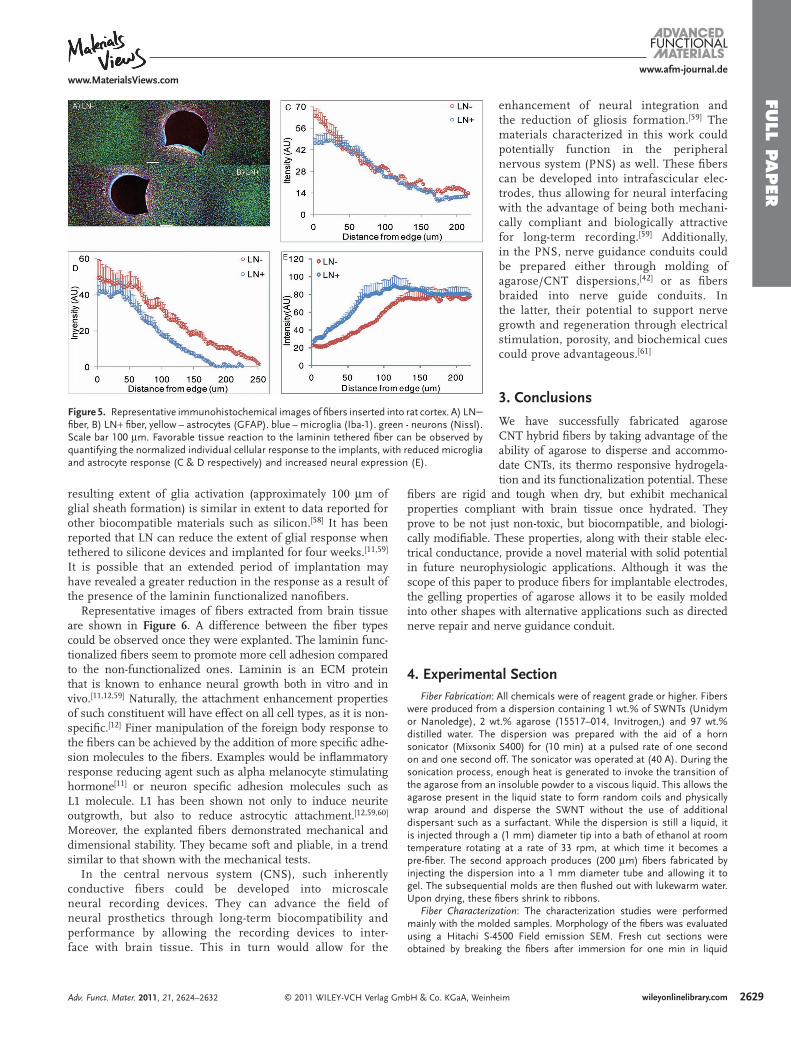
The insertion of fi bers into a rat cerebral cortex was performed to allow preliminary evaluation of the insertion ability of the fi bers into live tissue. This in turn permitted the acquisition of data with regard to the foreign body response infl icted by the presence of fi bers in the tissue. Brain tissue infl ammatory response to implanted materials is materialized through the presence of activated microglia and astrocytes at the vicinity of the implant site. [ 1 , 58 ] Representative immunohistochemical images from sites where LN− and LN + fi bers were inserted into rat cortex are shown in Figure 5 a and 5 b respectively. The intensities of microglia, astrocyte and neural expression measured for each fi ber type are shown in Figure 5 c, 5 d and 5 e respectively.
The in vivo evaluation as to the effect of the inserted fi bers on brain tissue reveals a slight effect to the functionalization with laminin on the formation of the glial response (gliosis). In both cases activation of microglia and astrocytes is observed corresponding to the formation of mild gliosis. [ 1 , 58 ] The
GmbH & Co. KGaA, Weinheim Adv. Funct. Mater. 2011, 21, 2624–2632
FULL P
APER
www.afm-journal.dewww.MaterialsViews.com
Figure 5 . Representative immunohistochemical images of fi bers inserted into rat cortex. A) LN− fi ber, B) LN + fi ber, yellow – astrocytes (GFAP). blue – microglia (Iba-1). green - neurons (Nissl). Scale bar 100 μ m. Favorable tissue reaction to the laminin tethered fi ber can be observed by quantifying the normalized individual cellular response to the implants, with reduced microglia and astrocyte response (C & D respectively) and increased neural expression (E).
resulting extent of glia activation (approximately 100 μ m of glial sheath formation) is similar in extent to data reported for other biocompatible materials such as silicon. [ 58 ] It has been reported that LN can reduce the extent of glial response when tethered to silicone devices and implanted for four weeks. [11,59 ] It is possible that an extended period of implantation may have revealed a greater reduction in the response as a result of the presence of the laminin functionalized nanofi bers.
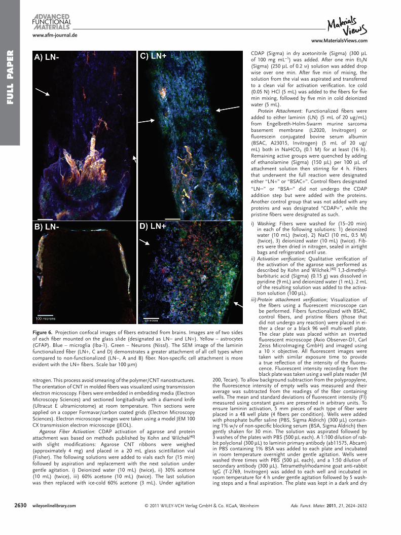
Representative images of fi bers extracted from brain tissue are shown in Figure 6 . A difference between the fi ber types could be observed once they were explanted. The laminin func-tionalized fi bers seem to promote more cell adhesion compared to the non-functionalized ones. Laminin is an ECM protein that is known to enhance neural growth both in vitro and in vivo. [ 11 ,12, 59 ] Naturally, the attachment enhancement properties of such constituent will have effect on all cell types, as it is non-specifi c. [ 12 ] Finer manipulation of the foreign body response to the fi bers can be achieved by the addition of more specifi c adhe-sion molecules to the fi bers. Examples would be infl ammatory response reducing agent such as alpha melanocyte stimulating hormone [ 11 ] or neuron specifi c adhesion molecules such as L1 molecule. L1 has been shown not only to induce neurite outgrowth, but also to reduce astrocytic attachment. [ 12 , 59 , 60 ] Moreover, the explanted fi bers demonstrated mechanical and dimensional stability. They became soft and pliable, in a trend similar to that shown with the mechanical tests.
In the central nervous system (CNS), such inherently conductive fi bers could be developed into microscale neural recording devices. They can advance the fi eld of neural prosthetics through long-term biocompatibility and performance by allowing the recording devices to inter-face with brain tissue. This in turn would allow for the
© 2011 WILEY-VCH Verlag GmbH & Co. KGaA, WeiAdv. Funct. Mater. 2011, 21, 2624–2632
enhancement of neural integration and the reduction of gliosis formation. [ 59 ] The materials characterized in this work could potentially function in the peripheral nervous system (PNS) as well. These fi bers can be developed into intrafascicular elec-trodes, thus allowing for neural interfacing with the advantage of being both mechani-cally compliant and biologically attractive for long-term recording. [ 59 ] Additionally, in the PNS, nerve guidance conduits could be prepared either through molding of agarose/CNT dispersions, [ 42 ] or as fi bers braided into nerve guide conduits. In the latter, their potential to support nerve growth and regeneration through electrical stimulation, porosity, and biochemical cues could prove advantageous. [ 61 ]
3. Conclusions
We have successfully fabricated agarose CNT hybrid fi bers by taking advantage of the ability of agarose to disperse and accommo-date CNTs, its thermo responsive hydrogela-tion and its functionalization potential. These
fi bers are rigid and tough when dry, but exhibit mechanical properties compliant with brain tissue once hydrated. They prove to be not just non-toxic, but biocompatible, and biologi-cally modifi able. These properties, along with their stable elec-trical conductance, provide a novel material with solid potential in future neurophysiologic applications. Although it was the scope of this paper to produce fi bers for implantable electrodes, the gelling properties of agarose allows it to be easily molded into other shapes with alternative applications such as directed nerve repair and nerve guidance conduit.
4. Experimental Section Fiber Fabrication : All chemicals were of reagent grade or higher. Fibers
were produced from a dispersion containing 1 wt.% of SWNTs (Unidym or Nanoledge), 2 wt.% agarose (15517–014, Invitrogen,) and 97 wt.% distilled water. The dispersion was prepared with the aid of a horn sonicator (Mixsonix S400) for (10 min) at a pulsed rate of one second on and one second off. The sonicator was operated at (40 A). During the sonication process, enough heat is generated to invoke the transition of the agarose from an insoluble powder to a viscous liquid. This allows the agarose present in the liquid state to form random coils and physically wrap around and disperse the SWNT without the use of additional dispersant such as a surfactant. While the dispersion is still a liquid, it is injected through a (1 mm) diameter tip into a bath of ethanol at room temperature rotating at a rate of 33 rpm, at which time it becomes a pre-fi ber. The second approach produces (200 μ m) fi bers fabricated by injecting the dispersion into a 1 mm diameter tube and allowing it to gel. The subsequential molds are then fl ushed out with lukewarm water. Upon drying, these fi bers shrink to ribbons.
Fiber Characterization : The characterization studies were performed mainly with the molded samples. Morphology of the fi bers was evaluated using a Hitachi S-4500 Field emission SEM. Fresh cut sections were obtained by breaking the fi bers after immersion for one min in liquid
nheim 2629wileyonlinelibrary.com
FULL
PAPER
2630
www.afm-journal.dewww.MaterialsViews.com
Figure 6 . Projection confocal images of fi bers extracted from brains. Images are of two sides of each fi ber mounted on the glass slide (designated as LN− and LN + ). Yellow – astrocytes (GFAP). Blue – microglia (Iba-1). Green – Neurons (Nissl). The SEM image of the laminin functionalized fi ber (LN + , C and D) demonstrates a greater attachment of all cell types when compared to non-functionalized (LN−, A and B) fi ber. Non-specifi c cell attachment is more evident with the LN + fi bers. Scale bar 100 μ m)
nitrogen. This process avoid smearing of the polymer/CNT nanostructures. The orientation of CNT in molded fi bers was visualized using transmission electron microscopy. Fibers were embedded in embedding media (Electron Microscopy Sciences) and sectioned longitudinally with a diamond knife (Ultracut E ultramicrotome) at room temperature. Thin sections were applied on a copper Formavar/carbon coated grids (Electron Microscopy Sciences). Electron microscope images were taken using a model JEM 100 CX transmission electron microscope (JEOL).
Agarose Fiber Activation : CDAP activation of agarose and protein attachment was based on methods published by Kohn and Wilchek [ 40 ] with slight modifi cations: Agarose CNT ribbons were weighed (approximately 4 mg) and placed in a 20 mL glass scintillation vial (Fisher). The following solutions were added to vials each for (15 min) followed by aspiration and replacement with the next solution under gentle agitation. i) Deionized water (10 mL) (twice), ii) 30% acetone (10 mL) (twice), iii) 60% acetone (10 mL) (twice). The last solution was then replaced with ice-cold 60% acetone (3 mL). Under agitation
© 2011 WILEY-VCH Verlag GmbH & Co. KGaA, Wewileyonlinelibrary.com
CDAP (Sigma) in dry acetonitrile (Sigma) (300 μ L of 100 mg mL − 1 ) was added. After one min Et 3 N (Sigma) (250 μ L of 0.2 M ) solution was added drop wise over one min. After fi ve min of mixing, the solution from the vial was aspirated and transferred to a clean vial for activation verifi cation. Ice cold (0.05 N) HCl (5 mL) was added to the fi bers for fi ve min mixing, followed by fi ve min in cold deionized water (5 mL).
Protein Attachment : Functionalized fi bers were added to either laminin (LN) (5 mL of 20 ug/mL) from Engelbreth-Holm-Swarm murine sarcoma basement membrane (L2020, Invitrogen) or fl uorescein conjugated bovine serum albumin (BSAC, A23015, Invitrogen) (5 mL of 20 ug/mL) both in NaHCO 3 (0.1 M) for at least (16 h). Remaining active groups were quenched by adding of ethanolamine (Sigma) (150 μ L) per 100 μ L of attachment solution then stirring for 4 h. Fibers that underwent the full reaction were designated either “LN + ” or “BSAC + ”. Control fi bers designated “LN−” or “BSA−” did not undergo the CDAP addition step but were added with the proteins. Another control group that was not added with any proteins and was designated “CDAP + ”, while the pristine fi bers were designated as such.
i) Washing : Fibers were washed for (15–20 min) in each of the following solutions: 1) deionized water (10 mL) (twice), 2) NaCl (10 mL, 0.5 M) (twice), 3) deionized water (10 mL) (twice). Fib-ers were then dried in nitrogen, sealed in airtight bags and refrigerated until use.
ii) Activation verifi cation; Qualitative verifi cation of the activation of the agarose was performed as described by Kohn and Wilchek. [ 40 ] 1,3-dimethyl-barbituric acid (Sigma) (0.15 g) was dissolved in pyridine (9 mL) and deionized water (1 mL). 2 mL of the resulting solution was added to the activa-tion solution (100 μ L).
iii) Protein attachment verifi cation ; Visualization of the fi bers using a fl uorescent microscope can be performed. Fibers functionalized with BSAC, control fi bers, and pristine fi bers (those that did not undergo any reaction) were placed in ei-ther a clear or a black 96 well multi-well plate. The clear plate was placed within an inverted fl uorescent microscope (Axio Observer-D1, Carl Zeiss MicroImaging GmbH) and imaged using a 10 × objective. All fl uorescent images were taken with similar exposure time to provide a true refl ection of the intensity of the fl uores-cence. Fluorescent intensity recording from the
black plate was taken using a well plate reader (M200, Tecan). To allow background subtraction from the polypropylene, the fl uorescence intensity of empty wells was measured and their average was subtracted from the readings of the fi ber containing wells. The mean and standard deviations of fl uorescent intensity (FI) measured using constant gains are presented in arbitrary units. To ensure laminin activation, 5 mm pieces of each type of fi ber were placed in a 48 well plate (4 fi bers per condition). Wells were added with phosphate buffer saline (PBS, Sigma Aldrich) (300 μ L) contain-ing 1% w/v of non-specifi c blocking serum (BSA, Sigma Aldrich) then gently shaken for 30 min. The solution was aspirated followed by 3 washes of the plates with PBS (500 μ L each). A 1:100 dilution of rab-bit polyclonal (300 μ L) to laminin primary antibody (ab11575, Abcam) in PBS containing 1% BSA was added to each plate and incubated in room temperature overnight under gentle agitation. Wells were washed three times with PBS (500 μ L each), and a 1:50 dilution of secondary antibody (300 μ L). Tetramethylrhodamine goat anti-rabbit IgG (T-2769, Invitrogen) was added to each well and incubated in room temperature for 4 h under gentle agitation followed by 5 wash-ing steps and a fi nal aspiration. The plate was kept in a dark and dry
inheim Adv. Funct. Mater. 2011, 21, 2624–2632
FULL P
APER
www.afm-journal.dewww.MaterialsViews.com
environment to allow evaporation of excess moisture. Fluorescent im-ages and intensity reading of the fi bers were taken as described for the BSAC functionalized fi bers.
Conductivity Measurements : Fibers were partitioned into three batches based on whether either CDAP and/or LN were added to the reaction. Within each batch, three fi bers were tested. Prior to testing, each end of the fi ber was dipped in liquid nitrogen and clipped to expose a rigid cross section. Droplets of gallium/indium eutectic (liquid metal) was placed on each end of the fi ber and the resistance was measured with a circuit-test DMR-5200 handheld multimeter. Eight measurements were taken and a statistical analysis was performed to compare the variance within each group and between groups. To test the fi bers in buffer, the same procedure was used. However, in order to do so, a basin of vacuum grease was placed around the body of the fi ber leaving the two fi ber ends protruding out and untouched by the grease. Then the basin was fi lled with PBS. Resistance measurements were taken one h after fi lling the basin with PBS and 48 h after. This was repeated three times with batches of three different fi bers.
Mechanical Testing : Tensile properties of the CNT fi bers were tested using an MTS model Sintech 5/D tension machine, fi tted with the (100 N) load cell at room temperature with 50% relative humidity. A minimum of 5 fi bers per sample were tested. To evaluate the effect of the activation on the agarose, samples were hydrated by immersing individual fi bers in PBS at (50 ° C) (close to the agarose mp) under gentle agitation for one h. The mechanical testing was terminated when fi bers reached their breakpoint. The mean and standard deviation of the secant modulus, yield stress and strain, and the maximal stress and strain are reported.
Cytotoxicity and Cell Attachment : Fibers were cut into 5 mm pieces with a razorblade and placed into the wells of a Costar 96-well tissue-culture treated polystyrene plate. The plate was sterilized for 1 h in UV. Four types of fi bers were used: CDAP + , LN−, LN + , and pristine fi bers. Rat astrocytes, kindly provided by John Frampton of the Wadsworth Center, were cultured in DMEM (Invitrogen), 10%FBS (Atlanta Biologicals), 1% Penicillin/Streptomycin at 37 ° C, 5% CO 2 . The cells were cultured to 90% confl uence and then trypsinized, centrifuged, and the pellet re-suspended in media and the cells counted. 15,000 astrocytes were seeded into each well containing fi ber and incubated for 18 h at (37 ° C). 15,000 astrocytes were added to the positive and negative control wells. After 18 h, the media was aspirated from each well and washed with PBS. A 1:10 dilution of Alamar Blue (ABD Serotec) to regular media was prepared and 100 uL of this mixture was added to each well. The cells were incubated for 5 h at (37 ° C) and then a fl uorescence measurement was recorded at 560 excitation and 590 emission using a Tecan Infi nite M200 Fluorescent Plate Reader. The data obtained was normalized to the positive controls. To allow the evaluation of cell attachment on functionalized agarose CNT composites, dispersion fi lms were prepared in the following manner: After sonication the CNT/agarose dispersion (90 μ L) was sandwiched between two 12 mm glass cover slips. Once cooled, fl at gel capsule were formed. These capsules, with a composition similar to that of the fi bers, underwent chemical modifi cation in the same manner described for the fi bers. Discs were placed in a 24 well plate, sterilized under UV for 15 min, then washed with serum free culture media. Primary rat astrocytes (20 μ L containing 100 000) were seeded onto the disks and incubated for 2 h to allow for cell attachment. Regular media was added to the wells containing the disks and the plates were incubated for three days. Afterward, the astrocyte-seeded disks were either 1) stained with Calcein AM (Invitrogen) followed by imaging using in the form of 3D data sets using a Leica SP2 confocal laser scanning inverted microscope with a 10 x dry objective, or 2) fi xed with 4% PFA for 15 min at (4 ° C). Following fi xation, the cells were stained with 1:500 v/v Hoechst 33258 (Anaspec) and imaged using a Zeiss Axio Observer Fluorescent Microscope
In Vivo Characterization:
i) Fiber sterilization and implantation; To allow accurate placement and smooth insertion of the fi bers, a new insertion method developed in our lab was used. First a 24G × 3/4” catheter (Terumo, Som-erset, NJ) was clipped. This allows the cannula and needle to be at the same length. The needle was withdrawn from the tip, and then the fi ber was manually threaded into the now empty lumen
© 2011 WILEY-VCH Verlag GmAdv. Funct. Mater. 2011, 21, 2624–2632
tip. To insert the fi bers into live tissue, the catheter was held above the insertion site using a mechanical arm, and a push of the needle drove the fi ber into the required area without the nee-dle penetrating the tissue. Prior to use, catheters with fi bers were placed in self-sealing sterilizable pouches and sterilized with eth-ylene oxide gas (Anprolene; Anderson Products, Chapel Hill, NC) followed by 10 days aeration. Animal procedures were performed under the approval of the Wadsworth Center Institutional Animal Care and Use Committee (IACUC). Insertions were performed in a manner previously described with slight modifi cations. [ 5 ] Briefl y, a (360 g) male Sprague–Dawley rat was anesthetized with 2.5% iso-fl urane with oxygen (1 L min − 1 ) for 5 min in a pre-exposed chamber, and then maintained with 2% isofl urane with oxygen for the dura-tion of the procedure (60 min) in a stereotaxic holder. Four holes were drilled using electric drill (two on each side of midline, one anterior to bregma and one posterior to lambda). The dura was transected from the area of interest. Using a stereotactic holder, catheters were accurately placed above the insertion area, and a manual push of the needle allowed for smooth insertion of the fi b-ers. Cellulose dialysis fi lm (Fisher Scientifi c) was cut to 5 mm × 5 mm squares and applied over the exposed tissue, adhered to the scull and the skin was closed using staples.
ii) Tissue processing and immunohistochemistry ; 14 days after implantation animal was sacrifi ced by fi rst anesthetizing with a ketamine/xylazine mixture, followed by transcardial perfusion. [ 62 ] Tissue processing was performed based on standard immunohistochemistry (IHC) proce-dures. [ 58 ] Horizontal 80- μ m-thick tissue slices were cut using a vibra-tory microtome (Vibratom, model 1000). Sections (900–1100 μ m) down from the dorsal surface of the brain were used. Once section-ing was completed, fi bers remaining in the intact tissue were gently removed and processed similarly to the brain slices. Histochemistry was performed on tissue slices and fi bers labeling 3 cell types using the following reagents: Primary antibodies: 1) Astrocytes, rat anti-GFAP (Invitrogen, 13 – 0300, dilution 1:200), 2) Microglia, rabbit anti-Iba1 (019 – 19741, dilution 1:800, Wako, Richmond, VA. Secondary an-tibodies and added stain: 1) Goat anti-rabbit (Alexa Flour 488 A11008, dilution 1:200, Invitrogen), 2) Goat anti-rat (Alexa Flour 546 A110081, dilution 1:200, Invitrogen), 3) NeuroTrace stain for Nissl substance (530/615 N21482, Invitrogen). Sections were mounted on glass slides with ProLong Gold (Invitrogen) for confocal imaging. Histological im-ages were collected in the form of 3D data sets using a Leica SP2 confocal laser scanning inverted microscope with a 10 x dry objective. Images were stacked into X, Y projections of the entire Z dimension of the sample to allow for evaluation of cellular populations surrounding insertion sites. Images of the insertion site and two adjacent lateral fi elds were collected. Composite images were formed by aligning and superimposing through-focused projections of individual images us-ing image-processing software (ImageJ, NIH). This allowed for obser-vation of changes in immunohistochemistry immediately around the insertion sites and in control regions farther away. Fiber samples were imaged on both sides of the mounting slide since the black opaque nature of the fi bers did not allow imaging of the full fi ber thickness. One or two fi elds were collected for each side.
iii) Image quantifi cation ; Using ImageJ, individual channels were convert-ed to 8 bit, followed by correction of the background and intensity. The radial profi le plugin (by Paul Baggethun) was used to produces a profi le plot of normalized integrated intensities around the implant site as a function of distance from the fi ber center. The averaged in-tensity gradient maximized at the fi bers edge is plotted along with the standard deviation.
Supporting Information Supporting Information is available from the Wiley Online Library or from the author.
Acknowledgements D.Y.L and J.L. contributed equally to this work. This work was funded by National Institutes of Health under grant R01 EB007467, the Center for Neural Communication Technology (CNCT funded by the National Institutes of Health under grant P41 EB000203) and the Graduate Assistance in Areas of National Need (GAANN) in pharmaceutical
bH & Co. KGaA, Weinheim 2631wileyonlinelibrary.com
FULL
PAPER
2632
www.afm-journal.dewww.MaterialsViews.com
[ 1 ] J. Leach , A. K. H. Achyuta , S. K. Murthy , Front. Neuroeng. 2010 , 3 , 12 . [ 2 ] S. I. Ryu , K. V. Shenoy , Neurosurg. Focus 2009 , 1 , 27 . [ 3 ] V. S. Polikov , P. A. Tresco , W. M. Reichert , J. Neurosci. Meth. 2005 , 148 , 1 . [ 4 ] W. Shain , L. Spataro , J. Dilgen , K. Haverstick , S. Retterer ,
M. Isaacson , M. Saltzman , J. N. Turner , Neural Systems and Rehabil. Eng., IEEE Transactions, 2003 , 11 , 186.
[ 5 ] L. Spataro , J . Dilgen , S. Retterer , A. J. Spence , M. Isaacson , J. N. Turner , W. Shain , Exp. Neurol. 2005 , 194 , 289 .
[ 6 ] Y. H. Zhong , R. V. Bellamkonda , Brain Res. 2007 , 1148 , 15 . [ 7 ] M. R. Abidian , D. C. Martin , Adv. Funct. Materials 2009 , 19 , 573 . [ 8 ] A. Mercanzini , S. T. Reddy , D. Velluto , P. Colin , A. Maillard ,
J.-C. Bensadoun , J. A. Hubbell , P. Renaud , J. Controlled Release 2010 , 145 , 196 .
[ 9 ] C. Marin , E. Fernandez , Front. Neuroengineering 2010 , 3 , 8 . [ 10 ] E. K. Purcell , D. E. Thompson , K. A. Ludwig , D. R. Kipke , J. Neurosci.
Meth. 2009 , 183 , 149 . [ 11 ] W. He , G. C. McConnell , T. M. Schneider , R. V. Bellamkonda , Adv.
Mater. 2007 , 19 , 3529 . [ 12 ] E. Azemi , W. R. Stauffer , M. S. Gostock , C. F. Lagenaur , X. T. Cui ,
Acta Biomater. 2008 , 4 , 1208 . [ 13 ] B. D. Winslow , M. B. Christensen , W.-K. Yang , F. Solzbacher ,
P. A. Tresco , Biomaterials 2010 , 31 , 9163 . [ 14 ] C.-H. Chen , S.-C. Chuang , Y.-T. Lee , Y.-C. Chang , S.-R. Yeh , D.-J. Yao ,
J. Micro/Nanolithogr., MEMS, MOEMS 2010 , 9 , 031007 . [ 15 ] T. D. Y. Kozai , D. R. Kipke , J. Neurosci. Meth. 2009 , 184 , 199 . [ 16 ] C. P. Foley , N. Nishimura , K. B. Neeves , C. B. Schaffer , W. L. Olbricht ,
Biomed. Microdevices 2009 , 11 , 915 . [ 17 ] J. P. Seymour , D. R. Kipke , Biomaterials 2007 , 28 , 3594 . [ 18 ] D. B. Jaroch , M. P. Ward , E. Y. Chow , J. L. Rickus , P. P. Irazoqui ,
J. Neurosci. Meth. 2009 , 183 , 213 . [ 19 ] D. R. Kipke , W. Shain , G. Buzsaki , E. Fetz , J. M. Henderson ,
J. F. Hetke , G. Schalk , J. Neurosci. 2008 , 28 , 11830 . [ 20 ] M. R. Freeman , Science 2010 , 330 , 774 . [ 21 ] A. B. Dalton , S. Collins , E. Munoz , J. M. Razal , V. H. Ebron ,
J. P. Ferraris , J. N. Coleman , B. G. Kim , R. H. Baughman , Nature 2003 , 423 , 703 .
[ 22 ] A. V. Neimark , S. Ruetsch , K. G. Kornev , P. I. Ravikovitch , Nano Lett. 2003 , 3 , 419 .
[ 23 ] B. Vigolo , A. Penicaud , C. Coulon , C. Sauder , R. Pailler , C. Journet , P. Bernier , P. Poulin , Science 2000 , 290 , 1331 .
[ 24 ] R. A. Dubin , G. C. Callegari , J. Kohn , A. V. Neimark , IEEE Trans. Nanobiosci. 2008 , 7 , 11 .
[ 25 ] E. Jan , N. A. Kotov , Nano Lett. 2007 , 7 , 1123 . [ 26 ] T. I. Chao , S. H. Xiang , C. S. Chen , W. C. Chin , A. J. Nelson ,
C. C. Wang , J. Lu , Biochem. Biophys. Res. Commun. 2009 , 384 , 426 . [ 27 ] C. Y. Tay , H. G. Gu , W. S. Leong , H. Y. Yu , H. Q. Li , B. C. Heng ,
H. Tantang , S. C. J. Loo , L. J. Li , L. P. Tan , Carbon 2010 , 48 , 1095 . [ 28 ] A. J. Granero , J. M. Razal , G. G. Wallace , M. I. H. Panhuis , Adv.
Funct. Mater. 2008 , 18 , 3759 . [ 29 ] C. Lynam , S. E. Moulton , G. G. Wallace , Adv. Mater. 2007 , 19 , 1244 . [ 30 ] W. Neri , M. Maugey , P. Miaudet , A. Derre , C. Zakri , P. Poulin ,
Macromol. Rapid Commun. 2006 , 27 , 1035 . [ 31 ] J. M. Razal , K. J. Gilmore , G. G. Wallace , Adv. Funct. Mater. 2008 , 18 , 61 . [ 32 ] G. A. Spinks , S. R. Shin , G. G. Wallace , P. G. Whitten , I. Y. Kim ,
S. I. Kim , S. J. Kim , Sens. Actuators, B 2007 , 121 , 616 .
engineering. The content is solely the responsibility of the authors and does not necessarily represent the offi cial views of the NIH, or GAANN. The authors would like to thank Anthony Ribaudo (TRI/Princeton) for expert guidance and assistance with SEM and Valery Starovoytov (Rutgers) for expert assistance with TEM.
Received: November 17, 2010 Revised: February 22, 2011
Published online: May 5, 2011
© 2011 WILEY-VCH Verlag wileyonlinelibrary.com
[ 33 ] B. C. Thompson , S. E. Moulton , K. J. Gilmore , M. J. Higgins , P. G. Whitten , G. G. Wallace , Carbon 2009 , 47 , 1282 .
[ 34 ] X. Z. Xu , A. J. Uddin , K. Aoki , Y. Gotoh , T. Saito , M. Yumura , Carbon 2010 , 48 , 1977 .
[ 35 ] M. R. Hynd , J. P. Frampton , N. Dowell-Mesfi n , J. N. Turner , W. Shain , J. Neurosci. Meth. 2007 , 162 , 255 .
[ 36 ] A. P. Rago , A. P. Napolitano , D. M. Dean , P. R. Chai , J. R. Morgan , Cytotechnology 2008 , 56 , 81 .
[ 37 ] D. R. Nisbet , K. E. Crompton , M. K. Horne , D. I. Finkelstein , J. S. Forsythe , J. Biomed. Mater. Res., Part B 2008 , 87B , 251 .
[ 38 ] Y. Luo , M. S. Shoichet , Nat. Mater. 2004 , 3 , 249 . [ 39 ] X. J. Yu , G. P. Dillon , R. V. Bellamkonda , Tissue Eng. 1999 , 5 , 291 . [ 40 ] J. Kohn , M. Wilchek , Appl. Biochem. Biotechnol. 1984 , 9 , 285 . [ 41 ] N. Pernodet , M. Maaloum , B. Tinland , Electrophoresis 1997 , 18 , 55 . [ 42 ] T. Gros , J. S. Sakamoto , A. Blesch , L. A. Havton , M. H. Tuszynski ,
Biomaterials 2010 , 31 , 6719 . [ 43 ] K. G. Kornev , G. Callegari , J. Kuppler , S. Ruetsch , A. V. Neimark ,
Phys. Rev. Lett. 2006 , 97 , 4 . [ 44 ] G. Cellot , E. Cilia , S. Cipollone , V. Rancic , A. Sucapane , S. Giordani ,
L. Gambazzi , H. Markram , M. Grandolfo , D. Scaini , F. Gelain , L. Casalis , M. Prato , M. Giugliano , L. Ballerini , Nat. Nanotechnol. 2009 , 4 , 126 .
[ 45 ] F. M. Blighe , K. Young , J. J. Vilatela , A. H. Windle , I. A. Kinloch , L. Deng , R. J. Young , J. N. Coleman , Adv. Funct. Mater. , 2011 21 , 364 .
[ 46 ] A. A. Sharp , A. M. Ortega , D. Restrepo , D. Curran-Everett , K. Gall , Biomed. Eng., IEEE Trans. on 2009 , 56 , 45 .
[ 47 ] M. Hrapko , J. A. van Dommelen , G. W. Peters , J. S. Wismans , Bior-heology 2008 , 45 , 663 .
[ 48 ] D. E. Shafer , B. Toll , R. F. Schuman , B. L. Nelson , J. J. Mond , A. Lees , Vaccine 2000 , 18 , 1273 .
[ 49 ] V. Normand , D. L. Lootens , E. Amici , K. P. Plucknett , P. Aymard , Biomacromolecules 2000 , 1 , 730 .
[ 50 ] D. G. Jones , E. R. Anderson , K. A. Galvin , Neurorehabilitation 2003 , 18 , 339 .
[ 51 ] M. K. Gheith , V. A. Sinani , J. P. Wicksted , R. L. Matts , N. A. Kotov , Adv. Mater. 2005 , 17 , 2663 .
[ 52 ] H. Hu , Y. C. Ni , V. Montana , R. C. Haddon , V. Parpura , Nano Lett. 2004 , 4 , 507 .
[ 53 ] V. Lovat , D. Pantarotto , L. Lagostena , B. Cacciari , M. Grandolfo , M. Righi , G. Spalluto , M. Prato , L. Ballerini , Nano Lett. 2005 , 5 , 1107 .
[ 54 ] A. Galano , Nanoscale 2010 , 2 , 373 . [ 55 ] M. HajjHassan , V. Chodavarapu , S. Musallam , Sensors 2008 , 8 ,
6704 . [ 56 ] A. A. Shvedova , V. Castranova , E. R. Kisin , D. Schwegler-Berry ,
A. R. Murray , V. Z. Gandelsman , A. Maynard , P. Baron , J. Toxicol. Env. Health, A 2003 , 66 , 1909 .
[ 57 ] A. A. Shvedova , E. R. Kisin , R. Mercer , A. R. Murray , V. J. Johnson , A. I. Potapovich , Y. Y. Tyurina , O. Gorelik , S. Arepalli , D. Schwegler-Berry , A. F. Hubbs , J. Antonini , D. E. Evans , B. K. Ku , D. Ramsey , A. Maynard , V. E. Kagan , V. Castranova , P. Baron , Am. J. Physiol. 2005 , 289 , L698 .
[ 58 ] D. H. Szarowski , M. D. Andersen , S. Retterer , A. J. Spence , M. Isaacson , H. G. Craighead , J. N. Turner , W. Shain , Brain Res. 2003 , 983 , 23 .
[ 59 ] W. M. Grill , S. E. Norman , R. V. Bellamkonda , Annu. Rev. Biomed. Eng. 2009 , 11 , 1 .
[ 60 ] K. Webb , E. Budko , T. J. Neuberger , S. Z. Chen , M. Schachner , P. A. Tresco , Biomaterials 2001 , 22 , 1017 .
[ 61 ] G. C. W. Ruiter , M. J. A. Malessy , M. J. Yaszemski , A. J. Windebank , R. J. Spinner , Neurosurg. Focus 2009 , 26 , 9 .
[ 62 ] C. S. Bjornsson , G. Lin , Y. Al-Kofahi , A. Narayanaswamy , K. L. Smith , W. Shain , B. Roysam , J. Neurosci. Methods 2008 , 170 , 165 .
GmbH & Co. KGaA, Weinheim Adv. Funct. Mater. 2011, 21, 2624–2632
Related Documents