Biocompounds from rapeseed oil industry co-stream as active ingredients for skin care applications D. Rivera, K. Rommi, M. M. Fernandes, R. Lantto and T. Tzanov Abstract: OBJECTIVE: Despite the great number of substances produced by the skin care industry, very few of them seem to truly have an effect on the skin. Therefore, given the social implications surrounding physical appearance, the search for new bioactive compounds to prevent or attenuate skin aging and enhance self-image is a priority of current research. In this context, being rich in valuable compounds such as proteins, phenolics, lipids and vitamins, this study is focused in the potential activity of rapeseed press cake hydrolyzates to be used as raw materials for skin care applications. METHODS: In this study, the protein-rich press residue from the rapeseed oil industry was converted enzymatically into short-chain biologically active peptides using four protease products with varying substrate specificity - Alcalase 2.4L FG, Protex 6L, Protamex and Corolase 7089. The antioxidant, anti-wrinkle and anti-inflammatory activity of the obtained hydrolyzates were evaluated in vitro while their biocompatibility with human skin firoblasts was tested. RESULTS: All hydrolyzates were biocompatible with skin fibroblasts after 24 h of exposure, while the non-hydrolyzed extract induced cell toxicity. Alcalase 2,4L FG and Protex 6L - obtained hydrolyzates were the most promising extracts showing improved bioactivities suitable for skin anti-ageing formulations, namely anti-oxidant activity, inhibiting approximately 80% cellular reactive oxidative species, anti-inflammatory and anti-wrinkle properties, inhibiting around 36% of myeloperoxidase activity and over 83% of elastase activity. CONCLUSION: The enzymatic technology applied to the rapeseed oil industry co-stream results in the release of bioactive compounds suitable for skin care applications.

Welcome message from author
This document is posted to help you gain knowledge. Please leave a comment to let me know what you think about it! Share it to your friends and learn new things together.
Transcript

Biocompounds from rapeseed oil industry co-stream as active ingredients for skin care applications D. Rivera, K. Rommi, M. M. Fernandes, R. Lantto and T. Tzanov
Abstract:
OBJECTIVE: Despite the great number of substances produced by the skin care industry, very
few of them seem to truly have an effect on the skin. Therefore, given the social implications
surrounding physical appearance, the search for new bioactive compounds to prevent or
attenuate skin aging and enhance self-image is a priority of current research. In this context,
being rich in valuable compounds such as proteins, phenolics, lipids and vitamins, this study is
focused in the potential activity of rapeseed press cake hydrolyzates to be used as raw materials
for skin care applications.
METHODS: In this study, the protein-rich press residue from the rapeseed oil industry was
converted enzymatically into short-chain biologically active peptides using four protease
products with varying substrate specificity - Alcalase 2.4L FG, Protex 6L, Protamex and
Corolase 7089. The antioxidant, anti-wrinkle and anti-inflammatory activity of the obtained
hydrolyzates were evaluated in vitro while their biocompatibility with human skin firoblasts
was tested.
RESULTS: All hydrolyzates were biocompatible with skin fibroblasts after 24 h of exposure,
while the non-hydrolyzed extract induced cell toxicity. Alcalase 2,4L FG and Protex 6L -
obtained hydrolyzates were the most promising extracts showing improved bioactivities suitable
for skin anti-ageing formulations, namely anti-oxidant activity, inhibiting approximately 80%
cellular reactive oxidative species, anti-inflammatory and anti-wrinkle properties, inhibiting
around 36% of myeloperoxidase activity and over 83% of elastase activity.
CONCLUSION: The enzymatic technology applied to the rapeseed oil industry co-stream
results in the release of bioactive compounds suitable for skin care applications.

Keywords: Chemical analysis, Spectroscopy, Enzymatic hydrolysis, Cell culture, Bioactive
peptides, Skin care.
INTRODUCTION
Rapeseed is one of the leading oilseed crops, ranking the first with respect to oil production in
the EU, and third after soybean and palm worldwide [14]. Processing rapeseeds into vegetable
oil generates a residue which accounts for 60 - 70% of the total seed mass, reaching around 13,1
million tons of rapeseed press cake per year [15]. Currently, this co-product is mainly used as an
ingredient in animal feed and fertilizers [5,22,31,47]. In order to ensure an increased economic
revenue in the production of rapeseed oil, the full potential of its co-streams should be
investigated [11,33]. Being rich in valuable compounds such as proteins, phenolics, essential
amino acids, vitamins and lipids, rapeseed press cake is an excellent raw material for skin care
ingredients. A common industrially used strategy to obtain value-added ingredients from
protein-based co-streams is their enzymatic conversion into smaller, biologically active peptides
[34]. Peptides obtained from the hydrolysis of rapeseed press cake proteins have shown high
solubility, emulsifying and foaming capacity [43] as well as biological activities, including anti-
hypertensive [47] and anti-oxidant [30] properties, exploited mainly in food applications [33].
Up to our knowledge, rapeseed hydrolyzates have never been used as ingredients for skin care
formulations. The only report found on their anti-ageing capacity relates to the application of a
non-hydrolyzed rapeseed meal extract [27].
One of the most important issues related to skin ageing and other conditions such as skin
cancer is the exposure to UV radiation. Reactive oxygen species (ROS) and lipid peroxides
formed under UV radiation damage cellular lipids, proteins and DNA, which finally leads to
alteration of skin structure. Neutrophils production is also stimulated, increasing the activity of
neutrophil-derived myeloperoxidase (MPO) - a ROS-generating enzyme, and proteases such as
elastase and collagenase [26]. A chronic state of oxidative stress is thus caused by the elevated
levels of these deleterious skin enzymes and ROS [4,37,41]. As a consequence, the regular skin
function can be inhibited, reducing collagen and elastin production, decreasing fibroblasts
activity, and inducing skin atrophy in both epidermis and dermis, as well as skin thinning [35].
These phenomena may cause different skin conditions: from the simple wrinkle formation and
premature skin ageing to other complicated skin diseases such as pruritus, psoriasis and skin
cancer [16]. Thus, anti-ageing ingredients should necessarily counteract ROS species and inhibit
overexpressed MPO and elastase activity.
In this study, the protein-rich press residue from the rapeseed oil industry was converted
enzymatically into short-chain biologically active peptides with potential for skin care
applications. Proteolysis has potential to liberate bioactive peptides, otherwise inactive within
the sequence of the parent protein. Furthermore, the peptides can be water-extracted to partially
separate them from undesirable press cake components [28,38]. The efficiency of the obtained
hydrolyzates to target specific mechanisms of the premature skin ageing were evaluated in vitro.
MATERIALS AND METHODS
Materials
Cold-pressed rapeseed (Brassica rapa ssp Oleifera) press cake was obtained from Kankaisten
Öljykasvit Oy (Turenki, Finland). Four commercial endoproteases were used for proteolysis of
the raw material - Protex 6L (from Genencor), Corolase 7089 (from AB Enzymes GmbH),
Protamex and Alcalase 2.4L FG (from Novozymes). Commercial rapeseed protein isolate,
Isolexx, was obtained from BioExx Proteins of Saskatoon Inc., SA (Canada). DCTM (detergent
compatible) Protein Assay Kit was purchased from BioRad AlamarBlue®. Cell Viability
Reagent and EnzChek® Gelatinase/Collagenase Assay kit were purchased from Invitrogen, Life
Technologies Corp. (Spain). Human foreskin fibroblasts cell line BJ-5ta (ATCC® CRL-4001™)
and Hanks’ Balanced Salt Solution (HBSS) were provided by the American Type Culture
Collection (LGC Standards S.L.U, Spain). All other reagents were from Sigma-Aldrich (Spain).
Production of a Protein-Enriched Fraction (PEF) from Rapeseed Press Cake

A protein-enriched fraction (PEF) of rapeseed press cake was used as a raw material for the
production of protein hydrolyzates. For PEF production, the press cake was ground at 1000 rpm
using an SM 300 cutting mill (Retsch GmbH, Germany) and defatted by supercritical carbon
dioxide (SC-CO2) extraction in a Nova Swiss extraction vessel (Nova Werke AG, Switzerland)
with a Chematur Ecoplanning compressor (Chematur Engineering Ltd., Finland). The vessel
was operated at 40°C, 300 bar for ca. 5 hours. After defatting the press cake was dry-milled
twice in a 100 UPZ-II fine impact mill (Hosokawa Alpine Ag, Germany) with stainless steel pin
discs at 17800 rpm. The milled material was air-classified in a Minisplit Air Classifier (British
Rema Manufacturing Company Ltd., UK) at 15000 rpm, 220 m3 h inlet air flow and 25 rpm
feed rate in order to separate the fine kernel fragments, for further use as PEF sample, from the
coarse hull particles.
Enzymatic Hydrolysis of PEF to Obtain Rapeseed Protein Hydrolyzates (RPH)
Four commercial endopeptidase preparations of microbial origin were used for PEF hydrolysis -
Protex 6L (alkaline serine endopeptidase from B. licheniformis, dosed at 1634 nKat g-1 dry
substrate), Protamex (alkaline serine and neutral metallo-endopeptidase from B. licheniformis/B.
amyloliquefaciens, 2052 nKat g-1), Corolase 7089 (neutral serine endopeptidase from B. subtilis,
2140 nKat g-1) and Alcalase 2.4L FG (alkaline serine/glutamyl endopeptidase from B.
licheniformis, 720.3 nKat g-1). Their proteolytic activities in nKat g-1 of dry substrate were
determined at pH 7.5, 30 °C using casein as a substrate, and applying identical experimental
conditions to allow for an activity comparison among the proteases. Commercial rapeseed
protein isolate Isolexx was used as a reference material for protein hydrolysis and hydrolyzates
characterization. The protein concentration of Isolexx and PEF was determined as the total
nitrogen content according to the Kjeldahl procedure [24]. Briefly, the proteins were digested
with sulfuric acid at 420°C using CuSO4 and TiO2 as catalysts. The obtained NH3 was distilled
and titrated using HCl. Protein concentration was calculated from the total nitrogen content
using a conversion factor of 6.25.The PEF and Isolexx were treated with proteases in a single or
two-step process as follows: 1) Protex 6L (sample code RPH_Px), 2) Protamex (RPH_Pm), 3)
Corolase 7089 (RPH_Co), 4) Alcalase 2.4L FG (RPH_Al), 5) Protex 6L followed by Protamex
(RPH_Px-Pm), 6) Protex 6L followed by Corolase 7089 (RPH_Px-Co), and 7) Protex 6L
followed by Alcalase 2.4L FG (RPH_Px-Al). Additionally, Isolexx and the PEF were subjected
to aqueous treatment without enzyme to obtain non-hydrolyzed reference protein extracts
(Ixx_NH and RP_NH respectively). The enzyme and reference treatments were performed at
the natural pH of the substrate (ca. pH 6) without pH adjustment. Each step was carried out for
2 hours at 50 °C, 10% (w/v) consistency in water, with 10 mg total enzyme protein per g dry
substrate. Before addition of the proteolytic enzymes, the substrate-water suspensions were
boiled for 10 min in order to inactivate endogenous enzymes. The enzymatic hydrolysis was
stopped by boiling the suspensions for 10 min. After the final treatments, the suspensions were
centrifuged for 15 min at 15,281 × g and the supernatants were freeze-dried to obtain RPH in
the form of fine powders. The samples were kept at - 20 °C until required for further use.
Free Amino Nitrogen (FAN) Content and Molecular Weight (MW) Distribution in RPH
Liberation of peptides and amino acids during protease treatment of rapeseed materials was
quantified by determining the free amino nitrogen (FAN) ends in the hydrolyzates, according to
the method described in Analytica-EBC [3]. The degree of hydrolysis (DH) was calculated as
the proportion of FAN out of total raw material nitrogen. MW range of proteins and peptides in
Isolexx, PEF, and in the non-hydrolyzed extracts and hydrolyzates from these substrates was
determined by reducing SDS-PAGE. The samples were dissolved in 1 M Tris-HCl buffer pH
6.8 containing 40% glycerol, 2% SDS and 1% mercaptoethanol by heating at 100 °C for 5 min,
and run on a 16.5% Tris-Tricine Precast peptide gel (Bio-Rad) at 125 V for 95 min in Tris-
Tricine buffer pH 8.3. The MW of the protein and peptide bands was determined based on the
migration of 1400-26600 Da polypeptide standards (Bio-Rad).
Chemical Composition of the RPH
The chemical composition of the RPH was analyzed in terms of protein, sugar and phenolic
content. To analyze protein content, the Lowry method was performed using DCTM Protein
Assay Kit. Briefly, the dried RPH were dissolved in water to 40 mg mL-1, and alkaline copper
tartrate solution and Folin reagent were added. Color development upon 20 min incubation was
determined by measuring absorbance at 750 nm. BSA 0.2-1.5 mg mL-1 (from Sigma) was used
to plot a standard curve from which the protein concentration of the hydrolyzates was
determined.
The total sinapic and ferulic acid content of the RPH was determined by liquid
chromatography according to the method by Vuorela et al. [44]. The samples were hydrolyzed
with 2 M NaOH to de-esterify bound phenolics, and phenolic acids were extracted with 0.6 M
ethyl acetate. The extracts were analyzed by ultra-performance liquid chromatography with
diode array detection (UPLC-DAD) using an AcquityTM Ultra Performance LC unit equipped
with an Acquity UPLC BEH C18 column (Waters, USA). Sinapic and ferulic acid
concentrations were calculated on the basis of their respective standard curves (0.1-1.0 mg mL-
1).
Neutral sugar (glucose, fructose, rhamnose, galactose, arabinose, mannose, xylose and
sucrose) concentrations in the RPH were determined by high-performance anion-exchange
chromatography with pulse amperometric detection (HPAEC-PAD) using a ICS-3000 ion
chromatography system equipped with a CarboPac PA1 column (Dionex, Sunnyvale, CA) based
on their respective standard curves [20].
Anti-oxidant activity of RPH in vitro
DPPH radical scavenging activity assay.
DPPH radical scavenging activity of RPH were determined using a modified method described
by Badakhshan et al. [29]. Briefly, 50 µL of RPH water solution (0.5 mg mL-1) and 5 mL of
0.004% (w/v) DPPH solution in methanol were incubated in dark during 30 min at room
temperature. The absorbance values were then measured at 517 nm using a microplate reader

TECAN infinite M200. Methanol was used as a blank and ascorbic acid (0.5 mg mL-1) and
sinapic acid were used as a positive control.
Oxygen radical absorbance capacity (ORAC) assay.
The ORAC assay was performed according to the method described by Alashi et al. [1] with
modifications. Briefly, RPH were dissolved in 75 mM sodium phosphate buffer pH 7.4. The
samples (final peptide concentration 0.5 mg mL-1) were then mixed with 300 nM fluorescein in
a 96-well tissue culture-treated polystyrene black plate (Nunc) followed by incubation of the
mixture in the dark at 37 °C for 15 min. Thereafter, 50µL (80 mM) 2, 2'-Azobis(2-
amidinopropane) dihydrochloride (AAPH) was added to the mixture and the change in
fluorescence due to AAPH-induced oxidation of fluorescein was measured at 1 min intervals
during 90 min at 485-535 nm excitation and emission wavelengths respectively, using a
fluorescence microplate reader.
Cell culture.
To determine the biocompatibility of RPH with skin, BJ-5ta cells were used. The cells were
maintained in 4 parts Dulbecco’s Modified Eagle’s Media (DMEM) containing 4 mM L-
glutamine, 4500 mg L-1 glucose, 1500 mg L-1 sodium bicarbonate, 1 mM sodium pyruvate and 1
part of Media 199, supplemented with 10% (v/v) of fetal bovine serum (FBS), and 10 g mL-1
Hygromycin B at 37 ºC, in a humidified atmosphere with 5% CO2, according to the
recommendations of the manufacturer. The culture media was replaced every 2 days. At pre-
confluence, cells were harvested using trypsin-EDTA (ATCC-30-2101, 0.25% (w/v)
trypsin/0.53 mM EDTA solution in HBSS without calcium or magnesium).
Cellular oxidative stress.
Cellular antioxidant activity (CAA) was determined as described by Wolfe et al. [45]. Growth
media per well, containing BJ-5ta cells, was seeded at a density of 1×10⁵ per well on a 96-well
tissue culture-treated polystyrene black plate clear bottom (Corning Costar Corp.). The growth
media was removed 24 h after seeding and the wells were washed with 100 μL of PBS. The
cells were then treated for 1 h with 100 µL of 1% FBS media containing RPH (2 mg mL-1) plus
25 µM DCFH-DA. After incubation, the wells were washed again with 100 μL of PBS followed
by the application of 100 µL of HBSS containing 600 µM AAPH and fluorescence was
immediately measured in a microplate reader at 37 °C. Emission at 538 nm was measured with
excitation at 485 nm every 5 min during 1 h. Negative control wells contained cells treated with
DCFH-DA and AAPH. Ascorbic acid (1 mg mL-1) was used as positive control.
Anti-inflammatory activity of RPH
The effect of RPH on MPO activity was detected spectrophotometrically using guaiacol as a
substrate. The samples were previously diluted in 50 mM PBS pH 6.6 at 0.2 mg mL-1 and were
further incubated for 1 h at 37 ºC, with 0.24 units MPO and 10 mM guaiacol to a final volume
of 180 μL. Thereafter, the reaction was initiated by adding 1 mM H2O2 (20 μL). The activity
was determined by the increase of the absorbance rate per min at 470 nm, and expressed as a
percentage of MPO inhibition.
Anti-ageing activity of RPH in vitro
The inhibitory effect of biopeptides on the HNE was determined according to the method
described by Vasconcelos et al. [42] with modifications. Briefly, 15 μL of HNE (20 μg mL-1)
were incubated with 15 μL of RPH (20–80 μM) for 30 min at 25 °C. The final volume of each
sample was adjusted to 300 μL with 0.1 M HEPES, 0.5 M NaCl pH 7.5 assay buffer. 5 mM of
N-methoxysuccinyl-Ala-Ala-Pro-Val-p-nitroanilide solution was prepared in DMSO. The
reaction was initiated with the addition of the substrate previously diluted in the assay buffer
(200 μM). Negative control was performed in the same conditions without inhibitor and 100 μM
phenylmethylsulfonyl fluoride (PMSF) was used as a positive control. The residual activity was
determined by the increase of the optical density at 410 nm over 10 min using a microplate
reader.
Biocompatibility of RPH
Cell viability was monitored using AlamarBlue® (Resazurin as an active compound) assay.
Briefly, cells in growth media were seeded at a density of 1×10⁵ cells per well into a 96-well
tissue culture-treated polystyrene plate (Nunc). 24 h after seeding, cells were exposed to 150 µL
of growth media containing RPH (0.2 and 2 mg mL-1) and incubated at 37 ºC in a humidified
atmosphere with 5% CO2. After 24 h of contact with cells, the RPH were removed and the cells
washed twice with PBS. Subsequently, 100 µL of 10% (v/v) AlamarBlue® in grow media was
added to each well as a reagent for detecting cell viability. The absorbance at 570 nm was
measured after 4 h of incubation at 37º C using 600 nm as a reference wavelength, in a
microplate reader. Wells with media were used as the blank, wells with 500 µM H2O2 was used
as a positive control of cell death, and those seeded with BJ-5ta in media were the negative
control. All tests were performed in duplicate. BJ-5ta cells relative viability percentage was
determined for each concentration of RPH based on the proportionality of resorufin formation
and the number of viable cells.
Statistical analysis
The experiments were performed in triplicate (MPO, DPPH, ORAC and CAA) or duplicate
(HNE and biocompatibility) and results are presented as means with error bars representing the
SD. The data were analyzed using the statistical software graph pad prism version 5.04 for
windows (Graph Pad Software, San Diego, CA, USA). Differences between treatments were
analyzed with one-way analysis of variance (ANOVA). Significant differences among means
were determined by Dunnett post-hoc test with a significance level of p <0.05.
RESULTS AND DISCUSSION
RPH generation by enzymatic hydrolysis of crude intermediate PEF
The commercial proteases Protex 6L, Protamex, Alcalase 2.4 FG and Corolase 7089 were used
to hydrolyze rapeseed PEF and a commercial rapeseed protein isolate (Ixx) containing ~46%
and ~90% protein respectively. The latter was used as a reference material to compare the
efficacy of the enzymes for RPH production from a rapeseed protein isolate and a crude
rapeseed PEF. Endopeptidases were selected due to their capability to cleave peptide bonds in
the middle parts of the polypeptide chains, generating small and medium-size peptides, the
expected size range for optimal bioactivity [12]. The hydrolyzates were analyzed for free amino
nitrogen content to monitor the efficiency of the enzymatic hydrolysis, and using a SDS-PAGE
gel electrophoresis to monitor the breakdown of the proteins.
The increase of the FAN levels in the hydrolyzates (Figure 1), in comparison with the non-
hydrolyzed extracts, indicated the liberation of peptides or amino acids during the protease
treatments. The hydrolysis was more pronounced on the commercial substrate Isolexx due to its
higher protein content. Two-stage enzyme treatments (Px-Pm, Px-Co and Px-Al) hydrolyzed
both substrates to a larger extent than single enzyme treatments, releasing more FAN in the
form of peptides and most probably free amino acids.
Protex 6L and Protamex applied individually were also efficient on both substrates. This might
be explained by their specificity towards hydrophobic amino acids which are the predominant
residues in the major rapeseed proteins: cruciferins, napins and lipid transfer proteins [7,9].
Alcalase 2.4L FG and Corolase 7089 caused low DH (9.96% and 8.48%, respectively)
towards PEF in comparison to other enzymes. Although majority of rapeseed protein
bioactivities have been reported for small molecular weight peptides and hydrolyzates with
relatively high DH [12,21,46], a low DH may be favorable for certain functional properties.
Partial DH (3-10%) has been found to result in improved angiotensin converting enzyme-
inhibitory activity and functional properties of the resulting hydrolyzates in comparison to non-
hydrolyzed protein [48]. In addition to the choice of enzyme, the selected hydrolysis conditions
are expected to have influenced the DH. In this study, all protease treatments were carried out in
water without pH adjustment to avoid introduction of ions into the hydrolyzates. Modification
of the hydrolysis conditions towards the pH optimum of each enzyme preparation would most
probably increase the hydrolysis efficiency, especially in case of the alkaline enzyme
preparations Alcalase 2.4L FG, Protamex and Protex 6L.
The proteolysis generated small molecular weight peptides from both Isolexx and PEF as
confirmed by SDS-PAGE (Figure 2). As a result of protease treatment, most of the bands with
MW above 26.6 kDa and between 26.6-6.5 kDa disappeared and a large and clear band in the
region below 1.4 kDa appeared, confirming that all enzymes and combinations thereof were
able to hydrolyze the original fractions (Ixx and PEF) to low MW peptides.
The overall protein composition of Isolexx and PEF was determined by SDS-PAGE after
dissolution of the proteins in SDS-PAGE sample buffer (2% SDS and 1% mercaptoethanol).
Some of the bands with MW above 14.4 kDa present in the raw materials (“Ixx” and “PEF”
columns in Figure 2) were not present in the non-hydrolyzed extracts (“No enzyme” columns in
Figure 2), suggesting that the corresponding proteins (cruciferins) remained partially insoluble
without enzymatic treatment and were thus not recovered in the non-hydrolyzed extracts.
The gel profile of the extract obtained from hydrolysis of PEF with Corolase 7089 showed
attenuated bands with MW above 26.6 kDa, which were not found in the original fractions (PEF
and Ixx column) or in the respective non-hydrolyzed extracts (“No enzyme” column). Similar
results were observed after hydrolysis of Isolexx with Alcalase. This behavior suggests the
breakdown of high MW proteins (e.g. cruciferins), not observed in the reference gel profile due
to range of the used standard marker, into proteins around 26.6 kDa.
The bands around 6.5 kDa in PEF, Isolexx and the Alcalase-obtained hydrolyzates,
corresponding to napin subunits [7], were attenuated or not present in the hydrolyzates obtained
after other enzyme treatments. This suggests that albumins remained partly undigested after
Alcalase treatment. In comparison to the other enzymes, Alcalase 2.4L FG may have possessed
a weaker catalytic efficiency towards albumins which are stabilized by their rigid structure with
four disulfide bonds and have been reported to be more resistant to hydrolysis by Alcalase than
rapeseed cruciferins [10].
Chemical composition of the RPH. The chemical properties of Isolexx and PEF-derived RPH
samples were assessed in terms of protein, neutral sugars and sinapic acid content (Table 1).
The non-hydrolyzed extract (NH) and hydrolyzates from Isolexx contained generally more
protein per dry matter than the respective extracts obtained from PEF.
Due to the presence of carbohydrates, lignin, lipids, ash and phenolic compounds in rapeseed
press cake [36], the PEF-derived extracts were expected to contain a larger share of co-
solubilized non-protein components than Isolexx-derived extracts. This was accordingly shown
as the higher content of neutral sugars in the PEF-derived extracts.
As a result of enzymatic hydrolysis, the obtained RPH were enriched in protein content while
the content of neutral sugars and sinapic acid was significantly reduced (Table 1). The improved
protein recovery is considered a common feature of the enzymatic hydrolysis, resulting from the
higher solubility of peptides in comparison to intact rapeseed protein.
Effect of RPH on the anti-oxidant activity in vitro. As the largest organ in the body, skin
provides a barrier against UV radiation, chemicals, microbes and physical pollutants [41].
Nevertheless, with advancing age, the cellular anti-oxidant potential as well as the absorption of
nutrients, including scavengers of harmful free radicals, gradually diminishes [18]. The
incorporation of anti-oxidants into skin conditioning products enhances the ability of other anti-
oxidants within the skin to protect skin tissues, hinders the UV-induced immunosuppression,
and could be used for the treatment and prevention of oxidative stress-mediated alterations [39].
The obtained RPH were studied for their antioxidant capacity using three different methods: i)
DPPH radical scavenging activity assay using the non-biological radical DPPH was performed
as a preliminary test, ii) ORAC assay, based on a hydrogen atom transfer (HAT) reaction
mechanism, was carried out as it better mimics human biology than DPPH, and finally iii) a

cell-based anti-oxidant activity that better represents the complexity of biological systems was
evaluated as it is shown in Figures 3, 4 y 5 respectively.
The anti-oxidant efficacy of RPH obtained from the CAA and ORAC assays was in good
agreement with that measured in the DPPH radical scavenging activity assay. In general, all
RPH showed remarkably higher radical scavenging ability when compared with the non-
hydrolyzed reference extract from Isolexx (Ixx_NH).
Interestingly, the non-hydrolyzed extract from PEF showed a similar level of DPPH activity
as the hydrolyzates. Knowing that this sample contained negligible amounts of low-Mw
peptides (Figure 2) and had a high content (3.5% d.m.) of sinapic acid (Table 1) in comparison
to the hydrolyzates, the presence of phenolic compounds, is suggested to play an important role
in its antioxidant activity. Phenolics have been extensively reported to have good anti-oxidant
properties neutralizing free radicals by acting as donors of a hydrogen atom to radicals [2], and
sinapic acid, the main phenolic compound in rapeseed press cake, is a well-known antioxidant
[23]. Hydrolyzates from RPH_Px and RPH_Al treatments showed the highest anti-oxidant
activities in CAA assay (~80%) at a concentration of 2 mg mL-1, while the same samples
reached 50-60% antioxidant activity in the ORAC assay at 0.5 mg mL-1 (Figure 5). These
results correlate well with a number of studies reporting potent anti-oxidant activities of
Alcalase-derived hydrolyzates when compared to other enzyme samples. The hydrolyzates
obtained using a combination of these two alkaline serine endopeptidases (Protex and Alcalase)
also contain a higher sinapic acid content (Table 1) which might have enhanced the overall
anti-oxidant activity of the hydrolyzates [34].
On the other hand, the two-stage protease treatments showed lower radical scavenging
activity. This was probably due to the extensive proteolysis, as observed in Figure 1, which
might have resulted in a decrease of the amount of active peptides and an increase of free amino
acids which are considered ineffective as anti-oxidants [17].

By analyzing the antioxidant properties of the obtained hydrolyzates, it could be concluded
that the presence of phenolic acids, found in both hydrolyzed and non-hydrolyzed PEF samples,
boosts the antioxidant properties of the proteins/peptides. Especially in the non-hydrolyzed
extracts, antioxidant activity can be assigned to the higher content of phenolic compounds. After
proteolysis, the antioxidant activity is believed to be mainly due to the presence of low MW
peptides that act synergistically with sinapic acid for antioxidant activity as observed for the
RPH_Px and RPH_Al hydrolyzates. The differences in the antioxidant properties of the other
hydrolyzates are suggested to be affected mainly by the specificity of the enzyme preparation
used and the extent of hydrolysis, influencing the nature and the composition of the obtained
peptides and consequently their functional properties.
Anti-inflammatory activity - MPO activity inhibition. Skin damage is a cosmetic issue but
also a medical problem. Severely damaged skin is prone to bruising and chronic inflammation,
which together with UV exposure and other environmental factors may trigger an imbalance
between ROS and endogenous anti-oxidant systems, stimulation the production of neutrophils
and thus increasing the activity of myeloperoxidase (MPO). This enzyme is involved in a wide
range of body-regulating activities, including infection protection, but when overexpressed
increases the susceptibility of skin to inflammation [13]. Previous studies have suggested that
the presence of MPO in inflamed skin was much higher compared with normal skin, providing
evidence for its role in the inflammatory process [26]. Moreover, the MPO activity is commonly
used as a measure of total infiltrating neutrophil content found in inflamed UV-irradiated skin
[8,25]. Therefore, a moderated MPO inhibition is desired to provide inflammation control,
without inducing unbalanced skin functions.
In general, all RPH inhibited moderately the MPO activity (Figure 6), showing potential in
counteracting skin inflammation. The RPH_Px and RPH_Al hydrolyzates inhibited MPO by
36%, which indicates that apart from being efficient antioxidants they could act as anti-
inflammatory agents as well. Surprisingly, the RP_NH inhibited around 80% of the MPO

activity, suggesting that the phenolic compounds present in high concentration in this non-
hydrolyzed sample may act as competitive substrates for MPO.
The propensity of plant polyphenolic extracts to bind proteins presumably accounts for the
fact that polyphenols inhibit virtually every enzyme tested in vitro [19]. However, such strong
inhibition is not desired due to the biological functions of MPO, and thus moderate inhibition by
peptides is considered more favorable. Out of the hydrolyzates, the RPH_Px-Al treated sample
showed strongest activity inhibiting the MPO enzyme up to 65%, a level which could be
considered a balanced MPO inhibition for treatment of skin irritation.
Effect of RPH on human neutrophil elastase (HNE) activity in vitro. The process of skin
ageing induces changes in the elasticity and thickness of the skin over time due to the
degradation of collagen and elastin components of the extracellular matrix (ECM). These
changes result in visible alterations such as wrinkles, pigmentation and skin thickness [2]. In
normal conditions, the presence of proteolytic enzymes such as elastase and collagenase on skin
are necessary to maintain skin balance [18]. Nevertheless, UV exposure and other
environmental conditions can over-induce their activity causing irreversible skin damage and
premature ageing [26]. In terms of anti-ageing, finding inhibitors of these proteolytic enzymes
could prevent the loss of skin elasticity and thus skin ageing. Therefore, the ability of natural
sources such as RPH to act as anti-ageing and skin repairing agents by blocking their activity
and altering or inhibiting their metabolic pathways was evaluated. RPH_Al, RPH_Pm and
RPH_Px hydrolyzates inhibited the elastase activity by 88%, 87% and 83%, respectively
(Figure 7). Nevertheless, good inhibitory activities were also found for the other enzyme-
obtained hydrolyzates, including the non-hydrolyzed sample (~65%). The low MW peptides
generated by the hydrolysis may act as competitive substrates for elastase, deviating its activity
from the elastin substrate.
During the last decades, an intense effort has been directed towards the development of
inhibitors to supplement the body’s elastase inhibitory capacity. The most pursued approach
was the development of low MW recombinant endogenous or synthetic elastase inhibitors that
act as competitive substrates [6,32]. Recently, a large number of natural compounds, comprising
phenolics, terpenoids, fatty acids and carbohydrates have also been reported as elastase
inhibitors [40]. Therefore, the higher inhibitory activity found for the reference treatment might
be related to the higher amount of phenolic acids compared to the hydrolyzates. On the other
hand, RPH_Px-Pm hydrolyzate induced only ~15% elastase inhibition, possibly due to its high
DH (Figure 1) which may have resulted in generation of free amino acids unable to act as
competitive substrates.
Biocompatibility of RPH with human foreskin fibroblasts. Aiming at cosmetic applications,
the biocompatibility of RPH is an essential parameter to be assessed. It is important to ensure
that they do not cause adverse effects upon interaction with human cells. The biocompatibility
of the obtained peptides was assessed in human skin fibroblasts. Two different RPH
concentrations (0.2 mg mL-1 and 2 mg mL-1) were used in the biological activity assays
performed after 24 h contact with the cells (Figure 8).
The results confirmed that both the lowest and highest sample concentrations used in the
assay did not damage the cells except for the non-hydrolyzed sample RP_NH. This one induced
toxicity to the cells at 2 mg mL-1 after 24 h contact (53%), probably due to the relatively high
concentration of sinapic acid (3.5% d.m.) or the expected presence of other phenolic compounds
deriving from PEF. In all other cases, the cell viability was above 80% indicating that at these
concentrations the RPH are safe for skin conditioning purposes.
In the present study, microbial endoproteases with different substrate specificity were applied
individually or in combination to hydrolyze proteins in the rapeseed press cake, generating
peptides which could probably act synergistically with the antioxidant phenolic acids and thus
boosting the anti-oxidant, anti-wrinkle and anti-inflammatory activity of the extracts. In
addition, the dry fractionation (i.e. SC-CO2 extraction, milling and air classification) eliminated
any potential toxic compounds encountered in rapeseed press cake, as confirmed by the high
biocompatibility of the hydrolyzates with skin fibroblasts. This study presents the rare example
of an underutilized natural product transformed into suitable bioactive ingredients that have
shown interesting activities and can be used in topical skin care applications for improving its
properties.
Conclusions
In the present work, bioactive peptide-rich extracts suitable for skin care applications
were obtained from a co-product of rapeseed oil processing industry using enzyme
technology. Microbial endoproteases with different substrate specificity, i.e. Alcalase
2.4L FG, Protex 6L, Protamex and Corolase 7089 were applied individually or in
combination to hydrolyze proteins in the rapeseed press cake. All enzymes liberated
peptides from the rapeseed press cake.-The DH was generally higher when a
combination of two enzymes, namely Protex 6L followed by another enzyme, was used.
Such extensive hydrolysis, however, resulted in less bioactive extracts when compared
with the hydrolyzates obtained using a single enzyme, most probably due to the
generation of non-active free amino acids rather than peptides. One-step single-enzyme
hydrolysis was thus found to generate peptides with better anti-oxidant, anti-wrinkle
and anti-inflammatory properties suitable for skin anti-ageing. The dry fractionation
(i.e. SC-CO2 extraction, milling and air classification) and hydrolysis process eliminated
any potential toxic compounds encountered in rapeseed press cake, as confirmed by the
high biocompatibility of the hydrolyzates with skin fibroblasts. Enrichment of protein
content and reduction of the amount of phenolic acids and neutral sugars were observed
after the proteolytic treatment.

Hydrolyzates obtained with either Alcalase 2.4L FG or Protex 6L showed potential as
anti-oxidants scavenging the cellular free radicals. This was most probably related to the
substrate specificity of these alkaline serine endopeptidases, promoting the liberation of
antioxidant peptides. The peptides may in addition act synergistically with the
antioxidant phenolic acids present at higher concentration in these extracts than in the
other hydrolyzates. Alcalase 2.4L FG and Protex 6L hydrolyzates also showed anti-
inflammatory and anti-wrinkle properties by inhibiting myeloperoxidase and elastase
activity, most probably acting as competitive substrates. Ruled by the same mechanism,
the non-hydrolyzed extracts showed good anti-inflammatory and anti-wrinkle properties
as well. However, toxicity of the non-hydrolyzed extract from the protein enriched
fraction of rapeseed press cake (PEF) towards human cells makes it unsuitable for skin
care applications. This study presents the rare example of an underutilized natural
product, obtained from a rapeseed oil pressing co-product, and subsequently
enzymatically transformed into suitable bioactive ingredients that have shown
interesting activities and can be used in topical skin care applications for improving its
properties.
ACKNOWLEDGEMENTS
The research leading to these results has received funding from the European Community’s
Seventh Framework Programme FP7/2007-2013 under grant agreement No. 289170 –
APROPOS.
REFERENCES
1. Alashi, A.M., Blanchard, C.L., Mailer, R.J., Agboola, S.O., Mawson, A.J., He, R., Girgih, A., and Aluko, R.E. Antioxidant properties of Australian canola meal protein hydrolysates. Food Chem. 146, 500–506 (2014).

2. Alhakmani, F., Kumar, S., and Khan, S.A. Estimation of total phenolic content, in-vitro antioxidant and anti-inflammatory activity of flowers of Moringa oleifera. Asian Pac. J. Trop. Biomed. 3, 623–627 (2013).
3. Analytica-EBC Free Amino Nitrogen of Malt by Spectrophotometry. Analytica-EBC (1997).
4. Aslam, M.N., Lansky, E.P., and Varani, J. Pomegranate as a cosmeceutical source: pomegranate fractions promote proliferation and procollagen synthesis and inhibit matrix metalloproteinase-1 production in human skin cells. J. Ethnopharmacol. 103, 311–318 (2006).
5. Avramidou, P., Evangelou, A., and Komilis, D. Use of municipal solid waste compost as a growth media for an energy plant (rapeseed). J. Environ. Manage. 121, 152–159 (2013).
6. Bernstein, P.R., Edwards, P.D., and Williams, J.C. Inhibitors of human leukocyte elastase. Prog. Med. Chem. 31, 59–120 (1994).
7. Bérot, S., Compoint, J.P., Larré, C., Malabat, C., and Guéguen, J. Large scale purification of rapeseed proteins (Brassica napus L.). J. Chromatogr. B 818, 35–42 (2005).
8. Casagrande, R., Georgetti, S.R., Verri Jr., W.A., Dorta, D.J., dos Santos, A., and Fonseca, M.J. V Protective effect of topical formulations containing quercetin against UVB-induced oxidative stress in hairless mice. J. Photochem. Photobiol. B Biol. 84, 21–27 (2006).
9. Chabanon, G., Alves da Costa, L., Farges, B., Harscoat, C., Chenu, S., Goergen, J.L., Marc, A., Marc, I., and Chevalot, I. Influence of the rapeseed protein hydrolysis process on CHO cell growth. Bioresour. Technol. 99, 7143–7151 (2008).
10. Chabanon, G., Chevalot, I., Framboisier, X., Chenu, S., and Marc, I. Hydrolysis of rapeseed protein isolates: Kinetics, characterization and functional properties of hydrolysates. Process Biochem. 42, 1419–1428 (2007).
11. Chakrabarti, S., Forough, J., and Wu, J. Food-derived bioactive peptides on inflammation and oxidative stress. Biomed Res. Int. 2014, DOI: 10.1155/2014/608979 (2014).
12. Cumby, N., Zhong, Y., Naczk, M., and Shahidi, F. Antioxidant activity and water-holding capacity of canola protein hydrolysates. Food Chem. 109, 144–148 (2008).
13. Díaz-González, M., Francesko, A., Fernandes, M.M., and Tzanov, T. Myeloperoxidase – a paradoxical enzyme – from host defense to disease. In: Lashinski EM (ed.). Enzymes and Enzyme Activity: Structure, Biology and Clinical Significance. 1, 1-38 (2013).
14. Fahs, A., and Louarn, G. Plant protein interactions studied using AFM force spectroscopy: nanomechanical and adhesion properties. Phys. Chem. Chem. Phys. 15, 11339–11348 (2013).
15. Ferchau, E. Equipment for decentralised cold pressing of oil seeds. 1–64 (2000).
16. Fuchs, J., Zollner, T., Kaufmann, R., and Podda, M. Redox-modulated pathways in inflammatory skin diseases. Free Radic. Biol. Med. 30, 337–353 (2001).
17. Gallegos Tintoré, S., Guerrero, L.C., Corzo Ríos, L.J., and Martínez Ayala, A.L. Péptidos con actividad antioxidante de proteínas Vegetales. In: Segura-Campos M, Guerrero LC, Betancur Ancona D (eds.). Bioactividad de péptidos derivados de proteínas alimentarias. Mexico: OmniaScience. 1, 111-122 (2013).
18. Giacomoni, P.U., Declercq, L., Hellemans, L., and Maes, D. Aging of human skin: review of a mechanistic model and first experimental data. IUBMB Life 49, 259–263 (2000).
19. Haslam, E. Natural polyphenols (vegetable tannins) as drugs: possible modes of action. J. Nat. Prod. 59, 205–215 (1996).
20. Hausalo, T. Analysis of wood and pulp carbohydrates by anion exchange chromatography with pulsed amperometric detection. In: Proceedings of the 8th International Symposium on Wood and Pulping Chemistry, June 6–9, Helsinki, Finland. 131-136 (1995).
21. He, R., Girgih, A.T., Malomo, S.A., Ju, X., and Aluko, R.E. Antioxidant activities of enzymatic rapeseed protein hydrolysates and the membrane ultrafiltration fractions. J. Funct. Foods 5, 219–227 (2013).
22. He, R., Ju, X., Yuan, J., Wang, L., Girgih, A.T., and Aluko, R.E. Antioxidant activities of rapeseed peptides produced by solid state fermentation. Food Res. Int. 49, 432–438 (2012).
23. Jalaludeen, A.M., and Pari, L. Studies on the antioxidant and free radical-scavenging effect of sinapic acid: An in vivo and in vitro model. J. Pharm. Sci. Res. 3, 1447–1455 (2011).
24. Kane, P.F. CuSO4-TiO2 as Kjeldahl digestion catalyst in manual determination of crude protein in animal feeds. J. Assoc. Off. Anal. Chem. 69, 664–666 (1986).
25. Katiyar, S.K., and Mukhtar, H. Green tea polyphenol (-)-epigallocatechin-3-gallate treatment to mouse skin prevents UVB-induced infiltration of leukocytes, depletion of antigen-presenting cells, and oxidative stress. J. Leukoc. Biol. 69, 719–726 (2001).
26. Kim, K.H., Park, S.J., Lee, Y.J., Lee, J.E., Song, C.H., Choi, S.H., Ku, S.K., and Kang, S.J. Inhibition of UVB-induced Skin Damage by Exopolymers from Aureobasidium pullulans SM-2001 in Hairless Mice. Basic Clin. Pharmacol. Toxicol. (2014).
27. Kim, S.M., and Na, M.S. A Study on Skin Care Effects of Rapeseed Meal Extract. Korean Soc. Biotechnol. Bioeng. 28, 177–184 (2013).
28. Korhonen, H., and Pihlanto, A. Food-derived Bioactive Peptides - Opportunities for Designing Future Foods. Curr. Pharm. Des. 9, 1297–1308 (2003).
29. Mahdi-Pour, B., Jothy, S.L., Latha, L.Y., Chen, Y., and Sasidharan, S. Antioxidant activity of methanol extracts of different parts of Lantana camara. Asian Pac. J. Trop. Biomed. 2, 960–965 (2012).
30. McCarthy, A., O’Callaghan, Y., and O’Brien, N. Protein Hydrolysates from Agricultural Crops—Bioactivity and Potential for Functional Food Development. Agriculture 3, 112–130 (2013).
31. Nyambura M., B. Valorisation of fish waste biomass through recovery of nutritional lipids and biogas. 137 (2011).
32. Ohbayashi, H. Current synthetic inhibitors of human neutrophil elastase. Expert Opin. Ther. Pat. 12, 65–84 (2002).
33. Pedroche, J., Yust, M.M., Megías, C., Lqari, H., Alaiz, M., Girón-calle, J., Millán, F., Vioque, J., and Pedroche, P.J. Utilisation of rapeseed protein isolates for production of peptides with angiotensin I-converting enzyme ( ACE )-inhibitory activity. Grasas y aceites 55, 354–358 (2004).
34. Power, O., Jakeman, P., and FitzGerald, R.J. Antioxidative peptides: enzymatic production, in vitro and in vivo antioxidant activity and potential applications of milk-derived antioxidative peptides. Amino Acids 44, 797–820 (2013).
35. Ramesh, T., Yoo, S. kwang, Kim, S. won S. kwan, Hwang, S.Y., Sohn, S. hyun, and Kim, I.W. Cordycepin (3’-deoxyadenosine) attenuates age-related oxidative stress and ameliorates antioxidant capacity in rats. Exp. Gerontol. 47, 979–987 (2012).
36. Rommi, K., Hakala, T.K., Holopainen, U., Nordlund, E., Poutanen, K., and Lantto, R. Effect of Enzyme-Aided Cell Wall Disintegration on Protein Extractability from Intact and Dehulled Rapeseed (Brassica rapa L. and Brassica napus L.) Press Cakes. J. Agric. Food Chem. 62, 7989–7997 (2014).
37. Ruiz, M.A., Clares, B., Morales, M.E., and Gallardo, V. Evaluation of the anti-wrinkle efficacy of cosmetic formulations with an anti-aging peptide ( Argireline® ). ARS Pharm. 50, 168–176 (2010).
38. Samaranayaka, A.G.P., and Li-Chan, E.C.Y. Food-derived peptidic antioxidants: A review of their production, assessment, and potential applications. J. Funct. Foods 3, 229–254 (2011).
39. Schueller, R., and Romanowski, P. Substantiating Cosmetic Product Claims. Cosmet. Toilet. 113, 73–78 (1998).
40. Siedle, B., Hrenn, A., and Merfort, I. Natural compounds as inhibitors of human neutrophil elastase. Planta Med. 73, 401–420 (2007).
41. Thring, T.S., Hili, P., and Naughton, D.P. Antioxidant and potential anti-inflammatory activity of extracts and formulations of white tea, rose, and witch hazel on primary human dermal fibroblast cells. J. Inflamm. 8, (2011).

42. Vasconcelos, A., Azoia, N.G., Carvalho, A.C., Gomes, A.C., Güebitz, G., and Cavaco-Paulo, A. Tailoring elastase inhibition with synthetic peptides. Eur. J. Pharmacol. 666, 53–60 (2011).
43. Vioque, J., Sánchez-Vioque, R., Clemente, A., Pedroche, J., and Millán, F. Partially hydrolyzed rapeseed protein isolates with improved functional properties. J. Am. Oil Chem. Soc. 77, 447–450 (2000).
44. Vuorela, S., Meyer, A.S., and Heinonen, M. Quantitative analysis of the main phenolics in rapeseed meal and oils processed differently using enzymatic hydrolysis and HPLC. Eur. Food Res. Technol. 217, 517–523 (2003).
45. Wolfe, K.L., and Liu, R.H. Cellular antioxidant activity (CAA) assay for assessing antioxidants, foods, and dietary supplements. J. Agric. Food Chem. 55, 8896–8907 (2007).
46. Wu, J., Aluko, R.E., and Muir, A.D. Purification of angiotensin I-converting enzyme-inhibitory peptides from the enzymatic hydrolysate of defatted canola meal. Food Chem. 111, 942–950 (2008).
47. Yoshie-Stark, Y., Wada, Y., Schott, M., and Wäsche, A. Functional and bioactive properties of rapeseed protein concentrates and sensory analysis of food application with rapeseed protein concentrates. LWT - Food Sci. Technol. 39, 503–512 (2006).
48. Yoshie-Stark, Y., Wada, Y., and Wäsche, A. Chemical composition, functional properties, and bioactivities of rapeseed protein isolates. Food Chem. 107, 32–39 (2008).

Figure captions
Figure 1. Degree of enzymatic hydrolysis in the hydrolyzates of commercial rapeseed protein
isolate Isolexx (Ixx) and crude rapeseed protein-enriched fraction (PEF) determined as the
proportion of FAN out of total raw material nitrogen (N) in the hydrolyzates.
Figure 2. SDS–PAGE in reducing conditions of Isolexx (Ixx) (left side) and PEF (right side)
and of extracts obtained from these materials under different enzymatic treatments: No enzyme:
non-hydrolyzed extract, Px: Protex 6L, Pm: Protamex, Co: Corolase 7089, Al: Alcalase 2.4L
FG, Px-Pm: Protex 6L followed by Protamex Px-Co: Protex 6L followed by Corolase 7089 and
Px-Al: Protex 6L followed by Alcalase 2.4L FG. Proteins were dissolved in SDS buffer and
disulfide bonds in proteins were reduced with 1% mercaptoethanol.
Figure 3. DPPH radical scavenging effect of RPH and non-hydrolyzed extracts from Isolexx
(Ixx_NH) and PEF (RP_NH).
Figure 4. Oxygen radical absorbance capacity (ORAC) of RPH and non-hydrolyzed extracts
from Isolexx (Ixx_NH) and PEF (RP_NH) at 0.5 mg mL-1.
Figure 5. Cellular anti-oxidant activity of RPH at a concentration of 2 mg mL-1 assessed in skin
fibroblasts (BJ-5ta) cells. Peroxyl radical-induced oxidation of DCFH to DCF (% of
dichlorofluorescein released) in skin fibroblasts cells, without anti-oxidant (C-), upon contact
with RPH, 1 mg mL-1 ascorbic acid and non-hydrolyzed sample.

Figure 6. Inhibition of MPO activity by RPH at 0.2 mg mL-1.
Figure 7. Inhibition of human neutrophil elastase by the RPH (0.5 mg mL-1).
Figure 8. Relative cell viability in presence of 0.2 mg mL-1 and 2 mg mL-1 of RPH after 24 h
incubation.
Tables
Table I. Chemical composition of Isolexx and PEF-derived RPH in terms of protein, neutral
sugars and sinapic acid content after enzymatic treatment
Hydrolyzate
Protein (% d.m.)
Neutral Sugars
(% d.m.)
Sinapic Acid
(% d.m.)
Ixx_NH 68.8 2.1 Na RP_NH 38.1 31.2 3.5 RPH_Px 68.5 23.1 2.0 RPH_Pm 59.1 21.7 1.6 RPH_Co 57.3 24.1 1.6 RPH_Al 63.2 25.5 2.2 RPH_Px-
Pm 54.8 17.8 1.4
RPH_Px-Co 59.2 21.9 1.5
RPH_Px-Al 65.8 16.3 1.7
Ixx_NH: Non-hydrolyzed extract from Isolexx. RP_NH: Non-hydrolyzed extract from protein-enriched fraction. RPH: Rapeseed protein hydrolyzates from protein-enriched fraction. Px: Protex 6L. Pm: Protamex. Co: Corolase 7089. Al: Alcalase 2.4L FG. Results are expressed as dry matter percentage (% d.m.). Na: not analyzed

Figures
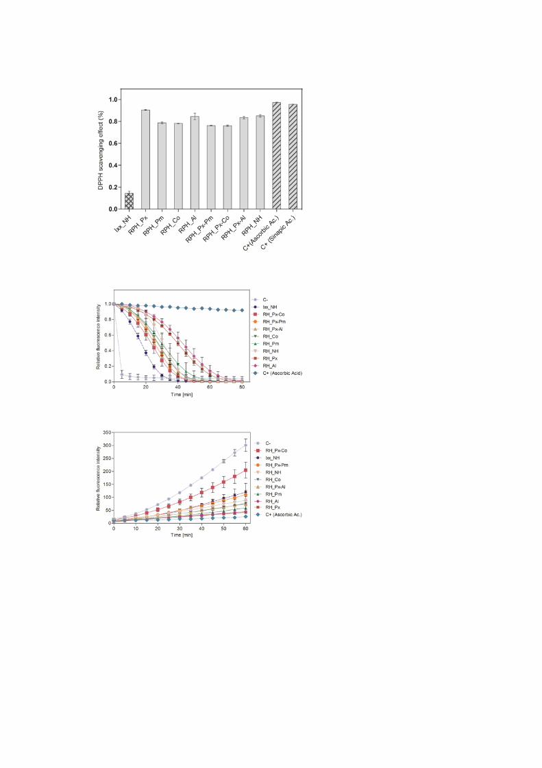


Table of Contents
Related Documents