Proc. Nati. Acad. Sci. USA Vol. 89, pp. 5527-5531, June 1992 Biochemistry Discrimination of DNA response elements for thyroid hormone and estrogen is dependent on dimerization of receptor DNA binding domains MARGARET A. HIRST*, LINDSAY HINCKt, MARK DANIELSENt, AND GORDON M. RINGOLD*§ *Institute of Cancer and Developmental Biology, Syntex Research USA, S3-1, 3401 Hillview Avenue, Palo Alto, CA 94304; tProgram in Cancer Biology, Stanford University, Stanford, CA 94305; and Department of Biochemistry and Molecular Biology, 3900 Reservoir Road NW, Georgetown University Medical School, Washington, DC 20007-2197 Communicated by Keith R. Yamamoto, December 23, 1991 (received for review August 2, 1991) ABSTRACT We and others have previously shown that a two-amino acid substitution in the base of the first zinc finger of the glucocorticoid receptor DNA binding domain (DBD) is sufficient to alter the receptor's target DNA from a glucocor- ticoid response element (GRE) to an estrogen response element (ERE). Activation of a thyroid hormone response element (TRE) has been shown to require an additional five-amino acid change in the second zinc finger of the thyroid hormone receptor (TR). Using closely related TRE and ERE sequences, we report that a receptor containing the TR DBD activates the ERE poorly, and receptors contalning essential amino acids of the estrogen receptor (ER) DBD activate the TRE poorly. The ER DBD (expressed in Escherichia col) selectively bound to a 32P-labeled ERE (32P-ERE) as a dimer and a 32P-TRE as a monomer, whereas the TR DBD bound 32P-TRE as a dimer and 32P-ERE as a monomer. When hybrid receptor DBDs were examined, we found that the five amino acids in the second zinc finger of the TR necessary for TRE activation were also essential for dimer formation on a TRE. Dimer formation of ER on an ERE was localized to the second half of the second zinc finger. These results suggest that the ability of ER and TR to functionally discriminate between an ERE and a TRE is a result of dimerization of their DBDs. Steroid hormone receptors are a class of transcriptional activators that produce their biological effects through dis- crete gene enhancers termed hormone response elements (HREs) (1, 2). Glucocorticoid receptors (GRs) and estrogen receptors (ERs) are composed of modular domains, each with ascribable functions related to ligand binding, DNA binding, and transcription activation (2-5). Binding of the receptor to a specific HRE occurs through a DNA binding domain (DBD), which consists of two fingers, each formed by the coordination of a single zinc atom with four cysteine residues (6, 7). This structural configuration is consistent with evi- dence that receptors interact with their response elements in the form of homodimers (8-10) or heterodimers (11, 12). To elucidate the mechanism by which a receptor recog- nizes its own response element, we and others recently constructed cDNAs carrying systematic substitutions of amino acid residues within the zinc fingers of either the GR (13, 14) or the ER (15). These studies identified seven amino acids that are critical for determining sequence-specific rec- ognition by the DBD. Selective activation from a glucocor- ticoid Kesponse element (GRE) vs. an estrogen response element (ERE) is dependent on two amino acids in the base of the first finger; activation from a thyroid hormone re- sponse element (TRE) requires an additional five amino acids in the second finger. Here we have focused on the ability of hybrid receptors to distinguish between an ERE and a TRE. Experiments indi- cated that while chimeric receptors containing the entire DBD of the thyroid hormone receptor (TR) are able to activate transcription from a TRE, they are unable to do so from an ERE. This was intriguing because the difference between some EREs and TREs is the spacing of the palin- dromic half sites (16). Using an Escherichia coli expression system, we synthesized the DBDs of hybrid receptors and examined their ability to bind HREs in vitro by an electro- phoretic mobility-shift assay (17). Our results suggest that the spacing of the TRE and ERE half sites is critical in the formation of DBD dimers. The portion of the TR DBD required for dimerization appears to be the first five amino acids in the second finger previously identified as essential for TRE activation (14). Dimerization of ER DBDs was also found to occur through the second finger; however, the critical residues are C-terminal to those identified for the TR. MATERIALS AND METHODS Construction of Reporter Plasmids and Chimeric Receptors in Expression Vectors. The first finger constructs, pGE9 and pGT1, have been described (13). The interfinger constructs, pGT2 and pGT3, were derived from pGT1 and pSV2Xrec and pGTR was derived from pGT3. For protein expression of the DBDs, the T7 E. coli ex- pression system was used (18). A blunted Sty I/Nar I (amino acids 386-581) fragment from pSV2Xrec, a modified version of the wild-type GR (13), was ligated into the BamH1 site of the pET3a expression vector. This yielded Xrec, a 237-amino acid fusion protein that contains 10 amino acids of T7 gene 10 on the N terminus and 31 amino acids of T7 gene 10 on the C terminus with a calculated molecular weight of 28,649. Amino acids 420-4% of pGT1, pGT2, pGT3, pGTR, and pGER were used to replace the Kpn I/Xho I fragment of Xrec. All constructs were confirmed by DNA sequencing and also examined by Western blot analysis using the monoclonal antibody BuGR1 (a generous gift of Robert Harrison, Vanderbilt University School of Medicine, Nashville, TN) to confirm that receptor protein was being expressed. Reporter plasmids were constructed from pMCAT5 (19), which lacks the endogenous GRE. pMECAT and pMTCAT were made by inserting either an ERE-containing oligonu- cleotide (GATCAAGCTTAGATCAGGTCACTGT- GACCTAGATCTAAGCTT) or a TRE-containing oligonu- Abbreviations: HRE, hormone response element; GR, glucocorti- coid receptor; ER, estrogen receptor; DBD, DNA binding domain; GRE, glucocorticoid response element; ERE, estrogen response element; TRE, thyroid hormone response element; TR, thyroid hormone receptor. §Present address: Affymax Research Institute, 4001 Miranda Ave- nue, Palo Alto, CA 94304. 5527 The publication costs of this article were defrayed in part by page charge payment. This article must therefore be hereby marked "advertisement" in accordance with 18 U.S.C. §1734 solely to indicate this fact.

Welcome message from author
This document is posted to help you gain knowledge. Please leave a comment to let me know what you think about it! Share it to your friends and learn new things together.
Transcript
Proc. Nati. Acad. Sci. USAVol. 89, pp. 5527-5531, June 1992Biochemistry
Discrimination of DNA response elements for thyroid hormone andestrogen is dependent on dimerization of receptor DNAbinding domainsMARGARET A. HIRST*, LINDSAY HINCKt, MARK DANIELSENt, AND GORDON M. RINGOLD*§*Institute of Cancer and Developmental Biology, Syntex Research USA, S3-1, 3401 Hillview Avenue, Palo Alto, CA 94304; tProgram in Cancer Biology,Stanford University, Stanford, CA 94305; and Department of Biochemistry and Molecular Biology, 3900 Reservoir Road NW, Georgetown University MedicalSchool, Washington, DC 20007-2197
Communicated by Keith R. Yamamoto, December 23, 1991 (received for review August 2, 1991)
ABSTRACT We and others have previously shown that atwo-amino acid substitution in the base of the first zinc fingerof the glucocorticoid receptor DNA binding domain (DBD) issufficient to alter the receptor's target DNA from a glucocor-ticoid response element (GRE) to an estrogen response element(ERE). Activation of a thyroid hormone response element(TRE) has been shown to require an additional five-amino acidchange in the second zinc finger of the thyroid hormonereceptor (TR). Using closely related TRE and ERE sequences,we report that a receptor containing the TR DBD activates theERE poorly, and receptors contalning essential amino acids ofthe estrogen receptor (ER) DBD activate the TRE poorly. TheER DBD (expressed in Escherichia col) selectively bound to a32P-labeled ERE (32P-ERE) as a dimer and a 32P-TRE as amonomer, whereas the TRDBD bound 32P-TRE as a dimer and32P-ERE as a monomer. When hybrid receptor DBDs wereexamined, we found that the five amino acids in the second zincfinger of the TR necessary for TRE activation were alsoessential for dimer formation on a TRE. Dimer formation ofER on an ERE was localized to the second half of the secondzinc finger. These results suggest that the ability ofER and TRto functionally discriminate between an ERE and a TRE is aresult of dimerization of their DBDs.
Steroid hormone receptors are a class of transcriptionalactivators that produce their biological effects through dis-crete gene enhancers termed hormone response elements(HREs) (1, 2). Glucocorticoid receptors (GRs) and estrogenreceptors (ERs) are composed ofmodular domains, each withascribable functions related to ligand binding, DNA binding,and transcription activation (2-5). Binding of the receptor toa specific HRE occurs through a DNA binding domain(DBD), which consists of two fingers, each formed by thecoordination ofa single zinc atom with four cysteine residues(6, 7). This structural configuration is consistent with evi-dence that receptors interact with their response elements inthe form of homodimers (8-10) or heterodimers (11, 12).To elucidate the mechanism by which a receptor recog-
nizes its own response element, we and others recentlyconstructed cDNAs carrying systematic substitutions ofamino acid residues within the zinc fingers of either the GR(13, 14) or the ER (15). These studies identified seven aminoacids that are critical for determining sequence-specific rec-ognition by the DBD. Selective activation from a glucocor-ticoid Kesponse element (GRE) vs. an estrogen responseelement (ERE) is dependent on two amino acids in the baseof the first finger; activation from a thyroid hormone re-sponse element (TRE) requires an additional five amino acidsin the second finger.
Here we have focused on the ability of hybrid receptors todistinguish between an ERE and a TRE. Experiments indi-cated that while chimeric receptors containing the entireDBD of the thyroid hormone receptor (TR) are able toactivate transcription from a TRE, they are unable to do sofrom an ERE. This was intriguing because the differencebetween some EREs and TREs is the spacing of the palin-dromic half sites (16). Using an Escherichia coli expressionsystem, we synthesized the DBDs of hybrid receptors andexamined their ability to bind HREs in vitro by an electro-phoretic mobility-shift assay (17). Our results suggest that thespacing of the TRE and ERE half sites is critical in theformation of DBD dimers. The portion of the TR DBDrequired for dimerization appears to be the first five aminoacids in the second finger previously identified as essentialfor TRE activation (14). Dimerization of ER DBDs was alsofound to occur through the second finger; however, thecritical residues are C-terminal to those identified for the TR.
MATERIALS AND METHODSConstruction of Reporter Plasmids and Chimeric Receptors
in Expression Vectors. The first finger constructs, pGE9 andpGT1, have been described (13). The interfinger constructs,pGT2 and pGT3, were derived from pGT1 and pSV2Xrec andpGTR was derived from pGT3.For protein expression of the DBDs, the T7 E. coli ex-
pression system was used (18). A blunted Sty I/Nar I (aminoacids 386-581) fragment from pSV2Xrec, a modified versionof the wild-type GR (13), was ligated into the BamH1 site ofthe pET3a expression vector. This yielded Xrec, a 237-aminoacid fusion protein that contains 10 amino acids ofT7 gene 10on the N terminus and 31 amino acids of T7 gene 10 on theC terminus with a calculated molecular weight of 28,649.Amino acids 420-4% of pGT1, pGT2, pGT3, pGTR, andpGER were used to replace the Kpn I/Xho I fragment ofXrec. All constructs were confirmed byDNA sequencing andalso examined by Western blot analysis using the monoclonalantibody BuGR1 (a generous gift of Robert Harrison,Vanderbilt University School ofMedicine, Nashville, TN) toconfirm that receptor protein was being expressed.
Reporter plasmids were constructed from pMCAT5 (19),which lacks the endogenous GRE. pMECAT and pMTCATwere made by inserting either an ERE-containing oligonu-cleotide (GATCAAGCTTAGATCAGGTCACTGT-GACCTAGATCTAAGCTT) or a TRE-containing oligonu-
Abbreviations: HRE, hormone response element; GR, glucocorti-coid receptor; ER, estrogen receptor; DBD, DNA binding domain;GRE, glucocorticoid response element; ERE, estrogen responseelement; TRE, thyroid hormone response element; TR, thyroidhormone receptor.§Present address: Affymax Research Institute, 4001 Miranda Ave-nue, Palo Alto, CA 94304.
5527
The publication costs of this article were defrayed in part by page chargepayment. This article must therefore be hereby marked "advertisement"in accordance with 18 U.S.C. §1734 solely to indicate this fact.
Proc. Natl. Acad. Sci. USA 89 (1992)
cleotide (GATCAAGCTTAGATCAGGTCATGACCTA-GATCTAAGCTT) into a Bgl II site.
Tissue Culture and Transfections. COS-7 cells grown tonear confluence were transfected by the lipofection proce-dure as described (13) using 0.5 ,ug of receptor DNA and 1.5,ug of the reporter plasmid. Cells were harvested after 3 daysand treated with vehicle (ethanol) or 1 uM dexamethasone 24hr before harvesting. Chloramphenicol acetyltransferase as-says were performed as described (13).
Protein Expression of Chimeric DBD. pET expression vec-tors with or without receptor sequence were transfected intoBL21DE3 E. coli carrying the LysS plasmid (obtained fromF. W. Studier; ref. 18). One-liter cultures in L broth weregrown to an OD600 of 0.3. Isopropyl ,-D-thiogalactopyrano-side was then added to a final concentration of 1 mM andcultures were grown an additional 5-6 hr. Extracts wereprepared as described (7).
Electrophoretic Mobility-Shift Assay. Three double-stranded oligomers, two 35 base pairs and one 34 base pairslong, were synthesized and used in the mobility-shift assay.These were called ERE (GATCGCTAATAGGTCACAGT-GACCTGATGCCGTCC), TRE (GATCGCTAATCAGGT-CATGACCTGGATGCCGTCC), and GRE (GATCGCTAA-TAGAACAAAATGTTCTGATGCCGTCC). When used asprobes, the oligonucleotides were end-labeled with[-32P]ATP (3000 Ci/mmol; 1 Ci = 37 GBq) using T4 poly-nucleotide kinase (20). In a 25-Mul binding assay mixture, eachsample contained buffer [10 mM Tris HCI, pH 7.5/0.5 mMEDTA/10% (vol/vol) glycerol/50mM sodium chloride/0.2%Nonidet P-40J, 100 ng of poly(dI-dC), 10-20 pg of labeledDNA probe (1-1.5 x 10⁹ dpm per ,ug of DNA), and (addedlast) 1-2 ,u of a 1:50-1:100 dilution of extract (100-500 ng ofprotein). Samples were incubated at room temperature for 20min and then applied to an 8% native gel [8% polyacrylamide/0.5x TBE (lx TBE = 89 mM Tris, pH 8.3/89 mM boricacid/8 mM EDTA)] equilibrated at 4°C and run =3 hr at 10V/cm.
Methylation Interference. The method used for this assay isdescribed elsewhere (21). End-labeled ERE and TRE frag-ments were prepared (7.5 x 10⁷ dpm per ,ug of DNA) andexposed to dimethyl sulfate as described (22). To obtainadequate radioactivity in the recovery, all components in theassay were increased 50-fold over our typical assay sample.The appropriate bands from the mobility-shift assay werelocalized by autoradiography, excised from the gel, andrecovered by electrophoresis onto DEAE paper. After ex-traction from the paper, the DNA was cleaved with piperi-dine (22), dried under vacuum, and resuspended in forma-mide; 50,000 cpm of each sample was run on a 10% sequenc-ing gel.
RESULTSTranscriptional Activity ofGRs Containing Chimeric DBDs.
We (13) and others (14, 15) have demonstrated that substi-tuting two amino acids in the base of the first zinc finger of
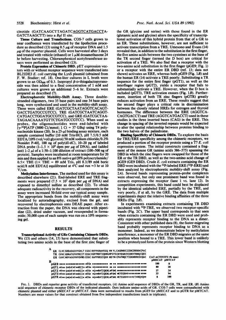
the GR (glycine and serine) with those found in the ER(glutamic acid and glycine) alters the specificity of transcrip-tional activation of this hybrid protein from that of a GR toan ER. These substitutions, however, are not sufficient toactivate transcription from a TRE. Umesono and Evans (14)revealed that, in addition to the substitution in the first finger,the five amino acids between the two cysteines at the base ofthe TR second finger (termed the D box) are critical foractivation of a TRE. We also find that a receptor with thetwo-amino acid substitution in the first finger (pGE9; Fig. 1)or a receptor with the entire ER DBD (ref. 13; data notshown) activates an ERE, whereas both pGE9 (Fig. 1B) andthe human ER (14) activate a TRE poorly. Substituting a TRsequence for the entire first finger (pGT1), as well as theinterfinger region (pGT2), yields a receptor that fails tosubstantially activate a TRE. However, when the D box isincluded (pGT3), TRE activation ensues (Fig. 1B). Further-more, insertion of both TR zinc fingers (pGTR) greatlyreduces activation from an ERE. These results suggest thatthe second finger plays a critical role in discriminationbetween the closely related HREs for estrogen and thyroidhormones. The difference between the ERE (AGGTCA-CAGTGACCT) and TRE (AGGTCATGACCT) used in thesestudies is the three inserted bases (CAG) in the ERE. Thischange in spacing of the inverted repeats would be expectedto alter the spatial relationship between proteins binding tothe two halves of the palindrome.
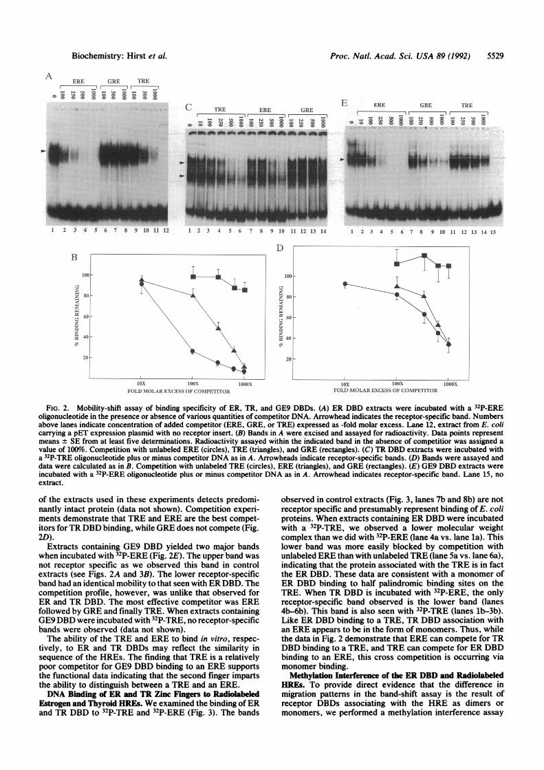
Binding Specifiity of Chimeric DBDs. To explore the basisfor TRE/ERE specificity among the chimeric receptors, weproduced a portion of the receptor protein using a T7 E. coliexpression system. The initial constructs contained a frag-ment of the mouse GR (amino acids 386-575) spanning theDBD in which the zinc fingers were swapped for those of theER or the TR DBD, as well as the two-amino acid change ofpGE9 (GE9 DBD). Crude E. coli extracts containing the ERDBD were incubated with the 32P-labeled ERE (32P-ERE) andthen analyzed by electrophoretic mobility-shift assay (Fig.2A). Several bands representing protein-probe complexeswere observed, but only one prominent band was found inextracts expressing the receptor (lane 1 vs. lane 12). Incompetition experiments, this band could best be displacedby the identical unlabeled ERE, partially by the TRE, andvery poorly, if at all, by the GRE. The data from multipleexperiments depict the relative binding affinities of the threeHREs (Fig. 2B). f
In experiments examining extracts containing TR DBDincubated with 32P-TRE, we observed two receptor-specificbands (Fig. 2C). The upper band corresponds to that seenwhen extracts containing the ER DBD were used and prob-ably represents receptor binding to the DNA as a dimer.Consistent with other published data (8), the faster-migratingband probably represents receptor binding to DNA as amonomer. Indeed, as we demonstrate below by methylationinterference, a monomer ofthe ER DBD migrates at the sameposition when bound to a TRE. This lower band is unlikelyto be a proteolyzed form ofthe protein since Western blotting
AGR CLVC SDEASGCHYGVLT CGSC KVFFKRAVEGQ HN YLCAGRNDCIIDKIRRKNCPACTR CVVC GDKATGYHYRCIT CEGC KGFFRRTIQKNLHPSYSCKYEGKCVIDKVTRNQCQECER CAVC NDYASGYHYGVWS CEGC KAFFKRSIQGH ND YM CPATNQCTIDKNRRKSCQAC CAT ACTIVITY (% max)
B pMECAT pMTCATpGE9 *ttt *********** oEGo ****v*** s so* -"""""""""" sees 100 8pGTl *Ve. G.K.TOYOORCIO .EG. GO*Ge*.eee ..es ... .so..8 0 20pGT2 *Ve. GOKOT*YORCI* sEG. *GeOR*TIQKNLOPSOS .*...*........*.. .0 27 24pGT3 *Ve. GeKeTeYOORCIO cEG. *Ge.R*TIQKNL*PSeS SKYEGKeVeeeeeee***e 33 94pGTR *Ve. GeKeTOYOeRCIO oEG. *GOR*TIQKNL*PSS OKYEGKoVoo*VTONQ*Qoo 8 100
FIG. 1. DBDs and reporter gene activity of transfected receptors. (A) Amino acid sequence of DBDs of the GR, TR, and ER. (B) Aminoacid sequence of chimeric receptor DBDs of the indicated plasmids. Dots indicate amino acids of GR. COS-7 cells were cotransfected withchimeric plasmid and either pMECAT or pMTCAT. Data were normalized to results from pGE9 for pMECAT and to pGTR for pMTCAT.Numbers are mean values for that construct obtained from five independent transfections (each in triplicate).
5528 Biochemistry: Hirst et al.
Proc. Natl. Acad. Sci. USA 89 (1992) 5529
ERE GRE TREI0 -II 0.00- Na tJI = - UA = - tA =
_ = th1 0 0 - C = = -
wM6,gm u .. .eni_
C TRE ERE GREI 1 - - -_It- A - - - -A = - t- -l -tiC$ l= =O n I= = tA =8 O= = = = = = = = = = = = CD
E ERE GRE TRE
C"u _ n O
= c c 0 g = cO g a 0O 0.~~~~ a
1 2 3 4 5 6 7 8 9 10 11 12 1 2 3 4 5 6 7 8 9 10 11 12 13 14
DB
.RiZt.:; 8o j-10.
-0!7. 6al
Z._ 40
2
20 ,-
loorI-
60H
_oI~- i0;-
: g 60
1 2 3 4 5 6 7 8 9 10 11 12 13 14 15
T -1
lox IOOX.FOLD NIOLAR EXCESS OF (OMPETITOR
lox bUOXFOLD MOLAR EXCESS OF COMPETITOR
FIG. 2. Mobility-shift assay of binding specificity of ER, TR, and GE9 DBDs. (A) ER DBD extracts were incubated with a 32P-EREoligonucleotide in the presence or absence of various quantities of competitor DNA. Arrowhead indicates the receptor-specific band. Numbersabove lanes indicate concentration of added competitor (ERE, GRE, or TRE) expressed as -fold molar excess. Lane 12, extract from E. colicarrying a pET expression plasmid with no receptor insert. (B) Bands in A were excised and assayed for radioactivity. Data points representmeans + SE from at least five determinations. Radioactivity assayed within the indicated band in the absence of competitior was assigned avalue of 100%.o Competition with unlabeled ERE (circles), TRE (triangles), and GRE (rectangles). (C) TR DBD extracts were incubated witha 32P-TRE oligonucleotide plus or minus competitor DNA as in A. Arrowheads indicate receptor-specific bands. (D) Bands were assayed anddata were calculated as in B. Competition with unlabeled TRE (circles), ERE (triangles), and GRE (rectangles). (E) GE9 DBD extracts wereincubated with a 32P-ERE oligonucleotide plus or minus competitor DNA as in A. Arrowhead indicates receptor-specific band. Lane 15, noextract.
of the extracts used in these experiments detects predomi-nantly intact protein (data not shown). Competition experi-ments demonstrate that TRE and ERE are the best compet-itors forTR DBD binding, while GRE does not compete (Fig.2D).
Extracts containing GE9 DBD yielded two major bandswhen incubated with 32P-ERE (Fig. 2E). The upper band wasnot receptor specific as we observed this band in controlextracts (see Figs. 2A and 3B). The lower receptor-specificband had an identical mobility to that seen with ER DBD. Thecompetition profile, however, was unlike that observed forER and TR DBD. The most effective competitor was EREfollowed by GRE and finally TRE. When extracts containingGE9DBD were incubated with 32P-TRE, no receptor-specificbands were observed (data not shown).The ability of the TRE and ERE to bind in vitro, respec-
tively, to ER and TR DBDs may reflect the similarity insequence of the HREs. The finding that TRE is a relativelypoor competitor for GE9 DBD binding to an ERE supportsthe functional data indicating that the second finger impartsthe ability to distinguish between a TRE and an ERE.DNA Binding of ER and TR Zinc Fingers to Radiolabeled
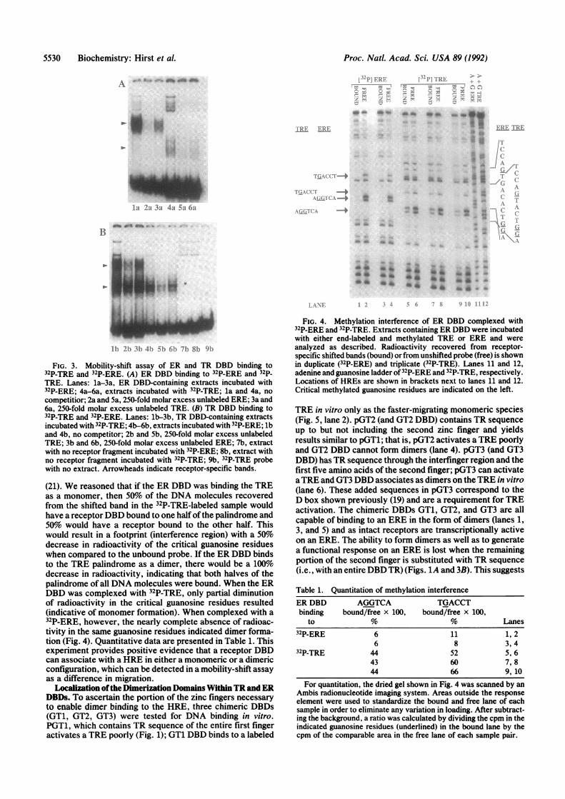
Estrogen and Thyroid HREs. We examined the binding ofERand TR DBD to 32P-TRE and 32P-ERE (Fig. 3). The bands
observed in control extracts (Fig. 3, lanes 7b and 8b) are notreceptor specific and presumably represent binding of E. coliproteins. When extracts containing ER DBD were incubatedwith a 32P-TRE, we observed a lower molecular weightcomplex than we did with 32P-ERE (lane 4a vs. lane la). Thislower band was more easily blocked by competition withunlabeled ERE than with unlabeled TRE (lane 5a vs. lane 6a),indicating that the protein associated with the TRE is in factthe ER DBD. These data are consistent with a monomer ofER DBD binding to half palindromic binding sites on theTRE. When TR DBD is incubated with 32P-ERE, the onlyreceptor-specific band observed is the lower band (lanes4b-6b). This band is also seen with 32P-TRE (lanes lb-3b).Like ER DBD binding to a TRE, TR DBD association withan ERE appears to be in the form of monomers. Thus, whilethe data in Fig. 2 demonstrate that ERE can compete for TRDBD binding to a TRE, and TRE can compete for ER DBDbinding to an ERE, this cross competition is occurring viamonomer binding.
Methylation Interference of the ER DBD and RadiolabeledHREs. To provide direct evidence that the difference inmigration patterns in the band-shift assay is the result ofreceptor DBDs associating with the HRE as dimers ormonomers, we performed a methylation interference assay
A 4 gg.p
1000x
Biochemistry: Hirst et al.
m-r
Mox)0
f.u
w i
Proc. Natl. Acad. Sci. USA 89 (1992)
A d *f
- I .
4W
lit 2a 3a 4a 5a 6a,L.iLL,~ -4 :.
'~~~~JALH -~~~~~~~~~-l
-'I;
lpJ b Ot
--V.
...
A*bb lb* -dXA b
11) 2) 3) -lb) )b 6b1 7bh 81 9i
FIG. 3. Mobility-shift assay of ER and TR DBD binding to32P-TRE and 32P-ERE. (A) ER DBD binding to 32P-ERE and 32p-TRE. Lanes: la-3a, ER DBD-containing extracts incubated with32P-ERE; 4a-6a, extracts incubated with 32P-TRE; la and 4a, no
competitior; 2a and 5a, 250-fold molar excess unlabeled ERE; 3a and6a, 250-fold molar excess unlabeled TRE. (B) TR DBD binding to32P-TRE and 32P-ERE. Lanes: lb-3b, TR DBD-containing extractsincubated with 32P-TRE; 4b-6b, extracts incubated with 32P-ERE; lband 4b, no competitor; 2b and Sb, 250-fold molar excess unlabeledTRE; 3b and 6b, 250-fold molar excess unlabeled ERE; 7b, extractwith no receptor fragment incubated with 32P-ERE; 8b, extract withno receptor fragment incubated with 32P-TRE; 9b, 32P-TRE probewith no extract. Arrowheads indicate receptor-specific bands.
(21). We reasoned that if the ER DBD was binding the TREas a monomer, then 50o of the DNA molecules recoveredfrom the shifted band in the 32P-TRE-labeled sample wouldhave a receptorDBD bound to one half ofthe palindrome and50% would have a receptor bound to the other half. Thiswould result in a footprint (interference region) with a 50%decrease in radioactivity of the critical guanosine residueswhen compared to the unbound probe. If the ER DBD bindsto the TRE palindrome as a dimer, there would be a 100%decrease in radioactivity, indicating that both halves of thepalindrome of all DNA molecules were bound. When the ERDBD was complexed with 32P-TRE, only partial diminutionof radioactivity in the critical guanosine residues resulted(indicative of monomer formation). When complexed with a32P-ERE, however, the nearly complete absence of radioac-tivity in the same guanosine residues indicated dimer forma-tion (Fig. 4). Quantitative data are presented in Table 1. Thisexperiment provides positive evidence that a receptor DBDcan associate with a HRE in either a monomeric or a dimericconfiguration, which can be detected in a mobility-shift assayas a difference in migration.
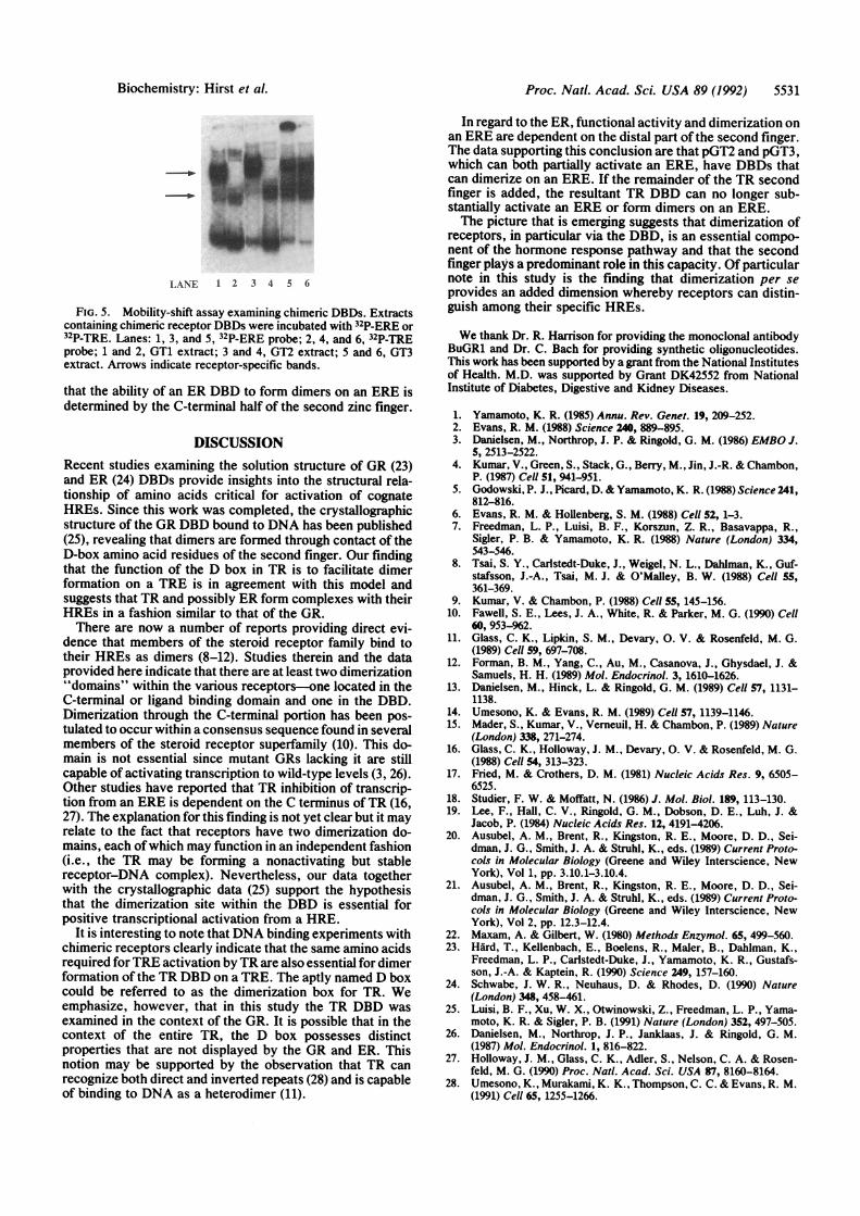
Lcalization ofthe Dimerization Domains WithinTR and ERDBDs. To ascertain the portion of the zinc fingers necessaryto enable dimer binding to the HRE, three chimeric DBDs(GT1, GT2, GT3) were tested for DNA binding in vitro.PGT1, which contains TR sequence of the entire first fingeractivates a TRE poorly (Fig. 1); GT1 DBD binds to a labeled
FIG. 4. Methylation interference of ER DBD complexed with32P-ERE and 32P-TRE. Extracts containing ER DBD were incubatedwith either end-labeled and methylated TRE or ERE and wereanalyzed as described. Radioactivity recovered from receptor-specific shifted bands (bound) or from unshifted probe (free) is shownin duplicate (32P-ERE) and triplicate (32P-TRE). Lanes 11 and 12,adenine and guanosine ladder of32P-ERE and 32P-TRE, respectively.Locations of HREs are shown in brackets next to lanes 11 and 12.Critical methylated guanosine residues are indicated on the left.
TRE in vitro only as the faster-migrating monomeric species(Fig. 5, lane 2). pGT2 (and GT2 DBD) contains TR sequenceup to but not including the second zinc finger and yieldsresults similar to pGTl; that is, pGT2 activates aTRE poorlyand GT2 DBD cannot form dimers (lane 4). pGT3 (and GT3DBD) has TR sequence through the interfinger region and thefirst five amino acids of the second finger; pGT3 can activateaTRE and GT3 DBD associates as dimers on the TRE in vitro(lane 6). These added sequences in pGT3 correspond to theD box shown previously (19) and are a requirement for TREactivation. The chimeric DBDs GT1, GT2, and GT3 are allcapable of binding to an ERE in the form of dimers (lanes 1,3, and 5) and as intact receptors are transcriptionally activeon an ERE. The ability to form dimers as well as to generatea functional response on an ERE is lost when the remainingportion of the second finger is substituted with TR sequence(i.e., with an entire DBD TR) (Figs. 1A and 3B). This suggests
Table 1. Quantitation of methylation interferenceER DBD AGGTCA TGACCTbinding bound/free x 100, bound/free x 100,
to t % Lanes32P-ERE 6 11 1, 2
6 8 3,432P-TRE 44 52 5, 6
43 60 7,844 66 9, 10
For quantitation, the dried gel shown in Fig. 4 was scanned by anAmbis radionucleotide imaging system. Areas outside the responseelement were used to standardize the bound and free lane of eachsample in order to eliminate any variation in loading. After subtract-ing the background, a ratio was calculated by dividing the cpm in theindicated guanosine residues (underlined) in the bound lane by thecpm of the comparable area in the free lane of each sample pair.
B1
5530 Biochemistry: Hirst et al.
1.AtI'.. ]I [e
Proc. Natl. Acad. Sci. USA 89 (1992) 5531
LANE 1 2 3 4 5 6
FIG. 5. Mobility-shift assay examining chimeric DBDs. Extractscontaining chimeric receptor DBDs were incubated with 32P-ERE or32P-TRE. Lanes: 1, 3, and 5, 32P-ERE probe; 2, 4, and 6, 32P-TREprobe; 1 and 2, GT1 extract; 3 and 4, GT2 extract; 5 and 6, GT3extract. Arrows indicate receptor-specific bands.
that the ability of an ER DBD to form dimers on an ERE isdetermined by the C-terminal half of the second zinc finger.
DISCUSSIONRecent studies examining the solution structure of GR (23)and ER (24) DBDs provide insights into the structural rela-tionship of amino acids critical for activation of cognateHREs. Since this work was completed, the crystallographicstructure of the GR DBD bound to DNA has been published(25), revealing that dimers are formed through contact of theD-box amino acid residues of the second finger. Our findingthat the function of the D box in TR is to facilitate dimerformation on a TRE is in agreement with this model andsuggests that TR and possibly ER form complexes with theirHREs in a fashion similar to that of the GR.There are now a number of reports providing direct evi-
dence that members of the steroid receptor family bind totheir HREs as dimers (8-12). Studies therein and the dataprovided here indicate that there are at least two dimerization"domains" within the various receptors-one located in theC-terminal or ligand binding domain and one in the DBD.Dimerization through the C-terminal portion has been pos-tulated to occur within a consensus sequence found in severalmembers of the steroid receptor superfamily (10). This do-main is not essential since mutant GRs lacking it are stillcapable of activating transcription to wild-type levels (3, 26).Other studies have reported that TR inhibition of transcrip-tion from an ERE is dependent on the C terminus ofTR (16,27). The explanation for this finding is not yet clear but it mayrelate to the fact that receptors have two dimerization do-mains, each of which may function in an independent fashion(i.e., the TR may be forming a nonactivating but stablereceptor-DNA complex). Nevertheless, our data togetherwith the crystallographic data (25) support the hypothesisthat the dimerization site within the DBD is essential forpositive transcriptional activation from a HRE.
It is interesting to note thatDNA binding experiments withchimeric receptors clearly indicate that the same amino acidsrequired forTRE activation by TR are also essential for dimerformation of the TR DBD on a TRE. The aptly named D boxcould be referred to as the dimerization box for TR. Weemphasize, however, that in this study the TR DBD wasexamined in the context of the GR. It is possible that in thecontext of the entire TR, the D box possesses distinctproperties that are not displayed by the GR and ER. Thisnotion may be supported by the observation that TR canrecognize both direct and inverted repeats (28) and is capableof binding to DNA as a heterodimer (11).
In regard to the ER, functional activity and dimerization onan ERE are dependent on the distal part of the second finger.The data supporting this conclusion are that pGT2 and pGT3,which can both partially activate an ERE, have DBDs thatcan dimerize on an ERE. If the remainder of the TR secondfinger is added, the resultant TR DBD can no longer sub-stantially activate an ERE or form dimers on an ERE.The picture that is emerging suggests that dimerization of
receptors, in particular via the DBD, is an essential compo-nent of the hormone response pathway and that the secondfinger plays a predominant role in this capacity. Of particularnote in this study is the finding that dimerization per seprovides an added dimension whereby receptors can distin-guish among their specific HREs.
We thank Dr. R. Harrison for providing the monoclonal antibodyBuGRi and Dr. C. Bach for providing synthetic oligonucleotides.This work has been supported by a grant from the National Institutesof Health. M.D. was supported by Grant DK42552 from NationalInstitute of Diabetes, Digestive and Kidney Diseases.
1. Yamamoto, K. R. (1985) Annu. Rev. Genet. 19, 209-252.2. Evans, R. M. (1988) Science 240, 889-895.3. Danielsen, M., Northrop, J. P. & Ringold, G. M. (1986) EMBO J.
5, 2513-2522.4. Kumar, V., Green, S., Stack, G., Berry, M., Jin, J.-R. & Chambon,
P. (1987) Cell 51, 941-951.5. Godowski, P. J., Picard, D. & Yamamoto, K. R. (1988) Science 241,
812-816.6. Evans, R. M. & Hollenberg, S. M. (1988) Cell 52, 1-3.7. Freedman, L. P., Luisi, B. F., Korszun, Z. R., Basavappa, R.,
Sigler, P. B. & Yamamoto, K. R. (1988) Nature (London) 334,543-546.
8. Tsai, S. Y., Carlstedt-Duke, J., Weigel, N. L., Dahlman, K., Guf-stafsson, J.-A., Tsai, M. J. & O'Malley, B. W. (1988) Cell 55,361-369.
9. Kumar, V. & Chambon, P. (1988) Cell 55, 145-156.10. Fawell, S. E., Lees, J. A., White, R. & Parker, M. G. (1990) Cell
60, 953-962.11. Glass, C. K., Lipkin, S. M., Devary, 0. V. & Rosenfeld, M. G.
(1989) Cell 59, 697-708.12. Forman, B. M., Yang, C., Au, M., Casanova, J., Ghysdael, J. &
Samuels, H. H. (1989) Mol. Endocrinol. 3, 1610-1626.13. Danielsen, M., Hinck, L. & Ringold, G. M. (1989) Cell 57, 1131-
1138.14. Umesono, K. & Evans, R. M. (1989) Cell 57, 1139-1146.15. Mader, S., Kumar, V., Verneuil, H. & Chambon, P. (1989) Nature
(London) 338, 271-274.16. Glass, C. K., Holloway, J. M., Devary, 0. V. & Rosenfeld, M. G.
(1988) Cell 54, 313-323.17. Fried, M. & Crothers, D. M. (1981) Nucleic Acids Res. 9, 6505-
6525.18. Studier, F. W. & Moffatt, N. (1986) J. Mol. Biol. 189, 113-130.19. Lee, F., Hall, C. V., Ringold, G. M., Dobson, D. E., Luh, J. &
Jacob, P. (1984) Nucleic Acids Res. 12, 4191-4206.20. Ausubel, A. M., Brent, R., Kingston, R. E., Moore, D. D., Sei-
dman, J. G., Smith, J. A. & Struhl, K., eds. (1989) Current Proto-cols in Molecular Biology (Greene and Wiley Interscience, NewYork), Vol 1, pp. 3.10.1-3.10.4.
21. Ausubel, A. M., Brent, R., Kingston, R. E., Moore, D. D., Sei-dman, J. G., Smith, J. A. & Struhl, K., eds. (1989) Current Proto-cols in Molecular Biology (Greene and Wiley Interscience, NewYork), Vol 2, pp. 12.3-12.4.
22. Maxam, A. & Gilbert, W. (1980) Methods Enzymol. 65, 499-560.23. Hard, T., Kellenbach, E., Boelens, R., Maler, B., Dahlman, K.,
Freedman, L. P., Carlstedt-Duke, J., Yamamoto, K. R., Gustafs-son, J.-A. & Kaptein, R. (1990) Science 249, 157-160.
24. Schwabe, J. W. R., Neuhaus, D. & Rhodes, D. (1990) Nature(London) 348, 458-461.
25. Luisi, B. F., Xu, W. X., Otwinowski, Z., Freedman, L. P., Yama-moto, K. R. & Sigler, P. B. (1991) Nature (London) 352, 497-505.
26. Danielsen, M., Northrop, J. P., Janklaas, J. & Ringold, G. M.(1987) Mol. Endocrinol. 1, 816-822.
27. Holloway, J. M., Glass, C. K., Adler, S., Nelson, C. A. & Rosen-feld, M. G. (1990) Proc. Natl. Acad. Sci. USA 87, 8160-8164.
28. Umesono, K., Murakami, K. K., Thompson, C. C. & Evans, R. M.(1991) Cell 65, 1255-1266.
Biochemistry: Hirst et al.
Related Documents