High-energy water sites determine peptide binding affinity and specificity of PDZ domains Thijs Beuming,* Ramy Farid, and Woody Sherman Schro ¨ dinger Inc., New York, New York 10036 Received 16 January 2009; Revised 20 March 2009; Accepted 27 April 2009 DOI: 10.1002/pro.177 Published online 28 May 2009 proteinscience.org Abstract: PDZ domains have well known binding preferences for distinct C-terminal peptide motifs. For most PDZ domains, these motifs are of the form [S/T]-W-[I/L/V]. Although the preference for S/T has been explained by a specific hydrogen bond interaction with a histidine in the PDZ domain and the (I/L/V) is buried in a hydrophobic pocket, the mechanism for Trp specificity at the second to last position has thus far remained unknown. Here, we apply a method to compute the free energies of explicit water molecules and predict that potency gained by Trp binding is due to a favorable release of high-energy water molecules into bulk. The affinities of a series of peptides for both wild-type and mutant forms of the PDZ domain of Erbin correlate very well with the computed free energy of binding of displaced waters, suggesting a direct relationship between water displacement and peptide affinity. Finally, we show a correlation between the magnitude of the displaced water free energy and the degree of Trp-sensitivity among subtypes of the HTRA PDZ family, indicating a water-mediated mechanism for specificity of peptide binding. Keywords: PDZ domain; peptide binding affinity; water thermodynamics; molecular dynamics; WaterMap Introduction PDZ domains are small modular protein domains of around 70–90 amino acids that play an important role in the assembly of complexes of proteins at the plasma membrane. 1 PDZ domains have been shown to interact with G-protein coupled receptors (GPCRs), ion chan- nels, transporters, and receptor tyrosine kinases, as well as with cytoskeletal proteins, extra-cellular matrix proteins, and components of tight junctions (claudins, occludins). Biological processes involving PDZ domains include the establishment and maintenance of epithelial polarity 2,3 ; the organization of signaling complexes at the synapse 4 and trafficking of proteins to the plasma membrane. 5 Many different types of PDZ domain-containing proteins have been identified, with as few as one and as many as 10 copies of the do- main, often in combination with other regulatory domains, including SH2, SH3, WW, guanylate kinase, PTB, or LRR domains. The combination of several domains in these proteins enables them to act as mo- lecular switchboards, bringing together components of signaling complexes. 6 In total, more than 500 PDZ domains in over 300 different proteins have been identified in the human genome. 7 Known PDZ do- main-mediated protein–protein interactions have been compiled in a PDZ specific database, named PDZBase. 8 Insight into the mechanism underlying affinity and specificity of these interactions has come from X- ray crystallography, 9 NMR spectroscopy, 10 sequence analysis, 11 computational biophysics, 12 and combi- natorial peptide experiments. 13–15 One particularly attractive experimental technique to study the binding Abbreviations: dvl, dishevelled; GPCR, G-protein coupled re- ceptor; HTRA, high temperature requirement A; MD, molecular dynamics; MM–GB/SA, molecular mechanics–Generalized Born surface area; NMR, nuclear magnetic resonance; PDZ, PSD- 95/Discs-large/ZO-1. *Correspondence to: Thijs Beuming, 120 West 45th street, 29th floor, Tower 45, New York, NY, 10036. E-mail: thijs.beuming@ schrodinger.com Published by Wiley-Blackwell. V C 2009 The Protein Society PROTEIN SCIENCE 2009 VOL 18:1609—1619 1609

Welcome message from author
This document is posted to help you gain knowledge. Please leave a comment to let me know what you think about it! Share it to your friends and learn new things together.
Transcript

High-energy water sites determinepeptide binding affinity and specificityof PDZ domains
Thijs Beuming,* Ramy Farid, and Woody Sherman
Schrodinger Inc., New York, New York 10036
Received 16 January 2009; Revised 20 March 2009; Accepted 27 April 2009DOI: 10.1002/pro.177
Published online 28 May 2009 proteinscience.org
Abstract: PDZ domains have well known binding preferences for distinct C-terminal peptidemotifs. For most PDZ domains, these motifs are of the form [S/T]-W-[I/L/V]. Although the
preference for S/T has been explained by a specific hydrogen bond interaction with a histidine in
the PDZ domain and the (I/L/V) is buried in a hydrophobic pocket, the mechanism for Trpspecificity at the second to last position has thus far remained unknown. Here, we apply a method
to compute the free energies of explicit water molecules and predict that potency gained by Trp
binding is due to a favorable release of high-energy water molecules into bulk. The affinities of aseries of peptides for both wild-type and mutant forms of the PDZ domain of Erbin correlate very
well with the computed free energy of binding of displaced waters, suggesting a direct relationship
between water displacement and peptide affinity. Finally, we show a correlation between themagnitude of the displaced water free energy and the degree of Trp-sensitivity among subtypes of
the HTRA PDZ family, indicating a water-mediated mechanism for specificity of peptide binding.
Keywords: PDZ domain; peptide binding affinity; water thermodynamics; molecular dynamics;WaterMap
IntroductionPDZ domains are small modular protein domains of
around 70–90 amino acids that play an important role
in the assembly of complexes of proteins at the plasma
membrane.1 PDZ domains have been shown to interact
with G-protein coupled receptors (GPCRs), ion chan-
nels, transporters, and receptor tyrosine kinases, as
well as with cytoskeletal proteins, extra-cellular matrix
proteins, and components of tight junctions (claudins,
occludins). Biological processes involving PDZ
domains include the establishment and maintenance
of epithelial polarity2,3; the organization of signaling
complexes at the synapse4 and trafficking of proteins
to the plasma membrane.5 Many different types of
PDZ domain-containing proteins have been identified,
with as few as one and as many as 10 copies of the do-
main, often in combination with other regulatory
domains, including SH2, SH3, WW, guanylate kinase,
PTB, or LRR domains. The combination of several
domains in these proteins enables them to act as mo-
lecular switchboards, bringing together components of
signaling complexes.6 In total, more than 500 PDZ
domains in over 300 different proteins have been
identified in the human genome.7 Known PDZ do-
main-mediated protein–protein interactions have been
compiled in a PDZ specific database, named
PDZBase.8
Insight into the mechanism underlying affinity
and specificity of these interactions has come from X-
ray crystallography,9 NMR spectroscopy,10 sequence
analysis,11 computational biophysics,12 and combi-
natorial peptide experiments.13–15 One particularly
attractive experimental technique to study the binding
Abbreviations: dvl, dishevelled; GPCR, G-protein coupled re-ceptor; HTRA, high temperature requirement A; MD, moleculardynamics; MM–GB/SA, molecular mechanics–Generalized Bornsurface area; NMR, nuclear magnetic resonance; PDZ, PSD-95/Discs-large/ZO-1.
*Correspondence to: Thijs Beuming, 120 West 45th street, 29thfloor, Tower 45, New York, NY, 10036. E-mail: [email protected]
Published by Wiley-Blackwell. VC 2009 The Protein Society PROTEIN SCIENCE 2009 VOL 18:1609—1619 1609

properties of these domains is phage display, whereby
large collections of peptides are screened and ampli-
fied in an in vivo selection process, revealing the opti-
mal binding motifs of each PDZ domain. The research
group of Sidhu and co-workers has determined opti-
mal binding profiles for a large number of PDZ
domains, initially for Erbin,16 ZO-1,17,18 and the HTRA
(high-temperature requirement A) family.19,20 More
recently, they profiled as many as 100 human and C.
elegans PDZ domains.21 Together with the experimen-
tally detected endogenous PDZ interactions, these
experiments have enabled a comprehensive analysis of
determinants of specificity and promiscuity of the
entire PDZ domain family.
C-terminal PDZ binding motifs are often classified
as Class I (S/T-X-U) or Class II (U-X-U), where U is
any hydrophobic residue (the terminal residue in the
ligand interacting with the PDZ domain is termed the
P-0 position, and the next residue towards the N-ter-
minus is designated as the P-1 position, etc.). Corre-
spondingly, PDZ domains have been classified as Class
I binding or Class II binding based on the observed
correlation between binding specificity and the occur-
rence of specific residues in the peptide binding
site.13,22 For example, most PDZ domains have a His
residue at the aB1 position in the PDZ domain (see
Fig. 1), and these domains prefer peptides with a Thr/
Ser at the P-2 position.9 The specific size and shape of
the P-0 hydrophobic pocket, formed by residues in the
aB and bB structural elements, has been correlated
with the observed preference for L or V at the P-0
position in NHERF and PSD-95 PDZ domains.23 More
distal interactions with P-3 and P-4 sites appear to be
mediated by the bB-bC loop, and the variability of this
loop in terms of length and sequence appears to mod-
ulate specificity through these positions.24 Large scale
phage display experiments have significantly extended
the dual classification scheme, showing that these
domains can be subdivided in as many as 16 different
classes and can recognize features of up to seven pep-
tide residues.21
Interestingly, the P-1 (second to last) position was
originally thought to be relatively unimportant for
specificity, based on the variability of amino acid types
at this position in known ligands of PDZ domains,13
and the exposure to solvent of the P-1 side chain in
crystal structures. However, more recent experiments,
most notably a phage-display study of the Erbin PDZ
domain,16 showed that certain side chains at P-1 can
have a major contribution to affinity. For example, the
Erbin phage display profile indicated that a WETWV
peptide was the most optimal binding motif. The affin-
ity of this super-binding peptide was determined to be
20 nM, a 1500-fold increase in potency compared to
the peptide in which the Trp P-1 residue is replaced by
Ala (i.e., WETAV). The NMR structure of Erbin with
the WETWV peptide revealed that a pocket formed by
Ser26, Arg51, and Gln49 residues accommodated the
Figure 1. (A) WaterMap of the Erbin PDZ domain (gray) complex with the WETWV peptide (green). The aB helix and the bBand bC strands form the peptide binding site. Peptide residues are numbered P-0 to P-4, with the numbering starting at the
C-terminal valine residue. All identified water sites in the peptide binding site are shown as spheres. Water sites with DG >
3.5 kcal/mol are shown in red, sites with 1.5 > DG > 3.5 kcal/mol are shown in orange, and sites with DG < 1.5 kcal/mol in
gray; The three highest energy water sites overlap with side-chains at the P-0 and P-1 positions. (B) Close-up of the P-1
pocket with three predicted water-sites, named w-1a (orange), w-1b (red), and w-1c (orange). The w-1b site is more
unfavorable (�TDS 2.6 kcal/mol, DH 3.2 kcal/mol, DG 5.8 kcal/mol) than the w-1a (�TDS 1.6 kcal/mol, DH �0.1 kcal/mol, DG1.5 kcal/mol) and w-1c sites (�TDS 1.7 kcal/mol, DH 0.6 kcal/mol, DG 2.3 kcal/mol). (C) WETFV-Erbin model. (D) WETPV-
Erbin model. (E) WETAV-Erbin model. Note that while the wild-type Trp almost completely displaces the unfavorable waters,
the Phe only has partial displacement and both Ala and Pro do not overlap with any of the three high-energy water sites, in
good agreement with the experimentally measured differences in affinity.
1610 PROTEINSCIENCE.ORG High-Energy Water Sites Determine PDZ-Peptide Affinity
Trp side chain. However, specific side-chain interac-
tions appeared to not be important, as Ala mutants at
position 26, 49, and 51 had little effect on binding af-
finity of the peptide. Thus, the authors suggested that
non-specific, backbone-mediated interactions were im-
portant for the high affinity of the Trp containing
peptide.
We have recently developed a method, called
WaterMap, for calculating the location and thermody-
namics of water sites in proteins.25,26 This method is
based on molecular dynamics (MD) simulation and
computes both the enthalpy and entropy of water sites
around a protein, relative to bulk water, from an anal-
ysis of explicit water molecules from the MD trajectory
(see Materials and Methods). It has been shown that
WaterMap can be used as a tool for qualitatively
understanding the properties of binding sites,25 and as
a quantitative tool for predicting small molecule bind-
ing affinities.26 Here, we have used WaterMap to
investigate the mechanism of high-affinity binding of
peptides to PDZ domains. In particular, the method
was used to investigate peptide binding affinity of the
structurally well-characterized PDZ domains of Erbin
and the HTRA family. Erbin (ERB-interacting protein)
is a member of the LAP (leucine-rich repeat and PDZ
containing) family, a class of proteins involved in the
maintenance of epithelial polarity.27 The PDZ domain
of Erbin was originally identified through yeast two-
hybrid experiments as an interacting partner of the
ERB-B2 receptor,28 and has been shown to interact in
vivo with p120-like catenin proteins.29 The HTRA
family of proteins are characterized by a combination
of a serine protease domain and a PDZ domain.30 The
best characterized member of the family is HTRA2
(also known as Omi), which is released from mito-
chondria in cellular stress and participates in the regu-
lation of apoptosis.31
Using WaterMap, we predict the presence of high-
energy water sites on the surface of several PDZ
domains, including Erbin and the HTRA family. In
Erbin, the high-energy water site at the P-1 position is
formed by the hydrophobic parts of the side chains
Ser26, Arg49, and Gln51, as well as the surface of the
bB-bC b-sheet. We show that the Trp dependence of
high-affinity peptides can be correlated to the ability
of Trp to displace this high-energy water, thus releas-
ing it into the bulk and resulting in a net free energy
gain. Additional predicted high-energy water sites on
the PDZ domain, including those in the P-0 pocket,
are also important for affinity, and we show that the
reported affinities of a series of peptides for Erbin cor-
relate well with the total energy of released water, sug-
gesting a direct relationship between water displace-
ment and peptide binding affinity. Finally, we
demonstrate a correlation between the degree of Trp
dependence and the magnitude of the water free
energy among subtypes of the HTRA family, linking
water displacement to the specificity of PDZ domains.
Results
Prediction of high-energy water sites in Erbin
The results from running WaterMap on the PDZ do-
main of Erbin are shown in Figure 1. Water sites are
predicted along the entire peptide binding site and
have been classified into low-energy (i.e., relatively sta-
ble with DG < 1.5 kcal/mol, gray spheres), medium-
energy (1.5 < DG < 3.5 kcal/mol, orange spheres), and
high-energy sites (highly unstable with DG > 3.5 kcal/
mol, large red spheres). High-energy water sites are
found only in the P-0 and P-1 pockets. This is consist-
ent with the experimental data, which shows that the
P-0 and P-1 sites are most important for high-affinity
binding of the WETWV peptide (see Table I). Substitu-
tions of the WETWV peptides at the P-1 and P-0 sites
lead to large reductions in affinity; at P-1, Trp to Ala
(WTEAV) leads to a 1500-fold loss of binding affinity
(corresponding to a DDG of 4.1 kcal/mol), while at P-
0, Val to Ala (WETWA) results in 3700-fold lower af-
finity (DDG 4.6 kcal/mol.) Ala substitutions at posi-
tions P-2, P-3, and P-4 only affect affinity 100-fold
(2.5 kcal/mol), 140-fold (2.7 kcal/mol), and 50-fold
(2.2 kcal/mol), respectively. Even a conservative sub-
stitution at the P-1 site of Trp to Phe causes a signifi-
cant loss of affinity (170-fold, 2.8 kcal/mol). Finally,
mutation to Pro at this position leads to the largest
loss in affinity (3300-fold, 4.5 kcal/mol).
Inspection of the WaterMap in the P-1 pocket sug-
gests a reason why Trp is a much better binder than
Phe, Ala, or Pro. Three water sites are predicted in the
pocket formed by the Ser26, Arg49, and Gln51 side
chains and the b-sheet backbone (see Fig. 1, panels B–
E). These water sites will be referred to hence forth at
w-1a, w-1b, and w-1c. The central water (w-1b, shown
in red) has the highest energy (5.8 kcal/mol), while
the two flanking waters (w-1a and w-1c, shown in or-
ange) have moderately unfavorable energies (1.5 and
2.3 kcal/mol). Amongst the experimentally tested var-
iants, only Trp [Fig. 1(B)] has the ability to displace
these waters effectively, while Phe [Fig. 1(C)] can only
partially displace them. Pro [Fig. 1(D)] and Ala [Fig.
1(E)] do not overlap with these water sites.
The presence of high-energy water molecules in
the P-0 pocket is consistent with the strongly hydro-
phobic character of this pocket; the P-0 Val residue is
in direct contact with the side chains of Leu23, Phe25,
Ile27 in the bB strand, and Val83, Leu86 in the aB he-
lix. However, the P-1 high-energy water site is more
surprising, given its partial exposure to solvent, and
the proximity of three polar residues Ser26, Arg49,
and Gln51. Closer inspection of the environment
reveals that aside from the Ser, no protein hydrogen
bonding partners are available, since the b-sheet al-
ready has satisfied all the backbone hydrogen bonds,
and the Arg and Gln polar groups point away from the
water site toward solvent. This also explains why dra-
matic affinity can be gained with Trp while at the
Beuming et al. PROTEIN SCIENCE VOL 18:1609—1619 1611

same time alanine scans of Erbin do not have a signifi-
cant effect on the affinity of the Trp P-1 variant (see
next section for details).
Prediction of peptide affinityTo determine whether WaterMap could be used to ret-
rospectively predict the peptide affinities in Table I,
the NMR structure was used as a template to create
models of all the PDZ-peptide complexes from Table I
(see Materials and Methods for details). The total
energies were then estimated by summing over all
water sites overlapping with the different peptides,
according to the formula described in Abel et al.,26
with no adjustable parameters (see Materials and
Methods). The WaterMap energies of the WETAV and
WETFV peptides calculated using this formula are
�27.8 kcal/mol and �31.3 kcal/mol, relative to �37.2
kcal/mol for WETWV. Thus, the trend of the calcu-
lated energy differences is in good agreement with the
experimental results. Not surprisingly, there is a signif-
icant difference between the absolute values of the
predicted and measured affinities, which is likely due
to the neglect of entropy loss and induced strain in
both the ligand and receptor. The WaterMap energies
for the entire set of peptides in Table I were calculated
and compared with experimental data. As can be seen
in Figure 2, there is a good correlation (R2 ¼ 0.67)
between WaterMap energies and experimental peptide
affinities, suggesting that displacement of water from
the surface of the PDZ domain is an important con-
tributor to the affinity of PDZ-peptides complexes.
We also computed peptide affinities using the mo-
lecular mechanics–Generalized Born/surface area
(MM–GB/SA) method, as implemented in Prime.32–35
This method has been used to successfully predict
binding affinities in diverse systems such as thrombin,
factor Xa, HIV-RT, and several kinases.34,36 In the
case of Erbin studied here, there is only modest corre-
lation between peptide affinities and MM–GB/SA
energies [R2 ¼ 0.33, see Fig. 2(B)]. Indeed, the MM–
Table I. Erbin-Peptide Experimental and Computational Binding Affinities
Peptide
Affinity,IC50(lM)
Relativeaffinity
RTln(IC50)
WaterMap-TDS
(kcal/mol)WaterMap DH(kcal/mol)
WaterMap DG(kcal/mol) MW
MM–GB/SA DG
(kcal/mol)
WETWV 0.02 � 0.002 1 �8.6 �30.4 �6.8 �37.2 718 �23.9FETWV 0.16 � 0.01 8.0 �7.5 �28.1 �6.6 �34.7 678 �22.7YETWV 0.16 � 0.01 8.0 �7.5 �28.1 �6.6 �34.7 694 �29.4WDTWV 0.41 � 0.06 21 �6.9 �28.9 �7.5 �36.4 704 �21.3WESWV 0.16 � 0.01 8.0 �7.5 �30.0 �6.3 �36.3 704 �26.2WEVWV 0.26 � 0.01 13 �7.2 �30.5 �6.9 �37.4 716 �23.2WETFV 3.3 � 0.2 170 �5.8 �27.1 �4.2 �31.3 679 �23.5WETPV 66 � 4 3300 �4.1 �24.5 �3.3 �27.8 630 �14.6WETWL 1.8 � 0.1 90 �6.1 �28.8 �3.9 �32.7 732 �29.0WETWI 1.7 � 0.1 85 �6.2 �30.4 �6.8 �37.2 732 �29.4AETWV 1.0 � 0.1 50 �6.4 �27.5 �6.6 �34.1 603 �22.0WATWV 2.8 � 0.7 140 �5.9 �28.1 �7.7 �35.8 661 �19.8WEAWV 2.0 � 0.1 100 �6.1 �29.8 �6.1 �35.9 688 �23.3WETAV 30 � 7 1500 �4.5 �24.5 �3.3 �27.8 603 �15.4WETWA 74 � 4 3700 �4.0 �28.0 �2.6 �30.6 690 �21.3
Experimental affinities were determined as IC50 using a glutathione S-transferase-based chemiluminescence assay.16 WaterMapaffinities were calculated from the overlap of the peptides with the water-sites shown in Figure 1, according to the protocoldescribed in Abel et al.26 MM–GB/SA energies were calculated using Prime,32,33,35 employing a brief minimization of the pro-tein/peptide structure; peptide strain energies were not included in the total energy. MW, molecular weight.
Figure 2. Correlations between computationally estimated energies and experimental peptide affinities [RTln (IC50)]. (a)
WaterMap (R2 ¼ 0.67), (b) MM–GB/SA (R2 ¼ 0.33), (c) molecular weight (R2 ¼ 0.25).
1612 PROTEINSCIENCE.ORG High-Energy Water Sites Determine PDZ-Peptide Affinity

GB/SA correlation only slightly exceeds the correlation
of molecular weight to affinity of the peptides [R2 ¼0.24, see Fig. 2(C)]. Thus, while implicit solvation and
non-bonded interaction energy terms provide some ex-
planation of the affinity of this series of peptides,
more reliable predictions are obtained when the role
of explicit water is considered.
Virtual amino acid screeningTo further assess the ability of WaterMap to explain
and predict relative binding affinities, we performed a
single residue modification to each of the 20 amino
acid types at the five different positions P-0 to P-4. As
shown in Figure 3, the WaterMap energies predict that
the optimal residues at the P-0, P-1, and P-4 sites are
V, W, and W, respectively, in agreement with the opti-
mal motif identified through phage display. For P-0,
the b-branched side-chains Ile and Thr have similar
energies to the optimal Val. For P-2 and P-3, there
appears to be no specific preference for Thr and Glu
respectively in terms of water displacement. Although
there is significant water overlap of these side chains
with water sites, many other amino acids can give
comparable or even improved WaterMap energies.
This suggests that other structural criteria are involved
in determining selectivity at these positions in Erbin
(e.g., hydrogen bond formation at P-2 or charge–
charge interactions at P-3).
Erbin mutants S26A, R49A, and Q51A
Skelton et al. (ref. 16) probed the dependence of the
peptide affinity on specific side-chain interactions with
the PDZ domain by mutating all residues in the pep-
tide binding site to Ala.16 They discovered that none of
the mutations in the proximity of the Trp (i.e., S26A,
R49A, and Q51A) strongly affected peptide affinity. In
particular, the S26A mutation resulted in only a 1.5-
fold loss of affinity and R49A affinity was 3.4-fold less,
while the Q51A mutant affinity was 9-fold higher than
wild-type. To investigate whether these variants con-
tain conserved high-energy water sites, we generated
models for all three mutants (see Materials and Meth-
ods). Locations and energies of water sites for the
mutants were calculated using WaterMap and the dif-
ference in affinity of the WETWV and WETAV pep-
tides was predicted. The results from WaterMap for
these three mutants reveal that the w-1b high-energy
water site predicted in the wild type is also present in
the structures of the Ala mutants (see Fig. 4). The
energies of the w-1b water site are 6.1 kcal/mol in
Figure 3. Virtual amino acid screening of the WETWV
peptide. All 20 amino-acid types were introduced in the
WETWV reference peptide at all five positions. For positions
P-0, P-1, and P-4, the phage-display derived optimal
residues (black) were identified among the most favorable
based on the WaterMap energies. For P-2 and P-3, many
different amino-acid types were comparable to or better
than native residues, suggesting that energetic factors other
than water displacement are important for affinity at these
positions.
Beuming et al. PROTEIN SCIENCE VOL 18:1609—1619 1613
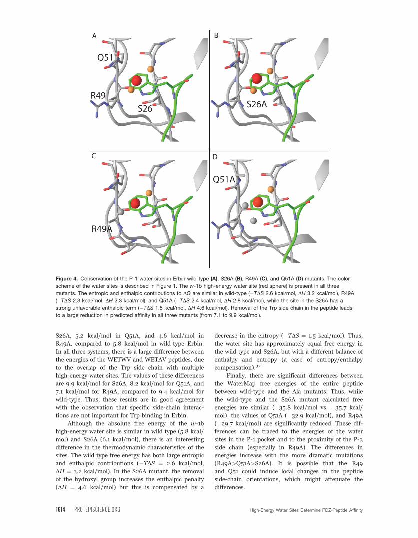
S26A, 5.2 kcal/mol in Q51A, and 4.6 kcal/mol in
R49A, compared to 5.8 kcal/mol in wild-type Erbin.
In all three systems, there is a large difference between
the energies of the WETWV and WETAV peptides, due
to the overlap of the Trp side chain with multiple
high-energy water sites. The values of these differences
are 9.9 kcal/mol for S26A, 8.2 kcal/mol for Q51A, and
7.1 kcal/mol for R49A, compared to 9.4 kcal/mol for
wild-type. Thus, these results are in good agreement
with the observation that specific side-chain interac-
tions are not important for Trp binding in Erbin.
Although the absolute free energy of the w-1b
high-energy water site is similar in wild type (5.8 kcal/
mol) and S26A (6.1 kcal/mol), there is an interesting
difference in the thermodynamic characteristics of the
sites. The wild type free energy has both large entropic
and enthalpic contributions (�TDS ¼ 2.6 kcal/mol,
DH ¼ 3.2 kcal/mol). In the S26A mutant, the removal
of the hydroxyl group increases the enthalpic penalty
(DH ¼ 4.6 kcal/mol) but this is compensated by a
decrease in the entropy (�TDS ¼ 1.5 kcal/mol). Thus,
the water site has approximately equal free energy in
the wild type and S26A, but with a different balance of
enthalpy and entropy (a case of entropy/enthalpy
compensation).37
Finally, there are significant differences between
the WaterMap free energies of the entire peptide
between wild-type and the Ala mutants. Thus, while
the wild-type and the S26A mutant calculated free
energies are similar (�35.8 kcal/mol vs. �35.7 kcal/
mol), the values of Q51A (�32.9 kcal/mol), and R49A
(�29.7 kcal/mol) are significantly reduced. These dif-
ferences can be traced to the energies of the water
sites in the P-1 pocket and to the proximity of the P-3
side chain (especially in R49A). The differences in
energies increase with the more dramatic mutations
(R49A>Q51A>S26A). It is possible that the R49
and Q51 could induce local changes in the peptide
side-chain orientations, which might attenuate the
differences.
Figure 4. Conservation of the P-1 water sites in Erbin wild-type (A), S26A (B), R49A (C), and Q51A (D) mutants. The color
scheme of the water sites is described in Figure 1. The w-1b high-energy water site (red sphere) is present in all three
mutants. The entropic and enthalpic contributions to DG are similar in wild-type (�TDS 2.6 kcal/mol, DH 3.2 kcal/mol), R49A
(�TDS 2.3 kcal/mol, DH 2.3 kcal/mol), and Q51A (�TDS 2.4 kcal/mol, DH 2.8 kcal/mol), while the site in the S26A has a
strong unfavorable enthalpic term (�TDS 1.5 kcal/mol, DH 4.6 kcal/mol). Removal of the Trp side chain in the peptide leads
to a large reduction in predicted affinity in all three mutants (from 7.1 to 9.9 kcal/mol).
1614 PROTEINSCIENCE.ORG High-Energy Water Sites Determine PDZ-Peptide Affinity

Conservation of the P-1 water sites in other
PDZ domains
Application of WaterMap to a series of PDZ domains
other than Erbin suggests that an unfavorable water
site at the P-1 pocket may be present in all PDZ
domains. The PDZ domains that were investigated
here include ZO-1, disheveled (dvl), AF-6, and the
HTRA family. The results for the HTRA family are
described in detail below. In the case of ZO-1, dvl, and
AF-6, the simulations indicate that the P-1 pocket
accommodates at least one moderately or highly
unfavorable water within 2 A of the Erbin w-1b site
(data not shown). Thus, the displacement of high-
energy water from the P-1 site might be a general
mechanism for affinity in PDZ domains. There are sig-
nificant differences in the magnitudes of the free ener-
gies of the water sites among the PDZ domains that
were tested, and it is possible that these differences
might correlate with differences in specificity.
To explore this further, WaterMap was run on the
HTRA family of PDZ domains, including HTRA1, 2,
and 3. Phage display peptide profiles are available for
the members of this family,19,20 and scanning Ala
mutations of the peptide have revealed differences in
sensitivity to Trp at P-1. As can be seen in Table II,
the mutation of Trp (P-1) to Ala from the HTRA2 and
HTRA3 super binding peptides (SWTMFWV and
FGRWV, respectively), leads to a dramatic reduction
in affinity (>300 fold and �450 fold, or 3.2 kcal/mol
and 3.4 kcal/mol, respectively). In contrast, mutation
of Trp (P-1) to Ala only attenuates the affinity of the
DSRIWWV peptide for HTRA1 by 6-fold (1.1 kcal/
mol). These differences correlate well with qualitative
and quantitative differences among the water site loca-
tions and energies of the three HTRA PDZ domains.
Similar to Erbin, calculations for HTRA2 and
HTRA3 suggest several unfavorable water molecules in
the P-1 pocket (see Fig. 5), with a central w-1b-like
water that is highly unfavorable (5.9 kcal/mol and 4.8
kcal, respectively). In HTRA2, mutation of Trp to Ala
leads to a computed loss of affinity of 11.5 kcal/mol,
while in HTRA3, the computed loss is 10.9 kcal/mol.
In HTRA1, there is only one moderately unfavorable
water site, with a free energy of 3.3 kcal/mol, and the
difference between Trp and Ala peptides is only
3.5 kcal/mol. Indeed, the rotameric states of the
Arg386 and Glu416 residues in HTRA1 change the
shape of the P-1 pocket relative to HTRA2/3, which
limits the number of water sites in the pocket. Thus,
differences in the energetics of bound water molecules
among members of the HTRA family are consistent
with the observed differences in specificity.
Discussion
The molecular and cellular basis of specificity of PDZ-
peptide interactions has been a field of intense
research in recent years. One aspect of elucidating the
mechanism of specificity is an accurate description of
the molecular determinants of affinity of PDZ domains
to peptides. A key technology to address this issue
experimentally has been the high-throughput genera-
tion of peptide profiles using phage display.21 An im-
portant insight that has resulted from these experi-
ments is that in addition to the P-0 and P-2 positions,
the P-1 position is responsible for high-affinity binding
of peptides. Specifically, several PDZ domains, includ-
ing Erbin, ZO-1, and members of the HTRA family
have been shown to bind optimally to peptides with
Trp residues at P-1. However, the mechanism of high-
affinity Trp binding has remained unknown, as no
specific side-chain interactions have been identified
that confer Trp specificity to the PDZ domain.16 The
prediction of a high-energy water site in the Trp bind-
ing pocket described here now suggests a molecular
basis for this effect.
The WaterMap technology used here has been
used previously to identify specific 3D-protein motifs
that generate high-energy water sites in proteins, for
example the hinge region of kinases and the binding
site of streptavidin.25 Here, we show that the exposed
surface of a b-sheet can create a particularly unfavora-
ble environment for a water molecule. Thus, in the
case of Erbin, the b-sheet and the polar groups of
Ser26, Arg49, and Gln51 contribute to a polar environ-
ment that might traditionally be classified as hydro-
philic, but this region is nonetheless unfavorable for
water because of the absence of specific hydrogen
bonding partners. Indeed, removal of these polar
side chains by mutation to Ala maintains the unfavora-
ble water site, in agreement with experimental
observations.
Strong correlations between WaterMap energies
and experimental binding affinities of small molecules
have been observed for factor Xa.26 It is encouraging
that in this work the methodology has proven to be
amenable to biologically endogenous protein-peptide
interactions. In fact, the PDZ domains appears to be
well-suited to this approach, since induced-fit effects
are minimal in the binding event of peptides,9 and the
Table II. Trp-Sensitivity of HTRA PDZ Domains
PDZdomain Peptide
IC50(lM)
RTln(IC50)
WaterMapDG (kcal/mol)
HTRA1 DSRIWWV 0.9 7.8 �28.3DSRIWAV 6 6.7 �24.6
HTRA2 SWTMFWV 3.3 7.1 �34.1SWTMFAV >1000 <3.9 �22.6
HTRA3 FGRWV 0.6 8.0 �28.5FGRAV 270 4.6 �17.6
Peptide affinities for super-binding peptides against the threeHTRA domains show a small difference between Trp and Alaat P-1 for HTRA1 (�6-fold, 0.9 kcal/mol), but a large dif-ference for HTRA2 (>300-fold, >3.2 kcal/mol) and HTRA3(�450-fold, 3.4 kcal/mol). Correspondingly, WaterMap DGvalues are much smaller for HTRA1 (3.7 kcal/mol) than forHTRA2 (11.5 kcal/mol) and HTRA3 (10.9 kcal/mol).
Beuming et al. PROTEIN SCIENCE VOL 18:1609—1619 1615

interactions are predominantly non-polar in nature.
While free energy trends in this case are captured by
WaterMap—leading to strong correlations between
computed free energies and experimental binding
affinities—the computed WaterMap energies are con-
siderably more favorable than the experimental pep-
tide affinities. Neglect of the ligand entropy terms
(translational, rotational, and configurational) associ-
ated with the binding event is likely to be the primary
source of these differences. Indeed, the magnitude of
this entropy term was estimated using quasi-harmonic
analysis to be between 5 and 24 kcal/mol for a series
of 12 PDZ/peptide complexes.38 Other neglected com-
ponents of the free energy that disfavor binding
include induced strain of the peptide and protein reor-
ganization energy of the PDZ domain. We also note a
general overestimation of the DDG between pairs of
peptides [i.e., the slope of the line in Fig. 2(A) is less
than one]. For example, the difference in experimental
binding between the WETWV and WETAV peptides
for Erbin is 4.1 kcal/mol while the calculated differ-
ence is 9.5 kcal/mol. It is likely that this overestima-
tion is caused by the neglect of second order terms in
the calculation of the entropy. In cases where multiple
water sites are found in close proximity (e.g., the P-1
pocket), this neglect of water–water correlation could
lead to overestimation of the free energies of these
sites. Regardless of the absolute values of the calcu-
lated binding affinity, the predicted trends in affinity
presented in this work suggest that the WaterMap
methodology may be useful for predicting relative
ligand binding affinities in other systems.
To estimate the free energy differences of the pep-
tides in Table I, substitutions were introduced in the
structure of the WETWV peptide by maintaining the
v1 and v2 angles. This appears to be a reasonable
assumption, since most mutations were either conserv-
ative or to Ala. Thus, the models of the peptide-PDZ
domain complex can be expected to be reasonable,
and indeed there is a good correlation between the
peptide affinities and the computed energies. The
introduction of mutations in the PDZ domains is more
complicated, especially in the case of R49A and Q51A.
While the measured affinities of WETWV against wild-
type and the three mutants are approximately con-
stant, there are differences in the total WaterMap
energies for the peptide. These differences can be
attributed not only to the energies of water sites in the
P-1 pocket, but also more distal effects. For example,
the R49A mutation changes the water environment
around Glu (P-3). However, the removal of a positive
charge from the protein surface undoubtedly has an
Figure 5. Predicted P-1 water-sites in the HTRA family.
The color scheme of the water sites is described in Figure
1. HTRA2 (middle) and HTRA3 (bottom) both have several
unfavorable water sites overlapping with the P-1 Trp side
chain, with the high-energy w-1b site present in both
pockets. HTRA1 (top) has only a single, moderately
unfavorable water site overlapping with Trp. This is in good
agreement with the observed sensitivity of HTRA2 and
HTRA3, but not HTRA1, to the mutation of Trp to Ala.
1616 PROTEINSCIENCE.ORG High-Energy Water Sites Determine PDZ-Peptide Affinity

effect on the orientation of Glu (P-3), and therefore,
the current model may be inappropriate to assess the
total water displacement free energy of the entire pep-
tide. In the absence of an X-ray structure for the
mutants, the induced changes in the binding mode of
peptides needs to be more carefully modeled, which is
beyond the scope of this study.
The differences in Trp sensitivity among members
of the HTRA family were found to be in good agree-
ment with the differences in energies of water sites in
the P-1 pocket. The structures of the HTRA family
reveal qualitative differences in the side-chain orienta-
tion of the P-1 pocket. In HTRA1, the b-branched side
chains of Ile415 and Ile418 pack in a manner that
appears to favor the formation of a salt bridge between
Arg386 and Glu416, and this creates a different shape
of the P-1 pocket. On the basis of our results, we sug-
gest that these differences have an effect on the pep-
tide affinity by modulating the water energies in the
unbound state of the PDZ domain.
Although the biological and structural properties of
PDZ domains are increasingly well described, the devel-
opment of small molecule inhibitors is still in its infancy.
PDZ domains are potentially relevant pharmaceutical
targets because of their important role in signaling path-
ways.39 Given their abundant interaction with GPCRs,
modulation of PDZ-receptor interactions could be an
indirect approach to target this important class of tar-
gets. Several PDZ domains have been implicated in dis-
ease, including the PDZ domain containing protein
PICK1 in schizophrenia40 and the disheveled (dvl) pro-
tein in cancer. Indeed, a dvl inhibitor named FJ9 was
shown to have anti-tumor properties.41 Several studies
have focused on developing improved compounds for in-
hibiting dvl and the structure-activity relationships for
some of these compounds suggest that water displace-
ment is an important mechanism for small molecule
binding as well. For example, compounds with large aro-
matic substituents at a position that is expected to over-
lap with the Trp-1 site were shown to have improved af-
finity over the lead compound.42,43 In general, the
methodology described in this work to quantify the ener-
getics of water molecules in the binding process and the
application thereof should aid in the understanding of
molecular recognition in PDZ domains and potentially
other pharmaceutically relevant targets.
Materials and Methods
The WaterMap methodology has been described in
detail in Abel et al.26 Molecular dynamics simulations
were performed with the Desmond molecular dynamics
engine44 using the OPLS2005 force field.45,46 The start-
ing structures were obtained from the Protein Data
Bank47 and prepared with the Protein Preparation Wiz-
ard in Maestro 8.5.48 The simulation system was gener-
ated using the System Builder module of Desmond.
Briefly, atoms of the protein were truncated beyond 15
A of the peptide binding site and the resulting protein
construct was solvated in a TIP4P water box extending
at least 5 A beyond the protein in all directions. The
peptide was not included in the system. For all simula-
tions, a 9 ns production MD simulation with positional
restraints on the protein non-hydrogen atoms was per-
formed following the default relaxation protocol, which
involved successive stages of minimization and heating.
Water molecules in the proximity of the peptide bind-
ing site from approximately 2000 equally spaced snap-
shots from the simulation were clustered to form
hydration sites. The enthalpy was computed from the
average non-bonded energy of each hydration site. The
excess entropy was computed by numerically integrat-
ing a local expansion of spatial and orientational corre-
lation functions49,50 as implemented in Abel et al.26 As
an approximation, only contributions from the first
order term of the expansion were included in the en-
tropy calculation. Ligand binding energies were then
estimated as the sum of hydration site (hs) free energies
that are displaced by ligand atoms (lig) upon binding.
The function for the DG of binding is
DGbind ¼Xlig;hs
DGhs
"1�
j~rlig �~rhsjRco
#HðRco � j~rlig �~rhsjÞ
(1)
where, Rco is the distance cutoff for a ligand atom to
begin displacing a hydration site, DGhs is the com-
puted free energy of transferring the water molecule in
a given hydration site from the active site to the bulk
fluid, and H is the Heaviside step function. The value
of Rco was chosen to be 2.24.26
The best representative conformer of the NMR en-
semble (the structure with the least violations of the ex-
perimental restraints) of the Erbin/peptide complex (PDB
ID: 1N7T,16) was used to calculate the Erbin WaterMap.
To assess consistency and reproducibility of the results,
calculations were repeated for additional structures from
the ensemble, as well as with a crystal structure of the
Erbin PDZ domain (PDB ID: 1MFG).51 The WaterMap
results (hydration site locations and corresponding free
energies) from these structures were quantitatively similar
and only the representative structure was used to estimate
the peptide energies. For the HTRA1/DSRIWWV com-
plex, the best representative NMR conformer of PDB
entry 2JOA19 was used, while for HTRA2/WTMFWV and
HTRA3/FGRWV complexes, X-ray structures were avail-
able (PDB ID: 2PZD20 and 2P3W19).
Models of the S26A, R49A, and Q51A mutants
were generated by deleting all side-chain atoms except
for the Cb carbon. The structural effects of these muta-
tions were assumed to be minimal; hence no optimiza-
tion of the backbone and proximal side chains was
performed. Models of PDZ/peptide complexes were
based on the Erbin/WETWV structure and substitu-
tions were introduced by keeping the v1 and v2
Beuming et al. PROTEIN SCIENCE VOL 18:1609—1619 1617

dihedral angles constant. For non-conservative muta-
tions, the side chains were manually reoriented to
maximize the overlap with the WETWV residues, while
minimizing steric overlap with the PDZ domain.
MM–GB/SA energies were calculated using
Prime32,33,35 by taking the difference between the
bound state and unbound state energies of the pro-
tein–peptide system after minimization.34 Reported
energies include the molecular mechanics energy
(intramolecular and non-bonded terms) as well as the
solvation energy (polar and non-polar terms). Peptide
and protein configurational entropies were not
included in these calculations; however, an approxima-
tion to the solvent entropy is included as an implicit
term in the Generalized Born methodology.
REFERENCES1. Nourry C, Grant SG, Borg JP (2003) PDZ domain pro-
teins: plug and play! Sci STKE RE7.2. Bilder D, Schober M, Perrimon N (2003) Integrated ac-
tivity of PDZ protein complexes regulates epithelial polar-ity. Nat Cell Biol 5:53–58.
3. Hurd TW, Gao L, Roh MH, Macara IG, Margolis B(2003) Direct interaction of two polarity complexesimplicated in epithelial tight junction assembly. Nat CellBiol 5:137–142
4. Kim E, Sheng M (2004) PDZ domain proteins of synap-ses. Nat Rev Neurosci 5:771–781.
5. Madsen KL, Eriksen J, Milan-Lobo L, Han DS, Niv MY,Ammendrup-Johnsen I, Henriksen U, Bhatia VK, Stamou D,Sitte HH, McMahon HT, Weinstein H, Gether U (2008)Membrane localization is critical for activation of the PICK1BAR domain. Traffic 9:1327–1343.
6. Harris BZ, Lim WA (2001) Mechanism and role of PDZdomains in signaling complex assembly. J Cell Sci 114:3219–3231.
7. Schultz J, Copley RR, Doerks T, Ponting CP, Bork P(2000) SMART: a web-based tool for the study of geneti-cally mobile domains. Nucleic Acids Res 28:231–234.
8. Beuming T, Skrabanek L, Niv MY, Mukherjee P, Wein-stein H (2005) PDZBase: a protein-protein interactiondatabase for PDZ-domains. Bioinformatics 21:827–828.
9. Doyle DA, Lee A, Lewis J, Kim E, Sheng M, MacKinnonR (1996) Crystal structures of a complexed and peptide-free membrane protein-binding domain: molecular basisof peptide recognition by PDZ. Cell 85:1067–1076.
10. Oschkinat H (1999) A new type of PDZ domain recogni-tion. Nat Struct Biol 6:408–410.
11. Lockless SW, Ranganathan R (1999) Evolutionarily con-served pathways of energetic connectivity in protein fami-lies. Science 286:295–299.
12. Niv MY, Weinstein H (2005) A flexible docking procedurefor the exploration of peptide binding selectivity to knownstructures and homology models of PDZ domains. J AmChem Soc 127:14072–14079.
13. Songyang Z, Fanning AS, Fu C, Xu J, Marfatia SM, Chis-hti AH, Crompton A, Chan AC, Anderson JM, Cantley LC(1997) Recognition of unique carboxyl-terminal motifs bydistinct PDZ domains. Science 275:73–77.
14. Wiedemann U, Boisguerin P, Leben R, Leitner D, KrauseG, Moelling K, Volkmer-Engert R, Oschkinat H (2004)Quantification of PDZ domain specificity, prediction ofligand affinity and rational design of super-binding pep-tides. J Mol Biol 343:703–718.
15. Kurakin A, Swistowski A, Wu SC, Bredesen DE (2007) ThePDZ domain as a complex adaptive system. PLoS ONE 2:e953.
16. Skelton NJ, Koehler MF, Zobel K, Wong WL, Yeh S, Pisa-barro MT, Yin JP, Lasky LA, Sidhu SS (2003) Origins ofPDZ domain ligand specificity. Structure determinationand mutagenesis of the Erbin PDZ domain. J Biol Chem278:7645–7654.
17. Appleton BA, Zhang Y, Wu P, Yin JP, Hunziker W, Skel-ton NJ, Sidhu SS, Wiesmann C (2006) Comparativestructural analysis of the Erbin PDZ domain and the firstPDZ domain of ZO-1. Insights into determinants of PDZdomain specificity. J Biol Chem 281:22312–22320
18. Zhang Y, Yeh S, Appleton BA, Held HA, Kausalya PJ,Phua DC, Wong WL, Lasky LA, Wiesmann C, HunzikerW, Sidhu SS (2006) Convergent and divergent ligandspecificity among PDZ domains of the LAP and zonulaoccludens (ZO) families. J Biol Chem 281:22299–22311.
19. Runyon ST, Zhang Y, Appleton BA, Sazinsky SL, Wu P,Pan B, Wiesmann C, Skelton NJ, Sidhu SS (2007) Struc-tural and functional analysis of the PDZ domains ofhuman HtrA1 and HtrA3. Protein Sci 16:2454–2471.
20. Zhang Y, Appleton BA, Wu P, Wiesmann C, Sidhu SS (2007)Structural and functional analysis of the ligand specificity ofthe HtrA2/Omi PDZ domain. Protein Sci 16:1738–1750
21. Tonikian R, Zhang Y, Sazinsky SL, Currell B, Yeh JH,Reva B, Held HA, Appleton BA, Evangelista M, Wu Y,Xin X, Chan AC, Seshagin S, Lasky LA, Sander C, BooneC, Bader GD, Sidhu SS (2008) A specificity map for thePDZ domain family. PLoS Biol 6:e239.
22. Bezprozvanny I, Maximov A (2001) Classification of PDZdomains. FEBS Lett 509:457–462.
23. Karthikeyan S, Leung T, Birrane G, Webster G, Ladias JA(2001) Crystal structure of the PDZ1 domain of humanNa(þ)/H(þ) exchanger regulatory factor provides insightsinto the mechanism of carboxyl-terminal leucine recogni-tion by class I PDZ domains. J Mol Biol 308:963–973.
24. Wang L, Piserchio A, Mierke DF (2005) Structural charac-terization of the intermolecular interactions of synapse-associated protein-97 with the NR2B subunit of N-methyl-D-aspartate receptors. J Biol Chem 280:26992– 26996.
25. Young T, Abel R, Kim B, Berne BJ, Friesner RA (2007)Motifs for molecular recognition exploiting hydrophobicenclosure in protein-ligand binding. Proc Natl Acad SciUSA 104:808–813.
26. Abel R, Young T, Farid R, Berne BJ, Friesner RA (2008)Role of the active-site solvent in the thermodynamics offactor Xa ligand binding. J Am Chem Soc 130:2817–2831
27. Bryant PJ, Huwe A (2000) LAP proteins: what’s up withepithelia? Nat Cell Biol 2:E141–E143.
28. Borg JP, Marchetto S, Le Bivic A, Ollendorff V, Jaulin-Bastard F, Saito H, Fournier E, Adelaide J, Margolis B,Birnbaum D (2000) ERBIN: a basolateral PDZ proteinthat interacts with the mammalian ERBB2/HER2 recep-tor. Nat Cell Biol 2:407–414.
29. Laura RP, Witt AS, Held HA, Gerstner R, Deshayes K,Koehler MF, Kosik KS, Sidhu SS, Lasky LA (2002) TheErbin PDZ domain binds with high affinity and specific-ity to the carboxyl termini of delta-catenin and ARVCF. JBiol Chem 277:12906–12914.
30. Kim DY, Kim KK (2005) Structure and function of HtrAfamily proteins, the key players in protein quality control.J Biochem Mol Biol 38:266–274
31. Ekert PG, Vaux DL (2005) The mitochondrial deathsquad: hardened killers or innocent bystanders? CurrOpin Cell Biol 17:626–630.
32. Jacobson MP, Friesner RA, Xiang Z, Honig B (2002) Onthe role of the crystal environment in determining pro-tein side-chain conformations. J Mol Biol 320:597–608.
1618 PROTEINSCIENCE.ORG High-Energy Water Sites Determine PDZ-Peptide Affinity
33. Jacobson MP, Kaminski GA, Friesner RA, Rapp CS(2002) Force field validation using protein side chainprediction. J Phys Chem B 106:11673–11680.
34. Lyne PD, Lamb ML, Saeh JC (2006) Accurate predictionof the relative potencies of members of a series of kinaseinhibitors using molecular docking and MM-GBSA scor-ing. J Med Chem 49:4805–4808.
35. Yu Z, Jacobson MP, Friesner RA (2006) What role dosurfaces play in GB models? A new-generation of surface-generalized born model based on a novel gaussian surfacefor biomolecules. J Comput Chem 27:72–89.
36. Guimaraes CRW, Cardozo M (2008) MM-GB/SA rescor-ing of docking poses in structure-based lead optimization.J Chem Inf Model 48:958–970.
37. Gallicchio E, Kubo MM, Levy RM (1998) Entropy-en-thalpy compensation in solvation and ligand bindingrevisited. J Am Chem Soc 120:4526–4527.
38. Basdevant N, Weinstein H, Ceruso M (2006) Thermody-namic basis for promiscuity and selectivity in protein-protein interactions: PDZ domains, a case study. J AmChem Soc 128:12766–12777.
39. Wang NX, Lee HJ, Zheng JJ (2008) Therapeutic use ofPDZ protein-protein interaction antagonism. Drug NewsPerspect 21:137–141.
40. Dev KK, Henley JM (2006) The schizophrenic faces ofPICK1. Trends Pharmacol Sci 27:574–579.
41. Fujii N, You L, Xu Z, Uematsu K, Shan J, He B, MikamiI, Edmondson LR, Neale G, Zheng J, Guy RK, JablonsDM (2007) An antagonist of dishevelled protein-proteininteraction suppresses beta-catenin-dependent tumor cellgrowth. Cancer Res 67:573–579.
42. Shan J, Shi DL, Wang J, Zheng J (2005) Identification ofa specific inhibitor of the dishevelled PDZ domain. Bio-chemistry 44:15495–15503.
43. Shan J, Zheng JJ (2009) Optimizing Dvl PDZ domaininhibitor by exploring chemical space. J Comput AidedMol Des 23:37–47.
44. Bowers KJ, Chow E, Xu H, Dror RO, Eastwood MP, Gre-gersen BA, Klepeis JL, Kolossvary I, Moraes MA, SacerdotiFD, Salmon JK, Shan Y, Shaw DE (2006) Scalable algo-rithms for molecular dynamics simulations on commodityclusters. In Proceedings of the ACM/IEEE Conference onSupercomputing (SC06). New York: ACM Press.
45. Jorgensen WL, Maxwell DS, Tirado-Rives J (1996) Devel-opment and testing of the OPLS all-atom force field onconformational energetics and properties of organicliquids. J Am Chem Soc 118:11225–11236.
46. Kaminski GA, Friesner RA, Tirado-Rives J, JorgensenWL (2001) Evaluation and reparametrization of theOPLS-AA force field for proteins via comparison withaccurate quantum chemical calculations on peptides. JPhys Chem B 105:6474–6487.
47. Deshpande N, Addess KJ, Bluhm WF, Merino-Ott JC,Townsend-Merino W, Zhang Q, Knezevich C, Xie L, ChenL, Feng Z, Green RK, Flippen-Anderson JL, Westbrook J,Berman HM, Bourne PE (2005) The RCSB protein databank: a redesigned query system and relational databasebased on the mmCIF schema. Nucleic Acids Res 33:D233–D237.
48. Maestro, 8.5 ed. (2008) Schrodinger: LLC.49. Lazaridis T (1998) Inhomogeneous fluid approach to solvation
thermodynamics. 1 theory. J Phys ChemB 102:3531–3541.50. Lazaridis T (1998) Inhomogeneous fluid approach to sol-
vation thermodynamics. 2. Applications to simple fluids.J Phys Chem B 102:3542–3550.
51. Birrane G, Chung J, Ladias JA (2003) Novel mode ofligand recognition by the Erbin PDZ domain. J BiolChem 278:1399–1402.
Beuming et al. PROTEIN SCIENCE VOL 18:1609—1619 1619
Related Documents