Benign Paroxysmal Positional Vertigo By Mick Benson

Welcome message from author
This document is posted to help you gain knowledge. Please leave a comment to let me know what you think about it! Share it to your friends and learn new things together.
Transcript

Benign Paroxysmal Positional Vertigo
By Mick Benson

Definition
• Benign - not life-threatening
• Paroxysmal - a sudden onset
• Positional - response provoked by change in head
position
• Vertigo - sensation of movement, usually
described as spinning or turning
• BPPV is the most common form of vertigo & inner ear vestibular disorders

Typical Presentation
• Transient episodes of vertigo (<1 minute)
• Initiated by position change
• Characterized by periods of exacerbation and remission
• Usually unilateral
• Symptoms include dizziness, imbalance, difficulty concentrating & nausea

What Triggers BPPV?
• Lying down or getting up• getting in and out of bed
• Rolling over in bed
• Bending over• picking something up
• Looking up• Shaving
• Washing hair in shower
• Going to dentist or beauty salon

How does BPPV cause Vertigo
• Semicircular Canals (SCC)
• Filled with endolymph
• Detect rotational movement
• Endolymph exerts pressure on Cupula (sensory receptor at SCC base) & sends impulses to brain
• Otolith in the semicircular canals shift causing the cupula to send false positional signals to the brain


Etiology
• Idiopathic (unknown causes)
• Natural age-related degeneration of otolithic membrane
• Head injuries (concussions, whiplash)
• Other possible causes
• Ear viruses, migraine, ear surgery

Incidence
Accounts for 20% of dizziness cases presenting to ENT office
Frequently seen in elderly
• 50% of all dizziness in elderly is due to BPPV
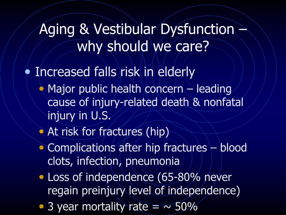
Aging & Vestibular Dysfunction –why should we care?
• Increased falls risk in elderly
• Major public health concern – leading cause of injury-related death & nonfatal injury in U.S.
• At risk for fractures (hip)
• Complications after hip fractures – blood clots, infection, pneumonia
• Loss of independence (65-80% never regain preinjury level of independence)
• 3 year mortality rate = ~ 50%

Types of BPPV
Cupulolithiasis- -otoconia in the utricle break
loose and adhere to the cupula of the
posterior semicircular canal
Canalithiasis--otoconia are free floating in the
posterior semicircular canal
• The most common form
• Accounts for 81-90% of all cases


Evaluation• Dix Hallpike (may use Frenzel Goggles)
• Patient sitting upright
• Turn head 45º to right
• Eyes remain open
• Assist patient into supine, head hanging position; maintain 45º head turn to right
• Patient focuses on target; observe eyes for nystagmus
• Maintain head hanging position for 30-40 seconds; if response occurs, wait for nystagmus to fatigue
• Patient centers head and returns to upright, seated position
• When seated, patient focuses on target; if response was demonstrated, may see nystagmus reversal
• Repeat with head hanging left

Frenzel Goggles
Used to detect nystagmus during Dix-Hallpike
evaluation


Diagnosis is based on a positive Dix-Hallpike
BPPV Nystagmus Classifications
• Counterclockwise – lateral canal BPPV
• Clockwise – lateral canal BPPV
• Down beating – superior canal BPPV
• Up beating – posterior canal BPPV

Typical Characteristics of Nystagmus
• Latency-10-40 seconds
• Paroxysmal
• Rotary nystagmus
• Duration < 1 minute
• Fatigues with repetition
• Nystagmus may reverse in upright position

http://www.youtube.com/watch?v=ZWnuAbBdKD0&feature=endscreen&NR=1
Nystagmus video

Interventions
• Wait/see – symptoms may subside within 2 months
• Medication (little benefit)
• Habituation exercises (Brandt-Daroff)
• Surgery
• Canalith Repositioning Procedures (CRP)
• Epley and Semont maneuvers
• Move otoconia from posterior canal into utricle (90% success rate)

• CRP/Epley is only done when a positive Dix-Hallpike is observed
• Should only be performed after a negative Cerebral Artery Screen
• Should only be performed by trained clinicians
• Not many therapists trained to treat BPPV: Certified Vestibular Rehab. Specialist
http://www.youtube.com/watch?v=7ZgUx9G0uEs&feature=related

Canalith Repositioning Procedure (CRP)
1. Supporting patient’s neck, quickly assist patient into supine, head hanging position; maintain 45º head position
• Otoconia move toward center of PSSC
2. Without lifting the patient’s head, help patient turn head to the opposite Hallpike position
• Otoconia reach common crus
3. Rotate head and body until patient is lying on side and nose is pointing to floor
• Otoconia pass through common crus
4. Maintaining head position from #3, assist patient to a seated position
• Otoconia enter utricle
5. Ask patient to center head and to tilt head down 20º
• Otoconia move into utricle
6. Repeat positions 1-5 until there is no nystagmus in any position

Canalith Repositioning Procedure/Epley
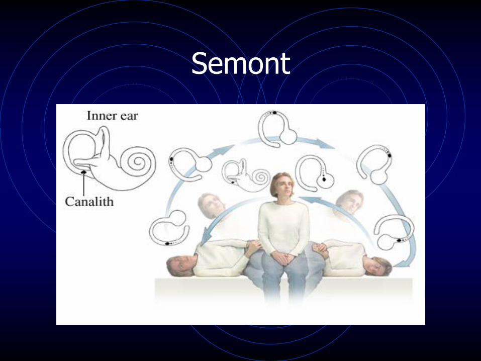
Semont

Patient instructions following CRP/Epley
• Sleep semi-recumbent for one night
• Avoid provoking head positions for one week
• Avoid moving head up and down
• Move head and body as a unit
• Can wear soft cervical collar as reminder for head movement
• Do not sleep on the side that was just treated

Bilateral BPPV
• Much less common
• If you see it, usually will see with head trauma
• Must treat one side at a time so you don’t ―undo‖ the side you just treated
• Harder to clear—generally will have multiple visits

Lateral Canal BPPV
• Otoconia migrate to the lateral canal
• Less common than posterior canal BPPV
• Can happen after CRP/Epley if head is lifted between first and second positions

Lateral Canal BPPV
• Roll test
• Body supine
• Head inclined 30º
• Turn head to either side

Lateral Canal BPPV
• Patients usually describe a strong and prolonged vertigo
• Often report dizziness when turning over in bed but not in other positions
• Can last up to or longer than a minute
• See a horizontal nystagmus, not rotary
• Nystagmus is typically present in both head positions but one is usually significantly worse
• Nystagmus can be geotropic (towards ground) or ageotropic (towards sky)
• Most commonly canalithiasis with geotropic nystagmus that is greater on the affected side

Maneuver for Lateral Canal BPPV

Summary
• Most common disorder of the inner ear’s vestibular system
• Etiology is idiopathic or head trauma
• More common in elderly – can have dramatic effect on quality of life
• Diagnosis is based on positive Dix-Hallpike
• CRP/Epley highly successful

www.neuropt.org/go/special-interest-groups/vestibular-rehabilitation

References:
• Boissonnault, W.G. (2011). Primary Care for the Physical Therapist Examination and Triage ,2nd Edition. St. Louis, MO: Elsevier Saunders
• Hain, T.C. (2011). Lateral Canal BPPV. http://www.dizziness-and-balance.com/disorders/bppv/bppv.html. Retrieved 03/03/12 from dizziness-and-balance.com
• Hain, T.C., Rodenbeek, M. (2009). BPPV. On the Level: Quarterly Newsletter of the VEDA, vol. 26 (No. 1) pp. 1-8
• Herdman, S.J. (2000). Vestibular Rehabilitation. Phila., PA: F.A. Davis Co.

References cont.:
• Shankman, G.A., Manske, R.C. (2011). Fundamental Orthopedic Management For The Physical Therapist Assistant, 3rd Edition. St. Louis, MO: Elsevier Mosby
Related Documents