Behavior and brain enzymatic changes after long-term intoxication with cadmium salt or contaminated potatoes Jamile F. Gonçalves a,⇑ , Fernando T. Nicoloso b , Pauline da Costa c , Júlia G. Farias b , Fabiano B. Carvalho c , Michelle M. da Rosa c , Jessié M. Gutierres c , Fátima H. Abdalla c , Juliana S.F. Pereira c , Glaecir R.M. Dias c , Nilda B.V. Barbosa c , Valderi L. Dressler c , Maribel A. Rubin c , Vera M. Morsch c , Maria Rosa C. Schetinger a,c,⇑ a Departamento de Bioquímica, Instituto de Ciências Básicas da Saúde, Universidade Federal do Rio Grande do Sul, Rua Ramiro Barcellos, 2600-Anexo, 90035-003 Porto Alegre, RS, Brazil b Departamento de Biologia, Centro de Ciências Naturais e Exatas, Universidade Federal de Santa Maria, Campus Universitário, Camobi, 97105-900 Santa Maria, RS, Brazil c Departamento de Química, Centro de Ciências Naturais e Exatas, Universidade Federal de Santa Maria, Campus Universitário, Camobi, 97105-900 Santa Maria, RS, Brazil article info Article history: Received 24 May 2012 Accepted 11 July 2012 Available online 20 July 2012 Keywords: AChE Anxiety Cd Na + ,K + -ATPase Memory Solanum tuberosum abstract This study investigated the cadmium (Cd) intoxication on cognitive, motor and anxiety performance of rats subjected to long-term exposure to diet with Cd salt or with Cd from contaminated potato tubers. Potato plantlets were micropropagated in MS medium and transplanted to plastic trays containing sand. Tubers were collected, planted in sand boxes and cultivated with 0 or 10 lM Cd and, after were oven- dried, powder processed and used for diet. Rats were divided into six groups and fed different diets for 5 months: control, potato, potato + Cd, 1, 5 or 25 mg/kg CdCl 2 . Cd exposure increased Cd concentration in brain regions. There was a significant decrease in the step-down latency in Cd-intoxicated rats and, elevated plus maze task revealed an anxiolytic effect in rats fed potato diet per se, and an anxiogenic effect in rats fed 25 mg/kg Cd. The brain structures of rats exposed to Cd salt or Cd from tubers showed an increased AChE activity, but Na + ,K + -ATPase decreased in cortex, hypothalamus, and cerebellum. There- fore, we suggest an association between the long-term diet of potato tuber and a clear anxiolytic effect. Moreover, we observed an impaired cognition and enhanced anxiety-like behavior displayed by Cd- intoxicated rats coupled with a marked increase of brain Cd concentration, and increase and decrease of AChE and Na + ,K + -ATPase activities, respectively. Ó 2012 Elsevier Ltd. All rights reserved. 1. Introduction Diet is the most important source of cadmium (Cd) exposure in the general population (non-occupational and nonsmoking) since Cd is not degraded in the environment and can also enter into the food chain (ATSDR, 2005). Both natural and anthropogenic sources of this heavy metal, including industrial emissions and the application of fertilizer and sewage sludge to farm land, may lead to the contamination of soils and to increased Cd uptake by crops and vegetables grown for human consumption (Järup and Akesson, 2009; Olsson et al., 2002). The uptake process of Cd from soil by plants is enhanced at low pH. Cd uptake by potatoes (Sola- num tuberosum) and carrots (Daucus carota) is sensitive to changes in pH (Öborn et al., 1995). Thus, the decrease of soil pH due to envi- ronmental acidification may further increase the Cd content in hu- man exposure pathway (Eriksson et al., 1996; Järup et al., 1998). Total human intake of Cd from food has been estimated as 2.8– 4.2 lg/kg body weight/week, which equates to approximately 40–60% of the current provisional tolerable weekly intake of 7 lg/kg body weight/week (Järup et al., 1998). Cd has a long biological half-life (15–30 years in humans) mainly due to its low rate of excretion from the body. Thus, pro- longed exposure to it has been linked to toxic effects since it gets accumulated over time in a variety of structures including kidneys, liver and central nervous system (CNS) (Zadorozhnaja et al., 2000). Cd is more toxic to newborn and young rats than to adult rats probably due to differences in the blood–brain barrier (BBB) integrity (Antonio et al., 2003; Yargiçoglu et al., 1997). However, this metal can increase BBB permeability, thus penetrating and accumulating in the brain of developing and adult rats (Gonçalves et al., 2010; Méndez-Armenta and Ríos, 2007; Pari and Murugavel, 2007; Shukla et al., 1996; Takeda et al., 1999) leading to brain intra- cellular accumulation, cellular dysfunction, and cerebral edema. Also, it can affect the degree and balance of excitation–inhibition 0278-6915/$ - see front matter Ó 2012 Elsevier Ltd. All rights reserved. http://dx.doi.org/10.1016/j.fct.2012.07.016 ⇑ Corresponding authors. Tel./fax: +55 55 3220 9557 (J.F. Gonçalves). E-mail addresses: [email protected] (J.F. Gonçalves), mariachitoli- [email protected] (Maria Rosa C. Schetinger). Food and Chemical Toxicology 50 (2012) 3709–3718 Contents lists available at SciVerse ScienceDirect Food and Chemical Toxicology journal homepage: www.elsevier.com/locate/foodchemtox

Welcome message from author
This document is posted to help you gain knowledge. Please leave a comment to let me know what you think about it! Share it to your friends and learn new things together.
Transcript
Food and Chemical Toxicology 50 (2012) 3709–3718
Contents lists available at SciVerse ScienceDirect
Food and Chemical Toxicology
journal homepage: www.elsevier .com/locate / foodchemtox
Behavior and brain enzymatic changes after long-term intoxicationwith cadmium salt or contaminated potatoes
Jamile F. Gonçalves a,⇑, Fernando T. Nicoloso b, Pauline da Costa c, Júlia G. Farias b, Fabiano B. Carvalho c,Michelle M. da Rosa c, Jessié M. Gutierres c, Fátima H. Abdalla c, Juliana S.F. Pereira c, Glaecir R.M. Dias c,Nilda B.V. Barbosa c, Valderi L. Dressler c, Maribel A. Rubin c, Vera M. Morsch c,Maria Rosa C. Schetinger a,c,⇑a Departamento de Bioquímica, Instituto de Ciências Básicas da Saúde, Universidade Federal do Rio Grande do Sul, Rua Ramiro Barcellos, 2600-Anexo, 90035-003 Porto Alegre,RS, Brazilb Departamento de Biologia, Centro de Ciências Naturais e Exatas, Universidade Federal de Santa Maria, Campus Universitário, Camobi, 97105-900 Santa Maria, RS, Brazilc Departamento de Química, Centro de Ciências Naturais e Exatas, Universidade Federal de Santa Maria, Campus Universitário, Camobi, 97105-900 Santa Maria, RS, Brazil
a r t i c l e i n f o
Article history:Received 24 May 2012Accepted 11 July 2012Available online 20 July 2012
Keywords:AChEAnxietyCdNa+,K+-ATPaseMemorySolanum tuberosum
0278-6915/$ - see front matter � 2012 Elsevier Ltd. Ahttp://dx.doi.org/10.1016/j.fct.2012.07.016
⇑ Corresponding authors. Tel./fax: +55 55 3220 955E-mail addresses: [email protected] (J
[email protected] (Maria Rosa C. Schetinger).
a b s t r a c t
This study investigated the cadmium (Cd) intoxication on cognitive, motor and anxiety performance ofrats subjected to long-term exposure to diet with Cd salt or with Cd from contaminated potato tubers.Potato plantlets were micropropagated in MS medium and transplanted to plastic trays containing sand.Tubers were collected, planted in sand boxes and cultivated with 0 or 10 lM Cd and, after were oven-dried, powder processed and used for diet. Rats were divided into six groups and fed different diets for5 months: control, potato, potato + Cd, 1, 5 or 25 mg/kg CdCl2. Cd exposure increased Cd concentrationin brain regions. There was a significant decrease in the step-down latency in Cd-intoxicated rats and,elevated plus maze task revealed an anxiolytic effect in rats fed potato diet per se, and an anxiogeniceffect in rats fed 25 mg/kg Cd. The brain structures of rats exposed to Cd salt or Cd from tubers showedan increased AChE activity, but Na+,K+-ATPase decreased in cortex, hypothalamus, and cerebellum. There-fore, we suggest an association between the long-term diet of potato tuber and a clear anxiolytic effect.Moreover, we observed an impaired cognition and enhanced anxiety-like behavior displayed by Cd-intoxicated rats coupled with a marked increase of brain Cd concentration, and increase and decreaseof AChE and Na+,K+-ATPase activities, respectively.
� 2012 Elsevier Ltd. All rights reserved.
1. Introduction
Diet is the most important source of cadmium (Cd) exposure inthe general population (non-occupational and nonsmoking) sinceCd is not degraded in the environment and can also enter intothe food chain (ATSDR, 2005). Both natural and anthropogenicsources of this heavy metal, including industrial emissions andthe application of fertilizer and sewage sludge to farm land, maylead to the contamination of soils and to increased Cd uptake bycrops and vegetables grown for human consumption (Järup andAkesson, 2009; Olsson et al., 2002). The uptake process of Cd fromsoil by plants is enhanced at low pH. Cd uptake by potatoes (Sola-num tuberosum) and carrots (Daucus carota) is sensitive to changesin pH (Öborn et al., 1995). Thus, the decrease of soil pH due to envi-ronmental acidification may further increase the Cd content in hu-
ll rights reserved.
7 (J.F. Gonçalves)..F. Gonçalves), mariachitoli-
man exposure pathway (Eriksson et al., 1996; Järup et al., 1998).Total human intake of Cd from food has been estimated as 2.8–4.2 lg/kg body weight/week, which equates to approximately40–60% of the current provisional tolerable weekly intake of7 lg/kg body weight/week (Järup et al., 1998).
Cd has a long biological half-life (15–30 years in humans)mainly due to its low rate of excretion from the body. Thus, pro-longed exposure to it has been linked to toxic effects since it getsaccumulated over time in a variety of structures including kidneys,liver and central nervous system (CNS) (Zadorozhnaja et al., 2000).Cd is more toxic to newborn and young rats than to adult ratsprobably due to differences in the blood–brain barrier (BBB)integrity (Antonio et al., 2003; Yargiçoglu et al., 1997). However,this metal can increase BBB permeability, thus penetrating andaccumulating in the brain of developing and adult rats (Gonçalveset al., 2010; Méndez-Armenta and Ríos, 2007; Pari and Murugavel,2007; Shukla et al., 1996; Takeda et al., 1999) leading to brain intra-cellular accumulation, cellular dysfunction, and cerebral edema.Also, it can affect the degree and balance of excitation–inhibition

3710 J.F. Gonçalves et al. / Food and Chemical Toxicology 50 (2012) 3709–3718
in synaptic neurotransmission as well as the antioxidant levels inanimal brain (Méndez-Armenta and Ríos, 2007; Minami et al.,2001; Provias et al., 1994). Exposure to Cd can produce long-termimpairment of neurobehavioral status such as alterations in atten-tion and memory as well as in the psychomotor, and visuomotorfunctioning and speed in workers (Hart et al., 1989; Viaene et al.,2000). Moreover, in rat studies have observed increased aggressiveand anxiety-like behaviours, impaired learning and memory pro-cesses, and changes in the development of the visual system (Desiet al., 1998; Gonçalves et al., 2010; Holloway and Thor, 1988; Terç-ariol et al., 2011; Yargiçoglu et al., 1997).
Some studies on Cd toxicity have found an association withbehavioral disturbances and cholinergic neurotransmission sincean increase or a decrease in the acetylcholinesterase (AChE, E.C.3.1.1.7) activity was verified in both animal models and humansthat showed behavioral impairments after exposure to Cd (Gonçal-ves et al., 2010; Pari and Murugavel, 2007). This enzyme hydro-lyses the neurotransmitter acetylcholine (ACh) in the synapticcleft of cholinergic synapses and neuromuscular junctions (Soreqand Seidman, 2001). Alterations in the AChE activity have beendemonstrated by our research group in various diseases and poi-sonings suggesting that this enzyme could be an important physi-ological and pathological parameter (Gonçalves et al., 2010; Kaizeret al., 2005; Schmatz et al., 2009).
In addition, Na+,K+-ATPase (E.C. 3.6.1.3), or sodium pump, isamong the enzymes particularly affected by Cd exposure (Antonioet al., 2002; Rajanna et al., 1983). This enzyme is one of the majormembrane proteins responsible for generating the membrane po-tential through the active transport of Na+ and K+ ions in the CNS(Aperia, 2007; Kaplan, 2002), consuming about 40–60% of ATP pro-duced in the brain (Erecinska et al., 2004). It is implicated in themetabolic energy production, in the uptake and release of seroto-nin and catecholamines as well as in the need to maintain cellularvolume control and neuronal excitability (Bogdanski et al., 1968;Hernandez, 1987; Mata et al., 1980; Mobasheri et al., 2000). Thus,disturbances in its activity could have serious consequences for theCNS functioning and play a relevant role in the learning and mem-ory mechanisms (Zhan et al., 2004).
In the present work we investigated the effect of Cd intoxicationon cognitive, motor and anxiety performance of rats subjected tolong-term exposure to diet with low levels of Cd salt or with Cdfrom contaminated potato tubers measured on inhibitory avoid-ance, open field and elevated plus maze tests. In addition, we alsoevaluated the influence of this metal on AChE and Na+,K+-ATPaseactivities in different brain structures.
2. Materials and methods
2.1. Chemicals
Acetylthiocholine iodide, cadmium chloride monohydrate, 5,50-dithio-bis-2-nitrobenzoic acid (DTNB), tris-(hydroxymethyl)-aminomethane GR, ouabain andCoomassie brilliant blue G were obtained from Sigma Chemical Co. (St. Louis, MO,USA). Agar–agar and thiamine chloride hydrochloride were obtained from MerkKGaA (Rockland, Massachusetts, USA). Myo inositol was obtained from Vetec Chem-ical Co. (Duque de Caxias, Rio de Janeiro, Brazil). All other reagents used in theexperiments were of analytical grade and of the highest purity.
2.2. Potato cultivation
Potato plantlets (S. tuberosum L.) for tissue culture were obtained from the Po-tato Breeding and Genetics Germplasm Program, Universidade Federal de SantaMaria, RS, Brazil. One potato genotype widely planted in Southern Brazil, Macaca,was used in this study. Nodal segments (1.0 cm long) without leaves were micro-propagated in MS medium (Murashige and Skoog, 1962), supplemented with30 g/L of sucrose, 0.1 g/L of myo-inositol and 6 g/L of agar in the absence growthregulators (Nicoloso et al., 2001). The plants were selected, comprising a homoge-neous population, and used as donor explants. The apical and basal regions ofshoots were not used.
After 21 days in MS medium, in vitro, plants were transplanted to plastic trayscontaining sand as substrate and cultivated for 27 days in a greenhouse with shad-ing (50%). A second hydroponic growth period was performed in sand box withspacing of 10 cm, reaching a density of 100 per m2 of surface seedlings. These plantsreceived three daily irrigations (15 min each) with complete nutrient solution con-taining (mg/L): 155.90 N; 46.40 P; 5271.00 S; 123.00 Ca; 30.00 Mg; 253.60 K;2622.00 B; 133.00 Na; 277.00 Mo; 2274.00 Zn; 636.00 Cu; 6501.00 Mn and 1.20 Fe.
The produced tubers were planted in sand box and cultivated during the springgrowing season (September to November) with the same experimental system pre-viously described. The Cd exposure was with the addition of the same nutrient solu-tion in the form of cadmium chloride monohydrate (CdCl2�H2O) at concentrations of0 (control) or 10 lM. At all stages of the experiment the pH of the solutions usedwas adjusted daily to 5.5 ± 0.1 by titration with solutions of HCl or NaOH 0.1 M.The plants were cultivated for 91 days.
At the end of the cycle, the plants were collected and tubers were washed threetimes with distilled water and dried with tissue paper. The produced tubers wereoven-dried at 65 �C to a constant mass for the determination of biomass as wellas Cd accumulation. Dried plant tissues (0.01–0.1 g) were ground and digested with4 mL of concentrated HNO3. Sample decomposition was carried out using a heatingblock Velp Scientifica (Milano, Italy). Heating was set at 130 �C for 2 h. Plastic capswere fitted to the vessels to prevent losses by volatilization. The final Cd concentra-tion in the produced tubers (powder) was 4.5 lg/g of dry weight.
2.3. Animals and dietary treatment
Three-week-old male Wistar rats weighing 85.5 ± 5.3 g from the Central AnimalHouse of the Universidade Federal de Santa Maria (UFSM) was used in this exper-iment. At weaning, the pups were acclimated for 10 days before the beginning ofthe experiments and had free access to drinking water and food (Supra�, Brazil)during the acclimatation period or diet during the experimental period. Rats weremaintained at a constant temperature (23 ± 1 �C) on a 12 h light/dark cycle. All ani-mal procedures were approved by the Animal Ethics Committee from UFSM.
Animals (10 rats per treatment) were randomly divided into six experimentalgroups and fed different diets (Table 1) for 5 months. The diet was prepared in anindustrial mixer to allow the uniformity of the mixture according to Barbosaet al. (2008). The choice of Cd concentrations was based on previous studies, whichshow that Cd diet intake causes overt signals of toxicity in rats (Callegaro et al.,2010a,b). After preparation, the diets were frozen (�20 �C) until use. The bodyweight of animals was obtained weekly until the end of the experiment period.
2.4. Behavior procedure
2.4.1. Inhibitory avoidanceOne day after the end of the treatment, animals were subjected to training in a
step-down inhibitory avoidance apparatus according to Guerra et al. (2006). Briefly,rats were subjected to a single training session in a step-down inhibitory avoidanceapparatus, which consisted of a 25 � 25 � 35 cm box with a grid floor whose leftportion was covered by a 7 � 25 cm platform, 2.5 cm height. The rat was placedgently on the platform facing the rear left corner and when the rat stepped downwith all four paws on the grid, a 2-s 0.4-mA shock was applied to the grid. Memoryretention was evaluated in a test session carried out 24 h after training, in whichtrained animals were placed on the training box platform, and the step-down la-tency was measured. A cut-off time of 300 s was imposed on step-down latencyduring testing session. The apparatus was cleaned with 30% ethanol before andafter each rat occupied it.
2.4.2. Open fieldImmediately after the inhibitory avoidance testing session, all animals were
transferred to an open field in order to assess exploratory activity. Animals wereplaced on the center quadrant of a round open field (56 � 40 � 30 cm) with thefloor divided into 12 squares measuring 12 � 12 cm each. An observer, who wasnot aware of the pharmacological treatments, manually recorded the number ofcrossing and rearing responses over 5 min. Crossing was defined as the total num-ber of areas crossed with the four paws and rearing was defined as the total numberof stand-up responses on two paws. This test was carried out to identify possiblemotor disabilities that might have influenced inhibitory avoidance performanceat testing.
2.4.3. Foot shock sensitivity testThe reactivity to shock was evaluated in the same apparatus used for inhibitory
avoidance, except that the platform was removed. The modified ‘‘up and down’’method by Rubin et al. (2004) was used to determine the flinch, jump and vocali-zation thresholds in experimentally naive animals. Animals were placed on the gridand allowed a 3 min habituation period before the start of a series of shocks (1 s)delivered at 10 s intervals. Shock intensities ranged from 0.1 to 0.6 mA in 0.1 mAincrements. Adjustments in the shock intensity were made in accordance with eachanimal response. The intensity was raised by 1 unit when no response occurred andlowered by 1 unit when there was a response. A flinch response was defined as thewithdrawal of one paw from the grid floor, and a jump response was defined as the

Table 1Experimental groups, diets and the concentration of Cd found in the diets.
Groups Diets Cd concentration (mg/kg diet)⁄
Control Only diet <0.03Pot 10% of diet composed of tuber powder from potatoes grown without Cd <0.03Pot + Cd 10% of diet composed of tuber powder from potatoes grown with Cd 0.64 ± 0.041 mg/kg Cd Diet mixed with 1 mg/kg of CdCl2 salt dissolved in water 0.88 ± 0.025 mg/kg Cd Diet mixed with 5 mg/kg of CdCl2 salt dissolved in water 3.62 ± 0.2325 mg/kg Cd Diet mixed with 25 mg/kg of CdCl2 salt dissolved in water 18.47 ± 0.41
Abbreviations: Pot (potato); Cd (cadmium).⁄Values are expressed as means ± SEM (n = 4). Measured by ICP OES with detection limit of 0.03 mg/kg.
J.F. Gonçalves et al. / Food and Chemical Toxicology 50 (2012) 3709–3718 3711
withdrawal of three or four paws. Two measurements of each threshold (flinch,jump and vocalization) were taken and the mean of each score was calculated foreach animal.
2.4.4. Elevated plus maze taskThe anxiolytic-like behavior was evaluated using the task of the elevated plus
maze as previously described (Frussa-Filho et al., 1999; Rubin et al., 2000). Theapparatus was made of wood and consisted of four arms, all with the same dimen-sions (50 � 10 cm) and elevated 50 cm above the floor. Two of these arms were en-closed by 40 cm high lateral walls with an open roof and located perpendicularly tothe other two opposed open arms. The four arms delimited a central area of100 cm2. Rats were placed in the center of the maze facing an open arm and wereallowed to explore the maze freely for 5 min. The time spent and the number of en-tries in open and closed-arms as well as the total number of arm entries were re-corded. The apparatus was thoroughly cleaned with 30% ethanol between eachsession.
2.5. Brain tissue preparation
After behavioral tests, animals were anesthetized and submitted to euthanasia.The cranium was opened and the structures were gently removed and separatedinto cerebral cortex (CO), hippocampus (HC), hypothalamus (HT), cerebellum (CE)and striatum (ST). To verify the cadmium concentration in brain structures, threeor four animals per group were randomly chosen. For the other animals, the brainstructures were homogenized in a glass potter in a solution of 10 mM Tris–HCl, pH7.4, on ice, at a proportion of 1:10 (w/v). The resulting homogenate was used todetermine the Na+,K+-ATPase activity. A fraction of homogenate was centrifugedat 1800 rpm for 10 min and the resulting supernatant was used to determine theAChE activity.
2.6. Cadmium concentration in brain regions
Brain structures were weighted in glass vessels and 3–8 mL of HNO3 was addedfor digestion. Digestion was performed using a block (Velp Scientifica, Milano, Italy)heated at 130 �C during 3 h. After this time, 2 mL of H2O2 was added and sampleswere heated again for more 1 h. Digested samples were then transferred to polypro-pylene flasks and the volume was completed with water to 30–50 mL. Cadmiumdetermination was performed by graphite furnace atomic absorption spectrometry(GF AAS, contrAA 700, Analytik Jena) using a xenonium lamp set at 28.8018 nm. Theheating program was performed using the temperatures of 120, 600, 1600 and2300 �C for drying, pyrolysis, atomization and cleaning steps, respectively. In addi-tion, Cd determination was also performed by inductively coupled plasma opticalemission spectrometry (ICP OES) using a concentric nebulizer (Meinhard Associ-ates, Golden, USA) and a cyclonic spray chamber (Glass Expansion, Inc., West Mel-bourne, Australia). However, in view of higher limit of detection obtained incomparison with the results obtained by GF AAS, Cd measurements were preferen-tially performed by GF AAS.
2.7. Cerebral AChE enzymatic assay
The AChE enzymatic assay was determined by a modification of the spectropho-tometric method of Ellman et al. (1961) as previously described by Rocha et al.(1993). The reaction mixture (2 mL final volume) contained 100 mM K+-phosphatebuffer, pH 7.5 and 1 mM 5,50-dithio-bis-nitrobenzoic acid (DTNB). The method isbased on the formation of the yellow anion, 5,50-dithio-bis-acid nitrobenzoic, mea-sured by absorbance at 412 nm during 2-min incubation at 25 �C. The enzyme waspre-incubated for 2 min. The reaction was initiated by adding 0.8 mM acetylthioch-oline iodide (AcSCh). Protein was determined previously and adjusted for eachstructure: CO (0.6–0.8 mg/mL), HC (0.8 mg/mL), HT (0.6 mg/mL), CE (0.5–0.6 mg/mL), and ST (0.4 mg/mL). All samples were run in duplicate or triplicate and the en-zyme activity was expressed in lmol AcSCh/h/mg of protein.
2.8. Na+,K+-ATPase enzymatic assay
The Na+,K+-ATPase activity was measured as previously described (Wyse et al.,2000) and modified by Carvalho et al. (2012). Briefly, the assay medium consisted of(in mM) 30 Tris–HCl buffer (pH 7.4), 0.1 EDTA, 50 NaCl, 5 KCl, 6 MgCl2 and 50 lg ofprotein in the presence or absence of ouabain (1 mM), in a final volume of 350 lL.The reaction was started by the addition of adenosine triphosphate to a final con-centration of 3 mM. After 30 min at 37 �C, the reaction was stopped by the additionof 70 lL of 50% (w/v) trichloroacetic acid. Saturating substrate concentrations wereused and the reaction was linear with protein and time. Appropriate controls wereincluded in the assays for non-enzymatic hydrolysis of ATP. The amount of inor-ganic phosphate (Pi) released was quantified colorimetrically as previously de-scribed (Fiske and Subbarow, 1925), using NaH2PO4 as reference standard.Specific the Na+,K+-ATPase activity was calculated by subtracting the ouabain-insensitive activity from the overall activity (in the absence of ouabain) and ex-pressed in nmol of Pi/min/mg of protein.
2.9. Protein determination
The protein content was determined colorimetrically according to the Bradfordmethod (1976) using bovine serum albumin (1 mg/mL) as standard solution.
2.10. Statistical analysis
Statistical analysis of the training and test step-down latencies was carried outby Kruskal–Wallis followed by post hoc analyses (nonparametric Dunn’s test). Allother parameters evaluated were analyzed by one-way ANOVA, followed by Stu-dent–Newman–Keuls (SNK) or Duncan’s multiple range tests, where p < 0.05 wasconsidered to represent a significant difference in all experiments.
3. Results
3.1. Cd or potato intake did not affect body weight measurements
Table 2 shows the body weight of the animals at the onset andat the end of the experiment as well as their body weight gain. Ascan be observed, there were no statistical differences in the bodyweight measurements between the groups.
3.2. Cd exposure caused memory deficits
Fig. 1 shows the effect of Cd and potato diet intake on step-down latencies. The statistical analysis of testing showed thatlong-term exposure to Cd salt (1, 5 and 25 mg/kg) as well as Cdaccumulated in potato tubers caused memory impairment. Statis-tical analysis of training showed no significant differences betweenthe groups.
Because the motivational disparities in the training session mayaccount for differences in inhibitory avoidance at testing, experi-ments were performed to assess whether Cd or potato intake af-fected the shock threshold or the locomotor ability of theanimals. Statistical analysis of open-field data revealed that neitherCd nor potato intake caused gross motor disabilities at testing asdemonstrated by the similar number of crossing or rearing re-sponses exhibited by the animals. Moreover, Cd or potato intakedid not alter foot shock sensitivity as demonstrated by the similarflinch, jump and vocalization thresholds exhibited by the rats(Table 3).

Table 2Effects of cadmium (Cd) and potato (pot) diet intake in the body weight gain (g) ofexperimental groups.
Groups Initial body weight(g)
Final body weight(g)
Body weight gain(g)
Control 81.5 ± 4.3 412.6 ± 3.1 +331.1Pot 87.8 ± 6.3 383.1 ± 0.4 +295.3Pot + Cd 83.3 ± 6.4 438.7 ± 1.1 +355.41 mg/kg Cd 85.0 ± 3.9 388.3 ± 1.6 +303.35 mg/kg Cd 87.9 ± 2.4 424.5 ± 9.1 +336.625 mg/kg Cd 87.3 ± 4.3 435.7 ± 2.8 +348.4
Values are expressed as means ± SEM. n = 6–10 rats per group (p < 0.05).
3712 J.F. Gonçalves et al. / Food and Chemical Toxicology 50 (2012) 3709–3718
3.3. Cd exposure or potato intake was anxiogenic and anxiolitic,respectively
Anxiety studies using an elevated plus maze showed a loweranxiety in rats fed potato diet since these rats spent more timein the open arms, spent less time in the closed arms, and enteredthe open arms more times compared with those of the othergroups (Fig. 2A, B and, C, respectively). On the other hand, ratsfed 25 mg/kg Cd diet showed a higher anxiety since these ratsspent less time in the open arms, spent more time in the closedarms, and entered the open arms less times compared with thoseof the other groups. There were no significant alterations in thenumber of entries in the closed arms or in the total number of en-tries in arms between the experimental groups (Fig. 2D and E).
3.4. Cd exposure increased Cd concentration in brain regions
Cd exposure increased Cd concentration in brain regions of ratsfed potato grown with Cd, 1, 5 and 25 mg/kg Cd diet in relation to
Fig. 1. Effect of cadmium (Cd) and potato (Pot) diet intake on step-down latencies. Aftermedian ± interquartile range of training and test. ⁄p < 0.05 compared with the other gro
Table 3Effects of cadmium (Cd) and potato (pot) diet intake on the behavior of rats in the openavoidance testing session and on foot shock sensitivity (flinch, jump and vocalization).
Group Crossing Rearing
Control 31.50 ± 4.76 25.75 ± 3.85Pot 33.75 ± 4.85 25.25 ± 2.84Pot + Cd 34.50 ± 3.55 26.40 ± 3.121 mg/kg Cd 23.20 ± 3.90 18.50 ± 3.045 mg/kg Cd 25.40 ± 3.46 20.80 ± 2.8025 mg/kg Cd 32.55 ± 2.31 23.70 ± 1.74Statistical analysis F(5,51) = 0.13 F(5,51) = 0.33
p > 0.05 p > 0.05
Data are means ± SEM for 8–10 animals per group.
the control (Table 4). Control rats and rats fed potato diet per seshowed Cd concentrations lower than the detection limit(0.009 lg/g) in different brain structures. Rats fed potato grownwith Cd, 1 and 5 mg/kg Cd diet showed small and similar Cd con-centrations of around 0.011 lg/g in different brain structures. Ratsfed 25 mg/kg Cd showed highest Cd concentrations in all brain re-gions when compared with the other groups.
3.5. Cd exposure increased AChE activity
The AChE activity in different brain structures is given in Fig. 3.There were no significant alterations in the AChE activity in cere-bral cortex (Fig. 3A). Hippocampus AChE activity increased 25%in rats fed 25 mg/kg Cd diet in relation to the control (Fig. 3B).Hipothalamus AChE activity increased 63% and 52% in rats fed 5and 25 mg/kg Cd diet in relation to the control, respectively(Fig. 3C). Cerebellum AChE activity increased 45%, 32% and 39%in rats fed 1, 5 and 25 mg/kg Cd diet in relation to the control,respectively (Fig. 3D). Striatum AChE activity increased 44%, 44%,57% and 66% in rats fed potato grown with Cd, 1, 5 and 25 mg/kgCd diet when compared with the control, respectively (Fig. 3E).
3.6. Cd exposure decreased Na+,K+-ATPase activity
Results obtained for Na+,K+-ATPase activity in different brainstructures are presented in Fig. 4. There was a decrease of 32%,25% and 39% in Na+,K+-ATPase activity in cerebral cortex of ratsfed potato grown with Cd, 5 and 25 mg/kg Cd diet in relation tothe control, respectively (Fig. 4A). There were no significant alter-ations in the Na+,K+-ATPase activity in hippocampus (Fig. 4B). Adecrease of 28% in Na+,K+-ATPase activity in hipothalamus of ratsfed 25 mg/kg Cd diet was observed in relation to the control(Fig. 4C). ‘‘There were no significant alterations in the Na+,K+-ATP-
one treatment-free day, animals were tested in a step-down latency test. Data areups at testing by the Dunn’s nonparametric multiple comparison task (n = 8–10).
-field (number of crossing and rearing responses) immediately after the inhibitory
Flinch (mA) Jump (mA) Vocalization (mA)
0.32 ± 0.03 0.36 ± 0.03 0.45 ± 0.010.31 ± 0.02 0.39 ± 0.01 0.47 ± 0.020.33 ± 0.02 0.36 ± 0.02 0.43 ± 0.010.33 ± 0.02 0.33 ± 0.01 0.45 ± 0.010.33 ± 0.02 0.37 ± 0.03 0.49 ± 0.010.34 ± 0.02 0.37 ± 0.02 0.46 ± 0.01F(5,51) = 0.88 F(5,51) = 0.61 F(5,51) = 0.15p > 0.05 p > 0.05 p > 0.05
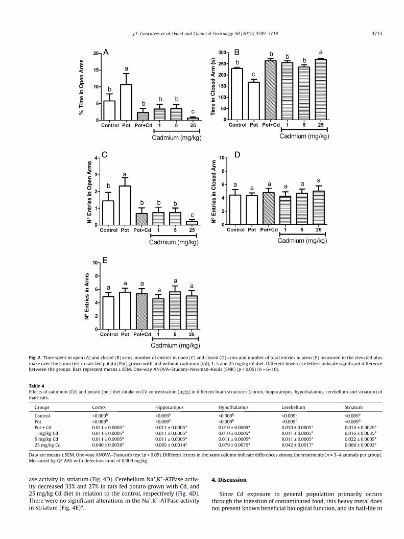
Fig. 2. Time spent in open (A) and closed (B) arms, number of entries in open (C) and closed (D) arms and number of total entries in arms (E) measured in the elevated plusmaze over the 5 min test in rats fed potato (Pot) grown with and without cadmium (Cd), 1, 5 and 25 mg/kg Cd diet. Different lowercase letters indicate significant differencebetween the groups. Bars represent means ± SEM. One-way ANOVA–Student–Newman–Keuls (SNK) (p < 0.05) (n = 6–10).
Table 4Effects of cadmium (Cd) and potato (pot) diet intake on Cd concentration (lg/g) in different brain structures (cortex, hippocampus, hypothalamus, cerebellum and striatum) ofmale rats.
Groups Cortex Hippocampus Hypothalamus Cerebellum Striatum
Control <0.009b <0.009b <0.009b <0.009b <0.009b
Pot <0.009b <0.009b <0.009b <0.009b <0.009b
Pot + Cd 0.011 ± 0.0005a 0.011 ± 0.0005a 0.010 ± 0.0005a 0.010 ± 0.0005a 0.014 ± 0.0029a
1 mg/kg Cd 0.011 ± 0.0005a 0.011 ± 0.0005a 0.010 ± 0.0005a 0.011 ± 0.0005a 0.016 ± 0.0035a
5 mg/kg Cd 0.011 ± 0.0005a 0.011 ± 0.0005a 0.011 ± 0.0005a 0.011 ± 0.0005a 0.022 ± 0.0005a
25 mg/kg Cd 0.040 ± 0.0058a 0.065 ± 0.0014a 0.079 ± 0.0075a 0.042 ± 0.0017a 0.060 ± 0.0092a
Data are means ± SEM. One-way ANOVA–Duncan’s test (p < 0.05). Different letters in the same column indicate differences among the treatments (n = 3–4 animals per group).Measured by GF AAS with detection limit of 0.009 mg/kg.
J.F. Gonçalves et al. / Food and Chemical Toxicology 50 (2012) 3709–3718 3713
ase activity in striatum (Fig. 4D). Cerebellum Na+,K+-ATPase activ-ity decreased 33% and 27% in rats fed potato grown with Cd, and25 mg/kg Cd diet in relation to the control, respectively (Fig. 4D).There were no significant alterations in the Na+,K+-ATPase activityin striatum (Fig. 4E)’’.
4. Discussion
Since Cd exposure to general population primarily occursthrough the ingestion of contaminated food, this heavy metal doesnot present known beneficial biological function, and its half-life in

Fig. 3. Acetylcholinesterase (AChE) activity in supernatant of cerebral cortex (A), hippocampus (B), hypothalamus (C), cerebellum (D) and striatum (E) of rats fed potatogrown with and without Cd, 1, 5 and 25 mg/kg Cd diet. Different lowercase letters indicate significant difference between the groups. Bars represent means ± SEM. One-wayANOVA–Duncan’s multiple range test (p < 0.05) (n = 4–6).
3714 J.F. Gonçalves et al. / Food and Chemical Toxicology 50 (2012) 3709–3718
the organisms is elevated, a long period of exposure to Cd levels viadiet may represent a great risk to human health. Thus, the presentinvestigation on the long-term exposure to diet with low levels ofCd salt or with Cd from contaminated potato tubers represents asignificant contribution to better understand the Cd toxicology.In this study Cd-exposed adult rats showed small amount of Cdin brain structures but significantly higher than control valuesand enough to cause brain injury, corroborating with the literaturedata (Gonçalves et al., 2010; Takeda et al., 1999; Wong and Klaas-sen, 1982). Moreover, although Cd insult did not alter the bodyweight measurements of the rats, it was harmful to the brain func-tions and consequently to animal behavior.
The inhibitory avoidance test is a classic model behavioral testwith a strong aversive component used to evaluate learning andmemory in rats and mice (Cahill et al., 1986). In the present study,we observed a significant decrease in the step-down latency in Cd-intoxicated rats by the inhibitory avoidance test, suggesting learn-
ing and memory impairment in these animals. This result corrob-orates our recently published data where rats intoxicated orallywith Cd showed impaired cognition (Gonçalves et al., 2010). In fact,some studies have demonstrated that Cd induces neurotoxicity inanimals with a wide spectrum of clinical entities such as changesin the normal brain neurochemistry as well as neurological andbehavioral disturbances (Gonçalves et al., 2010; Minami et al.,2001).
As reported by Schmatz et al. (2009), a major concern in shock-motivated learning tests, particularly in those investigating the ef-fect of drugs given before the acquisition of a given test, is whetherpharmacological treatments affect locomotor activities or motiva-tional aspects of learning, such as shock sensitivity. To rule out thispossibility, immediately after inhibitory avoidance testing ses-sions, the animals were subjected to an open-field test which iswidely used to evaluate motor disorders (Belzung and Griebel,2001). The open field session revealed that the pharmacological

Fig. 4. Activity of Na+K+-ATPase in homogenate of cerebral cortex (A), hippocampus (B), hypothalamus (C), cerebellum (D) and striatum (E) of rats fed potato (Pot) grown withand without cadmium (Cd), 1, 5 and 25 mg/kg Cd diet. Different lowercase letters indicate significant difference between the groups. Bars represent means ± SEM. One-wayANOVA–Student–Newman–Keuls (SNK) (p < 0.05) (n = 4–6).
J.F. Gonçalves et al. / Food and Chemical Toxicology 50 (2012) 3709–3718 3715
treatment did not alter spontaneous locomotor activity since ratsfed the different diets showed a similar number of crossing or rear-ing responses. Moreover, in our study we demonstrated that rats ofdifferent treatments did not show altered shock sensitivity as ver-ified by their similar flinch, jump and vocalization thresholds.Thus, these data suggest that neither Cd nor potato intake beforetraining of inhibitory avoidance caused motor disabilities nor al-tered foot shock sensitivity. This way, we can exclude the possibil-ity that these parameters may have contributed to the alteration instep-down latencies.
Besides learning and memory evaluation, we also measured theanxiolytic performance of the rats by the elevated plus maze, com-monly used to study anxiety-related behavior in rodents (Belzungand Griebel, 2001). Our results showed a lower anxiety in rats fedpotato diet per se. According to Allemão (1976), the word Solanumcomes from the Latin word Solari which means ‘‘to relieve’’, and thevegetable which receives this denomination has been employed forits tranquilizer property. It is well known that benzodiazepines are
among the most frequently prescribed drugs which present tran-quilizing, sedative-hypnotic, anxiolytic, and anticonvulsant effectsmediated by binding to a specific subtype of the GABA(A) receptor,i.e., the a1-type GABA(A) receptor, which is mainly expressed incortical areas and in the thalamus of all vertebrates (Rudolphet al., 1999). Interestingly, Kavvadias et al. (2000) reported theendogenous formation of benzodiazepines such as N-des-methyldiazepam (temazepam) and diazepam in amounts of about70–450 ng/g cell tissue of S. tuberosum. These contents are approx-imately 10- to 100-fold higher than those previously found in po-tato tuber (2–4 ng diazepam equivalents/g) (Wildmann, 1988).Moreover, Muceniece et al. (2008) postulated that potato juice ex-tracted from tuber marrow might contain GABA(A) receptor GABA-site active compounds since its low content of diazepam could notsustain the anticonvulsant activity of potato juice in vivo, suggest-ing that juice as well as potato taken as food may have the capacityof influencing brain GABA-ergic activity. Besides benzodiazepinederivatives, other compounds that exhibit anxiolytic effects were
3716 J.F. Gonçalves et al. / Food and Chemical Toxicology 50 (2012) 3709–3718
identified in potato such as a variety of flavonoids that could be asubtype-selective partial agonist of GABA(A) receptors (Giorgettiand Negri, 2011). Thus, our results reinforce the anxiolytic proper-ties of the Solanaceae family and to the best of our knowledge thisis the first approach associating the long-term diet of potato tuberto a clear anxiolytic effect in rodents. With regard to Cd-based diet,the present investigation showed that the highest Cd concentrationapplied to the diet (25 mg/kg) caused higher anxiety in rats. Leretet al. (2003) showed that the rats co-exposed to Cd (10 mg/L) andPb (300 mg/L) in drinking water from the beginning of pregnancyuntil weaning showed an increased anxiety-like behaviour andalthough the mechanism by which these metals are able to alterbehaviour in the elevated plus-maze is yet to be established, it ap-pears that there is a link with hippocampal serotoninergic anddopaminergic neurons. On the other hand, Minetti and Reale(2006) showed a lower anxiety in all the offspring prenatally ex-posed to 0.6 mg/kg Cd subcutaneously. It is important to pointout that their dose of Cd is about five times less than that appliedby us here and also the male offspring in all the groups studied byMinetti and Reale (2006) evidenced a clear anxious behavior withrespect to the female rats. Moreover, when the total number of armentries was evaluated, the absence of changes in this measure indi-cated that all groups have similar locomotor activity corroboratingour findings in the open field test. This indicates that the changesobserved in the plus maze are consistent with a decreased andan increased anxiety behavior produced by potato and Cd intake,respectively.
In the present study, all Cd diets increased the AChE activity inthe striatum. However, the activation of AChE in the hippocampus,hypothalamus and cerebellum was observed in a dependence ofthe Cd concentration administered and no effect in response toCd was detected in the cerebral cortex between the groups. Bothin vitro and in vivo effects of Cd on the AChE activity have been de-scribed in the literature for different animal models (Carageorgiouet al., 2004, 2005; Luchese et al., 2007; Pari and Murugavel, 2007).The mechanism of action of Cd on AChE enzyme has been hypoth-esized to be either the displacement of metal cofactors from the ac-tive site or the direct deactivation of the enzyme site (Casalinoet al., 1997). Moreover, Cd can leads to alteration of the structuralintegrity of lipids and secondarily affects membrane-bound en-zymes such as AChE and Na+,K+-ATPase. Thus, alterations in the li-pid membrane by oxidative stress could be a decisive factor in themodification of the conformational state of the AChE and Na+,K+-ATPase molecules, which would explain changes in their activities(Gonçalves et al., 2010; Pal et al., 1993; Schmatz et al., 2009; Srin-ivasan and Ramprasath, 2011). However, results are controversialand the activation as well as the inhibition of the AChE activityhas been reported. The activity of AChE in rat brain synaptosomalplasma membranes showed a considerable decrease after 6 h of Cdexposure, followed by a progressive increase up to 24 h (Fasitsaset al., 1991). Brain AChE activity was decreased in rats exposedto 1, 2 or 5 mg/kg Cd for 8 h (Carageorgiou et al., 2004) as wellas in mice exposed subcutaneously to 10 mmol/kg, five times/weekfor 4 weeks (Luchese et al., 2007). Decreased AChE activity wasfound in hippocampus, cerebellum and hypothalamus of ratsintoxicated with 2 mg/kg Cd by gavage every other day for 30 days(Gonçalves et al., 2010). However, an increase in the AChE activitywas found in brain of rats exposed to 1 mg/kg Cd intraperitoneallyfor 14 days or intramuscularly for 4 months (Carageorgiou et al.,2004, 2005). Interestingly, AChE activation leads to a fast ACh deg-radation and a subsequent down stimulation of ACh receptorscausing undesirable effects on cognitive functions (Soreq and Seid-man, 2001). Based on our results we can suggest that the increasein the AChE activity caused by Cd leads to a reduction of choliner-gic neurotransmission efficiency due to a decrease in ACh levels inthe synaptic cleft thus contributing to progressive cognitive
impairment. Furthermore, we may infer that the activator effectelicited by Cd diet on the AChE activity could be one of the mech-anisms involved in the memory impairment observed in the inhib-itory avoidance test in this study (Schmatz et al., 2009). In fact,there is a wealth of evidence that brain cholinergic systems partic-ipate in memory processes. Our research team has already re-ported that cerebral AChE activity was restored by theantioxidant N-acetylcysteine which has beneficial actions modu-lating cholinergic neurotransmission and improving cognition(Gonçalves et al., 2010).
Cholinergic pathways, which fulfil a pivotal role in the controlof cognitive-attentional, arousal and motivation function, ramifyextensively in the hippocampus, amygdala, septum, frontal cortexand several other limbic regions. They reciprocally interact withGABAergic, monoaminergic, and glutamatergic pathways playinga role in the control of anxious states (Milan, 2003). There isemerging evidence that increased ACh levels in brain reduce anxi-ety. More specifically there is evidence that some of these anxio-lytic effects of ACh are modulated by the hippocampus since anoverall enhancement of cholinergic transmission in the hippocam-pus was accompanied by anxiolytic properties (Degroot et al.,2001; Degroot and Treit, 2002). Degroot et al. (2001) found thatinfusions of the AChE inhibitor physostigmine (10 mg/mL) intothe dorsal hippocampus increased open-arm exploration in theplus-maze test and decreased burying behavior in the shock-probetest, demonstrating that general up-regulation of hippocampalcholinergic activity (increased ACh levels) resulted in a clear anxio-lytic effect in two different tests of anxiety. The anxiolytic effectsinduced by stimulating the hippocampal cholinergic system areconsistent with previous data indicating that intrahippocampalinfusions of both muscarinic and nicotinic antagonists increaseanxiety (File et al., 1998; Hess and Blozovski, 1987; Smytheet al., 1998). Our study produced consistent results that the cholin-ergic system may be involved in anxiety-like behaviour as well asmemory impairment observed in rats fed with the highest Cd diet(25 mg/kg) since we found that the enzyme AChE was activated,which probably decreases ACh levels in four cerebral structuresincluding the hippocampus, a region particularly important in anx-iety and memory processes.
The activity of Na+,K+-ATPase was decreased in cerebral cortex,hypothalamus, and cerebellum of rats exposed to Cd salt or Cdfrom tubers. In hippocampus and striatum there was no alterationin the Na+,K+-ATPase activity between the groups. Several studieshave demonstrated that certain divalent metal ions are potentinhibitors of brain Na+,K+-ATPase (Antonio et al., 2002; Chettyet al., 1992; Hexum, 1974; Prakash et al., 1973; Rajanna et al.,1983). In this sense, our results confirm that Cd may exert aninhibitory effect on the Na+,K+-ATPase activity. According to Anto-nio et al. (2002), this enzyme plays a vital role in linking the extra-cellular signals to the intracellular medium in neural tissues. Thusthe inhibition of this enzyme by Cd could be damaging to the braincells leading to an earlier stage of edema, followed by a later stageof degeneration and necrosis. Also, Na+,K+-ATPase inhibition byouabain impairs learning and memory in Morris water maze andstep-through passive avoidance tasks (Sato et al., 2004; Zhanet al., 2004), showing the main role of this enzyme on learningand memory processes which could be associated with Cd-inducedimpaired cognition observed in the present investigation. Rajannaet al. (1983) proposed that Cd may compete with ATP and Na+ siteson Na+,K+-ATPase inhibiting its activity in rat brain synaptosomes.Chetty et al. (1992) have reported that Cd interferes withphosphorylation state of the enzyme which results in its inhibition.Borges et al. (2005) postulated that the inhibition of cerebral Na+,K+-ATPase by organochalcogens may occur through a change inthe crucial thiol groups of this enzyme. In fact, Cd has high affinityfor the sulphydril groups in enzymes and proteins and its binding
J.F. Gonçalves et al. / Food and Chemical Toxicology 50 (2012) 3709–3718 3717
can alter their correct function in numerous and not relatedprocesses (Antonio et al., 2002). Moreover, the binding of Cd tothe -SH groups of glutathione could implicate in the induction ofoxidative stress associated with Cd poisoning (Antonio et al.,2002). In relation to this, Stefanello et al. (2011) showed that theadministration of antioxidants (vitamins E plus C) partially pre-vented the inhibition of the Na+,K+-ATPase activity caused by acuteand chronic hypermethioninemia which strongly suggests the oxi-dative damage as a possible mechanism involved in the reductionof this enzyme activity. On the other hand, Carageorgiou et al.(2004) have found a whole brain Na+,K+-ATPase stimulation as adose-dependent effect of acute Cd administration in rats possiblydue to Cd-induced acute oxidative stress. In fact, these controver-sies in AChE and Na+,K+-ATPase activities could be explained bythe different routes and doses of Cd administration, differencesin the biological samples assayed, and differing periods of exposure(short-term and long-term) (Carageorgiou et al., 2004).
5. Conclusions
Although all the proposed mechanisms by which Cd especiallyaffects behavior continue to engender controversy, from the pres-ent study it is clear that long-term exposure to diet with low dosesof Cd salt or with Cd from contaminated food increased Cd concen-tration in brain regions of rats, was harmful to the brain functionsand consequently to animal behavior. Our data suggest that im-paired cognition and enhanced anxiety-like behavior displayedby Cd-intoxicated rats is coupled with a marked increase in theAChE activity and a decrease in the Na+,K+-ATPase activity in thebrain structures, two important enzymes that can indicate markedalterations in the synaptic transmission. Moreover, additionalstudies about potato tubers and Cd toxicity would be desirable todetermine their influence on anxiety processes since there arescarce reports on the anxiolitic and anxiogenic properties exhib-ited by rats treated with potato tubers and Cd, respectively.
Conflict of Interest
The authors declare that there are no conflicts of interest.
Acknowledgements
This work is supported by the FINEP research grant (Rede Insti-tuto Brasileiro de Neurociência (IBN-Net) # 01.06.0842-00) andINCT for Excitotoxicity and Neuroprotection – MCT/CNPq. In addi-tion, the authors also gratefully acknowledge the Coordenação eAperfeiçoamento de Pessoal de Nível Superior (CAPES), ConselhoNacional de Desenvolvimento Científico e Tecnológico (CNPq),and Fundação de Amparo à Pesquisa do Estado do Rio Grande doSul (FAPERGS) for the research fellowships.
References
Allemão, M.F.C., 1976. In Gazeta Médica do Rio de Janeiro Tomo I (1862).Brasiliensia Documenta (X), São Paulo.
Antonio, M.T., López, N., Leret, M.L., 2002. Pb and Cd poisoning during developmentalters cerebellar and striatal function in rats. Toxicology 176, 59–66.
Aperia, A., 2007. New roles for an old enzyme: Na, K-ATPase emerges as aninteresting drug target. J. Intern. Med. 261, 44–52.
ATSDR, 2005. Agency for Toxic Substance and Disease Registry. U.S. ToxicologicalProfile for Cadmium. Department of Health and Humans Services, Public HealthService. Centers for Disease Control, Atlanta, GA, USA.
Barbosa, N.B.V., Nogueira, C.W., Guecheva, T.N., Bellinaso, M.L., Rocha, J.B.T., 2008.Diphenyl diselenide supplementation delays the development of N-nitroso-N-methylurea-induced mammary tumors. Arch. Toxicol. 82, 655–663.
Belzung, C., Griebel, G., 2001. Measuring normal and pathological anxiety-likebehaviour in mice: a review. Behav. Brain Res. 125, 141–149.
Bogdanski, D.F., Tissari, A., Brodie, B.B., 1968. Role of sodium, potassium, ouabainand reserpine in uptake, storage and metabolism of biogenic amines insynaptosomes. Life Sci. 7, 419–428.
Borges, V.C., Rocha, J.B.T., Nogueira, C.W., 2005. Effect of diphenyl diselenide,diphenyl ditelluride and ebselen on cerebral Na+, K+-ATPase activity in rats.Toxicology 215, 191–197.
Bradford, M.M., 1976. A rapid and sensitive method for the quantitation ofmicrogram quantities of protein utilizing the principle of protein-dye binding.Anal. Biochem. 72, 248–254.
Cahill, L., Brioni, J., Izquierdo, I., 1986. Retrograde memory enhancement bydiazepam: its relation to anterograde amnesia and some clinical implications.Psychopharmacology 90, 454–456.
Callegaro, M.G.K., Milbradt, B.G., Alves, E., Diettrich, T., Kemerich, D.M., Hausen, B.S.,Duarte, F.A., Flores, E.M.M., Dressler, V.L., Emanuelli, T., 2010a. Effect of wheatbran and flazseed on cadmium effects and retention in rats. Hum. Exp. Toxicol.30, 981–991.
Callegaro, M.G.K., Milbradt, B.G., Diettrich, T., Alves, E., Duarte, F.A., Flores, E.M.M.,Dressler, V.L., Silva, L.P., Emanuelli, T., 2010b. Influence of cereal bransupplement on cadmium effects in growing rats. Hum. Exp. Toxicol. 29, 467–476.
Carageorgiou, H., Tzotzes, V., Pantos, C., Mourouzis, C., Zarros, A., Tsakiris, S., 2005.Cadmium effects on brain acetylcholinesterase activity and antioxidants statusof adult rats: modulation by zinc, calcium and L-cysteine co-administration.Basic Clin. Pharmacol. Toxicol. 97, 320–324.
Carageorgiou, H., Tzotzes, V., Pantos, C., Mourouzis, C., Zarros, A., Tsakiris, S., 2004.In vivo and in vitro effects of cadmium on adult rat brain total antioxidantstatus, acetylcholinesterase, (Na+, K+)-ATPase and Mg2+-ATPase activities:protection by L-cysteine. Basic Clin. Pharmacol. Toxicol. 94, 112–118.
Carvalho, F.B., Mello, C.F., Marisco, P.C., Tonello, R., Girardi, B.A., Ferreira, J., Oliveira,M.S., Rubin, M.A., 2012. Spermidine decreases Na+, K+-ATPase activity throughNMDA receptor and protein kinase G activation in the hippocampus of rats. Eur.J. Pharmacol. 684, 79–86.
Casalino, E., Sblano, S., Landriscina, C., 1997. Enzyme activity alteration by cadmiumadministration to rats: the possibility of iron involvement in lipid peroxidation.Arch. Biochem. Biophys. 346, 171–179.
Chetty, C.S., Cooper, A., McNeil, C., Rajanna, B., 1992. The effects of cadmium in vitroon adenosine triphosphatase system and protection by thiol reagents in ratbrain microsomes. Arch. Environ. Contam. Toxicol. 22, 456–458.
Degroot, A., Treit, D., 2002. Dorsal and ventral hippocampal cholinergic systemsmodulate anxiety in the plus-maze and shock-probe tests. Brain Res. 949, 60–70.
Degroot, A., Kashluba, S., Treit, D., 2001. Septal GABAergic and hippocampalcholinergic systems modulate anxiety in the plus-maze and shock-probe tests.Pharmacol. Biochem. Behav. 69, 391–399.
Desi, I., Nagyrnajtenyi, L., Schulz, H., 1998. Behavioural and neurotoxicologicalchanges caused by cadmium treatment of rats during development. J. Appl.Toxicol. 18, 63–70.
Ellman, G.L., Courtney, D.K., Andres, V., Flatherstone, R.M., 1961. A new and rapidcolorimetric determination of acetylcholinesterase activity. Biochem.Pharmacol. 7, 88–95.
Erecinska, M., Cherian, S., Silver, I.A., 2004. Energy metabolism in mammalian brainduring development. Prog. Neurobiol. 73, 397–445.
Eriksson, J., Öborn, I., Jansson, G., Andersson, A., 1996. Factors influencing Cd-content in crops – results from Swedish Field investigations. Swed. J. Agric. Res.26, 1–8.
Fasitsas, C.D., Theocharis, S.E., Zoulas, D., Chrissimou, S., Deliconstantinos, G., 1991.Time-dependent cadmium-neurotoxicity in rat brain synaptosomal plasmamembranes. Comp. Biochem. Physiol. 100, 271–275.
File, S.E., Gonzalez, L.E., Andrews, N., 1998. Endogenous acetylcholine in the dorsalhippocampus reduces anxiety through actions on nicotinic and muscarinicreceptors. Behav. Neurosci. 112, 352–359.
Fiske, C.H., Subbarow, Y., 1925. The colorimetric determination of phosphorous. J.Biol. Chem. 66, 375–400.
Frussa-Filho, R., Barbosa-Junior, H., Silva, R.H., Da Cunha, C., Mello, C.F., 1999.Naltrexone potentiates the anxiolytic effects of chlordiazepoxide in ratsexposed to novel environments. Psychopharmacology 147, 168–173.
Giorgetti, M., Negri, G., 2011. Plants from Solanaceae family with possible anxiolyticeffect reported on 19th century’s Brazilian medical journal. Rev. Bras.Farmacogn. 21, 772–780.
Gonçalves, J.F., Fiorenza, A.M., Spanevello, R.M., Mazzanti, C.M., Bochi, G.V., Antes,F.G., Stefanello, N., Rubin, M.A., Dressler, V.L., Morsch, V.M., Schetinger, M.R.C.,2010. N-Acetylcysteine prevents memory deficits, the decrease inacetylcholinesterase activity and oxidative stress in rats exposed to cadmium.Chem. Biol. Interact. 186, 53–60.
Guerra, G.P., Mello, C.F., Sauzem, P.D., Berlese, D.B., Furian, A.F., Tabarelli, Z., Rubin,M.A., 2006. Nitric oxide is involved in the memory facilitation induced byspermidine in rats. Psychopharmacology 186, 150–158.
Hart, R.P., Rose, C.S., Hamer, R.M., 1989. Neuropsychological effect of occupationalexposure to cadmium. J. Clin. Exp. Neuropsychol. 11, 933–943.
Hernandez, J., 1987. Brain Na+, K+-ATPase activity possibly regulated by a specificserotonin receptor. Brain Res. 408, 399–402.
Hess, C., Blozovski, D., 1987. Hippocampal muscarinic cholinergic mediation ofspontaneous alternation and fear in the developing rat. Behav. Brain Res. 24,203–214.
Hexum, T.D., 1974. Studies on the reaction catalysed by transport (Na, K). Adenosinetriphosphatase-I. Effect of divalent metals. Biochem. Pharmacol. 23, 3441–3447.

3718 J.F. Gonçalves et al. / Food and Chemical Toxicology 50 (2012) 3709–3718
Holloway, W.R., Thor, D.H., 1988. Cadmium exposure in infancy: effects on activityand social behavior of juvenile rats. Neurotoxicol. Teratol. 10, 135–142.
Järup, L., Akesson, A., 2009. Current status of cadmium as an environmental healthproblem. Toxicol. Appl. Pharmacol. 238, 201–208.
Järup, L., Berglund, M., Elinder, C.G., Nordberg, G., Vahter, M., 1998. Health effects ofcadmium exposure – a review of the literature and a risk estimate. Scan. J. WorkEnviron. Health 42, 1–52.
Kaizer, R.R., Corrêa, M.C., Spanevello, R.M., Morsch, V.M., Mazzanti, C.M., Gonçalves,J.F., Schetinger, M.R.C., 2005. Acetylcholinesterase activation and enhanced lipidperoxidation after long-term exposure to low levels of aluminum on differentmouse brain regions. J. Inorg. Biochem. 99, 1865–1870.
Kaplan, J.H., 2002. Biochemistry of Na,K-ATPase. Annu. Rev. Biochem. 71, 511–535.Kavvadias, D., Abou-Mandour, A.A., Czygan, F.-C., Beckmann, H., Sand, P., Riederer,
P., Schreier, P., 2000. Identification of Benzodiazepines in Artemisia dracunculusand Solanum tuberosum rationalizing their endogenous formation in planttissue. Biochem. Biophys. Res. Commun. 269, 290–295.
Leret, M.L., Millán, J.A.S., Antonio, M., 2003. Perinatal exposure to lead and cadmiumaffects anxiety-like behaviour. Toxicology 186, 125–130.
Luchese, C., Brandão, R., Oliveira, R., Nogueira, C.W., Santos, F.W., 2007. Efficacy ofdiphenyl diselenide against cerebral and pulmonary damage induced bycadmium in mice. Toxicol. Lett. 173, 181–190.
Mata, M., Fink, D.J., Gainer, H., Smith, C.B., Davidsen, L., Savaki, H., Schwartz, W.J.,Sokoloff, L., 1980. Activity-dependent energy metabolism in rat posteriorpituitary primarily reflects sodium pump activity. J. Neurochem. 34, 213–215.
Méndez-Armenta, M., Ríos, C., 2007. Cadmium neurotoxicity. Environ. Toxicol. Appl.Pharmacol. 23, 350–358.
Milan, M.J., 2003. The neurobiology and control of anxious states. Progr. Neurobiol.70, 83–244.
Minami, A., Takeda, A., Nishibaba, D., Takefuta, S., Oku, N., 2001. Cadmium toxicityin synaptic neurotransmission in the brain. Brain Res. 894, 336–339.
Minetti, A., Reale, C.A., 2006. Sensorimotor developmental delays and lower anxietyin rats prenatally exposed to cadmium. J. Appl. Toxicol. 26, 35–41.
Mobasheri, A., Ávila, J., Cozar-Castellano, I., Brownleader, M.D., Trevan, M., Francis,M.J.O., Lamb, J.F., Martin-Vasallo, P., 2000. Na+, K+-ATPase isozyme diversity;comparative biochemistry and physiological implications of novel functionalinteractions. Biosci. Rep. 20, 51–91.
Muceniece, R., Saleniece, K., Krigere, L., Rumaks, J., Dzirkale, Z., Mezhapuke, R.,Kviesis, J., Mekss, P., Klusa, V., Schiöth, H.B., Dambrova, M., 2008. Potato(Solanum tuberosum) juice exerts an anticonvulsant effect in mice throughbinding to GABA receptors. Planta Med. 74, 491–496.
Murashige, T., Skoog, F., 1962. A revised médium for rapid growth and bioassayswith tobacco tissue cultures. Physiol. Plant. 15, 473–497.
Nicoloso, F.T., Erig, A.C., Martins, C.F., Russowski, D., Ferrão, G.E., Skrebsky, E.C.,2001. Micropropagação do Ginseng brasileiro [Pfaffia glomerata (Spreng.)Pedersen]. Rev. Bras. Plant. Med. 3, 11–18.
Öborn, I., Jansson, G., Johnsson, L., 1995. A field study on the influence of soil pH ontrace element levels in spring wheat (Triticum nestivum), potatoes (Solariumtuberosum) and carrots (Daucus carota). Water Air Soil Pollut. 85, 835–840.
Olsson, L.M., Bensryd, I., Lundh, T., Ottosson, H., Skerfving, S., Oskarsson, A., 2002.Cadmium in blood and urine-impact of sex, age, dietary, iron status, and formersmoking-association of renal effects. Environ. Health Perspect. 110, 1185–1190.
Pal, R., Nath, R., Gill, D.K., 1993. Influence of ethanol on cadmium accumulation andits impact on lipid peroxidation and membrane bound functional enzymes(Na+/K+ ATPase, acetylcholinesterase) in various regions of adult rat brain.Neurochem. Int. 23, 451–458.
Pari, L., Murugavel, P., 2007. Diallyl tetrasulfide improvescadmiuminducedalterations of acetylcholinesterase, ATPases and oxidative stress in brain ofrat. Toxicology 234, 44–50.
Prakash, N.J., Fontana, J., Henkin, R.I., 1973. Effect of transitional metal ions on(Na+K+)-ATPase activity and the uptake of norepinephrine and choline bysynaptosomes. Life Sci. 12, 249–269.
Provias, J.P., Ackerley, C.A., Smith, C., Becker, L.E., 1994. Cadmium encephalopathy: areport with elemental analysis and pathological findings. Acta Neuropathol.Berl. 88, 583–586.
Rajanna, B., Hobson, M., Bansal, S.K., Desaiah, D., 1983. Effect of cadmium chlorideon rat brain synaptosomal ATPases. Toxicol. Lett. 18, 331–336.
Rocha, J.B.T., Emanuelli, T., Pereira, M.E., 1993. Effects of early undernutrition on 15kinetic parameters of brain acetylcholinesterase from adult rats. ActaNeurobiol. Exp. 53, 431–437.
Rubin, M.A., Berlese, D.B., Stiegemeier, J.A., Volkweis, M.A., Oliveira, D.M.,dos Santos, T.L., Fenili, A.C., Mello, C.F., 2004. Intra-amygdala administrationof polyamines modulates fear conditioning in rats. J. Neurosci. 24, 2328–2334.
Rubin, M.A., Albach, C.A., Berlese, D.B., Bonacorso, H.G., Bittencourt, S.R., Queiroz,C.M., Maixner, A.E., Mello, C.F., 2000. Anxiolytic-like effects of 4-phenyl-2-trichloromethyl-3H-1,5-benzodiazepine hydrogen sulfate in mice. Braz. J. Med.Biol. Res. 33, 1069–1073.
Rudolph, U., Crestani, F., Benke, D., Brünig, I., Benson, J.A., Fritschy, J.-M., Martin, J.R.,Bluethmann, H., Möhler, H., 1999. Benzodiazepine actions mediated by specificc-aminobutyric acidA receptor subtypes. Nature 401, 796–800.
Sato, T., Tanaka, K., Ohnishi, Y., Teramoto, T., Irifune, M., Nishikawa, T., 2004. Effectsof steroid hormones on (Na+, K+)-ATPase activity inhibition-induced amnesia onthe step-through passive avoidance task in gonadectomized mice. Pharmacol.Res. 49, 151–159.
Schmatz, R., Mazzanti, C.M., Spanevello, R., Stefanello, N., Gutierres, J., Corrêa, M.,Rosa, M.M., Rubin, M.A., Schetinger, M.R.C., Morsch, V.M., 2009. Resveratrolprevents memory deficits and the increase in acetylcholinesterase activity instreptozotocin-induced diabetic rats. Eur. J. Pharmacol. 610, 42–48.
Shukla, A., Shukla, G.S., Srimal, R.C., 1996. Cadmium-induced alterations inblood–brain barrier permeability and its possible correlation withdecreased microvessel antioxidant potential in rat. Hum. Exp. Toxicol. 15,400–405.
Smythe, J.W., Murphy, D., Bhatnagar, S., Timothy, C., Costall, B., 1998. The effects ofintrahippocampal scopolamine infusions on anxiety in rats as measured by theblack–white box test. Brain Res. Bull. 45, 89–93.
Soreq, H., Seidman, S., 2001. Acetylcholinesterase – new roles for an old actor. Nat.Rev. Neurosci. 2, 294–302.
Srinivasan, R., Ramprasath, C., 2011. Protective role of silibinin in cadmium inducedchanges of acetylcholinesterase, ATPases and oxidative stress in brain of albinowistar rats. J. Ecobiotechnol. 3, 34–39.
Stefanello, F.M., Ferreira, A.G.K., Pereira, T.C.B., Cunha, M.J., Bonan, C.D., Bogo, M.R.,Wyse, A.T.S., 2011. Acute and chronic hypermethioninemia alter Na+, K+-ATPaseactivity in rat hippocampus: prevention by antioxidants. Int. J. Dev. Neurosci.29, 483–488.
Takeda, A., Takefuta, S., Ijiro, H., Okada, S., Oku, N., 1999. 109Cd transport in rat brain.Brain Res. Bull. 49, 453–459.
Terçariol, S.G., Almeida, A.A., Godinho, A.F., 2011. Cadmium and exposure to stressincrease aggressive behavior. Environ. Toxicol. Pharmacol. 32, 40–45.
Viaene, M.K., Masschelein, R., Leeders, J., De Groof, M., Swerts, L.J.V.C., Roels, H.A.,2000. Neurobehavioural effects of occupational exposure to cadmium: a crosssectional epidemiological study. Occup. Environ. Med. 57, 19–27.
Wildmann, J., 1988. Increase of natural benzodiazepines in wheat and potato duringgermination. Biochem. Biophys. Res. Commun. 157, 1436–1443.
Wong, K.L., Klaassen, C.D., 1982. Neurotoxic effects of cadmium in young rats.Toxicol. Appl. Pharmacol. 63, 330–337.
Wyse, A.T., Streck, E.L., Barros, S.V., Brusque, A.M., Zugno, A.I., Wajner, M., 2000.Methylmalonate administration decreases Na+, K+-ATPase activity in cerebralcortex of rats. Neuroreport 11, 2331–2334.
Yargiçoglu, P., Agar, A., Oguz, Y., Izgüt-Uysal, V.N., Sentürk, Ü.K., Öner, G., 1997. Theeffect of developmental exposure to cadmium (Cd) on visual evoked potentials(VEPs) and lipid peroxidation. Neurotoxicol. Teratol. 19, 213–219.
Zadorozhnaja, T.D., Little, R.E., Miller, R.K., Mendel, N.A., Taylor, R.J., Presley, B.J.,Gladen, B.C., 2000. Concentrations of arsenic, cadmium, copper, lead, mercury,and zinc in human placentas from two cities in Ukraine. J. Toxicol. Environ.Health 61, 255–263.
Zhan, H., Tada, T., Nakazato, F., Tanaka, Y., Hongo, K., 2004. Spatial learningtransiently disturbed by intraventricular administration of ouabain. Neurol.Res. 26, 35–40.
Related Documents