BDNF increases homotypic olivocerebellar reinnervation and associated fine motor and cognitive skill Melina L. Willson, 1,2 Catriona McElnea, 2 Jean Mariani, 1,3 Ann M. Lohof 1 and Rachel M. Sherrard 1,4 1 Universite¤ Pierre et Marie Curie-Paris 6, Unite¤ Mixte de Recherche (UMR) 7102^Neurobiologie des Processus Adaptatifs (NPA); Centre National de la Recherche Scientifique (CNRS), UMR 7102^NPA, F-75005 Paris, France, 2 School of Veterinary and Biomedical Sciences, James Cook University, Australia, 3 Assistance Publique, Ho“ pitaux de Paris, Ho“ pital Charles Foix, UEF, F-94200, Ivry sur Seine, France and 4 Developmental Neuroplasticity Laboratory, School of Anatomy and Human Biology, University of Western Australia, Australia Correspondence to: R.M. Sherrard, Labo DVSN UMR7102 NPA, Case 14, Universite¤ Pierre et Marie Curie, 9 quai St Bernard, 75005 Paris, France E-mail: [email protected] Recovery of complex neural function after injury to the adult CNS is limited by minimal spontaneous axonal regeneration and/or sprouting from remaining pathways. In contrast, the developing CNS displays spontaneous reorganization following lesion, in which uninjured axons can develop new projections to appropriate target neurons and provide partial recovery of complex behaviours. Similar pathways can be induced in the mature CNS, providing models to optimize post-injury recovery of complex neural functions. After unilateral transec- tion of a developing olivocerebellar path (pedunculotomy), remaining inferior olivary axons topographically rein- nervate the denervated hemicerebellum and compensate functional deficits. Brain-derived neurotrophic factor (BDNF) partly recreates such reinnervation in the mature cerebellum. However the function of this incomplete reinnervation and any unwanted behavioural effects of BDNF remain unknown. We measured olivocerebellar reinnervation and tested rotarod and navigation skills in Wistar rats treated with BDNF/vehicle and pedunculo- tomized on day 3 (Px3; with reinnervation) or 11 (Px11; without spontaneous reinnervation). BDNF treatment did not affect motor or spatial behaviour in normal (control) animals. Px11-BDNF animals equalled controls on the rotarod, outperforming Px11-vehicle animals. Moreover, Px3 -BDNF and Px11-BDNF animals achieved spatial learning and memory tasks as well as controls, with Px11-BDNF animals showing better spatial orientation than Px11-vehicle counterparts. BDNF slightly increased olivocerebellar reinnervation in Px3 animals and induced sparse (22% Purkinje cells) yet widespread reinnervation in Px11 animals. As reinnervation correlated with spatial function, these data imply that after injury even a small amount of reinnervation that is homotypic to correct target neurons compensates deficits in appropriate complex motor and spatial skills. As there was no effect in control animals, BDNF effectively induces this axon collateralisation without interfering with normal neuronal circuits. Keywords: climbing fibres; gait; reinnervation; spatial function Abbreviations: BDNF = brain-derived neurotrophic factor; CF = climbing fibre; CNS = central nervous system; LTD = long-term depression; PC = Purkinje cell; Px = pedunculotomy; SC = spinal cord Received November 13, 2007 . Revised January 16, 2008. Accepted January 25, 2008. Advance Access publication February 25, 2008 Introduction The adult mammalian CNS has limited repair after injury due to intrinsic neuronal properties (Neumann and Woolf, 1999) and inhibitory extracellular molecules (Fawcett, 2006) which prevent effective axonal regeneration. However, remaining uninjured axons can develop a few collaterals in grey matter that reinnervate denervated neurons (spinal cord = SC, Bareyre et al., 2004; hippocampus, Deller and Frotscher, 1997; cerebellum, Rossi et al., 1991); and this response is increased by neurotrophins (SC, Zhou and Shine, 2003; Vavrek et al., 2006). While neurotrophic factor treatment has reached clinical trials for neurodegenerative doi:10.1093/brain/awn024 Brain (2008), 131 , 1099 ^1112 ß The Author (2008). Published by Oxford University Press on behalf of the Guarantors of Brain. All rights reserved. For Permissions, please email: [email protected]

Welcome message from author
This document is posted to help you gain knowledge. Please leave a comment to let me know what you think about it! Share it to your friends and learn new things together.
Transcript

BDNF increases homotypic olivocerebellarreinnervation and associated fine motor andcognitive skillMelina L.Willson,1,2 Catriona McElnea,2 Jean Mariani,1,3 Ann M. Lohof1 and Rachel M. Sherrard1,4
1Universite¤ Pierre et Marie Curie-Paris 6, Unite¤ Mixte de Recherche (UMR) 7102^Neurobiologie des Processus Adaptatifs(NPA); Centre National de la Recherche Scientifique (CNRS), UMR 7102^NPA, F-75005 Paris, France, 2School of Veterinaryand Biomedical Sciences, James Cook University, Australia, 3Assistance Publique, Ho“ pitaux de Paris, Ho“ pital Charles Foix,UEF, F-94200, Ivry sur Seine, France and 4Developmental Neuroplasticity Laboratory, School of Anatomy and HumanBiology, University of Western Australia, Australia
Correspondence to: R.M. Sherrard, Labo DVSNUMR7102 NPA,Case14,Universite¤ Pierre et Marie Curie, 9 quai St Bernard,75005 Paris, FranceE-mail: [email protected]
Recovery of complex neural function after injury to the adult CNS is limited by minimal spontaneous axonalregeneration and/or sprouting from remaining pathways. In contrast, the developing CNS displays spontaneousreorganization following lesion, in which uninjured axons can develop new projections to appropriate targetneurons and provide partial recovery of complex behaviours. Similar pathways can be induced in the matureCNS, providing models to optimize post-injury recovery of complex neural functions. After unilateral transec-tion of a developing olivocerebellar path (pedunculotomy), remaining inferior olivary axons topographically rein-nervate the denervated hemicerebellum and compensate functional deficits. Brain-derived neurotrophic factor(BDNF) partly recreates such reinnervation in the mature cerebellum.However the function of this incompletereinnervation and any unwanted behavioural effects of BDNF remain unknown.We measured olivocerebellarreinnervation and tested rotarod and navigation skills inWistar rats treated with BDNF/vehicle and pedunculo-tomized on day 3 (Px3; with reinnervation) or 11 (Px11; without spontaneous reinnervation). BDNF treatmentdid not affect motor or spatial behaviour in normal (control) animals. Px11-BDNF animals equalled controls onthe rotarod, outperforming Px11-vehicle animals.Moreover, Px3-BDNF and Px11-BDNF animals achieved spatiallearning andmemory tasks as well as controls, with Px11-BDNF animals showing better spatial orientation thanPx11-vehicle counterparts. BDNF slightly increased olivocerebellar reinnervation in Px3 animals and inducedsparse (22% Purkinje cells) yet widespread reinnervation in Px11 animals. As reinnervation correlated withspatial function, these data imply that after injury even a small amount of reinnervation that is homotypic tocorrect target neurons compensates deficits in appropriate complex motor and spatial skills. As there was noeffect in control animals, BDNF effectively induces this axon collateralisation without interfering with normalneuronal circuits.
Keywords: climbing fibres; gait; reinnervation; spatial function
Abbreviations: BDNF=brain-derived neurotrophic factor; CF=climbing fibre; CNS=central nervous system;LTD= long-term depression; PC=Purkinje cell; Px=pedunculotomy; SC=spinal cord
Received November 13, 2007. Revised January16, 2008. Accepted January 25, 2008. Advance Access publication February 25, 2008
IntroductionThe adult mammalian CNS has limited repair after injurydue to intrinsic neuronal properties (Neumann andWoolf, 1999) and inhibitory extracellular molecules(Fawcett, 2006) which prevent effective axonal regeneration.However, remaining uninjured axons can develop a few
collaterals in grey matter that reinnervate denervated neurons(spinal cord = SC, Bareyre et al., 2004; hippocampus, Dellerand Frotscher, 1997; cerebellum, Rossi et al., 1991); and thisresponse is increased by neurotrophins (SC, Zhou andShine, 2003; Vavrek et al., 2006). While neurotrophic factortreatment has reached clinical trials for neurodegenerative
doi:10.1093/brain/awn024 Brain (2008), 131, 1099^1112
� The Author (2008). Published by Oxford University Press on behalf of the Guarantors of Brain. All rights reserved. For Permissions, please email: [email protected]

disease (Kordower et al., 2006; Price et al., 2007), neuro-trophin application to neurotrauma remains preclinical,principally because their capacity to repair neural circuitsand restore complex functions is unknown. In addition,denervation-induced axon collaterals often synapse hetero-typically on available post-synaptic sites creating incorrectneural circuits (SC, Vavrek et al., 2006; vision, Finlay et al.,1979) that only provide ‘adaptive’ function: e.g. in the injuredSC locomotor recovery depends on inappropriate musclesapproximating the required movement (Ballermann et al.,2006). To recover skilled actions (Smith et al., 2007; Willsonet al., 2007), axon collaterals must reinnervate the same typeof neuron, i.e. homotypically (cerebellum, Angaut et al., 1985;hippocampus, Deller and Frotscher 1997; red nucleus, Smithet al., 2007). Although neurotrophins promote axonal growththrough white matter (Lu et al., 2004), their capacity togenerate homotypic, topographically accurate collateralsthrough that milieu, which integrate into remaining neuralcircuits and remediate complex behaviours, e.g. fine motorskills or learning, is unknown.Axon collateral sprouting through white matter tracts to
re-form projections with appropriate afferent-target connec-tions can occur in the injured neonatal CNS (corticospinal,Hicks and D’Amato, 1970; corticorubral, Naus et al., 1987;vision, Spear, 1995; cerebellum, Zagrebelsky et al., 1997). Thenew circuits compensate motor (SC, Weber and Stelzner,1977; cerebellum, Dixon et al., 2005) and cognitive (Levineet al., 1987; Willson et al., 2007) deficits, in proportion to thespecificity with which they recreate the original circuit(vision, Finlay et al., 1979; cerebellum, Gramsbergen andIjkema-Paassen, 1982; Willson et al., 2007). Thus recreatingdevelopmental plasticity in the mature CNS may improverecovery following injury.We studied the behavioural sequelae of recreating
developmental plasticity in the relatively mature system,using the rat olivocerebellar projection as a model of axonalinjury. In the adult, olivocerebellar axons enter thecerebellum via the contralateral inferior peduncle andterminate on Purkinje cells (PCs) as climbing fibres (CFs)organized with precise parasagittal topography (Buisseret-Delmas and Angaut, 1993; Sugihara et al., 2001). This pathregulates motor learning (Apps and Lee, 2002) and spatialcognition (Meignin et al., 1999; Rondi-Reig and Burguiere,2005). After unilateral CF transection (pedunculotomy) earlyin development, the contralateral (axotomized) inferior olivedegenerates and new axons, arising from the remaininginferior olive, reinnervate PCs of the denervated hemicer-ebellum and partly recreate the olivo–cortico–nuclear circuit(Sugihara et al., 2003; Fig. 1A). Dense reinnervation of themedial hemicerebellum (Angaut et al., 1985) compensatesmotor deficits (Dixon et al., 2005), whereas sparse reinner-vation to the lateral hemicerebellum (Sugihara et al., 2003)partially restores spatial learning (Willson et al., 2007).However, these behavioural improvements may not dependon CF reinnervation, since there is extensive neuronalplasticity throughout the CNS of young animals.
Olivocerebellar reinnervation can be created in juvenileand young adult animals by injection into the denervatedhemicerebellum of a factor involved in olivocerebellardevelopment, brain-derived neurotrophic factor (BDNF:Sherrard and Bower, 2001; Dixon and Sherrard, 2006).However it is less dense than neonatal reinnervationand occurs predominantly in the vermis (Dixon andSherrard, 2006), raising a question about its behaviouralefficacy. Moreover, BDNF is a potent neuromodulator(Lang et al., 2006) that may induce unwanted effectson adjacent circuits and alter normal cerebellar function.
Fig. 1 (A and B) Schematic diagramsrepresenting the normalolivocerebellar pathway (filled-line) that originates in the inferiorolive, crosses themidline in themedulla and ascends to thecontralateral hemispherevia the inferior cerebellar peduncle.(A) If thepeduncle is transected (�) on P3, the contralateralolive degenerates andnew transcommissural fibres grow toreinnervate Purkinje cells densely in thevermis (bold dotted line)andmore sparsely in thehemisphere (fine line; Sherrardetal.,1986).BDNFwas injected at P6 into the intermediate/lateralhemicerebellum lateral to theparavermal vein to augmentreinnervation in thepartially reinnervatedhemisphere (greydotted line). (B) After peduncle transection (�) on P11, BDNFinjection into the leftmedial/intermediate hemicerebellum atP12 induces transcommissural reinnervation (greydotted line:Sherrard and Bower, 2001).
1100 Brain (2008), 131, 1099^1112 M. L.Willson et al.

We studied motor and spatial ability in animals with BDNF-induced reinnervation. We show that BDNF does not affectnormal animals and that increasing homotypic olivocere-bellar reinnervation commensurately improves appropriatecomplex neural functions, specifically fine motor andcognitive skills, thus suggesting that neurotrophins couldimprove recovery following neurotrauma.
Materials and MethodsOlivocerebellar axonal transectionWistar rat pups (176 from 18 litters) were used. Experiments were
performed under license from James Cook University (A732) and
University of Western Australian (AEC 04/100/359) in accordance
with regulations of the NH&MRC of Australia and the NIH. The
birthdate was designated P0 and pups were allocated to two
experimental groups: (1) pups lesioned on P3, after which
transcommissural olivocerebellar reinnervation occurs sponta-
neously; and (2) juveniles lesioned on P11 in which transcommis-
sural reinnervation does not develop spontaneously but is induced
by BDNF (Sherrard and Bower, 2001). After diethyl ether
anaesthesia (BDH, Poole UK), pups underwent unilateral transec-
tion of the left inferior cerebellar peduncle at P3 (Px3) or P11
(Px11). The skin over the neck was incised longitudinally and the
muscles retracted to expose the atlanto-occipital membrane.
A capsulotomy knife (MSP, 3mm blade) was inserted parallel to
the brainstem into the fourth ventricle and rotated to the left to cut
the left inferior cerebellar peduncle (Bower and Waddington, 1981;
Dixon et al., 2005). Some pups in each litter underwent a sham
operation leaving the peduncle intact. After recovery from the
anaesthetic, animals were returned to the dam. Food and water were
provided ad libitum.
Intracerebellar BDNF treatmentTo increase (Px3) or induce (Px11) reinnervation, animals were
treated with recombinant human BDNF (r-metHu BDNF, Amgen
Inc., CA, USA) in 0.1% bovine serum albumin in phosphate buffer.
The dose, 3.5 nmol/ml of cerebellar tissue, is optimal for inducing
transcommissural reinnervation (Sherrard and Bower, 2001). The
amount of BDNF for each age group is calculated from the left
hemicerebellar volume, which has been measured during develop-
ment (Heinsen, 1977). Vehicle contained cytochrome C (Sigma-
Aldrich), which has a similar charge and molecular weight as BDNF
(Caleo et al., 2003). Under ether anaesthesia, a craniotomy was
performed to expose the left hemicerebellum and 1 ml containingthe appropriate concentration of BDNF/vehicle was injected in
multiple (12–14) aliquots (Sherrard and Bower, 2001). After Px3,
BDNF/vehicle was injected into the left lateral hemicerebellum from
the lobulus simplex rostrally to crus II caudally, 72 h after lesion
(P6; Fig. 1A) when reinnervating axons first reach this region
(Zagrebelsky et al., 1997). In Px11 pups, BDNF/vehicle was injected
in the left medial/intermediate hemicerebellum (lobules VI-VIII) up
to 1.5mm from the midline at 24 h post-lesion (P12; Fig. 1B;
Sherrard and Bower, 2001). Sham-operated controls were also
injected, to control for the effect of an injection and/or BDNF on
normal behaviour. The incision was sutured and all animals were
allowed to recover and mature to P30 after which motor and spatial
functions were assessed.
Basic motor skillsThree motor tests were used to identify motor dysfunction thatcould affect the spatial learning. From P30 to P35, all animalsunderwent daily tests (3�/day for 6 days) assessing simplelocomotor skills (Petrosini et al., 1990; Dixon et al., 2005): tocross a narrow bridge (60� 3 cm, 60 cm above foam), ascend aladder (17 steps, 25� tilt) and progress along a wire (140� 0.3 cm,50 cm above foam) to an escape platform (Dixon et al., 2005). Foreach test, the time taken and success or failure to complete the taskwithin a 3min session were noted.
Complex motor synchronizationThe rotarod was used to test the animals’ ability to correctly matchstepping frequency and stride length to the speed of rotation, whichrelies in part on an intact CF path (Chen et al., 1995; Rondi-Reiget al., 1997). The rotarod is a horizontal cylinder (50 cm long and5 cm diameter), which rotates about its long axis at 10, 20 or 30revolutions per minute (rpm). The rat was placed on the rotatingcylinder so it had to walk forward, synchronizing its gait to therotation speed, to maintain its position on top of the rod. At eachspeed, there were 3 trials/day for 9 days with an inter-trial intervalof 3min (Dixon et al., 2005). For each trial, the error latency (fallingor clinging to the rod) or the upper limit of 180 s was recorded. Thislimit was chosen because animals that reach this time can walk formuch longer (Auvray et al., 1989).
Spatial training and testsThe water maze (Morris, 1984) is a circular pool (120 cm indiameter) filled with water (21�C). A clear Plexiglas escape platform(15 cm in diameter) was positioned in one (northeast) quadrantand submerged 2 cm below the water surface in the hidden platformtest. The animal’s starting position was randomly selected from oneof the four entry points (N, S, E and W) and the rat was releasedfacing the pool wall. The maze was in a room with numerous extra-maze cues (Rondi-Reig et al., 2002) and white noise. Three versionsof the spatial task were carried out in the following order: hiddenplatform training over three sessions (10 trials/session), probe testover two trials (removal of escape platform) and a retrieval test7 days after the probe test (four trials).For hidden platform training, the animal was given 120 s to find
the escape platform (Willson et al., 2007). The measured variablesin each trial were: (i) escape latency (ii) total quadrants crossed(iii) percentage of direct swims, defined as a swim path that did notdeviate outside a 20 cm wide corridor from the rat’s entry to theplatform (Day et al., 1999; Willson et al., 2007) and (iv) success tolocate the goal. From this data, a search score was calculated toquantify the swim trajectory (Burguiere et al., 2005). The probe testwas conducted 2 h after the last hidden platform training trial inwhich the escape platform was removed and the rat given 60 s tosearch the platform’s former (‘test’) location. The time spent in thetest quadrant and the same measures as training were taken.A learned spatial position was recorded if the swim time in the testquadrant was greater than chance (i.e. 1:4 = 15 s). Seven days afterthe probe test, animals underwent a retrieval test (the hiddenplatform was returned to its original position) and the sameprotocol as the hidden platform was used.
Swimming assessmentAlthough cerebellar lesions have less effect on swimming than onterrestrial locomotion (Federico et al., 2006), animals underwent
Functional BDNF-induced reinnervation Brain (2008), 131, 1099^1112 1101

four training sessions (5 trials/session) in a visible platform test, toensure that motor dysfunction did not impair the animalorientating and swimming to the platform. The escape platformwas 2 cm above the waterline in the south-west quadrant with a flagattached (proximal cue). Measured variables were escape latency,total quadrants crossed and search score. Since prior motor trainingand swimming experience can mask water maze abnormalities(Cain et al., 1996), these tests were made after the spatiallearning tests.
Olivocerebellar axonal tracingAfter behavioural testing, the presence or absence of CF reinner-vation was revealed using retrograde or anterograde tracing. Aftermotor testing Fast Blue (2% in distilled water, Illing, Germany) wasinjected into the left cerebellar hemisphere (Sherrard and Bower,2001). After spatial learning tests animals were injected in the leftinferior olive with Fluoro-Emerald (4% in distilled water; dextran-FITC conjugate; 10 000MW, Molecular Probes, OR, USA) toreveal reinnervating CFs, as described previously (Dixon andSherrard, 2006).
HistologySeven days after injections, animals were reanaesthetized withLethobarb (365mg/kg) and transcardially perfused with heparinizedsaline (5 units/ml) and 4% paraformaldehyde in phosphate buffer(pH 7.4; Dixon and Sherrard, 2006). The cerebellum and brainstemwere dissected free, post-fixed and cryoprotected in 30% bufferedsucrose. Three parallel sets of serial coronal or sagittal cerebellarsections were cut at 30 and 40 mm, respectively. Two parallel sets ofserial 30 mm coronal sections of the brainstem were also taken.To confirm complete pedunculotomy, one set of coronal
brainstem and cerebellar sections was stained with 0.5% methyleneblue. We only retained animals with: (i) total degeneration of theright inferior olive (Angaut et al., 1985; Sherrard et al., 1986)(ii) separation of the left hemicerebellum from the brainstem at thelevel of the inferior cerebellar peduncle (Sherrard et al., 1986) and(iii) residual left deep cerebellar nuclei to provide cerebellar output(Altman and Bayer, 1997). Forty animals were excluded due toeither incomplete pedunculotomy, degeneration of the left deepcerebellar nuclei or inadvertent brainstem damage, e.g. to thevestibular nuclei.
Quantification of olivocerebellar reinnervationDepending on the tracer injected, a second set of sections was usedto visualize either retrogradely Fast-Blue-labelled olivary neurons orFluoro-Emerald-filled reinnervating CF arbors and CF terminalslabelled by vesicular glutamate transporter VGLUT2 immunohis-tochemistry (Hioki et al., 2003; Miyazaki et al., 2003). To labelVGLUT2, sections were washed in phosphate-buffered salinecontaining 0.25% Triton-X100 (T-PBS) followed by blockingsolution 0.2% gelatin in T-PBS (T-PBS-G) for 1 h. Sections wereincubated overnight with guinea pig polyclonal anti-VGLUT2(Euromedex, 1 : 3000) in T-PBS-G. The VGLUT2 was revealed for2 h with Cy3-conjugated donkey anti-guinea pig (Beckman, 1 : 200)in T-PBS-G. After washes in T-PBS, sections were mounted inMowiol.In animals injected with Fast Blue, CF reinnervation was
quantified by counting all the retrogradely labelled neurons in theleft inferior olive. In animals injected with Fluoro-Emerald and
processed for VGLUT2 immunohistochemistry, the cerebellum ofpedunculotomized animals was divided into a series of parasagittalzones (500 mm wide) extending from the midline to the left lateralhemicerebellum. Within each zone, the amount of VGLUT2positive CF reinnervation was scored in each lobule using anarbitrary scale i.e. 1 = few strands, 2 = one-fourth CF-filled lobule,3 = half lobule, 4 = three-fourth lobule and 5= completely CF-filledlobule (Fig. 5A). Lobule scores in each 500mm zone were summedto generate a reinnervation value for each cerebellar cortical zoneand functional region. The functional regions were designatedmedial (0–1500 mm), intermediate (1500–2500 mm) and lateralhemicerebellum (42500mm; Sugihara et al., 2003) according tothe olivo–cortico–nuclear zones (Voogd and Glickstein, 1998).To compare the density of CF reinnervation between experimentalgroups, we used the reinnervation values of all animals ineach group for each zone to obtain an average score for eachparasagittal zone.In addition, a parallel series of animals were used to quantify the
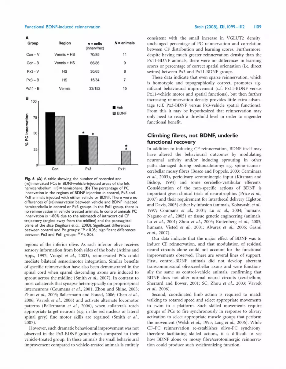
percentage of reinnervated PCs in the areas treated with BDNF: thehemisphere in Px3 groups and vermis in the Px11 group. Experi-mental animals were anaesthetized with isoflurane (Baxter S.A.,Maurepas, France), decapitated and cerebellar slices (300 mm) wereprepared from pedunculotomized and control hemicerebella fromanimals aged 24–36 days using standard procedures (Llano et al.,1991) as described in Sugihara et al. (2003). Whole-cell patch-clamprecordings were made from visually identified PCs, and CF currentswere elicited by stimulation in the granular layer. Patch pipetteswere filled with a solution containing (in mM) Cs D-gluconate, 120;biocytin, 13; HEPES, 10; BAPTA, 10; TEA Cl, 3; Na2ATP, 2;Mg ATP, 2; NaGTP, 0.2; pH 7.3, 290–300mOsm. Chemicals for theinternal solutions were from Sigma (St Quentin Fallavier, France).CF currents were identified by their all-or-none character andby the demonstration of paired-pulse depression. PCs wereconsidered to be non-innervated if no CF current could be elicitedby stimulation at several different stimulation locations andintensities.
Data analysesMotor testsFor each basic motor test, performance plateaued by day 5, thus thethree trials on day 5 were averaged to calculate each animal’s meantime for inter-group comparisons. For the rotarod, at each speedthe three trials on each day were averaged to obtain each animal’smean performance for intra-group comparisons and those from thelast (9th) day were used for inter-group analyses.
Spatial/swim testsFor each test the data for all trials within one session were averagedto calculate the mean performance (escape latency/quadrants/searchscore) of each animal, which was then used to calculate the meangroup performance during each session.
Correlation of reinnervation and behaviourTo correlate behaviour to reinnervation, each animal’s motorperformance and their escape latency/quadrants crossed/searchscore from the last session of the spatial tests (visible platform,hidden platform and retention) were converted to a scoredepending on how far performance differed from control animals(Willson et al., 2007). Thus a behavioural score was obtained for
1102 Brain (2008), 131, 1099^1112 M. L.Willson et al.
each animal in each test. The reinnervation value in each cortical
zone was normalized to a theoretical value if all the lobules wereCF-filled (5� number of lobules) and a reinnervation score was
calculated for each functional region. This reinnervation score was
correlated to the animal’s behavioural score.
StatisticsTransformed (logged) motor and water maze data revealed homo-
geneity of variance and inter-group comparisons were analysedusing analysis of variance (ANOVA) and either Bonferroni’s or
Dunnets T3 (if normality was not attained) post hoc tests. Intra-
group comparisons were made by repeated measures ANOVA. Forthe probe trials, the time spent in the ‘test’ quadrant was compared
to that predicted by chance with t-tests. The percentage success of atask, frequency of direct swims and percentage PC innervation were
assessed using the �2 test. Pearson’s correlation was used to test the
relation between reinnervation and behavioural scores. All valueswere stated as mean� SEM and a=0.05.
ResultsMinor effects of BDNF treatment onsimple locomotor tasksWe examined whether BDNF treatment in Px3 and Px11animals affected their ability to perform simple locomotortasks. Compared to age-matched vehicle-treated animals,BDNF treatment did not change the performance of sham-operated controls nor significantly improve the time taken byeither pedunculotomized group in the bridge, ladder or wiretests. The only exception was that the Px3-BDNF animalsperformed better than Px3-vehicle on the ladder[F(5, 31) = 6.9, P50.05]. Vehicle-treated groups essentiallyreplicated previous findings: Px3 and Px11 animals wereslower on the bridge and ladder tests and Px11-vehicleanimals were also slower on the wire (Fig. 2A, B and C;Willson et al., 2007).
BDNF improves synchronization of gait torotarod speed in Px11-lesioned animalsAs a more sensitive test of CF function (Rondi-Reig et al.,1997), some animals (Control-vehicle, n= 6; Px11-vehicle,n= 10; Px11-BDNF, n= 6) had their gait tested on therotarod. We did not examine a separate set of BDNF-treatedcontrols because, with an upper time limit of 180 s, they hadno opportunity to walk for longer than vehicle-treatedcontrols who reach the maximum time (Dixon et al., 2005,this study, Fig. 2D–G). There were no inter-group differencesat a slow speed (10 rpm). At 20 rpm, the Px11-BDNF groupwalked for longer than the Px11-vehicle group[F(3, 27) = 11.26, P50.05] and were similar to controls.Furthermore at 30 rpm, when Px11-vehicle animals wereworse than controls [F(3,24) = 25.3, P50.01], the Px11-BDNF group showed an intermediate performance notsignificantly different from control or from Px11-vehicle(Fig. 2D).
We also examined how the animals learned this task,because CFs are required for motor learning and the samecellular mechanisms are thought to underlie the learning ofboth motor and spatial tasks (Ito, 2001; Burguiere et al.,2005). Learning was examined at 10 rpm, since skills learnedat one speed are transferred to the next (Rondi-Reig et al.,1997). The Px11-BDNF group displayed learning at 10 rpmimproving over the first 2 days [F(8, 53)= 3.82, P50.01:Fig. 2E–G]. This contrasts with controls, which learned withinthe first day, and Px11-vehicle animals which did notdemonstrate any learning. These data suggest that BDNF-treatment in juvenile rats allows learning in the rotarod taskand better matching of gait with rotarod speed, compared torats without reinnervation. This is consistent with BDNFinducing transcommissural olivocerebellar reinnervation(as previously described: Sherrard and Bower, 2001).
BDNF treatment does not alter swimmingability in the cued water mazeSince prior sensorimotor training (e.g. the rotarod) can masksubtle water maze abnormalities (Cain et al., 1996), we useda different set of animals for spatial learning tasks (Control-vehicle, n= 6; Control-BDNF, n= 3; Px3-vehicle, n= 4; Px3-BDNF, n= 6; Px11-vehicle, n= 7; Px11-BDNF, n= 10). Wetested all groups in the visible platform task as this controlledfor any effect of motor impairment or BDNF on the animal’sability to orientate to and reach a target platform. All groups,irrespective of lesion or BDNF, had similar escape latenciesand total quadrants crossed (Fig. 3A and B). Moreover,escape latency correlated with search score (i.e. swimmingtrajectories) indicating that all animals swam at a similarspeed. The same correlation value (R2 = 0.79, P50.01 forboth; Fig. 3C) was found for vehicle and BDNF-treatedgroups, revealing similar search strategies for both groupsand confirming that BDNF did not affect how the animalbehaved in the maze.
BDNF-treated lesioned animals showimproved acquisition of a spatial taskThe hidden platform water maze was used to assess whetherBDNF-induced reinnervation improved spatial learning.In addition to confirming previous results for vehicle-injected groups (Willson et al., 2007), with Px3-vehicleanimals reaching control performance and Px11-vehicleanimals performing poorly in the maze, our present studyrevealed that BDNF facilitated spatial learning performancein pedunculotomized animals without significantly alteringthe results in control animals.
The BDNF-treated control and Px3 groups performed inthe maze equally to their vehicle treated counterparts. Theyimproved their performance in the maze between all sessionswith decreased escape latencies (P50.05; Fig. 4A), fewerquadrants crossed (P50.01; Fig. 4B) and better searchstrategies (P50.01; Fig. 4C). To ensure that the BDNF
Functional BDNF-induced reinnervation Brain (2008), 131, 1099^1112 1103

groups had reached their optimal performance within thetesting period, we analysed individual training trials withineach session (10 trials per session). The Px3-BDNF groupplateaued in their performance by the last two trials ofsession 2 (trials 19 and 20) while the control-BDNF groupreached a plateau at the mid-point of session 3 (trials 22–28).Also, both BDNF- and vehicle-treated Px11 groups improved
all parameters of their maze performance until session 2 andthereafter remained unchanged (P50.05 for escape latencies,quadrants crossed and search scores). Therefore, because thecontrol-BDNF animals continued to improve betweensessions 2 and 3, in session 3 they were better than BDNF-treated Px11 animals [escape latency F(5, 66) = 6.7, P50.01;quadrants crossed F(5, 66) = 6.5, P50.05; search score
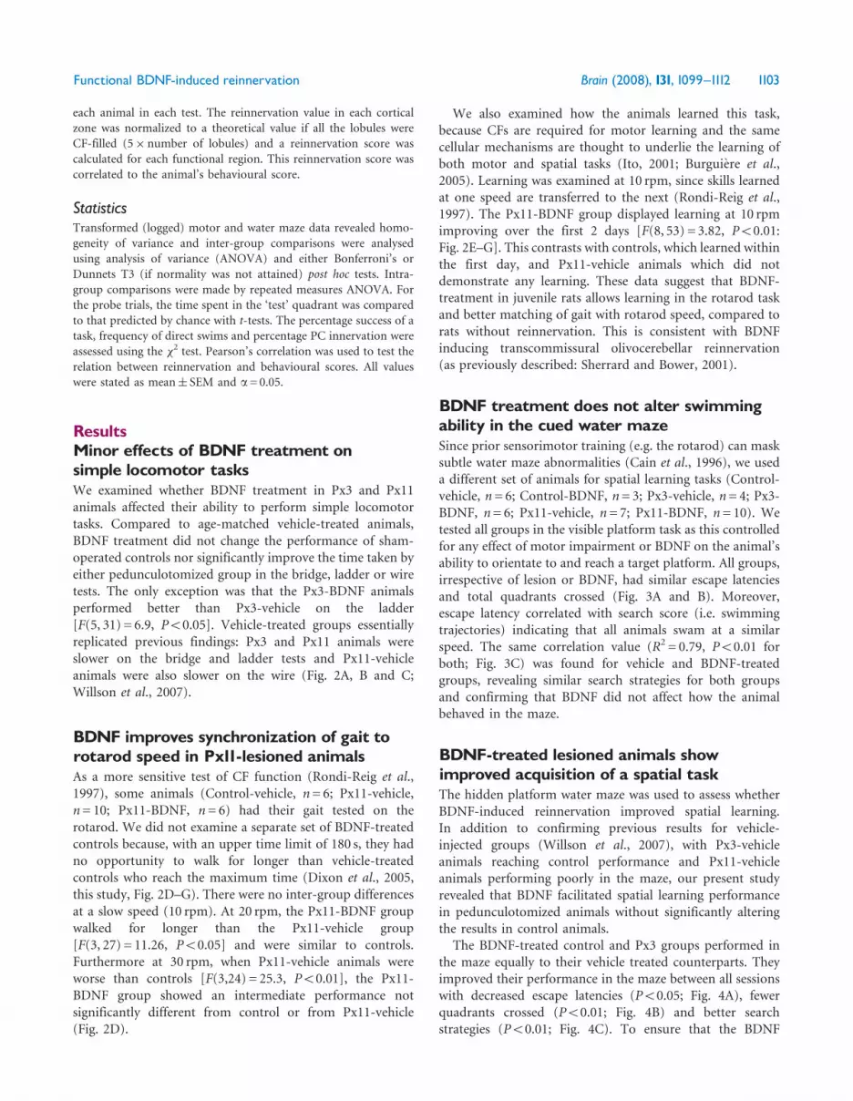
Fig. 2 Bar charts represent the time taken (mean� SEM) by vehicle and BDNF-treated groups to complete the bridge (A), ladder (B) andwire (C) during the fifth day of training and the error latencies on the rotarod (D^G). All lesioned groups were slower than control groupson the bridge (A), whilst only the Px3-vehicle (Px3V) group performed worse than controls on the ladder (B).On the rotarod (D), at 20and 30 rpm the Px11-vehicle (Px11V) group failed more quickly than control or Px11-BDNF (Px11B). Furthermore, control and Px11-BDNFgroups learned the task at 10 rpm within the first and third days, respectively while the Px11Vdid not (E). At higher speeds (F and G) nogroup significantly improved their error latency over the 9 days of training. Significant differences between control and lesioned: �P50.05,��P50.01; significant difference between Px3 and Px11 groups: #P50.05; significant difference between Px11V and Px11B groups: “P50.05.
1104 Brain (2008), 131, 1099^1112 M. L.Willson et al.

F(5, 66) = 5.7, P50.05]. Nevertheless, the Px11-BDNFgroup had the same performance as vehicle-treated controlsand both Px3 groups during training sessions 2 and 3(Fig. 4A–D), whereas the Px11-vehicle animals remained
significantly impaired (P50.05 for escape latency andquadrants crossed in both sessions 2 and 3).
The effect of BDNF on spatial function was furtherdemonstrated in the number of direct swims to the platform.Animals in both BDNF-treated pedunculotomized groupsmade more direct swims to the hidden platform com-pared to their vehicle-treated counterparts (�2 test: Px3,P50.05; Px11, P50.01) and equalled the control (control-vehicle) group (Table 1). In addition, the Px11-BDNFanimals also made as many direct swims as both Px3 groups(Table 1).
To discriminate whether the better motor performance(e.g. on the rotarod) of the BDNF-treated Px11 animalscontributed to their improved spatial function, we madethree further analyses. First, we tested for correlationsbetween the measured variables in the visible and hiddenplatform mazes (Cain et al., 1996). No correlations wereobserved for any group between these two versions of thewater maze, but only existed for each group within each test.Second, we re-compared our groups using the time taken inthe basic motor tests as a covariate of the escape latency inthe hidden platform (Martin et al., 2003). For the ladderthere was no covariate effect, although for the bridge andwire tests there was a significant effect of the covariate, i.e.better motor function [RANCOVA covariate: bridge,F(1, 5) = 8.75, P50.01; wire, F(1, 5) = 5.23, P50.05] butthe differences between groups for escape latency persisted[RANCOVA group: bridge, F(1, 5) = 3.19, P50.01; wire,F(1, 5) = 2.48, P50.05]. Third, we compared Px11 BDNF-and vehicle-treated groups using performance in the visibleplatform (cued maze) as covariate for the hidden platformresults (Martin et al., 2003) and found significant effects ofboth covariate [RANCOVA covariate: F(1, 2) = 5.7, P50.05]and group [RANCOVA group, F(1, 2) = 4.3, P50.05]. Thesedata indicate that motor ability does affect the performancein the hidden platform test. However, inter-group differencesremained when the motor effect was taken into account,indicating true differences in spatial learning.
BDNF treatment of lesioned-animalsfacilitates spatial memoryA probe test, in which the escape platform is removed,evaluates how accurately animals learned the spatial task. Allgroups showed similar spatial bias to the test quadrant(�32% of test time, 19/60 s). In addition, the number ofquadrant entries, total quadrants crossed and direct swimswere not significantly different between any groups.
A retrieval test assesses the animals’ memory of previouslylearned strategies. In this test, the platform was replaced intoits original location and performance was compared to thelast hidden platform training session. The intervening weekbetween the probe and retrieval test did not impair escapelatency, total quadrants crossed or search strategies forany group. However, BDNF-treated animals rememberedbetter, as the number of direct swims was similar for all
Fig. 3 The mean number of quadrants crossed to reach thevisible platform by all groups either with vehicle (A) or BDNF(B) treatment. There were no inter-group differences.(C) Correlation of search scores (quantified swim trajectory) andescape latency in the last training session.Vehicle and BDNF-treated animals had the same correlation value.
Functional BDNF-induced reinnervation Brain (2008), 131, 1099^1112 1105
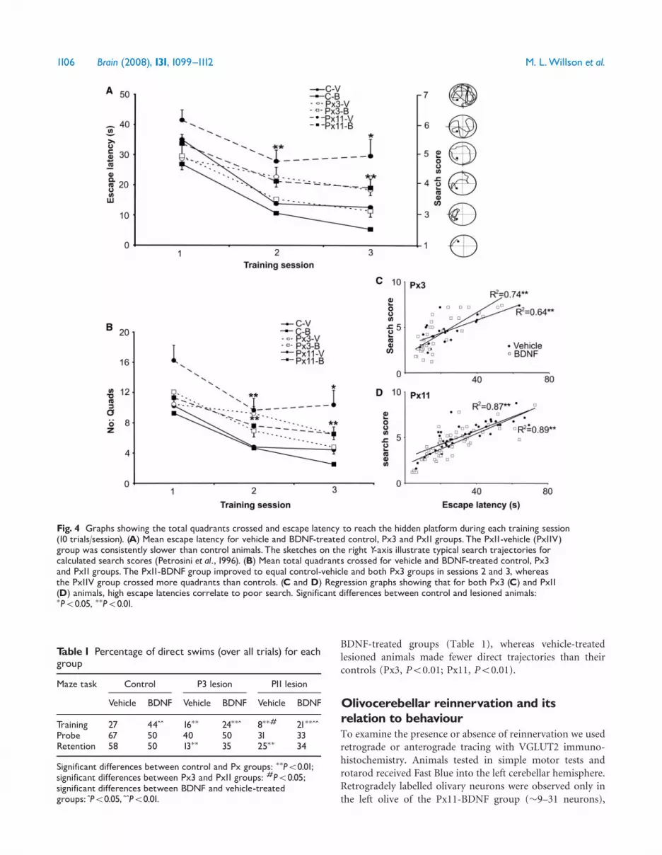
BDNF-treated groups (Table 1), whereas vehicle-treatedlesioned animals made fewer direct trajectories than theircontrols (Px3, P50.01; Px11, P50.01).
Olivocerebellar reinnervation and itsrelation to behaviourTo examine the presence or absence of reinnervation we usedretrograde or anterograde tracing with VGLUT2 immuno-histochemistry. Animals tested in simple motor tests androtarod received Fast Blue into the left cerebellar hemisphere.Retrogradely labelled olivary neurons were observed only inthe left olive of the Px11-BDNF group (�9–31 neurons),
Fig. 4 Graphs showing the total quadrants crossed and escape latency to reach the hidden platform during each training session(10 trials/session). (A) Mean escape latency for vehicle and BDNF-treated control, Px3 and Px11 groups. The Px11-vehicle (Px11V)group was consistently slower than control animals. The sketches on the right Y-axis illustrate typical search trajectories forcalculated search scores (Petrosini et al., 1996). (B) Mean total quadrants crossed for vehicle and BDNF-treated control, Px3and Px11 groups. The Px11-BDNF group improved to equal control-vehicle and both Px3 groups in sessions 2 and 3, whereasthe Px11V group crossed more quadrants than controls. (C and D) Regression graphs showing that for both Px3 (C) and Px11(D) animals, high escape latencies correlate to poor search. Significant differences between control and lesioned animals:�P50.05, ��P50.01.
Table 1 Percentage of direct swims (over all trials) for eachgroup
Maze task Control P3 lesion P11 lesion
Vehicle BDNF Vehicle BDNF Vehicle BDNF
Training 27 44 “ “ 16�� 24�� “ 8��# 21�� “ “Probe 67 50 40 50 31 33Retention 58 50 13�� 35 25�� 34
Significant differences between control and Px groups: ��P50.01;significant differences between Px3 and Px11 groups: #P50.05;significant differences between BDNF and vehicle-treatedgroups: “P50.05, “ “P50.01.
1106 Brain (2008), 131, 1099^1112 M. L.Willson et al.

confirming transcommissural olivocerebellar reinnervationand the lack of aberrant sprouting in intact animals(Sherrard and Bower, 2001; Dixon and Sherrard, 2006).In animals tested for simple motor tests and spatial
function, anterograde tracing revealed the normal crossedpath in control animals and the expected reinnervation withnormal morphology (data not shown) in Px3 and Px11-BDNF groups (Dixon and Sherrard, 2006; Letellier et al.,2007). However, the present study provides new detail on thedistribution and density of this reinnervation. VGLUT2-positive reinnervating CFs were distributed in the lefthemicerebellum up to 8mm from the midline, predomi-nately in vermal lobules III–VIII and in the hemisphere fromlobulus simplex to copula pyramidis (Fig. 5B). In Px3animals, VGLUT2 labelling appeared denser in the inter-mediate left hemicerebellum of BDNF- compared to vehicle-treated animals, although quantitatively the difference wasnot significant (P40.05). In contrast, in Px11 animalsBDNF-induced reinnervation was less extensive and muchless dense than in either vehicle or BDNF-treated Px3 groups(P50.05).In addition, to assess the effect of BDNF on CF–PC
reinnervation at a cellular level, we counted the percentage ofPCs from which CF-induced currents could be recorded inthose cerebellar regions which had been injected with BDNFor vehicle. BDNF did not alter the percentage of PCinnervation in control animals, nor in the left hemisphereof Px3 animals (Fig. 6A). BDNF induced reinnervation toonly 22% PCs in Px11 animals, which is less than in eitherPx3 group (Fig. 6B). Furthermore, none of the pedunculo-tomized groups was completely reinnervated. As there wasno reinnervation in Px11-vehicle animals, we did not recordfrom this group.To examine whether olivocerebellar reinnervation was
related to an animal’s behaviour, we correlated reinnervationscores for each functional region of the left hemicerebellum(medial, intermediate or lateral) and behaviour scores foreach motor and water maze test. For the basic motor tests,reinnervation (either by VGLUT2 mapping or retrogradeolivary analysis) did not correlate to motor score in anygroup, which is consistent with previous findings (Willsonet al., 2007). In addition, there was no correlation betweenthe number of retrogradely labelled olivary neurons (9, 9, 17,20, 24 and 31) and rotarod score at any speed (10 rpm,R2 = 0; 20 rpm, R2 =�0.46 and 30 rpm, R2 =�0.40).In contrast, in the hidden platform and retention tests, thetotal quadrants crossed and escape latency correlated withreinnervation in the intermediate and lateral hemicerebellumof Px3 groups (n= 5 animals, hidden platform: R2 = 0.72,P50.001; retention test: R2 = 0.65, P50.01; Fig. 5C).Furthermore, when all groups are considered, the reinnerva-tion score of the left hemicerebellum directly correlated withthe escape latencies in the hidden platform task (R2 = 0.40,P50.05). These results indicate that increasing olivocer-ebellar reinnervation aids spatial learning.
DiscussionWe used the olivocerebellar path as a model of axonal injuryto examine the effect of BDNF-induced collateral reinnerva-tion on complex motor and cognitive functions. We observedthat BDNF promoted transcommissural CF reinnervationin juvenile (Px11) animals and this was associated withenhanced CF-related functions: rotarod motor skills(Rondi-Reig et al., 1997), spatial learning (Dahhaoui et al.,1992; Meignin et al., 1999; Rondi-Reig et al., 2002) andmemory (Gasbarri et al., 2003). We also show that sparsereinnervation, when it is homotypic, compensates deficits incomplex and cognitive tasks, and that BDNF creates this effectwithout overtly disturbing undamaged paths.
Quality rather than quantity: BDNF-inducedCF reinnervation ameliorates relevantfunctional deficitsThis study provides new insights into neurotrophin-relatedrepair in the injured CNS. Transcommissural olivocerebellarreinnervation that takes place in the presence of BDNF wasassociated with improved CF-mediated behaviours: complexmotor and spatial skills. This may not seem surprisingbecause CF reinnervation ameliorates motor and spatialdeficits in neonatal animals (Dixon et al., 2005; Willson et al.,2007) and BDNF induces this reinnervation in young adultanimals (Dixon and Sherrard, 2006; Letellier et al., 2007).However in the injured neonatal brain before P10, there iswidespread anatomical plasticity (Weber and Stelzner, 1977;Gramsbergen and Ijkema-Paassen, 1982; Sherrard et al.,1986; Naus et al., 1987) whose contribution to behaviouralimprovement is unknown; but which can no longer con-tribute to the observed recovery in juvenile Px11-BDNFanimals.
Our data suggest that it is specificity rather than amountof reinnervation that is important. First, the Px11 BDNF-treated animals showed remarkable recovery in both motorskill and spatial function despite sparse transcommissuralreinnervation. Although reinnervation was light, with only�20% PCs responding to CF stimulation, it was homotypicwith normal arbors developing on reinnervated PCs (thisstudy; Dixon and Sherrard, 2006; Letellier et al., 2007). Also,reinnervation extended into the left hemisphere (Fig. 5),where spatial function is largely regulated (Joyal et al., 1996),which is consistent with the improved spatial function weobserved. Because our retrograde tracing only assessedolivocerebellar reinnervation to the injected left hemisphere,the lack of correlation between olivary labelling and motorskill in Px11-BDNF animals is consistent with reinnervatingCFs showing relatively normal topography, although on thewrong side of the midline (Sherrard et al., 1986; Zagrebelskyet al., 1997; Sherrard and Bower, 2001). This topographywould allow PCs of the medial hemicerebellum that normallyinter-connect with spinal circuits (Bosco and Poppele, 2003;Cerminara et al., 2003), to receive CFs from appropriate
Functional BDNF-induced reinnervation Brain (2008), 131, 1099^1112 1107

Fig. 5 The density and distribution of transcommissural CF reinnervation following left pedunculotomy at P3 or P11 and treatment withvehicle or BDNF. (A) Photomicrographs illustrating the arbitrary scale used to define the amount of VGLUT2 positive reinnervation in themolecular layer of each left hemicerebellar lobule. Scores were graded as: 1= strands of CFs, 2=¼ filled lobule etc until 5=completelyCF-filled lobule. Asterisks indicate regions with reinnervation.WM=white matter, GCL=granule cell layer, ML=molecular layer.Bar=100mm. (B) An unfolded cerebellum (adapted from Buisseret-Delmas and Angaut, 1993) showing the amount of VGLUT2-labelled CFreinnervation in 1mm wide parasagittal zones which is representative of each experimental group. Although Px3-BDNF animals appearmore densely labelled than vehicle-treated animals, only the widespread diffuse CF labelling of Px11-BDNF animals was quantitatively dif-ferent (�P50.05). CI=crus I, CII= crus II, CP=copula pyramidis, LS= lobulus simplex, PML=paramedian lobule, I-X=vermal lobules I-X.(C) Correlations between CF reinnervation and escape latency/total quadrants crossed by Px3 groups in the hidden platform and retentiontests. (��P50.01, ���P50.001).
1108 Brain (2008), 131, 1099^1112 M. L.Willson et al.
regions of the inferior olive. As each inferior olive receivessensory information from both sides of the body (Atkins andApps, 1997; Voogd et al., 2003), reinnervated PCs couldmediate bilateral sensorimotor integration. Similar benefitsof specific reinnervation have also been demonstrated in thespinal cord when spared descending axons are induced tosprout across the midline (Smith et al., 2007). In contrast tomost collaterals that synapse heterotypically on propriospinalinterneurons (Coumans et al., 2001; Zhou and Shine, 2003;Zhou et al., 2003; Ballermann and Fouad, 2006; Chen et al.,2006; Vavrek et al., 2006) and activate alternate locomotorpatterns (Ballermann et al., 2006), when collaterals reachappropriate target neurons (e.g. in the red nucleus or lateralspinal grey) fine motor skills are regained (Smith et al.,2007).However, such dramatic behavioural improvement was not
observed in the Px3-BDNF group when compared to theirvehicle-treated group. In these animals the small behaviouralimprovement compared to vehicle-treated animals is entirely
consistent with the small increase in VGLUT2 density,unchanged percentage of PC reinnervation and correlationbetween CF distribution and learning scores. Furthermore,despite having much greater reinnervation density than thePx11-BDNF animals, there were no differences in learningscores or percentage of correct spatial orientation (i.e. directswims) between Px3 and Px11-BDNF groups.
These data indicate that even sparse reinnervation, whichis homotypic and topographically correct, promotes sig-nificant behavioural improvement (c.f. Px11-BDNF versusPx11-vehicle motor and spatial functions), but then furtherincreasing reinnervation density provides little extra advan-tage (c.f. Px3-BDNF versus Px3-vehicle spatial functions).From this it may be hypothesized that reinnervation mayonly need to reach a threshold level in order to engenderfunctional benefit.
Climbing fibres, not BDNF, underliefunctional recoveryIn addition to inducing CF reinnervation, BDNF itself mayhave altered the behavioural outcomes by modulatingneuronal activity and/or inducing sprouting in otherpaths damaged during pedunculotomy: e.g. spino-/cuneo-cerebellar mossy fibres (Bosco and Poppele, 2003; Cerminaraet al., 2003), periolivary serotoninergic input (Kitzman andBishop, 1994) and some cerebello-vestibular efferents.Consideration of the non-specific actions of BDNF isimportant given clinical trials of neurotrophins (Price et al.,2007) and their requirement for intrathecal delivery (Egletonand Davis, 2005) either by infusion (animals, Kobayashi et al.,1997; Coumans et al., 2001; Lu et al., 2004; humans,Nagano et al., 2005) or tissue genetic engineering (animals,Lu et al., 2001; Zhou et al., 2003; Ruitenberg et al., 2005;humans, Visted et al., 2001; Alvarez et al., 2006; Gasmiet al., 2007).
Our data indicate that the major effect of BDNF was toinduce CF reinnervation, and that modulation of residualneural circuits alone could not account for the functionalimprovements observed. There are several lines of support.First, control-BDNF animals did not develop aberranttranscommissural olivocerebellar axons and were function-ally the same as control-vehicle animals, confirming thatBDNF does not alter normal neural circuits (cerebellum,Sherrard and Bower, 2001; SC, Zhou et al., 2003; Vavreket al., 2006).
Second, coordinated limb action is required to matchwalking to rotarod speed and select appropriate movementsto swim to a platform. Such skilled movements requiregroups of PCs to fire synchronously in response to olivaryactivation to select appropriate muscle groups that performthe movement (Welsh et al., 1995; Lang et al., 2006). WhileCF–PC reinnervation re-establishes olivo-PC synchrony,therefore facilitating skilled actions, it is difficult to seehow BDNF alone or mossy fibre/serotoninergic reinnerva-tion could produce such synchronizing function.
Fig. 6 (A) A table showing the number of recorded and(re)innervated PCs in BDNF/vehicle-injected areas of the lefthemicerebellum. HS=hemisphere. (B) The percentage of PCinnervation in the regions of BDNF injection in control, Px3 andPx11 animals injected with either vehicle or BDNF.There were nodifferences of (re)innervation between vehicle and BDNF injectedhemicerebella in control or Px3 groups. In the Px11 group, there isno reinnervation in vehicle treated animals. In control animals PCinnervation is �80% due to the mismatch of intracortical CFtrajectory (angled away from the midline) and the parasagittalplane of the slice (Sugihara et al., 2003). Significant differencesbetween control and Px groups: �P50.05; significant differencesbetween Px3 and Px11 groups: #P50.05.
Functional BDNF-induced reinnervation Brain (2008), 131, 1099^1112 1109
Third, in addition to synchronous firing of PC groups,motor and spatial functions also require correct patterns ofPurkinje cell activity (Dahhaoui et al., 1992; Le Marec et al.,1997; Meignin et al., 1999; Martin et al., 2003). CF-deprivedPCs have altered firing rates (Montarolo et al., 1982;Batini et al., 1985) which change activity in their targetdeep cerebellar nuclear neurons (Batini et al., 1985) anddisrupt sensorimotor processing (Hoebeek et al., 2005).Reinnervating CFs, which induce normal post-synapticcurrents in their target PCs (Sugihara et al., 2003; Letellieret al., 2007), should normalize PC firing and deep cerebellarnuclear outflow and hence improve sensorimotor regulation.In contrast, since BDNF is excitatory to PCs (Kafitz et al.,1999; Carter et al., 2002; Sadakata et al., 2007), any injectedBDNF that was still active at the time of behavioural testingwould increase PC firing and exacerbate the effects of CFdenervation. Likewise, serotonin inhibits deep cerebellarnuclear activity (Kitzman and Bishop, 1994); hence BDNF-induced serotoninergic reinnervation would also exacerbatethe effects of CF loss. Neither of these is compatible with theobserved functional improvement.Finally, the cerebellar (procedural) component of spatial
navigation involves learning the motor patterns required tomake a direct path to a specific target and linking them toinputs from the environment (Leggio et al., 1999; Burguiereet al., 2005). These processes involve PC long-term depres-sion (LTD; Burguiere et al., 2005), which is generated bysynaptic activity at the PC of both its CF and parallel fibreafferents (Ito, 2001). Since BDNF generally prevents thegeneration of LTD in CNS neurons (hippocampus, Aicardiet al., 2004; visual cortex, Kinoshita et al., 1999), any residualactivity of injected BDNF would impair spatial learning,which does require LTD (Burguiere et al., 2005). In fact, eventhough BDNF-induced mossy/serotoninergic fibre reinner-vation may facilitate parallel fibre-PC synapses and thereforeLTD, our covariate analyses demonstrate that improvedspatial navigation is related to CF reinnervation rather thanBDNF treatment (Px3-vehicle, Px3 and Px11 BDNF groups).This suggests that reinnervating CFs enable PC LTD toprovide the link between learned motor patterns andenvironmental cues. This view is supported by improvementin lesioned BDNF-treated groups of spatial knowledgeretention, which also involves CF function (Dahhaouiet al., 1992).In summary, despite its potent neuromodulatory effects
and potential to induce sprouting of non-CF afferents, ourdata suggest that BDNF promotes functional recovery byinducing CF reinnervation that can provide an anatomicalsubstrate for motor and spatial learning.
Olivocerebellar reinnervation: relevanceto the injured CNS?As discussed earlier, our data reveal that it is the specificity ofCF reinnervation that BDNF induces, which is mostimportant for recovery of spatial cognition and skilled
motor functions. When combined with recent similarfindings in the corticospinal tract (Smith et al., 2007), itmay be suggested that homotypic (re)innervation is a genericrequirement applicable to other neural systems and thereforepotentially to human neurological dysfunction. Importantly,our data also suggest a therapeutic threshold for reinnerva-tion, since even in a task as complex as spatial cognition(1) sparse input (Px11-BDNF), when specific, correlates withsignificant improvement, while (2) further increasing it(Px3-BDNF) provides minimal additional benefit. Since an‘innervation threshold’ is well known to precede symptomonset in Parkinson’s disease (Conley and Kirchner, 1999),our data imply the same principle can be applied toreinnervation, so that ‘less is more’, with its smaller risk of(re)connectivity errors. A threshold of reinnervation, rapidlyimproving peptide delivery to the CNS (Jain, 2007; de Boerand Gaillard, 2007) and the efficacy of a single post-lesion,albeit invasive, treatment broadens our understanding ofrecovery from neural injury/dysfunction. Moreover, ourstudy adds that a system-relevant growth-factor (BDNF) hasthis effect without also altering adjacent normal circuits, asindicated by the normal behaviour and olivocerebellarstructure in control-BDNF animals. Finally, as BDNFinduces similar reinnervation in young adult rats (Dixonand Sherrard, 2006), re-creating developmental plasticityfocuses effective treatment to the demography mostcommonly involved in neural injury; young adult males(National Hospital Morbidity Study, Australia; NINDSPublic Liaison Office).
AcknowledgementsWe would like to thank Prof. Adrian Bower for helpfuladvice about this study and insightful critiques on themanuscript. Also we thank Kelly Gifford and Marie-ClaudeMalidor for assistance with tissue sectioning and AmgenInc. for the generous donation of BDNF. This work wassupported by university funds from James Cook Universityand Notre Dame (Australia) to R.M.S., Universite Pierre etMarie Curie and CNRS funds to J.M., a Graduate ResearchScheme grant to M.L.W., and l’Institut pour la Recherchesur la Moelle Epiniere et l’Encephale to A.M.L. M.L.W. wassupported by an Australian Postgraduate Award and laFondation pour la Recherche Medicale. The internationalcollaboration was supported by a PICS grant from theCNRS to A.M.L.
ReferencesAicardi G, Argilli E, Cappello S, Santi S, Riccio M, Thoenen H, et al.
Induction of long-term potentiation and depression is reflected by
corresponding changes in secretion of endogenous brain-derived
neurotrophic factor. Proc Natl Acad Sci USA 2004; 101: 15788–92.
Altman J, Bayer SA. The development of the cerebellar system: in relation
to its evolution, structure and functions. Boca Raton: CRC Press; 1997.
Alvarez XA, Cacabelos R, Laredo M, Couceiro V, Sampedro C, Varela M,
et al. A 24-week, double-blind, placebo-controlled study of three dosages
of Cerebrolysin in patients with mild to moderate Alzheimer’s disease.
Eur J Neurol 2006; 13: 43–54.
1110 Brain (2008), 131, 1099^1112 M. L.Willson et al.
Angaut P, Alvarado-Mallart RM, Sotelo C. Compensatory climbing fibre
innervation after unilateral pedunculotomy in the newborn rat: origin
and topographic organisation. J Comp Neurol 1985; 236: 161–78.
Apps R, Lee S. Central regulation of cerebellar climbing fibre input during
motor learning. J Physiol 2002; 541: 301–17.
Atkins MJ, Apps R. Somatotopical organisation within the climbing fibre
projection to the paramedian lobule and copula pyramidis of the rat
cerebellum. J Comp Neurol 1997; 389: 249–63.
Auvray N, Caston J, Reber A, Stelz T. Role of the cerebellum in the
ontogenesis of the equilibrium behavior in the young rat: a behavioral
study. Brain Res 1989; 505: 291–301.
Ballermann M, Fouad K. Spontaneous locomotor recovery in spinal cord
injured rats is accompanied by anatomical plasticity of reticulospinal
fibers. Eur J Neurosci 2006; 23: 1988–96.
Ballermann M, Tse ADY, Misiaszek JE, Fouad K. Adaptations in the walking
pattern of spinal cord injured rats. J Neurotrauma 2006; 23: 897–907.
Bareyre FM, Kerschensteiner M, Raineteau O, Mettenleiter TC,
Weinmann O, Schwab ME. The injured spinal cord spontaneously
forms a new intraspinal circuit in adult rats. Nat Neurosci 2004; 7: 269–77.
Batini C, Billard JM, Daniel H. Long term modification of cerebellar
inhibition after inferior olive degeneration. Exp Brain Res 1985; 59:
404–9.
de Boer AG, Gaillard PJ. Drug targeting to the brain. Ann Rev Pharmacol
Toxicol 2007; 47: 323–55.
Bosco G, Poppele RE. Modulation of dorsal spinocerebellar responses to
limb movement. II. Effect of sensory input. J Neurophysiol 2003; 90:
3372–83.
Bower AJ, Waddington G. A simple operative technique for chronically
severing the cerebellar peduncles in neonatal rats. J Neurosci Methods
1981; 4: 181–8.
Buisseret-Delmas C, Angaut P. The cerebellar olivo-corticonuclear
connections in the rat. Prog Neurobiol 1993; 40: 63–87.
Burguiere E, Arleo A, Hojjati M, Elgersma Y, De Zeeuw CI, Berthoz A,
et al. Spatial navigation impairment in mice lacking cerebellar LTD: a
motor adaptation deficit? Nat Neurosci 2005; 8: 1292–4.
Cain DP, Saucier D, Hall J, Hargreaves EL, Boon F. Detailed behavioral
analysis of water maze acquisition under APV or CNQX: contribution
of sensorimotor disturbances to drug-induced acquisition deficits. Behav
Neurosci 1996; 110: 86–102.
Caleo M, Medini P, von Bartheld CS, Maffei L. Provision of brain-derived
neurotrophic factor via anterograde transport from the eye preserves the
physiological responses of axotomized geniculate neurons. J Neurosci
2003; 23: 297–6.
Carter AR, Chen C, Schwartz PM, Segal RA. Brain-derived neurotrophic
factor modulates cerebellar plasticity and synaptic ultrastructure.
J Neurosci 2002; 22: 1316–27.
Cerminara NL, Makarabhirom K, Rawson JA. Somatosensory properties of
cuneocerebellar neurones in the main cuneate nucleus of the rat.
Cerebellum 2003; 2: 131–45.
Chen C, Kano M, Abeliovich A, Chen L, Bao S, Kim JJ, et al.
Impaired motor coordination correlates with persistent multiple
climbing fiber innervation in PKC gamma mutant mice. Cell 1995;
83: 1233–42.
Chen Q, Zhou L, Shine HD. Expression of Neurotrophin-3 promotes
axonal plasticity in the acute but not chronic injured spinal cord.
J Neurotrauma 2006; 23: 1254–60.
Conley SC, Kirchner JT. Parkinson’s disease–the shaking palsy: underlying
factors, diagnostic considerations, and clinical course. Postgrad Med
1999; 106: 39–52.
Coumans JV, Lin TT, Dal HN, MacArthur L, McAtee M, Nash C, et al.
Axonal regeneration and functional recovery after complete spinal cord
transection in rats by delayed treatment with transplants and
neurotrophins. J Neurosci 2001; 21: 9334–44.
Dahhaoui M, Stelz T, Caston J. Effects of lesion of the inferior olivary
complex by 3-acetylpyridine on learning and memory in the rat. J Comp
Physiol A 1992; 171: 657–64.
Day LB, Weisend M, Sutherland RJ, Schallert T. The hippocampus is not
necessary for a place response but may be necessary for pliancy. Behav
Neurosci 1999; 193: 914–24.
Deller T, Frotscher M. Lesion-induced plasticity of central neurons:
sprouting of single fibres in the rat hippocampus after unilateral
entorhinal cortex lesion. Prog Neurobiol 1997; 53: 687–727.
Dixon KJ, Hilber W, Speare S, Willson ML, Bower AJ, Sherrard RM. Post-
lesion transcommissural olivocerebellar reinnervation improves motor
function following unilateral pedunculotomy in the neonatal rat. Exp
Neurol 2005; 196: 254–65.
Dixon KJ, Sherrard RM. Brain-derived neurotrophic factor induces post-
lesion transcommissural growth of olivary axons that develop normal
climbing fibers on mature Purkinje cells. Exp Neurol 2006; 202: 44–56.
Egleton RD, Davis TP. Development of neuropeptide drugs that cross the
blood-brain barrier. NeuroRx 2005; 2: 44–53.
Fawcett JW. Overcoming inhibition in the damaged spinal cord.
J Neurotrauma 2006; 23: 371–83.
Federico F, Leggio MG, Neri P, Mandolesi L, Petrosini L. NMDA receptor
activity in learning spatial procedural strategies II. The influence of
cerebellar lesions. Brain Res Bull 2006; 70: 356–67.
Finlay BL, Wilson KG, Schneider GE. Anomalous ipsilateral retinotectal
projections in Syrian hamsters with early lesions: topography and
functional capacity. J Comp Neurol 1979; 183: 721–40.
Gasbarri A, Pompili A, Pacitti C, Cicirata F. Comparative effects of lesions
to the ponto-cerebellar and olivo-cerebellar pathways on motor and
spatial learning in the rat. Neuroscience 2003; 116: 1131–40.
Gasmi M, Brandon EP, Herzog CD, Wilson A, Bishop KM, Hofer EK,
et al. AAV2-mediated delivery of human neurturin to the rat
nigrostriatal system: Long-term efficacy and tolerability of CERE-120
for Parkinson’s disease. Neurobiol Dis 2007; 27: 67–76.
Gramsbergen A, Ijkema-Paassen J. CNS plasticity after hemicerebellectomy
in the young rat. Quantitative relations between aberrant and normal
cerebello-rubral projections. Neurosci Lett 1982; 33: 129–34.
Heinsen H. Quantitative anatomical studies on the postnatal development
of the cerebellum of the albino rat. Anat Embryol 1977; 151: 201–18.
Hicks SP, D’Amato CJ. Motor-sensory and visual behaviour after
hemispherectomy in newborn and mature rats. Exp Neurol 1970; 29:
416–38.
Hioki H, Fujiyama F, Taki K, Tomioka R, Furuta T, Tamamaki N, et al.
Differential distribution of vesicular glutamate transporters in the rat
cerebellar cortex. Neuroscience 2003; 117: 1–6.
Hoebeek FE, Stahl JS, van Alphen AM, Schonewille M, Luo C,
Rutterman M, et al. Increased noise level of Purkinje cell activities
minimizes impact of their modulation during sensorimotor control.
Neuron 2005; 45: 953–65.
Ito M. Cerebellar long-term depression: characterization, signal transduc-
tion, and functional roles. Physiol Rev 2001; 81: 1143–95.
Jain KK. Nanotechnology-based drug delivery to the central nervous
system. Neurodegen Dis 2007; 4: 287–91.
Joyal CC, Meyer C, Jacquart G, Mahler P, Caston J, Lalonde R. Effects of
midline and lateral cerebellar lesions on motor coordination and spatial
orientation. Brain Res 1996; 739: 1–11.
Kafitz KW, Rose CR, Thoenen H, Konnerth A. Neurotrophin-evoked rapid
excitation through TrkB receptors. Nature 1999; 401: 918–21.
Kinoshita S, Yasuda H, Taniguchi N, Katoh-Semba R, Hatanaka H,
Tsumoto T. Brain-derived neurotrophic factor prevents low-frequency
inputs from inducing long-term depression in the developing visual
cortex. J Neurosci 1999; 19: 2122–30.
Kitzman PH, Bishop GA. The origin of serotoninergic afferents to the cat’s
cerebellar nuclei. J Comp Neurol 1994; 340: 541–50.
Kobayashi NR, Fan DP, Giehl KM, Bedard AM, Wiegand SJ, Tetzlaff W.
BDNF and NT-4/5 prevent atrophy of rat rubrospinal neurons after
cervical axotomy, stimulate GAP-43 and Talpha1-tubulin mRNA
expression, and promote axonal regeneration. J Neurosci 1997; 17:
9583–95.
Kordower JH, Herzog CD, Dass B, Bakay RAE, Stansell J 3rd, Gasmi M,
et al. Delivery of neurturin by AAV2 (cere-120)-mediated gene transfer
Functional BDNF-induced reinnervation Brain (2008), 131, 1099^1112 1111
provides structural and functional neuroprotection and neurorestoration
in MTPT-treated monkeys. Ann Neurol 2006; 60: 706–15.
Lang EJ, Sugihara I, Llinas R. Olivocerebellar modulation of motor cortex
ability to generate vibrissal movements in rat. J Physiol 2006; 571:
101–20.
Le Marec N, Dahhaoui M, Stelz T, Bakalian A, Delhaye-Bouchaud N,
Caston J, et al. Effect of cerebellar granule cell depletion on spatial
learning and memory and in an avoidance conditioning task: studies in
postnatally X-irradiated rats. Dev Brain Res 1997; 99: 20–8.
Leggio MG, Neri P, Graziano A, Mandolesi L, Molinari M, Petrosini L.
Cerebellar contribution to spatial event processing: characterization of
procedural learning. Exp Brain Res 1999; 127: 1–11.
Letellier M, Bailly Y, Demais V, Sherrard RM, Mariani J, Lohof AM.
Reinnervation of late post-natal Purkinje cells by climbing fibres:
neosynaptogenesis without transient multi-innervation. J Neurosci 2007;
27: 5373–83.
Levine SC, Huttenlocher P, Banich MT, Duda EE. Factors affecting
cognitive functioning of hemiplegic children. Dev Med Child Neurol
1987; 29: 27–35.
Llano I, Marty A, Armstrong CM, Konnerth A. Synaptic- and agonist-
induced excitatory currents of Purkinje cells in rat cerebellar slices.
J Physiol 1991; 434: 183–213.
Lu P, Blesch A, Tuszynski MH. Neurotrophism without neurotropism:
BDNF promotes survival but not growth of lesioned corticospinal
neurons. J Comp Neurol 2001; 436: 456–70.
Lu P, Yang H, Jones LL, Filbin MT, Tuszynski MH. Combinatorial therapy
with neurotrophins and cAMP promotes axonal regeneration beyond
sites of spinal cord injury. J Neurosci 2004; 24: 6402–9.
Martin LA, Goldowitz D, Mittleman G. The cerebellum and spatial ability:
dissection of motor and cognitive components with a mouse model
system. Eur J Neurosci 2003; 18: 2002–10.
Meignin C, Hilber P, Caston J. Influence of stimulation of the
olivocerebellar pathway by harmaline on spatial learning in the rat.
Brain Res 1999; 824: 277–83.
Miyazaki T, Fukaya M, Shimizu H, Watanabe M. Subtype switching of
vesicular glutamate transporters at parallel-fibre-Purkinje cell synapses
in developing mouse cerebellum. Eur J Neurosci 2003; 17: 2563–72.
Montarolo PG, Palestini M, Strata P. The inhibitory effect of the
olivocerebellar input on the cerebellar Purkinje cells in the rat.
J Physiol 1982; 332: 187–202.
Morris R. Developments of a water-maze procedure for studying spatial
learning in the rat. J Neurosci Methods 1984; 11: 47–60.
Nagano I, Shiote M, Murakami T, Kamada H, Hamakawa Y, Matsubara E,
et al. Beneficial effects of intrathecal IGF-1 administration in patients
with amyotrophic lateral sclerosis. Neurol Res 2005; 27: 768–72.
Naus CG, Flumerfelt BA, Hrycyshyn AW. Ultrastructural study of
remodeled rubral afferents following neonatal lesions in the rat.
J Comp Neurol 1987; 259: 131–9.
Neumann S, Woolf CJ. Regeneration of dorsal column fibers into and
beyond the lesion site following adult spinal cord Injury. Neuron 1999;
23: 83–91.
Petrosini L, Molinari M, Gremoli T. Hemicerebellectomy and motor
behaviour in rats. I Development of motor function after neonatal
lesion. Exp Brain Res 1990; 82: 472–82.
Petrosini L, Molinari M, Dell’Anna ME. Cerebellar contribution to spatial
event processing: Morris water maze and T-maze. Eur J Neurosci 1996;
8: 1882–96.
Price RD, Milne SA, Sharkey J, Matsuoka N. Advances in small molecules
promoting neurotrophic function. Pharm Ther 2007; 115: 292–306.
Rondi-Reig L, Burguiere E. Is the cerebellum ready for navigation? Prog
Brain Res 2005; 148: 212.
Rondi-Reig L, Delhaye-Bouchaud N, Mariani J, Caston J. Role of the
inferior olivary complex in motor skills and motor learning in the adult
rat. Neuroscience 1997; 77: 955–63.
Rondi-Reig L, Le Marec N, Caston J, Mariani J. The role of climbing and
parallel fibers inputs to cerebellar cortex in navigation. Behav Brain Res
2002; 132: 11–8.
Rossi F, Wiklund L, van der Want JJL, Strata P. Reinnervation
of cerebellar Purkinje cells by climbing fibres surviving a subtotal
lesion of the inferior olive in the adult rat. I. Development of new
collateral branches and terminal plexuses. J Comp Neurol 1991; 308:
513–35.
Ruitenberg MJ, Levison DB, Lee SV, Verhaagen J, Harvey AR, Plant GW.
NT-3 expression from engineered olfactory ensheathing glia promotes
spinal sparing and regeneration. Brain 2005; 128: 839–53.
Sadakata T, Takegawa W, Mizoguchi A, Washida M, Katoh-Semba R,
Shutoh F, et al. Impaired cerebellar development and function in mice
lacking CAPS2, a protein involved in neurotrophin release. J Neurosci
2007; 27: 2472–82.
Sherrard RM, Bower AJ. BDNF and NT3 extend the critical
period for developmental climbing fibre plasticity. Neuroreport 2001;
12: 2871–4.
Sherrard RM, Bower AJ, Payne JN. Innervation of the adult rat cerebellar
hemisphere by fibres from the ipsilateral inferior olive following
unilateral neonatal pedunculotomy: an autoradiographic and retrograde
fluorescent double-labelling study. Exp Brain Res 1986; 62: 411–21.
Smith JM, Lunga P, Story D, Harris N, Le Belle J, James MF, et al. Inosine
promotes recovery of skilled motor function in a model of focal brain
injury. Brain 2007; 130: 915–25.
Spear PD. Plasticity following neonatal visual cortex damage in cats. Can J
Physiol Pharmacol 1995; 73: 1389–97.
Sugihara I, Lohof AM, Letellier M, Mariani J, Sherrard RM. Post-lesion
transcommissural growth of olivary climbing fibres creates functional
synaptic microzones. Eur J Neurosci 2003; 18: 3027–36.
Sugihara I, Wu HS, Shinoda Y. The entire trajectories of single
olivocerebellar axons in the cerebellar cortex and their contribution to
cerebellar compartmentalisation. J Neurosci 2001; 21: 7715–23.
Vavrek R, Girgis J, Tetzlaf W, Hiebert GW, Fouad K. BDNF promotes
connections of corticospinal neurons onto spared descending inter-
neurons in spinal cord injured rats. Brain 2006; 129: 1534–45.
Visted T, Bjerkvig R, Enger PO. Cell encapsulation technology as a
therapeutic strategy for CNS malignancies. Neurooncology 2001; 3:
201–10.
Voogd J, Glickstein M. The anatomy of the cerebellum. Trends Neurosci
1998; 21: 370–5.
Voogd J, Pardoe J, Ruigrok TJH, Apps R. The distribution of climbing and
mossy fiber collateral branches from the copula pyramidis and the
paramedian lobule: congruence of climbing fiber cortical zones and the
pattern of zebrin banding within the rat cerebellum. J Neurosci 2003;
23: 4645–56.
Weber ED, Stelzner DJ. Behavioral effects of spinal cord transection in the
developing rat. Brain Res 1977; 125: 241–55.
Welsh JP, Lang EJ, Sugihara I, Llinas R. Dynamic organization of motor
control within the olivocerebellar system. Nature 1995; 374: 453–7.
Willson ML, Bower AJ, Sherrard RM. Developmental neural plasticity and
its cognitive benefits: olivocerebellar reinnervation compensates spatial
function in the cerebellum. Eur J Neurosci 2007; 25: 1475–83.
Zagrebelsky M, Strata P, Hawkes R, Rossi F. Reestablishment of the
olivocerebellar projection map by compensatory transcommmisural
reinnervation following unilateral transection of the inferior cerebellar
peduncle in the newborn rat. J Comp Neurol 1997; 379: 283–99.
Zhou L, Baumgartner BJ, Hill-Felberg SJ, McGowen LR, Shine DH.
Neurotrophin-3 expressed in situ induces axonal plasticity in the adult
spinal cord. J Neurosci 2003; 23: 1424–31.
Zhou L, Shine DH. Neurotrophic factors expressed in both cortex and
spinal cord induce axonal plasticity after spinal cord injury. J Neurosci
Res 2003; 74: 221–6.
1112 Brain (2008), 131, 1099^1112 M. L.Willson et al.
Related Documents