Cortical motor activity and reorganization following upper-limb amputation and subsequent targeted reinnervation Albert Chen a,b , Jun Yao b , Todd Kuiken c,d , Julius P.A. Dewald a,b,c, ⁎ a Department of Biomedical Engineering, Northwestern University, IL, USA b Department of Physical Therapy and Human Movement Sciences, Northwestern University, IL, USA c Department of Physical Medicine and Rehabilitation, Northwestern University, IL, USA d Sensory Motor Performance Program, Rehabilitation Institute of Chicago, IL, USA abstract article info Article history: Received 4 August 2013 Received in revised form 23 September 2013 Accepted 1 October 2013 Available online 11 October 2013 Keywords: Cortical activity Brain reorganization Amputation Motor Targeted reinnervation Electroencephalography Previous studies have postulated that the amount of brain reorganization following peripheral injuries may be correlated with negative symptoms or consequences. However, it is unknown whether restoring effective limb function may then be associated with further changes in the expression of this reorganization. Recently, targeted reinnervation (TR), a surgical technique that restores a direct neural connection from amputated sensorimotor nerves to new peripheral targets such as muscle, has been successfully applied to upper-limb amputees. It has been shown to be effective in restoring both peripheral motor and sensory functions via the reinnervated nerves as soon as a few months after the surgery. However, it was unclear whether TR could also restore normal cortical motor representations for control of the missing limb. To answer this question, we used high-density electroencephalography (EEG) to localize cortical activity related to cued motor tasks generated by the intact and missing limb. Using a case study of 3 upper-limb amputees, 2 of whom went through pre and post-TR experiments, we present unique quantitative evidence for the re-mapping of motor representations for the missing limb closer to their original locations following TR. This provides evidence that an effective restoration of peripheral function from TR can be linked to the return of more normal cortical expression for the missing limb. Therefore, cortical mapping may be used as a potential guide for monitoring rehabilitation following peripheral injuries. © 2013 The Authors. Published by Elsevier Inc. 1. Introduction It has become well-established in the literature that changes in cortical organization often occur following injury to the peripheral nervous system. Several animal studies have demonstrated that motor and somatosensory representations of neighboring intact body parts expand into cortical areas previously devoted to an injured or missing limb (Donoghue and Sanes, 1987; Kaas, 2000; Kaas et al., 1983; Merzenich et al., 1978; Wall et al., 1986). Similar trends in cortical reorganization have been observed in humans that have sustained peripheral injuries such as upper-limb amputations (Elbert et al., 1994; Flor et al., 1995; Karl et al., 2001; Weiss et al., 2000). For example, face, lip, chin, and neck stimulations in upper-limb amputee subjects have been found to map to cortical areas corresponding to the missing extremity (Borsook et al., 1998; Elbert et al., 1997; Ramachandran et al., 1992). Despite numerous reports of cortical reorganization, other studies have shown that the motor representations of the missing limb persist following amputation, although not necessarily in their original areas. When voluntary executed movements of the missing limb are attempted, neuroimaging techniques such as functional magnetic resonance imaging (fMRI), positron emission tomography (PET) and magnetoencephalography (MEG) show that in general, the resulting movement representations of the missing limb may move out of the original cortical area and into neighboring areas (Giraux et al., 2001; Karl et al., 2004; Kew et al., 1994; Lotze et al., 2001). Similarly, the magnetic stimulation of motor cortical areas previously linked to the missing limb was also able to elicit contractions or movements from residual muscles adjacent to the site of amputation (Chen et al., 1998; Cohen et al., 1991; Irlbacher et al., 2002; Kew et al., 1994; Ridding and Rothwell, 1999; Roricht et al., 1999). These results beg the question of whether the persistent rep- resentation of the missing limb could return to its original area, NeuroImage: Clinical 3 (2013) 498–506 ⁎ Corresponding author at: Department of Physical Therapy and Human Movement Sciences, Northwestern University, IL, USA. Tel.: +1 312 908 6788; fax: +1 312 908 0741. E-mail address: [email protected] (J.P.A. Dewald). 2213-1582 © 2013 The Authors. Published by Elsevier Inc. http://dx.doi.org/10.1016/j.nicl.2013.10.001 Contents lists available at ScienceDirect NeuroImage: Clinical journal homepage: www.elsevier.com/locate/ynicl Open access under CC BY-NC-ND license. Open access under CC BY-NC-ND license.

Welcome message from author
This document is posted to help you gain knowledge. Please leave a comment to let me know what you think about it! Share it to your friends and learn new things together.
Transcript

NeuroImage: Clinical 3 (2013) 498–506
Contents lists available at ScienceDirect
NeuroImage: Clinical
j ourna l homepage: www.e lsev ie r .com/ locate /yn ic l
Cortical motor activity and reorganization following upper-limbamputation and subsequent targeted reinnervation
Albert Chen a,b, Jun Yao b, Todd Kuiken c,d, Julius P.A. Dewald a,b,c,⁎a Department of Biomedical Engineering, Northwestern University, IL, USAb Department of Physical Therapy and Human Movement Sciences, Northwestern University, IL, USAc Department of Physical Medicine and Rehabilitation, Northwestern University, IL, USAd Sensory Motor Performance Program, Rehabilitation Institute of Chicago, IL, USA
⁎ Corresponding author at: Department of Physical TSciences, Northwestern University, IL, USA. Tel.:+1 312 9
E-mail address: [email protected] (J.P.A. De
2213-1582 © 2013 The Authors. Published by Elsevier Inchttp://dx.doi.org/10.1016/j.nicl.2013.10.001
a b s t r a c t
a r t i c l e i n f oArticle history:Received 4 August 2013Received in revised form 23 September 2013Accepted 1 October 2013Available online 11 October 2013
Keywords:Cortical activityBrain reorganizationAmputationMotorTargeted reinnervationElectroencephalography
Previous studies have postulated that the amount of brain reorganization following peripheral injuries may becorrelated with negative symptoms or consequences. However, it is unknown whether restoring effective limbfunctionmay then be associatedwith further changes in the expression of this reorganization. Recently, targetedreinnervation (TR), a surgical technique that restores a direct neural connection from amputated sensorimotornerves to new peripheral targets such as muscle, has been successfully applied to upper-limb amputees. It hasbeen shown to be effective in restoring both peripheral motor and sensory functions via the reinnervated nervesas soon as a fewmonths after the surgery. However, it was unclearwhether TR could also restore normal corticalmotor representations for control of the missing limb. To answer this question, we used high-densityelectroencephalography (EEG) to localize cortical activity related to cued motor tasks generated by the intactand missing limb. Using a case study of 3 upper-limb amputees, 2 of whom went through pre and post-TRexperiments, we present unique quantitative evidence for the re-mapping of motor representations for themissing limb closer to their original locations following TR. This provides evidence that an effective restorationof peripheral function from TR can be linked to the return of more normal cortical expression for the missinglimb. Therefore, cortical mapping may be used as a potential guide for monitoring rehabilitation followingperipheral injuries.
© 2013 The Authors. Published by Elsevier Inc. Open access under CC BY-NC-ND license.
1. Introduction
It has become well-established in the literature that changes incortical organization often occur following injury to the peripheralnervous system. Several animal studies have demonstrated that motorand somatosensory representations of neighboring intact body partsexpand into cortical areas previously devoted to an injured or missinglimb (Donoghue and Sanes, 1987; Kaas, 2000; Kaas et al., 1983;Merzenich et al., 1978; Wall et al., 1986). Similar trends in corticalreorganization have been observed in humans that have sustainedperipheral injuries such as upper-limb amputations (Elbert et al.,1994; Flor et al., 1995; Karl et al., 2001;Weiss et al., 2000). For example,face, lip, chin, and neck stimulations in upper-limb amputee subjects
herapy and Human Movement08 6788; fax:+1 312 908 0741.wald).
.Open access under CC BY-NC-ND licen
have been found to map to cortical areas corresponding to the missingextremity (Borsook et al., 1998; Elbert et al., 1997; Ramachandranet al., 1992).
Despite numerous reports of cortical reorganization, otherstudies have shown that the motor representations of the missinglimb persist following amputation, although not necessarily intheir original areas. When voluntary executed movements of themissing limb are attempted, neuroimaging techniques such asfunctional magnetic resonance imaging (fMRI), positron emissiontomography (PET) and magnetoencephalography (MEG) show thatin general, the resulting movement representations of the missinglimb may move out of the original cortical area and into neighboringareas (Giraux et al., 2001; Karl et al., 2004; Kew et al., 1994; Lotzeet al., 2001). Similarly, the magnetic stimulation of motor corticalareas previously linked to the missing limb was also able to elicitcontractions or movements from residual muscles adjacent to thesite of amputation (Chen et al., 1998; Cohen et al., 1991; Irlbacheret al., 2002; Kew et al., 1994; Ridding and Rothwell, 1999; Rorichtet al., 1999).
These results beg the question of whether the persistent rep-resentation of the missing limb could return to its original area,
se.

499A. Chen et al. / NeuroImage: Clinical 3 (2013) 498–506
perhaps via surgical interventions that attempt to restoremotor function.One such intervention is targeted reinnervation (TR), a technique thatimproves artificial limb function for amputees and successfully helpedthem to regain both motor and sensory functions related to the missinglimb. In TR, the inactivated, residual sensorimotor nerves previouslyresponsible for innervating the missing limb are surgically re-routed toalternative denervated muscle groups and skin areas over the chest oron the residual limb (Hijjawi et al., 2006; Kuiken, 2003; Kuiken et al.,2004). After a few months, new functional connections between thenerves, muscle and skin are created. The reinnervated muscles act asbiological amplifiers for efferent motor command signals; surfaceelectromyographic (EMG) signals at these new sites then providecontrol signals for an amputee to operate a motorized, myoelectricprosthesis (Kuiken et al., 2005). TR also returns sensations oftouch, pressure, vibration and temperature for the missing limb tothe skin overlying reinnervated muscles (Kuiken et al., 2007a,b).From a neural perspective, afferent traffic and efferent neural trafficare re-established for parts of the limb completely lost for months toyears following amputation.
The remarkable ability to completely rewire the peripheral neuralconnectivity of the arm and restore function of the lost limb makes TRa unique model for investigating a possible restoration of normalcortical expression in human amputees. In particular, we hypothesizethat the return of new peripheral motor targets and voluntary musclecontrol can restore the original cortical representations for the missinglimb. In this study, high-density electroencephalography (EEG) wasused to investigate the locations of cortical activity related to cuedmotor tasks generated by the intact andmissing limb.We present uniqueevidence for the restoration of cortical mapping that occur in themotor cortex from 2 individuals with upper-limb amputations whounderwent TR. More specifically, we found that motor representationswere re-mapped closer to presumed pre-amputation locations followingTR. This suggests that an effective rehabilitation intervention such as TR isassociated with the restoration of the cortical activity that normallycontrols them. Our results imply that cortical mapping can be usedas a potential monitor and guide for future rehabilitation of peripheralinjuries.
2. Methods
2.1. Subject selection
Three subjects with upper-limb amputations were screened andselected to undergo TR at least 6months following their injuries. Allsubjects suffered amputations due to trauma from automobile orelectrocution accidents and had non-painful phantom limb sen-sations. In addition, all subjects were consistent users of upper-arm prostheses — subject A1 used a cosmetic prosthesis while subjectsA2 and A3 mainly used myoelectric prostheses. Information about thesubjects, including amputation site and time of experimentation testingdates with respect to the date of amputation, is summarized in Table 1.Written consent was provided by each subject prior to participation inthe study that was approved by the Institutional Review Board ofNorthwestern University and in compliance with the principles ofthe Declaration of Helsinki.
Table 1Amputee subject information.
Subject Age Sex Dominant hand Amputation site
A1 38 F R Left transhumeralA2 39 M R Right shoulder disarticulA3 54 M R Bilateral shoulder disarti
Abbreviations: F= female, M=male, R= right, TR= targeted reinnervation, *=did not perf
2.2. Targeted reinnervation procedures
Graphical representations of each subject's TR surgical procedure areshown in Fig. 1 and are briefly described below.
2.2.1. Subject A1— left transhumeral amputeeSubject A1 was a 38 year old woman with a left transhumeral
amputation due to injuries sustained in an automobile collision. DuringTR surgery performed 6months after amputation (Fig. 1a), the mediannerve was transferred to the denervated medial biceps brachii muscle,and the distal radial nerve was transferred to the denervated lateraltriceps brachii muscle. The triceps long head and lateral biceps musclesremained intact for control of elbow extension and elbow flexion,respectively. Full muscle reinnervation took approximately 5 months.The intercostal brachial cutaneous nerve was also transected todenervate and numb the distal residual limb. Over the course of 7–8 months, median and distal radial nerve afferents reinnervated thisskin, producing sensations of her missing hand when touched atdifferent points. For example, sensation referring to the index fingerwas found at the end of the residual limb near the reinnervated sitefor the median nerve.
2.2.2. Subject A2— right shoulder disarticulation amputeeSubject A2 is a 39 year old man who had his right arm amputated
at the shoulder following an electrical accident. Four nerve transferswere made for the targeted reinnervation procedure (Fig. 1b). Themusculocutaneous nerve was transferred to the clavicular head of thepectoralis major to control elbow flexion. The median nerve wastransferred to the top half of the sternal head of the pectoralis majorto control hand closing, while the distal radial nerve was transferredto the pectoralis minor to control hand opening/wrist extension. Theulnar nerve was also transferred to the lower half of the sternal headof the pectoralis major. Similarly to subject A1, muscle reinnervationand sensations for his missing limb returned over the course of 5–7months.
2.2.3. Subject A3— bilateral shoulder disarticulation amputeeSubject A3 is a 54-year old man with a bilateral arm amputation at
the shoulder following electrical burn injuries. He underwent targetedreinnervation on the left side 9months following the amputation. Theremaining parts of 4 peripheral nerves were transferred to differentsegments of his left pectoral chest muscles (Fig. 1c). The musculocu-taneous nerve was transferred to the clavicular head of the pectoralismajor. The median nerve was transferred to the top half of the sternalhead of the pectoralis major, while the radial nerve was transferred tothe bottom half. The ulnar nerve was transferred to the pectoralisminor. Subcutaneous fat on the chest was removed to allow themuscleto come in close contact to the skin for producing stronger EMG signals.After 5 months, sensations for his missing limb returned to the skinoverlying the reinnervated sites (Kuiken et al., 2007a), includingfeelings of pressure, temperature, and pain.
2.3. Experimental protocol
In this study, cued motor tasks were used to determine motorcortical representations of themissing and intact limbs. In two subjects,
Time from amputation to TR Time from TR to post-TR testing
6months 10.5monthsation 18months 18.5monthsculation 9months* 4.5 years
orm pre-TR experiments.

Fig. 1. Schematics of the targeted reinnervation (TR) procedures for all three subjects. (a) Subject A1— left transhumeral amputee. (b) Subject A2— right shoulder disarticulation amputee.(c) Subject A3— bilateral shoulder disarticulation amputee.
500 A. Chen et al. / NeuroImage: Clinical 3 (2013) 498–506
these tasks were performed both pre-TR (1week before surgery) andafter TR. In subject A3 (bilateral amputee), the tasks were onlyperformed after TR had already been performed on one side— however,he still had one side that did not undergo TR, which served toapproximate a pre-TR representation for a missing limb. High-density(160 channels) EEG signals were recorded during performance of thetasks.
The amputees all performed a subset of the following proximal anddistal motor tasks with both sides of the body: shoulder abduction,elbow flexion or extension, and hand closing or opening. The specificmotor tasks for each subject are listed in Table 2. For unilateralamputees A1 and A2, the motor tasks involved making differentvoluntary intended movements with both the missing limb and theintact arm. For the bilateral shoulder disarticulation amputee A3,movements were made on both sides, separately. For the missinglimb, subjects were instructed to try to voluntarily execute the requiredmovement, which differs from making a purely imaginary movementsince it produces noticeable residual muscle activity. All movementswere held for 5 s, and subjects were asked to make comfortably strongand consistent, but not maximal, movements. Most importantly,subjects were also instructed to try to activate the primary musclesused for the tasks as selectively as possible. Exact force levels of exertionwere not required since they could not be measured at the amputatedside.
Pictures of the experimental setup are shown in Fig. 2. The amputeesubjects were seated in a chair with their arms relaxed at their sides orresting on their lap before performing the task (Fig. 2a). A visual displayin front of the subject was used to present timing information for eachmotor task (Fig. 2b). A yellow box with a countdown timer wasshown on the screen; at the beginning of each trial, it would countdown from 5 s to zero. When the timer reached zero, the box turnedgreen, and the subject was supposed to initiate the movement. All thesubjects went through a training session before data collection. Duringthe training session, we monitored up to 15 separate muscles usingelectromyogram (EMG) electrodes, including forearm, upper arm, and
Table 2Experimental motor tasks for each amputee subject (both arms).
Subject Pre-TR Post-TR
A1 Elbow extension Elbow extensionHand closing Hand closing
A2 Shoulder abduction Shoulder abductionHand closing Hand closingHand opening Hand opening
A3 N/A Shoulder abductionHand closing
Abbreviations: TR= targeted reinnervation, N/A=not available.
trunk muscles residing on the same side of the body performingmovements and from the contralateral side, depending on availability.For experiments conducted after TR, sites over the reinnervated targetmuscles were also recorded. For training of the missing limb, wewould first ask the subjects to bilaterally perform the movement andverify with the EMGs over the intact limb that they were performingthe correct movement. Then we would ask them to only move themissing limb in the same way, and verify that the EMGs over the intactlimbwere then silent. This trainingwas provided both pre-TR and post-TR.
EMG signals were filtered (high pass at 6Hz and low pass at 500Hz)and sampled at 1024Hz. After some initial training of about 5–10 trials,sets of 30 trials were performed in succession, with about 90–120 trialsperformed in total for each task. Breaks of about 10 s were givenbetween each trial, and longer breaks of 5–10min separated each setto prevent fatigue.
2.4. High-density EEG collection and MRI
Scalp voltage recordings weremade during eachmotor task trial witha 160-channel EEG system using active silver/silver-chloride (Ag/AgCl)electrodes (Biosemi, Amsterdam, The Netherlands). This particularsetup can be used concurrently with the EMG system and,advantageously, allows for overt and natural subject arm movementswithout needing to keep the head perfectly still, as is required withfMRI or MEG. The electrodes were evenly distributed on a stretchablefabric cap and fitted over the head of the subject. Electro-oculogram(EOG) electrodes were placed on the supra-orbital and infra-orbitalmargins for detection of eye movements. The skin under each electrodewas lightly scratched and conductive gel was applied. The EEG data wassampled at 1024 Hz and low pass filtered for anti-aliasing (cutofffrequency=400Hz) for all 160 channels.
EEG electrode positions and the anatomical landmarks (nasion andtwo preauricular points) were recorded using a 3D magnetic digitizer(Polhemus, Colchester, VT). The digitized electrode locations wereused to co-register the EEG data with the subject's anatomical MRI.T1-weighted MR images were taken with a 3 Tesla (3 T) SiemensMAGNETOM Trio scanner (Siemens AG, Erlangen, Germany) atNorthwestern Memorial Hospital. Approximately 176–192 contiguousimages in the sagittal plane were taken, with voxel dimensions of1.0×1.0×1.0mm and voxel matrix of 256×256.
2.5. EEG signal analysis
EEG signals were screened for the presence of eye and musclemovement artifacts in any of the channels, which eliminated that signalin an individual trial from further analysis. In general, about 5–10% of
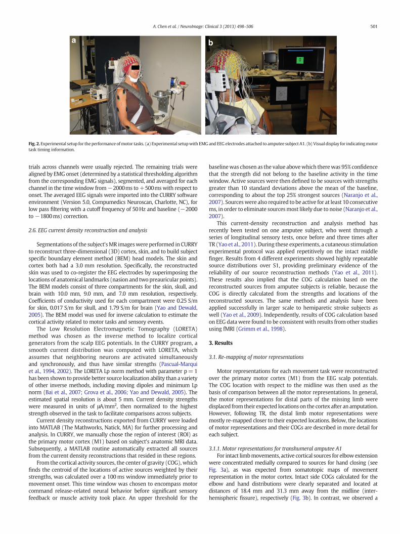
Fig. 2. Experimental setup for theperformance ofmotor tasks. (a) Experimental setupwith EMGand EEG electrodes attached to amputee subject A1. (b) Visual display for indicatingmotortask timing information.
501A. Chen et al. / NeuroImage: Clinical 3 (2013) 498–506
trials across channels were usually rejected. The remaining trials werealigned by EMGonset (determined by a statistical thresholding algorithmfrom the corresponding EMG signals), segmented, and averaged for eachchannel in the timewindow from−2000ms to+500mswith respect toonset. The averaged EEG signals were imported into the CURRY softwareenvironment (Version 5.0, Compumedics Neuroscan, Charlotte, NC), forlow pass filtering with a cutoff frequency of 50Hz and baseline (−2000to−1800ms) correction.
2.6. EEG current density reconstruction and analysis
Segmentations of the subject'sMR imageswere performed in CURRYto reconstruct three-dimensional (3D) cortex, skin, and to build subjectspecific boundary element method (BEM) head models. The skin andcortex both had a 3.0 mm resolution. Specifically, the reconstructedskin was used to co-register the EEG electrodes by superimposing thelocations of anatomical landmarks (nasion and two preauricular points).The BEM models consist of three compartments for the skin, skull, andbrain with 10.0 mm, 9.0 mm, and 7.0 mm resolution, respectively.Coefficients of conductivity used for each compartment were 0.25 S/mfor skin, 0.017 S/m for skull, and 1.79 S/m for brain (Yao and Dewald,2005). The BEM model was used for inverse calculation to estimate thecortical activity related to motor tasks and sensory events.
The Low Resolution Electromagnetic Tomography (LORETA)method was chosen as the inverse method to localize corticalgenerators from the scalp EEG potentials. In the CURRY program, asmooth current distribution was computed with LORETA, whichassumes that neighboring neurons are activated simultaneouslyand synchronously, and thus have similar strengths (Pascual-Marquiet al., 1994, 2002). The LORETA Lp norm method with parameter p=1has been shown to provide better source localization ability than a varietyof other inverse methods, including moving dipoles and minimum Lpnorm (Bai et al., 2007; Grova et al., 2006; Yao and Dewald, 2005). Theestimated spatial resolution is about 5 mm. Current density strengthswere measured in units of μA/mm2, then normalized to the higheststrength observed in the task to facilitate comparisons across subjects.
Current density reconstructions exported from CURRY were loadedinto MATLAB (The Mathworks, Natick, MA) for further processing andanalysis. In CURRY, we manually chose the region of interest (ROI) asthe primary motor cortex (M1) based on subject's anatomic MRI data.Subsequently, a MATLAB routine automatically extracted all sourcesfrom the current density reconstructions that resided in these regions.
From the cortical activity sources, the center of gravity (COG), whichfinds the centroid of the locations of active sources weighted by theirstrengths, was calculated over a 100ms window immediately prior tomovement onset. This time window was chosen to encompass motorcommand release-related neural behavior before significant sensoryfeedback or muscle activity took place. An upper threshold for the
baselinewas chosen as the value abovewhich therewas 95% confidencethat the strength did not belong to the baseline activity in the timewindow. Active sources were then defined to be sources with strengthsgreater than 10 standard deviations above the mean of the baseline,corresponding to about the top 25% strongest sources (Naranjo et al.,2007). Sourceswere also required to be active for at least 10 consecutivems, in order to eliminate sourcesmost likely due to noise (Naranjo et al.,2007).
This current-density reconstruction and analysis method hasrecently been tested on one amputee subject, who went through aseries of longitudinal sensory tests, once before and three times afterTR (Yao et al., 2011). During these experiments, a cutaneous stimulationexperimental protocol was applied repetitively on the intact middlefinger. Results from 4 different experiments showed highly repeatablesource distributions over S1, providing preliminary evidence of thereliability of our source reconstruction methods (Yao et al., 2011).These results also implied that the COG calculation based on thereconstructed sources from amputee subjects is reliable, because theCOG is directly calculated from the strengths and locations of thereconstructed sources. The same methods and analysis have beenapplied successfully in larger scale to hemiparetic stroke subjects aswell (Yao et al., 2009). Independently, results of COG calculation basedon EEG data were found to be consistentwith results from other studiesusing fMRI (Grimm et al., 1998).
3. Results
3.1. Re-mapping of motor representations
Motor representations for each movement task were reconstructedover the primary motor cortex (M1) from the EEG scalp potentials.The COG location with respect to the midline was then used as thebasis of comparison between all the motor representations. In general,the motor representations for distal parts of the missing limb weredisplaced from their expected locations on the cortex after an amputation.However, following TR, the distal limb motor representations weremostly re-mapped closer to their expected locations. Below, the locationsof motor representations and their COGs are described in more detail foreach subject.
3.1.1. Motor representations for transhumeral amputee A1For intact limbmovements, active cortical sources for elbowextension
were concentrated medially compared to sources for hand closing (seeFig. 3a), as was expected from somatotopic maps of movementrepresentation in the motor cortex. Intact side COGs calculated for theelbow and hand distributions were clearly separated and located atdistances of 18.4 mm and 31.3 mm away from the midline (inter-hemispheric fissure), respectively (Fig. 3b). In contrast, we observed a
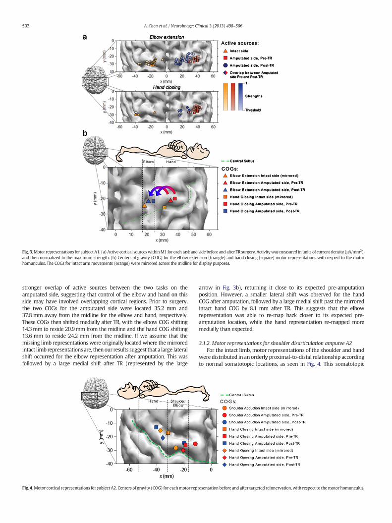
Fig. 3.Motor representations for subject A1. (a) Active cortical sourceswithinM1 for each task and side before and after TR surgery. Activitywasmeasured in units of current density (μA/mm2),and then normalized to the maximum strength. (b) Centers of gravity (COG) for the elbow extension (triangle) and hand closing (square) motor representations with respect to the motorhomunculus. The COGs for intact arm movements (orange) were mirrored across the midline for display purposes.
502 A. Chen et al. / NeuroImage: Clinical 3 (2013) 498–506
stronger overlap of active sources between the two tasks on theamputated side, suggesting that control of the elbow and hand on thisside may have involved overlapping cortical regions. Prior to surgery,the two COGs for the amputated side were located 35.2 mm and37.8 mm away from the midline for the elbow and hand, respectively.These COGs then shifted medially after TR, with the elbow COG shifting14.3mm to reside 20.9mm from the midline and the hand COG shifting13.6 mm to reside 24.2 mm from the midline. If we assume that themissing limb representations were originally located where themirroredintact limb representations are, thenour results suggest that a large lateralshift occurred for the elbow representation after amputation. This wasfollowed by a large medial shift after TR (represented by the large
Fig. 4.Motor cortical representations for subject A2. Centers of gravity (COG) for eachmotor rep
arrow in Fig. 3b), returning it close to its expected pre-amputationposition. However, a smaller lateral shift was observed for the handCOG after amputation, followed by a large medial shift past the mirroredintact hand COG by 8.1 mm after TR. This suggests that the elbowrepresentation was able to re-map back closer to its expected pre-amputation location, while the hand representation re-mapped moremedially than expected.
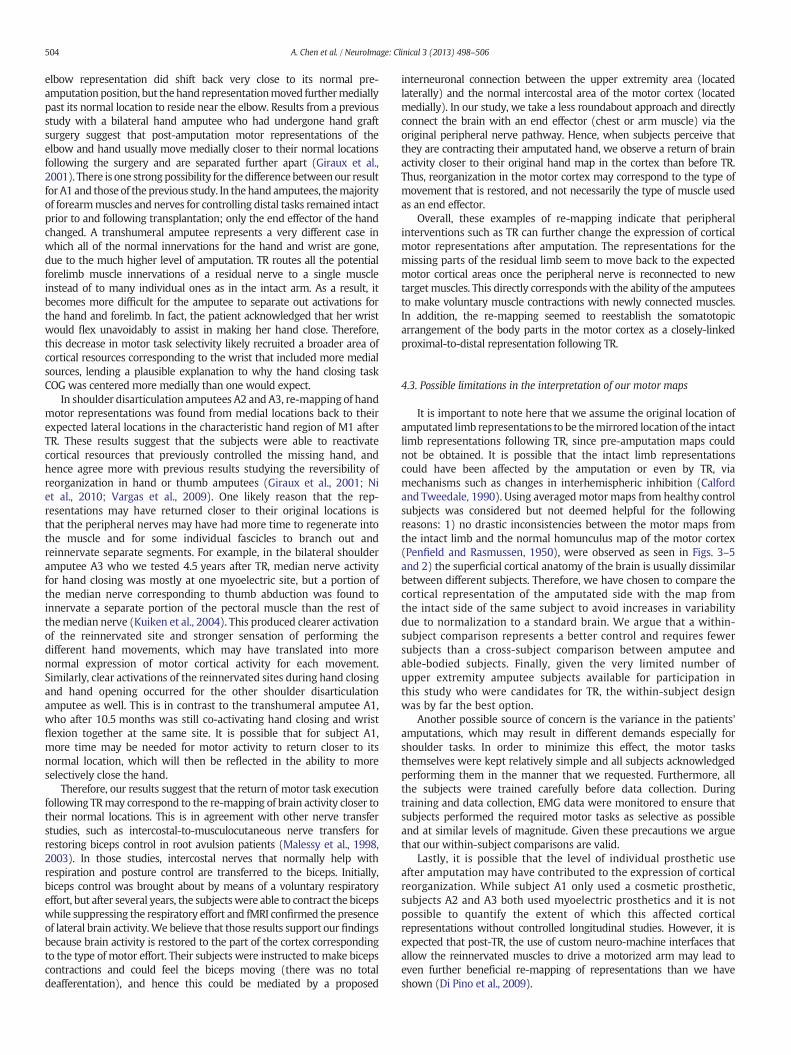
3.1.2. Motor representations for shoulder disarticulation amputee A2For the intact limb, motor representations of the shoulder and hand
were distributed in an orderly proximal-to-distal relationship accordingto normal somatotopic locations, as seen in Fig. 4. This somatotopic
resentation before and after targeted reinnervation,with respect to themotor homunculus.

503A. Chen et al. / NeuroImage: Clinical 3 (2013) 498–506
relationship was also preserved for the amputated limb before TR, butthe shoulder abduction COG was located more medially compared tothe mirrored intact shoulder COG (about 11.7 mm from the midlinecompared to 20.0 mm). Hand closing and opening before TR alsoactivated cortical areas that were located much more medially thanexpected, even further medially than the mirrored intact shoulderrepresentation. Distances between the midline and the hand COGswere 18.8mm for hand closing and 20.6mm for hand opening.
Following TR, a lateral shift occurred for all the reinnervated limb'srepresentations, bringing them closer to their expected locations. Theshoulder representation moved laterally closer to the mirrored intactshoulder representation, with the new shoulder COG located 20.1mmfrom the midline. The hand closing and opening representations alsomoved laterally to rest near the mirrored intact hand representations,about 35–40mm from themidline. These results suggest that followingTR, the shoulder and hand representations were able to move in amedial-to-lateral direction to return closer to their estimated originallocations, while preserving their somatotopic organization.
3.1.3. Motor representations for bilateral shoulder disarticulation amputeeA3
Motor representations for the bilateral shoulder disarticulationamputee A3 are displayed in Fig. 5. Representations for his non-reinnervated right amputated limb behaved similarly to those of subjectA2's pre-TR amputated limb. The shoulder abduction representationwas distributed more medially than the hand closing representation,with the shoulder COG about 24.4 mm from the midline and the handCOG about 31.9 mm away. For the reinnervated amputated limb, theshoulder abduction representation was located in approximately thesame location as themirrored non-reinnervated shoulder representation,with the COG also about 24mm from the midline. The reinnervatedhand closing representation was then found at a much more lateralsite along the precentral gyrus than the non-reinnervated handrepresentation, with the COG about 41 mm from the midline. TheCOG location was close to the characteristic knob-like bend of thecentral gyrus where motor control of the hand is normally expected(Penfield and Rasmussen, 1950). Therefore, these results suggestthat the shoulder representations for both amputated limbsremained about the same, even though TR was performed on oneof the limbs. However, the reinnervated hand representation wasable to re-map from a more medial location after injury to a morenormal, lateral location after TR.
Fig. 5.Motor cortical representations for subject A3. Centers of gravity (COG) are shown for eachomunculus.
4. Discussion
4.1. Plasticity of motor representations after amputation
Our results show that when voluntary movements of the missinglimb are attempted after amputation, but before TR, movement repre-sentations of the missing limb shift out of the original cortical areainto neighboring areas. Previous studies have also reported shifts inlocations of movement representations following amputation (Girauxet al., 2001; Karl et al., 2004; Kew et al., 1994; Lotze et al., 2001). Fortranshumeral subject A1, a lateral shift of both the elbow and handrepresentations occurred following amputation. The elbow represen-tation moved to encompass more of what used to be hand regions ofthe cortex, while the hand representation moved more laterally to that.In the shoulder disarticulation amputees (A2 and A3), hand represen-tations appeared to shift medially closer to shoulder representationsafter injury. The variability of remapping of the motor cortex the after-amputation could be a result of the combined effects of various factors,such as the levels of amputation, the extent of usage of a prosthesis, andso on. Regardless, our results confirm the perseverance of long-term plasticity at the motor cortex after amputation, thus per-mitting remapping of motor cortices after TR.
Another interesting result was that while missing limb representa-tions did shift into neighboring areas, there was still considerableoverlap with residual limb representations in the motor cortex afteramputation. In previous studies, magnetic stimulation over theseoriginal cortical areas using TMS not only produced movements ormuscle contractions in neighboring residual parts of the body (Cohenet al., 1991; Hall et al., 1990; Kew et al., 1994; Pascual-Leone et al.,1996; Roricht et al., 1999), but also produced a sense of phantom limbmovement (Amassian et al., 1989; Cohen et al., 1991; Hess et al.,1986; Mercier et al., 2006). Thus, there may indeed be somecohabitation of a movement representation of the missing limb and amovement representation of a residual muscle in the same corticalarea (Mercier et al., 2006; Reilly and Sirigu, 2008). However, our resultsshow that the sole use of TMSdoes not provide a total picture of how themovement representations may shift to include other sources on thecortex, as seen through neuroimaging.
4.2. Plasticity of motor representations following targeted reinnervation
Missing limb representations mostly shifted back closer to theiroriginal locations following TR. In the transhumeral amputee A1, the
hmotor representation before and after targeted reinnervation, with respect to themotor
504 A. Chen et al. / NeuroImage: Clinical 3 (2013) 498–506
elbow representation did shift back very close to its normal pre-amputation position, but thehand representationmoved furthermediallypast its normal location to reside near the elbow. Results from a previousstudy with a bilateral hand amputee who had undergone hand graftsurgery suggest that post-amputation motor representations of theelbow and hand usually move medially closer to their normal locationsfollowing the surgery and are separated further apart (Giraux et al.,2001). There is one strongpossibility for the difference between our resultfor A1 and those of the previous study. In the hand amputees, themajorityof forearmmuscles and nerves for controlling distal tasks remained intactprior to and following transplantation; only the end effector of the handchanged. A transhumeral amputee represents a very different case inwhich all of the normal innervations for the hand and wrist are gone,due to the much higher level of amputation. TR routes all the potentialforelimb muscle innervations of a residual nerve to a single muscleinstead of to many individual ones as in the intact arm. As a result, itbecomes more difficult for the amputee to separate out activations forthe hand and forelimb. In fact, the patient acknowledged that her wristwould flex unavoidably to assist in making her hand close. Therefore,this decrease in motor task selectivity likely recruited a broader area ofcortical resources corresponding to the wrist that included more medialsources, lending a plausible explanation to why the hand closing taskCOG was centered more medially than one would expect.
In shoulder disarticulation amputees A2 and A3, re-mapping of handmotor representations was found from medial locations back to theirexpected lateral locations in the characteristic hand region of M1 afterTR. These results suggest that the subjects were able to reactivatecortical resources that previously controlled the missing hand, andhence agree more with previous results studying the reversibility ofreorganization in hand or thumb amputees (Giraux et al., 2001; Niet al., 2010; Vargas et al., 2009). One likely reason that the rep-resentations may have returned closer to their original locations isthat the peripheral nerves may have had more time to regenerate intothe muscle and for some individual fascicles to branch out andreinnervate separate segments. For example, in the bilateral shoulderamputee A3 who we tested 4.5 years after TR, median nerve activityfor hand closing was mostly at one myoelectric site, but a portion ofthe median nerve corresponding to thumb abduction was found toinnervate a separate portion of the pectoral muscle than the rest ofthemedian nerve (Kuiken et al., 2004). This produced clearer activationof the reinnervated site and stronger sensation of performing thedifferent hand movements, which may have translated into morenormal expression of motor cortical activity for each movement.Similarly, clear activations of the reinnervated sites during hand closingand hand opening occurred for the other shoulder disarticulationamputee as well. This is in contrast to the transhumeral amputee A1,who after 10.5 months was still co-activating hand closing and wristflexion together at the same site. It is possible that for subject A1,more time may be needed for motor activity to return closer to itsnormal location, which will then be reflected in the ability to moreselectively close the hand.
Therefore, our results suggest that the return of motor task executionfollowing TRmay correspond to the re-mapping of brain activity closer totheir normal locations. This is in agreement with other nerve transferstudies, such as intercostal-to-musculocutaneous nerve transfers forrestoring biceps control in root avulsion patients (Malessy et al., 1998,2003). In those studies, intercostal nerves that normally help withrespiration and posture control are transferred to the biceps. Initially,biceps control was brought about by means of a voluntary respiratoryeffort, but after several years, the subjectswere able to contract the bicepswhile suppressing the respiratory effort and fMRI confirmed the presenceof lateral brain activity.We believe that those results support our findingsbecause brain activity is restored to the part of the cortex correspondingto the type of motor effort. Their subjects were instructed to make bicepscontractions and could feel the biceps moving (there was no totaldeafferentation), and hence this could be mediated by a proposed
interneuronal connection between the upper extremity area (locatedlaterally) and the normal intercostal area of the motor cortex (locatedmedially). In our study, we take a less roundabout approach and directlyconnect the brain with an end effector (chest or arm muscle) via theoriginal peripheral nerve pathway. Hence, when subjects perceive thatthey are contracting their amputated hand, we observe a return of brainactivity closer to their original hand map in the cortex than before TR.Thus, reorganization in the motor cortex may correspond to the type ofmovement that is restored, and not necessarily the type of muscle usedas an end effector.
Overall, these examples of re-mapping indicate that peripheralinterventions such as TR can further change the expression of corticalmotor representations after amputation. The representations for themissing parts of the residual limb seem to move back to the expectedmotor cortical areas once the peripheral nerve is reconnected to newtarget muscles. This directly correspondswith the ability of the amputeesto make voluntary muscle contractions with newly connected muscles.In addition, the re-mapping seemed to reestablish the somatotopicarrangement of the body parts in the motor cortex as a closely-linkedproximal-to-distal representation following TR.
4.3. Possible limitations in the interpretation of our motor maps
It is important to note here that we assume the original location ofamputated limb representations to be themirrored location of the intactlimb representations following TR, since pre-amputation maps couldnot be obtained. It is possible that the intact limb representationscould have been affected by the amputation or even by TR, viamechanisms such as changes in interhemispheric inhibition (Calfordand Tweedale, 1990). Using averaged motor maps from healthy controlsubjects was considered but not deemed helpful for the followingreasons: 1) no drastic inconsistencies between the motor maps fromthe intact limb and the normal homunculus map of the motor cortex(Penfield and Rasmussen, 1950), were observed as seen in Figs. 3–5and 2) the superficial cortical anatomy of the brain is usually dissimilarbetween different subjects. Therefore, we have chosen to compare thecortical representation of the amputated side with the map fromthe intact side of the same subject to avoid increases in variabilitydue to normalization to a standard brain. We argue that a within-subject comparison represents a better control and requires fewersubjects than a cross-subject comparison between amputee andable-bodied subjects. Finally, given the very limited number ofupper extremity amputee subjects available for participation inthis study who were candidates for TR, the within-subject designwas by far the best option.
Another possible source of concern is the variance in the patients'amputations, which may result in different demands especially forshoulder tasks. In order to minimize this effect, the motor tasksthemselves were kept relatively simple and all subjects acknowledgedperforming them in the manner that we requested. Furthermore, allthe subjects were trained carefully before data collection. Duringtraining and data collection, EMG data were monitored to ensure thatsubjects performed the required motor tasks as selective as possibleand at similar levels of magnitude. Given these precautions we arguethat our within-subject comparisons are valid.
Lastly, it is possible that the level of individual prosthetic useafter amputation may have contributed to the expression of corticalreorganization. While subject A1 only used a cosmetic prosthetic,subjects A2 and A3 both used myoelectric prosthetics and it is notpossible to quantify the extent of which this affected corticalrepresentations without controlled longitudinal studies. However, it isexpected that post-TR, the use of custom neuro-machine interfaces thatallow the reinnervated muscles to drive a motorized arm may lead toeven further beneficial re-mapping of representations than we haveshown (Di Pino et al., 2009).
505A. Chen et al. / NeuroImage: Clinical 3 (2013) 498–506
4.4. Central mechanisms underlying reorganization
To date, the mechanisms that allow for shifts in representationsfollowing peripheral injury and for re-mapping of representationsfollowing TR are not well understood. One of the most frequentlyaccepted mechanisms postulated to be responsible for changes inthe primary motor cortex (M1) following peripheral injury is theunmasking of latent synaptic connections (Jacobs and Donoghue,1991; Kaas et al., 1983; Sanes and Donoghue, 2000; Sanes et al.,1988; Schieber and Deuel, 1997). These latent connections consistof horizontal connections spanning M1 that combine with M1neurons to help construct a motor representation. This system ofinhibitory and excitatory connections within M1 may provide theunderlying connectivity responsible for the distributed nature ofM1 subregion organization (Sanes and Donoghue, 2000). Particularneurons in M1 influence multiple muscles at the periphery insteadof a single muscle. Thus, M1 regions controlling proximal and distalmusculature are not strictly segregated and can overlap (He et al.,1993; Nudo et al., 2001; Sanes and Donoghue, 2000; Schieber,1999; Schieber and Deuel, 1997).
Recent studies have shown that these latent connections provide apossible substrate on which plasticity can occur. Motor output mapscan rapidly change after procedures such as transient deafferentation(Brasil-Neto et al., 1993) or peripheral nerve transection (Donoghueet al., 1990). They can even rapidly change back soon after thetransient deafferentation procedure is ended. Therefore, theremust already be some underlying framework for rapid brainplasticity in place. The unmasking of existing connections mayinvolve increasing the efficacy of synapses, such as by increasingthe amount of excitatory neurotransmitter release, increasing the densityof post-synaptic receptors, changing membrane conductances,decreasing inhibitory inputs, or reducing inhibition from excitatoryinputs (Chen et al., 2002; Kaas, 1991; Wall, 1977). Of these, themain evidence has been that changes in motor maps can beproduced by using agents that reduce GABA-mediated intracorticalinhibition (Jacobs and Donoghue, 1991; Ridding and Rothwell,1999; Ziemann et al., 1998a). What these studies have demon-strated pharmacologically may potentially take place spontaneouslyin the brain to bring about plastic changes and reorganization afterperipheral injury. Further evidence to support the idea of a reduction ininhibition has been demonstrated in transient deafferentation and TMSstudies following amputation in humans (Ridding and Rothwell, 1999;Ziemann et al., 1998b).
Therefore, changes following upper-limb amputation may usethese mechanisms to shift weights of latent connections, such thatthe weights for remaining intact muscles are strengthened at theexpense of the missing parts of the upper limb (Giraux et al.,2001). In our study, we observed that representations for themissing parts of the limb were displaced following amputation forall subjects. After peripheral interventions to restore afferentation/efferentation, such as a hand graft or targeted reinnervation, theweights of these connections may then be restored to their normalstate over time.
Longer-term changes following amputation may be mediated byadditional mechanisms such as long-term potentiation (LTP) orlong-term depression (LTD). LTP and LTD involving NMDA receptoractivation or deactivation are longer-lasting mechanisms in whichsynapses stay strengthened or weakened. This may occur afterperipheral injuries since long-lasting decreases (greater than anhour) in intracortical inhibition normally found after repetitive TMS(Ridding and Rothwell, 1999) were reversed after administering anNMDA receptor blocker (Ziemann et al., 1998a). Therefore, a combinationof short and longer-termmechanismsmay be responsible for the changesin motor representations following amputation. Peripheral interventionssuch as TR would then induce further changes through these samemechanisms.
5. Conclusions
In this case study,wewere able tomeasure both the effects of a high-level amputation and subsequent TR procedure on the expressionof brain activity related to motor tasks of the upper limb. Primarily,we found that TR was able to re-map motor representations acrossa range of upper-limb amputees closer to their presumed pre-amputation states. These results have strong neural implications forrehabilitation, in particular highlighting the potential to assess theprogress and effectiveness of surgical interventions and novel neuro-prosthetics at restoring more natural control of motor functions forthe missing limb. As we have shown, cortical mapping can revealwhether the amputee is ideally able to regain similar patterns of corticalbehavior for the missing limb, such that the natural thought of movingthe missing hand can be relayed via the reinnervated nerves to movea motorized hand in a neuro-prosthetic. Over time, the control of theprosthetic arm is done by the same regions that originally controlledthe missing limb, which is more natural to amputees than otherprosthetics that rely on indirect methods of control. Hence, the use ofTR driven neuro-prosthetics that can take advantage of the brain'sremarkable ability to reorganize is expected to generate enormousstrides in restoring arm function to amputees in the future.
Disclosure/conflict of interest
None declared.
Funding
This study was supported by an American Heart AssociationPredoctoral Fellowship and National Institutes of Health grants N01-HD-5-3402 and R01HD047569-05.
Acknowledgments
The authors would like to thank Carolina Carmona and Dan Krainakfor assistance with experimental procedures and Paul Marasco forhelpful comments about the manuscript.
References
Amassian, V.E., Cracco, R.Q., Maccabee, P.J., 1989. A sense of movement elicited inparalyzed distal arm by focal magnetic coil stimulation of human motor cortex.Brain Res. 479, 355–360.
Bai, X., Towle, V.L., He, E.J., He, B., 2007. Evaluation of cortical current density imagingmethods using intracranial electrocorticograms and functional MRI. Neuroimage 35,598–608. http://dx.doi.org/10.1016/j.neuroimage.2006.12.026.
Borsook, D., Becerra, L., Fishman, S., et al., 1998. Acute plasticity in the humansomatosensory cortex following amputation. Neuroreport 9, 1013–1017.
Brasil-Neto, J.P., Valls-Sole, J., Pascual-Leone, A., et al., 1993. Rapid modulation of humancortical motor outputs following ischaemic nerve block. Brain 116 (Pt 3), 511–525.
Calford, M.B., Tweedale, R., 1990. Interhemispheric transfer of plasticity in the cerebralcortex. Science 249, 805–807.
Chen, R., Corwell, B., Yaseen, Z., Hallett, M., Cohen, L.G., 1998. Mechanisms of corticalreorganization in lower-limb amputees. J. Neurosci. 18, 3443–3450.
Chen, R., Cohen, L.G., Hallett, M., 2002. Nervous system reorganization following injury.Neuroscience 111, 761–773.
Cohen, L.G., Bandinelli, S., Findley, T.W., Hallett, M., 1991. Motor reorganization afterupper limb amputation in man. A study with focal magnetic stimulation. Brain 114(Pt 1B), 615–627.
Di Pino, G., Guglielmelli, E., Rossini, P.M., 2009. Neuroplasticity in amputees: mainimplications on bidirectional interfacing of cybernetic hand prostheses. Prog.Neurobiol. 88, 114–126. http://dx.doi.org/10.1016/j.pneurobio.2009.03.001 (S0301-0082(09)00035-5 [pii]).
Donoghue, J.P., Sanes, J.N., 1987. Peripheral nerve injury in developing rats reorganizesrepresentation pattern in motor cortex. Proc. Natl. Acad. Sci. U. S. A. 84, 1123–1126.
Donoghue, J.P., Suner, S., Sanes, J.N., 1990. Dynamic organization of primary motor cortexoutput to target muscles in adult rats. II. Rapid reorganization following motor nervelesions. Exp. Brain Res. 79, 492–503.
Elbert, T., Flor, H., Birbaumer, N., Knecht, S., Hampson, S., Larbig, W., Taub, E., 1994.Extensive reorganization of the somatosensory cortex in adult humans after nervoussystem injury. Neuroreport 5, 2593–2597.
506 A. Chen et al. / NeuroImage: Clinical 3 (2013) 498–506
Elbert, T., Sterr, A., Flor, H., et al., 1997. Input-increase and input-decrease types of corticalreorganization after upper extremity amputation in humans. Exp. Brain Res. 117,161–164.
Flor, H., Elbert, T., Knecht, S., et al., 1995. Phantom-limb pain as a perceptual correlate ofcortical reorganization following arm amputation. Nature 375, 482–484.
Giraux, P., Sirigu, A., Schneider, F., Dubernard, J.M., 2001. Cortical reorganization in motorcortex after graft of both hands. Nat. Neurosci. 4, 691–692. http://dx.doi.org/10.1038/89472.
Grimm, C., Schreiber, A., Kristeva-Feige, R., Mergner, T., Hennig, J., Lucking, C.H., 1998. Acomparison between electric source localisation and fMRI during somatosensorystimulation. Electroencephalogr. Clin. Neurophysiol. 106, 22–29.
Grova, C., Daunizeau, J., Lina, J.M., Benar, C.G., Benali, H., Gotman, J., 2006. Evaluation ofEEG localization methods using realistic simulations of interictal spikes. Neuroimage29, 734–753. http://dx.doi.org/10.1016/j.neuroimage.2005.08.053.
Hall, E.J., Flament, D., Fraser, C., Lemon, R.N., 1990. Non-invasive brain stimulation revealsreorganized cortical outputs in amputees. Neurosci. Lett. 116, 379–386.
He, S.Q., Dum, R.P., Strick, P.L., 1993. Topographic organization of corticospinal projectionsfrom the frontal lobe: motor areas on the lateral surface of the hemisphere.J. Neurosci. 13, 952–980.
Hess, C.W., Mills, K.R., Murray, N.M., 1986. Magnetic stimulation of the humanbrain: facilitation of motor responses by voluntary contraction of ipsilateral andcontralateral muscles with additional observations on an amputee. Neurosci. Lett.71, 235–240 (doi: 0304-3940(86)90565-3 [pii]).
Hijjawi, J.B., Kuiken, T.A., Lipschutz, R.D., Miller, L.A., Stubblefield, K.A., Dumanian, G.A.,2006. Improved myoelectric prosthesis control accomplished using multiplenerve transfers. Plast. Reconstr. Surg. 118, 1573–1578. http://dx.doi.org/10.1097/01.prs.0000242487.62487.fb.
Irlbacher, K., Meyer, B.U., Voss, M., Brandt, S.A., Roricht, S., 2002. Spatial reorganization ofcortical motor output maps of stump muscles in human upper-limb amputees.Neurosci. Lett. 321, 129–132 (doi: S0304394002000393 [pii]).
Jacobs, K.M., Donoghue, J.P., 1991. Reshaping the cortical motor map by unmasking latentintracortical connections. Science 251, 944–947.
Kaas, J.H., 1991. Plasticity of sensory and motor maps in adult mammals. Annu. Rev.Neurosci. 14, 137–167. http://dx.doi.org/10.1146/annurev.ne.14.030191.001033.
Kaas, J.H., 2000. The reorganization of somatosensory and motor cortex afterperipheral nerve or spinal cord injury in primates. Prog. Brain Res. 128, 173–179.http://dx.doi.org/10.1016/S0079-6123(00)28015-1.
Kaas, J.H., Merzenich,M.M., Killackey, H.P., 1983. The reorganization of somatosensory cortexfollowing peripheral nerve damage in adult and developing mammals. Annu. Rev.Neurosci. 6, 325–356. http://dx.doi.org/10.1146/annurev.ne.06.030183.001545.
Karl, A., Birbaumer, N., Lutzenberger, W., Cohen, L.G., Flor, H., 2001. Reorganization ofmotor and somatosensory cortex in upper extremity amputees with phantom limbpain. J. Neurosci. 21, 3609–3618.
Karl, A., Muhlnickel, W., Kurth, R., Flor, H., 2004. Neuroelectric source imaging of steady-statemovement-related cortical potentials in human upper extremity amputees withand without phantom limb pain. Pain 110, 90–102. http://dx.doi.org/10.1016/j.pain.2004.03.013.
Kew, J.J., Ridding, M.C., Rothwell, J.C., et al., 1994. Reorganization of cortical blood flowand transcranial magnetic stimulation maps in human subjects after upper limbamputation. J. Neurophysiol. 72, 2517–2524.
Kuiken, T.A., 2003. Consideration of nerve-muscle grafts to improve the control ofartificial arms. Technol. Disabil. 15, 105–111.
Kuiken, T.A., Dumanian, G.A., Lipschutz, R.D., Miller, L.A., Stubblefield, K.A., 2004. The useof targeted muscle reinnervation for improved myoelectric prosthesis control in abilateral shoulder disarticulation amputee. Prosthet. Orthot. Int. 28, 245–253.
Kuiken, T., Miller, L., Lipschutz, R., Stubblefield, K., Dumanian, G., 2005. Prostheticcommand signals following targeted hyper-reinnervation nerve transfer surgery.Conf. Proc. IEEE Eng. Med. Biol. Soc. 7, 7652–7655.
Kuiken, T.A., Marasco, P.D., Lock, B.A., Harden, R.N., Dewald, J.P., 2007a. Redirectionof cutaneous sensation from the hand to the chest skin of human amputeeswith targeted reinnervation. Proc. Natl. Acad. Sci. U. S. A. 104, 20061–20066.http://dx.doi.org/10.1073/pnas.0706525104.
Kuiken, T.A., Miller, L.A., Lipschutz, R.D., et al., 2007b. Targeted reinnervation for enhancedprosthetic arm function in a womanwith a proximal amputation: a case study. Lancet369, 371–380. http://dx.doi.org/10.1016/S0140-6736(07)60193-7.
Lotze, M., Flor, H., Grodd, W., Larbig, W., Birbaumer, N., 2001. Phantom movements andpain. An fMRI study in upper limb amputees. Brain 124, 2268–2277.
Malessy, M.J., van der Kamp, W., Thomeer, R.T., van Dijk, J.G., 1998. Cortical excitability ofthe biceps muscle after intercostal-to-musculocutaneous nerve transfer. Neurosurgery42, 787–794 (discussion 794–785).
Malessy, M.J., Bakker, D., Dekker, A.J., Van Duk, J.G., Thomeer, R.T., 2003.Functional magnetic resonance imaging and control over the biceps muscle
after intercostal-musculocutaneous nerve transfer. J. Neurosurg. 98, 261–268.http://dx.doi.org/10.3171/jns.2003.98.2.0261.
Mercier, C., Reilly, K.T., Vargas, C.D., Aballea, A., Sirigu, A., 2006. Mapping phantommovement representations in the motor cortex of amputees. Brain 129, 2202–2210.http://dx.doi.org/10.1093/brain/awl180.
Merzenich, M.M., Kaas, J.H., Sur, M., Lin, C.S., 1978. Double representation of the bodysurface within cytoarchitectonic areas 3b and 1 in “SI” in the owl monkey (Aotustrivirgatus). J. Comp. Neurol. 181, 41–73. http://dx.doi.org/10.1002/cne.901810104.
Naranjo, J.R., Brovelli, A., Longo, R., Budai, R., Kristeva, R., Battaglini, P.P., 2007. EEGdynamics of the frontoparietal network during reaching preparation in humans.Neuroimage 34, 1673–1682. http://dx.doi.org/10.1016/j.neuroimage.2006.07.049.
Ni, Z., Anastakis, D.J., Gunraj, C., Chen, R., 2010. Reversal of cortical reorganization inhuman primary motor cortex following thumb reconstruction. J. Neurophysiol. 103,65–73. http://dx.doi.org/10.1152/jn.00732.2009.
Nudo, R.J., Plautz, E.J., Frost, S.B., 2001. Role of adaptive plasticity in recovery of functionafter damage to motor cortex. Muscle Nerve 24, 1000–1019.
Pascual-Leone, A., Peris, M., Tormos, J.M., Pascual, A.P., Catala, M.D., 1996. Reorganizationof human cortical motor output maps following traumatic forearm amputation.Neuroreport 7, 2068–2070.
Pascual-Marqui, R.D., Michel, C.M., Lehmann, D., 1994. Low resolution electromagnetictomography: a new method for localizing electrical activity in the brain. Int.J. Psychophysiol. 18, 49–65.
Pascual-Marqui, R.D., Esslen, M., Kochi, K., Lehmann, D., 2002. Functional imaging withlow-resolution brain electromagnetic tomography (LORETA): a review. MethodsFind. Exp. Clin. Pharmacol. 24 (Suppl. C), 91–95.
Penfield, W., Rasmussen, T., 1950. The Cerebral Cortex of Man. The Macmillan Company,New York.
Ramachandran, V.S., Stewart, M., Rogers-Ramachandran, D.C., 1992. Perceptual correlatesof massive cortical reorganization. Neuroreport 3, 583–586.
Reilly, K.T., Sirigu, A., 2008. The motor cortex and its role in phantom limb phenomena.Neuroscientist 14, 195–202. http://dx.doi.org/10.1177/1073858407309466.
Ridding, M.C., Rothwell, J.C., 1999. Afferent input and cortical organisation: a study withmagnetic stimulation. Exp. Brain Res. 126, 536–544.
Roricht, S., Meyer, B.U., Niehaus, L., Brandt, S.A., 1999. Long-term reorganization of motorcortex outputs after arm amputation. Neurology 53, 106–111.
Sanes, J.N., Donoghue, J.P., 2000. Plasticity and primarymotor cortex. Annu. Rev. Neurosci.23, 393–415. http://dx.doi.org/10.1146/annurev.neuro.23.1.393.
Sanes, J.N., Suner, S., Lando, J.F., Donoghue, J.P., 1988. Rapid reorganization of adult ratmotor cortex somatic representation patterns after motor nerve injury. Proc. Natl.Acad. Sci. U. S. A. 85, 2003–2007.
Schieber, M.H., 1999. Somatotopic gradients in the distributed organization of the humanprimary motor cortex hand area: evidence from small infarcts. Exp. Brain Res. 128,139–148.
Schieber, M.H., Deuel, R.K., 1997. Primary motor cortex reorganization in a long-termmonkey amputee. Somatosens. Mot. Res. 14, 157–167.
Vargas, C.D., Aballea, A., Rodrigues, E.C., et al., 2009. Re-emergence of hand-musclerepresentations in human motor cortex after hand allograft. Proc. Natl. Acad. Sci. U.S. A. 106, 7197–7202. http://dx.doi.org/10.1073/pnas.0809614106.
Wall, P.D., 1977. The presence of ineffective synapses and the circumstances whichunmask them. Philos. Trans. R. Soc. Lond. B Biol. Sci. 278, 361–372.
Wall, J.T., Kaas, J.H., Sur, M., Nelson, R.J., Felleman, D.J., Merzenich, M.M., 1986. Functionalreorganization in somatosensory cortical areas 3b and 1 of adult monkeysafter median nerve repair: possible relationships to sensory recovery in humans.J. Neurosci. 6, 218–233.
Weiss, T., Miltner, W.H., Huonker, R., Friedel, R., Schmidt, I., Taub, E., 2000. Rapidfunctional plasticity of the somatosensory cortex after finger amputation. Exp. BrainRes. 134, 199–203.
Yao, J., Dewald, J.P., 2005. Evaluation of different cortical source localization methodsusing simulated and experimental EEG data. Neuroimage 25, 369–382.http://dx.doi.org/10.1016/j.neuroimage.2004.11.036.
Yao, J., Chen, A., Carmona, C., Dewald, J.P., 2009. Cortical overlap of joint representationscontributes to the loss of independent joint control following stroke. Neuroimage45, 490–499. http://dx.doi.org/10.1016/j.neuroimage.2008.12.002 (S1053-8119(08)01263-9 [pii]).
Yao, J., Carmona, C., Chen, A., Kuiken, T., Dewald, J., 2011. Sensory cortical re-mappingfollowing upper-limb amputation and subsequent targeted reinnervation: a casereport. Conf. Proc. IEEE Eng. Med. Biol. Soc. 2011, 1065–1068. http://dx.doi.org/10.1109/IEMBS.2011.6090248.
Ziemann, U., Corwell, B., Cohen, L.G., 1998a. Modulation of plasticity in human motorcortex after forearm ischemic nerve block. J. Neurosci. 18, 1115–1123.
Ziemann, U., Hallett, M., Cohen, L.G., 1998b. Mechanisms of deafferentation-inducedplasticity in human motor cortex. J. Neurosci. 18, 7000–7007.
Related Documents


