BULLOUS DISEASES

B ULLOUS DISEASES. Vesicles and bullae are accumulations of fluid within or under the epidermis. Subepidermal blisters Occur between the dermis and the.
Jan 02, 2016
Welcome message from author
This document is posted to help you gain knowledge. Please leave a comment to let me know what you think about it! Share it to your friends and learn new things together.
Transcript
BULLOUS DISEASES
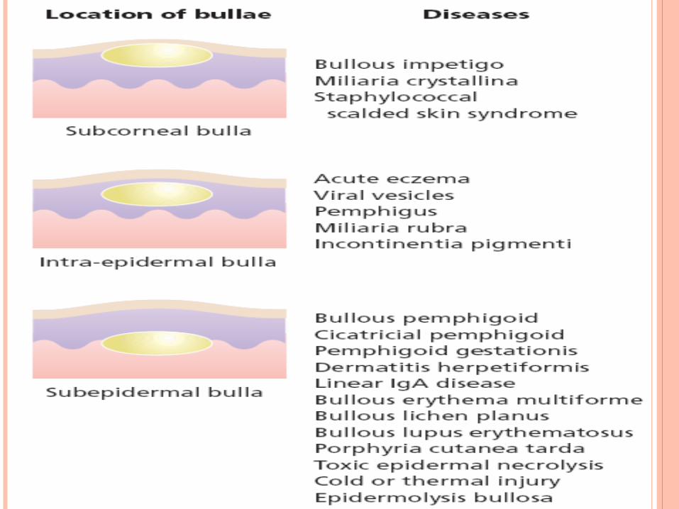
Vesicles and bullae are accumulations of fluid within or under the epidermis.
Subepidermal blisters Occur between the dermis and the epidermis. Their roofs are relatively thick and so they tend to be
tense and intact. They may contain blood.
Intra-epidermal blisters appear within the spinosum cell layer of the
epidermis So have thin roofs and rupture easily Leave an oozing denuded surface.
Subcorneal blisters Form just beneath the stratum corneum at the
outermost edge of the epidermis Have even thinner roofs Tendency to break is more marked
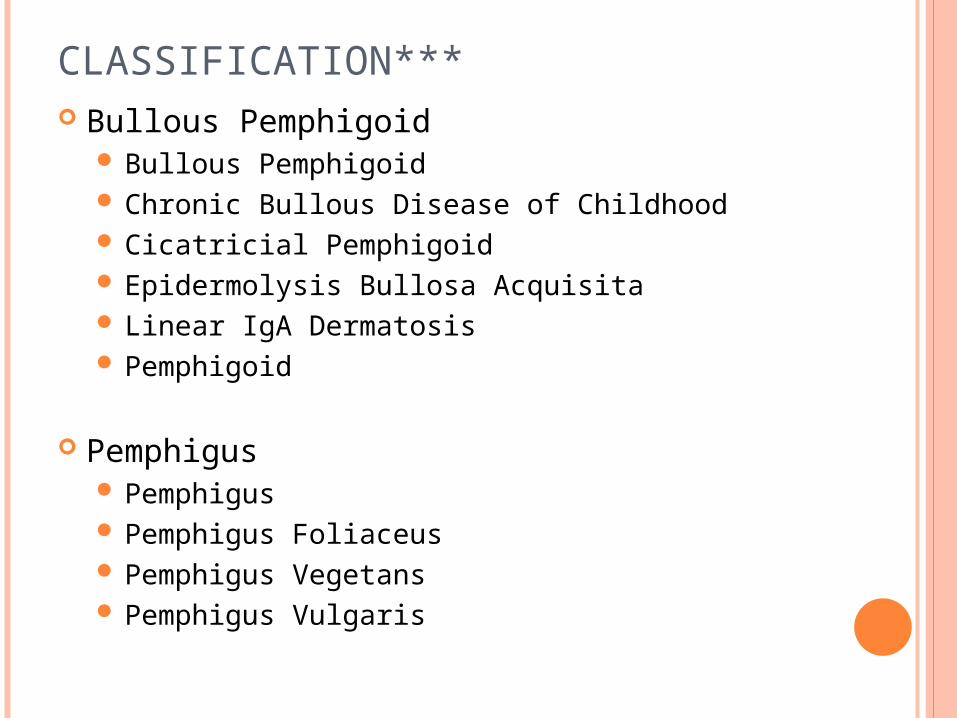
CLASSIFICATION*** Bullous Pemphigoid
Bullous Pemphigoid Chronic Bullous Disease of Childhood Cicatricial Pemphigoid Epidermolysis Bullosa Acquisita Linear IgA Dermatosis Pemphigoid
Pemphigus Pemphigus Pemphigus Foliaceus Pemphigus Vegetans Pemphigus Vulgaris
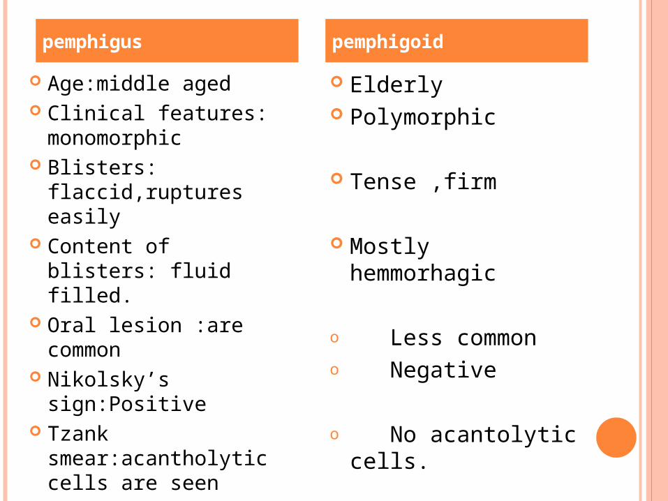
Age:middle aged Clinical features:
monomorphic Blisters:
flaccid,ruptures easily
Content of blisters: fluid filled.
Oral lesion :are common
Nikolsky’s sign:Positive
Tzank smear:acantholytic cells are seen
Elderly Polymorphic
Tense ,firm
Mostly hemmorhagic
o Less commono Negative
o No acantolytic cells.
pemphigus pemphigoid

BULLOUS PEMPHIGOID
It is an autoimmune disorder, meaning it is caused when the body's immune system malfunctions.
The immune system is meant to defend the body against bacteria, viruses, and disease, but instead produces antibodies against healthy tissue, cells and organs.
Some patients with BP have other diseases such diabetes and rheumatoid arthritis.
Other factors triggering BP include drugs (Furosemide, penicillin), mechanical trauma, and physical traumas (burns from radiation, sun or heat)
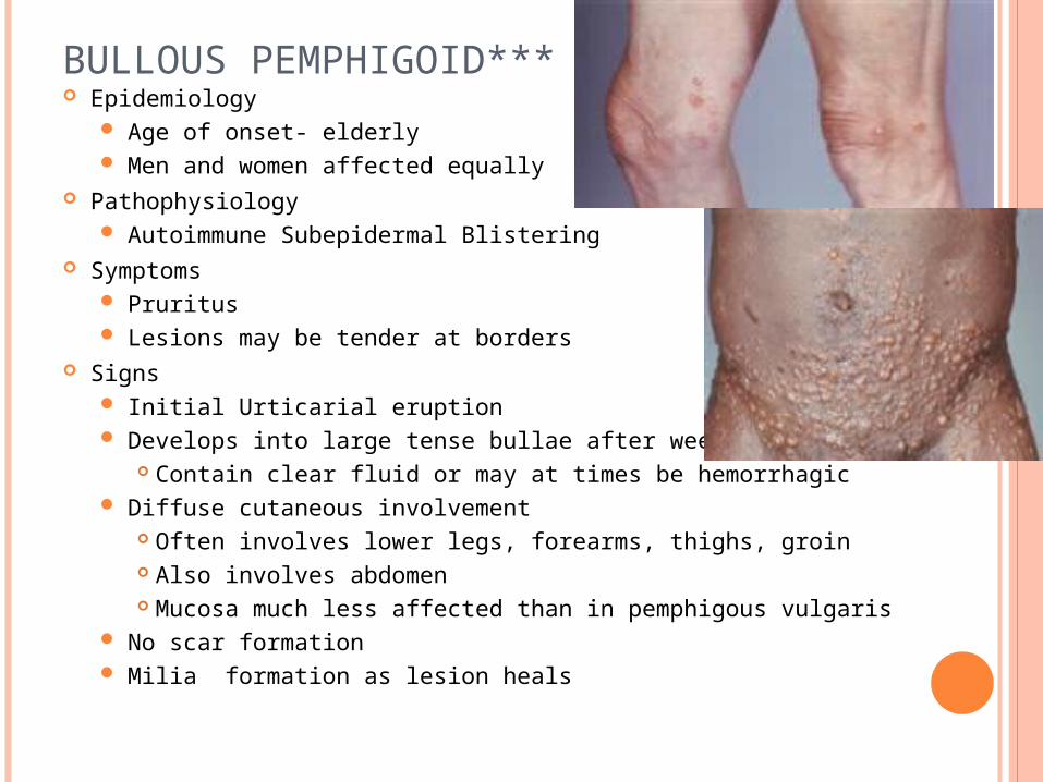
BULLOUS PEMPHIGOID*** Epidemiology
Age of onset- elderly Men and women affected equally
Pathophysiology Autoimmune Subepidermal Blistering
Symptoms Pruritus Lesions may be tender at borders
Signs Initial Urticarial eruption Develops into large tense bullae after weeks to months
Contain clear fluid or may at times be hemorrhagic Diffuse cutaneous involvement
Often involves lower legs, forearms, thighs, groin Also involves abdomen Mucosa much less affected than in pemphigous vulgaris
No scar formation Milia formation as lesion heals
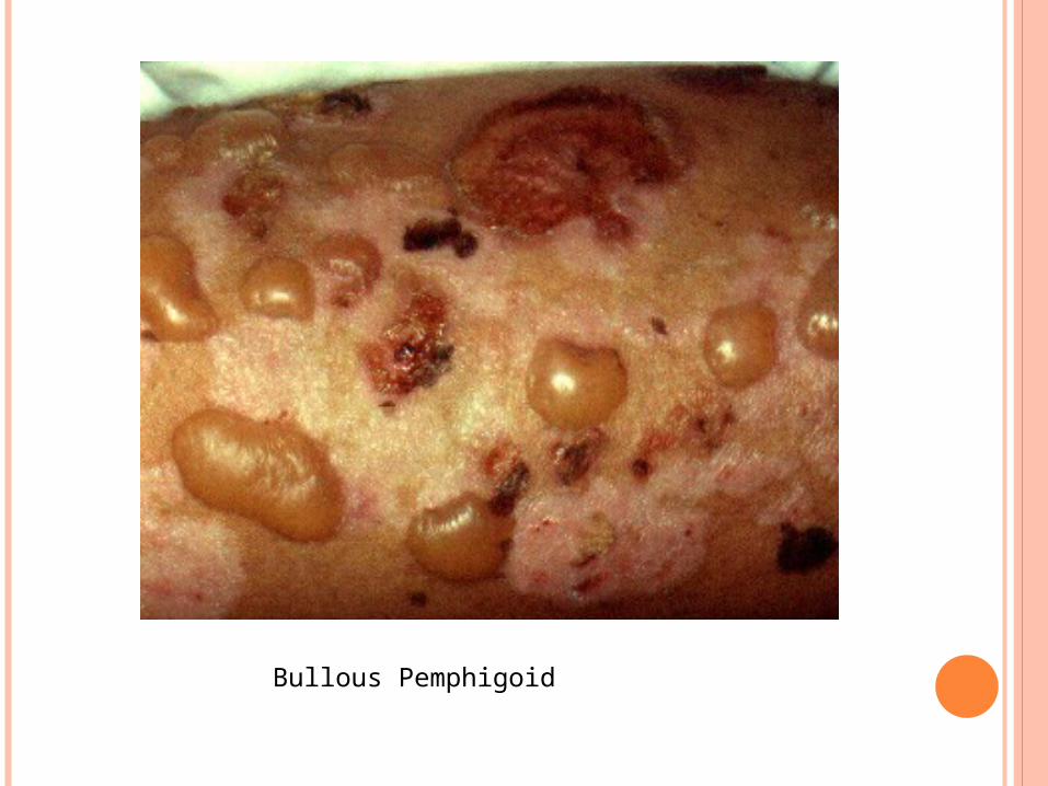
Bullous Pemphigoid
Labs Histology
Supepidermal Blister Superficial dermal inflammation
Immunofluorescence Ig G and C3 deposition along basement membrane zone
Management First-Line
Prednisone Methotrexate may be used for prednisone intolerance Topical corticosteroids in localized mild cases
Adjunctive agents Azathioprine Mycophenolate mofeti Tetracycline
Course Self-limited condition Remits with treatment by 6 years in 50 % of cases
CHRONIC BULLOUS DISEASE OF CHILDHOOD Epidemiology
Age of Onset: Under age 5 years Pathophysiology
Autoimmune bullous disorder Variant of Linear IgA Dermatosis (seen in adults)
Symptoms Pruritus and burning sensation
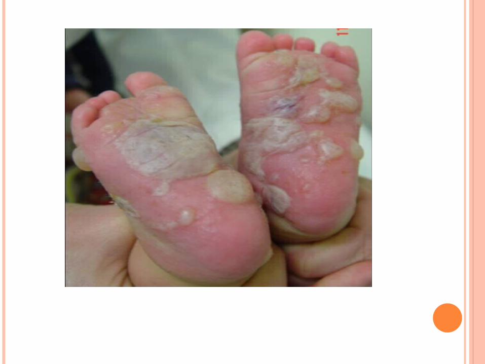
Signs Tense inflamed Blisters over red base
May appear as collarettes of Blisters Distribution
Most commonly found on genitalia Also may involves face and perioral skin Oral Mucosa involved in 50% of cases
Course Resolves within 2 years of onset in most cases
Histology Subepidermal Blisters Inflammatory infiltrates at basement membrane zone
Immunofluorescence IgA deposition along basement membrane
Management First Line: Dapsone or Sulfapyridine
(requires G6PD and monitoring of CBC) Adjunct: Low dose prednisone initially
CICATRICIAL PEMPHIGOID ***Epidemiology
Incidence (rare): 1 in 12,000 to 20,000 Ages affected: 60 to 80 years Gender predominance: Women by male ratio of 2:1
Pathophysiology Autoimmune bullous condition Associated conditions
Stevens-Johnson Syndrome Medications
Topical Glaucoma medications Practolol Clonidine
Signs Blistering of mucus membranes
Blister ruptures within hours of formation Painful Erosion

Most Erosions heal without scarring
Most common sites of involvement Oral Mucosa (most often buccal and palatal mucosa)
Results in erosive Gingivitis Conjunctiva (usually bilateral)
Results in chronic Conjunctivitis and scarring Other sites of involvement
Skin (usually non-scarring) Scalp (Cicatricial Alopecia) Pharynx and larynx External genitalia Nasal mucosa Anus Esophagus
Labs: Histology Subepidermal bulla Perivascular inflammatory cell infiltrates Fibrosis distinguishes from Bullous Pemphigoid Immunofluorescence with IgG, C3, IgA deposits
Forms linear band at Basement Membrane Zone
Differential Diagnosis Bullous Pemphigoid Epidermolysis Bullosa Acquisita(EBA) Linear IgA Bullous Dermatosis(LABD) Systemic Lupus Erythematosus Pseudopemphigoid Pemphigus
MANAGEMENT: Early aggressive treatment
First-Line: Prednisone 0.5 to 0.75 mg/kg/day x6 months Ocular Involvement: Azathioprine or Cyclophosphamide Skin Involvement: Topical Corticosteroids Oral Involvement
Dapsone Corticosteroid gel Dexamethasone mouthwash
Complications Ocular Lesions may result in blindness Laryngeal stricture Esophageal Stricture Supraglottic Stenosis
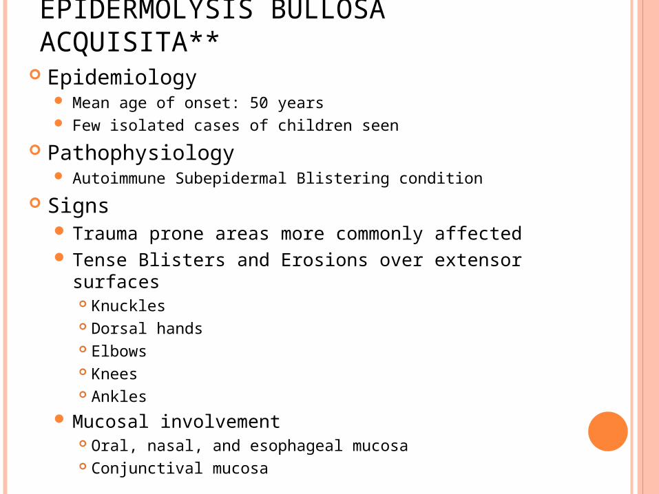
EPIDERMOLYSIS BULLOSA ACQUISITA**
Epidemiology Mean age of onset: 50 years Few isolated cases of children seen
Pathophysiology Autoimmune Subepidermal Blistering condition
Signs Trauma prone areas more commonly affected Tense Blisters and Erosions over extensor surfaces
Knuckles Dorsal hands Elbows Knees Ankles
Mucosal involvement Oral, nasal, and esophageal mucosa Conjunctival mucosa
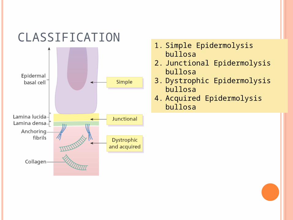
CLASSIFICATION1. Simple Epidermolysis bullosa2. Junctional Epidermolysis bullosa3. Dystrophic Epidermolysis
bullosa4. Acquired Epidermolysis bullosa
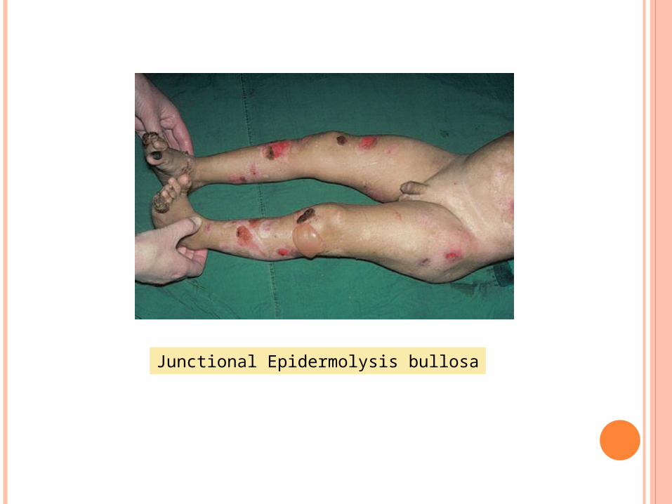
Junctional Epidermolysis bullosa
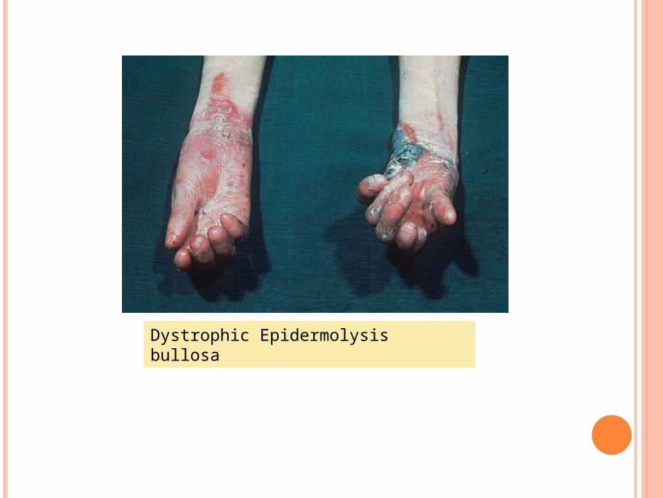
Dystrophic Epidermolysisbullosa
LINEAR IGA DERMATOSIS Epidemiology
Onset over age 30 years Pathophysiology IgA mediated dermatosis mediated by IgA
and causes "sausage-like" lobulated blisters arranged at the outer edges of flattened erythematous patches. Autoimmune bullous disorder Variant of Chronic Bullous Disease of Childhood
Symptoms Pruritus
Signs Annular Lesions
Papules (may be excoriated and crusted) Vesicles Bullae
Distribution Symmetric involvement on extensor surfaces Elbows, knees, and buttocks affected Mucosa involvement may occur (Conjunctiva, oral)
Labs Histology
Subepidermal Blister with inflammatory infiltrate Immunofluorescence
IgG deposits at dermal-epidermal junction
Complications Scarring skin lesions (with associated Milia)
Differential Diagnosis Porphyria cutanea tarda Hereditary Epidermolysis bullosa Bullous Pemphigoid Course Chronic waxing and waning course
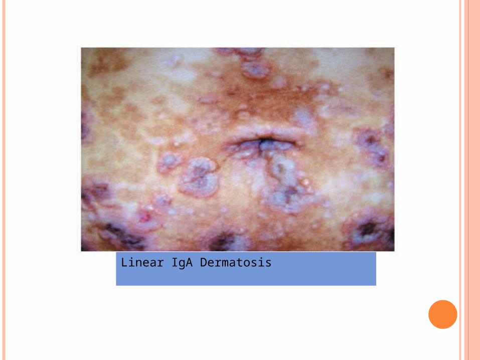
Linear IgA Dermatosis
Related Documents