Welcome message from author
This document is posted to help you gain knowledge. Please leave a comment to let me know what you think about it! Share it to your friends and learn new things together.
Transcript

Autoimmunity • Autoimmunity arises because of defects in central
or peripheral tolerance of lymphocytes to self-antigens
– Autoimmune disease can be caused to primary defects
in B cells, T cells and possibly dendritic cells
– To study autoimmune disease, researchers make use of human samples from normal and autoimmune patients as well as both spontaneous autoimmune and induced animal models of autoimmune disease
What Causes Autoimmune Disease?
With human diseases, this is often not clear as human patients may have a constellation of genetic changes that predispose them to disease combined with various environmental influences that trigger the disease Animal models can help us understand what causes autoimmune disease as we can control the genetic defects and the environmental exposures. Animal models have taught us that genetic defects in
T cells or B cells can drive autoimmunity and that environmental influences can exacerbate disease.

Figure 13-6

Mechanisms by which autoimmune responses can damage tissues

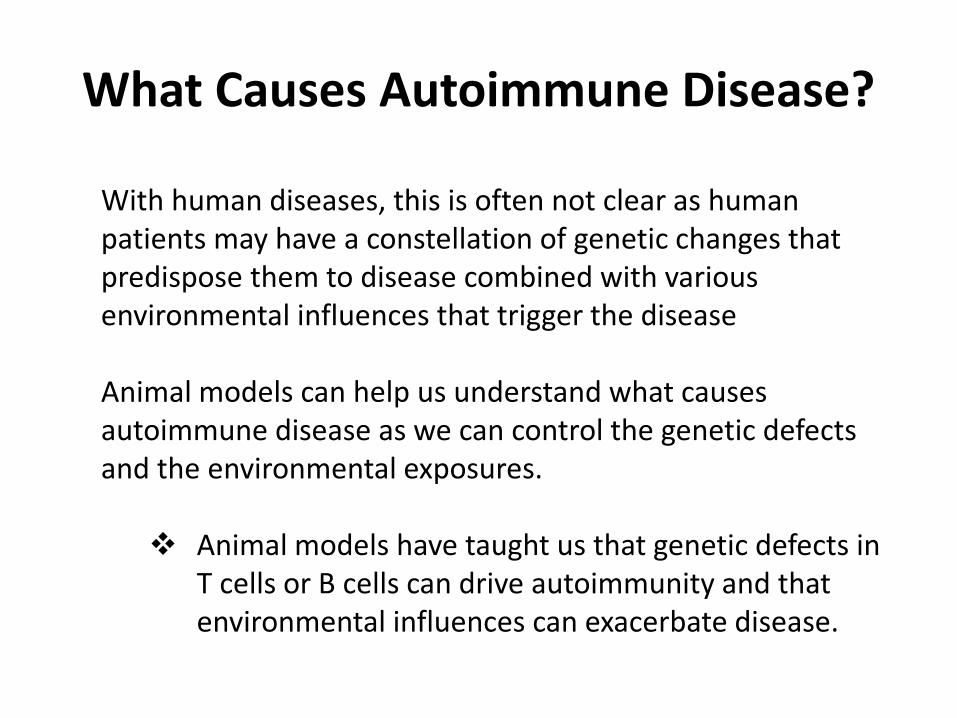
Autoantibodies Trigger Disease
Figure 13-28
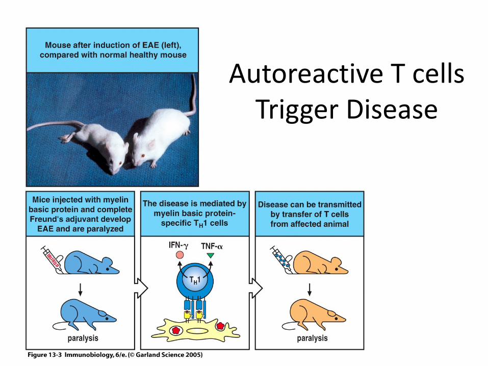
Figure 13-3 Autoreactive T cells
Trigger Disease

Role of T cells can be seen by disease associations with MHC haplotype
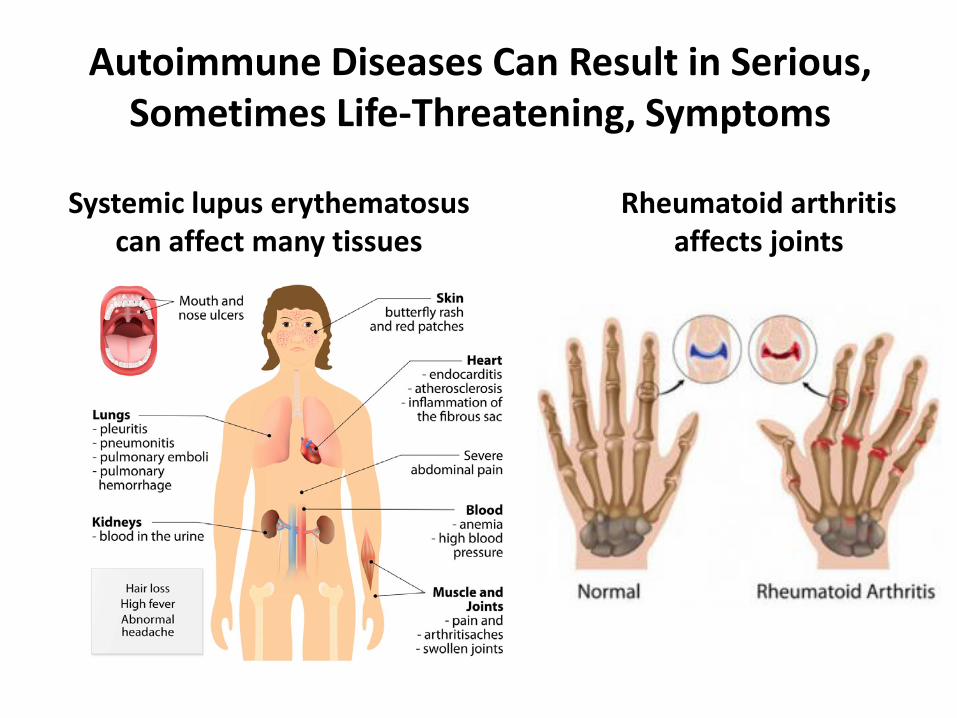
Rheumatoid arthritis affects joints
Autoimmune Diseases Can Result in Serious, Sometimes Life-Threatening, Symptoms
Systemic lupus erythematosus can affect many tissues
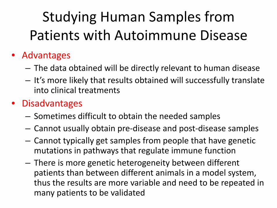
Studying Human Samples from Patients with Autoimmune Disease
• Advantages – The data obtained will be directly relevant to human disease – It’s more likely that results obtained will successfully translate
into clinical treatments • Disadvantages
– Sometimes difficult to obtain the needed samples – Cannot usually obtain pre-disease and post-disease samples – Cannot typically get samples from people that have genetic
mutations in pathways that regulate immune function – There is more genetic heterogeneity between different
patients than between different animals in a model system, thus the results are more variable and need to be repeated in many patients to be validated

Most well-studied autoim-mune diseases

Using Animal Models of Autoimmune Disease
• Advantages – Can obtain samples from organs such as spleen and thymus that are
difficult to obtain from humans (where most studies use either peripheral blood lymphocytes or tonsil-derived lymphocytes)
– Can obtain samples both before and after disease development to see how things change over time.
– Can use genetically modified mice to study the role of certain proteins in the autoimmune response
– Inbred strains of rats and mice are genetically homogeneous, making it easier to get statistically-meaningful data from smaller numbers of animals
– Can explore the effects of treatments which cannot be used in humans for various ethical reasons
• Disadvantages – Humans are not the same as mice or other animals and sometimes
respond differently – Treatments that work in mice to suppress autoimmune symptoms may not
work in humans
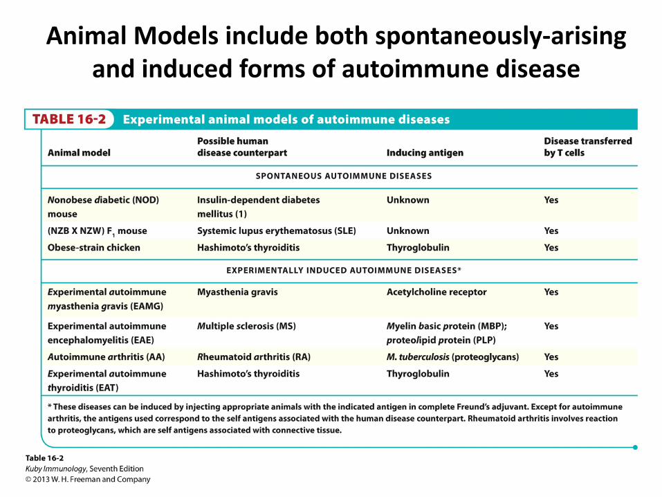
Animal Models include both spontaneously-arising and induced forms of autoimmune disease

Advantages/Disadvantages of Induced versus Spontaneous Animal Models of Autoimmune Disease
INDUCED
SPONTANEOUS
Advantages
1.) Can mimic diseases for which there may not be an appropriate spontaneous model 2.) Can have a large cohort of animals at the same stage of disease at the same time
1.) More like a human autoimmune syndrome with complex genetic factors 2.) Allows the study of autoimmunity at different life-stages
Disadvantages
1.) The genetic composition of the mice does not pre-dispose to autoimmunity. 2.) Normal immune suppressive mechanisms may inhibit the response 3.) Requires immunization with antigen mixed with an adjuvant
1.) There may not be an appropriate spontaneous model of the disease you want to study 2.) Spontaneous mutations in mice may not be the same as the mutations that promote human disease.

Many, but not all, human autoimmune diseases are more common in women

Possible reasons women may have increased susceptibility to Autoimmune Disease
Hormonal environment – for instance estrogens or other female hormones may promote immune cell activation or male hormones such as testosterone may inhibit Immune activation
Women carry two copies of the X chromosome, which contains many immunologically-relevant genes, while men only have 1. Women may express some X chromosome genes from both chromosomes in some cells (leading to increased amounts of those proteins). Many immunologically important genes are found on the X chromosome, whose altered expression might cause autoimmunity:
•TLR7 (recognizes viral and human RNA) •TLR8 (recognizes viral and human RNA) •CD40L (CD40 ligand) •FoxP3 (needed for development of Treg cells) •Btk (involved in BCR signaling) •NEMO (involved in NF-κB pathway) •Was (Wasp protein – downstream effector of BCR and TCR signaling) • IL2RG (Common gamma chain for numerous cytokines)
One X chromosome is randomly chosen for inactivation (Barr body)

Some Examples of Autoimmune Disease in Humans and Animal Models

Blocking Antibodies- Human Disease
Some autoantibodies can bind to receptors on cells and block their function

Stimulating Antibodies – Human Disease
Some autoantibodies can bind to receptors on cells and stimulate their function resulting in over-active pathways
Grave’s Disease

Many autoimmune patients (especially those with SLE) produce antibodies that bind the the nuclei of cultured cells and can cause cell damage
Anti-nuclear autoantibodies (ANA)

Figure 13-33
Antinuclear antibodies and other auto-antibodies form immune complexes with their autoantigens that deposit in tissues like kidney and induce damage
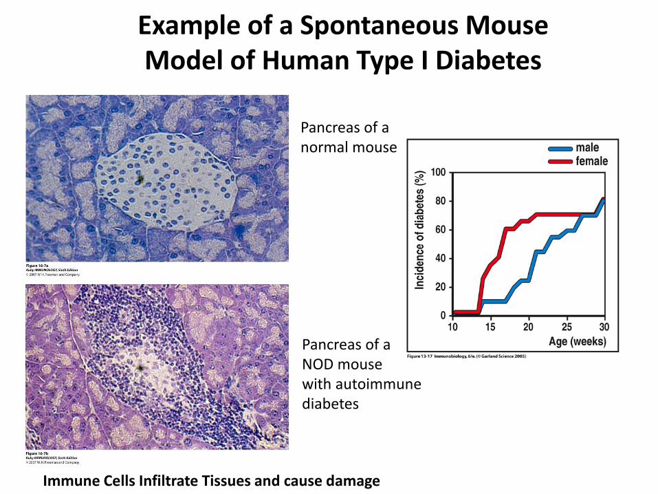
Pancreas of a normal mouse
Pancreas of a NOD mouse with autoimmune diabetes
Immune Cells Infiltrate Tissues and cause damage
Example of a Spontaneous Mouse Model of Human Type I Diabetes

Major Immune Cells and Pathways Implicated in Autoimmune Disease
Naïve T cell
Dendritic Cell
B cell
Plasma Cell
Treg
Th1
Th2
Th17
TFH
Autoantibodies Inhibit other T cells
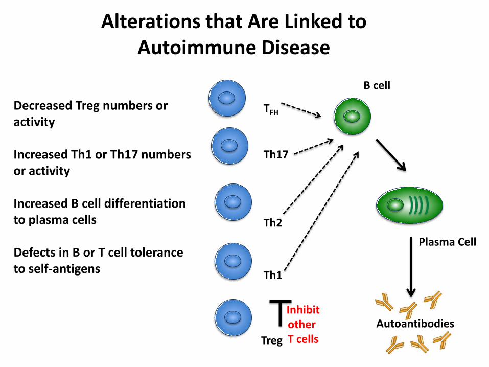
Alterations that Are Linked to Autoimmune Disease
B cell
Plasma Cell
Treg
Th1
Th2
Th17
TFH
Autoantibodies Inhibit other T cells
Decreased Treg numbers or activity Increased Th1 or Th17 numbers or activity Increased B cell differentiation to plasma cells Defects in B or T cell tolerance to self-antigens

T cells are normally tolerized
Defects in production of Tregs can lead to autoimmune disease

Autoreactive T cells can activate autoreactive B cells
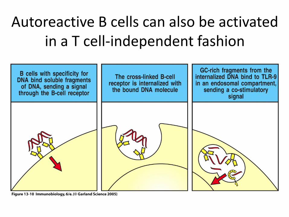
Autoreactive B cells can also be activated in a T cell-independent fashion

Immune Complexes can also Activate Autoreactive B cells
DNA or RNA
Antibody-secreting Plasma cell
Blimp - 1 hi
Ets - 1 lo
BCR
Late Endosome
TLR7 or
TLR9
IgG
Similarly B cells whose BCR recognizes IgG (called rheumatoid factor B cells) can also be activated via TLR7 or TLR9 because they can internalize immune
complexes containing TLR7 or TLR9 ligands

Certain auto antigens are frequently targeted
Antigens associated with DNA • Double or single-stranded DNA • Histones • Chromatin • Type I topoisomerase
Antigens associated with RNA • SSA-Ro • SSB-La • Jo1 • RNP • Smith antigen
Antigens associated with immune complexes • IgG
The relevance of this specificity will be explored later in this lecture

Plasmacytoid Dendritic Cells DNA or RNA
Type I interferon secreting cell
Blimp - 1 hi
Ets - 1 lo
Fc Receptor
Late Endosome
TLR7 or
TLR9
IgG
Plasmacytoid Dendritic Cells are a specialized dendritic cell subset that produces interferon in response to TLR7 or TLR9 stimulation. They usually participate in viral
infections, but in autoimmunity can respond to immune complexes containing TLR ligands. Interferon stimulates further immune activation.
Interferon α or β
Immune cell activation

Some examples of Gene Knockouts with autoimmune disease

Example #1 - Genetic Pre-disposition to Autoimmune Disease Due to a Gene Expressed in T cells
FoxP3 – The gene FoxP3 is almost exclusively expressed in T cells, not B cells or other immune cells FoxP3 controls the development and function of regulatory T cells (Treg) Loss of FoxP3 leads to severe autoimmune disease in both animals models (Scurfy mice) and in human patients (IPEX Syndrome (Immunodisreg- ulation, Polyendocrinopathy and Enteropathy, X-linked syndrome) Young Scurfy Mouse with Eye and Ear inflammation
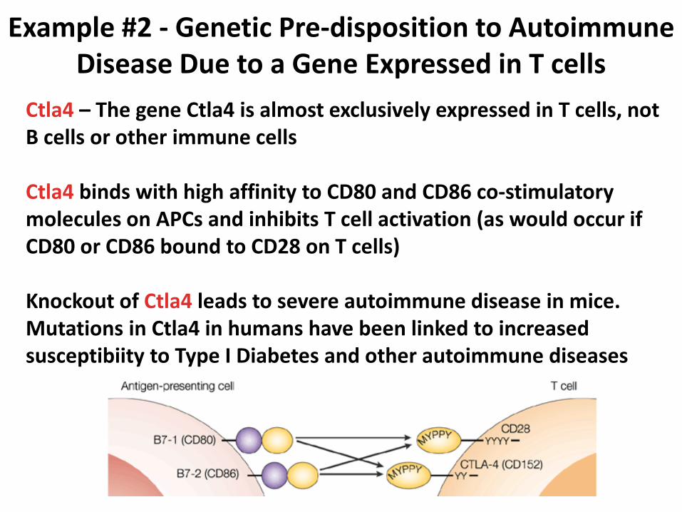
Example #2 - Genetic Pre-disposition to Autoimmune Disease Due to a Gene Expressed in T cells
Ctla4 – The gene Ctla4 is almost exclusively expressed in T cells, not B cells or other immune cells Ctla4 binds with high affinity to CD80 and CD86 co-stimulatory molecules on APCs and inhibits T cell activation (as would occur if CD80 or CD86 bound to CD28 on T cells) Knockout of Ctla4 leads to severe autoimmune disease in mice. Mutations in Ctla4 in humans have been linked to increased susceptibiity to Type I Diabetes and other autoimmune diseases

Example #1 - Genetic Pre-disposition to Autoimmune Disease Due to a Gene Expressed in B cells
CD22– The gene CD22 is exclusively expressed in B cells, not T cells or other immune cells CD22 binds with to glycoproteins containing sialic acid residues as part of the sugar structure Knockout of CD22 (also called Siglec-2) leads to autoimmune disease in mice. The SiglecG receptor (closely related to CD22) is also expressed almost exclusively on B cells and cooperates with CD22 to inhibit B cell activation. Mice lacking both CD22 and SiglecG have a more severe autoimmune syndrome. Human B cells have several similar proteins CD22, Siglec-10 and Siglec-11, which may all have roles in B cells to limit autoimmune disease

Example #2 - Genetic Pre-disposition to Autoimmune Disease Due to a Gene Expressed in B cells
Bank1 – The gene Bank1 is almost exclusively expressed in B cells Bank1 is a scaffold protein that functions in B-cell receptor-induced calcium mobilization from intracellular stores Bank1 knockout mice have enhanced spontaneous germinal center formation and increased basal IgG2a production There are disease-associated polymorphisms in Bank1 in human SLE and RA patients.
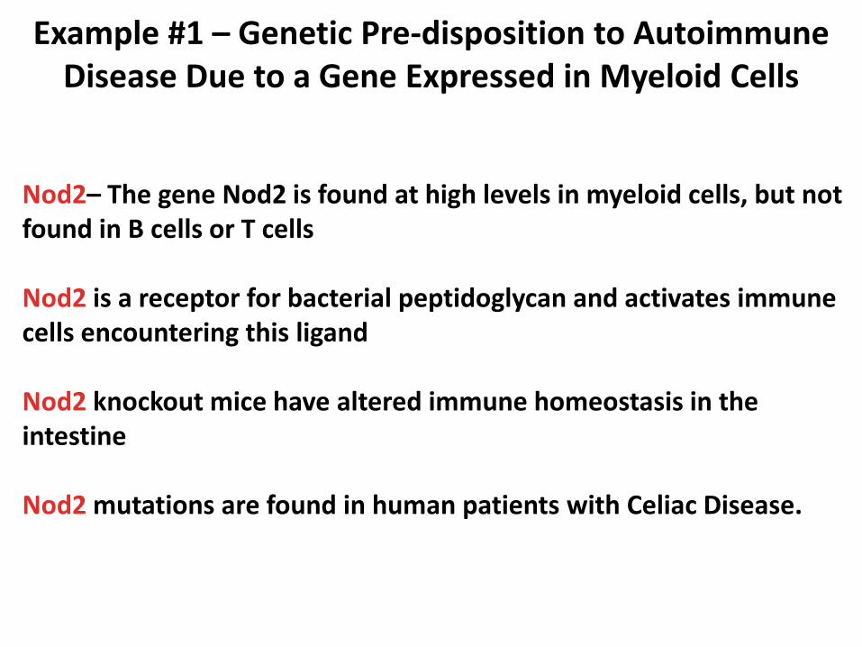
Example #1 – Genetic Pre-disposition to Autoimmune Disease Due to a Gene Expressed in Myeloid Cells
Nod2– The gene Nod2 is found at high levels in myeloid cells, but not found in B cells or T cells Nod2 is a receptor for bacterial peptidoglycan and activates immune cells encountering this ligand Nod2 knockout mice have altered immune homeostasis in the intestine Nod2 mutations are found in human patients with Celiac Disease.

Do we know what genetic differences lead to predisposition to autoimmune disease?
Genome-wide association studies have identified single nucleotide changes in genes that are linked to autoimmune disease susceptibility.
The Table above shows some examples (there are many more).
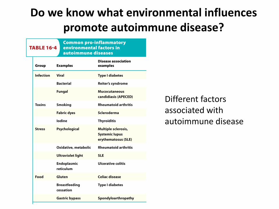
Do we know what environmental influences promote autoimmune disease?
Different factors associated with autoimmune disease
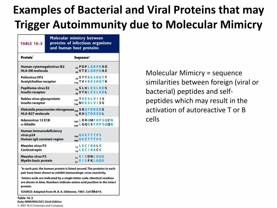
Examples of Bacterial and Viral Proteins that may Trigger Autoimmunity due to Molecular Mimicry
Molecular Mimicry = sequence similarities between foreign (viral or bacterial) peptides and self-peptides which may result in the activation of autoreactive T or B cells
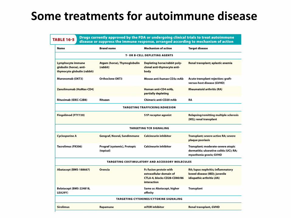
Some treatments for autoimmune disease

Possible Autoimmunity in Tumor Immunotherapy
Immunotherapy of a melanoma tumor in this mouse causes immune response against normal melanocytes and depigmentation
Related Documents