Auditory processing abnormalities in schizotypal personality disorder: An fMRI experiment using tones of deviant pitch and duration Chandlee C. Dickey a,b,c,d, ⁎ , Istvan A. Morocz d , Margaret A. Niznikiewicz b,c , Martina Voglmaier b,e , Sarah Toner b , Usman Khan c , Mark Dreusicke b , Seung-Schik Yoo d , Martha E. Shenton b,c,d , Robert W. McCarley b a Serious Mental Illness Program, VA Boston Healthcare System, Psychiatry 116A-RC, 940 Belmont St., Brockton, MA 02401, United States b Laboratory of Neuroscience, VA Boston Healthcare System, 940 Belmont St., Brockton, MA 02401, United States c Psychiatry Neuroimaging Laboratory, Brigham & Women's Hospital, 1249 Boylston St, Boston, MA 02215, United States d Surgical Planning Laboratory, Brigham & Women's Hospital, 75 Francis St., Boston, MA 02215, United States e Cambridge Health Alliance, Cambridge, MA, United States Received 31 January 2008; received in revised form 18 April 2008; accepted 24 April 2008 Available online 13 June 2008 Abstract Background: One of the cardinal features of schizotypal personality disorder (SPD) is language abnormalities. The focus of this study was to determine whether or not there are also processing abnormalities of pure tones differing in pitch and duration in SPD. Methods: Thirteen neuroleptic-naïve male subjects met full criteria for SPD and were group-matched on age and parental socio- economic status to 13 comparison subjects. Verbal learning was measured with the California Verbal Learning Test. Heschl's gyrus volumes were measured using structural MRI. Whole-brain fMRI activation patterns in an auditory task of listening to tones including pitch and duration deviants were compared between SPD and control subjects. In a second and separate ROI analysis we found that peak activation in superior temporal gyrus (STG), Brodmann Areas 41 and 42, was correlated with verbal learning and clinical measures derived from the SCID-II interview. Results: In the region of the STG, SPD subjects demonstrated more activation to pitch deviants bilaterally ( p b 0.001); and to duration deviants in the left hemisphere ( p = 0.005) (two-sample t). SPD subjects also showed more bilateral parietal cortex activation to duration deviants. In no region did comparison subjects activate more than SPD subjects in either experiment. Exploratory correlations for SPD subjects suggest a relationship between peak activation on the right for deviant tones in the pitch experiment with odd speech and impaired verbal learning. There was no difference between groups on Heschl's gyrus volume. Available online at www.sciencedirect.com Schizophrenia Research 103 (2008) 26 – 39 www.elsevier.com/locate/schres ⁎ Corresponding author. Serious Mental Illness Program, VA Boston Healthcare System, Psychiatry 116A-RC, 940 Belmont St., Brockton, MA 02401, United States. Psychiatry Neuroimaging Laboratory, 1249 Boylston St, Boston, MA 02115, United States. Tel.: +1 857 225 0093 (Cell); fax: +1 617 525 6150. E-mail addresses: [email protected] (C.C. Dickey), [email protected] (I.A. Morocz), [email protected] (M.A. Niznikiewicz), [email protected] (M. Voglmaier), [email protected] (S. Toner), [email protected] (U. Khan), [email protected] (M. Dreusicke), [email protected] (S.-S. Yoo), [email protected] (M.E. Shenton), [email protected] (R.W. McCarley). 0920-9964/$ - see front matter. Published by Elsevier B.V. doi:10.1016/j.schres.2008.04.041

Welcome message from author
This document is posted to help you gain knowledge. Please leave a comment to let me know what you think about it! Share it to your friends and learn new things together.
Transcript

Available online at www.sciencedirect.com
103 (2008) 26–39www.elsevier.com/locate/schres
Schizophrenia Research
Auditory processing abnormalities in schizotypal personalitydisorder: An fMRI experiment using tones of deviant
pitch and duration
Chandlee C. Dickey a,b,c,d,⁎, Istvan A. Morocz d, Margaret A. Niznikiewicz b,c,Martina Voglmaier b,e, Sarah Toner b, Usman Khan c, Mark Dreusicke b,
Seung-Schik Yoo d, Martha E. Shenton b,c,d, Robert W. McCarley b
a Serious Mental Illness Program, VA Boston Healthcare System, Psychiatry 116A-RC, 940 Belmont St., Brockton, MA 02401, United Statesb Laboratory of Neuroscience, VA Boston Healthcare System, 940 Belmont St., Brockton, MA 02401, United States
c Psychiatry Neuroimaging Laboratory, Brigham & Women's Hospital, 1249 Boylston St, Boston, MA 02215, United Statesd Surgical Planning Laboratory, Brigham & Women's Hospital, 75 Francis St., Boston, MA 02215, United States
e Cambridge Health Alliance, Cambridge, MA, United States
Received 31 January 2008; received in revised form 18 April 2008; accepted 24 April 2008Available online 13 June 2008
Abstract
Background: One of the cardinal features of schizotypal personality disorder (SPD) is language abnormalities. The focus of thisstudy was to determine whether or not there are also processing abnormalities of pure tones differing in pitch and duration inSPD.Methods: Thirteen neuroleptic-naïve male subjects met full criteria for SPD and were group-matched on age and parental socio-economic status to 13 comparison subjects. Verbal learning was measured with the California Verbal Learning Test. Heschl's gyrusvolumes were measured using structural MRI. Whole-brain fMRI activation patterns in an auditory task of listening to tonesincluding pitch and duration deviants were compared between SPD and control subjects. In a second and separate ROI analysis wefound that peak activation in superior temporal gyrus (STG), Brodmann Areas 41 and 42, was correlated with verbal learning andclinical measures derived from the SCID-II interview.Results: In the region of the STG, SPD subjects demonstrated more activation to pitch deviants bilaterally (pb0.001); and toduration deviants in the left hemisphere (p=0.005) (two-sample t). SPD subjects also showed more bilateral parietal cortexactivation to duration deviants. In no region did comparison subjects activate more than SPD subjects in either experiment.Exploratory correlations for SPD subjects suggest a relationship between peak activation on the right for deviant tones in the pitchexperiment with odd speech and impaired verbal learning. There was no difference between groups on Heschl's gyrus volume.
⁎ Corresponding author. Serious Mental Illness Program, VA Boston Healthcare System, Psychiatry 116A-RC, 940 Belmont St., Brockton, MA02401, United States. Psychiatry Neuroimaging Laboratory, 1249 Boylston St, Boston, MA 02115, United States. Tel.: +1 857 225 0093 (Cell);fax: +1 617 525 6150.
E-mail addresses: [email protected] (C.C. Dickey), [email protected] (I.A. Morocz),[email protected] (M.A. Niznikiewicz), [email protected] (M. Voglmaier), [email protected] (S. Toner),[email protected] (U. Khan), [email protected] (M. Dreusicke), [email protected] (S.-S. Yoo), [email protected](M.E. Shenton), [email protected] (R.W. McCarley).
0920-9964/$ - see front matter. Published by Elsevier B.V.doi:10.1016/j.schres.2008.04.041

27C.C. Dickey et al. / Schizophrenia Research 103 (2008) 26–39
Conclusions: These data suggest that SPD subjects have inefficient or hyper-responsive processing of pure tones both in terms ofpitch and duration deviance that is not attributable to smaller Heschl's gyrus volumes. Finally, these auditory processingabnormalities may have significance for the odd speech heard in some SPD subjects and downstream language and verbal learningdeficits.Published by Elsevier B.V.
Keywords: Schizotypal personality disorder; Auditory; Schizophrenia; fMRI; MRI; Imaging; Tone processing; Evoked potential; ERP; Mismatchnegativity; Pitch; Frequency; Duration; MMN
1. Introduction
Auditory sensory processing has been found to beimpaired in schizophrenia (Salisbury et al., 1998; Javittet al., 2000) and correlate with clinical features, parti-cularly negative symptoms (Javitt et al., 2000), (Kasaiet al., 2002) (Leitman et al., 2005) and cognitive im-pairment (Baldeweg et al., 2004). Abnormalities in thesuperior temporal gyrus (STG) have been implicated inthe processing of pure tones, a fundamental element ofcomplex sounds and language, using fMRI (Wible et al.,2001) and event-related potentials (Salisbury et al.,1998). Hallucinations, which may be considered anerror in auditory sensory processing, have been asso-ciated with the STG (Dierks et al., 1999) (Cleghornet al., 1992), with the STG more activated during hallu-cinations than actual speech (David et al., 1996). TheSTG has also been implicated in verbal learning deficitsin schizophrenia using PET (Ragland et al., 2001).However, research in schizophrenia has been con-founded by potential modulating effects of neurolepticmedications on fMRI signal (Stephan et al., 2001;Brassen et al., 2003). Neuroleptic-naïve subjects areneeded to ensure that research findings are due to un-derlying neuropathology rather than iatrogenic effects.
Subjects with schizotypal personality disorder (SPD)may provide an ideal population of neuroleptic-naïvesubjects for fMRI studies. Although SPD shares manyfeatures with schizophrenia, it is not a direct proxy forschizophrenia, as SPD subjects are not psychotic.Nonetheless, SPD and schizophrenia have traditionallybeen considered part of the schizophrenia spectrumdisorders based on epidemiological data (Kety et al.,1967) (Kendler et al., 1993), shared clinical featuressuch as thought disorder (Dickey et al., 1999) and pa-ranoia (Dickey et al., 2005), similar biological markers(Siever and Davis, 2004), comparable cognitive deficitsin verbal learning (Voglmaier et al., 1997) (Voglmaieret al., 2000) (Voglmaier et al., 2005), and overlappingmorphometric abnormalities (Dickey et al., 2002a,b).
Indeed, one brain region critical for early sensoryauditory processing, Heschl's gyrus (Yoo et al., 2005),
has been shown to have reduced volumes in subjectswith SPD (Dickey et al., 2002a,b), similar to what hasbeen shown in schizophrenia (Hirayasu et al., 2000).Heschl's gyrus lies on the plane of the STG, a regionfound to have small volumes in males with SPD (Dickeyet al., 1999; Downhill et al., 2001), and in females withSPD with a family history of mental illness (Dickeyet al., 2003). Of particular interest to this report, how-ever, Heschl's gyrus is noted to have marked inter-subject morphometric volume and shape variability(Leonard et al., 1998; Knaus et al., 2006), thus com-plicating the interpretation of volume data (Knaus et al.,2006).
As in schizophrenia, abnormalities of auditoryprocessing at multiple stages have been shown in SPDincluding the P50 (Cadenhead et al., 2000), P300(Salisbury et al., 1996), and N400 (Niznikiewicz et al.,1999). Similarly, in subjects with high scores of schi-zotypal features but not frank SPD, auditory abnorm-alities have been shown in the P300b (Klein et al.,1999), N400 (Kimble et al., 2000), and in phonemicdiscrimination (Li et al., 2003). Finally, in one paperexamining mismatch negativity (MMN) in subjectsclinically diagnosed with schizotypy but for whom aformal diagnostic interview was not performed, schizo-typal subjects were found to have increased amplitudesin the Fz and Cz electrodes to pitch deviants (Liu et al.,2007). Hence, it appears that in the schizophreniaspectrum there is a range of auditory processing abnor-malities, albeit with some negative findings (Brenneret al., 2003). The current report seeks to build on thisliterature by examining tone processing of pitch andduration deviance in SPD subjects using fMRI.
One question in the literature is how to best measuredeficits in early auditory sensory processing in schizo-phrenia and SPD, whether through event-relatedpotential (ERP) or fMRI studies. The ERP methodologyaffords good temporal resolution while fMRI offers agood spatial resolution. ERP components often used toexamine processing auditory pre-attentive and attentiveabnormalities in schizophrenia are mismatch negati-vity (MMN) and P300. A MMN ERP paradigm, which
28 C.C. Dickey et al. / Schizophrenia Research 103 (2008) 26–39
elicits an early negative deflection following a deviantstimulus, results in a less negative deflection in schizo-phrenic subjects and has been used frequently to assesssubjects' pre-attentive ability to detect changes in tonefeatures (e.g.:Salisbury et al., 1998). In contrast, to ourknowledge (Medline search 1/10/08), there havebeen only two published fMRI experiments utilizingthe mismatch design in patients with schizophrenia(Wible et al., 2001; Kircher et al., 2004). Adequatenumbers of deviants are required to produce a detectablecontrast-to-noise ratio, yet this must be balanced withexperimental length, as subjects' tolerance to longscanning session is limited. For these reasons, weemployed a significantly modified mismatch experimentwith larger differentials between standard and devianttones and more frequent deviants as compared withprototypic mismatch paradigms.
Therefore, whether the STG in SPD exhibits normalfunctioning as measured by hemodynamic responseto early auditory sensory information is the centralquestion driving the current report. Structural MRI andneuropsychological testing procedures are also in-cluded. The possible relationship between early auditoryprocessing and downstream language and other cogni-tive functions, as well as the highly complex clinicalmanifestations of SPD, is also evaluated in an ex-ploratory fashion. These questions are important to askin SPD subjects as they are neuroleptic-naïve, thus, acomplicated confound in similar studies of auditory func-tion with schizophrenic subjects is removed (Umbrichtet al., 1998).
2. Methods
2.1. Subject recruitment
All subjects were male; right-handed; between 18and 55 years old; neuroleptic-naïve; on no psychotropicmedications; had no history of ECT, neurologicdisorder, substance abuse in the last 1 year or substancedependence in the last 5 years; and were recruited fromthe community through newspaper and subway adver-tisements (for recruitment specifics, see Dickey et al.,2005). Note that data for past psychotropic use of anykind or substance dependence beyond five years agowas not available. SPD subjects met DSM-IV criteria forSPD using the SCID and SCID-II interviews and had nopersonal history of bipolar disorder nor psychosis.Thirteen SPD subjects were group-matched on age,parental socio-economic status, and estimated IQ to 13comparison subjects who had additional exclusionarycriteria of no personal history with Axis I or Axis II
disorder as determined by SCID, or first-degree relativehistory of Axis I disorder.
2.2. Clinical measures
SPD criteria were from SCID-II interview. IQ wasassessed through the WAIS-R Vocabulary and BlockDesign sub-scales (Brooker and Cyr, 1986). Verbal learn-ing was assessed using the California Verbal LearningTest (CVLT), total words learned trials 1–5 (Delis et al.,1987). This test was selected as a test of verbal workingmemory and because it has been shown by our laboratoryto be abnormal in SPD subjects (Voglmaier et al., 1997;Voglmaier et al., 2000; Voglmaier et al., 2005).
2.3. Structural MRI
Heschl's gyrus was manually delineated on resolutionSPGR images obtained within a year of the fMRIprotocol, except one SPD subject for which no structuralMRI was available. The protocol for the drawing re-sembled that of a previously published report on Heschl'svolumes (Dickey et al., 2002a,b). The anterior boundarywas the temporal stem; the posterior boundary was thecomplete crux of the fornix; the lateral boundary wasdetermined by a horizontal line extending laterally fromthe superior most white matter track of the STG. Therewas one major methodological difference in the drawingbetween the two reports, however. In this report axialviews were used initially to guide the definition of theextent of Heschl's. Axial views were not availablepreviously. With the use of Slicer software (www.slicer.org) one could now visualize whether there was a singletransverse gyrus, a commonmedial stem branching to twomore laterally in which case both would be included perSteinmetz's criteria (Steinmetz et al., 1986), or twomedialstems joining more laterally in which case only themore anterior portion would be selected. This ability tovisualize and draw in three dimensions represents asignificant technological advancement compared withprevious capabilities and is important given the markedinter-subject and even inter-hemispheric variability of thisgyrus (Knaus et al., 2006). Volumes were corrected fortotal intracranial contents using a regression procedure(Dickey et al., 2002a,b). Inclusion of Heschl's volumemeasurement was important for the interpretation of thefunctional data.
2.4. fMRI acquisition and post processing
Whole-brain images were acquired on a 3.0T GEsystem in the oblique axial plane parallel to the superior

29C.C. Dickey et al. / Schizophrenia Research 103 (2008) 26–39
temporal plane prescribed from the localizer images.Functional image parameters included whole-braincoverage, 30 slices, 5 mm thick with 1 mm gap, 162acquisitions the first 6 of which were removed, FOV 24,matrix 64x64, TR 2.5, TE 35. The same prescription(orientation/slice thickness) was applied to obtain a low-resolution SPGR sequence. These low-resolution SPGRimages were reoriented, realigned, and normalized tothe Montreal Neurological Institute (MNI) T1 templatewith the resulting matrix files applied to the functionalimages using SPM2. Functional images were sub-sequently smoothed (using 12 FWHM 3-dimensionalGaussian kernel).
2.5. Stimuli
Two experiments were employed to activate theauditory cortex, one using the pitch deviants and the
Fig. 1. Diagrams of stimuli presentation and processing. a. For the pitch expernext tone. b. For the duration experiment, the standard tone is 50 ms in durexperiment the deviant tone is 200 ms in duration with 100 ms of silence beforwere presented in block design. e. Hemodynamic response curves were generaand for only the deviant tones (deviant condition). ISI = interstimulus intervintermixed; rest = silence.
second using the duration deviants. Tones were trans-mitted via sound-insulated and cushioned earphones(Silent Scan, Avotec, Jensen Beach, FL) at 80 db SPL(Sound Pressure Level). All tones had 10 ms rise and falltimes and an interstimulus interval of 300 ms and wereplayed at 80 db. Experiment 1: Pitch. The standard tonewas 500 Hz and the deviant tone was 2000 Hz, all tones100 ms in duration (Fig. 1a). Experiment 2: Duration.The standard tone was 50 ms in duration, the devianttone was 200 ms in duration, with a frequency of 500 Hzfor all tones (Fig. 1b and c). Presentation was blockdesign with 30 s blocks of tones alternating with 30 srest (Fig. 1d). Each tone block consisted of 100 tonepresentations, alternating between blocks of 100% stan-dard tones and mixed blocks of 75% standard tones and25% deviant tones. In the mixed standard and deviantblocks the order of standard and deviant tones was ran-domly determined. Only one deviant (pitch or duration)
iment all tones are 100 ms in duration with 200 ms of silence before theation with 250 ms of silence before the next tone. c. For the duratione the next tone. This variation decreases the expectancy factor. d. Tonested for all tones together (both standard and deviant, all tone condition)al; ms = milliseconds; s = standard; s/d = standard and deviant tones

Table 1Subject demographics and absolute Heschl's gyrus volumes inmilliliters
SPD NC p
N 13 13Age 36.8 (10.8) 30.4 (11.4) b0.2PSES 4.0 (1.2) 3.8 (1.4) 0.08SES 3.0 (1.4) 3.2 (1.4) 0.7IQ 116 (9.7) 120.4 (12.4) b0.4Education 15.2 (1.9) 18.0 (3.8) 0.02SSPT score 55.2 56.2 (N=8) b0.5Left Heschl's gyrus a 2.23 (0.9) 2.35 (0.6) 0.74Right Heschl's gyrus b 1.95 (0.51) 1.92 (0.2) 0.29
Note that statistics were performed on regressed not absolute volumesin order to account for confounding effect of head size. PSES =parental socio-economic status, SES = socio-economic status, SSPT =Speech Sound Perception Test.a F(1,24)=0.109.b F(1,24)=1.128.
30 C.C. Dickey et al. / Schizophrenia Research 103 (2008) 26–39
was presented in a given run. Run duration was 6′45″.There were two runs for pitch and two for duration for atotal of four runs, order counterbalanced among subjects.Subjects heard 1050 standard tones and 150 devianttones for a total of 1200 tones per experiment (Fig. 1e).For technical reasons, data were acquired on only 11SPD subjects for the duration experiment, therefore, thesample size differs in the two fMRI experiments. Taskfor both experiments was passive listening with eyesclosed. Following each run subjects were queried as towhether they heard the tones to ensure adequate hearingand wakefulness. All subjects affirmed that they didindeed hear the tones after each run. Subjects were notasked nor expected to differentiate between the tones.
2.6. fMRI statistical methods
2.6.1. Whole-brain analysisThis was the primary statistical analysis for the pitch
and duration fMRI experiments. Whole-brain event-related procedures were employed statistically in orderto single out the effect of all tones (standard+deviant),and in a separate analysis, the differential effect ofhearing only deviant tones. Restated, the hemodynamicresponses were modeled first in order to examine theeffect of being in the scanner and hearing all tones, bothstandard and deviant, regardless of block type (maineffect) (Fig. 1e, in blue and green). Subsequently, thehemodynamic effect of hearing only deviant tones(parametric effect) was modeled (Fig. 1e, in green). Thisisolates the effects of hearing deviant tones “on top of”the effect of hearing all tones, that is, standard anddeviant (green only “on top of” green and blue). Spe-cifically, the main effect of hearing all tones (standard+deviant, regardless of block) vs. rest were modeledusing one regressor (effect of hearing deviant tonesonly) in the General Linear Model for each subject.Second, the differential effect of hearing the devianttones was modeled as a parametric regressor (devianttones as 1 and standard tones with value 0) in order to
Fig. 2. Whole-brain statistical parametric maps with consistent p scale shown.tone set heard (all tones or deviants only), there is no area of the brain in whichall tones (standard+deviant) heard analyzed together. b. Pitch experiment dcompared with control subjects in the region of the STG bilaterally. Thresholdat 240 voxels. MNI coordinates and cluster significance by two-tailed t tepb0.001), (54, −46, 18, T=4.85, pb0.001). c. Duration experiment, all twith an extent of activation cluster size limit set at 0 voxels to ensure all visibby two-tailed t tests were: (2, 22, 10, T=5.67, pb0.0005) and (−14, 4, 24subjects activated more to deviant tones than comparison subjects in left tempan extent of activation cluster size limit set at 240 voxels for temporal lobe regin that area. MNI coordinates and cluster significance by two-tailed t tests w−22, −88, 2, T=3.1, p=0.003); for parietal lobe regions, (−26, −42, 38,T=3.36, p=0.001). Note standardized color bars across images reflect relati
measure the deviant-related modulation of the BOLDsignal time course for each subject. Thus the effect ofhearing all tones (both standard and deviant combined,the effect of being in a scanner and hearing tones, themain effect) could be compared as well as the effect ofonly hearing deviant tones (parametric effect). Subjectspecific whole-brain contrast images from the two groupswere pooled and were compared using one-sample (alltone and deviant tone conditions for both experiments)and two-sample t tests (all tone and deviant tone condi-tions for both experiments) for the random-effects ana-lysis. Note that these analyses were whole-brain analyses.
2.6.2. Secondary ROI analysisIn order to perform exploratory correlations with
clinical/cognitive measures, a second procedure, asecond analysis, a voxel-wise restricted ROI analysis,was employed in order to isolate peak activation in theregion of the STG for individual subjects. Note that thissecondary ROI analysis was used for correlations onlyand that the main fMRI analysis used a whole-brainanalysis. Therefore, for secondary and exploratory
Note that regardless of experiment (pitch or duration) and regardless ofcontrol subjects activated more than SPD subjects. a. Pitch experiment,eviant condition. Note significantly more activation in SPD subjectsed at the pb0.001 level with an extent of activation cluster size limit setsts were: (−52, −22, −2, T=5.3, pb0.001), (54, −46, −2, T=5.3,ones (standard+deviant) condition. Thresholded at the pb0.01 levelle super-threshold activation. MNI coordinates and cluster significance, T=5.3, pb0.0005). d. Duration experiment, deviant condition. SPDoral and bilateral parietal regions. Thresholded at the pb0.01 level withions, 0 voxels for parietal regions in order not to lose relevant findingsere: for temporal lobe regions, (−48, −28, −4, T=2.83, p=0.005 andT=4.0, pb0.001), (−24, −20, 42, T=3.2, p=0.002), (28, −40, 38,ve T scores. Neurological convention (left is left).

31C.C. Dickey et al. / Schizophrenia Research 103 (2008) 26–39

Table 2Clinical/cognitive/functional correlations
Condition ROI RHO p
SPD criteria of oddthinking or speech
Pitch Right 41 0.639 b0.02Right 42 0.534 b0.06
CVLT Pitch Right 41 −0.633 b0.03Right 42 −0.670 b0.02Right STG −0.830 0.001
Correlations between peak t activation values for the ROI, namely,STG, Brodmann Areas 41 and 42, with clinical criteria, and verballearning (CVLT) are given.
32 C.C. Dickey et al. / Schizophrenia Research 103 (2008) 26–39
correlations, ROI were selected as defined by WFUPickAtlas (www.fmri.wfubmc.edu). Specifically, Pick-Atlas defined ROI masks were applied and peak t valueswere generated for left and right Brodmann Areas 41,42, and STG for the deviant condition (three regions ofinterest (ROI) per side per experiment, 12 in total).These regions were selected a priori as they likely arethe regions involved in tone processing (Yoo et al.,2005; Kropotov et al., 1995; Alho, 1995). Note thatBrodmann Area 41 derived from PickAtlas does notdirectly correspond to the manual Heschl's drawingabove. They are slightly different measurements butboth roughly correspond to presumed areas of pure toneprocessing. Threshold was set at 0.05 corrected.
2.7. Clinical/cognitive/functional correlations
Exploratory Pearson correlations with peak t valuesand clinical measures were obtained. The clinicalmeasures that included the nine SPD diagnostic criteriaand CVLTwere correlated with the 12 ROI for a total of120 correlations. To limit the number of correlationsconsidered significant post hoc, only those which weresignificant in two of the three regions per side in asingle condition were accepted (one of the two regionscould have a correlation significant at the trend level).For example, a significant correlation with a clinicalmeasure and right Brodmann Areas 41 and 42 would beaccepted, but not right 41 and left 42, nor just right 41.We appreciate that even with these more stringent rules,a large number of correlations were performed.However, given that this is the first fMRI paper onauditory processing in SPD to our knowledge (Medlinesearch performed 1/10/08), an attempt to understand theclinical significance of the potential findings wasimportant.
3. Results
3.1. Subject demographics
There were no group differences in age, parentalsocio-economic status (PSES) (scale 1–5, with 5 ashighest PSES), personal socio-economic status (SES),or IQ although SPD subjects had fewer years of edu-cation (Table 1). SPD subjects met criteria foradditional co-morbid personality disorders including:avoidant (N=1), paranoid (N=6), borderline (N=2),obsessive compulsive (N=3), narcissistic (N=2), pas-sive aggressive (N=1), and schizoid (N=1), similar towhat others (McGlashan, 1986) and our laboratorypreviously published (Dickey et al., 2005) with the
exception of a higher percentage of paranoid subjectshere (46%). The mean number of DSM-IV SPD criteriamet for the SPD group was 5.62 (S.D.=0.768) out of apossible nine criteria.
3.2. Heschl's volumes
There were no group differences in left or rightHeschl's gyrus gray matter volumes manually drawn onhigh-resolution SPGR images and corrected for intra-cranial contents using a regression procedure (Dickeyet al., 1999) (Table 1). Indeed the effect size on the leftwas small, effect size=0.18, the effect size on the rightwas 0.03. The lack of volume difference, however,strongly suggested that differences in functional activa-tion were not due to a primary volume difference.
3.3. Pitch experiment
There were no areas in which comparison subjectsdemonstrated statistically significantly more activationthan SPD subjects in neither the all tones (standard+deviant) condition nor the deviant tone condition. Forthe differential effect of deviance, there were three areasin which SPD subjects demonstrated more activation(two-sample t test) all in the region of the STG bi-laterally (Fig. 2a and b).
3.4. Duration experiment
There were no areas in which comparison subjectsdemonstrated statistically significantly more activationthan SPD subjects in neither the all tones (standard+deviant) nor the deviant tone condition. For the additiveeffect of hearing deviant tones only, there were areas ofsignificantly more activation in both the temporal andparietal lobes in SPD subjects compared with controls(Fig. 2c and d).
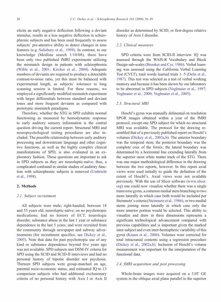
Fig. 3. Scatterplots of peak activation and clinical and cognitive measures. a. SPD subjects with greater impairment due to odd thinking or speech(higher score) demonstrated more activation while passively hearing tones in BA 41 and 42 on the right. b. SPD subjects who learned fewer words onthe CVLT had greater peak activation in the right BA 41, 42, and STG. BA = Brodmann Area.
33C.C. Dickey et al. / Schizophrenia Research 103 (2008) 26–39
3.5. Clinical correlations with fMRI activation patternsin the duration experiment
Of note, correlations were found on the right sidebetween the clinical symptom of odd thinking/speech inthe pitch experiment (Table 2). The more abnormal theactivation (t-score) while hearing deviant pitch tones,the more the impairment due to the clinical symptom.Verbal learning, a key abnormality in SPD, correlatedwith all right-sided regions such that the fewer wordslearned in trials 1–5, the more abnormal the activationwhile hearing deviant pitch tones. The number of cor-relations performed was high given the exploratorynature of the study and results would not have withstooda Bonferroni correction. However, they are includedhere to generate future hypotheses regarding the func-tional/anatomic relationships in SPD. As stated in theMethods section, included in Table 2 are only thosecorrelations which were found in at least 2/3 regions/side (Fig 3).
4. Discussion
The main finding of this report is increased activationin the region of the STG bilaterally in SPD subjectscompared with controls while subjects heard devianttones regardless of whether the deviance was in the pitch
or duration. In the whole-brain analysis, in no region ofthe brain did comparison subjects activate more thanSPD subjects. These findings suggest that SPD subjectscompared with controls had inefficient or hyper-res-ponsive processing of two of the most basic aspects ofauditory sensory stimuli. These data cannot unequi-vocally address the question as to whether the SPDsubjects have inefficient processing and, thus, needed torecruit more neurons to process the tones with a re-sulting larger hemodynamic response, or, the reverse,that SPD subjects have an exaggerated response tosubtle changes in sensory inputs with a resulting largerhemodynamic response curve. More basic research isrequired to differentiate those two possible interpreta-tions. Nonetheless, these current findings cannot beattributed to small Heschl's gyrus volume as the groupsdid not differ on that measure.
Deficits in early auditory sensory processing, in-cluding N1, MMN, and P300, have been shown usingERP in schizophrenic subjects (Baldeweg et al., 2004;Kasai et al., 2002; Salisbury et al., 1998; Javitt et al.,2000; Javitt et al., 1995; Elvevag et al., 2004), in pro-dromal subjects (Brockhaus-Dumke et al., 2005), and inSPD subjects (Salisbury et al., 1996; Niznikiewicz et al.,2000; Liu et al., 2007). Moreover, progressive changesin schizophrenic subjects can be indexed using a MMNparadigm in the ERP environment (Salisbury et al.,

34 C.C. Dickey et al. / Schizophrenia Research 103 (2008) 26–39
2007). Indeed, ERP has certain advantages in testingauditory sensory processing in that tones can be playedin sound-proof laboratories with little to no extraneousnoise, experimental design allows for a large number ofdeviants, greater than 160 in most studies (Shelley et al.,1991; Javitt et al., 1995; Umbricht et al., 1998; Michieet al., 2000; Baldeweg et al., 2004; Brockhaus-Dumkeet al., 2005) (exception, Kirino and Inoue, 1999) andallows for precise time recordings on the order of milli-seconds at the expense of poor spatial resolution.
In contrast, fMRI allows for more accurate anatomiclocalization at the expense of poor time resolution andhigh ambient scanner noise, although some have utilizedthe scanner noise as stimuli (Mathiak et al., 2002;Kircher et al., 2004). Applying a prototypic mismatchparadigm has been challenging in the fMRI environmentresulting in few papers on schizophrenia (Wible et al.,2001; Kircher et al., 2004). Even using scanner noiseas the stimulus causes limitations as variation in pitchcannot be tested (Mathiak et al., 2002; Kircher et al.,2004).
How one might measure the effect of tone deviantsalso differs between ERP and fMRI. ERP MMN studiesrely on traditional subtraction of waveforms (i.e.: wave-forms from standard tones subtracted from waveformsfrom deviant tones) (Javitt et al., 1995; Umbricht et al.,1998). Unfortunately, in fMRI aberrant activation pat-terns can result from subtraction. These areas of unusualactivation may reflect “spontaneous neuronal activity”,that is, areas not predicted by task demands, which can-not be experimentally controlled (Binder et al., 1999). Amismatch task is not cognitively demanding, in fact it isarguably pre-attentive (Naatanen 1990), possibly allow-ing for more “task-unrelated thoughts” (Binder et al.,1999). These “task-unrelated thoughts” can result inchanges in blood flow detected byBOLDmethod (Binderet al., 1999). In fact, the hemodynamic effect of suchthoughts using the subtraction method has been demon-strated in a tone task (Binder et al., 1999). Moreover,depending on the statistical approach, subtraction or other,results can differ markedly (Friston et al., 1996). Thesubtraction method assumes “pure insertion”, that thecognitive process is “irrespective of the cognitive orphysiological context” (Friston et al., 1996), an assump-tion which may not be valid. Indeed, the two fMRIpapers in schizophrenia compared groups on standard anddeviant tones separately, thus avoiding the difficulties ofthe subtraction method in an fMRI environment (Wibleet al., 2001; Kircher et al., 2004).
In the current fMRI study the effect of deviantswas measured using a parametric analysis. Specifically,the differential processing of deviant tones against the
background of hearing all tones was measured (i.e.: thefirst parameter is the effect of hearing all tones, bothstandard and deviant; and the second parameter is theeffect of hearing deviant tones only) (Fig. 1e). This hasthe benefit of making no assumption of “pure insertion”(Friston et al., 1996) and isolates the effect of hearingdeviant tones “on top of” hearing all tones. In addition,this method may possibly minimize the hemodynamiceffect of “task-unrelated thoughts” (Binder et al., 1999).
Another aspect of this study which differed from theMMN ERP literature was the use of disparate fre-quencies and durations between the standard and de-viant tones. Stimuli used across studies have varied withdifferences between standard and deviant pitches assmall as 24 Hz (1000–1024 Hz) (Javitt et al., 1995) to aslarge as 1000 Hz (1000–2000 Hz) (Kirino and Inoue,1999) and differences in duration as small as 25 ms(25–50 ms) (Baldeweg et al., 2004). Nonetheless, thepresence of the MMN-like deflection in both conditionssuggests that these experimental parameters engage theearly auditory sensory processing stream. Indeed, sti-mulus presentation features including probability anddegree of separation of the deviants, and interstimulusinterval, are all important variants which may affect theresults (Michie et al., 2000). However, as SPD hasdemonstrated less severe abnormalities than schizo-phrenia on electrophysiological measures (Trestmanet al., 1996), more extreme differences in terms ofpitch and duration between the standard and devianttones were selected to amplify the fMRI signal. More-over, a larger differentiation between standard and de-viant tones would help to compensate for any potentialdeficit in tone discrimination processing described in theschizophrenia literature (Javitt et al., 2000; Leitmanet al., 2005). Although a lower percentage of devianttones would have similarly increased an electrophysio-logical signal (Javitt et al., 2000), in order to achieveenough trials for an adequate hemodynamic response, ahigher percentage of deviant tones was selected.
Although the paradigm used in this study is notstandard mismatch, evidence from the MMN literaturemay inform the current findings. For example, gen-erators for the MMN are thought to be from the auditorycortex (Kropotov et al., 1995) with physical separationof foci for processing of pitch and duration (Molholmet al., 2005), similar to the current findings. Someworkers have also noted activation from frontalgenerators thought to reflect a shift of attention towardthe deviant, although recent work suggests that the roleof the frontal generator may not depend on attentionrequirements (Shalgi and Deouell, 2007). Indeed, arecent review by Naatanen et al. (2007) suggests that
35C.C. Dickey et al. / Schizophrenia Research 103 (2008) 26–39
frontal activity may be due to the sum of supratemporalgenerators (Naatanen et al., 2007). Our report did not findevidence of fMRI activation in either group in the frontallobe, possibly due to the use of largely disparate standardand deviant tone features (Shalgi and Deouell, 2007). Theactivation of the parietal lobe in the duration experiment isconsistent with the hypothesis that the parietal lobe isimportant for time perception (Harrington et al., 1998)and change-detection (Molholm et al., 2005). Indeed,bilateral activation has been demonstrated with deviantsin duration (Molholm et al., 2005). Finally, abnormalitiesin MMN have been documented in clinically diagnosedpatients with SPD (Liu et al., 2007).
In this report, in contrast to our previous report(Dickey et al., 2002a,b), we did not show any differencein Heschl's gray matter volume. We would argue,however, that, in general, in the peri-Heschl's/ STGregion, SPD subjects likely have smaller volumes(Dickey et al., 2002a,b, 2003, 1999; Downhill et al.,2001 {Koo, 2006 #3366}). There are several differencesbetween our two reports on Heschl's gyrus ((Dickeyet al., 2002a,b) and current) and that volumes measureddepend on many factors including subject demo-graphics, co-morbidity, subject N, image viewing toolswhich can affect landmark detection, and inter-subjectand inter-hemispheric variability of Heschl's morphol-ogy, particularly in schizophrenia spectrum disorders.First, in the earlier report there was a significantdifference between IQ and personal socio-economicstatus between groups suggesting that cohort may haverepresented a more impaired group of SPD subjects.Second, and delving deeper into the issue of subjectcharacteristics, the percentage of subjects meetingDSM-IV criteria for other Axis II disorders differsbetween samples. In the prior report there were moreborderline personality disorder subjects (31% vs. 15%in current sample) and fewer who met criteria for para-noid personality disorder (37.5% vs. 46% in currentreport). Little is known about brain morphology of theSTG region in paranoid and borderline personality dis-orders (no papers found per PubMed search performed4/17/08). Whether the presence of co-morbid person-ality disorders has an effect on Heschl's measurementcannot be addressed. Third, in this current sample, thesubject N is smaller, leading to possibly less stable data.Fourth, the current study used Slicer as the image pro-cessing software tool, a more sophisticated tool thanpreviously available (www.slicer.org) as this tool nowallows for simultaneous 3D viewing. As noted under theMethods section above, the delineation of Heschl'spreviously may have included more peri-Heschl'sarea posteriorly thus leading to the differences between
groups in that report (Dickey et al., 2002a,b). In thecurrent report the more posterior transverse gyri may nothave been included. Fifth, there is marked inter-subjectand even inter-hemispheric variability of the STG,particularly, Heschl's gyrus (Lange et al., 1997; Leonardet al., 1998; Sweet et al., 2005; Knaus et al., 2006). Toclarify, it is quite common for there to be two paralleltransverse or Heschl's gyri (Knaus et al., 2006; Sweetet al., 2005). However, these parallel transverse gyri canbe completely separate or partially separate. If they arepartially separate they can be merged medially, part waydown the gyrus, or laterally. What should one consideras Heschl's gyrus is not clear. Should one include both,part of both, or only the more anterior one? Boththe prior report and this paper used Steinmetz's crit-eria (Steinmetz et al., 1986) as the posterior boundary.This arbitrary criteria state that if the gyri are mergedmedially with a common stem, then one can includeboth. If they are merged part way down the gyrus orlaterally, then they should be divided into two separategyri and only include the more anterior gyri as Heschl's.Note that this criteria is helpful, but arbitrary. Subtledifferences in visualization abilities or in morphometrycan result in significant volume differences which maynot be meaningful functionally. Indeed, the functionalanatomy of AI (primary auditory cortex) may not berestricted to medial Heschl's gyrus, regardless of itsdefinition or criteria (Sweet et al., 2005). Indeed, recentwork suggests that there may be two primary auditoryreceptive fields in the human auditory system, one moremedial, one more lateral (Engelien et al., 2002). Closeinspection of the receptive field maps (Engelien et al.,2002) suggests that the more lateral field may bepartially located on what may be considered bySteinmetz's criteria to be the second transverse gyrus,an area more likely captured in our first report but lesslikely captured in this current report. That functionallyimportant area (Sweet et al., 2005) may represent thedifference in these two reports. Indeed, other workerssuggest that volume asymmetries in the region of theprimary auditory cortex do not correspond to functionalactivation while hearing tones and word pairs (Yooet al., 2005), possibly due to the poor matching of gyralsulcal patterns and cytoarchitechtonically definedacoustic regions (Morosan et al., 2001), and markedvariation in the morphology of Heschl's gyrus (Leonardet al., 1998) (Sweet et al., 2005). Furthermore, regionaldifferences in STG volumes are more marked inschizophrenia than in controls (Park et al., 2004). Insum, the lack of replication of our previous report islikely due to a combination of factors: differences insubject demographics, presence of co-morbid disorders,

36 C.C. Dickey et al. / Schizophrenia Research 103 (2008) 26–39
subject N, difference in measurement tools, and inter-subject morphometric variability of Heschl's gyrusparticularly in the schizophrenia spectrum.
Despite these limitations to direct comparison be-tween the two samples, the lack of difference betweengroups in Heschl's volume in this study is informative:one cannot attribute differences in activation in thecurrent report to subtle volume differences of Heschl'sgyrus per se. There may be subtle differences in volumesin the adjacent cortical regions, perhaps in areas 41 and42 or the larger STG not measured by the current report.However, even if there were smaller volumes in theadjacent cortex in SPD subjects, one would not havepredicted that smaller volumes lead to larger areas ofactivation, more likely, smaller volumes would predictsmaller areas of activation. Therefore, we do not believethat possible volume differences in peri-Heschl's regionscan account for the between group differences inactivation patterns.
The possible relationship between abnormal auditorysensory processing and clinical measures was also in-vestigated. Exploratory correlations between ROI andclinical/cognitive symptoms suggest that these earlyauditory sensory processing problems may have implica-tions for downstream language functioning. Specifically,in SPD subjects, there was a correlation betweenabnormal activation on the right during the pitch expe-riment with odd speech. Odd speech and formal thoughtdisorder likely represent a symptom continuum. Ourlaboratory previously demonstrated thought disordercorrelating with left STG volumes in female SPD sub-jects (Dickey et al., 2003). Similarly, in schizophrenicsubjects, correlations have been shown between thoughtdisorder and hallucinations with bilateral measures ofvolume and function of temporal lobe regions (Shentonet al., 1992; Dierks et al., 1999; Kircher et al., 2001; Bartaet al., 1990; Woodruff et al., 1997). In this report,however, the correlation with odd speech occurred withright-sided activation. Odd speech in SPD may becharacterized by impoverished prosody (Dickey et al.,unpublished data). Consensus suggests a right hemisphereadvantage for emotional (non-semantic) aspects oflanguage and prosody (Mitchell and Crow, 2005),particularly in the right STS and MTG, whereas moresemantic aspects of language recruit more left-sided re-gions (Mitchell et al., 2003). This hemispheric pattern hasbeen seen using fMRI (Mitchell et al., 2003) (Wildgruberet al., 2005), transcranial Doppler ultrasonography(Vingerhoets et al., 2003), repetitive transcranial mag-netic stimulation (van Rijin et al., 2005), event-relatedpotential (Eckstein and Friederici, 2005), and in lesionstudies (Pell, 2006). Therefore, it is possible, that the tone
processing impairment on the right in the SPD subjects isrelated to non-semantic aspects of their odd speech.
Other aspects of language, specifically verbal learning,also correlatedwith inappropriate activation of right-sidedROI for the current pitch experiment. Again, this is similarto findings in schizophrenia. Using PET, Ragland et al.demonstrated impaired verbal learning correlating withSTG activation abnormalities (Ragland et al., 2001).
There are several limitations to the current study. Onelimitation is the large number of correlations performed asdiscussed in the Methods section. However, includingthese exploratory findings in this paper may serve togenerate future hypotheses about the relationship betweenauditory processing and clinical features in this under-studied schizophrenia spectrum disorder. Second, onlymale subjects were included in this study. Future work willneed to include females to test whether there is a gendereffect (Dickey et al., 2003; Knaus et al., 2006). This isparticularly critical as our laboratory recently completed apitch MMN investigation in a group of male and femaleSPD individuals and the results suggest that there is noimpairment of early sensory processing as indexed byMMN in males. Instead, the significant pitch MMNreduction was found in females (Niznikiewicz et al., insubmission). Third, the whole sample size is small relativeto fMRI studies in schizophrenia. This reflects the inherentdifficulty in recruiting SPD subjects who are not part of aclinical population for a research study. Had we had accessto a larger group of SPD subjects then the findings could bemore generalized to all SPD subjects. Nonetheless, thesedata suggest that for at least a sub-population of SPDsubjects, there may be deficits in early sensory processingof tones. Fourth, there was a difference between groups interms of education. However, we are not aware of anypaper suggesting that education plays a role in simple toneprocessing per se. Fifth, shifting gradients in the MRIscanner are loud and theoretically may have caused moresensory interference for SPD subjects than for controlsubjects. Although all subjects used earphones, one cannotrule out systematic affect of scanner noise. We note that asilent event-related design, in which the scanner would notscan during stimuli presentation, would more fully removethe confound of interference from scanner noise. Unfortu-nately, such a design would also significantly increase thescanning time (a mean of 14 s between stimuli as proposedby Amaro et al., 2002). In addition, one cannot modulatescanner frequency or pitch, thus limiting the exploration ofdifferential pitch discrimination in SPD subjects. Sixth,there is no behavioral output measure during theexperiments. This limits the ability to determine what thesubject is doing while hearing the tones and, therefore,limits the ability to draw firm interpretations of the results.

37C.C. Dickey et al. / Schizophrenia Research 103 (2008) 26–39
That is, one cannot definitively conclude that the SPDsubjects exhibit inefficient processing vs. exaggeratedresponses to deviant tones. However, one can state that itappears that SPD subjects recruit more neurons whilehearing simple tones than comparison subjects. Having abehavioral response, however, would have added a layer ofcomplexity by introducing other potential confounds suchas SPD subjects' ability to make decisions, processingspeed, andmotor speed. Finally, one limitation common tothe fMRI literature is that data were collected for eachsubject on only one time point. Recent work has shownmarked intersession variability for the extent of activationemanating from listening to tones or word pairs (Yoo et al.,2005).
Nonetheless, these data suggest that neuroleptic-naïve SPD subjects, compared with matched comparisonsubjects, demonstrate inefficient or hyper-responsiveearly processing of pure tones. This atypical processingmay be correlated with some of the core features of SPD,namely, odd speech and impairment in verbal learning.
Role of funding sourceThis study was funded by awards to CCD (VA Advanced Career
Development Award and Brigham & Women's Hospital TranslationalNeuroscience Award), and to RWM (NIMH RO1 MH52807). The VA,BWH, and NIMH had no involvement in the study design, data col-lection or interpretation, nor in the writing or submitting of the report.
ContributorsDickey: designed the study, collected the fMRI data, analyzed the
clinical and fMRI data, and wrote the manuscript.Morocz: analyzed the fMRI data.Niznikiewicz: collected and analyzed the ERP data, and contributed
to the manuscript.Voglmaier: collected and analyzed CVLT data, and performed SCID
interview.Dreusicke: analyzed the fMRI data.Toner: aided in fMRI data collection.Yoo: aided in initial fMRI data collection.Khan: aided in final fMRI data presentation.Shenton: reviewed the manuscript.McCarley: reviewed the manuscript.
Conflict of interestAll authors declare that they have no conflict of interest.
AcknowledgementsWe thank Dr. Dean Salisbury for his careful review of this
manuscript. We thank Marie Fairbanks, Nancy Maxwell, and LoriBenjamin for their administrative support.
References
Alho, K., 1995. Cerebral generators of mismatch negativity (MMN)and its magnetic counterpart (MMNm) elicited by sound changes.Ear Hear. 16 (1), 38–51.
Amaro Jr., E., Williams, S.C., Shergill, S.S., Fu, C.H., MacSweeney,M., Picchioni, M.M., Brammer, M.J., McGuire, P.K., 2002.Acoustic noise and functional magnetic resonance imaging:current strategies and future prospects. J. Magn. Reson. Imaging16 (5), 497–510.
Baldeweg, T., Klugman, A., Gruzelier, J., Hirsch, S.R., 2004. Mis-match negativity potentials and cognitive impairment in schizo-phrenia. Schizophr. Res. 69 (2–3), 203–217.
Barta, P.E., Pearlson, G.D., Powers, R.E., Richards, S.S., Tune, L.E.,1990. Auditory hallucinations and smaller superior temporal gyralvolume in schizophrenia. Am. J. Psychiatry. 147 (11), 1457–1462.
Binder, J.R., Frost, J.A., Hammeke, T.A., Bellgowan, P.S., Rao, S.M.,Cox,R.W., 1999.Conceptual processing during the conscious restingstate. A functional MRI study. J. Cogn. Neurosci. 11 (1), 80–95.
Brassen, S., Tost, H., Hoehn, F., Weber-Fahr, W., Klein, S., Braus,D.F., 2003. Haloperidol challenge in healthy male humans: afunctional magnetic resonance imaging study. Neurosci. Lett.340 (3), 193–196.
Brenner, C.A., Sporns, O., Lysaker, P.H., O'Donnell, B.F., 2003. EEGsynchronization to modulated auditory tones in schizophrenia,schizoaffective disorder, and schizotypal personality disorder.Am. J. Psychiatry 160 (12), 2238–2240.
Brockhaus-Dumke, A., Tendolkar, I., Pukrop, R., Schultze-Lutter, F.,Klosterkotter, J., Ruhrmann, S., 2005. Impaired mismatchnegativity generation in prodromal subjects and patients withschizophrenia. Schizophr. Res. 73 (2–3), 297–310.
Brooker, B.H., Cyr, J.J., 1986. Tables for clinicians to use to convertWAIS-R short forms. J. Clin. Psychol. 42, 983–986.
Cadenhead, K., Light, G., Geyer, M., Braff, D., 2000. Sensory gatingdeficits assessed by the P50 event-related potential in subjects withschizotypal personality disorder. Am. J. Psychiatry 157 (1), 55–59.
Cleghorn, J.M., Franco, S., Szechtman, B., Kaplan, R.D., Szechtman,H., Brown, G.M., Nahmias, C., Garnett, E.S., 1992. Toward abrain map of auditory hallucinations [see comments]. Am. J.Psychiatry 149 (8), 1062–1069.
David, A.S., Woodruff, P.W., Howard, R., Mellers, J.D., Brammer, M.,Bullmore, E., Wright, I., Andrew, C., Williams, S.C., 1996.Auditory hallucinations inhibit exogenous activation of auditoryassociation cortex. NeuroReport 7 (4), 932–936.
Delis, D., Kramer, J., Kaplan, E., Ober, B., 1987. California VerbalLearning Test Manual—Research Edition. The PsychologicalCorporation, San Diego.
Dickey, C.C., McCarley, R.W., Voglmaier, M.M., Niznikiewicz, M.A.,Seidman, L.J., Hirayasu, Y., Fischer, I., Teh, E.K., Van Rhoads, R.,Jakab, M., Kikinis, R., Jolesz, F.A., Shenton, M.E., 1999.Schizotypal personality disorder and MRI abnormalities oftemporal lobe gray matter. Biol. Psychiatry 45 (11), 1393–1402.
Dickey, C., McCarley, R., Shenton, M., 2002a. The brain inschizotypal personality disorder: a review of the structural MRIand CT findings. Harv. Rev. Psychiatry 10, 1–15.
Dickey, C.C., McCarley, R.W., Voglmaier, M.M., Frumin, M.,Niznikiewicz, M.A., Hirayasu, Y., Fraone, S., Seidman, L.J.,Shenton, M.E., 2002b. Smaller left Heschl's gyrus volume inpatients with schizotypal personality disorder. Am. J. Psychiatry159 (9), 1521–1527.
Dickey, C.C., McCarley, R.W., Voglmaier, M.M., Niznikiewicz, M.A.,Seidman, L.J., Demeo, S., Frumin, M., Shenton, M.E., 2003. AnMRI study of superior temporal gyrus volume in women withschizotypal personality disorder. Am. J. Psychiatry 160 (12),2198–2201.
Dickey, C.C., McCarley, R.W., Niznikiewicz, M.A., Voglmaier, M.M.,Seidman, L.J., Kim, S., Shenton, M.E., 2005. Clinical, cognitive,

38 C.C. Dickey et al. / Schizophrenia Research 103 (2008) 26–39
and social characteristics in a sample of neuroleptic-naive personswith schizotypal personality disorder. Schizophr. Res. 78 (2–3),297–308.
Dierks, T., Linden, D.E., Jandl, M., Formisano, E., Goebel, R.,Lanfermann, H., Singer, W., 1999. Activation of Heschl's gyrusduring auditory hallucinations [see comments]. Neuron 22 (3),615–621.
Downhill, J., Buchsbaum, M., Hazlett, E., Barth, S., Roitman, S.,Nunn, M., Lekarev, O., Wei, T., Shihabuddin, L., Mitropoulou, V.,Silverman, J., Siever, L., 2001. Temporal lobe volume determinedby magnetic resonance imaging in schizotypal personality disorderand schizophrenia. Schizophr. Res. 48, 187–199.
Eckstein, K., Friederici, A.D., 2005. Late interaction of syntactic andprosodic processes in sentence comprehension as revealed by ERPs.Cogn. Brain Res. 25 (1), 130–143.
Elvevag, B., Brown, G.D., McCormack, T., Vousden, J.I., Goldberg,T.E., 2004. Identification of tone duration, line length, and letterposition: an experimental approach to timing and working me-mory deficits in schizophrenia. J. Abnorm. Psychology 113 (4),509–521.
Engelien, A., Yang, Y., Engelien, W., Zonana, J., Stern, E.,Silbersweig, D.A., 2002. Physiological mapping of humanauditory cortices with a silent event-related fMRI technique.NeuroImage 16 (4), 944–953.
Friston, K.J., Price, C.J., Fletcher, P., Moore, C., Frackowiak, R.S.,Dolan, R.J., 1996. The trouble with cognitive subtraction. Neuro-Image 4 (2), 97–104.
Harrington, D., Haaland, H., Knight, R., 1998. Cortical networksunderlying mechanisms of time perception. J. Neurosci. 18 (3),1085–1095.
Hirayasu, Y., McCarley, R., Salisbury, D., Tanaka, S., Kwon, J.,Frumin, M., Snyderman, D., Yurgelun-Todd, D., Kikinis, R.,Jolesz, F., Shenton, M., 2000. Planum temporale and Heschl gyrusvolume reduction in schizophrenia. Arch. Gen. Psychiatry 57,692–699.
Javitt, D., Shelley, A.-M., Ritter, W., 2000. Associated deficits inmismatch negativity generation and tone matching in schizo-phrenia. Clin. Neurophysiol. 111, 1733–1737.
Javitt, D.C., Doneshka, P., Grochowski, S., Ritter, W., 1995. Impairedmismatch negativity generation reflects widespread dysfunction ofworking memory in schizophrenia. Arch. Gen. Psychiatry 52 (7),550–558.
Kasaki, K., Nakagome, K., Itoh, K., Koshida, I., Hata, A., Iwanami,A., Fukuda, M., Kato, N., 2002. Impaired cortical network forpreattentive detection of change in speech sounds in schizophrenia:a high-resolution event-related study. Am. J. Psychiatry 159 (4),546–553.
Kendler, K.S., McGuire, M., Gruenberg, A.M., O'Hare, A., Spellman,M., Walsh, D., 1993. The Roscommon family study I. Methods,diagnosis of probands, and risk of schizophrenia in relatives. Arch.Gen. Psychiatry 50, 527–540.
Kety, S.S., Rosenthal, D., Wender, P.H., Schulsinger, F., 1967. Thetypes and prevalence of mental illness in the biological andadoptive families of adopted schizophrenics. Second ResearchConference of the Foundation's Fund for Research in Psychiatry,Dorado, Puerto Rico. Pergamon Press.
Kimble, M., Lyons, M., O'Donnell, B., Nestor, P., Niznikiewicz, M.,Toomey, R., 2000. The effect of family status and schizotypy onelectrophysiologic measures of attention and semantic processing.Biol. Psychiatry 47 (5), 402–412.
Kircher, T.T., Liddle, P.F., Brammer, M.J., Williams, S.C., Murray,R.M., McGuire, P.K., 2001. Neural correlates of formal thought
disorder in schizophrenia: preliminary findings from a func-tional magnetic resonance imaging study. Arch. Gen. Psychiatry58 (8), 769–774.
Kircher, T.T., Rapp, A., Grodd, W., Buchkremer, G., Weiskopf, N.,Lutzenberger, W., Ackermann, H., Mathiak, K., 2004. Mismatchnegativity responses in schizophrenia: a combined fMRI andwhole-head MEG study. Am. J. Psychiatry 161 (2), 294–304.
Kirino, E., Inoue, R., 1999. Relationship of mismatch negativity tobackground EEG and morphological findings in schizophrenia.Neuropsychobiology 40, 14–20.
Klein, C., Berg, P., Rockstroh, B., Andresen, B., 1999. Topography of theauditory P300 in schizotypal personality. Biol. Psychiatry 45 (12),1612–1621.
Knaus, T.A., Bollich, A.M., Corey, D.M., Lemen, L.C., Foundas, A.L.,2006. Variability in perisylvian brain anatomy in healthy adults.Brain Lang. 97 (2), 219–232.
Kropotov, J., Naatanen, R., Sevostianov, A., Alho, K., Reinikainen, K.,Kropotov, O., 1995. Mismatch negativity to auditory stimuluschange recorded directly from the human temporal cortex.Psychophysiology 32, 418–422.
Lange, N., Giedd, J.N., Castellanos, F.X., Vaituzis, A.C., Rapoport, J.L.,1997. Variability of human brain structure size: ages 4–20 years.Psych. Res. 74 (1), 1–12.
Leitman, D.I., Foxe, J.J., Butler, P.D., Saperstein, A., Revheim, N.,Javitt, D., 2005. Sensory contributions to impaired prosodicprocessing in schizophrenia. Biol. Psychiatry 58, 56–61.
Leonard, C.M., Puranik, C., Kuldau, J.M., Lombardino, L.J., 1998.Normal variation in the frequency and location of human auditorycortex landmarks. Heschl's gyrus: where is it? Cereb. Cortex 8 (5),397–406.
Li, C.S., Yang, Y.Y., Chen, M.C., Chen, W.J., Liu, J.L., 2003. Auditorydiscrimination in female adolescents varying in schizotypal features:preliminary findings. Psychiatry Clin. Neurosci. 57 (4), 391–397.
Liu, Y., Shen, X., Zhu, Y., Xu, Y., Cai, W., Shen, M., Yu, R., Wang, W.,2007. Mismatch negativity in paranoid, schizotypal, and antisocialpersonality disorders. Neurophysiol. Clin. 37 (2), 89–96.
Mathiak, K., Rapp, A., Kircher, T.T., Grodd, W., Hertrich, I.,Weiskopf, N., Lutzenberger, W., Ackermann, H., 2002. Mismatchresponses to randomized gradient switching noise as reflected byfMRI and whole-head magnetoencephalography. Hum. BrainMapp. 16 (3), 190–195.
McGlashan, T.H., 1986. Schizotypal personality disorder. ChestnutLodge follow-up study: VI. Long-term follow-up perspectives.Arch. Gen. Psychiatry 43 (4), 329–334.
Michie, P., Budd, T., Todd, J., Rock, D., Wichmann, H., Box, J.,Jabelensky, A., 2000. Duration and frequency mismatch negativityin schizophrenia. Clin. Neurophysiol. 111, 1054–1065.
Mitchell, R.L., Crow, T.J., 2005. Right hemisphere language functionsand schizophrenia: the forgotten hemisphere? Brain 128 (Pt 5),963–978.
Mitchell, R.L.C., Elliott, R., Barry,M., Cruttenden, A.,Woodruff, P.W.R.,2003. The neural response to emotional prosody, as revealed byfunctional magnetic resonance imaging. Neuropsychologia 41,1410–1421.
Molholm, S., Martinez, A., Ritter, W., Javitt, D.C., Foxe, J.J., 2005.The neural circuitry of pre-attentive auditory change-detection: anfMRI study of pitch and duration mismatch negativity generators.Cereb. Cortex 15 (5), 545–551.
Morosan, P., Rademacher, J., Schleicher, A., Amunts, K., Schormann,T., Zilles, K., 2001. Human primary auditory cortex: cytoarchi-tectonic subdivisions and mapping into a spatial reference system.NeuroImage 13, 684–701.

39C.C. Dickey et al. / Schizophrenia Research 103 (2008) 26–39
Naatanen, R., 1990. The role of attention in auditory informationprocessing as revealed by event-related potentials and othermeasures of cognitive function. Behav. Brain Sci. 13, 201–288.
Naatanen, R., Paavilainen, P., Rinne, T., Alho, K., 2007. The mismatchnegativity (MMN) in basic research of central auditory processing:a review. Clin. Neurophysiol. 118 (12), 2544–2590.
Niznikiewicz, M., Shenton, M., Voglmaier, M., Seidman, L., Dickey,C., Rhoads, R., Teh, E., McCarley, R., 1999. Electrophysiologicalcorrelates of language abnormality in schizotypal personalitydisorder. Am. J. Psychiatry 156, 1052–1058.
Niznikiewicz, M.A., Voglmaier, M.M., Shenton, M.E., Dickey, C.C.,Seidman, L.J., Teh, E., Van Rhoads, R., McCarley, R.W., 2000.Lateralized P3 deficit in schizotypal personality disorder. Biol.Psychiatry 48 (7), 702–705.
Park, H.-J., Levitt, J.J., Shenton, M.E., Salisbury, D.F., Kubiki, M.,Kikinis, R., Jolesz, F.A., McCarley, R., 2004. An MRI study ofspatial probability brain map differences between first episodeschizophrenia and normal controls. NeuroImage 22 (3), 1231–1246.
Pell, M.D., 2006. Cerebral mechanisms for understanding emotionalprosody in speech. Brain Lang. 96 (2), 221–234.
Ragland, J.D., Gur, R.C., Raz, J., Schroeder, L., Kohler, C.G., Smith, R.J.,Alavi, A., Gur, R.E., 2001. Effect of schizophrenia on fronto-temporal activity during word encoding and recognition: a PETcerebral blood flow study. Am. J. Psychiatry 158 (7), 1114–1125.
Salisbury, D., Voglmaier, M., Seidman, L., McCarley, R., 1996.Topographic abnormalities of P3 in schizotypal personalitydisorder. Biol. Psychiatry 40, 165–172.
Salisbury, D., Farrell, D., Shenton, M., Fisher, I., Zarate, C., McCarley,R., 1998. Mismatch negativity is reduced in chronic but not firstepisode schizophrenia. Biol. Psychiatry 45, 38 (Suppl.).
Salisbury, D.F., Kuroki, N., Kasai, K., Shenton, M.E., McCarley,R.W., 2007. Progressive and interrelated functional and struc-tural evidence of post-onset brain reduction in schizophrenia.Arch. Gen. Psychiatry. 64 (5), 521–529.
Shalgi, S., Deouell, L.Y., 2007. Direct evidence for differential roles oftemporal and frontal components of auditory change detection.Neuropsychologia 45 (8), 1878–1888.
Shelley, A., Ward, P., Catts, S., Michie, P., Andrews, S., McConaghy, N.,1991. Mismatch negativity: an index of a preattentive processingdeficit in schizophrenia. Biol. Psychiatry 30, 1059–1062.
Shenton, M.E., Kikinis, R., Jolesz, F.A., Pollak, S.D., LeMay, M.,Wible, C.G., Hokama, H., Martin, J., Metcalf, D., Coleman, M.,et al., 1992. Abnormalities of the left temporal lobe and thoughtdisorder in schizophrenia. A quantitative magnetic resonanceimaging study. N. Engl. J. Med. 327 (9), 604–612.
Siever, L.J., Davis, K.L., 2004. The pathophysiology of schizophreniadisorders: perspectives from the spectrum. Am. J. Psychiatry 161 (3),398–413.
Steinmetz, H., Rademacher, J., H.Y., 1986. Cerebral asymmetry: MRplanimetry of the human planum temporale. J. Comput. Assist.Tomogr. 13 (6), 996–1005.
Stephan, K.E.,Magnotta, V.A.,White, T., Arndt, S., Flaum,M., O'Leary,D.S., Andreasen, N.C., 2001. Effects of olanzapine on cerebellarfunctional connectivity in schizophrenia measured by fMRI during asimple motor task. Psychol. Med. 31 (6), 1065–1078.
Sweet, R.A., Dorph-Petersen, K.A., Lewis, D.A., 2005. Mappingauditory core, lateral belt, and parabelt cortices in the humansuperior temporal gyrus. J. Comp. Neurol. 491 (3), 270–289.
Trestman, R.L., Horvath, T., Kalus, O., Peterson, A.E., Coccaro, E.,Mitropoulou, V., Apter, S., Davidson, M., Siever, L.J., 1996.Event-related potentials in schizotypal personality disorder.J. Neuropsychiatry Clin. Neurosci. 8 (1), 33–40.
Umbricht, D., Javitt, D., Novak, G., Bates, J., Pollack, S., Lieberman,J., Kane, J., 1998. Effects of clozapine on auditory event-relatedpotentials in schizophrenia. Biol. Psychiatry 44, 716–725.
van Rijin, S., Aleman, A., van Diessen, E., Berckmoes, C.,Vingerhoets, G., Kahn, R.S., 2005. What is said or how it is saidmakes a difference: role of the right fronto-parietal operculum inemotional prosody as revealed by repetitive TMS. Eur. J. Neurosci.21, 3195–3200.
Vingerhoets, G., Berckmoes, C., Stroobant, N., 2003. Cerebral hemo-dynamics during discrimination of prosodic and semantic emo-tion in speech studied by transcranial Doppler ultrasonography.Neuropsychology 17 (1), 93–99.
Voglmaier, M., Seidman, L., Salisbury, D., McCarley, R., 1997.Neuropsychological dysfunction in schizotypal personality dis-order: a profile analysis. Biol. Psychiatry 41, 530–540.
Voglmaier, M., Seidman, L., Niznikiewicz, M., Dickey, C., Shenton,M., McCarley, R., 2000. Verbal and nonverbal neuropsychologicaltest performance in subjects with schizotypal personality disorder.Am. J. Psychiatry 157, 787–793.
Voglmaier, M.M., Seidman, L.J., Niznikiewicz, M.A., Dickey, C.C.,Shenton, M.E., McCarley, R.W., 2005. A comparative profileanalysis of neuropsychological function in men and women withschizotypal personality disorder. Schizophr. Res. 74 (1), 43–49.
Wible, C., Kubiki, M., Yoo, S.-S., Kacher, D., Salisbury, D., Anderson,J., Shenton, M., Hirayasu, Y., Kikinis, R., Jolesz, F., McCarley, R.,2001. A functional magnetic resonance imaging study of auditorymismatch in schizophrenia. Am. J. Psychiatry 158 (6), 938–943.
Wildgruber, D., Riecker, A., Hertrich, I., Erb, M., Grodd, W., Ethofer,T., Ackerman, H., 2005. Identification of emotional intonationevaluated by fMRI. NeuroImage 24, 1233–1241.
Woodruff, P.W., Wright, I.C., Bullmore, E.T., Brammer, M., Howard,R.J., Williams, S.C., Shapleske, J., Rossell, S., David, A.S.,McGuire, P.K., Murray, R.M., 1997. Auditory hallucinations andthe temporal cortical response to speech in schizophrenia: afunctional magnetic resonance imaging study. Am. J. Psychiatry154 (12), 1676–1682.
Yoo, S.S., O'Leary, H.M., Dickey, C.C., Wei, X.C., Guttmann, C.R.,Park, H.W., Panych, L.P., 2005. Functional asymmetry in humanprimary auditory cortex: identified from longitudinal fMRI study.Neurosci. Lett. 383 (1–2), 1–6.
Related Documents