ADHD-Derived Coding Variation in the Dopamine Transporter Disrupts Microdomain Targeting and Trafficking Regulation Dhananjay Sakrikar 1 , Michelle S. Mazei-Robison 1,* , Marc A. Mergy 1 , Nathan W. Richtand, Qiao Han 1 , Peter J. Hamilton 2 , Erica Bowton 2 , Aurelio Galli 2 , Jeremy Veenstra- VanderWeele 1,3 , Michael Gill 4 , and Randy D. Blakely 1,3 1 Department of Pharmacology, Vanderbilt University Medical Center, Nashville, TN 37232-8548 2 Department of Molecular Physiology and Biophysics, Vanderbilt University Medical Center, Nashville, TN 37232-8548 3 Department of Psychiatry, Vanderbilt University Medical Center, Nashville, TN 37232-8548 4 Department of Psychiatry, Trinity Centre for Health Sciences, Dublin 8, Ireland Abstract Attention-Deficit Hyperactivity Disorder (ADHD) is the most commonly diagnosed disorder of school-age children. Although genetic and brain imaging studies suggest a contribution of altered dopamine (DA) signaling in ADHD, evidence of signaling perturbations contributing to risk is largely circumstantial. The presynaptic, cocaine and amphetamine (AMPH)-sensitive DA transporter (DAT) constrains DA availability at pre- and post-synaptic receptors following vesicular release and is targeted by the most commonly prescribed ADHD therapeutics. Using polymorphism discovery approaches with an ADHD cohort, we identified a human DAT (hDAT) coding variant, R615C, located in the transporter’s distal C-terminus, a region previously implicated in constitutive and regulated transporter trafficking. Here we demonstrate that whereas wildtype DAT proteins traffic in a highly regulated manner, DAT 615C proteins recycle constitutively, and demonstrate insensitivity to the endocytic effects of AMPH and protein kinase C (PKC) activation. The disrupted regulation of DAT 615C parallels a redistribution of the transporter variant away from GM1 ganglioside- and flotillin1-enriched membranes, and is accompanied by altered calcium/calmodulin-dependent protein kinase II (CaMKII) and flotillin-1 interactions. Using C-terminal peptides derived from wildtype DAT and the R615C variant, we establish that the DAT 615C C-terminus can act dominantly to preclude AMPH regulation of wildtype DAT. Mutagenesis of DAT C-terminal sequences suggest that phosphorylation of T613 may be important in sorting DAT between constitutive and regulated pathways. Together, our studies support a coupling of DAT microdomain localization with transporter regulation and provide evidence of perturbed DAT activity and DA signaling as a risk determinant for ADHD. Keywords ADHD; dopamine; transporter; amphetamine; PKC; trafficking Correspondence To: Randy D. Blakely, Ph.D., Vanderbilt Brain Institute, Suite 7140, MRBIII, Vanderbilt University Medical Center, Nashville, TN 37232-8548, Tel: 615-936-1700, FAX: 615-936-3745, [email protected]. * Current Address: Fishberg Department of Neuroscience, Mt. Sinai School of Medicine, New York, NY 10029 NIH Public Access Author Manuscript J Neurosci. Author manuscript; available in PMC 2012 October 18. Published in final edited form as: J Neurosci. 2012 April 18; 32(16): 5385–5397. doi:10.1523/JNEUROSCI.6033-11.2012. NIH-PA Author Manuscript NIH-PA Author Manuscript NIH-PA Author Manuscript

Welcome message from author
This document is posted to help you gain knowledge. Please leave a comment to let me know what you think about it! Share it to your friends and learn new things together.
Transcript
ADHD-Derived Coding Variation in the Dopamine TransporterDisrupts Microdomain Targeting and Trafficking Regulation
Dhananjay Sakrikar1, Michelle S. Mazei-Robison1,*, Marc A. Mergy1, Nathan W. Richtand,Qiao Han1, Peter J. Hamilton2, Erica Bowton2, Aurelio Galli2, Jeremy Veenstra-VanderWeele1,3, Michael Gill4, and Randy D. Blakely1,3
1Department of Pharmacology, Vanderbilt University Medical Center, Nashville, TN 37232-85482Department of Molecular Physiology and Biophysics, Vanderbilt University Medical Center,Nashville, TN 37232-8548 3Department of Psychiatry, Vanderbilt University Medical Center,Nashville, TN 37232-8548 4Department of Psychiatry, Trinity Centre for Health Sciences, Dublin8, Ireland
AbstractAttention-Deficit Hyperactivity Disorder (ADHD) is the most commonly diagnosed disorder ofschool-age children. Although genetic and brain imaging studies suggest a contribution of altereddopamine (DA) signaling in ADHD, evidence of signaling perturbations contributing to risk islargely circumstantial. The presynaptic, cocaine and amphetamine (AMPH)-sensitive DAtransporter (DAT) constrains DA availability at pre- and post-synaptic receptors followingvesicular release and is targeted by the most commonly prescribed ADHD therapeutics. Usingpolymorphism discovery approaches with an ADHD cohort, we identified a human DAT (hDAT)coding variant, R615C, located in the transporter’s distal C-terminus, a region previouslyimplicated in constitutive and regulated transporter trafficking. Here we demonstrate that whereaswildtype DAT proteins traffic in a highly regulated manner, DAT 615C proteins recycleconstitutively, and demonstrate insensitivity to the endocytic effects of AMPH and protein kinaseC (PKC) activation. The disrupted regulation of DAT 615C parallels a redistribution of thetransporter variant away from GM1 ganglioside- and flotillin1-enriched membranes, and isaccompanied by altered calcium/calmodulin-dependent protein kinase II (CaMKII) and flotillin-1interactions. Using C-terminal peptides derived from wildtype DAT and the R615C variant, weestablish that the DAT 615C C-terminus can act dominantly to preclude AMPH regulation ofwildtype DAT. Mutagenesis of DAT C-terminal sequences suggest that phosphorylation of T613may be important in sorting DAT between constitutive and regulated pathways. Together, ourstudies support a coupling of DAT microdomain localization with transporter regulation andprovide evidence of perturbed DAT activity and DA signaling as a risk determinant for ADHD.
KeywordsADHD; dopamine; transporter; amphetamine; PKC; trafficking
Correspondence To: Randy D. Blakely, Ph.D., Vanderbilt Brain Institute, Suite 7140, MRBIII, Vanderbilt University Medical Center,Nashville, TN 37232-8548, Tel: 615-936-1700, FAX: 615-936-3745, [email protected].*Current Address: Fishberg Department of Neuroscience, Mt. Sinai School of Medicine, New York, NY 10029
NIH Public AccessAuthor ManuscriptJ Neurosci. Author manuscript; available in PMC 2012 October 18.
Published in final edited form as:J Neurosci. 2012 April 18; 32(16): 5385–5397. doi:10.1523/JNEUROSCI.6033-11.2012.
NIH
-PA Author Manuscript
NIH
-PA Author Manuscript
NIH
-PA Author Manuscript
INTRODUCTIONThe neurotransmitter DA provides critical modulatory influences over circuits subservingreward, locomotor activity, and attention (Carlsson, 1987, Robbins, 2003). As such,alterations in DA signaling contribute to multiple neurological and psychiatric disordersincluding Parkinson’s disease (Chase et al., 1998), attention-deficit hyperactivity disorder(ADHD) (Mazei-Robison et al., 2005), and addiction (Ritz et al., 1987). The re-uptake ofDA through presynaptic DATs is a primary mechanism for terminating DA action atpresynaptic and postsynaptic receptors and is a major target for psychostimulants, such ascocaine and AMPH.
Multiple studies point to a contribution of variation in the genes encoding DAT, catechol-O-methyl transferase (COMT) and/or DA receptors as influencing risk for ADHD (Gill et al.,1997, Qian et al., 2003, Bobb et al., 2005, Mazei-Robison et al., 2005), the most commonlydiagnosed disorder of school age children in the U.S. A further link between DAT functionand ADHD is suggested from the therapeutic utility of DAT-interacting psychostimulants,including methylphenidate (Ritalin®) and AMPH preparations (e.g. Adderall®). Brainimaging studies also point to deficits in DA signaling as a key feature of ADHD (Swansonet al., 2007). Genetic elimination of DAT expression in mice reduces presynaptic DA stores,elevates extracellular DA, and produces hyperactivity in a novel environment (Giros et al.,1996). However, humans that are homozygous for loss of function DAT (SLC6A4) allelesexhibit infantile neonatal dystonia rather than ADHD (Kurian et al., 2009), raising questionsas to the relevance of compromised DAT function and DA signaling as a key feature inADHD.
Reasoning that genetic alterations that perturb DAT regulation, as opposed to DATelimination, could reconcile data from rodent and human studies, we screened ADHDsubjects for rare variation producing coding variation in DAT. Previously, we described theanomalous, non-vesicular release of DA by the DAT A559V coding variant (Mazei-Robisonet al., 2008), leading to the hypothesis that dysregulated DA availability may be adeterminant of risk for ADHD. Here we provide additional evidence for this idea throughstudy of the properties of a second DAT variant, R615C. Our studies reveal novelcontributions of the DAT C-terminus in orchestrating the transporter’s membranecompartmentalization, trafficking pathways and protein associations. The functional impactof the DAT R615C variant on DAT localization and trafficking encourages furtherevaluation of perturbations of DAT regulatory networks in ADHD.
MATERIALS AND METHODSMaterials
DMEM and FBS were from Gibco. Mouse anti CaMKII antibody (MA1-048), TCEP,Sulpho-NHS-SS-Biotin, and streptavidin agarose resin were from Thermo Fisher. [3H] DAand [32P] orthophosphoric acid were from PerkinElmer. Alexa Fluor 647 CTxB was fromInvitrogen. KN-93, KN-92 and β-PMA were from EMD biosciences. Rat and rabbit anti-hDAT antibodies were from Millipore (MAB369 and AB5803 respectively). Mouse anti-flotillin-1 antibody was from BD Biosciences (Catalog # 610820). Secondary antibodieswere obtained from Jackson Laboratories. Other reagents were obtained from Sigma.
ADHD subject collection and ascertainmentSubject recruitment and ascertainment for U.S. ADHD probands has been previouslydescribed (Mazei-Robison et al., 2005, Mazei-Robison et al., 2008). Additional subjectsfrom Ireland were also enrolled from child guidance clinics and ADHD support groupsaround Ireland. The age range of the probands was between 5 and 14 years, with males
Sakrikar et al. Page 2
J Neurosci. Author manuscript; available in PMC 2012 October 18.
NIH
-PA Author Manuscript
NIH
-PA Author Manuscript
NIH
-PA Author Manuscript
accounting for 88% of the cases. To establish DSM-IV diagnoses, one or both parents wereinterviewed using the Child and Adolescent Psychiatric Assessment (CAPA). To fulfillDSM-IV ADHD criteria for symptom pervasiveness, information regarding ADHDsymptoms at school was obtained from teachers using a semistructured teacher telephoneinterview. ADHD symptom dimensions, as well as comorbid disorders such as OppositionalDefiant Disorder (ODD) and Conduct Disorder (CD) were obtained from the Child andAdolescent Psychiatric Assessment (CAPA). A positive family history of ADHD wasdefined by at least one parent scoring 36 or greater on the 25 item subscale of the WenderUtah Rating Scale (WURS). A 25-item sub scale was used in with a cutoff score of 36 orgreater being 96% sensitive and specific for a retrospective diagnosis of childhood ADHD inthe parent.
PCR amplification of hDAT exons and polymorphisms screening via temperature gradientcapillary electrophoresis (TGCE)
PCR amplification, screening and sequence confirmation was performed as describedpreviously (Mazei-Robison et al., 2005).
Cell culture, transfections, and stable cell line generationFlp-In™ 293 cells were used to generate stable lines expressing either DAT 615R or DAT615C following the manufacturer’s protocol (Invitrogen™). Selection media contained 100μg/ml Hygroycin B. HEK 293T cells used for transient transfections were cultured,maintained and transfected as described previously (Mazei-Robison and Blakely, 2005).
DA transport assays[3H] DA transport assays were performed in triplicate or quadruplet in 24 well plates asdescribed previously (Apparsundaram et al., 1998). Briefly, cells were washed with Krebs-Ringers-HEPES (KRH) (130 mM NaCl, 1.3 mM KCl, 2.2 mM CaCl2, 1.2 mM MgSO4, 1.2mM KH2PO4, 10 mM HEPES, pH 7.4) buffer and were incubated in the uptake assay buffer(KRH, 10 mM glucose, 100 μM pargyline, ascorbic acid, and 10 μM tropolone) beforeincubation with [3H] DA for 10 min followed by three 4°C washes. To determine non-specific uptake, parallel wells were incubated with 1 μM GBR 12909 5 min prior addition of[3H] DA. For single point uptake assays, 50 nM [3H] DA was used. For saturation kineticanalyses, six different DA concentrations were used (0.05, 0.1, 0.5, 1, 2, and 3 μM or 0.05,0.1, 0.5, 1, 3, and 6 μM). Serial dilution from a stock made with 5% [3H] DA was used forsaturation analyses, except for the 50 nM concentration where 100% tritiated substrate wasused. For all inhibitors, six concentrations (0.001, 0.01, 0.1, 0.5, 1, 10 μM) were tested todetermine IC50 values. With AMPH treatments, cells were rapidly washed with ice-coldKRH buffer and warmed back up to 37°C using uptake assay buffer for 5 min beforeaddition of [3H] DA for a further 3 min incubation. The shorter uptake time was used toreduce confounds from DA efflux due to the presence of intracellular AMPH. MicroScintscintillation cocktail was added to the wells at the end of the assay and DA uptake wasmeasured using a TopCount Scintillation Counter. Mean +/− SEM values reported derivefrom 3–6 independent experiments.
Cell surface biotinylation, biotinylation internalization, and biotinylation recycling assaysCells were seeded in 6 well plates and incubated for 36–48hrs before experiments. Cellswere washed twice with warm KRH buffer before drug treatments. At the end of thetreatment time, cells were placed on ice and washed rapidly twice with ice-cold PBS (136mM NaCl, 2.5 mM KCl, 1.5 mM KH2PO4, 6.5 mM Na2PO4, 2.8 mM glucose) containing0.1mM CaCl2 and 1mM MgSO4 Cells were incubated with sulphosuccinimidyl-1-2-(biotinamido)ethyl-1,3-dithioproprionate-biotin (Sulpho-NHS-SS-Biotin) for 30 min at 4°C.
Sakrikar et al. Page 3
J Neurosci. Author manuscript; available in PMC 2012 October 18.
NIH
-PA Author Manuscript
NIH
-PA Author Manuscript
NIH
-PA Author Manuscript
Excess biotin was quenched by 2 washes with 0.1 M glycine in PBS and cells weresolubilized using radioimmunopreciptation assay (RIPA) (100 mM Tris pH 7.4, 150 mMNaCl, 1 mM EDTA, 0.1 % SDS, 1 % TritonX-100, 1 % sodium deoxycholate) buffer for 30min at 4°C.
To determine the internalization rate, we used a modified version of a previously publishedprotocol (Holton et al., 2005). Briefly, cells seeded on 2 different plates were labeled withSulpho-NHS-SS-Biotin for 30 min at 4°C and quenched with 0.1 M glycine in PBS. Oneplate was mainlined at 4°C to determine total surface expression and stripping efficiency.The other plate was warmed by 3 rapid washed with warm PBS and cells were incubatedwith warm PBS at 37°C for 30 min either with or without AMPH to allow trafficking of thesurface proteins. At the end of incubation, plates were transferred onto ice and cells andwashed 3 times with ice-cold PBS followed by 3 washes with NT buffer (150 mM NaCl, 1mM EDTA, 0.2% BSA, 20 mM Tris pH 8.6). The biotin signal remaining on the surfacefrom the 37°C plates and stripping control wells from 4°C plates were stripped using 50 mMtris(2-carboxyethyl)phosphine (TCEP) in NT buffer for 60 min at 4°C. Fresh strippingsolution was added after the first 30 min to ensure optimal stripping efficiency. Cells werewashed rapid three times with ice-cold NT buffer followed by ice-cold PBS and solubilizedusing RIPA buffer for 30 min at 4°C. Protein concentration was determined using the BCAprotein assay and equal protein amounts were incubated with pre-washed streptavidin beadsfor 2hr or overnight at 4°C. Streptavidin beads were washed three times with RIPA bufferand bound proteins were eluted using Laemmli sample buffer (LSB) and analyzed usingSDS-PAGE, followed by western blotting. A rat anti-hDAT antibody (Millipore-MAB 369,1:5000) was used to visualize DAT and mouse anti β-actin (Sigma-a5316, 1:5000) antibodywas used to detect β-actin as a loading control. HRP-conjugated goat anti rat and mousesecondary antibodies (1:10000) were used.
Recycling rates were estimated using a biotinylation-recycling assay. Cells were labeledwith Sulpho-NHS-SS-Biotin for 60 min at 37°C to achieve labeling of both surface andrecycling intracellular proteins. Cells were placed on ice and cooled by washing with ice-cold PBS, followed by NT buffer three times each. Surface biotin labeling was strippedusing 50 mM TCEP in NT buffer for 90 min at 4°C. After an initial 60 min of stripping,fresh TCEP solution was added for the remaining 30 min to ensure efficient stripping,assessed via western blots. Following stripping, cells were washed rapidly three times withice-cold NT buffer followed by three times with ice-cold PBS buffer. One plate wasmaintained at 4°C, and the other plate was warmed by washing three times with 37°C PBS.Cells were then incubated for 30 min either with or without AMPH at 37°C. At the end ofthe incubation, cells were again placed on ice and washed three times with ice-cold PBSfollowed by NT buffer. Biotin signal from intracellular DAT that recycled to the surface wasstripped by incubating cells in 50 mM TCEP in NT buffer on ice for 60 min, with freshstripping buffer added after 30 min. Cells were then washed three times with ice-cold NTbuffer followed by three washes with ice-cold PBS buffer and solubilized using RIPA bufferfor 30 min at 4°C. Streptavidin pulldown and western blots were performed as describedabove.
Co-immunoprecipitation (co-IP) assaysDAT-CaMKII co-IP—HEK 293T cells were seeded on 10 cm dishes and co-transfectedwith pEYFP-HA-hDAT-WT (615R) (gift from Dr. Alexander Sorkin) or the pEYFP-HA-hDAT 615C variant and CaMKIIα (gift from Dr. Roger Colbran) 24hrs later. The pEYFP-HA-hDAT-WT construct has an N-terminal YFP tag and the HA tag derives frommodifications of extracellular loop 2, as described elsewhere (Sorkina et al., 2003, Sorkinaet al., 2006). The presence of YFP- or HA- tag does not interfere with expression, function
Sakrikar et al. Page 4
J Neurosci. Author manuscript; available in PMC 2012 October 18.
NIH
-PA Author Manuscript
NIH
-PA Author Manuscript
NIH
-PA Author Manuscript
and trafficking of DAT (Sorkina et al., 2006). Cells were lysed using co-IP buffer (50 mMTris pH 7.4, 150 mM NaCl, 1% TritonX-100) containing protease inhibitors 48hr post-transfection. Immunoprecipitation was performed overnight using goat anti CaMKIIantibody (gift from Dr. Roger Colbran) and co-immunoprecipitated DAT was visualizedusing rat anti-hDAT antibody as for western blots noted above. Mouse anti-CaMKIIantibody (1:5000) was used to visualize immunoprecipitated CaMKII. Goat IgG was used inparallel co-IP experiments to insure specificity of immunoprecipitation.
DAT-flotillin-1 co-IP—Flp-in™ HEK stable cells were seeded on 10 cm dishes. Cells werelysed using co-IP buffer 48hr after plating. Immunoprecipitation was done using rat anti-hDAT antibody and co-precipitated flotillin-1 was visualized using a mouse anti flotillin-1antibody (1:1000). Rabbit anti-hDAT antibody (1:5000) was used to visualizeimmunoprecipitated DAT. Extracts from Flp-In HEK parent line were used as a negativecontrol.
Metabolic labeling to assess DAT phosphorylation[32P] orthophosphate metabolic labeling was performed as described previously (Bowton etal., 2010). Briefly, Flp-In HEK stable cells were seeded on 6 well plates and cultured for48hrs. Cells were washed once with phosphate free DMEM and incubated in the samemedium for 1hr pool at 37°C to deplete the intracellular ATP. Cells were then supplementedwith 1 mCi/ml of [32P] orthophosphoric acid and incubated for 4hrs at 37°C. Cells were thenwashed 3 times with ice-cold PBS and lysed with RIPA buffer containing protease inhibitorsand 1 μM okadaic acid and microcystin each for 30 min at 4°C. Lysates wereimmunoprecipitated with rat anti-DAT antibody overnight at 4°C. ImmunoprecipitatedDATs were visualized using SDS-PAGE, followed by autoradiography and densitometry.
Cholera toxin B (CTxB) labeling and confocal microscopyHEK 293T cells were plated on glass bottom MatTek plates. Cells were transfected withpEYFP-HA-hDAT-WT (615R) or pEYFP-HA-hDAT 615C 24hrs later and cultured for 48hrs. Cells were washed 3 times with ice-cold PBS containing 0.1 mM CaCl2 and 1 mMMgSO4 and incubated with 1 μg/mL Alexa 647 conjugated CTxB for 30 min at 4°C. Cellswere washed 3 times with ice-cold PBS and fixed using 4% paraformaldehyde for 10 min atroom temperature. Fixed cells were washed with PBS and stored in PBS until imaging at4°C. Confocal imaging was performed in the VUMC Cell Imaging Shared Resource(supported by NIH grants CA68485, DK20593, DK58404, HD15052, DK59637 andEY08126). Laser output was adjusted to assure that all fluorescence monitored was non-saturating with respect to fluorophore emission. Intensity correlation quotient (ICQ) toassess colocalization of DAT with CTxB labeled membranes was obtained as previouslydescribed (Steiner et al., 2009, Baucum et al., 2010) using the Integration ColocalizationAnalysis section of WCIF Image J http://www.uhnresearch.ca/facilities/wcif/imagej/. Thisanalysis computes a correlation in the intensity of two probes across pixels in regions ofinterest, with ICQ values distributed between −0.5 and +0.5. ICQ values that report evidencefor colocalization fall between 0 and + 0.5 and are evaluated for significance using a Onesample t-test comparing against a zero ICQ value. Genotype differences in ICQ values weredetermined by a two tailed, Student’s t-test.
Detection of AMPH using High-Pressure Liquid Chromatography (HPLC)Flp-In HEK cells were seeded in 6 well plates and incubated for 36–48hrs beforeexperiments. Cells were washed twice with warm KRH buffer before incubation with 10μM AMPH for 5 minutes at 37°C. Cells were rapidly washed 3 times using ice-cold KRHbuffer and lysed using 10 mM TRIS/1 mM EDTA, pH 8.0 buffer (TE) containing protease
Sakrikar et al. Page 5
J Neurosci. Author manuscript; available in PMC 2012 October 18.
NIH
-PA Author Manuscript
NIH
-PA Author Manuscript
NIH
-PA Author Manuscript

inhibitors. Total protein concentration was determined using BCA protein assay and used tonormalize the AMPH uptake. AMPH uptake was determined using HPLC analysis and dataare expressed as nmol AMPH transported per μg protein.
AmperometryUnpatched amperometric currents were recorded as previously described(Bowton et al.,2010) using Flp-In™ HEK stable cells. Briefly, cells were plated at a density of 103 per 35-mm culture dish. To preload cells with DA, attached cells were washed with KRH assaybuffer containing 10 mM D-glucose, 100 μM pargyline, 1 mM tropolone, and 100 μMascorbic acid, and then incubated with 1 μM DA in assay buffer for 20 min at 37°C. Dishescontaining DA loaded cells were then washed three times with external solution (130 mMNaCl, 10 mM HEPES, 34 mM D-glucose, 1.5 mM CaCl2, 0.5 mM MgSO4, 1.3 mMKH2PO4, adjusted pH to 7.35, and 300 mOsm). A carbon fiber electrode (ProCFE; fiberdiameter of 5 μm; obtained from Dagan Corporation), juxtaposed to the plasma membraneand held at +700 mV (a potential greater than the oxidation potential of DA), was used tomonitor basal and AMPH-evoked DA efflux through DAT as a consequence of DAoxidation. To determine basal DAT-dependent efflux, cells were treated with 10 μM cocainefollowing establishment of a stable recording baseline. Cells were not voltage-clamped inbasal or AMPH (10 μM)-evoked DA efflux experiments. Amperometric currents inresponse to an addition of AMPH were recorded using an Axopatch 200B amplifier(Molecular Devices, Union City, CA) with a low-pass Bessel filter set at 1 kHz. Traces weredigitally filtered offline at 1 Hz using Clampex9 software (Molecular Devices). DA effluxwas quantified as the mean peak amperometric current (in picoamperes) +/− SEM.
Quantification and statisticsWestern blots were quantified using NIH Image J software, with multiple exposures taken toinsure linearity of signal detection. Student’s t-test or one-way ANOVA with Bonferroni’spost hoc test was used wherever appropriate. GraphPad Prism was used to determine Vmaxand Km for saturation kinetic analyses and IC50 for inhibition analyses. Significantdifferences between DAT 615R and 615C values of Vmax, Km, and IC50 were determinedusing Student’s t-test and a P value of 0.05 was considered as evidence of significance.
RESULTSIdentification of a functional DAT coding variant in an ADHD subject
Using polymorphism discovery methods (Li et al., 2002), we screened coding exons andintron-exon junctions of the SLC6A4 gene in 417 ADHD subjects. Although a number ofvariants have been identified that alter the coding of DAT, all known variants are rare (allelefrequency <1%) (Mazei-Robison et al., 2005). In an Irish cohort of 100 subjects receiving aDSM-IV diagnosis of ADHD (Bellgrove et al., 2009), we identified a nonsynonymous,single nucleotide polymorphism (SNP, 2026 T/C) that converts a highly conserved Argresidue at position 615 to Cys (R615C). R615 is completely conserved in mammalian DATsand resides in a generally well-conserved region of the DAT C-terminus (Fig. 1A), a regionimplicated in transporter somatic export, surface trafficking and protein-protein interactions(Torres et al., 2001, Bjerggaard et al., 2004, Fog et al., 2006). The subject, a Caucasian maleof European origin, 13 years old at the time of assessment, had a strong clinical and researchdiagnosis of combined type ADHD (Connor’s parent rating scale-inattentive symptoms,T=70; hyperactive symptoms, T=75, total score=74, WISK-III UK IQ=141) and was foundto be heterozygous for the R615C variant. Pedigree genotyping revealed that the mutationwas transmitted from the proband’s mother who is retrospectively suspected to havesuffered from ADHD as a child (Wender-Utah=59), though no clinical diagnosis is currentlyavailable. The subject’s father and two older sisters who do not carry the variant are
Sakrikar et al. Page 6
J Neurosci. Author manuscript; available in PMC 2012 October 18.
NIH
-PA Author Manuscript
NIH
-PA Author Manuscript
NIH
-PA Author Manuscript

unaffected (Fig. 1B). The R615C proband has been successfully treated withmethylphenidate.
To search for functional evidence that the R615C variant may be causal in the proband’sADHD, we compared the activities of DAT 615R and DAT 615C in stably transfected HEKcells. To limit confounds associated with different sites of integration, we created stable Flp-In™ HEK lines that express either of the two transporters from a common locus, under thecontrol of the same promoter (Sauer, 1994). Saturation kinetic analysis revealed a significantreduction in DA transport Vmax for the R615C variant (615R, 1.6±0.2 vs. 615C, 1.0±0.1pmol/μg protein/min, P<0.005, two-tailed Student’s t-test) without a significant change inDA Km (1.3±0.4 vs. 1.0±0.3 μM) (Fig. 1C). The reduced DA transport capacity wasassociated with a reduction in steady-state DAT protein levels (86±3% of DAT 615R; n=6,P<0.005, two-tailed Student’s t test) and diminished surface transporter expression (50±7%of DAT 615R; n=6, P<0.0001, two-tailed Student’s t test) (Fig. 1D), the latter determined bywhole cell biotinylation. However, we observed no significant differences in the IC50 valuesfor cocaine (615R, 7.7±2.0 vs. 615C, 10.0±2.1 nM), GBR 12909 (615R, 9.7±3.0 vs. 615C,10.9±2.0 nM), or AMPH (615R, 329±135 vs. 615C, 226±101 nM), and a small, butstatistically significant loss of potency for methylphenidate (615R 585±290 vs. 615C,821±160 nM, P<0.05, two-tailed Student’s t-test). Thus, although surface levels and DAtransport capacity are reduced, the R615C variant exhibits normal interactions with DA andDAT antagonists.
Anomalous modulation of DAT 615C by AMPHAMPH produces two alterations in DAT-expressing cells (Sulzer et al., 2005). First, AMPHinduces (sec-min) nonvesicular DA release (efflux), mediated by reverse transport ofcytoplasmic DA supported by CaMKII-mediated phosphorylation of the DAT N-terminus(Khoshbouei et al., 2003, Fog et al., 2006). Second, AMPH treatment of DAT-expressingcells has been reported to produce both a rapid translocation of the transporter to the cellsurface (Furman et al., 2009), and, with more prolonged treatment (10–30 min), producesnet transporter internalization (Saunders et al., 2000, Chen et al., 2010). Using carbon fiberamperometry (Bowton et al., 2010) we monitored basal efflux from DAT 615R and DAT615C cells preloaded with DA (data not shown), and found no significant difference. Wealso monitored AMPH-triggered DA efflux from DA-preloaded cells and, as shown in Fig.2A, 615R and 615C expressing cells exhibited comparable AMPH-evoked DA efflux,measured using the peak amplitude of amperometric responses. This finding was surprisingdue to the reduced steady-state DAT surface expression of the R615C variant noted above.We found a possible answer to this conundrum in examining the impact of AMPH ontransporter surface expression. Whereas treatment of 615R expressing cells with 10 μMAMPH for 30 min produced the expected reduction in transporter surface expression,without any change total DAT protein levels (Fig. 2B), the same treatment failed to reducesurface expression of DAT 615C. Consistent with these findings, AMPH reduced DAtransport from DAT 615R cells, but not from DAT 615C cells (Fig. 2C). Equivalent findingswere also obtained following transient transfection of DAT 615R or DAT 615C intoHEK-293T cells (data not shown) or CAD cells, a catecholamine-producing neuronal cellline (Qi et al., 1997) (Fig. 2D, E). DAT internalization is known to occur following PKCactivation via phorbol esters, such as β-PMA(Loder and Melikian, 2003). We also foundthat the DAT 615C displayed no net endocytosis (109±9% of vehicle treated cells) anddisplayed no reduction in DA transport activity (98±2% of control), following β-PMAtreatment (200 nM, 30 min). On the other hand, similar β-PMA treatment causedinternalization of DAT 615R (72±4% of vehicle treated cells, P<0.005, two-tailed Student’st-test) and a significant reduction in DA transport activity (61±6% of control, P<0.001, two-tailed Student’s t-test)”
Sakrikar et al. Page 7
J Neurosci. Author manuscript; available in PMC 2012 October 18.
NIH
-PA Author Manuscript
NIH
-PA Author Manuscript
NIH
-PA Author Manuscript
AMPH modulates DAT trafficking upon entering the cell, as demonstrated by studies of atransport incompetent DAT mutant where intracellular injection of AMPH causestransporter internalization (Kahlig et al., 2006). Possibly, the R615C variant could transportAMPH more efficiently than wildtype DA, leading to the appearance of equivalent DAefflux despite reduced surface expression. However, HPLC assays of DAT 615C cellstreated with AMPH under the same conditions used for efflux assays revealed similar levelsof intracellular AMPH for DAT 615R and DAT 615C cells (615R, 4.9±1.5 nM vs. 615Cm5.1±1.5 nM, n=4, P>0.05, two-tailed Student’s t-test).
DAT R615C exhibits accelerated rates of constitutive endocytosis and recyclingTo define the mechanisms supporting loss of AMPH-induced surface trafficking of the DATR615C variant, we implemented kinetic surface biotinylation assays that report rates ofmembrane protein endocytosis and recycling (Deken et al., 2003, Holton et al., 2005). Bymonitoring the amount of surface biotinylated DAT that displays resistance to a biotin-stripping reagent, we quantified the extent of transporter internalization under basal andAMPH-treated conditions. Here, we discovered that the DAT R615C variant displays anaccelerated, constitutive internalization rate relative to the wildtype transporter (Fig. 3A).Moreover, whereas AMPH significantly increased the rate of internalization for wildtypeDAT, we observed no change in DAT 615C internalization rate over that seen under basalconditions.
Possibly, accelerated internalization of DAT 615C under basal conditions might explain itssignificantly diminished basal surface expression. Alternatively, a change in surfacerecycling could also explain this difference, and also account for insensitivity to AMPH. Wetherefore determined the extent of DAT 615R and 615C recycling by biotinylating the fullpool of recycling transporter molecules at 37°C, followed by stripping away any surfaceresident transporters at 4°C. Upon warming the cells back to 37°C, we determined the rateof reappearance of either wildtype or DAT 615C. Thirty minutes after stripping and shiftingback to 37°C, approximately 40% of wildtype DAT remained intracellular under basalconditions, and the extent of recycling was not further decreased by AMPH. In contrast,approximately 90% of the DAT R615C variant recycled under basal conditions. Thiscapacity for recycling was also not diminished by AMPH (Fig. 3B). Thus, AMPH treatmentproduced a net internalization of wildtype DAT protein by enhancing transporterendocytosis rates without a compensatory increase in recycling rates. DAT 615Cconstitutively endocytosis and recycles at a much faster rate than wildtype transporters, andthis difference is not impacted by AMPH treatment. Thus, the failure of DAT 615C toexhibit AMPH-triggered internalization and a commensurate reduction in DA uptake (Fig.2B,C) is due to the transporter’s efficient recycling through a pathway that cannot supportacceleration of trafficking rates, which are already elevated.
DAT 615C reveals a CaMKII-dependent state of functional inactivationAMPH acts to mobilize intracellular calcium (Ca2+) in a CaMKII-dependent manner (Gnegyet al., 2004, Wei et al., 2007) and Ca2+ mobilization/CaMKII activation has been shown tobe critical for AMPH evoked DA efflux, likely through phosphorylation of the DAT N-terminus (Khoshbouei et al., 2004a, Fog et al., 2006). Most relevant to our traffickingstudies, CaMKII inhibition has also been shown to preclude net AMPH-mediated DATsurface redistribution (Wei et al., 2007). Since DAT 615C effluxes DA in response toAMPH, yet fails to traffic, we investigated whether CaMKII/DAT interactions, whichdepend on residues 615–617, remain intact (Fog et al., 2006). As shown in Fig. 4A, wedetected an ~2 fold increase in recovery of DAT 615C/CaMKII vs. 615R/CaMKIIcomplexes. Since CaMKII binding to the DAT C-terminus results in phosphorylation ofDAT N-terminal serines (Fog et al., 2006), we asked whether the increased CaMKII
Sakrikar et al. Page 8
J Neurosci. Author manuscript; available in PMC 2012 October 18.
NIH
-PA Author Manuscript
NIH
-PA Author Manuscript
NIH
-PA Author Manuscript

association of the R615C variant is paralleled by a change in transporter phosphorylation.Indeed, immunoprecipitation of DAT 615R or 615C from [32P] orthophosphate labeled cellsrevealed a significantly increased basal phosphorylation of the R615C variant (Fig. 4B)when data were normalized by total protein input. Given that, in the stable cell lines used,total DAT 615C protein levels are modestly, but significantly reduced when compared tototal WT DAT protein, the elevation in DAT 615C phosphorylation is likely to be actuallysomewhat higher than illustrated.
To assess whether the increased CaMKII association and basal phosphorylation associatedwith the R615C variant has functional consequences, we treated DAT 615R or 615C cellswith the CaMKII inhibitor KN-93, either by itself, or as a pretreatment before AMPHaddition. Treatment of cells with KN-93 alone did not impact the DA transport activity ofwildtype, 615R cells (Fig. 5A). However this treatment blocked the reduction imposed onDA transport activity by AMPH treatment. As shown above, AMPH imposed no significanteffect on DA transport activity for DAT 615C cells (Fig. 5A). Surprisingly, KN-93 alonesignificantly decreased the DA transport activity of 615C cells and this effect was morepronounced combining KN-93 with AMPH. The specificity of these drug treatments wasconfirmed through the use of KN-92, a structural analog of KN-93 that does not inhibitCaMKII (Fig. 5B). Thus it is possible that excessive, constitutive CaMKII regulation ofDAT 615C contributes to the shift of transporters to an AMPH-insensitive endocytosis andrecycling pathway. Indeed, biotinylation studies demonstrated that CaMKII blockade withKN-93 antagonized AMPH-induced reductions in cell surface expression cell of wildtypeDAT (Fig. 5C), similar to its ability to block AMPH-induced reductions in DA uptake. Incontrast to its effect on DAT 615C-supported DA uptake, KN-93 treatment produced noeffect on transporter internalization alone or in the presence of AMPH (Fig. 5C). These datareveal a CaMKII-dependent capacity to modify the function of DAT R615C in a trafficking-independent manner.
The ability of CaMKII to enhance DAT 615C DA transport activity could arise from either ashift of transporters to high-affinity DA recognition or from a concentration-independentincrease in DA transport capacity. To address these possibilities, we conducted saturationkinetic analysis for the DAT R615C variant, in the presence or absence of KN-93+AMPH(Fig. 5D). These drug treatments produced no significant change in DA transport Km (basal,5.4±1.5 vs. KN-93+AMPH, 3.8±0.6 μM), but generated a significant reduction in DAtransport Vmax (basal, 65±10 vs. KN-93+AMPH, 42±4 pmol/well/min, P<0.05, two-tailedStudent’s t-test). These findings support the hypothesis that CaMKII activity is required tosustain R615C in an active state.
DAT 615C demonstrates altered localization to membrane microdomainsMembrane microdomains have been reported to associate with (Foster et al., 2008) andinfluence DAT conformations (Hong and Amara, 2010), to constrain DAT lateral mobility(Adkins et al., 2007), to be required for PKC-mediated internalization, and to dictateCaMKII-mediated DA efflux (Cremona et al., 2011). DAT has been shown to localize tomicrodomains enriched for the protein flotillin-1(Cremona et al., 2011). Co-immunoprecipitation studies revealed reduced levels of the flottilin-1 in extracts of cellsstably transfected with DAT 615C versus extracts from cells transfected with wildtype DAT(Fig. 6A). Since reduced association of flotillin-1 could reflect diminished surfaceexpression of DAT (no data has been published as to whether DAT/flotillin-1 associationsare present in recycling endosomes or limited to the plasma membrane), as opposed to achange in microdomain localization, we used confocal imaging to compare thecolocalization of YFP-tagged DAT 615R or 615C proteins with Alexa 647-conjugatedcholera toxin B subunit (CTxB). The latter probe detects GM1 ganglioside, anothermolecule known to localize to discrete membrane subdomains (Simons and Ikonen, 1997).
Sakrikar et al. Page 9
J Neurosci. Author manuscript; available in PMC 2012 October 18.
NIH
-PA Author Manuscript
NIH
-PA Author Manuscript
NIH
-PA Author Manuscript
Moreover, in transfected N2a cells, DAT has been found to exhibit a greater degree ofcolocalization with CTxB labeled membranes, when compared to the transferrin receptor(Adkins et al., 2007). As shown in Fig. 6B, colocalization of the DAT R615C variant withCTxB labeled membranes was significantly lower than that seen with wildtype DAT.
DAT 615C acts dominantly via generation of local negative charge to disrupt AMPHactions
The ADHD subject with whom we initiated our studies is heterozygous for the R615Cvariant, suggesting that if the variant is a significant risk determinant for the disorder, itlikely acts dominantly to alter DAT function. One mechanism by which dominance could beestablished is from an ability of the DAT R615C variant to preclude association ofmolecules needed for normal DAT trafficking and function, possibly as a consequence ofthe transporter’s ability to dimerize (Sorkina et al., 2003, Torres et al., 2003). To explore thisidea, we synthesized peptides that comprise the last C-terminal 24 amino acids of eitherDAT 615R or 615C attached to the membrane permeable TAT sequence (Schwarze et al.,1999). Incubation of DAT 615R cells with the TAT-C24615R peptide failed to impactAMPH-mediated downregulation of DA uptake (Fig. 7A). However, incubations of thesecells with the TAT-C24615C peptide eliminated the effects of AMPH on the wildtypetransporter (Fig. 7A). Addition of either TAT-C24615R or TAT-C24615C peptide (or nopeptide) produced no effects on either basal or AMPH-modulated DA transport of DAT615C cells (Fig. 7B). These findings suggest that rather than attracting molecules that candrive DAT 615C out of GM1 and flotillin-1 containing membrane microdomains, the 615C-substituted C-terminus may preclude interactions needed for successful residency in thesecompartments.
Next we sought to determine whether the 615C substitution per se perturbs DAT regulation,or whether the loss of Arg residue at this position confers AMPH insensitivity. Followingsite-directed mutagenesis of the wildtype DAT C-terminus at residue 615, we found thatcells transfected with DAT 615A presented a pattern of AMPH regulation indistinguishablefrom those of DAT 615R (Fig. 7C), indicating that Cys addition versus Arg loss determinesAMPH insensitivity. Cytoplasmic Cys residues can be modified by palmitoylation ornitrosylation (Nagahara et al., 2009), modifications that have both been suggested to occurwith catecholamine transporters (Kaye et al., 2000, Foster and Vaughan, 2011). However,we detected no differences in palmitoylation between wildtype DAT and the R615C variant,and NOS inhibition did not restore AMPH regulation (data not shown). Cys residues alsoparticipate in disulphide bond formation, though this seems unlikely given the reducingenvironment of the cytosol where the DAT C-terminus resides. Cytoplasmic Cys residuescan form acidic thiolates (Nagahara et al., 2009) and as such could confer a charge inversioncompared to the wildtype 615R residue. Therefore, we asked whether DAT 615E would alsoconfer insensitivity to AMPH. Indeed, DAT 615E displayed no reductions in DA uptake inresponse to AMPH treatment (Fig. 7C). Since the mutants 615K, 615A, 615Q, and 615S allrespond to AMPH (Fig. 7C), we suggest that a negative thiolate arising from Cyssubstitution at the 615 position may be responsible for the shift of the DAT R615C variantto AMPH insensitivity.
The Arg residue at 615 of wildtype DAT forms a canonical phosphorylation site for multiplekinases with Thr 613 (Amanchy et al., 2007). However, since the R615A substitution is stillAMPH-sensitive, phosphorylation at this site is not required for the psychostimulant’simpact on DAT regulation. Consistent with this finding, cells transfected with the T613Amutant retained AMPH sensitivity (Fig. 7D). Moreover, cells transfected with a T613Emutant on the wildtype 615R background to mimic the charge that would be induced byThr613 phosphorylation lost AMPH sensitivity (Fig. 7D). These findings suggest thatThr613 may need to remain dephosphorylated to sustain psychostimulant (and PKC)
Sakrikar et al. Page 10
J Neurosci. Author manuscript; available in PMC 2012 October 18.
NIH
-PA Author Manuscript
NIH
-PA Author Manuscript
NIH
-PA Author Manuscript

regulation and that a nearby 615C thiol/thiolate may sufficiently mimic the structure ornegative charge of phosphorylated Thr613 to preclude DAT regulation by AMPH.
DISCUSSIONADHD is the most commonly diagnosed disorder of childhood (Smith et al., 2009) andmultiple lines of evidence suggest that alterations in DA signaling, including changes inDAT expression or function (Mazei-Robison et al., 2005), can contribute to both cognitiveand hyperactive traits of this disorder. In a meta-analysis of association studies examiningDAT polymorphisms in ADHD, (Gizer et al., 2009) concluded that although replicableassociations are evident, the SLC6A3 locus is likely to harbor multiple functional variantswhose variable influence across families can account for differences in effect sizes detectedacross studies. To date, however, the majority of genetic studies implicating DAT genevariation with ADHD, or the efficacy of ADHD medications, derive from analysis of avariable number tandem repeat (VNTR) in the 3′-untranslated region. As the functionalimpact of this VNTR remains ill defined (Winsberg and Comings, 1999, Kirley et al., 2003),aligning these studies with specific alterations in DA signaling is difficult. Rather than focuson common DAT variation of uncertain function, we sought insights into the DAcontributions to ADHD via a search for highly penetrant, DAT coding variants. Studies ofrare, heritable forms of Alzheimer’s disease (Lemere et al., 1996) and Parkinson’s disease(Groen et al., 2004) provide two cogent examples of how the elucidation of rare genevariation can lead to novel pathophysiological concepts. Moreover, intensive study of rarevariants is justified as such studies can help define broader networks that may elucidate acommon underlying pathophysiology.
Recently, we described properties of the DAT coding variant, A559V, identified in two malesiblings with ADHD (Mazei-Robison et al., 2005). We found A559V displays increasedDAT channel activity and spontaneous, nonvesicular DA release that can be greatlyenhanced by membrane depolarization (Mazei-Robison et al., 2008, Bowton et al., 2010).Moreover, spontaneous DA efflux can be attenuated by the ADHD therapeutic AMPH.Here, we describe a second, rare, ADHD-associated DAT coding variant, R615C, wheresubstitution establishes profound basal and regulatory alterations. Although the pedigreeharboring the R615C variant is small, with only a single affected carrier, the regulatorydisruption we report provides further evidence that changes in DAT-dependent DA signalingcontributes to risk for ADHD. Studies with knock-in mice that harbor the R615C variant,similar to those underway in our lab involving the A559V variant (Mergy M.A. and BlakelyR.D., Unpublished observations) should allow us to assess which of the multiple in vitroalterations in trafficking and regulation we have identified occurs in vivo, and how these orother changes impact synaptic DA inactivation.
Multiple elements of DAT’s cytoplasmic N- and C-termini have been implicated in basaland regulated control of DAT surface expression, stability and activity. Whereas the DATN-terminus has been the focus of much research investigating AMPH-induced DA efflux(Khoshbouei et al., 2004b, Fog et al., 2006, Binda et al., 2008), Holton et al., (2005) haveproposed that sequences in the C-terminus (residues 587–596) influence basal and PKC-modulated transporter trafficking. The distal C-terminus (residues 618–620) bears a type IIPDZ domain interaction motif that has been shown to dictate interactions of the transporterwith the PDZ domain protein, PICK1, and possibly enhance DAT surface expression(Torres et al., 2001). Interestingly, Bjerggaard et al., (2004) found that substitution of AAAfor the RHW sequence immediately upstream of the PDZ recognition motif leads to ERretention, but preserves the ability of mutant DAT to bind PICK1. The R615C mutation liesin the RHW sequence, and thus an effect on ER/Golgi export may contribute to thetransporter’s reduced surface expression and DA transport Vmax.
Sakrikar et al. Page 11
J Neurosci. Author manuscript; available in PMC 2012 October 18.
NIH
-PA Author Manuscript
NIH
-PA Author Manuscript
NIH
-PA Author Manuscript
As previously reported, we found AMPH treatments produce net transporter endocytosis(Saunders et al., 2000, Kahlig et al., 2004, Boudanova et al., 2008a). Activation of PKCisoforms with β-PMA also induces net transporter internalization (Miranda et al., 2007,Boudanova et al., 2008b, Cremona et al., 2011), an effect proposed to be mediated by DATC-terminal sequences (Holton et al., 2005). Remarkably, we found that the R615C variantdemonstrates complete resistance to the trafficking effects of both AMPH and PKCactivation. These findings led us to monitor the kinetics of DAT 615C basal and regulatedsurface trafficking. We found that DAT 615C displays a significantly accelerated rate ofboth endocytosis and recycling as compared to wildtype DAT. In our cells, treatment withAMPH accelerates the endocytic rates of wildtype DATs, with no detectible change intransporter recycling. Thus, it seems likely that DAT 615C lacks the dynamic range neededto display regulated endo/exocytosis due its high rate of constitutive trafficking.
DAT proteins have been found to reside within cholesterol and GM1 ganglioside-enrichedmembrane microdomains often referred to as “lipid or membrane rafts” (Sandvig and vanDeurs, 2000, Adkins et al., 2007, Foster et al., 2008), and to associate with the raft-associated protein flotillin-1 (Cremona et al., 2011). Also, DATs localized to CTxB-labeledmembrane microdomains demonstrate restricted mobility and can be mobilized bymembrane cholesterol extraction (Adkins et al., 2007). We found a reduced colocalization ofDAT 615C with CTxB-labeled membranes as well as a reduced association of themembrane raft-associated protein flotillin-1. Since quantitation of our co-immunoprecipitation data was normalized for total DAT protein, the reduced flotillin-1association of the R615C variant may reflect, at least in part, the reduced surface expressionof the transporter. However, at present we do not know whether DAT and flotillin-1 haveconstitutive or dynamic interactions during normal transporter recycling. In the context ofour immunofluorescence findings of a surface redistribution of R615C between GM1+ andGM1- compartments, we should also consider that redistribution on the surface might resultin targeting DAT to a compartment where opportunities for flotillin-1 interactions are lost.Cremona et al. (2011) provided evidence that interactions with flotillin-1 are required forboth PKC-dependent DAT trafficking and AMPH-induced DA efflux. Consistent with thesefindings, we observed a loss of PKC-induced transporter trafficking with DAT 615C, thoughAMPH-induced DA efflux was maintained. These findings indicate that DAT/flotillin-1associations support, but may not be necessary, for AMPH-triggered DA efflux. SinceCaMKII associates with DAT through C-terminal sequences that overlay the R615 residueand is critical for AMPH-triggered DA efflux (Fog et al., 2006), the constitutively elevatedDAT/CaMKII association may compensate for the loss of flotillin-1 associations, placingmutant transporters in a more DA efflux-competent state. Alternatively, since DAT respondsrapidly to AMPH action via increased surface expression (Furman et al., 2009), the R615Csubstitution may also enhance DAT levels transiently in response to AMPH, during theperiod of DA efflux measurements. Regardless, these findings raise the possibility thatflotillin-1 and CaMKII interactions may be exclusive, with their exchange being a keyfeature of dynamic DAT regulation. Recently, a Ras-like GTPase, Rin, was shown toassociate with DAT C-terminus in membrane rafts and regulate PKC-mediated DATdownregulation (Navaroli et al., 2011). Possibly, the mislocalization and regulatoryperturbations we observe with DAT 615C may derive from altered Rin associations, but thisissue requires further studies.
As we further explored the role of CaMKII in the insensitivity of the R615C variant toAMPH-induced internalization, we discovered a capacity for the kinase to support atrafficking-independent mode of transporter functional regulation. Specifically, CaMKIIactivity appears to maintain basal activity of DAT 615C when transporters are localizedaway from flotillin-1 rich membrane microdomains. Possibly, wildtype DAT residence inmembrane microdomains may interfere with the activity of CaMKII, providing a mechanism
Sakrikar et al. Page 12
J Neurosci. Author manuscript; available in PMC 2012 October 18.
NIH
-PA Author Manuscript
NIH
-PA Author Manuscript
NIH
-PA Author Manuscript

whereby physiological stimuli could produce enhanced DAT activity, possibly during statesof high DA release. Both norepinephrine and serotonin transporters exhibit similar states oftrafficking-independent catalytic regulation (Apparsundaram et al., 2001, Steiner et al.,2008).
Studies examining the physical requirements for AMPH regulation of wildtype DAT inrelation to the R615C variant provide key insights into mechanisms supporting transporterregulation. Using TAT-C24615R and TAT-C24615C peptides, we demonstrated that themutant C-terminus acts dominantly to eliminate AMPH actions on wildtype DAT. Thesefindings are important given the carrier status of our ADHD proband from which the R615Cvariant was identified. They also indicate that the R615C may compete in the hemizygousstate with the wild-type DAT C-terminus for key interactions needed for AMPH-inducedtransporter regulation. Additionally, we found that whereas 615C precludes AMPH-inducedtransporter downregulation, Ala, Lys, Gln, and Ser substitutions for R615 fail to perturbregulation. In further pursuit of the structural basis for the 615C effect, we found that 615Erecapitulated the impact of 615C, suggesting that the generation of negative charge in thedistal C-terminus may play a role in disrupting transporter regulation. Cys residues can existas thiolates and thereby create a local negative charge (Nagahara et al., 2009). Whereas thepKa of a free Cys thiol is estimated at ~8, leaving the Cys side chain of 615C largelyprotonated, the pKa of proteinaceous Cys residues is known to be quite sensitive to its localenvironment, where the thiolate anion can be stabilized by surrounding residues (Netto etal., 2007).
Since R615 forms a phosphorylation site motif with T613, we considered the possibility that615C gains its capacity to disrupt regulation by interfering with T613 phosphorylation.However, we found that a T613E mutation, created to produce the negative chargeassociated with Thr phosphorylation, recapitulates the behavior of 615C. These findingssuggest that dephosphorylation of T613 may be a critical step in sorting DAT into regulatedversus constitutive endocytic pathways. Further studies are needed to determine whether,and under what conditions, kinases and phosphatases may target this residue, eitherconstitutively or as part of a rapid, regulatory mechanism, or whether other mechanisms,such as charge-dependent protein associations, impart sorting decisions.
In toto, our findings lead us to propose a two-compartment model that organizes ourfindings (Figure 8). DAT exists at the cell surface in GM1/flotillin-1 enriched microdomainsthat restrict transporter lateral mobility and provide for regulated trafficking. Alternatively,the transporter can reside in membrane depleted of these raft components and here trafficslargely via constitutive endocytosis and rapid recycling. Decisions dictating the localizationand trafficking of DAT are dependent on sequences in the distal C-terminus of DAT, whichwe suspect involves phosphorylation/dephosphorylation reactions at T613. Our model hasparallels with the differential trafficking of insulin-responsive (GLUT4) and nonresponsive(GLUT1) glucose transporters. GLUT4 and GLUT1 equivalently support glucose uptake,but GLUT4 traffics through a limited capacity, regulated pathway whereas, GLUT1, trafficsin the same cells through higher-capacity, constitutive mechanisms (Zorzano et al., 1997).Wildtype DAT has the capacity to occupy either a regulated or constitutive pathway, but inour model systems is biased normally toward GM1/flotillin-1 enriched domains. As shownby our studies with the DAT615C peptide, the DAT-C terminus can play a dominant role inthe transporter’s capacity for regulated trafficking. The DNA encoding DAT 615 and theother mutants generated in this study, as well as the DAT615C peptide should be useful infurther elucidating the mechanisms by which DAT sorts between constitutive and regulatedtrafficking pathways. Although the R615C variant is rare, our findings suggest that a moreintensive analysis of proteins that influence DAT localization and function within membranemicrodomains may provide important insights for idiopathic ADHD.
Sakrikar et al. Page 13
J Neurosci. Author manuscript; available in PMC 2012 October 18.
NIH
-PA Author Manuscript
NIH
-PA Author Manuscript
NIH
-PA Author Manuscript
AcknowledgmentsThis research was supported by National Institute of Health (NIH) predoctoral fellowships MH067472 (M.S.M.-R)MH090738 (M.M.) MH064913 (E.B.), and a National Science Foundation (NSF) fellowship DGE0909667 (P.J.H.).R.D.B. was supported by NIH award HL56693. A.G. was supported by NIH awards DA013975 and DA014684.We thank Mark Stein (University of Illinois, Chicago) and Irwin Waldman (Emory University) for contribution ofDNAs from ADHD subjects for this study. We thank Chris Svitek, Jane Wright, and Angela Steele for laboratoryoversight and technical assistance. D.S. thanks the Vanderbilt Brain Institute for graduate training support andRoger Colbran for critical review of the manuscript.
ReferencesAdkins EM, Samuvel DJ, Fog JU, Eriksen J, Jayanthi LD, Vaegter CB, Ramamoorthy S, Gether U.
Membrane mobility and microdomain association of the dopamine transporter studied withfluorescence correlation spectroscopy and fluorescence recovery after photobleaching.Biochemistry. 2007; 46:10484–10497. [PubMed: 17711354]
Amanchy R, Periaswamy B, Mathivanan S, Reddy R, Tattikota SG, Pandey A. A curated compendiumof phosphorylation motifs. Nat Biotechnol. 2007; 25:285–286. [PubMed: 17344875]
Apparsundaram S, Galli A, DeFelice LJ, Hartzell HC, Blakely RD. Acute regulation of norepinephrinetransport: I. protein kinase C-linked muscarinic receptors influence transport capacity andtransporter density in SK-N-SH cells. J Pharmacol Exp Ther. 1998; 287:733–743. [PubMed:9808704]
Apparsundaram S, Sung U, Price RD, Blakely RD. Trafficking-dependent and -independent pathwaysof neurotransmitter transporter regulation differentially involving p38 mitogen-activated proteinkinase revealed in studies of insulin modulation of norepinephrine transport in SK-N-SH cells. JPharmacol Exp Ther. 2001; 299:666–677. [PubMed: 11602680]
Baucum AJ 2nd, Jalan-Sakrikar N, Jiao Y, Gustin RM, Carmody LC, Tabb DL, Ham AJ, Colbran RJ.Identification and validation of novel spinophilin-associated proteins in rodent striatum using anenhanced ex vivo shotgun proteomics approach. Mol Cell Proteomics. 2010; 9:1243–1259.[PubMed: 20124353]
Bellgrove MA, Johnson KA, Barry E, Mulligan A, Hawi Z, Gill M, Robertson I, Chambers CD.Dopaminergic haplotype as a predictor of spatial inattention in children with attention-deficit/hyperactivity disorder. Arch Gen Psychiatry. 2009; 66:1135–1142. [PubMed: 19805704]
Binda F, Dipace C, Bowton E, Robertson SD, Lute BJ, Fog JU, Zhang M, Sen N, Colbran RJ, GnegyME, Gether U, Javitch JA, Erreger K, Galli A. Syntaxin 1A interaction with the dopaminetransporter promotes amphetamine-induced dopamine efflux. Molecular pharmacology. 2008;74:1101–1108. [PubMed: 18617632]
Bjerggaard C, Fog JU, Hastrup H, Madsen K, Loland CJ, Javitch JA, Gether U. Surface targeting ofthe dopamine transporter involves discrete epitopes in the distal C terminus but does not requirecanonical PDZ domain interactions. The Journal of neuroscience : the official journal of the Societyfor Neuroscience. 2004; 24:7024–7036. [PubMed: 15295038]
Bobb AJ, Castellanos FX, Addington AM, Rapoport JL. Molecular genetic studies of ADHD: 1991 to2004. Am J Med Genet B Neuropsychiatr Genet. 2005; 132B:109–125. [PubMed: 15700344]
Boudanova E, Navaroli DM, Melikian HE. Amphetamine-induced decreases in dopamine transportersurface expression are protein kinase C-independent. Neuropharmacology. 2008a; 54:605–612.[PubMed: 18164041]
Boudanova E, Navaroli DM, Stevens Z, Melikian HE. Dopamine transporter endocytic determinants:carboxy terminal residues critical for basal and PKC-stimulated internalization. Mol CellNeurosci. 2008b; 39:211–217. [PubMed: 18638559]
Bowton E, Saunders C, Erreger K, Sakrikar D, Matthies HJ, Sen N, Jessen T, Colbran RJ, Caron MG,Javitch JA, Blakely RD, Galli A. Dysregulation of dopamine transporters via dopamine D2autoreceptors triggers anomalous dopamine efflux associated with attention-deficit hyperactivitydisorder. The Journal of neuroscience : the official journal of the Society for Neuroscience. 2010;30:6048–6057. [PubMed: 20427663]
Carlsson A. Perspectives on the discovery of central monoaminergic neurotransmission. Annu RevNeurosci. 1987; 10:19–40. [PubMed: 3032064]
Sakrikar et al. Page 14
J Neurosci. Author manuscript; available in PMC 2012 October 18.
NIH
-PA Author Manuscript
NIH
-PA Author Manuscript
NIH
-PA Author Manuscript
Chase TN, Oh JD, Blanchet PJ. Neostriatal mechanisms in Parkinson’s disease. Neurology. 1998;51:S30–35. [PubMed: 9711978]
Chen R, Furman CA, Gnegy ME. Dopamine transporter trafficking: rapid response on demand. FutureNeurol. 2010; 5:123. [PubMed: 20174452]
Cremona ML, Matthies HJ, Pau K, Bowton E, Speed N, Lute BJ, Anderson M, Sen N, Robertson SD,Vaughan RA, Rothman JE, Galli A, Javitch JA, Yamamoto A. Flotillin-1 is essential for PKC-triggered endocytosis and membrane microdomain localization of DAT. Nat Neurosci. 2011;14:469–477. [PubMed: 21399631]
Deken SL, Wang D, Quick MW. Plasma membrane GABA transporters reside on distinct vesicles andundergo rapid regulated recycling. The Journal of neuroscience : the official journal of the Societyfor Neuroscience. 2003; 23:1563–1568. [PubMed: 12629157]
Fog JU, Khoshbouei H, Holy M, Owens WA, Vaegter CB, Sen N, Nikandrova Y, Bowton E,McMahon DG, Colbran RJ, Daws LC, Sitte HH, Javitch JA, Galli A, Gether U. Calmodulin kinaseII interacts with the dopamine transporter C terminus to regulate amphetamine-induced reversetransport. Neuron. 2006; 51:417–429. [PubMed: 16908408]
Foster JD, Adkins SD, Lever JR, Vaughan RA. Phorbol ester induced trafficking-independentregulation and enhanced phosphorylation of the dopamine transporter associated with membranerafts and cholesterol. J Neurochem. 2008; 105:1683–1699. [PubMed: 18248623]
Foster JD, Vaughan RA. Palmitoylation controls dopamine transporter kinetics, degradation, andprotein kinase C-dependent regulation. The Journal of biological chemistry. 2011; 286:5175–5186.[PubMed: 21118819]
Furman CA, Chen R, Guptaroy B, Zhang M, Holz RW, Gnegy M. Dopamine and amphetaminerapidly increase dopamine transporter trafficking to the surface: live-cell imaging using totalinternal reflection fluorescence microscopy. The Journal of neuroscience : the official journal ofthe Society for Neuroscience. 2009; 29:3328–3336. [PubMed: 19279270]
Gill M, Daly G, Heron S, Hawi Z, Fitzgerald M. Confirmation of association between attention deficithyperactivity disorder and a dopamine transporter polymorphism. Mol Psychiatry. 1997; 2:311–313. [PubMed: 9246671]
Giros B, Jaber M, Jones SR, Wightman RM, Caron MG. Hyperlocomotion and indifference to cocaineand amphetamine in mice lacking the dopamine transporter. Nature. 1996; 379:606–612.[PubMed: 8628395]
Gizer IR, Ficks C, Waldman ID. Candidate gene studies of ADHD: a meta-analytic review. HumGenet. 2009; 126:51–90. [PubMed: 19506906]
Gnegy ME, Khoshbouei H, Berg KA, Javitch JA, Clarke WP, Zhang M, Galli A. Intracellular Ca2+regulates amphetamine-induced dopamine efflux and currents mediated by the human dopaminetransporter. Molecular pharmacology. 2004; 66:137–143. [PubMed: 15213305]
Groen JL, Kawarai T, Toulina A, Rivoiro C, Salehi-Rad S, Sato C, Morgan A, Liang Y, Postuma RB,St George-Hyslop P, Lang AE, Rogaeva E. Genetic association study of PINK1 codingpolymorphisms in Parkinson’s disease. Neurosci Lett. 2004; 372:226–229. [PubMed: 15542245]
Holton KL, Loder MK, Melikian HE. Nonclassical, distinct endocytic signals dictate constitutive andPKC-regulated neurotransmitter transporter internalization. Nat Neurosci. 2005; 8:881–888.[PubMed: 15924135]
Hong WC, Amara SG. Membrane cholesterol modulates the outward facing conformation of thedopamine transporter and alters cocaine binding. The Journal of biological chemistry. 2010;285:32616–32626. [PubMed: 20688912]
Kahlig KM, Javitch JA, Galli A. Amphetamine regulation of dopamine transport. Combinedmeasurements of transporter currents and transporter imaging support the endocytosis of an activecarrier. The Journal of biological chemistry. 2004; 279:8966–8975. [PubMed: 14699142]
Kahlig KM, Lute BJ, Wei Y, Loland CJ, Gether U, Javitch JA, Galli A. Regulation of dopaminetransporter trafficking by intracellular amphetamine. Molecular pharmacology. 2006; 70:542–548.[PubMed: 16684900]
Kaye DM, Gruskin S, Smith AI, Esler MD. Nitric oxide mediated modulation of norepinephrinetransport: identification of a potential target for S-nitrosylation. Br J Pharmacol. 2000; 130:1060–1064. [PubMed: 10882390]
Sakrikar et al. Page 15
J Neurosci. Author manuscript; available in PMC 2012 October 18.
NIH
-PA Author Manuscript
NIH
-PA Author Manuscript
NIH
-PA Author Manuscript
Khoshbouei H, Sen N, Guptaroy B, Johnson L, Lund D, Gnegy ME, Galli A, Javitch JA. N-terminalphosphorylation of the dopamine transporter is required for amphetamine-induced efflux. PLoSBiol. 2004a; 2:E78. [PubMed: 15024426]
Khoshbouei H, Sen N, Guptaroy B, Johnson L, Lund D, Gnegy ME, Galli A, Javitch JA. N-terminalphosphorylation of the dopamine transporter is required for amphetamine-induced efflux. PLoSbiology. 2004b; 2:E78. [PubMed: 15024426]
Khoshbouei H, Wang H, Lechleiter JD, Javitch JA, Galli A. Amphetamine-induced dopamine efflux.A voltage-sensitive and intracellular Na+-dependent mechanism. The Journal of biologicalchemistry. 2003; 278:12070–12077. [PubMed: 12556446]
Kirley A, Lowe N, Hawi Z, Mullins C, Daly G, Waldman I, McCarron M, O’Donnell D, Fitzgerald M,Gill M. Association of the 480 bp DAT1 allele with methylphenidate response in a sample of Irishchildren with ADHD. Am J Med Genet B Neuropsychiatr Genet. 2003; 121B:50–54. [PubMed:12898575]
Kurian MA, Zhen J, Cheng SY, Li Y, Mordekar SR, Jardine P, Morgan NV, Meyer E, Tee L, Pasha S,Wassmer E, Heales SJ, Gissen P, Reith ME, Maher ER. Homozygous loss-of-function mutationsin the gene encoding the dopamine transporter are associated with infantile parkinsonism-dystonia.J Clin Invest. 2009; 119:1595–1603. [PubMed: 19478460]
Lemere CA, Lopera F, Kosik KS, Lendon CL, Ossa J, Saido TC, Yamaguchi H, Ruiz A, Martinez A,Madrigal L, Hincapie L, Arango JC, Anthony DC, Koo EH, Goate AM, Selkoe DJ. The E280Apresenilin 1 Alzheimer mutation produces increased A beta 42 deposition and severe cerebellarpathology. Nat Med. 1996; 2:1146–1150. [PubMed: 8837617]
Li Q, Liu Z, Monroe H, Culiat CT. Integrated platform for detection of DNA sequence variants usingcapillary array electrophoresis. Electrophoresis. 2002; 23:1499–1511. [PubMed: 12116161]
Loder MK, Melikian HE. The dopamine transporter constitutively internalizes and recycles in aprotein kinase C-regulated manner in stably transfected PC12 cell lines. The Journal of biologicalchemistry. 2003; 278:22168–22174. [PubMed: 12682063]
Mazei-Robison MS, Blakely RD. Expression studies of naturally occurring human dopaminetransporter variants identifies a novel state of transporter inactivation associated with Val382Ala.Neuropharmacology. 2005; 49:737–749. [PubMed: 16212992]
Mazei-Robison MS, Bowton E, Holy M, Schmudermaier M, Freissmuth M, Sitte HH, Galli A, BlakelyRD. Anomalous dopamine release associated with a human dopamine transporter coding variant.The Journal of neuroscience : the official journal of the Society for Neuroscience. 2008; 28:7040–7046. [PubMed: 18614672]
Mazei-Robison MS, Couch RS, Shelton RC, Stein MA, Blakely RD. Sequence variation in the humandopamine transporter gene in children with attention deficit hyperactivity disorder.Neuropharmacology. 2005; 49:724–736. [PubMed: 16171832]
Miranda M, Dionne KR, Sorkina T, Sorkin A. Three ubiquitin conjugation sites in the amino terminusof the dopamine transporter mediate protein kinase C-dependent endocytosis of the transporter.Mol Biol Cell. 2007; 18:313–323. [PubMed: 17079728]
Nagahara N, Matsumura T, Okamoto R, Kajihara Y. Protein cysteine modifications: (1) medicalchemistry for proteomics. Curr Med Chem. 2009; 16:4419–4444. [PubMed: 19835564]
Navaroli DM, Stevens ZH, Uzelac Z, Gabriel L, King MJ, Lifshitz LM, Sitte HH, Melikian HE. Theplasma membrane-associated GTPase rin interacts with the dopamine transporter and is requiredfor protein kinase C-regulated dopamine transporter trafficking. The Journal of neuroscience : theofficial journal of the Society for Neuroscience. 2011; 31:13758–13770. [PubMed: 21957239]
Netto LE, de Oliveira MA, Monteiro G, Demasi AP, Cussiol JR, Discola KF, Demasi M, Silva GM,Alves SV, Faria VG, Horta BB. Reactive cysteine in proteins: protein folding, antioxidant defense,redox signaling and more. Comp Biochem Physiol C Toxicol Pharmacol. 2007; 146:180–193.[PubMed: 17045551]
Qi Y, Wang JKT, McMillian M, Chikaraishi DM. Characterization of a CNS cell line, CAD, in whichmorphological differentiation is initiated by serum deprivation. The Journal of Neuroscience.1997; 17(4):1217–1225. [PubMed: 9006967]
Qian Q, Wang Y, Zhou R, Li J, Wang B, Glatt S, Faraone SV. Family-based and case- controlassociation studies of catechol-O-methyltransferase in attention deficit hyperactivity disorder
Sakrikar et al. Page 16
J Neurosci. Author manuscript; available in PMC 2012 October 18.
NIH
-PA Author Manuscript
NIH
-PA Author Manuscript
NIH
-PA Author Manuscript

suggest genetic sexual dimorphism. Am J Med Genet B Neuropsychiatr Genet. 2003; 118B:103–109. [PubMed: 12627475]
Ritz MC, Lamb RJ, Goldberg SR, Kuhar MJ. Cocaine receptors on dopamine transporters are relatedto self-administration of cocaine. Science. 1987; 237:1219–1223. [PubMed: 2820058]
Robbins TW. Dopamine and cognition. Curr Opin Neurol. 2003; 16(Suppl 2):S1–2. [PubMed:15129843]
Sandvig K, van Deurs B. Entry of ricin and Shiga toxin into cells: molecular mechanisms and medicalperspectives. Embo J. 2000; 19:5943–5950. [PubMed: 11080141]
Sauer B. Site-specific recombination: developments and applications. Curr Opin Biotechnol. 1994;5:521–527. [PubMed: 7765467]
Saunders C, Ferrer JV, Shi L, Chen J, Merrill G, Lamb ME, Leeb-Lundberg LM, Carvelli L, JavitchJA, Galli A. Amphetamine-induced loss of human dopamine transporter activity: aninternalization-dependent and cocaine-sensitive mechanism. Proc Natl Acad Sci U S A. 2000;97:6850–6855. [PubMed: 10823899]
Schwarze SR, Ho A, Vocero-Akbani A, Dowdy SF. In vivo protein transduction: delivery of abiologically active protein into the mouse. Science. 1999; 285:1569–1572. [PubMed: 10477521]
Simons K, Ikonen E. Functional rafts in cell membranes. Nature. 1997; 387:569–572. [PubMed:9177342]
Smith AK, Mick E, Faraone SV. Advances in genetic studies of attention-deficit/hyperactivitydisorder. Curr Psychiatry Rep. 2009; 11:143–148. [PubMed: 19302768]
Sorkina T, Doolen S, Galperin E, Zahniser NR, Sorkin A. Oligomerization of dopamine transportersvisualized in living cells by fluorescence resonance energy transfer microscopy. The Journal ofbiological chemistry. 2003; 278:28274–28283. [PubMed: 12746456]
Sorkina T, Miranda M, Dionne KR, Hoover BR, Zahniser NR, Sorkin A. RNA interference screenreveals an essential role of Nedd4-2 in dopamine transporter ubiquitination and endocytosis. TheJournal of neuroscience : the official journal of the Society for Neuroscience. 2006; 26:8195–8205. [PubMed: 16885233]
Steiner JA, Carneiro AM, Blakely RD. Going with the flow: trafficking-dependent and -independentregulation of serotonin transport. Traffic. 2008; 9:1393–1402. [PubMed: 18445122]
Steiner JA, Carneiro AM, Wright J, Matthies HJ, Prasad HC, Nicki CK, Dostmann WR, BuchananCC, Corbin JD, Francis SH, Blakely RD. cGMP-dependent protein kinase Ialpha associates withthe antidepressant-sensitive serotonin transporter and dictates rapid modulation of serotoninuptake. Mol Brain. 2009; 2:26. [PubMed: 19656393]
Sulzer D, Sonders MS, Poulsen NW, Galli A. Mechanisms of neurotransmitter release byamphetamines: a review. Prog Neurobiol. 2005; 75:406–433. [PubMed: 15955613]
Swanson JM, Kinsbourne M, Nigg J, Lanphear B, Stefanatos GA, Volkow N, Taylor E, Casey BJ,Castellanos FX, Wadhwa PD. Etiologic subtypes of attention-deficit/hyperactivity disorder: brainimaging, molecular genetic and environmental factors and the dopamine hypothesis. NeuropsycholRev. 2007; 17:39–59. [PubMed: 17318414]
Torres GE, Carneiro A, Seamans K, Fiorentini C, Sweeney A, Yao WD, Caron MG. Oligomerizationand trafficking of the human dopamine transporter. Mutational analysis identifies critical domainsimportant for the functional expression of the transporter. The Journal of biological chemistry.2003; 278:2731–2739. [PubMed: 12429746]
Torres GE, Yao WD, Mohn AR, Quan H, Kim KM, Levey AI, Staudinger J, Caron MG. Functionalinteraction between monoamine plasma membrane transporters and the synaptic PDZ domain-containing protein PICK1. Neuron. 2001; 30:121–134. [PubMed: 11343649]
Wei Y, Williams JM, Dipace C, Sung U, Javitch JA, Galli A, Saunders C. Dopamine transporteractivity mediates amphetamine-induced inhibition of Akt through a Ca2+/calmodulin-dependentkinase II-dependent mechanism. Mol Pharmacol. 2007; 71:835–842. [PubMed: 17164407]
Winsberg BG, Comings DE. Association of the dopamine transporter gene (DAT1) with poormethylphenidate response. J Am Acad Child Adolesc Psychiatry. 1999; 38:1474–1477. [PubMed:10596245]
Zorzano A, Sevilla L, Camps M, Becker C, Meyer J, Kammermeier H, Munoz P, Guma A, Testar X,Palacin M, Blasi J, Fischer Y. Regulation of glucose transport, and glucose transporters expression
Sakrikar et al. Page 17
J Neurosci. Author manuscript; available in PMC 2012 October 18.
NIH
-PA Author Manuscript
NIH
-PA Author Manuscript
NIH
-PA Author Manuscript
and trafficking in the heart: studies in cardiac myocytes. Am J Cardiol. 1997; 80:65A–76A.[PubMed: 9205022]
Sakrikar et al. Page 18
J Neurosci. Author manuscript; available in PMC 2012 October 18.
NIH
-PA Author Manuscript
NIH
-PA Author Manuscript
NIH
-PA Author Manuscript

Figure 1. Reduced surface expression and DA transport Vmax associated with the R615C variant(A) Sequence alignment showing conservation of arginine 615 across mammalian DATs.(B) Pedigree of ADHD proband carrying the R615C variant. The subject (black box) washeterozygous for the R615C-DAT variant. Transmission of the mutant allele occurred frommother (grey circle). Other family members (father and two older sisters) were unaffected.(C) DA transport Vmax and Km are expressed as pmol/μg protein/min and μM respectively.Km & Vmax values are expressed as ± the SEM. A significant decrease in the DA transportVmax was observed for DAT 615C without a significant change Km. (D) Left:Representative western blot showing reduction in the surface and total DAT for the R615Cvariant compared to 615R. Total protein levels of β-actin, an intracellular loading control,were similar for both 615R and 615C. Right: Quantification from six independentexperiments indicating significant reduction in the R615C variant compared to 615R surfaceand total expression. Total protein samples were diluted 5 fold compared to the surfaceprotein fraction to retain signal linearity.
Sakrikar et al. Page 19
J Neurosci. Author manuscript; available in PMC 2012 October 18.
NIH
-PA Author Manuscript
NIH
-PA Author Manuscript
NIH
-PA Author Manuscript
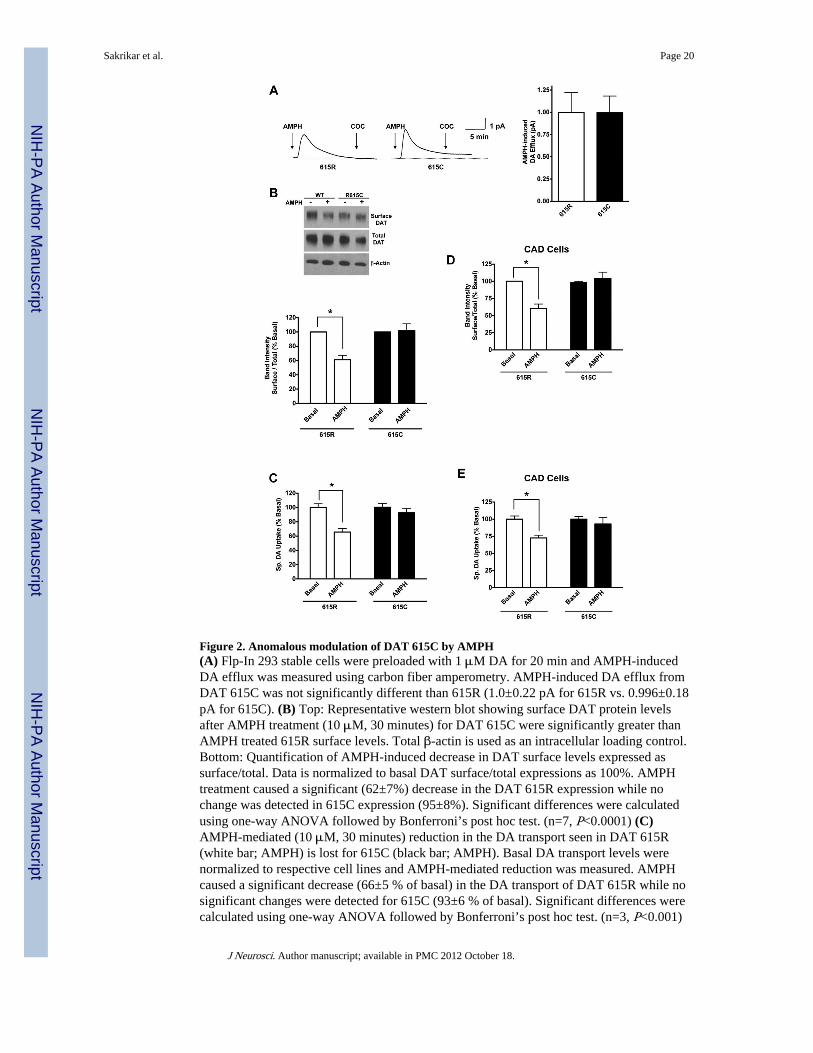
Figure 2. Anomalous modulation of DAT 615C by AMPH(A) Flp-In 293 stable cells were preloaded with 1 μM DA for 20 min and AMPH-inducedDA efflux was measured using carbon fiber amperometry. AMPH-induced DA efflux fromDAT 615C was not significantly different than 615R (1.0±0.22 pA for 615R vs. 0.996±0.18pA for 615C). (B) Top: Representative western blot showing surface DAT protein levelsafter AMPH treatment (10 μM, 30 minutes) for DAT 615C were significantly greater thanAMPH treated 615R surface levels. Total β-actin is used as an intracellular loading control.Bottom: Quantification of AMPH-induced decrease in DAT surface levels expressed assurface/total. Data is normalized to basal DAT surface/total expressions as 100%. AMPHtreatment caused a significant (62±7%) decrease in the DAT 615R expression while nochange was detected in 615C expression (95±8%). Significant differences were calculatedusing one-way ANOVA followed by Bonferroni’s post hoc test. (n=7, P<0.0001) (C)AMPH-mediated (10 μM, 30 minutes) reduction in the DA transport seen in DAT 615R(white bar; AMPH) is lost for 615C (black bar; AMPH). Basal DA transport levels werenormalized to respective cell lines and AMPH-mediated reduction was measured. AMPHcaused a significant decrease (66±5 % of basal) in the DA transport of DAT 615R while nosignificant changes were detected for 615C (93±6 % of basal). Significant differences werecalculated using one-way ANOVA followed by Bonferroni’s post hoc test. (n=3, P<0.001)
Sakrikar et al. Page 20
J Neurosci. Author manuscript; available in PMC 2012 October 18.
NIH
-PA Author Manuscript
NIH
-PA Author Manuscript
NIH
-PA Author Manuscript
(D) Quantification of AMPH-induced decrease in DAT surface levels expressed as surface/total from transiently transfected CAD cells. Data is normalized to basal DAT surface/totalexpressions as 100%. AMPH treatment caused a significant (63±6%) decrease in the DAT615R expression while no change was detected in 615C expression (104±9%). Significantdifferences were calculated using one-way ANOVA followed by Bonferroni’s post hoc test.(n=4, P<0.0001) (E) Transiently transfected, neuronally derived CAD cells display similarloss of AMPH-mediated reduction in DA transport seen in Flp-In HEK stable cells lines.Basal DA transport levels were normalized to respective cell lines and AMPH-mediatedreduction was measured. AMPH caused a significant decrease (72±4 % of basal) in the DAtransport of DAT 615R while no significant changes were detected for 615C (93±9 % ofbasal). Significant differences were calculated using one-way ANOVA followed byBonferroni’s post hoc test. (n=4, P<0.01)
Sakrikar et al. Page 21
J Neurosci. Author manuscript; available in PMC 2012 October 18.
NIH
-PA Author Manuscript
NIH
-PA Author Manuscript
NIH
-PA Author Manuscript
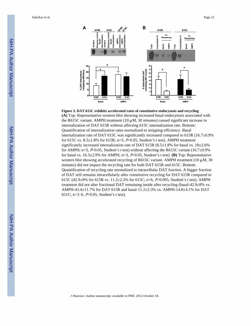
Figure 3. DAT 615C exhibits accelerated rates of constitutive endocytosis and recycling(A) Top: Representative western blot showing increased basal endocytosis associated withthe R615C variant. AMPH treatment (10 μM, 30 minutes) caused significant increase ininternalization of DAT 615R without affecting 615C internalization rate. Bottom:Quantification of internalization rates normalized to stripping efficiency. Basalinternalization rate of DAT 615C was significantly increased compared to 615R (16.7±0.9%for 615C vs. 8.5±1.8% for 615R; n=3, P<0.05, Student’s t test). AMPH treatmentsignificantly increased internalization rate of DAT 615R (8.5±1.8% for basal vs. 18±2.6%for AMPH; n=3, P<0.05, Student’s t test) without affecting the R615C variant (16.7±0.9%for basal vs. 16.3±2.9% for AMPH; n=3, P>0.05, Student’s t test). (B) Top: Representativewestern blot showing accelerated recycling of R615C variant. AMPH treatment (10 μM, 30minutes) did not impact the recycling rate for both DAT 615R and 615C. Bottom:Quantification of recycling rate normalized to intracellular DAT fraction. A bigger fractionof DAT still remains intracellularly after constitutive recycling for DAT 615R compared to615C (42.9±8% for 615R vs. 11.2±2.3% for 615C; n=6, P<0.005, Student’s t test). AMPHtreatment did not alter fractional DAT remaining inside after recycling (basal-42.9±8% vs.AMPH-43.4±11.7% for DAT 615R and basal-11.2±2.3% vs. AMPH-14.8±4.1% for DAT651C; n=3–6, P>0.05, Student’s t test).
Sakrikar et al. Page 22
J Neurosci. Author manuscript; available in PMC 2012 October 18.
NIH
-PA Author Manuscript
NIH
-PA Author Manuscript
NIH
-PA Author Manuscript

Figure 4. Increased basal CaMKII association and phosphorylation of the R615C variant(A) Top: Representative western blot showing increased association between DAT 615Cand CaMKII compared to 615R. Bottom: Quantification of DAT co-IP with CaMKIInormalized to respective inputs. Significantly larger pool of 615C is co-precipitated withCaMKII (179±26 % of 615R; n=3, P<0.05, Student’s t test). (B) Top: Representativeautoradiograph indicating increased basal phosphorylation of DAT 615C compared to 615RBottom: Quantification of DAT bands from the autoradiograph showing significant increasein basal phosphorylation levels of DAT 615C (125±1.6 % of 615R; n=3, P<0.05, Student’s ttest). Equal total protein amounts were used for IP following the metabolic labeling.
Sakrikar et al. Page 23
J Neurosci. Author manuscript; available in PMC 2012 October 18.
NIH
-PA Author Manuscript
NIH
-PA Author Manuscript
NIH
-PA Author Manuscript
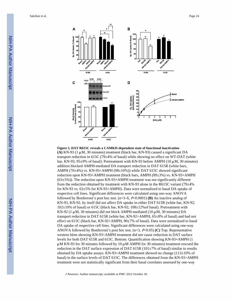
Figure 5. DAT R615C reveals a CAMKII-dependent state of functional inactivation(A) KN-93 (1 μM, 30 minutes) treatment (black bar, KN-93) caused a significant DAtransport reduction in 615C (79±4% of basal) while showing no effect on WT-DAT (whitebar, KN-93, 95±9% of basal). Pretreatment with KN-93 before AMPH (10 μM, 30 minutes)addition blocked AMPH-mediated DA transport reduction in DAT 615R (white bars,AMPH (70±4%) vs. KN-93+AMPH (98±10%)) while DAT 615C showed significantreduction upon KN-93+AMPH treatment (black bars, AMPH (89±3%) vs. KN-93+AMPH(63±5%)). The reduction upon KN-93+AMPH treatment was not significantly differentfrom the reduction obtained by treatment with KN-93 alone in the R615C variant (79±4%for KN-93 vs. 63±5% for KN-93+AMPH). Data were normalized to basal DA uptake ofrespective cell lines. Significant differences were calculated using one-way ANOVAfollowed by Bonferroni’s post hoc test. (n=3–6, P<0.0001) (B) An inactive analog ofKN-93, KN-92, by itself did not affect DA uptake in either DAT 615R (white bar, KN-92;102±10% of basal) or 615C (black bar, KN-92; 108±12%of basal). Pretreatment withKN-92 (1 μM, 30 minutes) did not block AMPH-mediated (10 μM, 30 minutes) DAtransport reduction in DAT 615R (white bar, KN-92+AMPH, 65±8% of basal) and had noteffect on 615C (black bar, KN-92+AMPH, 90±7% of basal). Data were normalized to basalDA uptake of respective cell lines. Significant differences were calculated using one-wayANOVA followed by Bonferroni’s post hoc test. (n=3, P<0.05) (C) Top: Representativewestern blots showing KN-93+AMPH treatment did not cause reduction in DAT surfacelevels for both DAT 615R and 615C. Bottom: Quantification showing KN-93+AMPH (1μM KN-93 for 30 minutes followed by 10 μM AMPH for 30 minutes) treatment rescued thereduction in the DAT surface expression of DAT 615R (101±7% of basal) similar to resultsobtained by DA uptake assays. KN-93+AMPH treatment showed no change (113±10% ofbasal) in the surface levels of DAT 615C. The differences obtained from the KN-93+AMPHtreatment were not statistically significant from their basal correlates assessed by one-way
Sakrikar et al. Page 24
J Neurosci. Author manuscript; available in PMC 2012 October 18.
NIH
-PA Author Manuscript
NIH
-PA Author Manuscript
NIH
-PA Author Manuscript

ANOVA. (n=7, P>0.05) (D) KN-93+AMPH (1 μM KN-93 for 30 minutes followed by 10μM AMPH for 30 minutes) treatment resulted in significant reduction in DA transport Vmaxfor the R615C variant. DA transport Vmax and Km are expressed as pmol/well/min and μMrespectively. A significant decrease in the DA transport Vmax was observed afterKN-93+AMPH treatment (65±10, basal vs. 42±3, KN-93+AMPH; n=4, P<0.05, Student’s ttest) without a significant change in Km (5.4±1.5, basal vs. 3.8±0.6, KN-93+AMPH; n=4,P>0.05, Student’s t test).
Sakrikar et al. Page 25
J Neurosci. Author manuscript; available in PMC 2012 October 18.
NIH
-PA Author Manuscript
NIH
-PA Author Manuscript
NIH
-PA Author Manuscript

Figure 6. Altered membrane raft microdomain association of the R615C variant(A) Top: Representative western blot showing reduced association of DAT 615C withflotillin-1. Bottom: Quantification of flotillin-1 co-IP with DAT normalized to respectiveinputs. DAT 615C displays significantly reduced association with flotillin-1 (33±8 % of615R; n=5, P<0.001, Student’s t test). (B) Top: HEK 293T cells are transiently transfectedwith YFP-HA-hDATs (green). Alexa 647 conjugated cholera toxin B is used to mark GM1ganglioside (red). Representative cells are shown with white arrows indicating lack of DATassociation with the raft fraction of R615C variant. Bottom: ICQ analysis is performed todefine colocalization of YFP-DAT with CTx-B labeled lipid rafts. Quantificationdemonstrated that although both 615R and 615C DAT are significantly colocalized withCTx-B, 615C exhibits a significantly reduced ICQ value compared to 615R (615R,0.1±0.006 vs. 615C, 0.06±0.006; n=22, P<0.01, Student’s t test).
Sakrikar et al. Page 26
J Neurosci. Author manuscript; available in PMC 2012 October 18.
NIH
-PA Author Manuscript
NIH
-PA Author Manuscript
NIH
-PA Author Manuscript
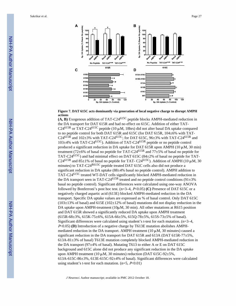
Figure 7. DAT 615C acts dominantly via generation of local negative charge to disrupt AMPHactions(A, B) Exogenous addition of TAT-C24615C peptide blocks AMPH-mediated reduction inthe DA transport for DAT 615R and had no effect on 615C. Addition of either TAT-C24615R or TAT-C24615C peptide (10 μM, 18hrs) did not alter basal DA uptake comparedto no peptide control for both DAT 615R and 615C (for DAT 615R, 104±6% with TAT-C24615R and 102±3% with TAT-C24615C; for DAT 615C, 96±3% with TAT-C24615R and103±4% with TAT-C24615C). Addition of TAT-C24615R peptide or no peptide controlproduced a significant reduction in DA uptake for DAT 615R upon AMPH (10 μM, 30 min)treatment (72±6% of basal no peptide for TAT-C24615R and 77±5% of basal no peptide forTAT-C24615C) and had minimal effect on DAT 615C (84±2% of basal no peptide for TAT-C24615R and 85±1% of basal no peptide for TAT- C24615C). Addition of AMPH (10 μM, 30minutes) to TAT-C24R615C peptide treated DAT 615C cells also did not produce asignificant reduction in DA uptake (88±4% basal no peptide control). AMPH addition toTAT-C24615C treated WT-DAT cells significantly blocked AMPH-mediated reduction inthe DA transport seen in TAT-C24615R treated and no peptide control conditions (91±3%basal no peptide control). Significant differences were calculated using one-way ANOVAfollowed by Bonferroni’s post hoc test. (n=3–4, P<0.05) (C) Presence of DAT 615C or anegatively charged aspartic acid (613E) blocked AMPH-mediated reduction in the DAtransport. Specific DA uptake values are expressed as % of basal control. Only DAT 615C(103±13% of basal) and 615E (102±12% of basal) mutations did not display reduction in theDA uptake upon AMPH-treatment (10μM, 30 min). All other mutations at R615 positionand DAT 615R showed a significantly reduced DA uptake upon AMPH treatment(615R-68±3%, 615K-75±6%, 615A-66±5%, 615Q-78±5%, 615S-73±5% of basal).Significant differences were calculated using student’s t-test for each mutation. (n=3–4,P<0.05) (D) Introduction of a negative charge by T613E mutation abolishes AMPH-mediated reduction in the DA transport. AMPH treatment (10 μM, 30 minutes) caused asignificant reduction in the DA transport for DAT 615R and 613A (DAT 615R-77±5%,613A-81±3% of basal) T613E mutation completely blocked AMPH-mediated reduction inthe DA transport (97±4% of basal). Mutating T613 to either A or E on DAT 615Cbackground and 615C alone did not produce any significant reduction in the DA uptakeupon AMPH treatment (10 μM, 30 minutes) reduction (DAT 615C-92±5%,613A-615C-86±3%, 613E-615C-92±4% of basal). Significant differences were calculatedusing student’s t-test for each mutation. (n=5, P<0.01)
Sakrikar et al. Page 27
J Neurosci. Author manuscript; available in PMC 2012 October 18.
NIH
-PA Author Manuscript
NIH
-PA Author Manuscript
NIH
-PA Author Manuscript

Figure 8. Model describing differential trafficking of DAT 615R and DAT 615C to the regulatedand constitutive endocytic pathways and biased localization toward GM1/flotillin-1 rich ordepleted membrane microdomainsDAT molecules are depicted as located in trafficking vesicles or at the plasma membrane ashomomeric or heteromeric dimers (though other configurations are acceptable). Transporterslocalize to two types of membrane domains, one domain enriched for GM1 ganglioside andflotillin-1, with the other domain relatively depleted of these molecules. Homo-multimers ofWT DAT (615R) subunits target preferentially to GM1 ganglioside/flotillin-1 enrichedcompartment as compared to DAT 615C subunit containing dimers. The heterodimer isshown preferentially targeting domains that are depleted of GM1 and flotillin-1, in keepingwith the dominant action of the 615C mutation in the ADHD proband and the dominantaction of the DAT C-terminal peptide containing 615C. DAT 615C subunits are diminishedin surface levels relative to WT subunits. Microdomains enriched for GM1 and flotillin-1support regulated trafficking of DATs, and under basal conditions exhibit slow rates ofendocytosis, whereas membranes depleted of GM-1 and flottilin-1 support more rapid,constitutive endocytosis and recycling.
Sakrikar et al. Page 28
J Neurosci. Author manuscript; available in PMC 2012 October 18.
NIH
-PA Author Manuscript
NIH
-PA Author Manuscript
NIH
-PA Author Manuscript
Related Documents