doi:10.1182/blood-2007-04-085399 Prepublished online October 11, 2007; Testi, Yaniv Lerenthal, Enrico Cundari and Daniela Barila Venturina Stagni, Maria Giovanna di Bari, Silvia Cursi, Ivano Condo, Maria Teresa Cencioni, Roberto FLIP in lymphoid cells ATM kinase activity modulates Fas sensitivity through the regulation of (4217 articles) Neoplasia Articles on similar topics can be found in the following Blood collections http://bloodjournal.hematologylibrary.org/site/misc/rights.xhtml#repub_requests Information about reproducing this article in parts or in its entirety may be found online at: http://bloodjournal.hematologylibrary.org/site/misc/rights.xhtml#reprints Information about ordering reprints may be found online at: http://bloodjournal.hematologylibrary.org/site/subscriptions/index.xhtml Information about subscriptions and ASH membership may be found online at: articles must include the digital object identifier (DOIs) and date of initial publication. priority; they are indexed by PubMed from initial publication. Citations to Advance online prior to final publication). Advance online articles are citable and establish publication yet appeared in the paper journal (edited, typeset versions may be posted when available Advance online articles have been peer reviewed and accepted for publication but have not Copyright 2011 by The American Society of Hematology; all rights reserved. Washington DC 20036. by the American Society of Hematology, 2021 L St, NW, Suite 900, Blood (print ISSN 0006-4971, online ISSN 1528-0020), is published weekly For personal use only. by guest on May 29, 2013. bloodjournal.hematologylibrary.org From

Welcome message from author
This document is posted to help you gain knowledge. Please leave a comment to let me know what you think about it! Share it to your friends and learn new things together.
Transcript
doi:10.1182/blood-2007-04-085399Prepublished online October 11, 2007;
Testi, Yaniv Lerenthal, Enrico Cundari and Daniela BarilaVenturina Stagni, Maria Giovanna di Bari, Silvia Cursi, Ivano Condo, Maria Teresa Cencioni, Roberto FLIP in lymphoid cellsATM kinase activity modulates Fas sensitivity through the regulation of
(4217 articles)Neoplasia �Articles on similar topics can be found in the following Blood collections
http://bloodjournal.hematologylibrary.org/site/misc/rights.xhtml#repub_requestsInformation about reproducing this article in parts or in its entirety may be found online at:
http://bloodjournal.hematologylibrary.org/site/misc/rights.xhtml#reprintsInformation about ordering reprints may be found online at:
http://bloodjournal.hematologylibrary.org/site/subscriptions/index.xhtmlInformation about subscriptions and ASH membership may be found online at:
articles must include the digital object identifier (DOIs) and date of initial publication. priority; they are indexed by PubMed from initial publication. Citations to Advance online prior to final publication). Advance online articles are citable and establish publicationyet appeared in the paper journal (edited, typeset versions may be posted when available Advance online articles have been peer reviewed and accepted for publication but have not
Copyright 2011 by The American Society of Hematology; all rights reserved.Washington DC 20036.by the American Society of Hematology, 2021 L St, NW, Suite 900, Blood (print ISSN 0006-4971, online ISSN 1528-0020), is published weekly
For personal use only. by guest on May 29, 2013. bloodjournal.hematologylibrary.orgFrom

1
ATM kinase activity modulates Fas sensitivity through the regulation of FLIP in
lymphoid cells
Venturina Stagni # (1, 2), Maria Giovanna di Bari # &(1, 2), Silvia Cursi (1, 2,), Ivano
Condò (3), Maria Teresa Cencioni (4), Roberto Testi (3), Yaniv Lerenthal (5) Enrico
Cundari (6) and Daniela Barilà (1, 2)*
(1) Dulbecco Telethon Institute, Department of Experimental Medicine and Biochemical Sciences, University of Rome "Tor Vergata", Via Montpellier, 1 00133 Rome, Italy. (2) Laboratory of Cell Signaling, Istituto di Ricovero e Cura a Carattere Scientifico (IRCCS) Fondazione Santa Lucia, 00179, Rome, Italy (3) Laboratory of Immunology and Signal Transduction, Department of Experimental Medicine and Biochemical Sciences, University of Rome "Tor Vergata", Via Montpellier,1 00133 Rome, Italy. (4) Laboratory of Neuroimmunology, Istituto di Ricovero e Cura a Carattere Scientifico (IRCCS) Fondazione Santa Lucia, 00179, Rome, Italy (5) The David and Inez Myers Laboratory for Genetic Research, Department of Human Genetics and Biochemistry, Sackler School of Medicine, Tel Aviv University, Tel Aviv 69978, Israel. (6) Istituto di Biologia e Patologia Molecolari Consiglio Nazionale delle Ricerche, 00185 Rome, Italy. & Present Address: Mammary Biology and Tumorigenesis Laboratory, National Cancer Institute, National Institutes of Health, Bethesda, MD 20892-1402, USA. # These authors equally contributed to this work * To whom correspondence should be addressed E-mail: [email protected], [email protected] Phone: +39-06-501703168 Fax: +39-06-501703330 Running Title: ATM kinase regulates Fas-induced apoptosis Keywords: Ataxia Telangiectasia, ATM kinase, Fas-induced apotosis, FLIP, lymphoma.
Blood First Edition Paper, prepublished online October 11, 2007; DOI 10.1182/blood-2007-04-085399
Copyright © 2007 American Society of Hematology
For personal use only. by guest on May 29, 2013. bloodjournal.hematologylibrary.orgFrom
2
ABSTRACT Ataxia Telangiectasia (A-T) is a rare cancer-predisposing genetic disease, caused
by the lack of functional ATM kinase, a major actor of the DSB DNA-damage response. A-
T patients show a broad and diverse phenotype, which includes an increased rate of
lymphoma and leukemia development. Fas-induced apoptosis plays a fundamental role in
the homeostasis of the immune system and its defects have been associated with
autoimmunity and lymphoma development.
We therefore investigated the role of ATM kinase in Fas-induced apoptosis. Using
A-T lymphoid cells we could show that ATM deficiency causes resistance to Fas-induced
apoptosis. A-T cells upregulate FLIP protein levels, a well-known inhibitor of Fas-induced
apoptosis. Reconstitution of ATM kinase activity was sufficient to decrease FLIP levels and
to restore Fas sensitivity. Conversely, genetic and pharmacological ATM kinase
inactivation resulted in FLIP protein upregulation and Fas resistance.
Both ATM and FLIP are aberrantly regulated in Hodgkin lymphoma. Importantly, we
found that reconstitution of ATM kinase activity decreases FLIP protein levels and restores
Fas sensitivity in Hodgkin lymphoma derived cells. Overall, these data identify a novel
molecular mechanism through which ATM kinase may regulate the immune system
homeostasis and impair lymphoma development.
For personal use only. by guest on May 29, 2013. bloodjournal.hematologylibrary.orgFrom
3
INTRODUCTION
Ataxia telangiectasia (A-T) is an autosomal recessive disorder characterized by
cerebellar progressive neurodegeneration leading to ataxia, dilatation of blood vessels in
the eye and facial area (telangiectasia), sensitivity to γ-irradiation, high incidence of
tumorigenesis in the lymphoid system and deficiency in immunoresponses. A-T pathology
is characterized by the loss of functional ATM protein kinase. Following DNA damage,
ATM is rapidly activated, (auto)phosphorylated 1 and, in turn, it phosphorylates a number
of substrates which all contribute to cell growth arrest or, alternatively, apoptosis (reviewed
in 2). The higher cancer predisposition of A-T patients has been associated with the lack of
DNA damage response, which results in genomic instability 3. The immune system is the
major target of tumor development in these patients, and lymphoma and leukemia are very
frequent 4,5. This clinical feature is consistent with the central role of ATM in the
management of the DNA DSBs generated during the immune system development and
function in physiological conditions6. Indeed most of the lymphoma developed in A-T
patients are characterized by aberrant VDJ recombination6. More interestingly, ATM
expression is aberrantly low in several B and T cell lymphomas irrespective of A-T
genotype7-10.
Fas (CD95/APO-1) is a transmembrane protein belonging to the tumor necrosis
factor superfamily. Upon binding of Fas ligand or agonistic antibodies, the Fas receptor
recruits several cytosolic proteins to form the death-inducing signalling complex (DISC).
This is necessary to catalyze dimerization, and processing of Procaspase-8 to generate
the active Caspase-8 tetramer, composed of two p18 and two p10 subunits, which initiates
the caspase cascade11. Procaspase-8 activation is absolutely required to trigger receptor-
activated apoptotic response12 and its catalytic activity has to be tightly regulated to avoid
inappropriate activation and undesired cell death13. FLIP protein is structurally similar to
Procaspase-8 and can therefore compete with Procaspase-8 for binding to DISC, thus
For personal use only. by guest on May 29, 2013. bloodjournal.hematologylibrary.orgFrom
4
preventing Caspase-8 activation and the following apoptotic cascade. Two isoforms of
FLIP, arising from alternative splicing, are normally present in most of the cells. FLIP-Long
(FLIP-L), similarly to Procaspase-8, has two DED domains that mediate the recruitment to
the DISC, as well as a p18 and a p10 subunit but it lacks the Cys residue in the active site
and is therefore catalytically impaired. However, in some contexts FLIP-L can also
dimerize and therefore promote Caspase-8 activation14. Conversely, FLIP-Short (FLIP-S)
contains only the DED domains and it behaves as a pure inhibitor of Procaspase-8
activation and Fas-induced apoptosis13.
The death receptor system is essential for the regulation of the lymphoid system
homeostasis15. It is assumed that the negative selection process of B as well as T cells in
the germinal center (GC) and thymus, respectively, depends on Fas system16,17. Several
lines of evidence indicate the importance of this system for the balance between B cell
proliferation and apoptosis18. Indeed, mice lacking functional Fas expression suffer from
autoimmunity and increased incidence of B cell lymphomas19,20. Patients with mutations
that impair the function of proteins involved in Fas-dependent apoptosis develop the
autoimmune lymphoproliferative syndrome (ALPS), which predisposes them to
autoimmune disorders and to lymphoma development21,22. Finally, Fas mutations where
identified in lymphomas, in particular those deriving from GC B cells(reviewed in23).
Classical Hodgkin’s lymphoma (cHL), a common human lymphoma, has been
proposed to derive, most frequently, from GC cells24. Currently, the molecular
pathogenesis of cHL remains unclear. Interestingly, Hodgkin/Reed Sternberg (HRS) cells,
the malignant cells of classical Hodgkin’s lymphoma (cHL), resist to Fas-induced
apoptosis25 and Fas resistance has been proposed to play an active role in the
development of HRS cells. Indeed, these cells evade the control of the immune system
and initiate the tumour growth. Recently, Fas resistance of HL-derived cell lines has been
proposed to be caused by the aberrant upregulation of FLIP proteins in these cells.
For personal use only. by guest on May 29, 2013. bloodjournal.hematologylibrary.orgFrom
5
Indeed, the specific downregulation of FLIP expression by siRNA sensitizes these cells to
Fas-induced apoptosis26,27. Remarkably, immunohistochemistry studies have shown that
most cases of Hodgkin’s disease are ATM negative7, although ATM loss of heterozygosity
is a rare event28, and therefore alternative mechanisms may account for ATM
downregulation29.
Taking into account the linkage between Fas impairment and the development of
those tumors that are more frequent in A-T patients the question arises as to whether any
relationship exists between Fas and ATM signaling pathways.
Here we show that ATM deficiency results in a significant resistance of lymphoid
cells derived from AT patients to Fas-induced apoptosis. Interestingly, loss of endogenous
ATM kinase activity results in the aberrant upregulation of FLIP protein levels.
Consistently, ATM kinase activation downregulates FLIP protein levels providing a novel
mechanism to modulate Fas sensitivity. Furthermore Hodgkin Lymphoma cells that are
characterized by Fas-resistance, may be sensitized to Fas upon ATM kinase expression.
These data point to ATM as a novel player in Fas-induced apoptosis and suggest a novel
molecular mechanism for the increased lymphoma susceptibility of A-T patients and for the
development of B cell lymphoma.
MATERIALS AND METHODS
DNA constructs
pcDNA3-Flag-ATM-wt, pcDNA3-Flag-ATM-Kin- were kindly provided by M. Kastan.
shFLIP construct and its control were kindly provided by H. Walczak30.
Antibodies and other reagents
The following antibodies and reagents were used: anti-phosphoSer1981-ATM
(Rockland), anti-ATM (MAT3, generously provided by Y.Shiloh), anti-phosphoSer15-p53
For personal use only. by guest on May 29, 2013. bloodjournal.hematologylibrary.orgFrom
6
(Cell Signaling), anti-p53 (Santa Cruz, Pab240), anti-phosphoThr68-Chk2 (Cell Signaling),
anti-Chk2 (kindly provided by D. Delia), anti-pS139 H2A.X (UBI), anti-Fas IgM monoclonal
antibody (CH11; UBI), anti-Flag (Sigma), anti-Caspase-8 (clone 5F7, MBL), anti-FLIP(S
and L) (H-202 Santa Cruz), anti-active Caspase-3 (Cell Signaling), caspase-inhibitor zVAD
(Biomol), NCS (kindly provided by Y.Shiloh), KU-55933 (kindly provided by KUDOS) .
Cell culture and transfections
C3ABR and L6 cells (kindly provided by M. Lavin and Y. Shiloh) as well as GM-
03189, GM-02782 cell lines, were cultured in RPMI 1640 medium with 10 mM HEPES, 1.0
mM sodium pyruvate, 10% fetal bovine serum. C3ABR and L6 cells were stably
transfected by electroporation using 20 μg of the indicated constructs. Stably transfected
cells where selected in the presence of 500 μg/ml G418. HL-derived cell line, L428, kindly
provided by H. Kashkar and M. Kronke, were transfected by electroporation.
Analysis of apoptosis
C3ABR, L6, L6pCDNA, L6-Flag-ATM-wt, L6-Flag-ATM-Kin- and L6-shFLIP cells
lines were treated to undergo apoptosis with 250-500 ng/ml anti-Fas antibody. Where
indicated in western blot and immunofluorescence analysis cells were also treated with
NCS (100ng/ml for 1h) or stimulated in the presence of 40 μM zVAD caspase-inhibitor,
which was added 30 min before stimulation with Fas.
Apoptosis was quantified by propidium iodide (Sigma) nuclear staining or by the
analysis of Annexin V (Pharmigen) exposure using a FACScanto (Becton Dickinson).
Specific apoptosis was determined as follows: (% of apoptotic cells with anti-Fas - % of
apoptotic cells without anti -Fas) / (100 - % of apoptotic cells without anti -Fas).
Analysis of Fas-receptor levels
To analyze the expression of Fas protein cells were incubated for 30’ RT with
mouse anti-human Fas antibody (APO1,Transduction Laboratories). Next, cells were
For personal use only. by guest on May 29, 2013. bloodjournal.hematologylibrary.orgFrom
7
reacted with PE-conjugated goat anti-mouse IgG(H+L) (Pharmigen) for 30 min at RT. Cells
were analyzed using a flow-cytometer. For each cell line incubation with PE-conjugated
alone served as negative controls. Mean fluorescence intensity of cell stained with anti-
Fas was used to compare the level of Fas expression.
Flow cytometry of phosho-Ser1981-ATM in apoptotic cells
Our protocol is a variation of a recent method used to evaluate phosphoepitope status by
flow cytometry31. 5 x 10⁵ cells were fixed in 4% formaldehyde and incubated 15 min at 4
oC. They were then permeabilized by resuspending with vigorous vortexing in 1 ml ice-cold
MeOH and incubated at -20°C O/N. Cells were washed and resuspended in PBS-Tween
0.5% containing 5% Normal Goat Serum (NGS) containing anti-mouse-phospho-Ser1981-
ATM and rabbit-active-Caspase-3 primary antibodies and incubated for 1 h at room
temperature. After washing and repeating the process with anti-mouse-AlexaFluor488 and
anti-rabbit-AlexaFluor633 conjugated secondary antibodies, flow cytometry was evaluated
in a FACScanto (Beckton Dickinson).
Immunofluorescence analysis
C3ABR, L6-pCDNA, L6-FlagATM-wt and L6-Flag-ATM-Kin- cells line were fixed,
permeabilized and immunofluorescence were carried out as previously described32. Flag-
ATM protein was visualized with monoclonal anti-Flag (Sigma) diluted 1:500 followed by
fluorescein-conjugated anti-mouse antibody (Alexis) diluted 1:200 in blocking buffer.
Phospho-S1981ATM was labeled with anti-pS1981 ATM (Rockland) diluted 1:1000
followed by rhodamine-conjugated anti-rabbit diluted 1:600 (Alexis) or by fluorescein-
conjugated anti-rabbit antibody diluted 1:200. Nuclei were visualized with Hoechst 33342
(Molecular Probes) diluted 1:20,000 in PBS-0.1% Triton X-100.
Immunoblotting
Cell extracts were prepared in IP buffer (50 mM Tris-HCl (pH 7.5), 250 mM NaCl,
1% NP-40, 5 mM EDTA, 5 mM EGTA, 1 mM phenylmethylsulfonyl fluoride, 25 mM NaF, 1
For personal use only. by guest on May 29, 2013. bloodjournal.hematologylibrary.orgFrom
8
mM orthovanadate, 10 μg/ml TPCK, 5 μg/ml TLCK, 1 μg/ml leupeptin, 10 μg/ml soybean
trypsin inhibitor, 1 μg/ml aprotinin)33. For immunoblotting, 100-200 μg of protein extract
were separated by SDS-PAGE, blotted onto nitrocellulose membrane and detected with
specific antibodies. All immunoblots were revealed by ECL (Amersham).
Caspase-8 activity assay.
To determine Caspase-8 activity in C3ABR, L6pCDNA, L6-FlagATM-WT and L6-
FlagATM-KD cells line, cells were induced to undergo apoptosis with 500 ng/ml of �nti-
Fas mAb. Protein extracts were assayed for caspase-8 activity using IETD-AMC as a
substrate Ac-IETD-AMC at 37 ºC in 200 μl assay buffer (20 mM Tris, pH 7.4, 0.1 M NaCl,
10% sucrose, 0.1% CHAPS, 10 mM DTT) containing 700 μg protein extract. Reaction was
started by the addition of 10 μM Ac-IETD-AMC. Cleavage of the substrate as a function of
time was monitored reading the absorbance at 460 nm upon excitation at 390 nm. The
enzymatic activity was determined from the linear portion of the curve.
Reverse transcription-polymerase chain reaction (RT-PCR)
Total cellular RNAs were isolated using Trizol reagent (Invitrogen) and subjected to
RT using oligo(dT) primer and M-MLV reverse transcriptase (Invitrogen) according to the
manufacturer’s protocol. RT reaction was then amplified by PCR using the primers
described in34. Amplification of actin was performed in the same PCR reaction as internal
control. PCR products were run on a 2% agarose gel and visualized by ethidium bromide
staining.
Statistical methods
All data were analyzed and presented as mean ± SD (n<10). The significance of
differences between populations of data were assessed according to the Student’s two
tailed T-test with a level of significance of at least p < 0.05 (alpha conventionally equal to
0.05). This analysis arises in the problem of estimating the mean of a normally distributed
population when the sample size is small.
For personal use only. by guest on May 29, 2013. bloodjournal.hematologylibrary.orgFrom
9
RESULTS
ATM deficient cells are resistant to Fas-induced apoptosis To investigate whether ATM could participate in Fas-mediated apoptosis, we
compared the sensitivity to Fas of two lymphoblastoid cell lines widely used in studies on
ATM activity, one established from an AT patient (L6)35, the other one from a healthy
control donor (C3ABR) 36. Fas was stimulated with agonistic anti-Fas antibodies that mimic
the binding of Fas-Ligand and triggers the apoptotic response. Interestingly L6 cells, which
lack the expression of ATM protein, were significantly resistant to Fas-induced apoptosis
(Fig. 1A, B). Similar results were obtained also with other A-T lymphoblastoid cell lines,
such as GM-03189, GM-02782 (Supplementary Fig. 1). To address the question of
whether ATM kinase activity is required for Fas sensitivity, we stably reconstituted ATM
expression in L6 cells. For this purpose L6 cells were stably transfected with constructs
that allow the expression of either FLAG-ATM-wt protein (L6-ATM-wt) or the kinase dead
FLAG-ATM-Kin- protein (L6-ATM-Kin-), or with the empty vector as control (L6-pCDNA).
ATM expression was monitored by immunoblotting with specific antibodies. (Fig. 1C). L6-
ATM-wt and L6-ATM-Kin- cells expressed same levels of ATM protein. Interestingly, the
reconstitution of the expression of ATM in the L6-ATM-wt cells dramatically sensitized
these cells to Fas-induced apoptosis (Fig. 1 D, E and Supplementary Fig. 2). The
expression of the ATM-kinase-defective mutant, FLAG-ATM-Kin-, completely failed to
restore Fas sensitivity. Overall these results, suggest that ATM kinase activity enhances
Fas-induced apoptosis.
Caspase activition is a pre-requisite of Fas-dependent ATM activation
We therefore asked the question of whether Fas stimulation triggers ATM kinase
activation and whether this may contribute to Fas sensitivity. To evaluate the effect of Fas-
stimulation on ATM kinase activity, protein extracts at different times of stimulation were
For personal use only. by guest on May 29, 2013. bloodjournal.hematologylibrary.orgFrom
10
analyzed. Similarly to what has been described following DNA damage37-41, Fas
stimulation resulted in the typical ATM-dependent phosphorylation cascade. In particular,
ATM was phosphorylated on its autophosphorylating activating site i.e. Ser19811, and p53,
Chk2 and H2AX became phosphorylated at Ser15, Thr68 and Ser139 respectively(Fig. 2A
and Supplementary Fig. 3). These data indicate that Fas stimulation results in ATM
activation. Importantly, ATM activation was completely prevented by preincubation with the
general caspase-inhibitor z-VAD (Fig. 2A), thus suggesting that, in the absence of caspase
activation, no Fas-induced ATM activation occurs. To evaluate this possibility, we
established a new flow cytometry–based assay that allowed us to analyze the levels of
phospho-Ser1981-ATM vs Caspase-3 activation, which accounts for apoptotic response.
This analysis revealed that ATM activation mainly occurs in cells that activate Caspase-3,
supporting the hypothesis that Fas-dependent ATM activation is downstream Caspase-3
activation, and therefore most likely does not play a major role in Fas sensitivity (Fig. 2B).
Fas stimulation also resulted in the cleavage of ATM protein (Fig. 2A), similarly to other
apoptotic stimuli, which trigger ATM kinase cleavage most likely through Caspase-3
activity42. Moreover the uncleavable mutant of ATM, ATM-D863A, previously characterized
42, sensitized A-T cells to Fas-induced apoptosis to the same extent as ATM-wt, further
confirming that the cleavage per se does not modulate Fas sensitivity (data not shown).
Finally, Fas stimulation triggered ATM phosphorylation on Ser1981 only on those cells that
showed apoptotic morphology characterized by nucleus condensation or fragmentation
(Fig. 2C). Overall, these findings strongly suggest that ATM activation upon Fas
stimulation is a passive event subsequent to DNA fragmentation, and therefore most likely
does not contribute significantly to cell fate.
ATM kinase activity downregulates c-FLIP protein levels
To get more insight in the molecular mechanism by which ATM modulates Fas-
induced apoptosis, we analyzed the expression profile of those proteins that are relevant
For personal use only. by guest on May 29, 2013. bloodjournal.hematologylibrary.orgFrom

11
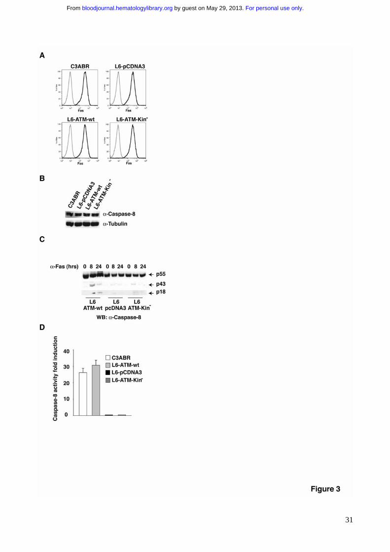
for this signaling. FACS analysis showed that ATM expression and activity do not
modulate the levels of Fas (Fig. 3A). Immunoblotting analysis showed that all cell lines
express comparable levels of Caspase-8, independently on ATM activity (Fig. 3B).
Remarkably, despite the observation that Caspase-8 is equally expressed in all cell lines,
its activation following Fas crosslinking is significantly delayed in the ATM deficient cells
(L6-pCDNA) as well as in the ATM kinase activity deficient cells (L6-ATM-Kin-) (Fig. 3C,
3D). Full activation of Caspase-8 upon Fas stimulation requires its processing, essential to
get a stable active caspase-8 tetramer and to allow its release from the DISC and
subsequent cleavage of cytoplasmic substrates, such as executioner caspases11.
Immunoblotting experiments, using an anti-Caspase-8 antibody raised against the p18
subunit, showed that the lack of ATM results in the delayed accumulation of the
intermediate processing product p43 and of the p18 subunit (Fig. 3C). Moreover, ATM
deficiency delayed Fas-induced Caspase-8 activation, measured as its ability to cleave its
substrate peptide IETD (Fig. 3D).
Being c-FLIP a well characterized inhibitor of Fas signaling, we wanted to
investigate the possible relationships between ATM activity and c-FLIP expression.
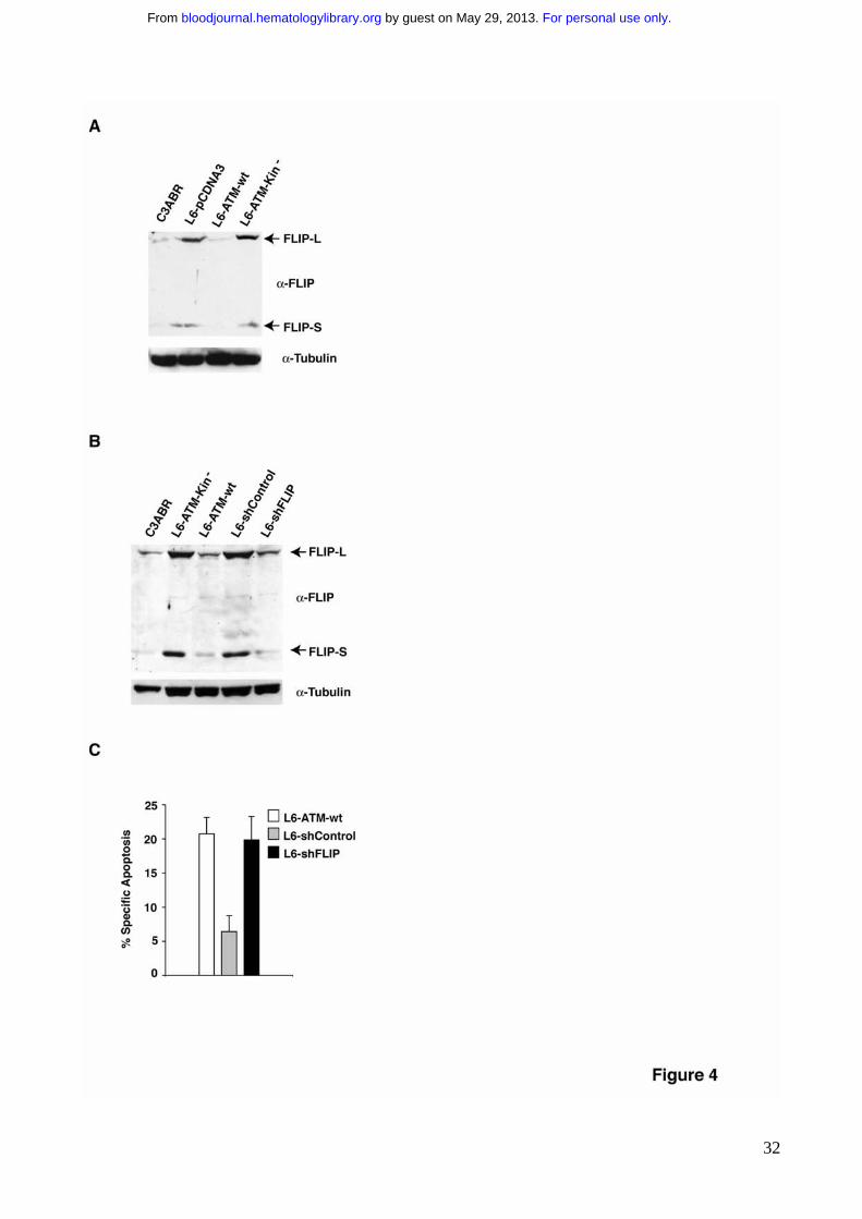
Importantly, the lack of ATM expression triggers the upregulation of c-FLIP (Fig. 4A),
which may account for Fas resistance of AT cells (Fig. 1). Reconstitution of ATM kinase
activity in L6-ATM-wt cells significantly decreased FLIP-L and FLIP-S expression levels,
which may account for the recovery of Fas sensitivity (Fig. 1). Again, the ATM-Kin- mutant
completely failed to downregulate FLIP (Fig. 4A). To test whether indeed ATM activity
modulates Fas sensitivity via the regulation of FLIP levels, we generated a stable A-T cell
line, L6-shFLIP, where FLIP expression has been genetically reduced through specific
shRNA that selectively targets FLIP-L and FLIP-S isoforms. These cells express low levels
of FLIP proteins comparable to the endogenous level of ATM kinase reconstituted cells
(Fig. 4B). Indeed, the reduction of FLIP sensitizes A-T cells to Fas-induced apoptosis (Fig.
For personal use only. by guest on May 29, 2013. bloodjournal.hematologylibrary.orgFrom

12
4C and Supplementary Fig. 2), indicating that the aberrant levels of FLIP proteins may be
responsible for Fas resistance in A-T cells. Overall these experiments show that ATM
kinase sensitizes cells to Fas-induced apoptosis through the modulation of FLIP levels.
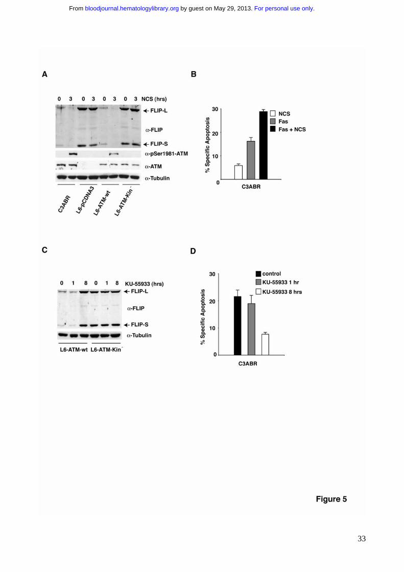
This observation suggests that ATM kinase activity may downregulate FLIP. Indeed
stimulation with neocarzinostatin (NCS), that classically triggers ATM kinase activation,
resulted in a reduction of the levels of FLIP protein (Fig. 5A). This effect is completely
abrogated in cells that lack ATM protein or reconstituted with the ATM kinase defective
mutant (Fig. 5A). According to these data, NCS treatment significantly sensitized cells to
Fas-induced apoptosis (Fig. 5B).
The observation that FLIP is aberrantly upregulated in A-T cells as well as in A-T
cells reconstituted with inactive ATM (Fig. 4A) suggests that the endogenous basal activity
of ATM is sufficient to downregulate FLIP protein levels. To unambiguously address this
issue, ATM proficient cells were incubated in the presence of the ATM kinase inhibitor KU-
5593343. Indeed, this treatment triggered FLIP upregulation (Fig. 5C). Interestingly,
preincubation with KU-55933 for 1 hour is not sufficient to increase FLIP protein levels
(Fig. 5C) and fails to protect cells from Fas-induced apoptosis (Fig. 5D). Conversely,
preincubation with KU-55933 for 8 hours, which is sufficient to trigger FLIP protein
accumulation, dramatically impairs Fas-induced apoptosis to the same extent of A-T cells
(Fig. 5D). These data clearly show that ATM kinase activity is required to modulate Fas
sensitivity through the control of FLIP protein levels.
ATM kinase activity modulates FLIP protein stability
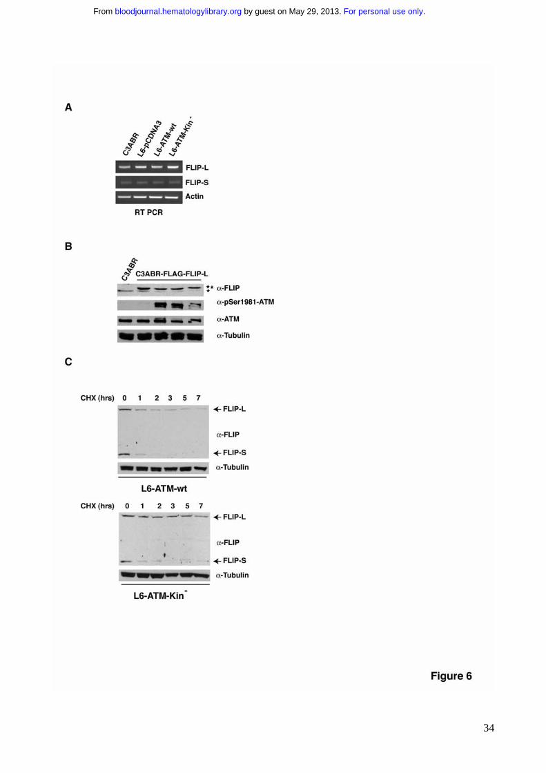
To evaluate whether ATM modulates the mRNA levels of FLIP, we analyzed FLIP
mRNA levels in ATM proficient and ATM deficient cell lines. RT-PCR experiments showed
that the levels of FLIP transcripts are comparable in all cell lines independently on ATM
expression and activity (Fig. 6A), suggesting that FLIP regulation does not occur at
transcriptional level. In agreement with this assumption, an exogenous FLAG-tagged FLIP-
For personal use only. by guest on May 29, 2013. bloodjournal.hematologylibrary.orgFrom
13
L driven by a heterologous promoter was repressed similarly to the endogenous FLIP-L
when stably transfected C3ABR-FLAG-FLIP-L cells were stimulated with NCS to trigger
ATM kinase activity (Fig. 6B). We therefore tested whether ATM kinase activity
accelerates FLIP proteins degradation by blocking nascent translation with cycloheximide
(CHX). Cells where pretreated with the ATM kinase inhibitor KU-55933 for 8 hrs to have
the same initial levels of FLIP proteins and then, upon KU-55933 removal, they were
incubated for different times with CHX. The degradation of both FLIP forms was
significantly faster when L6 cells where reconstituted with ATM-wt than with its kinase-
dead homologue (Fig. 6C). This approach allowed us to conclude that FLIP-protein
degradation is significantly increased dependently on ATM kinase activition and that ATM
kinase downregulates FLIP protein stability.
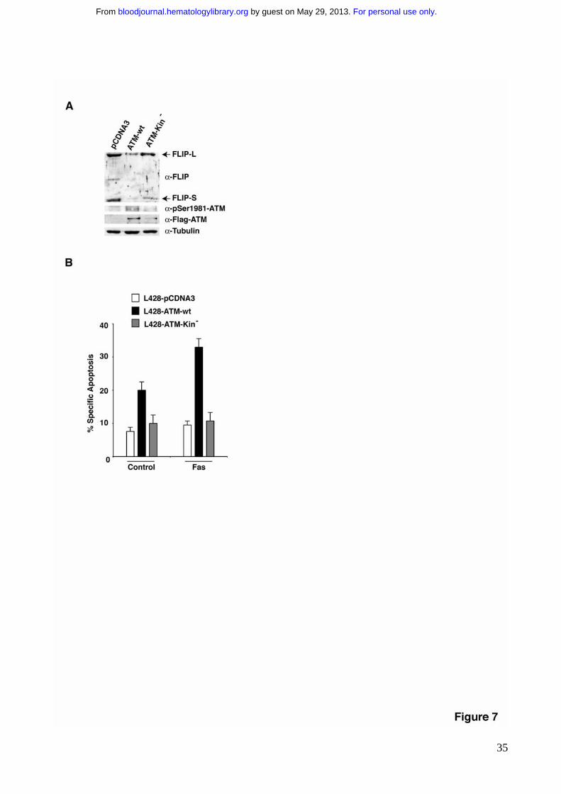
ATM kinase activity sensitizes Hodgkin Lymphoma cells to Fas-induced apoptosis
Resistance to death-receptor-mediated apoptosis is supposed to be important for
the deregulated growth of B cell lymphoma. Hodgkin/Reed Sternberg (HRS) cells, the
malignant cells of classical Hodgkin’s lymphoma (cHL), resist to Fas induced apoptosis.
Fas resistance in this system is due to the aberrant upregulation of FLIP proteins26,27.
Conversely, ATM expression and function is impaired in many HL cases 7 and in several
HL-derived cell lines9,29. To test whether ATM loss of function may contribute to Fas
resistance through FLIP protein upregulation, we took advantage of a lymphoma cell line,
L428, that has been previously characterized for the aberrant downregulation of ATM
activity29 and for the aberrant upregulation of FLIP protein levels26,27. Transient transfection
of ATM downregulates FLIP levels (Fig. 7A) and restores Fas sensitivity (Fig. 7B),
suggesting that targeting of ATM kinase activity significantly contributes to death receptor
resistance of HL cell lines and most likely plays a functional role in this pathology.
DISCUSSION
For personal use only. by guest on May 29, 2013. bloodjournal.hematologylibrary.orgFrom
14
Important defects of the immune system, leading to a significant increase of
lymphoma and leukemia development, are one of the major feature of A-T syndrome4,5.
Since ATM kinase plays a central role in the DSB DNA damage response and this
response is required in some physiological context such as the immune system
homeostasis, the lack of ATM activity has been proposed to be responsible for aberrant
chromosomal translocations originated as a consequence of a failure of the DNA damage
response and indeed associated to several lymphomas and leukemias6
Fas-dependent apoptosis plays a fundamental role in the regulation of the
homeostasis of the lymphoid system15. Failure in the Fas signaling causes, both in mice
and in humans, autoimmunity as well as aberrant proliferation and lymphoma
development19-22.
We reasoned that since defects in Fas-induced apoptosis result in defects in the
immune system that partially resemble some of the abnormalities characteristic of the
immune system of A-T patients, ATM kinase may play a role in Fas-induced apoptosis.
According to our hypothesis, the present article shows that cells that lack ATM kinase are
significantly resistant to Fas induced apoptosis (Fig.1). Reconstitution experiments showed
that ATM catalytic activity is required to sensitize cells to Fas (Fig. 1 D, E). We could show
that Fas stimulation triggers ATM kinase activation. However, our data strongly suggest
that ATM activation upon Fas stimulation occurs when the apoptotic signaling is already
irreversible, as a consequence of DNA condensation and fragmentation during the
apoptotic response. Therefore ATM activation does not seem to play a major role in the
sensitivity to Fas-induced apoptosis. This apparent paradox prompted us to investigate
whether basal ATM kinase may modulate the level and/or the activity of any central player
of Fas signaling. Fas sensitivity mainly relies on Fas-receptor expression on cell surface
and on Caspase-8 activity, which is absolutely required to drive the caspase cascade and
execute the apoptotic program. Importantly, ATM protein does not modulate Fas-receptor
For personal use only. by guest on May 29, 2013. bloodjournal.hematologylibrary.orgFrom
15
or Caspase-8 protein levels (Fig. 3A, B). However, A-T cells are impaired in Caspase-8
activation consistently with their resistance to Fas (Fig.3C,D). It has been clearly
established that FLIP proteins may modulate Caspase-8 activation in vitro and in vivo
(reviewed in13). Moreover FLIP level is tightly regulated during T and B cell activation and
its decrease parallels the enhancement of Fas sensitivity (reviewed in44). Importantly, we
could show that A-T cells significantly accumulate FLIP proteins (Fig. 4A). Reconstitution
of ATM kinase activity downregulates FLIP proteins. Conversely a catalitically inactive
ATM fails to downregulate FLIP. Importantly, there is a strict relationship between FLIP
levels and the sensitivity of the different cell lines to Fas-induced apoptosis. To further test
the hypothesis that ATM kinase sensitizes cells to Fas-induced apoptosis through the
downregulation of FLIP proteins, we interfered FLIP expression in A-T cells, by specific
shRNA constructs. Following this approach it was possible to downregulate FLIP to the
same levels observed in ATM proficient cells which, in turn resulted in the restoration of
Fas sensitivity in A-T cells (Fig. 4B, C). Therefore we concluded that ATM kinase activity
modulates Fas sensitivity through the regulation of FLIP protein levels.
The observation that FLIP levels decrease in A-T cells upon reconstitution with kinase
active ATM but not with a kinase defective mutant (Fig. 4A) suggests that a basal ATM
kinase activity may be sufficient to downregulate FLIP levels. The presence of an
endogenous basal ATM activity, which may be further induced upon DNA damage has
been already described45,46. Consistently with the presence of such a basal activity, NCS
treatment, which triggers ATM activation, downregulates FLIP in the presence of a kinase
competent ATM protein (Fig. 5A). Conversely, the treatment of ATM proficient cells with
the ATM kinase inhibitor KU-55933 triggers FLIP upregulation (Fig. 5C). These data allow
us to propose that ATM kinase activity modulates FLIP protein levels. Consistently, while
the decrease of FLIP levels following NCS treatment sensitizes cells to Fas induced
apoptosis (Fig. 5B), the upregulation of FLIP levels after 8 hours preincubation with KU-
For personal use only. by guest on May 29, 2013. bloodjournal.hematologylibrary.orgFrom
16
55933 protects cells from Fas-induced apoptosis (Fig. 5D). Overall, we provide evidence
for A-T cell resistance to Fas-induced apoptosis and we demonstrate that ATM kinase
activity may modulate Fas sensitivity through the regulation of FLIP proteins level.
Moreover, we could show that ATM modulates FLIP protein stability (Fig. 6). Further
experiments will clarify the molecular mechanism beyond this regulation.
Overall, these findings points to the upregulation of FLIP protein levels as a putative novel
marker of A-T cell lines. We are currently investigating the levels of FLIP protein in
heterozygous derived A-T cell lines. This study along with further experiments on
peripheral blood cells from A-T patients will address the question whether FLIP
upregulation could be used as a novel A-T prognostic marker.
Importantly, A-T patients have an increased rate of lymphoma and leukemia
development, with a frequent occurrence of B-cell lymphomas such as Hodgkin
Lymphomas4,5. Interestingly, several independent studies on HL reported the aberrant
downregulation of ATM activity as a common event, thus suggesting that ATM loss may
promote HL development7,9. Furthermore it has been clearly shown that HL are very
resistant to Fas- and TRAIL-induced apoptosis and this correlates clearly with the aberrant
upregulation of FLIP levels. Indeed the downregulation of FLIP is sufficient to sensitize
back these cells to death-receptor-induced apoptosis26,27. It has been reported that NF�B
transcription factor up-regulates FLIP expression47. Interestingly, the transcription factors
NF�B and AP1 are aberrantly activated in HL and have been proposed to be responsible
for the modulation of the levels of most of the proteins aberrantly expressed in HL48-50.
Indeed, repression of NF�B activity triggers FLIP protein downregulation in HSR cells27.
We have shown that ATM kinase activity modulates FLIP protein levels. To test whether
the lack of ATM kinase activity in HSR cells may contribute to FLIP downregulation we
restored ATM activity in L428 cells, an HL-derived cell line previously characterized for
aberrantly low ATM function29 and for aberrantly high FLIP levels26,27. Using this approach
For personal use only. by guest on May 29, 2013. bloodjournal.hematologylibrary.orgFrom
17
we could show that ATM activity is sufficient to decrease FLIP levels and to sensitize L428
cells to Fas-induced apoptosis (Fig. 7). This finding, along with the data in literature on
ATM deficiency in HL7,9, allows us to speculate that AT deficiency could also contribute to
lymphoma development via the loss of control on FLIP levels which in turn triggers Fas
resistance.
In summary, we identified a novel function for ATM kinase as a regulator of FLIP levels
and of Fas sensitivity and suggested that this signaling may contribute to the homeostasis
of the immune system. It is also tempting to speculate that failure of the ATM-dependent
FLIP regulation, might be at least partially responsible for the increased frequency of
lymphomas associated to A-T, as well as for the development of lymphoma in those
situations where ATM kinase activity is downregulated through alternative mechanisms
other than homozygous deletion. Furthermore, the induction of ATM activation may
provide a novel tool to downregulate FLIP protein levels and to sensitize those lymphomas
where endogenous ATM is still functional to death receptor induced apoptosis. Importantly,
treatment of tumor cells with DNA-damaging drugs like 5-FU has been shown to
downregulate cFLIP and thereby to sensitize cells to death receptor-induced apoptosis30.
We observed that ATM kinase activity is required for this effect (data not shown),
suggesting that indeed this mechanism might be diagnostically and therapeutically
relevant.
Finally, we provide novel evidence for a basal endogenous activity of ATM kinase
independent of the exogenous DNA damage induction, which probably accounts for
differences in the level of expression of FLIP protein. This basal activity of ATM could be
relevant also in other cellular processes and contribute, at least in part, to the complexity
of A-T phenotype. Therefore, investigations comparing different structural and functional
features of wt and A-T cells in the absence of DNA damage, may strongly contribute to
broaden current knowledge on ATM kinase function and A-T pathology.
For personal use only. by guest on May 29, 2013. bloodjournal.hematologylibrary.orgFrom

18
ACKNOWLEDGMENTS
We acknowledge Y. Shiloh, M. Kastan, D. Delia, H. Walczak, H. Kashkar, M.
Kronke, L. Chessa and KUDOS for kindly providing reagents, A. Diamantini for FACS
sorting (MOFLO), M.P. Paronetto for RT PCR experiment support and C. Sette and all his
lab members for helpful discussion and critical reading of the manuscript. D.B. is an
Assistant Telethon Scientist and is supported by the Italian Telethon Grant (TCP00061),
V.S., M.G., and S.C. have been supported by the Italian Telethon Foundation and by the
Italian Compagnia di San Paolo-Imi Bank Foundation. V.S. is presently supported by
Fondazione Santa Lucia. This work has been supported by grants from the Italian
Telethon Foundation (TCP00061), from the Italian Association for Cancer Research
(AIRC), from the Italian Compagnia di San Paolo-Imi Bank Foundation, and from the AT
Childrens’ Project, to D.B. This work was also supported by a grant from AIRC to R.T.
AUTHORSHIP
Contribution: V.S. performed the research and analyzed the data, M.G.d.B performed the
research, S.C. generated the shFLIP cell lines, I.C. supervised the work with the ATM
reconstituted cell lines, M.T.C. assisted with the flow cytometry and apoptosis analysis,
R.T. analyzed the data, Y.L. analyzed the data, E.C. wrote the paper, D.B. designed the
research and wrote the paper.
Conflict-of-interest disclosure: The authors declare no competing financial interests.
For personal use only. by guest on May 29, 2013. bloodjournal.hematologylibrary.orgFrom
19
REFERENCES
1. Bakkenist CJ, Kastan MB. DNA damage activates ATM through intermolecular
autophosphorylation and dimer dissociation. Nature. 2003;421:499-506
2. Shiloh Y. ATM and related protein kinases: safeguarding genome integrity. Nat
Rev Cancer. 2003; 3 :155-167
3. Khanna KK, Jackson SP. DNA double-strand breaks: signaling, repair and the
cancer connection. Nat Genet. 2001; 27: 247-254
4. Gumy-Pause F, Wacker P, Sappino AP. ATM gene and lymphoid malignancies.
Leukemia. 2004;18:238-242
5. Taylor AM, Metcalfe JA, Thick J, Mak YF. Leukemia and lymphoma in ataxia
telangiectasia. Blood. 1996; 87:423-438
6. Matei IR, Guidos CJ, Danska JS. ATM-dependent DNA damage surveillance in
T-cell development and leukemogenesis: the DSB connection. Immunol Rev. 2006; 209:
142-158
7. Starczynski J, Simmons W, Flavell JR, Byrd PJ, Stewart GS, Kullar HS, Groom
A, Crocker J, Moss PA, Reynolds GM, Glavina-Durdov M, Taylor AM, Fegan C, Stankovic
T, Murray PG. Variations in ATM protein expression during normal lymphoid differentiation
and among B-cell-derived neoplasias. Am J Pathol. 2003; 163: 423-432
8. Greiner TC, Dasgupta C, Ho VV, Weisenburger DD, Smith LM, Lynch JC, Vose
JM, Fu K, Armitage JO, Braziel RM, Campo E, Delabie J, Gascoyne RD, Jaffe ES, Muller-
Hermelink HK, Ott G, Rosenwald A, Staudt LM, Im MY, Karaman MW, Pike BL, Chan WC,
Hacia JG. Mutation and genomic deletion status of ataxia telangiectasia mutated (ATM)
and p53 confer specific gene expression profiles in mantle cell lymphoma. Proc Natl Acad
Sci U S A. 2006; 103: 2352-2357
9. Takagi M, Tsuchida R, Oguchi K, Shigeta T, Nakada S, Shimizu K, Ohki M, Delia
D, Chessa L, Taya Y, Nakanishi M, Tsunematsu Y, Bessho F, Isoyama K, Hayashi Y,
For personal use only. by guest on May 29, 2013. bloodjournal.hematologylibrary.orgFrom
20
Kudo K, Okamura J, Mizutani S. Identification and characterization of polymorphic
variations of the ataxia telangiectasia mutated (ATM) gene in childhood Hodgkin disease.
Blood. 2004; 103: 283-290
10. Oguchi K, Takagi M, Tsuchida R, Taya Y, Ito E, Isoyama K, Ishii E, Zannini L,
Delia D, Mizutani S. Missense mutation and defective function of ATM in a childhood acute
leukemia patient with MLL gene rearrangement. Blood. 2003; 101: 3622-3627
11. Wallach D, Varfolomeev EE, Malinin NL, Goltsev YV, Kovalenko AV, Boldin
MP. Tumor necrosis factor receptor and Fas signaling mechanisms. Annu. Rev. Immunol.
1999; 17: 331-367
12. Juo P, Kuo CJ, Yuan J, Blenis J. Essential requirement for caspase-8/FLICE in
the initiation of the Fas-induced apoptotic cascade. Curr Biol. 1998; 8: 1001-1008
13. Peter ME. The flip side of FLIP. Biochem J. 2004; 382:e1-3
14. Chang DW, Xing Z, Pan Y, Algeciras-Schimnich A, Barnhart BC, Yaish-Ohad
S, Peter ME, Yang X. c-FLIP(L) is a dual function regulator for caspase-8 activation and
CD95-mediated apoptosis. Embo J. 2002; 21: 3704-3714
15. Krammer PH. CD95's deadly mission in the immune system. Nature. 2000;
407: 789-795
16. Takahashi Y, Ohta H, Takemori T. Fas is required for clonal selection in
germinal centers and the subsequent establishment of the memory B cell repertoire.
Immunity. 2001; 14 :181-192
17. Siegel RM, Chan FK, Chun HJ, Lenardo MJ. The multifaceted role of Fas
signaling in immune cell homeostasis and autoimmunity. Nat Immunol. 2000; 1: 469-474
18. Defrance T, Casamayor-Palleja M, Krammer PH. The life and death of a B cell.
Adv Cancer Res. 2002; 86: 195-225
For personal use only. by guest on May 29, 2013. bloodjournal.hematologylibrary.orgFrom
21
19. Adachi M, Suematsu S, Kondo T, Ogasawara J, Tanaka T, Yoshida N, Nagata
S. Targeted mutation in the Fas gene causes hyperplasia in peripheral lymphoid organs
and liver. Nat Genet. 1995; 11: 294-300
20. Davidson WF, Giese T, Fredrickson TN. Spontaneous development of
plasmacytoid tumors in mice with defective Fas-Fas ligand interactions. J Exp Med. 1998;
187: 1825-1838
21. Fleisher TA, Puck JM, Strober W, Dale JK, Lenardo MJ, Siegel RM, Straus SE,
Bleesing JJ. The autoimmune lymphoproliferative syndrome. A disorder of human
lymphocyte apoptosis. Clin Rev Allergy Immunol. 2001; 20: 109-120
22. Straus SE, Jaffe ES, Puck JM, Dale JK, Elkon KB, Rosen-Wolff A, Peters AM,
Sneller MC, Hallahan CW, Wang J, Fischer RE, Jackson CM, Lin AY, Baumler C, Siegert
E, Marx A, Vaishnaw AK, Grodzicky T, Fleisher TA, Lenardo MJ. The development of
lymphomas in families with autoimmune lymphoproliferative syndrome with germline Fas
mutations and defective lymphocyte apoptosis. Blood. 2001; 98: 194-200
23. Muschen M, Rajewsky K, Kronke M, Kuppers R. The origin of CD95-gene
mutations in B-cell lymphoma. Trends Immunol. 2002; 23: 75-80
24. Kuppers R, Schwering I, Brauninger A, Rajewsky K, Hansmann ML. Biology of
Hodgkin's lymphoma. Ann Oncol. 2002; 13 Suppl 1:11-18
25. Re D, Hofmann A, Wolf J, Diehl V, Staratschek-Jox A. Cultivated H-RS cells
are resistant to CD95L-mediated apoptosis despite expression of wild-type CD95. Exp
Hematol. 2000; 28:348
26. Dutton A, O'Neil JD, Milner AE, Reynolds GM, Starczynski J, Crocker J, Young
LS, Murray PG. Expression of the cellular FLICE-inhibitory protein (c-FLIP) protects
Hodgkin's lymphoma cells from autonomous Fas-mediated death. Proc Natl Acad Sci U S
A. 2004; 101: 6611-6616
For personal use only. by guest on May 29, 2013. bloodjournal.hematologylibrary.orgFrom
22
27. Mathas S, Lietz A, Anagnostopoulos I, Hummel F, Wiesner B, Janz M, Jundt F,
Hirsch B, Johrens-Leder K, Vornlocher HP, Bommert K, Stein H, Dorken B. c-FLIP
mediates resistance of Hodgkin/Reed-Sternberg cells to death receptor-induced apoptosis.
J Exp Med. 2004; 199: 1041-1052
28. Lespinet V, Terraz F, Recher C, Campo E, Hall J, Delsol G, Al Saati T. Single-
cell analysis of loss of heterozygosity at the ATM gene locus in Hodgkin and Reed-
Sternberg cells of Hodgkin's lymphoma: ATM loss of heterozygosity is a rare event. Int J
Cancer. 2005; 114: 909-916
29. Dutton A, Woodman CB, Chukwuma MB, Last JI, Wei W, Vockerodt M,
Baumforth KR, Flavell JR, Rowe M, Taylor AM, Young LS, Murray PG. BMI-1 is induced
by the Epstein-Barr virus oncogene LMP1, and regulates the expression of viral target
genes in Hodgkin's lymphoma cells. Blood. 2007; 109: 2597-603.
30. Ganten TM, Haas TL, Sykora J, Stahl H, Sprick MR, Fas SC, Krueger A,
Weigand MA, Grosse-Wilde A, Stremmel W, Krammer PH, Walczak H. Enhanced
caspase-8 recruitment to and activation at the DISC is critical for sensitisation of human
hepatocellular carcinoma cells to TRAIL-induced apoptosis by chemotherapeutic drugs.
Cell Death Differ. 2004; 11 Suppl 1: S86-96
31. Perez OD, Krutzik PO, Nolan GP. Flow cytometric analysis of kinase signaling
cascades. Methods Mol. Biol. 2004; 263: 67-94
32. Tritarelli A, Oricchio E, Ciciarello M, Mangiacasale R, Palena A, Lavia P, Soddu
S, Cundari E. p53 localization at centrosomes during mitosis and postmitotic checkpoint
are ATM-dependent and require serine 15 phosphorylation. Mol Biol Cell. 2004; 15: 3751-
3757
33. Barilà D, Rufini A, Condò I, Ventura N, Dorey K, Superti-Furga G, Testi R.
Caspase-dependent cleavage of c-Abl contributes to apoptosis. Mol Cell Biol. 2003; 23:
2790-2799
For personal use only. by guest on May 29, 2013. bloodjournal.hematologylibrary.orgFrom
23
34. Salon C, Eymin B, Micheau O, Chaperot L, Pluman J, Brambilla C, Brambilla E,
Gazzeri S. E2F1 induced apoptosis and sensitizes human lung adenocarcinoma cells to
death-receptor-mediated apoptosis through specific downregulation of c-FLIP short. Cell
Death Differ. 2006; 13: 260-272
35. Gilad S, Bar-Shira A, Harnik R, Shkedy D, Ziv Y, Khosravi R, Brown K,
Vanagaite L, Xu G, Frydman M, Lavin MF, Hill D, Tagle DA, Shiloh Y. Ataxia-
telangiectasia: founder effect among north African Jews. Hum Mol Genet. 1996; 5: 2033-
2037
36. Lavin MF, Davidson M. Repair of strand breaks in superhelical DNA of ataxia
telangiectasia lymphoblastoid cells. J Cell Sci. 1981; 48: 383-391
37. Ahn JY, Schwarz JK, Piwnica-Worms H, Canman CE. Threonine 68
phosphorylation by ataxia telangiectasia mutated is required for efficient activation of Chk2
in response to ionizing radiation. Cancer Res. 2000; 60: 5934-5936
38. Khanna KK, Keating KE, Kozlov S, Scott S, Gatei M, Hobson K, Taya Y,
Gabrielli B, Chan D, Lees-Miller SP, Lavin MF. ATM associates with and phosphorylates
p53: mapping the region of interaction. Nat Genet. 1998; 20: 398-400
39. Matsuoka S, Rotman G, Ogawa A, Shiloh Y, Tamai K, Elledge SJ. Ataxia
telangiectasia-mutated phosphorylates Chk2 in vivo and in vitro. Proc Natl Acad Sci U S A.
2000; 97: 10389-10394
40. Burma S, Chen BP, Murphy M, Kurimasa A, Chen DJ. ATM phosphorylates
histone H2AX in response to DNA double-strand breaks. J Biol Chem. 2001; 276: 42462-
42467
41. Wang H, Wang M, Bocker W, Iliakis G. Complex H2AX phosphorylation
patterns by multiple kinases including ATM and DNA-PK in human cells exposed to
ionizing radiation and treated with kinase inhibitors. J Cell Physiol. 2005; 202: 492-502
For personal use only. by guest on May 29, 2013. bloodjournal.hematologylibrary.orgFrom
24
42. Smith GCM, D'Adda di Fagagna, F., Lakin, N.D. and Jackson,S.P. Cleavage
and inactivation of ATM during apoptosis. Mol.Cell.Biol. 1999; 19: 6076-6084
43. Hickson I, Zhao Y, Richardson CJ, Green SJ, Martin NM, Orr AI, Reaper PM,
Jackson SP, Curtin NJ, Smith GC. Identification and characterization of a novel and
specific inhibitor of the ataxia-telangiectasia mutated kinase ATM. Cancer Res. 2004; 64:
9152-9159
44. Thome M, Tschopp J. Regulation of lymphocyte proliferation and death by
FLIP. Nat Rev Immunol. 2001; 1: 50-58
45. Banin S, Shieh SY, Taya Y, Anderson CW, Chessa, L., Smorodinsky NI, Prives
C, Reiss Y, Shiloh Y, Ziv Y. Enhanced phosphorylation of p53 by ATM in response to DNA
damage. Science. 1998; 281: 1674-1677
46. Kozlov S, Gueven N, Keating K, Ramsay J, Lavin MF. ATP activates ataxia-
telangiectasia mutated (ATM) in vitro. Importance of autophosphorylation. J Biol Chem.
2003; 278: 9309-9317
47. Kreuz S, Siegmund D, Scheurich P, Wajant H. NF-kappaB inducers upregulate
cFLIP, a cycloheximide-sensitive inhibitor of death receptor signaling. Mol Cell Biol. 2001;
21: 3964-3973
48. Hinz M, Lemke P, Anagnostopoulos I, Hacker C, Krappmann D, Mathas S,
Dorken B, Zenke M, Stein H, Scheidereit C. Nuclear factor kappaB-dependent gene
expression profiling of Hodgkin's disease tumor cells, pathogenetic significance, and link to
constitutive signal transducer and activator of transcription 5a activity. J Exp Med. 2002;
196: 605-617
49. Mathas S, Hinz M, Anagnostopoulos I, Krappmann D, Lietz A, Jundt F,
Bommert K, Mechta-Grigoriou F, Stein H, Dorken B, Scheidereit C. Aberrantly expressed
c-Jun and JunB are a hallmark of Hodgkin lymphoma cells, stimulate proliferation and
synergize with NF-kappa B. Embo J. 2002; 21: 4104-4113
For personal use only. by guest on May 29, 2013. bloodjournal.hematologylibrary.orgFrom
25
50. Hinz M, Loser P, Mathas S, Krappmann D, Dorken B, Scheidereit C.
Constitutive NF-kappaB maintains high expression of a characteristic gene network,
including CD40, CD86, and a set of antiapoptotic genes in Hodgkin/Reed-Sternberg cells.
Blood. 2001; 97: 2798-2807
51. Uziel T, Lerenthal Y, Moyal L, Andegeko Y, Mittelman L, Shiloh Y. Requirement
of the MRN complex for ATM activation by DNA damage. Embo J. 2003; 22: 5612-5621
FIGURE LEGENDS
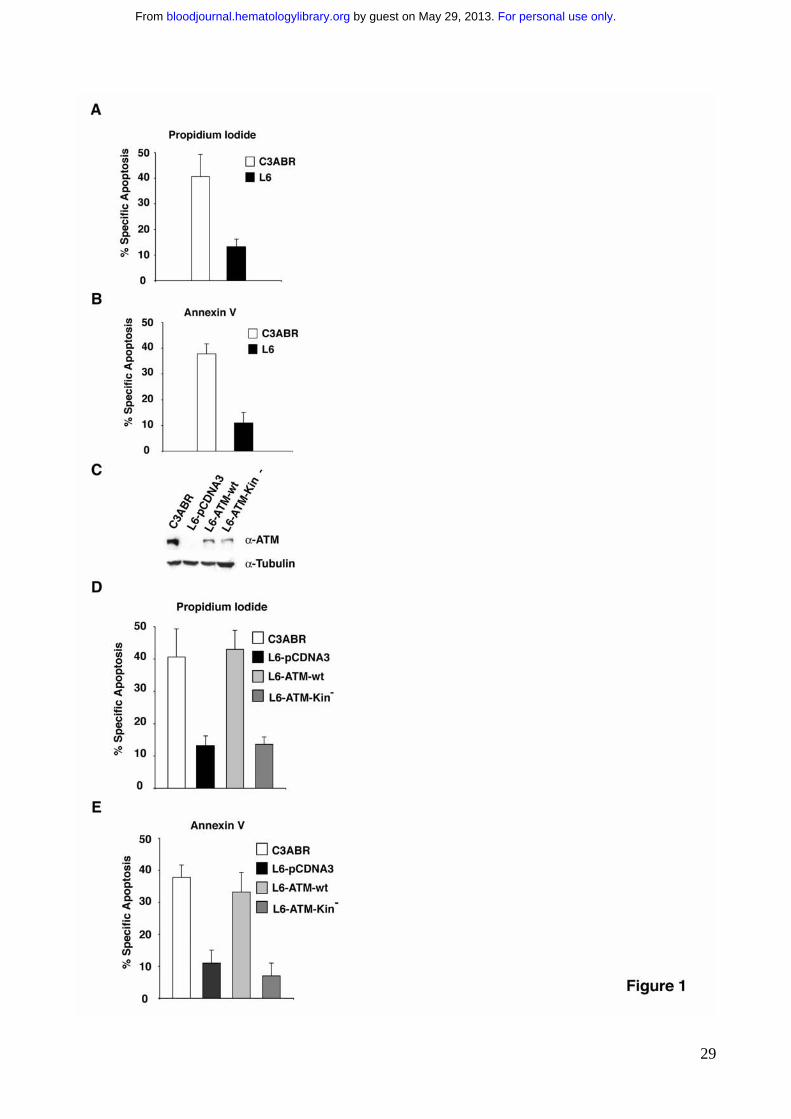
Figure 1. ATM deficient cells are resistant to Fas-induced apoptosis. ATM proficient
cells (C3ABR) and ATM deficient cells (L6) were treated with 250ng/ml of anti-Fas mAb.
Apoptosis was determined by the analysis of DNA fragmentation in propidium iodide
stained cells (P.I) (A) or by the analysis of Annexin V binding (B), 24 hrs after anti-Fas
treatment. (C) ATM deficient cells (L6) were stably transfected with ATM-wt, ATM-Kin- or
with empty vector as control using 20 μg of the indicated constructs. For immunoblotting,
80-100 μg of protein extract were separated by SDS-PAGE, and transferred on
nitrocellulose. ATM protein was revealed with anti-ATM (MAT3) antibodies. (D, E) Cells
were treated to undergo apotosis with 250ng/ml of anti-Fas mAb. Apoptosis was
determined by the analysis of DNA fragmentation upon propidium iodide nuclear staining
(P.I) (D) or by the analysis of Annexin V exposure (E) , at 24 hrs after anti-Fas treatment.
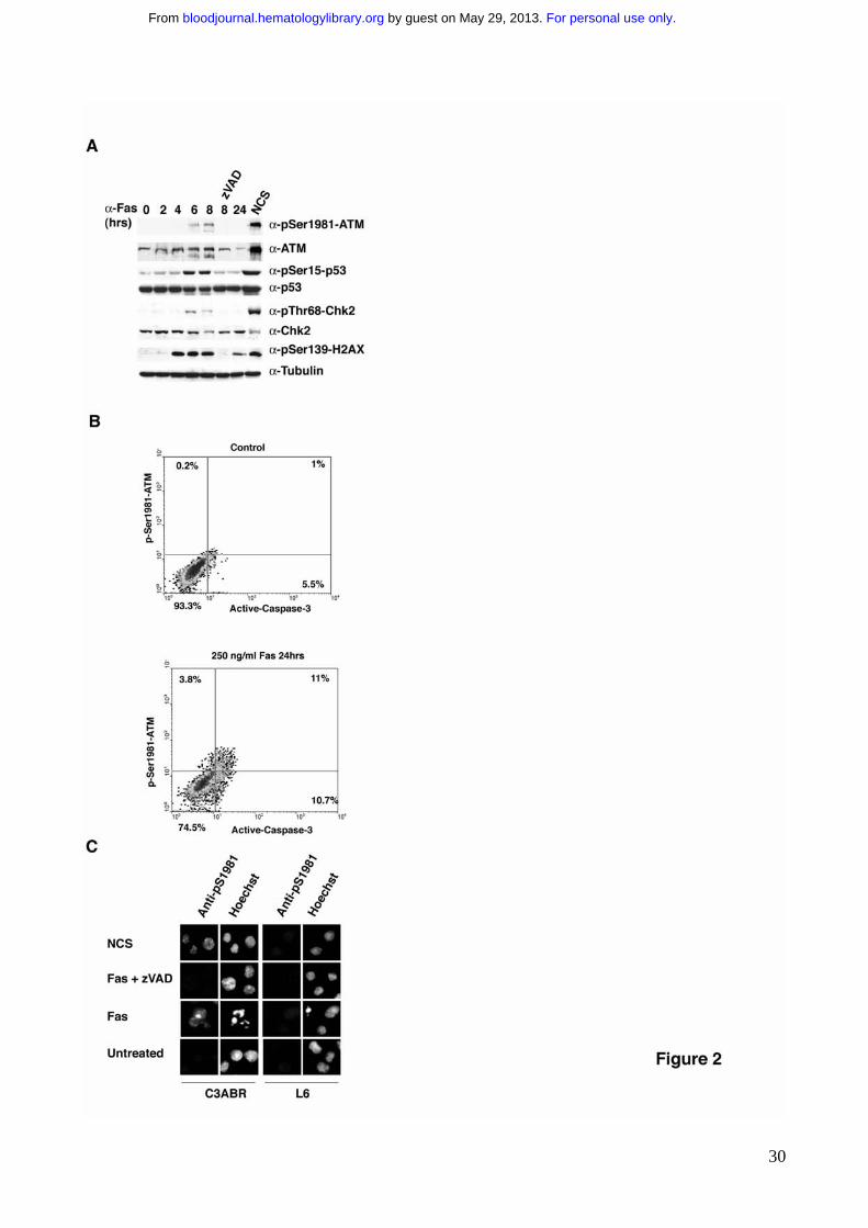
Figure 2. ATM kinase activation following Fas-induced apoptosis is a late passive
event A) C3ABR cells were induced to apoptosis with 250 ng/ml anti-Fas IgM monoclonal
antibody. Untreated and NCS-treated cells that triggers DSB and classically induces ATM
activation51, were used as controls. For immunoblotting, 80-100 μg of protein extract were
separated by SDS-PAGE, and transferred on nitrocellulose. The proteins of interest and
For personal use only. by guest on May 29, 2013. bloodjournal.hematologylibrary.orgFrom
26
their phosphorylation were revealed by immunoblotting with specific antibodies. B) C3ABR
cells were treated to undergo apoptosis with 250 ng/ml anti Fas IgM monoclonal antibody
(CH11; UBI). Untreated and treated cells were analyzed with by flow-cytometry for active
caspase-3 and phospho-Ser1981-ATM. C) C3ABR cells were treated to undergo
apoptosis as in B. Untreated and NCS-treated cells were used as controls. Cells were
fixed, permebilized and immunofluorescences were carried out as previously described 32.
Nuclear condensation and fragmentation has been evaluated by Hoechst staining.
Figure 3. ATM kinase activity promotes Caspase-8 activation. A) Fas receptor levels
were detected by flow-cytometry analysis. Cells were incubated with anti-Fas antibodies
followed by PE-conjugated secondary antibodies (dark lines). For each cell line an
incubation with PE-conjugated alone served as negative controls (light lines). B) Caspase-
8 expression was revealed by immunoblotting on extracts obtained by the indicated cell
lines. 80-100 μg of protein extract were separated by SDS-PAGE, transferred on
nitrocellulose and Caspase-8 expression revealed using specific antibodies. C) Protein
extracts from the indicated cell lines stimulated to undergo apoptosis with anti-Fas
antibodies, have been separated by SDS-PAGE and Caspase-8 revealed by
immunoblotting with specific antibodies. The arrows point to the entire protein, p55, as well
as to the processing products p43 and p18. D) Caspase-8 activity from the same extracts
was measured by the hydrolysis of the Caspase-8 substrate Ac-IETD-AMC.
Figure 4. Basal ATM kinase activity regulates FLIP protein levels. A) FLIP expression
was revealed by immunoblotting on extracts obtained from the indicated cell lines. 80-100
μg of protein extract were separated by SDS-PAGE, transferred on nitrocellulose and FLIP
expression revealed using specific antibodies. The arrows point to FLIP-L and FLIP-S
isoforms. B) ATM deficient L6 cells were stably transfected with shFLIP or with a
scrambled oligo as control. Protein extracts from the indicated cell lines were probed for
For personal use only. by guest on May 29, 2013. bloodjournal.hematologylibrary.orgFrom
27
FLIP expression by immunoblotting as described in A. The arrows point to endogenous
FLIP-L and FLIP-S. C) The indicated cell lines were stimulated to undergo apoptosis with
250ng/ml of anti-Fas mAb. Apoptosis was determined by the analysis of DNA
fragmentation upon propidium iodide nuclear staining (P.I) 24 hrs after anti-Fas treatment.
Figure 5. Modulation of ATM kinase activity results in the regulation of FLIP protein
levels which in turn determines Fas sensitivity. A) Different cell lines were treated with
NCS for 3 hrs to trigger ATM kinase activation. 80-100 μg of protein extract were
separated by SDS-PAGE, transferred on nitrocellulose and FLIP expression revealed
using specific antibodies. B) C3ABR cells were stimulated to undergo apoptosis with
250ng/ml of �nti-Fas mAb in the presence or in the absence of NCS pretreatment for 3
hrs. Apoptosis was determined by the analysis of DNA fragmentation in propidium iodide
stained cells (P.I) 24 hrs after anti-Fas treatment. C) The indicated cell lines were
incubated in the presence of the specific ATM kinase inhibitor KU-55933 (10 μM) for 1 or 8
hrs. 80-100 μg of protein extract were separated by SDS-PAGE, transferred on
nitrocellulose and FLIP expression revealed using specific antibodies. D) C3ABR cells
were preincubated for 1 or 8 hrs with the specific ATM kinase inhibitor KU-55933 (10 μM),
to allow endogenous ATM kinase inactivation and FLIP levels upregulation and then
stimulated to undergo apoptosis with 250ng/ml of anti-Fas mAb. Apoptosis was
determined by the analysis of DNA fragmentation in propidium iodide stained cells (P.I) 24
hrs after anti-Fas treatment.
Figure 6. ATM kinase activity downregulates FLIP protein stability. A) RT-PCR
analysis of FLIP-L and FLIP-S RNA expression levels was performed in the indicated cell
lines. Amplified actin was used as an internal control. B) C3ABR-FLAG-FLIP-L stably
transfected cells were incubated with NCS for different times to trigger ATM kinase
activation. 80-100 μg of protein extract were separated by SDS-PAGE, transferred on
nitrocellulose and endogenous (*) and transfected FLIP (**) expression revealed using
For personal use only. by guest on May 29, 2013. bloodjournal.hematologylibrary.orgFrom
28
specific anti-FLIP antibodies. C) Cells were pretreated with KU-55933 O/N, washed, and
then incubated with CHX for the indicated times.
Figure 7. ATM kinase activity downregulates FLIP protein levels and sensitizes
Hodgkin Lymphoma cell lines to Fas induced apoptosis. A) L428 HL cells were
transiently transfected with the indicated constructs along with GFP. 24 hrs after
transfection GFP positive cells were isolated by FACS sorting, and incubated for additional
12 hrs. For immunoblotting, 80-100 μg of protein extract were separated by SDS-PAGE,
and transferred on nitrocellulose. The proteins of interest and their phosphosphorylation
were revealed by immunoblotting with specific antibodies. B) L428 HL cells were
transiently transfected with the indicated constructs along with GFP. 24 hrs after
transfection cells were stimulated to undergo apoptosis with 250ng/ml of �nti-Fas mAb.
Apoptosis was determined by the analysis of Annexin V binding 24 hrs after �nti-Fas
treatment, upon FACS sorting of GFP positive cells.
For personal use only. by guest on May 29, 2013. bloodjournal.hematologylibrary.orgFrom
29
For personal use only. by guest on May 29, 2013. bloodjournal.hematologylibrary.orgFrom
30
For personal use only. by guest on May 29, 2013. bloodjournal.hematologylibrary.orgFrom
31
For personal use only. by guest on May 29, 2013. bloodjournal.hematologylibrary.orgFrom
32
For personal use only. by guest on May 29, 2013. bloodjournal.hematologylibrary.orgFrom
33
For personal use only. by guest on May 29, 2013. bloodjournal.hematologylibrary.orgFrom
34
For personal use only. by guest on May 29, 2013. bloodjournal.hematologylibrary.orgFrom
35
For personal use only. by guest on May 29, 2013. bloodjournal.hematologylibrary.orgFrom
Related Documents