Introduction The interaction between FasL and the Fas (Apo-1/CD95) receptor plays an essential role in the control of T- and B-cell activity and maintenance of immunological tolerance. Repeated antibody stimulation of CD4 + T-cells results in high levels of Fas and FasL expression and, as a consequence, these cells die (Abbas, 1996; Cornall et al., 1995; Matiba et al., 1997; Nagata, 1997). Likewise, Fas is involved in the elimination of active or autoreactive B-lymphocytes (Cornall et al., 1995; Nagata, 1997). FasL binding stabilizes the trimeric form of the Fas receptor, thereby allowing recruitment of the Fas-associated death domain (DD)-containing protein (FADD). FADD then binds to and activates caspase-8, an initiator caspase, which in turn activates downstream effector caspases, including caspase-3 (Martins and Earnshaw, 1997; Nagata and Golstein, 1995; Suda et al., 1993). As a consequence, many cellular proteins are degraded, leading to cell death. Depending on the cell type and the stimulus, a cell may die either by apoptosis or necrosis. Apoptosis is characterized by chromatin condensation, internucleosomal degradation of the DNA, cell shrinkage and disassembly into membrane-enclosed vesicles as a consequence of caspase activation (Rathmell and Thompson, 1999). Apoptotic and necrotic cells are both recognized by phagocytes, but only apoptotic cells are eliminated without release of cytosolic components to the environment, thereby preventing an inflammatory response (Fadok et al., 2000; Sauter et al., 2000). Necrosis, on the other hand, is characterized by swelling of the cells and organelles, resulting ultimately in disruption of the cell membrane and cell lysis (Majno and Joris, 1995). Initially, FasL was only thought to trigger cell death by apoptosis. Recently, however, inhibition of Fas-induced apoptosis in L929 fibrosarcoma cells by a caspase inhibitor lead to necrosis mediated by oxygen radicals (Vercammen et al., 1998a; Vercammen et al., 1998b). Also, primary activated T cells can be efficiently killed by FasL, TNF-α and TRAIL in the absence of active caspases (Holler et al., 2000). These results suggested that Fas, like TNFR-1 (Laster et al., 1988), triggers apoptotic or necrotic death. Treatment with FasL, TNF-α or IL-1β leads to formation of ceramide, in addition to caspase-3 activation (Garcia-Ruiz et 4671 Engagement of the Fas receptor promotes apoptosis by activation of caspases. In addition, alterations in plasma membrane lipid orientation and intracellular ceramide levels are often observed. In A20 B-lymphoma cells, FasL- induced cell death and phosphatidylserine (PS) externalization were completely prevented by the generic caspase inhibitor z-VAD-fmk. By contrast, the caspase-3 inhibitor Ac-DEVD-cho only partially restored cell viability and had no effect on surface exposure of PS. Flow cytometric analysis after FasL treatment identified two populations of dead cells. In one, death was dependent on caspase-3 and paralleled by DNA fragmentation and cell shrinkage. In the second, death occurred in the absence of caspase-3 activity and apoptotic features but was also blocked by zVAD-fmk. By morphological criteria these were identified as apoptotic and necrotic cells, respectively. Using fluorescent substrates, caspase-3 activity was detected only in the apoptotic cell population, whereas caspase-8 activity was detected in both. Both forms of caspase-8-dependent cell death were also detected downstream of Fas in Jurkat T-cells, where Fas- dependent PS externalization and delayed ceramide production, which is similar to results shown here in A20 cells, have been reported. However, for Raji B-cells, lacking lipid scrambling and ceramide production in response to Fas activation, only apoptosis was detected. Short-chain C2- or C6-ceramides, but not the respective inactive dihydro compounds or treatment with bacterial sphingomyelinase, induced predominantly necrotic rather than apoptotic cell death in A20 B-, Raji B- and Jurkat T- cells. Thus, delayed elevation of ceramide is proposed to promote necrosis in those Fas-stimulated cells where caspase-8 activation was insufficient to trigger caspase-3- dependent apoptosis. Key words: Lymphoid cells, Fas, Apoptosis, Necrosis, Caspases, Ceramide Summary Caspase-dependent initiation of apoptosis and necrosis by the Fas receptor in lymphoid cells: onset of necrosis is associated with delayed ceramide increase Claudio A. Hetz 1 , Martin Hunn 2 , Patricio Rojas 1 , Vicente Torres 1 , Lisette Leyton 1 and Andrew F. G. Quest 1, * 1 Instituto de Ciencias Biomédicas, Facultad de Medicina, Universidad de Chile, Santiago, Chile 2 Institute of Biochemistry, University of Lausanne, Switzerland *Author for correspondence (e-mail: [email protected]) Accepted 4 September 2002 Journal of Cell Science 115, 4671-4683 © 2002 The Company of Biologists Ltd doi:10.1242/jcs.00153 Research Article

Welcome message from author
This document is posted to help you gain knowledge. Please leave a comment to let me know what you think about it! Share it to your friends and learn new things together.
Transcript
IntroductionThe interaction between FasL and the Fas (Apo-1/CD95)receptor plays an essential role in the control of T- and B-cellactivity and maintenance of immunological tolerance.Repeated antibody stimulation of CD4+ T-cells results in highlevels of Fas and FasL expression and, as a consequence, thesecells die (Abbas, 1996; Cornall et al., 1995; Matiba et al., 1997;Nagata, 1997). Likewise, Fas is involved in the elimination ofactive or autoreactive B-lymphocytes (Cornall et al., 1995;Nagata, 1997).
FasL binding stabilizes the trimeric form of the Fas receptor,thereby allowing recruitment of the Fas-associated deathdomain (DD)-containing protein (FADD). FADD then binds toand activates caspase-8, an initiator caspase, which in turnactivates downstream effector caspases, including caspase-3(Martins and Earnshaw, 1997; Nagata and Golstein, 1995;Suda et al., 1993). As a consequence, many cellular proteinsare degraded, leading to cell death.
Depending on the cell type and the stimulus, a cell may dieeither by apoptosis or necrosis. Apoptosis is characterized bychromatin condensation, internucleosomal degradation of the
DNA, cell shrinkage and disassembly into membrane-enclosedvesicles as a consequence of caspase activation (Rathmell andThompson, 1999). Apoptotic and necrotic cells are bothrecognized by phagocytes, but only apoptotic cells areeliminated without release of cytosolic components to theenvironment, thereby preventing an inflammatory response(Fadok et al., 2000; Sauter et al., 2000). Necrosis, on the otherhand, is characterized by swelling of the cells and organelles,resulting ultimately in disruption of the cell membrane and celllysis (Majno and Joris, 1995).
Initially, FasL was only thought to trigger cell death byapoptosis. Recently, however, inhibition of Fas-inducedapoptosis in L929 fibrosarcoma cells by a caspase inhibitorlead to necrosis mediated by oxygen radicals (Vercammen etal., 1998a; Vercammen et al., 1998b). Also, primary activatedT cells can be efficiently killed by FasL, TNF-α and TRAILin the absence of active caspases (Holler et al., 2000). Theseresults suggested that Fas, like TNFR-1 (Laster et al., 1988),triggers apoptotic or necrotic death.
Treatment with FasL, TNF-α or IL-1β leads to formation ofceramide, in addition to caspase-3 activation (Garcia-Ruiz et
4671
Engagement of the Fas receptor promotes apoptosis byactivation of caspases. In addition, alterations in plasmamembrane lipid orientation and intracellular ceramidelevels are often observed. In A20 B-lymphoma cells, FasL-induced cell death and phosphatidylserine (PS)externalization were completely prevented by the genericcaspase inhibitor z-VAD-fmk. By contrast, the caspase-3inhibitor Ac-DEVD-cho only partially restored cellviability and had no effect on surface exposure of PS. Flowcytometric analysis after FasL treatment identified twopopulations of dead cells. In one, death was dependent oncaspase-3 and paralleled by DNA fragmentation and cellshrinkage. In the second, death occurred in the absenceof caspase-3 activity and apoptotic features but was alsoblocked by zVAD-fmk. By morphological criteria thesewere identified as apoptotic and necrotic cells,respectively. Using fluorescent substrates, caspase-3activity was detected only in the apoptotic cell population,whereas caspase-8 activity was detected in both. Both
forms of caspase-8-dependent cell death were alsodetected downstream of Fas in Jurkat T-cells, where Fas-dependent PS externalization and delayed ceramideproduction, which is similar to results shown here in A20cells, have been reported. However, for Raji B-cells,lacking lipid scrambling and ceramide production inresponse to Fas activation, only apoptosis was detected.Short-chain C2- or C6-ceramides, but not the respectiveinactive dihydro compounds or treatment with bacterialsphingomyelinase, induced predominantly necrotic ratherthan apoptotic cell death in A20 B-, Raji B- and Jurkat T-cells. Thus, delayed elevation of ceramide is proposed topromote necrosis in those Fas-stimulated cells wherecaspase-8 activation was insufficient to trigger caspase-3-dependent apoptosis.
Key words: Lymphoid cells, Fas, Apoptosis, Necrosis, Caspases,Ceramide
Summary
Caspase-dependent initiation of apoptosis andnecrosis by the Fas receptor in lymphoid cells: onset ofnecrosis is associated with delayed ceramide increaseClaudio A. Hetz 1, Martin Hunn 2, Patricio Rojas 1, Vicente Torres 1, Lisette Leyton 1 and Andrew F. G. Quest 1,*1Instituto de Ciencias Biomédicas, Facultad de Medicina, Universidad de Chile, Santiago, Chile2Institute of Biochemistry, University of Lausanne, Switzerland*Author for correspondence (e-mail: [email protected])
Accepted 4 September 2002Journal of Cell Science 115, 4671-4683 © 2002 The Company of Biologists Ltddoi:10.1242/jcs.00153
Research Article
4672
al., 1997; Gudz et al., 1997). Ceramide reportedly modulatesthe activity of a large number of proteins (Heinrich et al., 1999;Venkataraman and Futerman, 2000) and, in doing so, maypromote apoptosis. In addition, ceramide directly modulatesmitochondria function, for instance by inhibiting themitochondrial respiratory complex III (Garcia-Ruiz et al.,1997; Gudz et al., 1997; Quillet-Mary et al., 1997).Sphingomyelin hydrolysis by either neutral (nSMase) or acidicsphingomyelinases (aSMase) is generally implicated inceramide production (Hannun et al., 1996; Kolesnick andKronke, 1998).
Treatment of lymphoid cells with FasL induces elevationof intracellular ceramide levels. However, data are conflictingwith regard to the kinetics of the ceramide response and theSMases involved downstream of Fas. Some authors havefound a rapid transient response within minutes to an hourthat depends on initiator caspases, like caspase-8, and hasbeen attributed to activation of an aSMase (Cifone et al.,1995; Genestier et al., 1998). Alternatively, others havereported that apoptosis of lymphoid cells is generallyaccompanied by caspase-8-dependent, delayed ceramideproduction (after several hours) owing to the hydrolysis ofplasma membrane sphingomyelin as a consequence ofphospholipid scrambling. Increases in ceramide levelsfollowed the kinetics of nuclear fragmentation and occurredafter cytochrome c release or caspase-3 activation. Inaddition, Raji B-cells, which do not produce ceramides uponFas activation, display apoptotic features in mitochondria andthe nucleus (Tepper et al., 1997; Tepper et al., 2000). Theseobservations argue strongly against a role for ceramides intriggering apoptosis. Also, data obtained using aSMaseknock-out mice and overexpression of a nSMase have failedto implicate these SMases in either Fas-induced ceramideproduction or apoptosis of B- and T-cells (Brenner et al.,1997; Cock et al., 1998; Tepper et al., 2001). Thus, althoughevidence abounds suggesting that ceramide can promoteapoptosis, the extent to which ceramide formation is essentialremains controversial and appears to depend largely on thecellular system used (Cifone et al., 1995; Hannun et al., 1996;Hofmann and Dixit, 1998).
Here we investigated some of these questions, with a focuson A20 B- and Jurkat T-lymphoma cells, where cell death isreadily induced with recombinant, soluble FasL. We observedthat FasL stimulated two distinct types of caspase-8-dependent cell death: apoptosis that occurred via activationof caspase-3 and necrosis which, like phosphatidylserine(PS)-externalization, was caspase-3 independent. On theother hand, FasL also significantly increased ceramide levelsafter 3 hours in A20, as previously described for Jurkat T-cells. Treatment with cell-permeable ceramides or bacterialSMase led to necrosis in both cell types. In Raji B-cells,lacking ceramide production owing to absence of lipidscrambling (Tepper et al., 2000), Fas activation triggered onlyapoptosis at all antibody concentrations tested, whereas cell-permeable ceramides and bacterial SMase promoted necrosis.In the presence of FasL, addition of cell-permeable ceramidesonly promoted necrosis in A20 and Jurkat cells when addedwithin the first 4 hours after FasL. Thus, Fas-induced lipidscrambling and delayed elevation of intracellular ceramidelevels are suggested to promote necrosis in cells where FasLfailed to trigger caspase-3-dependent apoptosis.
Materials and MethodsMaterialsThe detergents nonidet P-40 (NP-40), sodium deoxycholate and 3-[(3-Cholamidopropyl)-dimethylammonio]-1-propanesulfonate (CHAPS)and bacterial SMase (Staphylococcus aureus) were from Sigma(Buchs, Switzerland). The fluorogenic caspase substrates (Ac-DEVD-amc, Ac-YVAD-amc) and caspase inhibitors (Ac-DEVD-cho, Ac-YVAD-cho, zVAD-fmk) were from Alexis Biochemicals(Läufelfingen, Switzerland). The cell-permeable caspase-3 substrateFAM-DEVD-fmk was from Promega (Madison, WI) and the cell-permeable caspase-8 substrate FAM-LETD-fmk from Intergen (NewYork, NY). C6-ceramide, dihydro-C6-ceramide, C2-ceramide anddihydro-C2-ceramide were from Biomol Research Laboratories Inc.(Wangen, Switzerland). Brain ceramides and dioctanoylglycerol werefrom Avanti Polar Lipids Inc. (Alabaster, AL). n-Octyl-β-D-glycopyranoside ULTROL grade was from Calbiochem (Lucerne,Switzerland). Protease inhibitors, RNAse A and annexin-V bindingkit were from Boehringer Mannheim/Roche Biochemicals (Basel,Switzerland). Cell medium, fetal calf serum and antibiotics were fromGIBCO-BRL (Basel, Switzerland). Organic solvents of the highestquality available were from Fluka (Buchs, Switzerland). Solublerecombinant human FasL was from Apotech SA (Geneva,Switzerland). Anti-human Fas antibodies (clone IPO-4) were fromKamaya Biochemical Company (Seattle, WA).
Cell cultureA20, A20R and M12 cells (kindly provided by Jürg Tschopp, Instituteof Biochemistry, University of Lausanne, Switzerland) were culturedin DMEM supplemented with 10% fetal calf serum, antibiotics(10,000 U/ml Penicillin, 10 µg/ml streptomycin) and 50 µM ethanol-2-thiol at 37°C and 5% CO2. Ramos, Daudi and Raji human B-lymphoma lines and the EBV-transformed human B-lymphoblast cellsGES and LM, all provided by Maria-Rosa Bono (Faculty of BasicSciences, University of Chile), were cultured in RPMI supplementedwith 10% fetal calf serum and antibiotics at 37°C, 5% CO2.
In vitro caspase-3/caspase-1 assayA20 and Jurkat cells (0.5×10⁶ cells) were treated with FasL for 0-8hours at 37°C. To test the effect of C6-ceramide or caspase inhibitors,cells were preincubated for 30 minutes. Then caspase activity wasmeasured using a previously described protocol (Hetz et al., 2002)modified from Boldin et al. (Boldin et al., 1996).
In situ caspase-3/caspase-8 assayCaspase activity was detected using the caspase-3 substrate FAM-DEVD-fmk (Promega, Madison, WI) or the caspase-8 substrate FAM-LETD-fmk (Intergen, New York, NY), following instructions from themanufacturer, by flow cytometry (FACS; Becton Dickinson, MountainView, CA) and the Cell Quest program.
Viability assaysCell viability was analyzed by FACS as described before (Hetz et al.,2002). A20 cells were incubated with either C6-ceramide or dihydro-C6-ceramide (DH-C6-ceramide) at the concentrations indicated for upto 16 hours at 37°C. For inhibition experiments, cells werepreincubated for 30 minutes without or with the caspase inhibitorsAc-DEVD-cho (100 µM), Ac-YVAD-cho (100 µM) or zVAD-fmk (10µM). Then FasL was added, and cells were incubated for another 16hours at 37°C. Cells were harvested and stained with 10 µg/ml ofpropidium iodide (PI) for determination of cell viability. For DNAcontent analysis cells were permeabilized with methanol and stainedwith PI. Samples containing roughly 1×10⁴ cells were analyzed byFACS using the Cell Quest program. Alternatively, cells were sorted
Journal of Cell Science 115 (23)
4673Ceramide in FasL-induced necrosis
by FACS using PI fluorescence emission as the parameter for selectionand nuclear morphology was analyzed by confocal microscopy asdescribed below.
Cell viability was also quantified by alternative methods using thereagents 3-(4,5-dimethylthazol-2-yl)-5-3-carboxymethoxy-phenyl)-2-(4-sulfophenyl)-2H-tetrazolium (MTS) and phenazine methosulfate(PMS) according to the recommendations of the supplier (Promega,CellTiter96® Aqueous).
Diacylglycerol kinase assayCeramide concentrations were determined in 5×10⁶ cells as previouslydescribed (Preiss et al., 1987; Walsh and Bell, 1986) using brainceramide (Avanti Polar Lipids, Alabaster, AL) as a standard.Determinations were done in triplicate in each experiment. On average(n=6), basal ceramide and diacylglycerol levels in A20 or A20R cellswere 2.9±1.0 pmol/nmol lipid phosphate (or 132±55 pmol per 5×10⁶
cells) and 14±3.2 pmol/nmol lipid phosphate (or 630±184 pmol per5×106 cells), respectively. These values are referred to as 100%.
DNA fragmentation assayA20 cells (1×10⁶/ml) were incubated in complete medium with FasL,C6-ceramide, dihydro-C6-ceramide or combinations thereof for 4hours at 37°C. For the inhibition experiments, cells were preincubatedfor 30 minutes with caspase inhibitors. Subsequently, cells wereharvested by brief centrifugation and lysed by addition of 100 µlphenol/chloroform/isoamylalcohol (25:24:1) and centrifuged. Then,17.5 µl of the aqueous phase were mixed with 2 µl 10× buffer H (50mM Tris-HCl pH 7.5, 10 mM MgCl2, 100 mM NaCl, 1 mMdithioerythrol) and 0.5 µl RNase A solution (500 mg/ml), incubatedfor 30 minutes at 37°C and analyzed by electrophoresis on a 2%agarose gel containing 0.5 mg/ml ethidium bromide. DNA bands werevisualized by exposure to UV light.
Assessment of chromatin condensation and morphologicalchangesCells were treated as described previously (viability assays) andstained after 16 hours cells with PI (1 µg/ml) for 5 minutes. Afterwashing twice in PBS, cells were treated with glycerol-DABCO andviewed by an SLM-400 Carl Zeiss confocal microscopy uponexcitation at 543 nm using a 570 nm emission filter (UACI, ICBM,University of Chile). As a control, cells were permeabilized byaddition of 500 µl ice-cold ethanol and incubated for 10 minutes at–20°C before staining with PI. A total of 200 cells were analyzed.Alternatively, cells were washed in PBS and fixed with 3%glutaraldehyde, 100 mM Na-cacodylate for 1.5 hours at 4°C. Afterpost-fixation in 1% AsO4 and dehydration, cells were embedded inEPSON 812 resin. Sections were stained with uranyl acetate and leadcitrate and were observed in a Zeiss TEM 109 Electron Microscope(Electron Microscopy Center, ICBM, Department of Morphology,University of Chile).
Quantification of phosphatidylserine exposurePresence of phosphatidylserine in the outer leaflet of the plasmamembrane was detected following instructions of the manufacturer byflow cytometry using FITC-coupled annexin V (annexin V-FITC) and5×103 cells per experiment.
ResultsFasL-induced caspase-3 activation and apoptosis in A20cells.To study the signaling events in FasL-induced apoptosis, the
murine B-lymphoma cell line A20 was initially employed as amodel system, together with FasL-insensitive A20R cells ascontrols. Resistance to FasL-induced cytotoxicity in the lattercase is probably due to downregulation of Fas (Hahne et al.,1996). Incubation of A20 cells with recombinant solublehuman FasL for 16 hours induced cell death in a concentration-dependent fashion (not shown), whereby 90% dead cells wereobserved with 100 ng/ml (Fig. 1A). No decrease in cellviability was observed in A20R cells under similar conditions(data not shown).
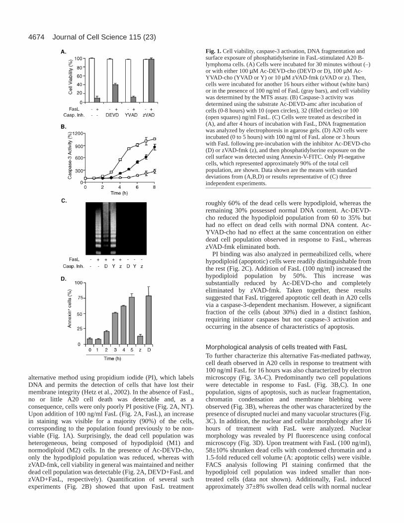
Addition of 100 µM of the caspase-3 inhibitor Ac-DEVD-cho following FasL treatment increased cell viability by 30%,whereas incubation with 100 µM of the caspase-1 inhibitor Ac-YVAD-cho did not protect A20 cells. Pre-incubation of cellswith the broad-range caspase inhibitor zVAD-fmk (10 µM)completely protected cells against FasL-induced cell death(Fig. 1A, see below Fig. 5). These observations are consistentwith the interpretation that activation of caspase-8, an initiatorcaspase upstream of caspase-3, represents an early eventtriggered by Fas, leading to cell death. Interestingly, inhibitionof caspase-3 and related caspases only partially restored cellviability upon treatment with FasL.
Caspase-3 activation, measured directly by monitoringcleavage of the fluorogenic substrate Ac-DEVD-amc, wasdetectable within 2 hours and rose until 8 hours after additionof FasL to over 10-fold, eight-fold or two-fold baseline valuesin the presence 100, 32 or 10 ng/ml FasL, respectively (Fig.1B). During the same period, no activation of caspase-1 wasdetectable using the fluorogenic substrate Ac-YVAD-amc (datanot shown). Western blot analysis, using a caspase-3-specificantibody revealed that pro-caspase-3 protein levels began todecline within 1 hour of treatment with FasL (100 ng/ml) andessentially disappeared within 2-3 hours, as did polyADP-ribose polymerase, a caspase-3 target (data not shown).
A hallmark of apoptosis is the generation of DNA fragmentsof defined length, which lead to a ladder-like pattern afterseparation by size. Also in A20 B-lymphoma cells, incubationwith FasL led to DNA laddering (Fig. 1C), and this effect wasconcentration dependent (data not shown). Pretreatment ofcells with the caspase-3 inhibitor Ac-DEVD-cho or the broad-spectrum caspase inhibitor zVAD-fmk reduced DNAdegradation or blocked it, respectively. By contrast, Ac-YVAD-cho did not protect A20 cells from Fas-induced DNAdegradation (Fig. 1C). PS exposure on the cell surface wasdetermined by flow fluorocytometric analysis of annexin-V-FITC binding. Appearance of PS on the cell surface, observedfor about 80% of the cells after treatment with 100 ng/ml FasLfor 5 hours, was inhibited by zVAD-fmk but not by Ac-DEVD-cho (Fig. 1D) or Ac-YVAD-cho (data not shown). Thus, byseveral criteria, caspase-3 was implicated in the execution ofapoptosis in FasL-stimulated A20 cells; however, FasL-induced cell death could only be partially blocked by thecaspase-3 inhibitor Ac-DEVD-cho, suggesting the existence ofan alternative caspase-3-independent pathway leading to A20cell death.
Ac-DEVD-cho treatment of A20 B-lymphoma cellsrevealed an alternative death signaling pathwayoriginating from FasCell viability after FasL treatment was investigated by an
4674
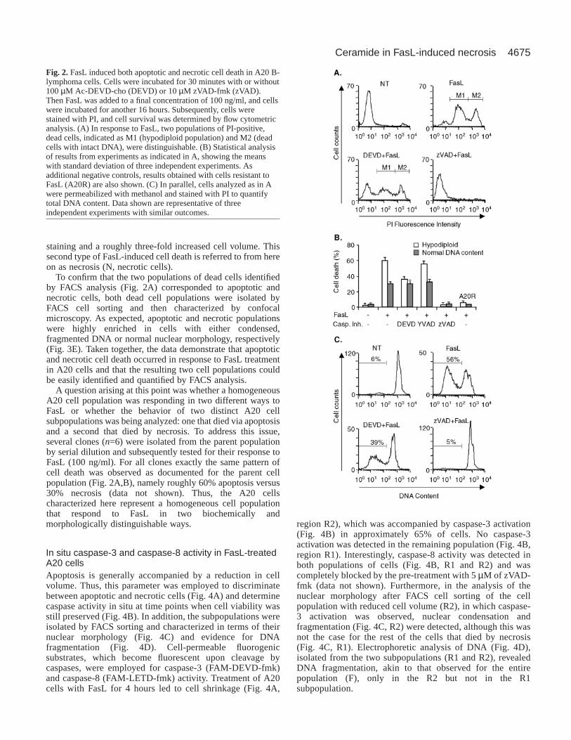
alternative method using propidium iodide (PI), which labelsDNA and permits the detection of cells that have lost theirmembrane integrity (Hetz et al., 2002). In the absence of FasL,no or little A20 cell death was detectable and, as aconsequence, cells were only poorly PI positive (Fig. 2A, NT).Upon addition of 100 ng/ml FasL (Fig. 2A, FasL), an increasein staining was visible for a majority (90%) of the cells,corresponding to the population found previously to be non-viable (Fig. 1A). Surprisingly, the dead cell population washeterogeneous, being composed of hypodiploid (M1) andnormodiploid (M2) cells. In the presence of Ac-DEVD-cho,only the hypodiploid population was reduced, whereas withzVAD-fmk, cell viability in general was maintained and neitherdead cell population was detectable (Fig. 2A, DEVD+FasL andzVAD+FasL, respectively). Quantification of several suchexperiments (Fig. 2B) showed that upon FasL treatment
roughly 60% of the dead cells were hypodiploid, whereas theremaining 30% possessed normal DNA content. Ac-DEVD-cho reduced the hypodiploid population from 60 to 35% buthad no effect on dead cells with normal DNA content. Ac-YVAD-cho had no effect at the same concentration on eitherdead cell population observed in response to FasL, whereaszVAD-fmk eliminated both.
PI binding was also analyzed in permeabilized cells, wherehypodiploid (apoptotic) cells were readily distinguishable fromthe rest (Fig. 2C). Addition of FasL (100 ng/ml) increased thehypodiploid population by 50%. This increase wassubstantially reduced by Ac-DEVD-cho and completelyeliminated by zVAD-fmk. Taken together, these resultssuggested that FasL triggered apoptotic cell death in A20 cellsvia a caspase-3-dependent mechanism. However, a significantfraction of the cells (about 30%) died in a distinct fashion,requiring initiator caspases but not caspase-3 activation andoccurring in the absence of characteristics of apoptosis.
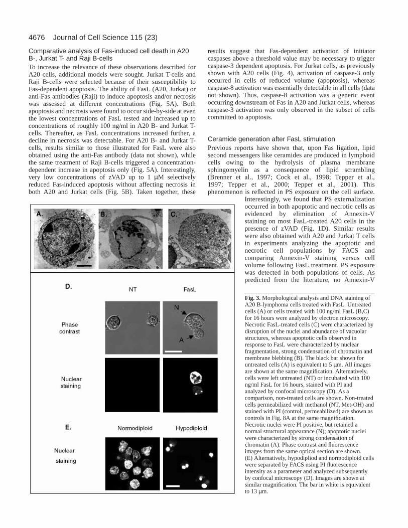
Morphological analysis of cells treated with FasLTo further characterize this alternative Fas-mediated pathway,cell death observed in A20 cells in response to treatment with100 ng/ml FasL for 16 hours was also characterized by electronmicroscopy (Fig. 3A-C). Predominantly two cell populationswere detectable in response to FasL (Fig. 3B,C). In onepopulation, signs of apoptosis, such as nuclear fragmentation,chromatin condensation and membrane blebbing wereobserved (Fig. 3B), whereas the other was characterized by thepresence of disrupted nuclei and many vacuolar structures (Fig.3C). In addition, the nuclear and cellular morphology after 16hours of treatment with FasL were analyzed. Nuclearmorphology was revealed by PI fluorescence using confocalmicroscopy (Fig. 3D). Upon treatment with FasL (100 ng/ml),58±10% shrunken dead cells with condensed chromatin and a1.5-fold reduced cell volume (A: apoptotic cells) were visible.FACS analysis following PI staining confirmed that thehypodiploid cell population was indeed smaller than non-treated cells (data not shown). Additionally, FasL inducedapproximately 37±8% swollen dead cells with normal nuclear
Journal of Cell Science 115 (23)
Fig. 1. Cell viability, caspase-3 activation, DNA fragmentation andsurface exposure of phosphatidylserine in FasL-stimulated A20 B-lymphoma cells. (A) Cells were incubated for 30 minutes without (–)or with either 100 µM Ac-DEVD-cho (DEVD or D), 100 µM Ac-YVAD-cho (YVAD or Y) or 10 µM zVAD-fmk (zVAD or z). Then,cells were incubated for another 16 hours either without (white bars)or in the presence of 100 ng/ml of FasL (gray bars), and cell viabilitywas determined by the MTS assay. (B) Caspase-3 activity wasdetermined using the substrate Ac-DEVD-amc after incubation ofcells (0-8 hours) with 10 (open circles), 32 (filled circles) or 100(open squares) ng/ml FasL. (C) Cells were treated as described in(A), and after 4 hours of incubation with FasL, DNA fragmentationwas analyzed by electrophoresis in agarose gels. (D) A20 cells wereincubated (0 to 5 hours) with 100 ng/ml of FasL alone or 3 hourswith FasL following pre-incubation with the inhibitor Ac-DEVD-cho(D) or zVAD-fmk (z), and then phosphatidylserine exposure on thecell surface was detected using Annexin-V-FITC. Only PI-negativecells, which represented approximately 90% of the total cellpopulation, are shown. Data shown are the means with standarddeviations from (A,B,D) or results representative of (C) threeindependent experiments.
4675Ceramide in FasL-induced necrosis
staining and a roughly three-fold increased cell volume. Thissecond type of FasL-induced cell death is referred to from hereon as necrosis (N, necrotic cells).
To confirm that the two populations of dead cells identifiedby FACS analysis (Fig. 2A) corresponded to apoptotic andnecrotic cells, both dead cell populations were isolated byFACS cell sorting and then characterized by confocalmicroscopy. As expected, apoptotic and necrotic populationswere highly enriched in cells with either condensed,fragmented DNA or normal nuclear morphology, respectively(Fig. 3E). Taken together, the data demonstrate that apoptoticand necrotic cell death occurred in response to FasL treatmentin A20 cells and that the resulting two cell populations couldbe easily identified and quantified by FACS analysis.
A question arising at this point was whether a homogeneousA20 cell population was responding in two different ways toFasL or whether the behavior of two distinct A20 cellsubpopulations was being analyzed: one that died via apoptosisand a second that died by necrosis. To address this issue,several clones (n=6) were isolated from the parent populationby serial dilution and subsequently tested for their response toFasL (100 ng/ml). For all clones exactly the same pattern ofcell death was observed as documented for the parent cellpopulation (Fig. 2A,B), namely roughly 60% apoptosis versus30% necrosis (data not shown). Thus, the A20 cellscharacterized here represent a homogeneous cell populationthat respond to FasL in two biochemically andmorphologically distinguishable ways.
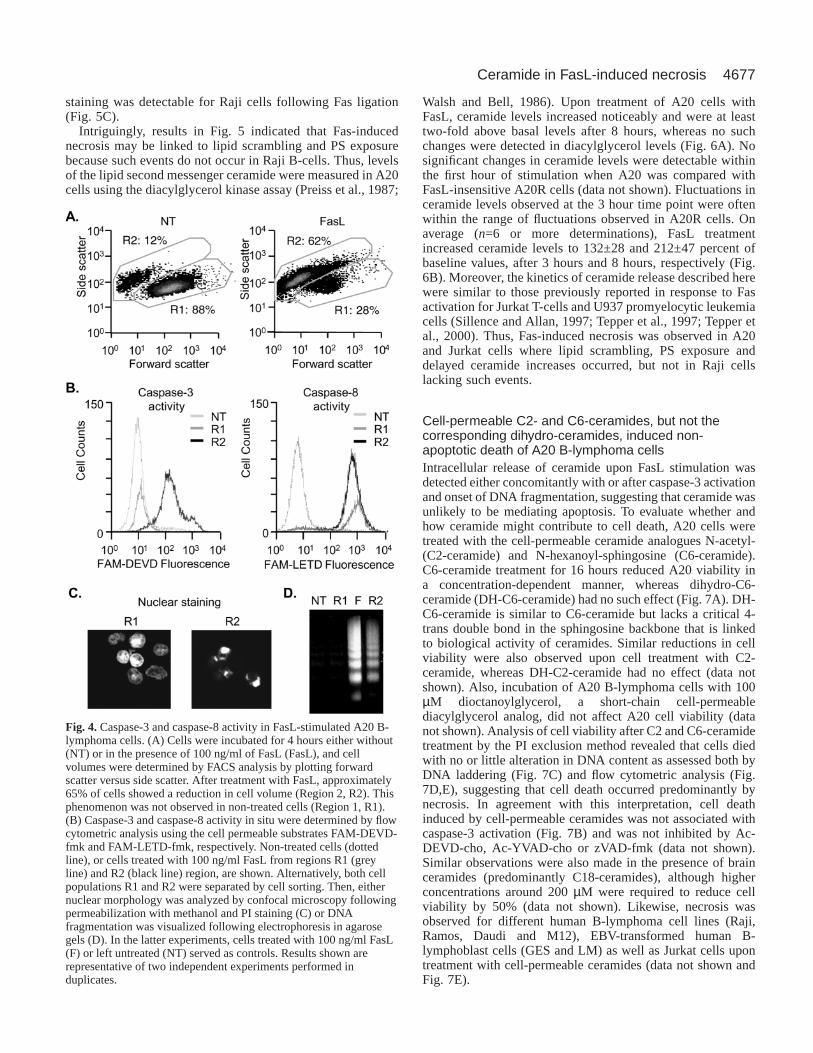
In situ caspase-3 and caspase-8 activity in FasL-treatedA20 cellsApoptosis is generally accompanied by a reduction in cellvolume. Thus, this parameter was employed to discriminatebetween apoptotic and necrotic cells (Fig. 4A) and determinecaspase activity in situ at time points when cell viability wasstill preserved (Fig. 4B). In addition, the subpopulations wereisolated by FACS sorting and characterized in terms of theirnuclear morphology (Fig. 4C) and evidence for DNAfragmentation (Fig. 4D). Cell-permeable fluorogenicsubstrates, which become fluorescent upon cleavage bycaspases, were employed for caspase-3 (FAM-DEVD-fmk)and caspase-8 (FAM-LETD-fmk) activity. Treatment of A20cells with FasL for 4 hours led to cell shrinkage (Fig. 4A,
region R2), which was accompanied by caspase-3 activation(Fig. 4B) in approximately 65% of cells. No caspase-3activation was detected in the remaining population (Fig. 4B,region R1). Interestingly, caspase-8 activity was detected inboth populations of cells (Fig. 4B, R1 and R2) and wascompletely blocked by the pre-treatment with 5 µM of zVAD-fmk (data not shown). Furthermore, in the analysis of thenuclear morphology after FACS cell sorting of the cellpopulation with reduced cell volume (R2), in which caspase-3 activation was observed, nuclear condensation andfragmentation (Fig. 4C, R2) were detected, although this wasnot the case for the rest of the cells that died by necrosis(Fig. 4C, R1). Electrophoretic analysis of DNA (Fig. 4D),isolated from the two subpopulations (R1 and R2), revealedDNA fragmentation, akin to that observed for the entirepopulation (F), only in the R2 but not in the R1subpopulation.
Fig. 2. FasL induced both apoptotic and necrotic cell death in A20 B-lymphoma cells. Cells were incubated for 30 minutes with or without100 µM Ac-DEVD-cho (DEVD) or 10 µM zVAD-fmk (zVAD).Then FasL was added to a final concentration of 100 ng/ml, and cellswere incubated for another 16 hours. Subsequently, cells werestained with PI, and cell survival was determined by flow cytometricanalysis. (A) In response to FasL, two populations of PI-positive,dead cells, indicated as M1 (hypodiploid population) and M2 (deadcells with intact DNA), were distinguishable. (B) Statistical analysisof results from experiments as indicated in A, showing the meanswith standard deviation of three independent experiments. Asadditional negative controls, results obtained with cells resistant toFasL (A20R) are also shown. (C) In parallel, cells analyzed as in Awere permeabilized with methanol and stained with PI to quantifytotal DNA content. Data shown are representative of threeindependent experiments with similar outcomes.
4676
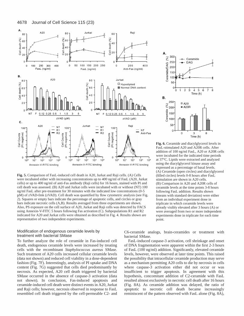
Comparative analysis of Fas-induced cell death in A20B-, Jurkat T- and Raji B-cellsTo increase the relevance of these observations described forA20 cells, additional models were sought. Jurkat T-cells andRaji B-cells were selected because of their susceptibility toFas-dependent apoptosis. The ability of FasL (A20, Jurkat) oranti-Fas antibodies (Raji) to induce apoptosis and/or necrosiswas assessed at different concentrations (Fig. 5A). Bothapoptosis and necrosis were found to occur side-by-side at eventhe lowest concentrations of FasL tested and increased up toconcentrations of roughly 100 ng/ml in A20 B- and Jurkat T-cells. Thereafter, as FasL concentrations increased further, adecline in necrosis was detectable. For A20 B- and Jurkat T-cells, results similar to those illustrated for FasL were alsoobtained using the anti-Fas antibody (data not shown), whilethe same treatment of Raji B-cells triggered a concentration-dependent increase in apoptosis only (Fig. 5A). Interestingly,very low concentrations of zVAD up to 1 µM selectivelyreduced Fas-induced apoptosis without affecting necrosis inboth A20 and Jurkat cells (Fig. 5B). Taken together, these
results suggest that Fas-dependent activation of initiatorcaspases above a threshold value may be necessary to triggercaspase-3 dependent apoptosis. For Jurkat cells, as previouslyshown with A20 cells (Fig. 4), activation of caspase-3 onlyoccurred in cells of reduced volume (apoptosis), whereascaspase-8 activation was essentially detectable in all cells (datanot shown). Thus, caspase-8 activation was a generic eventoccurring downstream of Fas in A20 and Jurkat cells, whereascaspase-3 activation was only observed in the subset of cellscommitted to apoptosis.
Ceramide generation after FasL stimulationPrevious reports have shown that, upon Fas ligation, lipidsecond messengers like ceramides are produced in lymphoidcells owing to the hydrolysis of plasma membranesphingomyelin as a consequence of lipid scrambling(Brenner et al., 1997; Cock et al., 1998; Tepper et al.,1997; Tepper et al., 2000; Tepper et al., 2001). Thisphenomenon is reflected in PS exposure on the cell surface.
Interestingly, we found that PS externalizationoccurred in both apoptotic and necrotic cells asevidenced by elimination of Annexin-Vstaining on most FasL-treated A20 cells in thepresence of zVAD (Fig. 1D). Similar resultswere also obtained with A20 and Jurkat T cellsin experiments analyzing the apoptotic andnecrotic cell populations by FACS andcomparing Annexin-V staining versus cellvolume following FasL treatment. PS exposurewas detected in both populations of cells. Aspredicted from the literature, no Annexin-V
Journal of Cell Science 115 (23)
Fig. 3. Morphological analysis and DNA staining ofA20 B-lymphoma cells treated with FasL. Untreatedcells (A) or cells treated with 100 ng/ml FasL (B,C)for 16 hours were analyzed by electron microscopy.Necrotic FasL-treated cells (C) were characterized bydisruption of the nuclei and abundance of vacuolarstructures, whereas apoptotic cells observed inresponse to FasL were characterized by nuclearfragmentation, strong condensation of chromatin andmembrane blebbing (B). The black bar shown foruntreated cells (A) is equivalent to 5 µm. All imagesare shown at the same magnification. Alternatively,cells were left untreated (NT) or incubated with 100ng/ml FasL for 16 hours, stained with PI andanalyzed by confocal microscopy (D). As acomparison, non-treated cells are shown. Non-treatedcells permeabilized with methanol (NT, Met-OH) andstained with PI (control, permeabilized) are shown ascontrols in Fig. 8A at the same magnification.Necrotic nuclei were PI positive, but retained anormal structural appearance (N); apoptotic nucleiwere characterized by strong condensation ofchromatin (A). Phase contrast and fluorescenceimages from the same optical section are shown.(E) Alternatively, hypodipliod and normodiploid cellswere separated by FACS using PI fluorescenceintensity as a parameter and analyzed subsequentlyby confocal microscopy (D). Images are shown atsimilar magnification. The bar in white is equivalentto 13 µm.
4677Ceramide in FasL-induced necrosis
staining was detectable for Raji cells following Fas ligation(Fig. 5C).
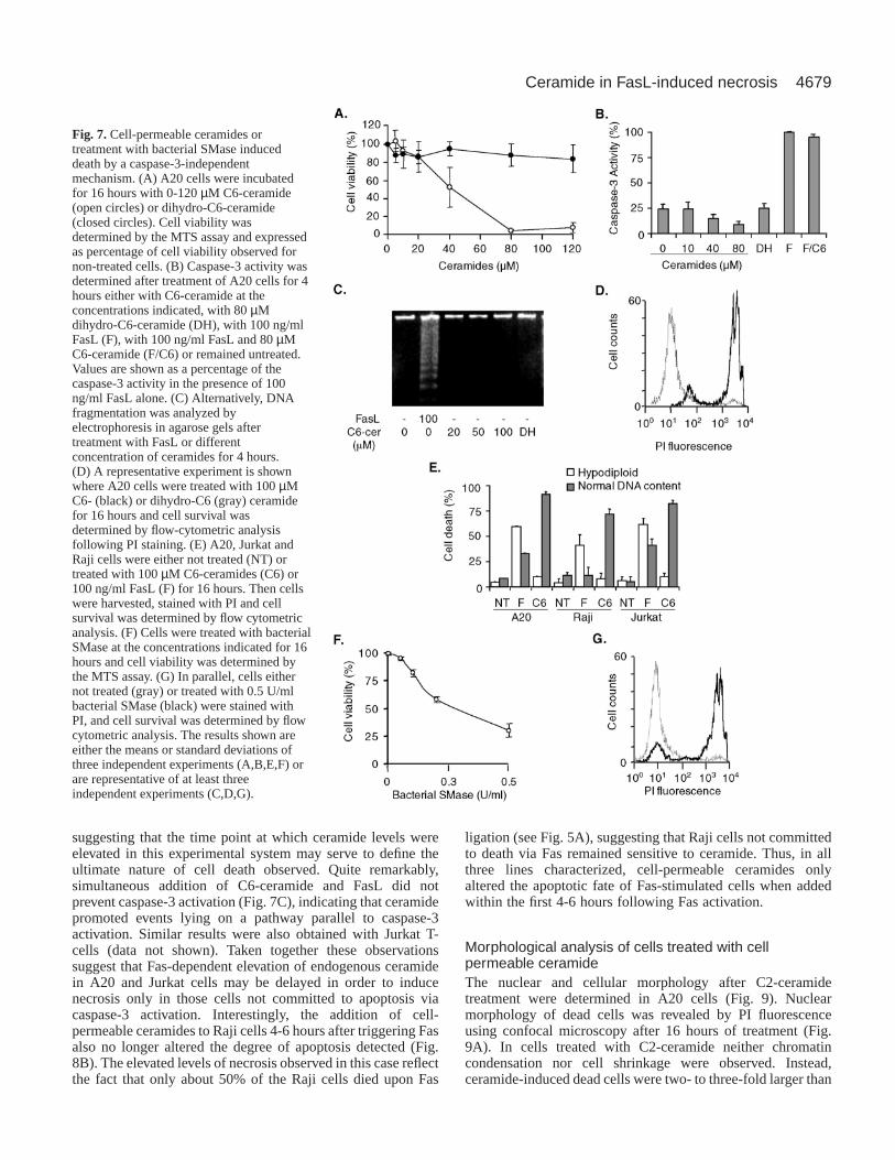
Intriguingly, results in Fig. 5 indicated that Fas-inducednecrosis may be linked to lipid scrambling and PS exposurebecause such events do not occur in Raji B-cells. Thus, levelsof the lipid second messenger ceramide were measured in A20cells using the diacylglycerol kinase assay (Preiss et al., 1987;
Walsh and Bell, 1986). Upon treatment of A20 cells withFasL, ceramide levels increased noticeably and were at leasttwo-fold above basal levels after 8 hours, whereas no suchchanges were detected in diacylglycerol levels (Fig. 6A). Nosignificant changes in ceramide levels were detectable withinthe first hour of stimulation when A20 was compared withFasL-insensitive A20R cells (data not shown). Fluctuations inceramide levels observed at the 3 hour time point were oftenwithin the range of fluctuations observed in A20R cells. Onaverage (n=6 or more determinations), FasL treatmentincreased ceramide levels to 132±28 and 212±47 percent ofbaseline values, after 3 hours and 8 hours, respectively (Fig.6B). Moreover, the kinetics of ceramide release described herewere similar to those previously reported in response to Fasactivation for Jurkat T-cells and U937 promyelocytic leukemiacells (Sillence and Allan, 1997; Tepper et al., 1997; Tepper etal., 2000). Thus, Fas-induced necrosis was observed in A20and Jurkat cells where lipid scrambling, PS exposure anddelayed ceramide increases occurred, but not in Raji cellslacking such events.
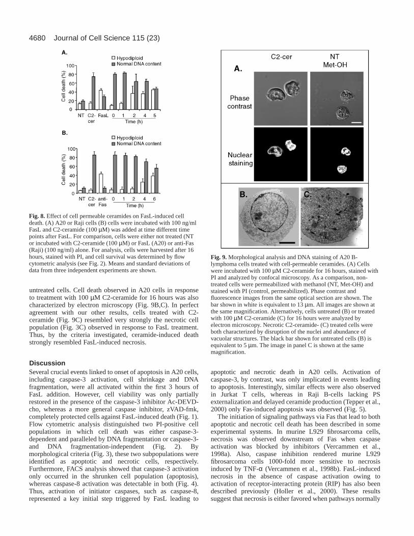
Cell-permeable C2- and C6-ceramides, but not thecorresponding dihydro-ceramides, induced non-apoptotic death of A20 B-lymphoma cellsIntracellular release of ceramide upon FasL stimulation wasdetected either concomitantly with or after caspase-3 activationand onset of DNA fragmentation, suggesting that ceramide wasunlikely to be mediating apoptosis. To evaluate whether andhow ceramide might contribute to cell death, A20 cells weretreated with the cell-permeable ceramide analogues N-acetyl-(C2-ceramide) and N-hexanoyl-sphingosine (C6-ceramide).C6-ceramide treatment for 16 hours reduced A20 viability ina concentration-dependent manner, whereas dihydro-C6-ceramide (DH-C6-ceramide) had no such effect (Fig. 7A). DH-C6-ceramide is similar to C6-ceramide but lacks a critical 4-trans double bond in the sphingosine backbone that is linkedto biological activity of ceramides. Similar reductions in cellviability were also observed upon cell treatment with C2-ceramide, whereas DH-C2-ceramide had no effect (data notshown). Also, incubation of A20 B-lymphoma cells with 100µM dioctanoylglycerol, a short-chain cell-permeablediacylglycerol analog, did not affect A20 cell viability (datanot shown). Analysis of cell viability after C2 and C6-ceramidetreatment by the PI exclusion method revealed that cells diedwith no or little alteration in DNA content as assessed both byDNA laddering (Fig. 7C) and flow cytometric analysis (Fig.7D,E), suggesting that cell death occurred predominantly bynecrosis. In agreement with this interpretation, cell deathinduced by cell-permeable ceramides was not associated withcaspase-3 activation (Fig. 7B) and was not inhibited by Ac-DEVD-cho, Ac-YVAD-cho or zVAD-fmk (data not shown).Similar observations were also made in the presence of brainceramides (predominantly C18-ceramides), although higherconcentrations around 200 µM were required to reduce cellviability by 50% (data not shown). Likewise, necrosis wasobserved for different human B-lymphoma cell lines (Raji,Ramos, Daudi and M12), EBV-transformed human B-lymphoblast cells (GES and LM) as well as Jurkat cells upontreatment with cell-permeable ceramides (data not shown andFig. 7E).
Fig. 4. Caspase-3 and caspase-8 activity in FasL-stimulated A20 B-lymphoma cells. (A) Cells were incubated for 4 hours either without(NT) or in the presence of 100 ng/ml of FasL (FasL), and cellvolumes were determined by FACS analysis by plotting forwardscatter versus side scatter. After treatment with FasL, approximately65% of cells showed a reduction in cell volume (Region 2, R2). Thisphenomenon was not observed in non-treated cells (Region 1, R1).(B) Caspase-3 and caspase-8 activity in situ were determined by flowcytometric analysis using the cell permeable substrates FAM-DEVD-fmk and FAM-LETD-fmk, respectively. Non-treated cells (dottedline), or cells treated with 100 ng/ml FasL from regions R1 (greyline) and R2 (black line) region, are shown. Alternatively, both cellpopulations R1 and R2 were separated by cell sorting. Then, eithernuclear morphology was analyzed by confocal microscopy followingpermeabilization with methanol and PI staining (C) or DNAfragmentation was visualized following electrophoresis in agarosegels (D). In the latter experiments, cells treated with 100 ng/ml FasL(F) or left untreated (NT) served as controls. Results shown arerepresentative of two independent experiments performed induplicates.
4678
Modification of endogenous ceramide levels bytreatment with bacterial SMaseTo further analyze the role of ceramide in Fas-induced celldeath, endogenous ceramide levels were increased by treatingcells with the recombinantStaphylococcus aureusSMase.Such treatment of A20 cells increased cellular ceramide levels(data not shown) and reduced cell viability in a dose-dependentfashion (Fig. 7F). Interestingly, analysis of PI uptake and DNAcontent (Fig. 7G) suggested that cells died predominantly bynecrosis. As expected, A20 cell death triggered by bacterialSMase occurred in the absence of caspase-3 activation (datanot shown). In conclusion, Fas-induced apoptosis andceramide-induced cell death were distinct events in A20, Jurkatand Raji cells; however, necrosis observed in response to FasLresembled cell death triggered by the cell-permeable C2- and
C6-ceramide analogs, brain-ceramides or treatment withbacterial SMase.
FasL-induced caspase-3 activation, cell shrinkage and onsetof DNA fragmentation were apparent within the first 2-3 hoursof FasL (100 ng/ml) addition. Significantly, elevated ceramidelevels, however, were observed at later time points. This raisedthe possibility that intracellular ceramide production may serveas a mechanism permitting A20 cells to die by necrosis in cellswhere caspase-3 activation either did not occur or wasinsufficient to trigger apoptosis. In agreement with thishypothesis, concomitant addition of C2-ceramide with FasLresulted almost exclusively in necrotic cell death after 16 hours(Fig. 8A). As ceramide addition was delayed, the ratio ofapoptotic to necrotic cell death became increasinglyreminiscent of the pattern observed with FasL alone (Fig. 8A),
Journal of Cell Science 115 (23)
Fig. 5. Comparison of FasL-induced cell death in A20, Jurkat and Raji cells. (A) Cellswere incubated either with increasing concentrations up to 400 ng/ml of FasL (A20, Jurkatcells) or up to 400 ng/ml of anti-Fas antibody (Raji cells) for 16 hours, stained with PI andcell death was assessed. (B) A20 and Jurkat cells were incubated with or without (NT) 100ng/ml FasL after pre-treatment for 30 minutes with the indicated low concentrations (0-5µM) of zVAD-fmk (zVAD). Cell death was quantified by flow cytometric analysis (see Fig.2). Squares or empty bars indicate the percentage of apoptotic cells, and circles or graybars indicate necrotic cells (A,B). Results averaged from three experiments are shown.Also, PS exposure on the cell surface of A20, Jurkat and Raji cells was detected by FACSusing Annexin-V-FITC 5 hours following Fas activation (C). Subpopulations R1 and R2indicated for A20 and Jurkat cells were obtained as described in Fig. 4. Results shown arerepresentative of two independent experiments.
Fig. 6. Ceramide and diacylglycerol levels inFasL-stimulated A20 and A20R cells. Afteraddition of 100 ng/ml FasL, A20 or A20R cellswere incubated for the indicated time periodsat 37°C. Lipids were extracted and analyzedusing the diacylglycerol kinase assay andexpressed as a percentage of basal levels.(A) Ceramide (open circles) and diacylglycerol(filled circles) levels 0-8 hours after FasLstimulation are shown in A20 cells.(B) Comparison in A20 and A20R cells ofceramide levels at the time points 3-8 hoursfollowing FasL addition. Results shown(means with standard deviation) were eitherfrom an individual experiment done intriplicate in which ceramide levels werealready visibly elevated after 3 hours (A) orwere averaged from two or more independentexperiments done in triplicate for each timepoint.
4679Ceramide in FasL-induced necrosis
suggesting that the time point at which ceramide levels wereelevated in this experimental system may serve to define theultimate nature of cell death observed. Quite remarkably,simultaneous addition of C6-ceramide and FasL did notprevent caspase-3 activation (Fig. 7C), indicating that ceramidepromoted events lying on a pathway parallel to caspase-3activation. Similar results were also obtained with Jurkat T-cells (data not shown). Taken together these observationssuggest that Fas-dependent elevation of endogenous ceramidein A20 and Jurkat cells may be delayed in order to inducenecrosis only in those cells not committed to apoptosis viacaspase-3 activation. Interestingly, the addition of cell-permeable ceramides to Raji cells 4-6 hours after triggering Fasalso no longer altered the degree of apoptosis detected (Fig.8B). The elevated levels of necrosis observed in this case reflectthe fact that only about 50% of the Raji cells died upon Fas
ligation (see Fig. 5A), suggesting that Raji cells not committedto death via Fas remained sensitive to ceramide. Thus, in allthree lines characterized, cell-permeable ceramides onlyaltered the apoptotic fate of Fas-stimulated cells when addedwithin the first 4-6 hours following Fas activation.
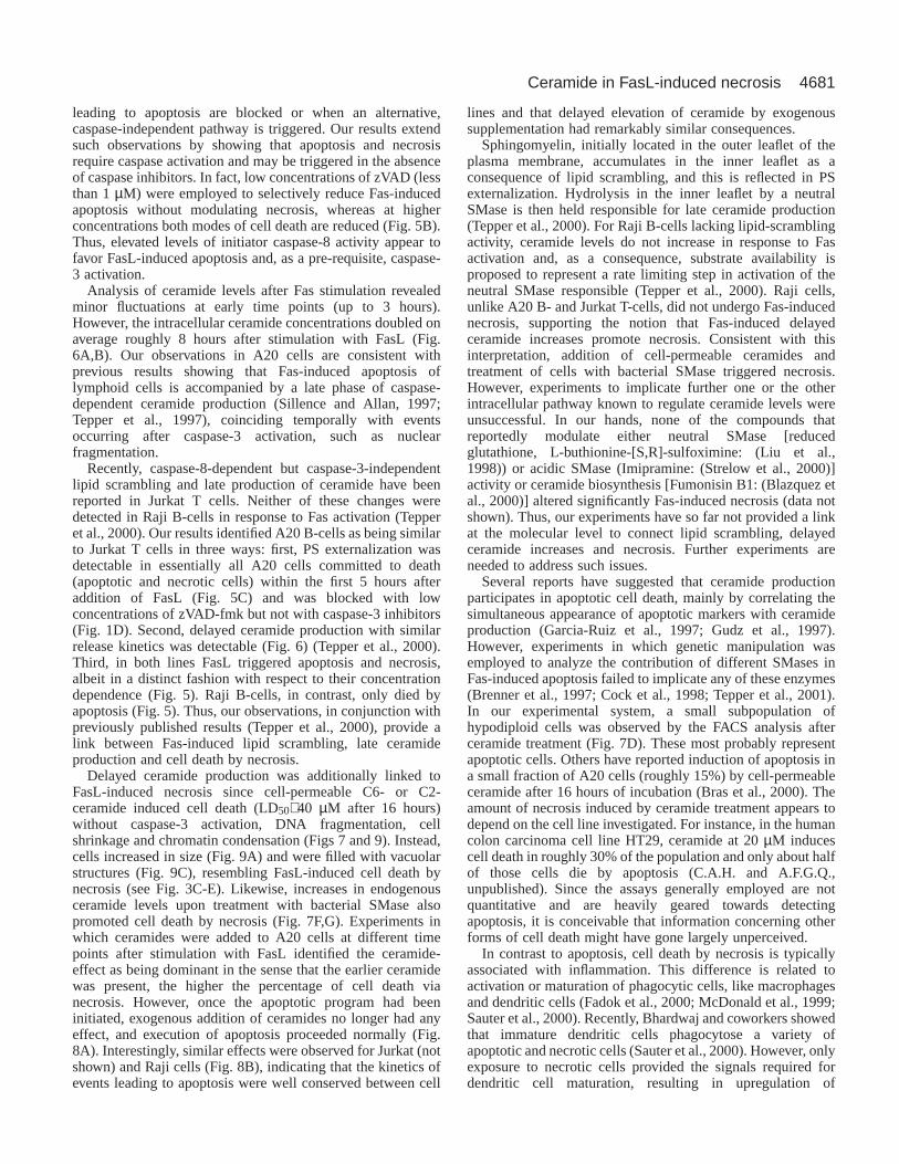
Morphological analysis of cells treated with cellpermeable ceramideThe nuclear and cellular morphology after C2-ceramidetreatment were determined in A20 cells (Fig. 9). Nuclearmorphology of dead cells was revealed by PI fluorescenceusing confocal microscopy after 16 hours of treatment (Fig.9A). In cells treated with C2-ceramide neither chromatincondensation nor cell shrinkage were observed. Instead,ceramide-induced dead cells were two- to three-fold larger than
Fig. 7. Cell-permeable ceramides ortreatment with bacterial SMase induceddeath by a caspase-3-independentmechanism. (A) A20 cells were incubatedfor 16 hours with 0-120 µM C6-ceramide(open circles) or dihydro-C6-ceramide(closed circles). Cell viability wasdetermined by the MTS assay and expressedas percentage of cell viability observed fornon-treated cells. (B) Caspase-3 activity wasdetermined after treatment of A20 cells for 4hours either with C6-ceramide at theconcentrations indicated, with 80 µMdihydro-C6-ceramide (DH), with 100 ng/mlFasL (F), with 100 ng/ml FasL and 80 µMC6-ceramide (F/C6) or remained untreated.Values are shown as a percentage of thecaspase-3 activity in the presence of 100ng/ml FasL alone. (C) Alternatively, DNAfragmentation was analyzed byelectrophoresis in agarose gels aftertreatment with FasL or differentconcentration of ceramides for 4 hours.(D) A representative experiment is shownwhere A20 cells were treated with 100 µMC6- (black) or dihydro-C6 (gray) ceramidefor 16 hours and cell survival wasdetermined by flow-cytometric analysisfollowing PI staining. (E) A20, Jurkat andRaji cells were either not treated (NT) ortreated with 100 µM C6-ceramides (C6) or100 ng/ml FasL (F) for 16 hours. Then cellswere harvested, stained with PI and cellsurvival was determined by flow cytometricanalysis. (F) Cells were treated with bacterialSMase at the concentrations indicated for 16hours and cell viability was determined bythe MTS assay. (G) In parallel, cells eithernot treated (gray) or treated with 0.5 U/mlbacterial SMase (black) were stained withPI, and cell survival was determined by flowcytometric analysis. The results shown areeither the means or standard deviations ofthree independent experiments (A,B,E,F) orare representative of at least threeindependent experiments (C,D,G).
4680
untreated cells. Cell death observed in A20 cells in responseto treatment with 100 µM C2-ceramide for 16 hours was alsocharacterized by electron microscopy (Fig. 9B,C). In perfectagreement with our other results, cells treated with C2-ceramide (Fig. 9C) resembled very strongly the necrotic cellpopulation (Fig. 3C) observed in response to FasL treatment.Thus, by the criteria investigated, ceramide-induced deathstrongly resembled FasL-induced necrosis.
DiscussionSeveral crucial events linked to onset of apoptosis in A20 cells,including caspase-3 activation, cell shrinkage and DNAfragmentation, were all activated within the first 3 hours ofFasL addition. However, cell viability was only partiallyrestored in the presence of the caspase-3 inhibitor Ac-DEVD-cho, whereas a more general caspase inhibitor, zVAD-fmk,completely protected cells against FasL-induced death (Fig. 1).Flow cytometric analysis distinguished two PI-positive cellpopulations in which cell death was either caspase-3-dependent and paralleled by DNA fragmentation or caspase-3-and DNA fragmentation-independent (Fig. 2). Bymorphological criteria (Fig. 3), these two subpopulations wereidentified as apoptotic and necrotic cells, respectively.Furthermore, FACS analysis showed that caspase-3 activationonly occurred in the shrunken cell population (apoptosis),whereas caspase-8 activation was detectable in both (Fig. 4).Thus, activation of initiator caspases, such as caspase-8,represented a key initial step triggered by FasL leading to
apoptotic and necrotic death in A20 cells. Activation ofcaspase-3, by contrast, was only implicated in events leadingto apoptosis. Interestingly, similar effects were also observedin Jurkat T cells, whereas in Raji B-cells lacking PSexternalization and delayed ceramide production (Tepper et al.,2000) only Fas-induced apoptosis was observed (Fig. 5).
The initiation of signaling pathways via Fas that lead to bothapoptotic and necrotic cell death has been described in someexperimental systems. In murine L929 fibrosarcoma cells,necrosis was observed downstream of Fas when caspaseactivation was blocked by inhibitors (Vercammen et al.,1998a). Also, caspase inhibition rendered murine L929fibrosarcoma cells 1000-fold more sensitive to necrosisinduced by TNF-α (Vercammen et al., 1998b). FasL-inducednecrosis in the absence of caspase activation owing toactivation of receptor-interacting protein (RIP) has also beendescribed previously (Holler et al., 2000). These resultssuggest that necrosis is either favored when pathways normally
Journal of Cell Science 115 (23)
Fig. 8. Effect of cell permeable ceramides on FasL-induced celldeath. (A) A20 or Raji cells (B) cells were incubated with 100 ng/mlFasL and C2-ceramide (100 µM) was added at time different timepoints after FasL. For comparison, cells were either not treated (NTor incubated with C2-ceramide (100 µM) or FasL (A20) or anti-Fas(Raji) (100 ng/ml) alone. For analysis, cells were harvested after 16hours, stained with PI, and cell survival was determined by flowcytometric analysis (see Fig. 2). Means and standard deviations ofdata from three independent experiments are shown.
Fig. 9. Morphological analysis and DNA staining of A20 B-lymphoma cells treated with cell-permeable ceramides. (A) Cellswere incubated with 100 µM C2-ceramide for 16 hours, stained withPI and analyzed by confocal microscopy. As a comparison, non-treated cells were permeabilized with methanol (NT, Met-OH) andstained with PI (control, permeabilized). Phase contrast andfluorescence images from the same optical section are shown. Thebar shown in white is equivalent to 13 µm. All images are shown atthe same magnification. Alternatively, cells untreated (B) or treatedwith 100 µM C2-ceramide (C) for 16 hours were analyzed byelectron microscopy. Necrotic C2-ceramide- (C) treated cells wereboth characterized by disruption of the nuclei and abundance ofvacuolar structures. The black bar shown for untreated cells (B) isequivalent to 5 µm. The image in panel C is shown at the samemagnification.
4681Ceramide in FasL-induced necrosis
leading to apoptosis are blocked or when an alternative,caspase-independent pathway is triggered. Our results extendsuch observations by showing that apoptosis and necrosisrequire caspase activation and may be triggered in the absenceof caspase inhibitors. In fact, low concentrations of zVAD (lessthan 1 µM) were employed to selectively reduce Fas-inducedapoptosis without modulating necrosis, whereas at higherconcentrations both modes of cell death are reduced (Fig. 5B).Thus, elevated levels of initiator caspase-8 activity appear tofavor FasL-induced apoptosis and, as a pre-requisite, caspase-3 activation.
Analysis of ceramide levels after Fas stimulation revealedminor fluctuations at early time points (up to 3 hours).However, the intracellular ceramide concentrations doubled onaverage roughly 8 hours after stimulation with FasL (Fig.6A,B). Our observations in A20 cells are consistent withprevious results showing that Fas-induced apoptosis oflymphoid cells is accompanied by a late phase of caspase-dependent ceramide production (Sillence and Allan, 1997;Tepper et al., 1997), coinciding temporally with eventsoccurring after caspase-3 activation, such as nuclearfragmentation.
Recently, caspase-8-dependent but caspase-3-independentlipid scrambling and late production of ceramide have beenreported in Jurkat T cells. Neither of these changes weredetected in Raji B-cells in response to Fas activation (Tepperet al., 2000). Our results identified A20 B-cells as being similarto Jurkat T cells in three ways: first, PS externalization wasdetectable in essentially all A20 cells committed to death(apoptotic and necrotic cells) within the first 5 hours afteraddition of FasL (Fig. 5C) and was blocked with lowconcentrations of zVAD-fmk but not with caspase-3 inhibitors(Fig. 1D). Second, delayed ceramide production with similarrelease kinetics was detectable (Fig. 6) (Tepper et al., 2000).Third, in both lines FasL triggered apoptosis and necrosis,albeit in a distinct fashion with respect to their concentrationdependence (Fig. 5). Raji B-cells, in contrast, only died byapoptosis (Fig. 5). Thus, our observations, in conjunction withpreviously published results (Tepper et al., 2000), provide alink between Fas-induced lipid scrambling, late ceramideproduction and cell death by necrosis.
Delayed ceramide production was additionally linked toFasL-induced necrosis since cell-permeable C6- or C2-ceramide induced cell death (LD50≅ 40 µM after 16 hours)without caspase-3 activation, DNA fragmentation, cellshrinkage and chromatin condensation (Figs 7 and 9). Instead,cells increased in size (Fig. 9A) and were filled with vacuolarstructures (Fig. 9C), resembling FasL-induced cell death bynecrosis (see Fig. 3C-E). Likewise, increases in endogenousceramide levels upon treatment with bacterial SMase alsopromoted cell death by necrosis (Fig. 7F,G). Experiments inwhich ceramides were added to A20 cells at different timepoints after stimulation with FasL identified the ceramide-effect as being dominant in the sense that the earlier ceramidewas present, the higher the percentage of cell death vianecrosis. However, once the apoptotic program had beeninitiated, exogenous addition of ceramides no longer had anyeffect, and execution of apoptosis proceeded normally (Fig.8A). Interestingly, similar effects were observed for Jurkat (notshown) and Raji cells (Fig. 8B), indicating that the kinetics ofevents leading to apoptosis were well conserved between cell
lines and that delayed elevation of ceramide by exogenoussupplementation had remarkably similar consequences.
Sphingomyelin, initially located in the outer leaflet of theplasma membrane, accumulates in the inner leaflet as aconsequence of lipid scrambling, and this is reflected in PSexternalization. Hydrolysis in the inner leaflet by a neutralSMase is then held responsible for late ceramide production(Tepper et al., 2000). For Raji B-cells lacking lipid-scramblingactivity, ceramide levels do not increase in response to Fasactivation and, as a consequence, substrate availability isproposed to represent a rate limiting step in activation of theneutral SMase responsible (Tepper et al., 2000). Raji cells,unlike A20 B- and Jurkat T-cells, did not undergo Fas-inducednecrosis, supporting the notion that Fas-induced delayedceramide increases promote necrosis. Consistent with thisinterpretation, addition of cell-permeable ceramides andtreatment of cells with bacterial SMase triggered necrosis.However, experiments to implicate further one or the otherintracellular pathway known to regulate ceramide levels wereunsuccessful. In our hands, none of the compounds thatreportedly modulate either neutral SMase [reducedglutathione, L-buthionine-[S,R]-sulfoximine: (Liu et al.,1998)) or acidic SMase (Imipramine: (Strelow et al., 2000)]activity or ceramide biosynthesis [Fumonisin B1: (Blazquez etal., 2000)] altered significantly Fas-induced necrosis (data notshown). Thus, our experiments have so far not provided a linkat the molecular level to connect lipid scrambling, delayedceramide increases and necrosis. Further experiments areneeded to address such issues.
Several reports have suggested that ceramide productionparticipates in apoptotic cell death, mainly by correlating thesimultaneous appearance of apoptotic markers with ceramideproduction (Garcia-Ruiz et al., 1997; Gudz et al., 1997).However, experiments in which genetic manipulation wasemployed to analyze the contribution of different SMases inFas-induced apoptosis failed to implicate any of these enzymes(Brenner et al., 1997; Cock et al., 1998; Tepper et al., 2001).In our experimental system, a small subpopulation ofhypodiploid cells was observed by the FACS analysis afterceramide treatment (Fig. 7D). These most probably representapoptotic cells. Others have reported induction of apoptosis ina small fraction of A20 cells (roughly 15%) by cell-permeableceramide after 16 hours of incubation (Bras et al., 2000). Theamount of necrosis induced by ceramide treatment appears todepend on the cell line investigated. For instance, in the humancolon carcinoma cell line HT29, ceramide at 20 µM inducescell death in roughly 30% of the population and only about halfof those cells die by apoptosis (C.A.H. and A.F.G.Q.,unpublished). Since the assays generally employed are notquantitative and are heavily geared towards detectingapoptosis, it is conceivable that information concerning otherforms of cell death might have gone largely unperceived.
In contrast to apoptosis, cell death by necrosis is typicallyassociated with inflammation. This difference is related toactivation or maturation of phagocytic cells, like macrophagesand dendritic cells (Fadok et al., 2000; McDonald et al., 1999;Sauter et al., 2000). Recently, Bhardwaj and coworkers showedthat immature dendritic cells phagocytose a variety ofapoptotic and necrotic cells (Sauter et al., 2000). However, onlyexposure to necrotic cells provided the signals required fordendritic cell maturation, resulting in upregulation of
4682
maturation-specific markers, co-stimulatory molecules and thecapacity to induce antigen-specific CD4+ and CD8+ T-cells.Thus, dendritic cells are able to distinguish between the twotypes of dead cells and respond in a distinct manner. Althoughthe interaction between apoptotic and phagocytic cells inducesan anti-inflammatory response (Fadok et al., 2000), necrosisappears to be critical for initiation of an immune response(Holler et al., 2000; Sauter et al., 2000). Interestingly, theresults presented here reinforce the notion that lipid scramblingand PS externalization do not provide the molecular basis todistinguish between cells dying by apoptosis or necrosis and,as a consequence, elicit different responses of the immunesystem.
Simultaneous or delayed activation of pathways leading tonecrotic and apoptotic death in the same cells is likely to occurfairly frequently (Ankarcrona et al., 1995; Barros et al., 2001;Dypbukt et al., 1994; Hetz et al., 2002; Jonas et al., 1994). Ithas been proposed that the modification of intracellular ATPconcentrations may serve as one possible mechanismpermitting the switch from apoptotic to necrotic cell death(Leist et al., 1997). Indeed, ceramides have been shown todirectly modulate mitochondria function, for instance, byinhibiting the mitochondrial respiratory complex III (Garcia-Ruiz et al., 1997; Gudz et al., 1997; Quillet-Mary et al., 1997).Thus, ceramide-induced necrosis may result from a decreasein ATP levels due to mitochondrial dysfunction. Consistentwith this notion, a decrease in mitochondrial membranepotential was detected in A20 cells after at least one hour ofincubation with cell permeable ceramides (data not shown).However, further experiments are required to determine howceramide promotes necrosis in the cells characterized here.
In summary, the results presented show that two differentpathways emerge from the Fas-receptor, one leading tocaspase-3-dependent apoptosis and the other favoring necrosisin a manner dependent upon activation of caspase-8, but notexecution caspases, like caspase-3. In addition, Fas-dependent,delayed production of ceramide was observed. The evidenceavailable suggests that the time point at which ceramide levelswere substantially elevated dictated the extent to whichnecrosis was observed. Thus, Fas-induced ceramide release isproposed to permit cells to undergo necrosis when caspase-8activation occurred but was insufficient to trigger caspase-3-dependent apoptosis. Ceramide production in A20 B-cells andJurkat T-cells downstream of caspase-8 may be temporallydelayed to trigger necrosis only in those cells not alreadycommitted to apoptosis.
Salvatore Valitutti, Felipe Barros, Andres Stutzin, Bruno Antonssonare thanked for careful revision and thoughtful comments concerningthe manuscript. Pascal Schneider and Jürg Tshopp are gratefullyacknowledged for providing recombinant human FasL and polyADP-ribose polymerase antibodies, Cecilia Sepúlveda for use of the FACSmachine and Nancy Olea for cell analysis by electron microscopy.Parts of these results have been presented previously in abstract form(Abstract#21, XV Annual Meeting of the Cell Biology Society ofChile, Valdivia, Nov. 2001). The paper is dedicated to Mrs. O.E. Questwho died of cancer while the work described was in progress.
ReferencesAbbas, A. K. (1996). Die and let live: eliminating dangerous lymphocytes.
Cell 84, 655-657.
Ankarcrona, M., Dypbukt, J. M., Bonfoco, E., Zhivotovsky, B., Orrenius,S., Lipton, S. A. and Nicotera, P.(1995). Glutamate-induced neuronaldeath: a succession of necrosis or apoptosis depending on mitochondrialfunction. Neuron15, 961-973.
Barros, L. F., Stutzin, A., Calixto, A., Catalan, M., Castro, J., Hetz, C. andHermosilla, T. (2001). Nonselective cation channels as effectors of freeradical-induced rat liver cell necrosis. Hepatology33, 114-122.
Blazquez, C., Galve-Roperh, I. and Guzman, M.(2000). De novo-synthesized ceramide signals apoptosis in astrocytes via extracellular signal-regulated kinase. FASEB J.14, 2315-2322.
Boldin, M. P., Goncharov, T. M., Goltsev, Y. V. and Wallach, D.(1996).Involvement of MACH, a novel MORT1/FADD-interacting protease, inFas/APO-1- and TNF receptor-induced cell death. Cell 85, 803-815.
Bras, A., Albar, J. P., Leonardo, E., de Buitrago, G. G. and Martinez, A.C. (2000). Ceramide-induced cell death is independent of the Fas/Fas ligandpathway and is prevented by Nur77 overexpression in A20 B cells. CellDeath Differ.7, 262-271.
Brenner, B., Koppenhoefer, U., Weinstock, C., Linderkamp, O., Lang, F.and Gulbins, E.(1997). Fas- or ceramide-induced apoptosis is mediated bya Rac1-regulated activation of Jun N-terminal kinase/p38 kinases andGADD153. J. Biol. Chem.272, 22173-22181.
Cifone, M. G., Roncaioli, P., de Maria, R., Camarda, G., Santoni, A.,Ruberti, G. and Testi, R. (1995). Multiple pathways originate at theFas/APO-1 (CD95) receptor: sequential involvement ofphosphatidylcholine-specific phospholipase C and acidic sphingomyelinasein the propagation of the apoptotic signal. EMBO J.14, 5859-5868.
Cock, J. G., Tepper, A. D., de Vries, E., van Blitterswijk, W. J. and Borst,J. (1998). CD95 (Fas/APO-1) induces ceramide formation and apoptosis inthe absence of a functional acid sphingomyelinase. J. Biol. Chem.273,7560-7565.
Cornall, R. J., Goodnow, C. C. and Cyster, J. G.(1995). The regulation ofself-reactive B cells. Curr. Opin. Immunol.7, 804-811.
Dypbukt, J. M., Ankarcrona, M., Burkitt, M., Sjoholm, A., Strom, K.,Orrenius, S. and Nicotera, P.(1994). Different prooxidant levels stimulategrowth, trigger apoptosis, or produce necrosis of insulin-secreting RINm5Fcells. The role of intracellular polyamines. J. Biol. Chem.269, 30553-30560.
Fadok, V. A., Bratton, D. L., Rose, D. M., Pearson, A., Ezekewitz, R. A.and Henson, P. M. (2000). A receptor for phosphatidylserine-specificclearance of apoptotic cells. Nature405, 85-90.
Garcia-Ruiz, C., Colell, A., Mari, M., Morales, A. and Fernandez-Checa,J. C. (1997). Direct effect of ceramide on the mitochondrial electrontransport chain leads to generation of reactive oxygen species. Role ofmitochondrial glutathione. J. Biol. Chem.272, 11369-11377.
Genestier, L., Prigent, A. F., Paillot, R., Quemeneur, L., Durand, I.,Banchereau, J., Revillard, J. P. and Bonnefoy-Berard, N.(1998).Caspase-dependent ceramide production in Fas- and HLA class I-mediatedperipheral T cell apoptosis. J. Biol. Chem.273, 5060-5066.
Gudz, T. I., Tserng, K. Y. and Hoppel, C. L.(1997). Direct inhibition ofmitochondrial respiratory chain complex III by cell-permeable ceramide. J.Biol. Chem.272, 24154-24158.
Hahne, M., Renno, T., Schroeter, M., Irmler, M., French, L., Bornard, T.,MacDonald, H. R. and Tschopp, J.(1996). Activated B cells expressfunctional Fas ligand. Eur. J. Immunol.26, 721-724.
Hannun, Y. A., Obeid, L. M. and Dbaibo, G. S. (1996). Ceramide. A novelsecond messenger and lipid mediator. In Handbook of Lipid Research, vol.8 (ed. R. M. Bell, J. H. Exton and S. M. Prescott), pp. 177-204. New York:Plenum Press.
Heinrich, M., Wickel, M., Schneider-Brachert, W., Sandberg, C., Gahr, J.,Schwandner, R., Weber, T., Saftig, P., Peters, C., Brunner, J. et al.(1999). Cathepsin D targeted by acid sphingomyelinase-derived ceramides.EMBO J.18, 5252-5263.
Hetz, C., Bono, M. R., Barros, L. F. and Lagos, R.(2002). Microcin E492,a channel-forming bacteriocin from Klebsiella pneumoniae, inducesapoptosis in some human cell lines. Proc. Natl. Acad. Sci. USA99, 2696-2701.
Hofmann, K. and Dixit, V. M. (1998). Ceramide in apoptosis–does it reallymatter? Trends Biochem. Sci.23, 374-377.
Holler, N., Zaru, R., Micheau, O., Thome, M., Attinger, A., Valitutti, S.,Bodmer, J. L., Schneider, P., Seed, B. and Tschopp, J.(2000). Fas triggersan alternative, caspase-8-independent cell death pathway using the kinaseRIP as effector molecule. Nat. Immunol.1, 489-495.
Jonas, D., Walev, I., Berger, T., Liebetrau, M., Palmer, M. and Bhakdi, S.(1994). Novel path to apoptosis: small transmembranes pores created by
Journal of Cell Science 115 (23)
4683Ceramide in FasL-induced necrosis
staphylococcal alpha-toxin in T lymphocytes evoke internucleosomal DNAdegradation. Infect. Immun.62, 1304-1312.
Kolesnick, R. N. and Kronke, M.(1998). Regulation of ceramide productionand apoptosis. Annu. Rev. Physiol.60, 643-665.
Laster, S. M., Wood, J. G. and Gooding, L. R.(1988). Tumor necrosis factorcan induce both apoptotic and necrotic cell lysis. J. Immunol.141, 2629-2634.
Leist, M., Single, B., Castoldi, A. F., Kuhnle, S. and Nicotera, P.(1997).Intracellular adenosine triphosphate (ATP) concentration: a switch in thedecision between apoptosis and necrosis. J. Exp. Med.185, 1481-1486.
Liu, B., Andrieu-Abadie, N., Levade, T., Zhang, P., Obeid, L. M. andHannun, Y. A. (1998). Glutathione regulation of neutral sphingomyelinasein tumor necrosis factor-alpha-induced cell death. J. Biol. Chem.273,11313-11320.
Majno, G. and Joris, I. (1995). Apoptosis, oncosis, and necrosis. An overviewof cell death. Am. J. Pathol.146, 3-15.
Martins, L. M. and Earnshaw, W. C. (1997). Apoptosis: alive and kickingin 1997. Trends Cell Biol.7, 111-114.
Matiba, B., Mariani, S. M. and Krammer, P. H. (1997). The CD95 systemand the death of a lymphocyte. Semin. Immunol.9, 59-68.
McDonald, P. P., Fadok, V. A., Bratton, D. and Henson, P. M.(1999).Transcriptional and translational regulation of inflammatory mediatorproduction by endogenous TGF-beta in macrophages that have ingestedapoptotic cells. J. Immunol.163, 6164-6172.
Nagata, S.(1997). Apoptosis by death factor. Cell 88, 355-365.Nagata, S. and Golstein, P.(1995). The Fas death factor. Science267, 1451-
1456.Preiss, J. E., Loomis, C. R., Bell, R. M. and Niedel, J. E.(1987). Quantitative
measurement of sn-1,2-diacylglycerols. Methods Enzymol.141, 294-300.Quillet-Mary, A., Jaffrezou, J. P., Mansat, V., Bordier, C., Naval, J. and
Laurent, G. (1997). Implication of mitochondrial hydrogen peroxidegeneration in ceramide-induced apoptosis. J. Biol. Chem.272, 21388-21395.
Rathmell, J. C. and Thompson, C. B.(1999). The central effectors of celldeath in the immune system. Annu. Rev. Immunol.17, 781-828.
Sauter, B., Albert, M. L., Francisco, L., Larsson, M., Somersan, S. andBhardwaj, N. (2000). Consequences of cell death: exposure to necrotic
tumor cells, but not primary tissue cells or apoptotic cells, induces thematuration of immunostimulatory dendritic cells. J. Exp. Med.191, 423-433.
Sillence, D. J. and Allan, D.(1997). Evidence against an early signalling rolefor ceramide in Fas-mediated apoptosis. Biochem. J.324, 29-32.
Strelow, A., Bernardo, K., Adam-Klages, S., Linke, T., Sandhoff, K.,Kronke, M. and Adam, D. (2000). Overexpression of acid ceramidaseprotects from tumor necrosis factor- induced cell death. J. Exp. Med.192,601-612.
Suda, T., Takahashi, T., Golstein, P. and Nagata, S.(1993). Molecularcloning and expression of the Fas ligand, a novel member of the tumornecrosis factor family. Cell 75, 1169-1178.
Tepper, A. D., Cock, J. G., de Vries, E., Borst, J. and van Blitterswijk, W.J. (1997). CD95/Fas-induced ceramide formation proceeds with slowkinetics and is not blocked by caspase-3/CPP32 inhibition. J. Biol. Chem.272, 24308-24312.
Tepper, A. D., Ruurs, P., Borst, J. and van Blitterswijk, W. J.(2001). Effectof overexpression of a neutral sphingomyelinase on CD95-induced ceramideproduction and apoptosis. Biochem. Biophys. Res. Commun.280, 634-639.
Tepper, A. D., Ruurs, P., Wiedmer, T., Sims, P. J., Borst, J. and vanBlitterswijk, W. J. (2000). Sphingomyelin hydrolysis to ceramide duringthe execution phase of apoptosis results from phospholipid scrambling andalters cell-surface morphology. J. Cell Biol.150, 155-164.
Venkataraman, K. and Futerman, A. H. (2000). Ceramide as a secondmessenger: sticky solutions to sticky problems. Trends Cell Biol10, 408-412.
Vercammen, D., Beyaert, R., Denecker, G., Goossens, V., van Loo, G.,Declercq, W., Grooten, J., Fiers, W. and Vandenabeele, P.(1998a).Inhibition of caspases increases the sensitivity of L929 cells to necrosismediated by tumor necrosis factor. J. Exp. Med.187, 1477-1485.
Vercammen, D., Brouckaert, G., Denecker, G., van de Craen, M.,Declercq, W., Fiers, W. and Vandenabeele, P.(1998b). Dual signaling ofthe Fas receptor: initiation of both apoptotic and necrotic cell deathpathways. J. Exp. Med.188, 919-930.
Walsh, J. P. and Bell, R. M. (1986). sn-1,2-Diacylglycerol kinase ofEscherichia coli. Mixed micellar analysis of the phospholipid cofactorrequirement and divalent cation dependence. J. Biol. Chem.261, 6239-6247.
Related Documents