JPP 2008, 60: 1365–1374 ß 2008 The Authors Received February 27, 2008 Accepted June 5, 2008 DOI 10.1211/jpp/60.10.0013 ISSN 0022-3573 Department of Biofunctional Evaluation, Molecular Pharmacology, Gifu Pharmaceutical University, 5-6-1 Mitahora-higashi, Gifu 502-8585, Japan Yoshimi Nakajima, Yuta Inokuchi, Masamitsu Shimazawa, Hideaki Hara Fine Chemicals Marketing Department, Fine Chemicals Division, Asahi Kasei Pharma Corporation, 9-1 Kanda Mitoshiro-cho, Chiyoda-ku, Tokyo 101-8481, Japan Kazumasa Otsubo Biotechnology Business Section, Merchandise Business Department, Nippon Oil Corporation, 3-12 Nishi Shimbashi 1-chome, Minato-ku, Tokyo 105-8412, Japan Takashi Ishibashi Correspondence: H. Hara, Department of Biofunctional Evaluation, Molecular Pharmacology Gifu Pharmaceutical University, 5-6-1 Mitahora-higashi, Gifu 502-8585, Japan. E-mail: [email protected] Astaxanthin, a dietary carotenoid, protects retinal cells against oxidative stress in-vitro and in mice in-vivo Yoshimi Nakajima, Yuta Inokuchi, Masamitsu Shimazawa, Kazumasa Otsubo, Takashi Ishibashi and Hideaki Hara Abstract We have investigated whether astaxanthin exerted neuroprotective effects in retinal ganglion cells in-vitro and in-vivo. In-vitro, retinal damage was induced by 24-h hydrogen peroxide (H 2 O 2 ) exposure or serum deprivation, and cell viability was measured using a WST assay. In cultured retinal ganglion cells (RGC-5, a rat ganglion cell-line transformed using E1A virus), astaxanthin inhibited the neurotoxicity induced by H 2 O 2 or serum deprivation, and reduced the intracellular oxidation induced by various reactive oxygen species (ROS). Furthermore, astaxanthin decreased the radical generation induced by serum deprivation in RGC-5. In mice in-vivo, astaxanthin (100 mg kg -1 , p.o., four times) reduced the retinal damage (a decrease in retinal ganglion cells and in thickness of inner plexiform layer) induced by intravitreal N-methyl-D-aspartate (NMDA) injection. Furthermore, astaxanthin reduced the expressions of 4-hydroxy-2-nonenal (4-HNE)-modified protein (indicator of lipid peroxidation) and 8-hydroxy-deoxyguanosine (8-OHdG; indicator of oxidative DNA damage). These findings indicated that astaxanthin had neuroprotective effects against retinal damage in-vitro and in-vivo, and that its protective effects may have been partly mediated via its antioxidant effects. Introduction Astaxanthin, a dietary carotenoid, is present in many biological systems, often decreasing the formation of products of oxidative damage induced by biological molecules. Astaxanthin is a powerful biological antioxidant occurring naturally in a wide variety of living organisms (Clarke et al 1990), and is present in many well-known sea foods such as salmon, trout, red sea-bream, shrimp, lobster and fish eggs. Astaxanthin possesses various pharmacological activities, including antioxidative activity (Kurashige et al 1990; O’Connor & O’Brien 1998; Iwamoto et al 2000; Kobayashi 2000; Aoi et al 2003), antitumour effects (Chew et al 1999a; Jyonouchi et al 2000), an anti-inflammatory action (Ohgami et al 2003), antidiabetic (Uchiyama et al 2002) and hepatoprotective effects (Kang et al 2001), and immunomodulatory activity (Okai & Higashi-Okai 1996; Chew et al 1999b). Thus, astaxanthin has considerable potential for applications in human health and nutrition. Retinal ganglion cell (RGC) death is a common feature of many ophthalmic disorders, such as glaucoma, optic neuropathies, and various retinovascular diseases (diabetic retinopathy and retinal vein occlusions) (Bocker-Meffert et al 2002). RGC death may occur via a variety of mechanisms involving, for example, reactive oxygen species (ROS) (Bonne et al 1998), excitatory amino acids (Dreyer 1998), nitric oxide (Neufeld 1999) and apoptosis (McKinnon 1997). Previous studies on the protective effects of carotenoids (including astaxanthin) against retinal damage have primarily focused on age-related maculopathy (Parisi et al 2008). Moreover, to our knowledge no examination has been made of the in-vitro neuroprotective effects of astaxanthin against oxidative stress using retinal ganglion cells or in-vivo models of retinal damage. The purpose of this study was to examine the effects of astaxanthin on retinal damage in-vitro and in-vivo. We studied its effects on hydrogen peroxide (H 2 O 2 )-induced or serum deprivation-induced neurotoxicity in RGC-5 (a rat ganglion cell-line transformed using E1A virus) cultures, on the intracellular oxidation induced by various ROS in RGC-5 cultures, and on in-vivo N-methyl-D-aspartate (NMDA)-induced retinal damage in mice. 1365

Welcome message from author
This document is posted to help you gain knowledge. Please leave a comment to let me know what you think about it! Share it to your friends and learn new things together.
Transcript

JPP 2008, 60: 1365–1374
� 2008 The Authors
Received February 27, 2008
Accepted June 5, 2008
DOI 10.1211/jpp/60.10.0013
ISSN 0022-3573
Department of Biofunctional
Evaluation, Molecular
Pharmacology, Gifu
Pharmaceutical University,
5-6-1 Mitahora-higashi,
Gifu 502-8585, Japan
Yoshimi Nakajima,
Yuta Inokuchi,
Masamitsu Shimazawa,
Hideaki Hara
Fine Chemicals Marketing
Department, Fine Chemicals
Division, Asahi Kasei Pharma
Corporation, 9-1 Kanda
Mitoshiro-cho, Chiyoda-ku,
Tokyo 101-8481, Japan
Kazumasa Otsubo
Biotechnology Business Section,
Merchandise Business
Department, Nippon Oil
Corporation, 3-12 Nishi
Shimbashi 1-chome, Minato-ku,
Tokyo 105-8412, Japan
Takashi Ishibashi
Correspondence: H. Hara,
Department of Biofunctional
Evaluation, Molecular
Pharmacology Gifu
Pharmaceutical University,
5-6-1 Mitahora-higashi,
Gifu 502-8585, Japan.
E-mail: [email protected]
Astaxanthin, a dietary carotenoid, protects retinal cells
against oxidative stress in-vitro and in mice in-vivo
Yoshimi Nakajima, Yuta Inokuchi, Masamitsu Shimazawa,
Kazumasa Otsubo, Takashi Ishibashi and Hideaki Hara
Abstract
We have investigated whether astaxanthin exerted neuroprotective effects in retinal ganglion cells
in-vitro and in-vivo. In-vitro, retinal damage was induced by 24-h hydrogen peroxide (H2O2) exposure
or serum deprivation, and cell viability was measured using a WST assay. In cultured retinal ganglion
cells (RGC-5, a rat ganglion cell-line transformed using E1A virus), astaxanthin inhibited the
neurotoxicity induced by H2O2 or serum deprivation, and reduced the intracellular oxidation induced
by various reactive oxygen species (ROS). Furthermore, astaxanthin decreased the radical generation
induced by serum deprivation in RGC-5. In mice in-vivo, astaxanthin (100 mg kg-1, p.o., four times)
reduced the retinal damage (a decrease in retinal ganglion cells and in thickness of inner plexiform
layer) induced by intravitreal N-methyl-D-aspartate (NMDA) injection. Furthermore, astaxanthin
reduced the expressions of 4-hydroxy-2-nonenal (4-HNE)-modified protein (indicator of lipid
peroxidation) and 8-hydroxy-deoxyguanosine (8-OHdG; indicator of oxidative DNA damage). These
findings indicated that astaxanthin had neuroprotective effects against retinal damage in-vitro
and in-vivo, and that its protective effects may have been partly mediated via its antioxidant effects.
Introduction
Astaxanthin, a dietary carotenoid, is present in many biological systems, often decreasingthe formation of products of oxidative damage induced by biological molecules.Astaxanthin is a powerful biological antioxidant occurring naturally in a wide variety ofliving organisms (Clarke et al 1990), and is present in many well-known sea foods such assalmon, trout, red sea-bream, shrimp, lobster and fish eggs. Astaxanthin possesses variouspharmacological activities, including antioxidative activity (Kurashige et al 1990;O’Connor & O’Brien 1998; Iwamoto et al 2000; Kobayashi 2000; Aoi et al 2003),antitumour effects (Chew et al 1999a; Jyonouchi et al 2000), an anti-inflammatory action(Ohgami et al 2003), antidiabetic (Uchiyama et al 2002) and hepatoprotective effects(Kang et al 2001), and immunomodulatory activity (Okai & Higashi-Okai 1996; Chew et al1999b). Thus, astaxanthin has considerable potential for applications in human health andnutrition.
Retinal ganglion cell (RGC) death is a common feature of many ophthalmic disorders,such as glaucoma, optic neuropathies, and various retinovascular diseases (diabeticretinopathy and retinal vein occlusions) (Bocker-Meffert et al 2002). RGC death may occurvia a variety of mechanisms involving, for example, reactive oxygen species (ROS)(Bonne et al 1998), excitatory amino acids (Dreyer 1998), nitric oxide (Neufeld 1999) andapoptosis (McKinnon 1997). Previous studies on the protective effects of carotenoids(including astaxanthin) against retinal damage have primarily focused on age-relatedmaculopathy (Parisi et al 2008). Moreover, to our knowledge no examination has beenmade of the in-vitro neuroprotective effects of astaxanthin against oxidative stressusing retinal ganglion cells or in-vivo models of retinal damage.
The purpose of this study was to examine the effects of astaxanthin on retinal damagein-vitro and in-vivo. We studied its effects on hydrogen peroxide (H2O2)-induced or serumdeprivation-induced neurotoxicity in RGC-5 (a rat ganglion cell-line transformed usingE1A virus) cultures, on the intracellular oxidation induced by various ROS in RGC-5cultures, and on in-vivo N-methyl-D-aspartate (NMDA)-induced retinal damage in mice.
1365
In addition, we examined its effects on the accumulation oflipid peroxidation and oxidative DNA damage observed at12 h after NMDA intravitreal injection in mice.
Materials and Methods
Materials
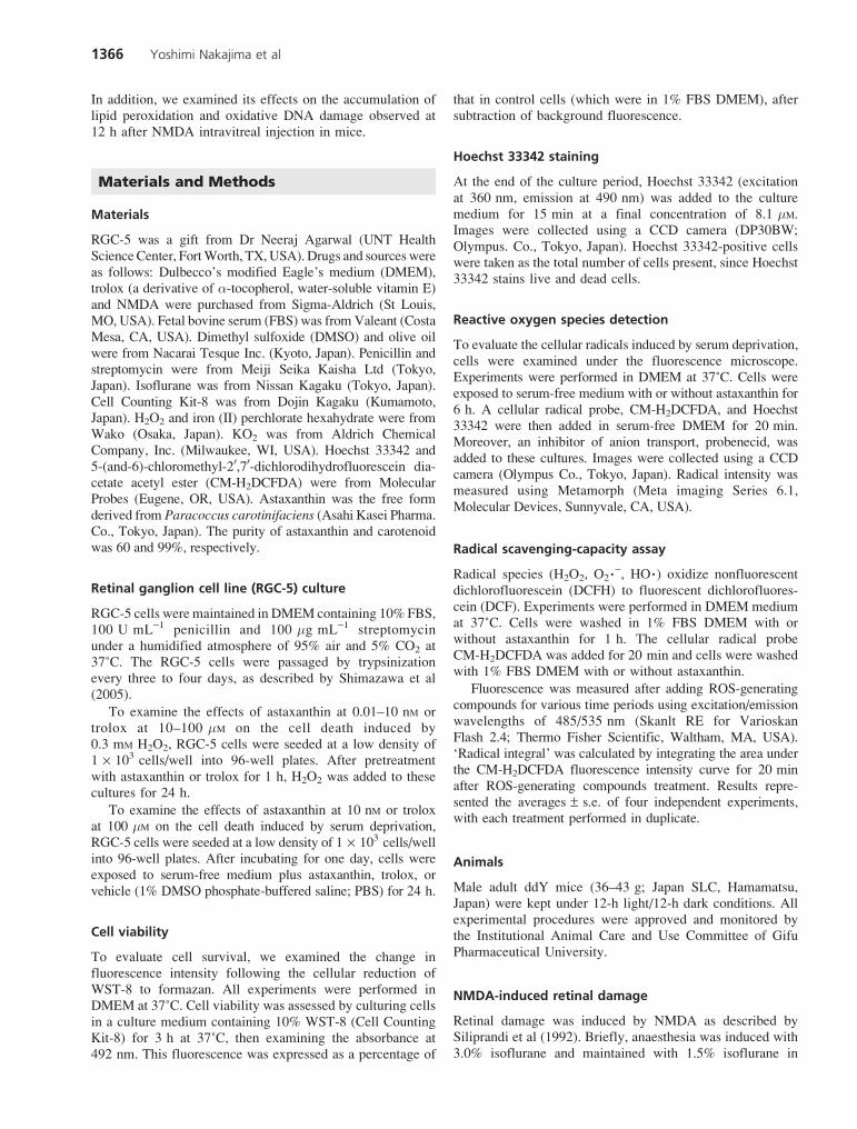
RGC-5 was a gift from Dr Neeraj Agarwal (UNT HealthScienceCenter, FortWorth, TX,USA). Drugs and sourceswereas follows: Dulbecco’s modified Eagle’s medium (DMEM),trolox (a derivative of a-tocopherol, water-soluble vitamin E)and NMDA were purchased from Sigma-Aldrich (St Louis,MO, USA). Fetal bovine serum (FBS) was fromValeant (CostaMesa, CA, USA). Dimethyl sulfoxide (DMSO) and olive oilwere from Nacarai Tesque Inc. (Kyoto, Japan). Penicillin andstreptomycin were from Meiji Seika Kaisha Ltd (Tokyo,Japan). Isoflurane was from Nissan Kagaku (Tokyo, Japan).Cell Counting Kit-8 was from Dojin Kagaku (Kumamoto,Japan). H2O2 and iron (II) perchlorate hexahydrate were fromWako (Osaka, Japan). KO2 was from Aldrich ChemicalCompany, Inc. (Milwaukee, WI, USA). Hoechst 33342 and5-(and-6)-chloromethyl-20,70-dichlorodihydrofluorescein dia-cetate acetyl ester (CM-H2DCFDA) were from MolecularProbes (Eugene, OR, USA). Astaxanthin was the free formderived from Paracoccus carotinifaciens (Asahi Kasei Pharma.Co., Tokyo, Japan). The purity of astaxanthin and carotenoidwas 60 and 99%, respectively.
Retinal ganglion cell line (RGC-5) culture
RGC-5 cells were maintained in DMEM containing 10% FBS,100 U mL-1 penicillin and 100 mg mL-1 streptomycinunder a humidified atmosphere of 95% air and 5% CO2 at37˚C. The RGC-5 cells were passaged by trypsinizationevery three to four days, as described by Shimazawa et al(2005).
To examine the effects of astaxanthin at 0.01–10 nM ortrolox at 10–100 mM on the cell death induced by0.3 mM H2O2, RGC-5 cells were seeded at a low density of1 ¥ 103 cells/well into 96-well plates. After pretreatmentwith astaxanthin or trolox for 1 h, H2O2 was added to thesecultures for 24 h.
To examine the effects of astaxanthin at 10 nM or troloxat 100 mM on the cell death induced by serum deprivation,RGC-5 cells were seeded at a low density of 1 ¥ 103 cells/wellinto 96-well plates. After incubating for one day, cells wereexposed to serum-free medium plus astaxanthin, trolox, orvehicle (1% DMSO phosphate-buffered saline; PBS) for 24 h.
Cell viability
To evaluate cell survival, we examined the change influorescence intensity following the cellular reduction ofWST-8 to formazan. All experiments were performed inDMEM at 37˚C. Cell viability was assessed by culturing cellsin a culture medium containing 10% WST-8 (Cell CountingKit-8) for 3 h at 37˚C, then examining the absorbance at492 nm. This fluorescence was expressed as a percentage of
that in control cells (which were in 1% FBS DMEM), aftersubtraction of background fluorescence.
Hoechst 33342 staining
At the end of the culture period, Hoechst 33342 (excitationat 360 nm, emission at 490 nm) was added to the culturemedium for 15 min at a final concentration of 8.1 mM.Images were collected using a CCD camera (DP30BW;Olympus. Co., Tokyo, Japan). Hoechst 33342-positive cellswere taken as the total number of cells present, since Hoechst33342 stains live and dead cells.
Reactive oxygen species detection
To evaluate the cellular radicals induced by serum deprivation,cells were examined under the fluorescence microscope.Experiments were performed in DMEM at 37˚C. Cells wereexposed to serum-free medium with or without astaxanthin for6 h. A cellular radical probe, CM-H2DCFDA, and Hoechst33342 were then added in serum-free DMEM for 20 min.Moreover, an inhibitor of anion transport, probenecid, wasadded to these cultures. Images were collected using a CCDcamera (Olympus Co., Tokyo, Japan). Radical intensity wasmeasured using Metamorph (Meta imaging Series 6.1,Molecular Devices, Sunnyvale, CA, USA).
Radical scavenging-capacity assay
Radical species (H2O2, O2�-, HO�) oxidize nonfluorescentdichlorofluorescein (DCFH) to fluorescent dichlorofluores-cein (DCF). Experiments were performed in DMEM mediumat 37˚C. Cells were washed in 1% FBS DMEM with orwithout astaxanthin for 1 h. The cellular radical probeCM-H2DCFDA was added for 20 min and cells were washedwith 1% FBS DMEM with or without astaxanthin.
Fluorescence was measured after adding ROS-generatingcompounds for various time periods using excitation/emissionwavelengths of 485/535 nm (Skanlt RE for VarioskanFlash 2.4; Thermo Fisher Scientific, Waltham, MA, USA).‘Radical integral’ was calculated by integrating the area underthe CM-H2DCFDA fluorescence intensity curve for 20 minafter ROS-generating compounds treatment. Results repre-sented the averages ± s.e. of four independent experiments,with each treatment performed in duplicate.
Animals
Male adult ddY mice (36–43 g; Japan SLC, Hamamatsu,Japan) were kept under 12-h light/12-h dark conditions. Allexperimental procedures were approved and monitored bythe Institutional Animal Care and Use Committee of GifuPharmaceutical University.
NMDA-induced retinal damage
Retinal damage was induced by NMDA as described bySiliprandi et al (1992). Briefly, anaesthesia was induced with3.0% isoflurane and maintained with 1.5% isoflurane in
1366 Yoshimi Nakajima et al
70% N2O and 30% O2 via an animal general anaesthesiamachine (Soft Lander; Sin-ei Industry Co., Ltd, Saitama,Japan). Body temperature was maintained at between37.0 and 37.5˚C with the aid of a heating pad and heatinglamp. Retinal damage was induced by the injection(2 mL/eye) of NMDA dissolved at 20 mM in 0.01 M PBS.This was injected into the vitreous body of the lefteye under the above anaesthesia. One drop of 0.01%levofloxacin ophthalmic solution (Santen PharmaceuticalsCo. Ltd, Osaka, Japan) was applied topically to the treatedeye immediately after the intravitreal injection. Seven daysafter the NMDA injection, eyeballs were enucleated forhistological analysis.
Astaxanthin (100 mg kg-1) was dissolved in olive oilimmediately before use, and was orally administered fourtimes (at 6 h before, and at 0, 6, and 24 h after the NMDAinjection) for histological analysis, or three times (at 6 hbefore, and at 0 and 6 h after the NMDA injection) forTUNEL staining and immunostaining analysis with avolume of 0.1 mL/10 g body weight.
Histological analysis of mouse retina
Mice were anaesthetized by an intraperitoneal injection ofsodium pentobarbital (80 mg kg-1). Each eye was enucleatedand kept immersed for at least 24 h at 4˚C in a fixativesolution containing 4% paraformaldehyde. Six paraffin-embedded sections (thickness, 4 mm) cut through the opticdisc of each eye were prepared in a standard manner, andstained with haematoxylin and eosin. Retinal damage wasevaluated as described by Yoneda et al (2001), three sectionsfrom each eye being used for the morphometric analysis.Light-microscope images were photographed, and thecell count in the ganglion cell layer (GCL) at a distancebetween 375 and 625 mm from the optic disc, and thethickness of the inner plexiform layer (IPL) were measuredon the photographs in a masked fashion by a singleobserver (Y. Nakajima). Data from three sections (selectedrandomly from the six sections) were averaged for each eye,and these were used to evaluate the GCL cell count and IPLthickness.
TUNEL staining
TUNEL staining was performed according to the manufac-turer’s protocol (In Situ Cell Death Detection Kit; RocheBiochemicals, Mannheim, Germany) to detect the retinal celldeath induced by NMDA. The mice (n = 8) were anaes-thetized with pentobarbital sodium (80 mg kg-1, i.p.) at 24 hafter intravitreal injection of NMDA at 5 nmol/eye. The eyeswere enucleated, fixed overnight in 4% paraformaldehydesolution in 0.1 M phosphate buffer (pH 7.4), and immersedfor two days in PBS containing 25% sucrose. The eyes werethen embedded in a supporting medium for frozen-tissuespecimens (OCT compound; Tissue-Tek, Tokyo, Japan).Retinal sections 10-mm thick were cut on a cryostat at -25˚C,and stored at -80˚C until staining. After twice washing withPBS, sections were incubated with terminal deoxyribonu-cleotidyl transferase (TdT) enzyme at 37˚C for 1 h. Thesections were washed three times in PBS for 1 min at room
temperature. Sections were subsequently incubated with ananti-fluorescein antibody–peroxidase (POD) conjugate atroom temperature in a humidified chamber for 30 min, andthen developed using diaminobenzidine(DAB) tetrahy-drochloride peroxidase substrate. Light-microscope imageswere photographed (COOLPIX4500; Nikon, Tokyo), and thelabelled cells were counted in the GCL at a distance between375 and 625 mm from the optic disc in two central areas ofthe retina. The numbers of TUNEL-positive cells wereaveraged for the two areas, and this value was plotted as thenumber of TUNEL-positive cells.
Immunostaining
To detect 4-HNE (4-hydroxy-2-nonenal) and 8-OHdG(8-hydroxy-20-deoxyguanosine) protein in the retina,immunostaining was performed. For this, the followingprimary antibodies were used: anti-4-HNE monoclonalantibody (clone HNEJ-2) and anti-8-OHdG monoclonalantibody (clone N45.1) (JaICA, Shizuoka, Japan). A totalof 24 animals were used, and each eye was enucleated asdescribed in ‘Histological analysis’, and then post-fixedovernight in 4% paraformaldehyde solution in 0.1 M
phosphate buffer (pH 7.4) at 4˚C, and embedded in paraffin.Cross sections (4 mm) through the optic nerve were obtainedfrom the paraffin-embedded eyes. Such sections weredeparaffinized with xylene and dehydrated through a gradedethanol series. Immunohistochemical staining was performedin accordance with the following protocol. Briefly, tissuesections were washed in 0.01 M PBS for 10 min, and thenendogenous peroxidase was quenched by treating thesections with 3% hydrogen peroxide in absolute methanolfor 10 min, followed by pre-incubation with VECTOR M.O.M. Immunodetection Kit (Vector Laboratories, Burlingame,CA, USA). A mouse monoclonal antibody against 4-HNE or8-OHdG was added at a dilution of 1:1000. Sections werethen incubated with primary antibodies overnight at 4˚C. Theslides were washed and incubated with biotinylated anti-mouse IgG. They were subsequently incubated with avidin–biotin–peroxidase complex for 30 min and developed usingDAB peroxidase substrate. Light-microscope images werephotographed, and the labelled cells were counted in the GCLat a distance between 375 and 625 mm from the optic disc intwo central areas of the retina. The number of8-OHdG-positive cells was averaged for the two areas, andthis value was plotted as the number of 8-OHdG-positive cells.Light-microscope images were photographed (COOLPIX4500; Nikon, Tokyo), and the DAB-labelled cells in theGCL and IPL at a distance between 475 and 525 mm(50 ¥ 50 mm) from the optic disc were counted in two centralareas of the retina. The retinal DAB-labelled density wasevaluated by means of an appropriately calibrated computer-ized image analysis (Image J).
Statistical analysis
Data were presented as means ± s.e.m. Statistical signifi-cance, as indicated (*P < 0.05, **P < 0.01), was determinedby one-way analysis of variance followed by a post-hoc
Astaxanthin: protecting retinal cells against oxidative stress 1367

Dunnett and Tukey test, either of which compared with thevehicle as indicated in the figure.
Results
Effects of astaxanthin on cell damage induced
by H2O2 in RGC-5 culture
Typical photographs of Hoechst 33342 staining are shownin Figure 1A–C. Non-treated control cells displayed normalnuclear morphology (Figure 1A). Cells treated with H2O2 for24 h revealed shrinkage and condensation of their nuclei(Figure 1B). Astaxanthin (10 nM) decreased the nuclearcondensation induced by H2O2 (Figure 1C). From ourevaluation of cell viability (using Cell Counting Kit-8),0.3 mM H2O2 treatment for 24 h reduced cell viability toapproximately 40% of control. Astaxanthin at 0.1–10 nMsignificantly inhibited this decrease (Figure 1D), and thepotency of astaxanthin was much the same as that of trolox(10 mM).
Effects of astaxanthin on cell damage induced
by serum deprivation in RGC-5 culture
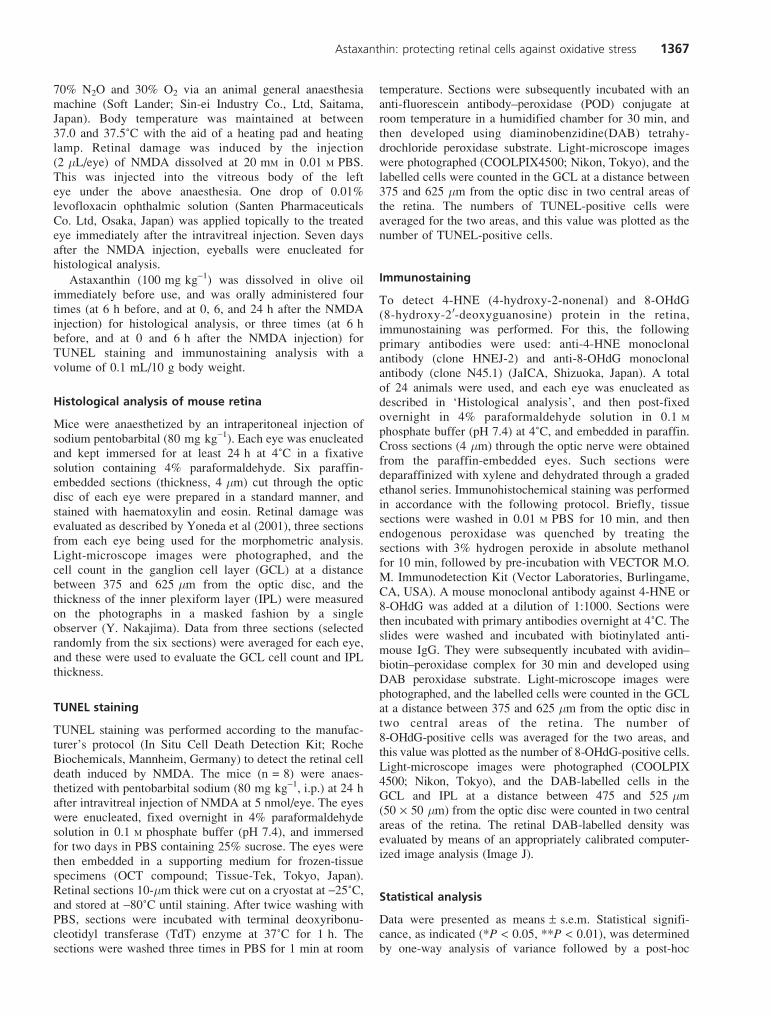
Astaxanthin at a concentration of 10 nM inhibited serumdeprivation-induced cell death in RGC-5 cell culture(Figure 2). Trolox (100 mM) also inhibited this cell death. Toinvestigate the neuroprotective effects of astaxanthin againstserum deprivation-induced oxidative stress, we determined thelevel of ROS in RGC-5 cells using a ROS-sensitive probe,CM-H2DCFDA. Non-treated control cells supplementedwith 1% FBS displayed little fluorescence intensity in the totalcells (Figure 3A). Total cell numberwas on average 300–400 perone sight using Metamorph. Serum deprivation resulted in anincrease in ROS production, as shown by increased DCFfluorescence (Figure 3B). Treatment with astaxanthin at 10 nMreduced the serum deprivation-induced ROS production(Figure 3C). For the evaluation of ROS production per cell,cellular radical intensity was quantified. Serum deprivationresulted in an8-fold increase inROSproduction (vs control), andastaxanthin (10 nM) reduced this ROS production to a similarextent as trolox (100 mM) (Figure 3D).
Effects of astaxanthin on the intracellular oxidation
of DCFH induced by various types of ROS
To investigate the effect of astaxanthin on hydrogen peroxide(H2O2), superoxide anion (O2�-), and hydroxyl radical(HO�) production, we employed a radical scavenging-capacity assay using a ROS-sensitive probe, CM-H2DCFDA.
50
110
100
90
80
70
60
Cel
l via
bili
ty (
% c
on
tro
l)
10 nM
Serum deprivation
ControlAstaxanthin Trolox
100 �M
**
*
Figure 2 Effects of astaxanthin on cell damage induced by serum
deprivation in RGC-5. Treatment with astaxanthin (10 nM) or trolox
(100 mM) significantly reduced the retinal cell damage induced by serum
deprivation. Cell viability was assessed by immersing cells in WST-8
solution for 3 h at 37˚C, with fluorescence being recorded
at 492/660 nm. Each column represents the mean ± s.e.m., n = 6.
*P < 0.05, **P < 0.01 vs serum deprivation alone.
A B C
D
H2O2
Cel
l via
bili
ty (
% c
on
tro
l)
110
100
90
80
70
60
50
40
30
Astaxanthin Trolox0.01 0.1 1 10 �M10 nMControl
****
** **
Figure 1 Effects of astaxanthin on cell damage induced by H2O2 in
RGC-5 culture. (A–C) Representative fluorescence microscopy of Hoechst
33342 staining at 24 h after addition of H2O2. A. Non-treated cells showed
normal nuclear morphology. B. H2O2 (0.3 mM)-induced neurotoxicity,
with cells showing nuclear condensation (arrows). C. Pretreatment with
10 nM astaxanthin at 1 h before H2O2-treatment reduced nuclear
condensation (arrows). D. Cell viability was assessed by immersing
cells in WST-8 solution for 3 h at 37˚C, with fluorescence being recorded
at 492/660 nm. H2O2 induced a decrease in cell viability. Astaxanthin
(0.1–10 nM) and trolox (10 mM) each significantly inhibited the H2O2-
induced cell damage. Each column represents the mean ± s.e.m., n = 6.
**P < 0.01 v. H2O2-treatment alone. Scale bar = 50 mm.
1368 Yoshimi Nakajima et al
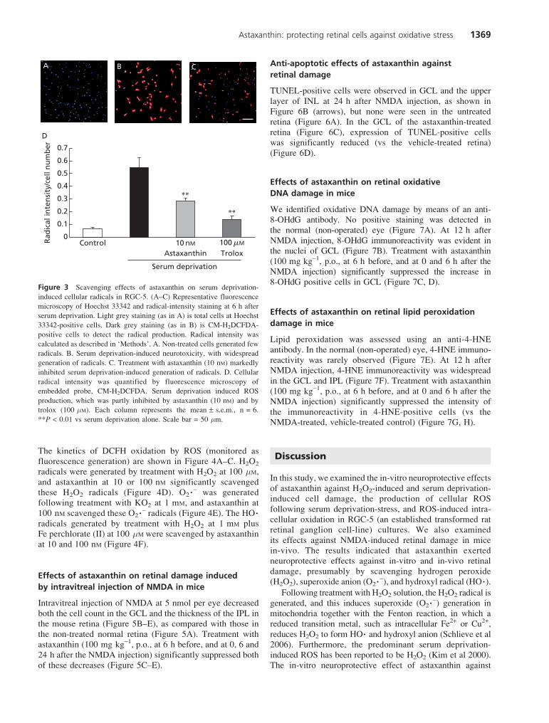
The kinetics of DCFH oxidation by ROS (monitored asfluorescence generation) are shown in Figure 4A–C. H2O2
radicals were generated by treatment with H2O2 at 100 mM,and astaxanthin at 10 or 100 nM significantly scavengedthese H2O2 radicals (Figure 4D). O2�- was generatedfollowing treatment with KO2 at 1 mM, and astaxanthin at100 nM scavenged these O2�- radicals (Figure 4E). The HO�radicals generated by treatment with H2O2 at 1 mM plusFe perchlorate (II) at 100 mM were scavenged by astaxanthinat 10 and 100 nM (Figure 4F).
Effects of astaxanthin on retinal damage induced
by intravitreal injection of NMDA in mice
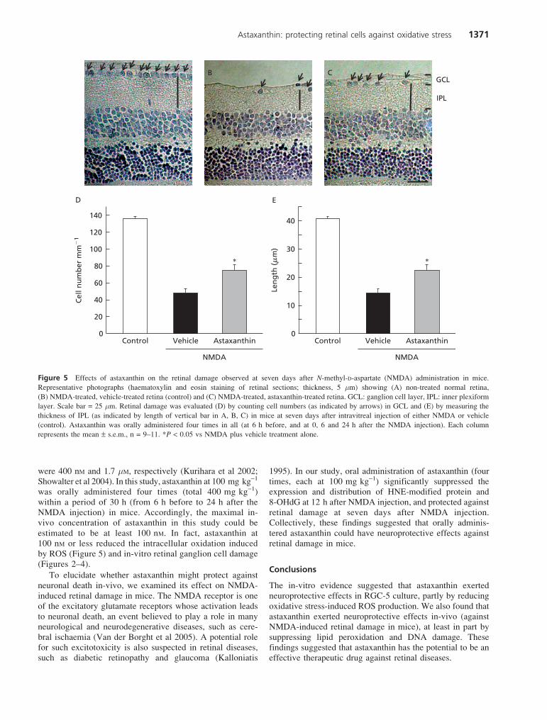
Intravitreal injection of NMDA at 5 nmol per eye decreasedboth the cell count in the GCL and the thickness of the IPL inthe mouse retina (Figure 5B–E), as compared with those inthe non-treated normal retina (Figure 5A). Treatment withastaxanthin (100 mg kg-1, p.o., at 6 h before, and at 0, 6 and24 h after the NMDA injection) significantly suppressed bothof these decreases (Figure 5C–E).
Anti-apoptotic effects of astaxanthin against
retinal damage
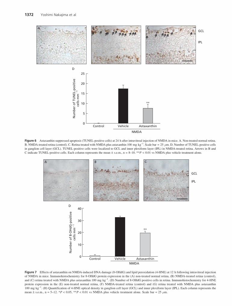
TUNEL-positive cells were observed in GCL and the upperlayer of INL at 24 h after NMDA injection, as shown inFigure 6B (arrows), but none were seen in the untreatedretina (Figure 6A). In the GCL of the astaxanthin-treatedretina (Figure 6C), expression of TUNEL-positive cellswas significantly reduced (vs the vehicle-treated retina)(Figure 6D).
Effects of astaxanthin on retinal oxidative
DNA damage in mice
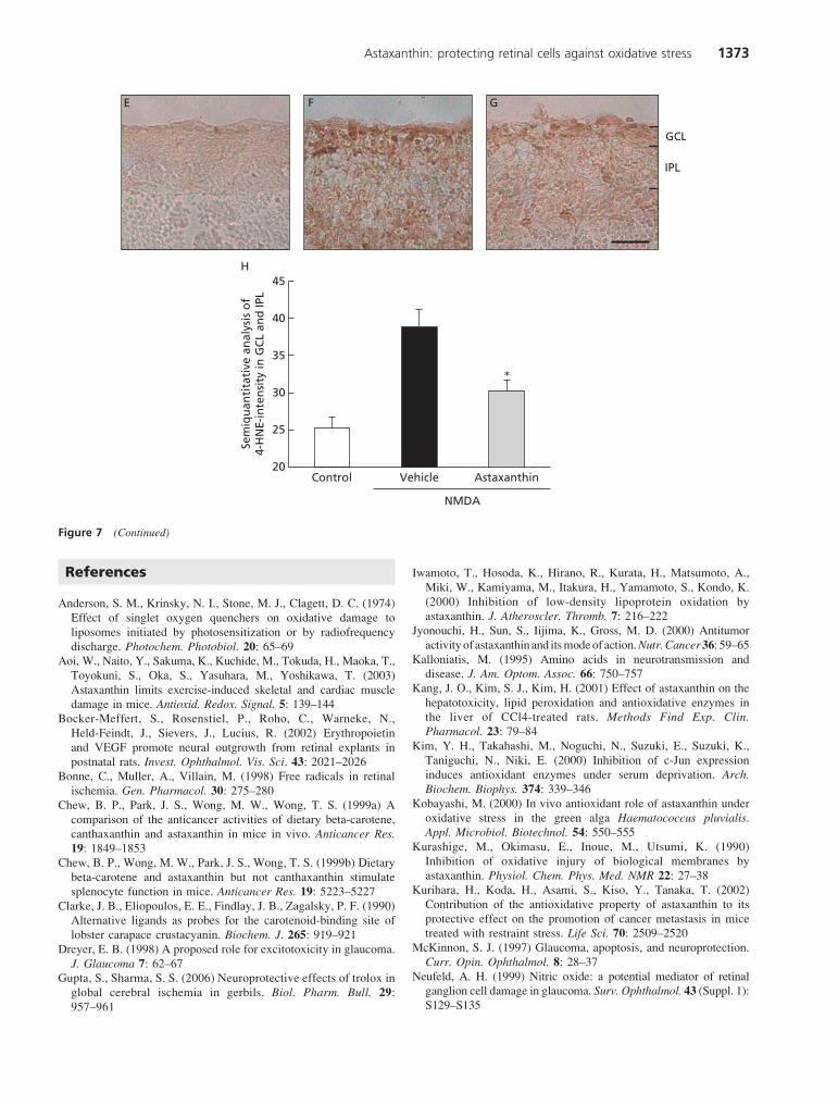
We identified oxidative DNA damage by means of an anti-8-OHdG antibody. No positive staining was detected inthe normal (non-operated) eye (Figure 7A). At 12 h afterNMDA injection, 8-OHdG immunoreactivity was evident inthe nuclei of GCL (Figure 7B). Treatment with astaxanthin(100 mg kg-1, p.o., at 6 h before, and at 0 and 6 h after theNMDA injection) significantly suppressed the increase in8-OHdG positive cells in GCL (Figure 7C, D).
Effects of astaxanthin on retinal lipid peroxidation
damage in mice
Lipid peroxidation was assessed using an anti-4-HNEantibody. In the normal (non-operated) eye, 4-HNE immuno-reactivity was rarely observed (Figure 7E). At 12 h afterNMDA injection, 4-HNE immunoreactivity was widespreadin the GCL and IPL (Figure 7F). Treatment with astaxanthin(100 mg kg-1, p.o., at 6 h before, and at 0 and 6 h after theNMDA injection) significantly suppressed the intensity ofthe immunoreactivity in 4-HNE-positive cells (vs theNMDA-treated, vehicle-treated control) (Figure 7G, H).
Discussion
In this study, we examined the in-vitro neuroprotective effectsof astaxanthin against H2O2-induced and serum deprivation-induced cell damage, the production of cellular ROSfollowing serum deprivation-stress, and ROS-induced intra-cellular oxidation in RGC-5 (an established transformed ratretinal ganglion cell-line) cultures. We also examinedits effects against NMDA-induced retinal damage in micein-vivo. The results indicated that astaxanthin exertedneuroprotective effects against in-vitro and in-vivo retinaldamage, presumably by scavenging hydrogen peroxide(H2O2), superoxide anion (O2�-), and hydroxyl radical (HO�).
Following treatment with H2O2 solution, the H2O2 radical isgenerated, and this induces superoxide (O2�-) generation inmitochondria together with the Fenton reaction, in which areduced transition metal, such as intracellular Fe2+ or Cu2+,reduces H2O2 to form HO� and hydroxyl anion (Schlieve et al2006). Furthermore, the predominant serum deprivation-induced ROS has been reported to be H2O2 (Kim et al 2000).The in-vitro neuroprotective effect of astaxanthin against
A B C
D
0.5
0.4
0.3
0.2
0.1
Rad
ical
inte
nsi
ty/c
ell n
um
ber
0.6
0
0.7
ControlAstaxanthin Trolox
10 nM
Serum deprivation
100 �M
∗∗
∗∗
Figure 3 Scavenging effects of astaxanthin on serum deprivation-
induced cellular radicals in RGC-5. (A–C) Representative fluorescence
microscopy of Hoechst 33342 and radical-intensity staining at 6 h after
serum deprivation. Light grey staining (as in A) is total cells at Hoechst
33342-positive cells. Dark grey staining (as in B) is CM-H2DCFDA-
positive cells to detect the radical production. Radical intensity was
calculated as described in ‘Methods’. A. Non-treated cells generated few
radicals. B. Serum deprivation-induced neurotoxicity, with widespread
generation of radicals. C. Treatment with astaxanthin (10 nM) markedly
inhibited serum deprivation-induced generation of radicals. D. Cellular
radical intensity was quantified by fluorescence microscopy of
embedded probe, CM-H2DCFDA. Serum deprivation induced ROS
production, which was partly inhibited by astaxanthin (10 nM) and by
trolox (100 mM). Each column represents the mean ± s.e.m., n = 6.
**P < 0.01 vs serum deprivation alone. Scale bar = 50 mm.
Astaxanthin: protecting retinal cells against oxidative stress 1369
H2O2-induced cell damage was stronger than that of trolox(Figure 1D), which is a powerful free-radical scavenger, mainlyof peroxynitrite (ONOO-), H2O2, O2�-, and HO� (Gupta &Sharma 2006). Astaxanthin protected cells against the celldamage inducedby singlet oxygen (Schroeder&Johnson1995),and singlet oxygen generated superoxide, hydrogen peroxide,and peroxyl radicalswithin the cell (Andersonet al 1974).Takentogether, all this suggested that the neuroprotective effect ofastaxanthin against serumdeprivation-induced cell damagemayhave been due to a reduction in the ROS production induced byserum deprivation.
Astaxanthin protected retinal ganglion cells (RGC-5)against H2O2-induced and serum deprivation-induced celldeath. The concentrations at which it exerted these neuropro-tective effects were consistent with those which had exertedprotective effects against ROS-induced intracellular oxidation(Figures 1–4). Here, we found evidence of in-vivo effects ofastaxanthin against the retinal damage induced by intravitrealinjection of NMDA in mice. We detected the effects ofastaxanthin against the accumulation of 4-hydroxy-2-nonenal
(4-HNE)-modified protein and 8-hydroxy-deoxyguanosine(8-OHdG) expression at 12 h after NMDA injection, and orallyadministered astaxanthin partly prevented the in-vivo retinaldamage induced by NMDA in mice. The effect of astaxanthinmay be attributable to attenuations of lipid peroxidation(4-HNE) and oxidative DNA damage (8-OHdG). 4-HNE, amarker of lipid peroxidation, is useful for following theprogress of lipid peroxidation at the cellular level after NMDAinjury. 8-OHdG is an oxidative form of the guanine nucleotidefound in DNA, and so DNA that has suffered oxidative damagedue to NMDA expresses 8-OHdG. Our results, therefore,indicated that astaxanthin inhibited neuronal damage in-vitroand in-vivo, and that these effects may have been partlymediated via a suppression of oxidative stress.
In this study, we administered free astaxanthin to mice.The concentrations of free astaxanthin in the plasma and liverafter single-dose oral gavage with free astaxanthin have beenmeasured by others (Showalter et al 2004). After adminis-tration of free astaxanthin (500 mg kg-1, p.o.) in an emulsionvehicle to mice, the concentrations in plasma and liver tissue
Astaxanthin
OH·
C
D E F
Flu
ore
scen
ce
Reaction time (min)
A
–0.05
0.05
0 5 10 15 20
0.15
0.25
0.35
0.45
0.55
10 nM
Control
Vehicle
100 nM
Reaction time (min)
–0.05
0.550.350.15
0.750.951.151.351.551.75
0 5 10 15 20
10 nM
Control
Vehicle
100 nM
H2O2
Rad
ical
inte
gra
l
Control
Astaxanthin
10 100 nM0
20
40
60
80
100
120
140
Control 10 Control 10
240 600
550
500
450
400
350
190
140
140
90
40
O2·–Astaxanthin
–10
Reaction time (min)
B
–0.05
–0.15
0.25
0.45
0.65
0.85
1.05
1.25
0 5 10 15 20
10 nM
Control
Vehicle
100 nM
100 nM 100 nM
**
***
Figure 4 Astaxanthin scavenged various radical species (H2O2, O2�- and HO�) in RGC-5. (A–C) Time-kinetics and concentration–response
relationships for antioxidant activity of astaxanthin. Astaxanthin was added to RGC-5 cultures for 1 h, then CM-H2DCFDA (10 mM) was added for
20 min. ROS production was stimulated with H2O2 at 100 mM, or with KO2 at 1 mM, or with H2O2 at 1 mM plus ferrous perchlorate (II) at 100 mM.
Fluorescence was measured for various time periods. A. H2O2-induced oxidation of DCFH in RGC-5. B. O2�--induced oxidation of DCFH in RGC-5.
C. HO�-induced oxidation of DCFH in RGC-5. D–F. Integral of ROS production from time-kinetic curves. Radical integral was calculated from
A–C, as described in ‘Methods’. Radical species were (D) H2O2, (E) O2�-, and (F) HO�. Each column represents the mean ± s.e.m., n = 6–12.
*P < 0.05, **P < 0.01 vs vehicle-treatment alone.
1370 Yoshimi Nakajima et al
were 400 nM and 1.7 mM, respectively (Kurihara et al 2002;Showalter et al 2004). In this study, astaxanthin at 100 mg kg-1
was orally administered four times (total 400 mg kg-1)within a period of 30 h (from 6 h before to 24 h after theNMDA injection) in mice. Accordingly, the maximal in-vivo concentration of astaxanthin in this study could beestimated to be at least 100 nM. In fact, astaxanthin at100 nM or less reduced the intracellular oxidation inducedby ROS (Figure 5) and in-vitro retinal ganglion cell damage(Figures 2–4).
To elucidate whether astaxanthin might protect againstneuronal death in-vivo, we examined its effect on NMDA-induced retinal damage in mice. The NMDA receptor is oneof the excitatory glutamate receptors whose activation leadsto neuronal death, an event believed to play a role in manyneurological and neurodegenerative diseases, such as cere-bral ischaemia (Van der Borght et al 2005). A potential rolefor such excitotoxicity is also suspected in retinal diseases,such as diabetic retinopathy and glaucoma (Kalloniatis
1995). In our study, oral administration of astaxanthin (fourtimes, each at 100 mg kg-1) significantly suppressed theexpression and distribution of HNE-modified protein and8-OHdG at 12 h after NMDA injection, and protected againstretinal damage at seven days after NMDA injection.Collectively, these findings suggested that orally adminis-tered astaxanthin could have neuroprotective effects againstretinal damage in mice.
Conclusions
The in-vitro evidence suggested that astaxanthin exertedneuroprotective effects in RGC-5 culture, partly by reducingoxidative stress-induced ROS production. We also found thatastaxanthin exerted neuroprotective effects in-vivo (againstNMDA-induced retinal damage in mice), at least in part bysuppressing lipid peroxidation and DNA damage. Thesefindings suggested that astaxanthin has the potential to be aneffective therapeutic drug against retinal diseases.
E
0
10
20
30
40
Len
gth
(�
m)
NMDA
Control Vehicle Astaxanthin0
20
40
60
80
100
120
140
Cel
l nu
mb
er m
m�
1
NMDA
Control Vehicle Astaxanthin
D
IPL
GCLA B C
∗∗
Figure 5 Effects of astaxanthin on the retinal damage observed at seven days after N-methyl-D-aspartate (NMDA) administration in mice.
Representative photographs (haematoxylin and eosin staining of retinal sections; thickness, 5 mm) showing (A) non-treated normal retina,
(B) NMDA-treated, vehicle-treated retina (control) and (C) NMDA-treated, astaxanthin-treated retina. GCL: ganglion cell layer, IPL: inner plexiform
layer. Scale bar = 25 mm. Retinal damage was evaluated (D) by counting cell numbers (as indicated by arrows) in GCL and (E) by measuring the
thickness of IPL (as indicated by length of vertical bar in A, B, C) in mice at seven days after intravitreal injection of either NMDA or vehicle
(control). Astaxanthin was orally administered four times in all (at 6 h before, and at 0, 6 and 24 h after the NMDA injection). Each column
represents the mean ± s.e.m., n = 9–11. *P < 0.05 vs NMDA plus vehicle treatment alone.
Astaxanthin: protecting retinal cells against oxidative stress 1371
D
NMDA
Control Vehicle Astaxanthin
5
10
15
20
0
25N
um
ber
of
TUN
EL-p
osi
tive
cells
mm
�1
IPL
GCLB CA
∗∗
Figure 6 Astaxanthin suppressed apoptosis (TUNEL-positive cells) at 24 h after intravitreal injection of NMDA in mice. A. Non-treated normal retina.
B. NMDA-treated retina (control). C. Retina treated with NMDA plus astaxanthin 100 mg kg-1. Scale bar = 25 mm. D. Number of TUNEL-positive cells
in ganglion cell layer (GCL). TUNEL-positive cells were localized to GCL and inner plexiform layer (IPL) in NMDA-treated retina. Arrows in B and
C indicate TUNEL-positive cells. Each column represents the mean ± s.e.m., n = 8–10. **P < 0.01 vs NMDA plus vehicle treatment alone.
C
D
Control Vehicle Astaxanthin
Nu
mb
er o
f 8-
OH
dG
-po
siti
vece
lls m
m�
1
BA
IPL
GCL
0
10
20
30
40
NMDA
∗∗
Figure 7 Effects of astaxanthin on NMDA-induced DNA damage (8-OHdG) and lipid peroxidation (4-HNE) at 12 h following intravitreal injection
of NMDA in mice. Immunohistochemistry for 8-OHdG protein expression in the (A) non-treated normal retina, (B) NMDA-treated retina (control),
and (C) retina treated with NMDA plus astaxanthin 100 mg kg-1. (D) Number of 8-OHdG positive cells in retina. Immunohistochemistry for 4-HNE
protein expression in the (E) non-treated normal retina, (F) NMDA-treated retina (control) and (G) retina treated with NMDA plus astaxanthin
100 mg kg-1. (H) Quantification of 4-HNE optical density in ganglion cell layer (GCL) and inner plexiform layer (IPL). Each column represents the
mean ± s.e.m., n = 5–12. *P < 0.05, **P < 0.01 vs NMDA plus vehicle treatment alone. Scale bar = 25 mm.
1372 Yoshimi Nakajima et al
References
Anderson, S. M., Krinsky, N. I., Stone, M. J., Clagett, D. C. (1974)
Effect of singlet oxygen quenchers on oxidative damage to
liposomes initiated by photosensitization or by radiofrequency
discharge. Photochem. Photobiol. 20: 65–69Aoi, W., Naito, Y., Sakuma, K., Kuchide, M., Tokuda, H., Maoka, T.,
Toyokuni, S., Oka, S., Yasuhara, M., Yoshikawa, T. (2003)
Astaxanthin limits exercise-induced skeletal and cardiac muscle
damage in mice. Antioxid. Redox. Signal. 5: 139–144Bocker-Meffert, S., Rosenstiel, P., Roho, C., Warneke, N.,
Held-Feindt, J., Sievers, J., Lucius, R. (2002) Erythropoietin
and VEGF promote neural outgrowth from retinal explants in
postnatal rats. Invest. Ophthalmol. Vis. Sci. 43: 2021–2026Bonne, C., Muller, A., Villain, M. (1998) Free radicals in retinal
ischemia. Gen. Pharmacol. 30: 275–280Chew, B. P., Park, J. S., Wong, M. W., Wong, T. S. (1999a) A
comparison of the anticancer activities of dietary beta-carotene,
canthaxanthin and astaxanthin in mice in vivo. Anticancer Res.
19: 1849–1853Chew, B. P., Wong, M. W., Park, J. S., Wong, T. S. (1999b) Dietary
beta-carotene and astaxanthin but not canthaxanthin stimulate
splenocyte function in mice. Anticancer Res. 19: 5223–5227Clarke, J. B., Eliopoulos, E. E., Findlay, J. B., Zagalsky, P. F. (1990)
Alternative ligands as probes for the carotenoid-binding site of
lobster carapace crustacyanin. Biochem. J. 265: 919–921Dreyer, E. B. (1998) A proposed role for excitotoxicity in glaucoma.
J. Glaucoma 7: 62–67Gupta, S., Sharma, S. S. (2006) Neuroprotective effects of trolox in
global cerebral ischemia in gerbils. Biol. Pharm. Bull. 29:957–961
Iwamoto, T., Hosoda, K., Hirano, R., Kurata, H., Matsumoto, A.,
Miki, W., Kamiyama, M., Itakura, H., Yamamoto, S., Kondo, K.
(2000) Inhibition of low-density lipoprotein oxidation by
astaxanthin. J. Atheroscler. Thromb. 7: 216–222Jyonouchi, H., Sun, S., Iijima, K., Gross, M. D. (2000) Antitumor
activityof astaxanthinand itsmodeof action.Nutr.Cancer36: 59–65Kalloniatis, M. (1995) Amino acids in neurotransmission and
disease. J. Am. Optom. Assoc. 66: 750–757Kang, J. O., Kim, S. J., Kim, H. (2001) Effect of astaxanthin on the
hepatotoxicity, lipid peroxidation and antioxidative enzymes in
the liver of CCl4-treated rats. Methods Find Exp. Clin.
Pharmacol. 23: 79–84Kim, Y. H., Takahashi, M., Noguchi, N., Suzuki, E., Suzuki, K.,
Taniguchi, N., Niki, E. (2000) Inhibition of c-Jun expression
induces antioxidant enzymes under serum deprivation. Arch.
Biochem. Biophys. 374: 339–346Kobayashi, M. (2000) In vivo antioxidant role of astaxanthin under
oxidative stress in the green alga Haematococcus pluvialis.
Appl. Microbiol. Biotechnol. 54: 550–555Kurashige, M., Okimasu, E., Inoue, M., Utsumi, K. (1990)
Inhibition of oxidative injury of biological membranes by
astaxanthin. Physiol. Chem. Phys. Med. NMR 22: 27–38Kurihara, H., Koda, H., Asami, S., Kiso, Y., Tanaka, T. (2002)
Contribution of the antioxidative property of astaxanthin to its
protective effect on the promotion of cancer metastasis in mice
treated with restraint stress. Life Sci. 70: 2509–2520McKinnon, S. J. (1997) Glaucoma, apoptosis, and neuroprotection.
Curr. Opin. Ophthalmol. 8: 28–37Neufeld, A. H. (1999) Nitric oxide: a potential mediator of retinal
ganglion cell damage in glaucoma. Surv. Ophthalmol. 43 (Suppl. 1):S129–S135
Control Vehicle Astaxanthin
Sem
iqu
anti
tati
ve a
nal
ysis
of
4-H
NE-
inte
nsi
ty in
GC
L an
d IP
L
E F
H
G
IPL
GCL
20
25
35
30
40
45
NMDA
∗
Figure 7 (Continued)
Astaxanthin: protecting retinal cells against oxidative stress 1373
O’Connor, I., O’Brien, N. (1998) Modulation of UVA light-induced
oxidative stress by beta-carotene, lutein and astaxanthin in
cultured fibroblasts. J. Dermatol. Sci. 16: 226–230Ohgami, K., Shiratori, K., Kotake, S., Nishida, T., Mizuki, N.,
Yazawa, K., Ohno, S. (2003) Effects of astaxanthin on
lipopolysaccharide-induced inflammation in vitro and in vivo.
Invest. Ophthalmol. Vis. Sci. 44: 2694–2701Okai, Y., Higashi-Okai, K. (1996) Possible immunomodulating
activities of carotenoids in in vitro cell culture experiments. Int. J.
Immunopharmacol. 18: 753–758Parisi, V., Tedeschi, M., Gallinaro, G., Varano, M., Saviano, S.,
Piermarocchi, S. (2008) Carotenoids and antioxidants in age-
related maculopathy Italian study: multifocal electroretinogram
modifications after 1 year. Ophthalmology 115: 324–333Schlieve, C. R., Lieven, C. J., Levin, L. A. (2006) Biochemical
activity of reactive oxygen species scavengers do not predict
retinal ganglion cell survival. Invest. Ophthalmol. Vis. Sci. 47:3878–3886
Schroeder, W. A., Johnson, E. A. (1995) Singlet oxygen and peroxyl
radicals regulate carotenoid biosynthesis in Phaffia rhodozyma.
J. Biol. Chem. 270: 18374–18379
Shimazawa, M., Yamashima, T., Agarwal, N., Hara, H. (2005)
Neuroprotective effects of minocycline against in vitro and in
vivo retinal ganglion cell damage. Brain Res. 1053: 185–194Showalter, L. A., Weinman, S. A., Osterlie, M., Lockwood, S. F.
(2004) Plasma appearance and tissue accumulation of non-
esterified, free astaxanthin in C57BL/6 mice after oral dosing of a
disodium disuccinate diester of astaxanthin (Heptax). Comp.
Biochem. Physiol. C Toxicol. Pharmacol. 137: 227–236Siliprandi, R., Canella, R., Carmignoto, G., Schiavo, N., Zanellato,
A., Zanoni, R., Vantini, G. (1992) N-methyl-D-aspartate-induced
neurotoxicity in the adult rat retina. Vis. Neurosci. 8: 567–573Uchiyama, K., Naito, Y., Hasegawa, G., Nakamura, N., Takahashi, J.,
Yoshikawa, T. (2002) Astaxanthin protects beta-cells against
glucose toxicity in diabetic db/db mice. Redox. Rep. 7: 290–293Van der Borght, K., Mulder, J., Keijser, J. N., Eggen, B. J., Luiten,
P. G., Van der Zee, E. A. (2005) Input from the medial septum
regulates adult hippocampal neurogenesis. Brain Res. Bull. 67:117–125
Yoneda, S., Tanihara, H., Kido, N., Honda, Y., Goto, W., Hara, H.,
Miyawaki, N. (2001) Interleukin-1beta mediates ischemic injury
in the rat retina. Exp. Eye. Res. 73: 661–667
1374 Yoshimi Nakajima et al
Related Documents