Nitrogen-depleted Chlorella zofingiensis produces astaxanthin, ketolutein and their fatty acid esters: a carotenoid metabolism study Kim J. M. Mulders & Yannick Weesepoel & Pierre Bodenes & Packo P. Lamers & Jean-Paul Vincken & Dirk E. Martens & Harry Gruppen & René H. Wijffels Received: 3 February 2014 /Revised and accepted: 4 May 2014 # Springer Science+Business Media Dordrecht 2014 Abstract Natural carotenoids such as astaxanthin, β,β-caro- tene and lutein are pigments with a high market value. We studied the effects of nitrogen depletion on the carotenoid metabolism of Chlorella zofingiensis (Chlorophyta) and the subsequent treatment with diphenylamine (DPA), an inhibitor of the biosynthesis of secondary ketocarotenoids. Pigments were identified and quantified based on reversed phase ultra- high performance liquid chromatography photodiode array tandem mass spectrometry (RP-UHPLC-PDA-MS n ). Nitrogen depletion (without DPA) resulted in a degradation of chlorophylls and primary carotenoids and an accumulation of astaxanthin, ketolutein, canthaxanthin, adonixanthin and β,β-carotene. The DPA treatment decreased the overall pro- duction of β,β-carotene derivatives (sum of astaxanthin, can- thaxanthin, echinenone and adonixanthin); however, the pro- duction of ketolutein and degradation of primary carotenoids were not modified. This suggests that the regulatory mecha- nisms controlling the flux towards ketolutein and primary carotenoids were not affected by the decreased levels of β,β-carotene derivatives. In addition, DPA increased produc- tion of the individual carotenoids, adonixanthin and echinenone. Insight into the regulation of microalgal caroten- oid biosynthesis as demonstrated in this paper is essential when a large-scale carotenoid production process is to be optimised or a recombinant C. zofingiensis strain is to be designed with the intention of excessively producing primary or secondary carotenoids. Keywords Chlorella zofingiensis . Nitrogen depletion . Diphenylamine . Enzyme inhibitor . Carotenoid metabolism . Astaxanthin . Ketolutein Introduction Natural carotenoids such as astaxanthin, β,β-carotene and lutein are pigments with a high market value. It has been claimed that these carotenoids beneficially affect health due to their anti-oxidative activity (Spolaore et al. 2006). Possible production organisms of naturally derived carotenoids that have gained significant attention are microalgae, because they contain an extensive number of various carotenoids, part of which can be produced in concentrations exceeding those ascertained in higher plants by one or more orders of magni- tude. Nevertheless, economically feasible microalgal caroten- oid production will require process and/or strain optimisation (Mulders et al. 2014). Microalgal carotenoids can be categorised into primary and secondary carotenoids. Primary carotenoids are functionally bound to the photosystems inside the chloroplast, whereas secondary carotenoids are not bound to the photosystems. Kim J.M. Mulders and Yannick Weesepoel contributed equally to the manuscript. Electronic supplementary material The online version of this article (doi:10.1007/s10811-014-0333-3) contains supplementary material, which is available to authorized users. K. J. M. Mulders : P. P. Lamers : D. E. Martens : R. H. Wijffels Bioprocess Engineering, AlgaePARC, Wageningen University, P.O. Box 16, 6700 AA Wageningen, The Netherlands Y. Weesepoel : J.<P. Vincken : H. Gruppen Laboratory of Food Chemistry, Wageningen University, P.O. Box 17, 6700 AA Wageningen, The Netherlands K. J. M. Mulders (*) : Y. Weesepoel FeyeCon Development and Implementation, Rijnkade 17a, 1382 GS Weesp, The Netherlands e-mail: [email protected] P. Bodenes Polytech Graduate School of Engineering, Nantes University, Bd. de l’Université, 44600 Saint-Nazaire, France J Appl Phycol DOI 10.1007/s10811-014-0333-3

Welcome message from author
This document is posted to help you gain knowledge. Please leave a comment to let me know what you think about it! Share it to your friends and learn new things together.
Transcript
Nitrogen-depleted Chlorella zofingiensis produces astaxanthin,
ketolutein and their fatty acid esters: a carotenoid
metabolism study
Kim J. M. Mulders & Yannick Weesepoel & Pierre Bodenes &
Packo P. Lamers & Jean-Paul Vincken & Dirk E. Martens &
Harry Gruppen & René H. Wijffels
Received: 3 February 2014 /Revised and accepted: 4 May 2014# Springer Science+Business Media Dordrecht 2014
Abstract Natural carotenoids such as astaxanthin, β,β-caro-
tene and lutein are pigments with a high market value. We
studied the effects of nitrogen depletion on the carotenoid
metabolism of Chlorella zofingiensis (Chlorophyta) and the
subsequent treatment with diphenylamine (DPA), an inhibitor
of the biosynthesis of secondary ketocarotenoids. Pigments
were identified and quantified based on reversed phase ultra-
high performance liquid chromatography photodiode array
tandem mass spectrometry (RP-UHPLC-PDA-MSn).
Nitrogen depletion (without DPA) resulted in a degradation
of chlorophylls and primary carotenoids and an accumulation
of astaxanthin, ketolutein, canthaxanthin, adonixanthin and
β,β-carotene. The DPA treatment decreased the overall pro-
duction of β,β-carotene derivatives (sum of astaxanthin, can-
thaxanthin, echinenone and adonixanthin); however, the pro-
duction of ketolutein and degradation of primary carotenoids
were not modified. This suggests that the regulatory mecha-
nisms controlling the flux towards ketolutein and primary
carotenoids were not affected by the decreased levels of
β,β-carotene derivatives. In addition, DPA increased produc-
tion of the individual carotenoids, adonixanthin and
echinenone. Insight into the regulation of microalgal caroten-
oid biosynthesis as demonstrated in this paper is essential
when a large-scale carotenoid production process is to be
optimised or a recombinant C. zofingiensis strain is to be
designed with the intention of excessively producing primary
or secondary carotenoids.
Keywords Chlorella zofingiensis . Nitrogen depletion .
Diphenylamine . Enzyme inhibitor . Carotenoidmetabolism .
Astaxanthin . Ketolutein
Introduction
Natural carotenoids such as astaxanthin, β,β-carotene and
lutein are pigments with a high market value. It has been
claimed that these carotenoids beneficially affect health due
to their anti-oxidative activity (Spolaore et al. 2006). Possible
production organisms of naturally derived carotenoids that
have gained significant attention are microalgae, because they
contain an extensive number of various carotenoids, part of
which can be produced in concentrations exceeding those
ascertained in higher plants by one or more orders of magni-
tude. Nevertheless, economically feasible microalgal caroten-
oid production will require process and/or strain optimisation
(Mulders et al. 2014).
Microalgal carotenoids can be categorised into primary and
secondary carotenoids. Primary carotenoids are functionally
bound to the photosystems inside the chloroplast, whereas
secondary carotenoids are not bound to the photosystems.
Kim J.M. Mulders and Yannick Weesepoel contributed equally to the
manuscript.
Electronic supplementary material The online version of this article
(doi:10.1007/s10811-014-0333-3) contains supplementary material,
which is available to authorized users.
K. J. M. Mulders : P. P. Lamers :D. E. Martens : R. H. Wijffels
Bioprocess Engineering, AlgaePARC, Wageningen University, P.O.
Box 16, 6700 AAWageningen, The Netherlands
Y. Weesepoel : J.<P. Vincken :H. Gruppen
Laboratory of Food Chemistry, Wageningen University, P.O. Box 17,
6700 AAWageningen, The Netherlands
K. J. M. Mulders (*) :Y. Weesepoel
FeyeCon Development and Implementation, Rijnkade 17a, 1382
GS Weesp, The Netherlands
e-mail: [email protected]
P. Bodenes
Polytech Graduate School of Engineering, Nantes University,
Bd. de l’Université, 44600 Saint-Nazaire, France
J Appl Phycol
DOI 10.1007/s10811-014-0333-3
An even more significant difference is their concentration
under various growth conditions. Under prolonged limited-
light growth conditions, secondary carotenoids are generally
absent or present only in extremely low concentrations, and
primary carotenoids are generally present in their maximal
concentration (typically, below 0.5 % dry weight (DW) (w/w))
(Mulders et al. 2014). Under adverse growth conditions (re-
ferred to as ‘stress’ conditions), primary carotenoids are gen-
erally degraded, and in certain green algae, secondary carot-
enoids are produced in excessive amounts (up to 10 % DW
(w/w)) (Lamers et al. 2008). As a consequence of these dif-
ferent responses, overproduction of the primary and second-
ary carotenoids requires different approaches. Secondary
carotenoid overproduction necessitates optimisation of the
process (stress) conditions, which may be enhanced by
additional genetic engineering. Although subsaturating light
conditions result in minor increases in primary pigment
concentrations, actual overproduction of primary carotenoids
requires modification of the carotenoid biosynthesis routes,
i.e. with genetic engineering (Mulders et al. 2014). Both
approaches share that insight into pigment metabolic regu-
lation is crucial.
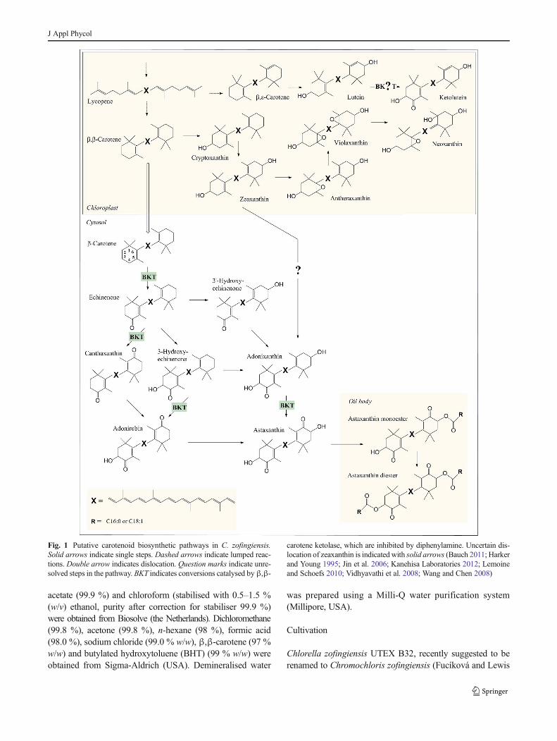
Chlorella zofingiensis is one of the most important
carotenogenic green algae in relationship to biotechnological
applications (Solovchenko 2013). In this species, lycopene,
which can be converted into β,ε-carotene (α-carotene) or
β,β-carotene (Fig. 1), is the precursor of all primary and
secondary carotenoids. Hydroxylation of α-carotene yields
lutein, the most abundant primary carotenoid of
C. zofingiensis (Del Campo et al. 2004). Hydroxylation of
β,β-carotene provides zeaxanthin, which can be epoxidised
twice, resulting in violaxanthin which can then be isomerised,
leading to neoxanthin. Under adverse growth conditions,
C. zofingiensis can accumulate multiple secondary caroten-
oids including astaxanthin, canthaxanthin and adonixanthin
(Bar et al. 1995; Orosa et al. 2001). Presumably, β,β-carotene
is transferred first to the cytosol where it is subsequently
converted into astaxanthin (Fig. 1). This conversion requires
two oxygenation and two hydroxylation reactions.
Oxygenations are catalysed by β,β-carotene ketolase
(BKT), and hydroxylations are catalysed by a β,β-carotene
hydroxylase (CHYb). Esterification of astaxanthin results in
monoesters and diesters which appear to accumulate in
triacylglyceride (TAG) oil bodies (Solovchenko 2013)
(Fig. 1). It has been postulated that in C. zofingiensis, zeaxan-
thin can also be a precursor of astaxanthin through conversion
into adonixanthin (Fig. 1). However, this pathway has not yet
been fully resolved (Huang et al. 2006;Wang and Chen 2008).
Furthermore, the accumulation of canthaxanthin in a relatively
high concentration has led to the speculation that this carot-
enoid is an end product rather than a precursor of astaxanthin
(Huang et al. 2006). This speculation requires further confir-
mation. Finally, a recent discovery by Bauch (2011) suggests
that BKT converts the primary carotenoid lutein into the
secondary carotenoid ketolutein under adverse growth condi-
tions. As this is contradictory with all other available litera-
ture, this requires confirmation. Thus, whereas primary carot-
enoid biosynthesis pathways in C. zofingiensis are thoroughly
investigated, those for secondary carotenoids require further
elucidation. Furthermore, the metabolic regulation of primary
and secondary carotenoid biosynthesis is extensively under-
investigated which is, as stated, of utmost importance when
designing a metabolically engineered carotenoid production
strain or optimising a large-scale carotenoid production pro-
cess. The aim of this paper was to obtain additional insight
into the pathways and regulation of carotenoid metabolism of
C. zofingiensis.
This was accomplished by depleting C. zofingiensis of ni-
trogen, which effectively induces secondary carotenogenesis
and subsequently adding diphenylamine (DPA), an inhibitor of
BKT (Harker and Young 1995; Fan et al. 1995; Wang and
Chen 2008). Since BKT catalyses multiple secondary caroten-
oid conversions (Fig. 1), inhibition was expected to lead to
insight into the pathway of astaxanthin biosynthesis as it did,
for example, in the green alga Haematococcus pluvialis
(Collins et al. 2011; Fan et al. 1995; Harker and Young 1995;
Zhekisheva et al. 2005). Additionally, as primary and second-
ary carotenoids of C. zofingiensis possess a common metabolic
precursor and BKT catalyses the first step of secondary
carotenogenesis, inhibition with DPA was expected to alter
the primary pigment metabolism (e.g. lutein biosynthesis) and
reveal insights in carotenoid metabolic regulation. To ensure
that the entire collection of carotenoids ofC. zofingiensiswould
be revealed, carotenoids were identified employing reversed
phase liquid chromatography and additional mass
spectrometry.
Materials and methods
Materials
(rac.)-Adonirubin (95 %), (3S,3′R)-adonixanthin (99 %),
(rac./meso)-astaxanthin (97 %), (rac./meso)-astaxanthin
monopalmitate (97 %), (rac./meso)-astaxanthin dipalmitate
(98 %), canthaxanthin (98 %), echinenone (98 %), (rac.)-3-
hydroxyechinenone (97 %), lutein (96 %) and (3R,3′R)-zea-
xanthin (97 %) (all w/w) were purchased from CaroteNature
(Lupsingen, Switzerland). Violaxanthin (98 %), lycopene
(92 %), cryptoxanthin (β-cryptoxanthin) (98 %),
antheraxanthin (97 %), α-cryptoxanthin (98 %), and β,ε-
carotene (α-carotene) (88 %) (all w/w) were purchased in
approximately 1 mg mL−1 ethanol or acetone solutions from
DHI laboratory products (Denmark). Chlorophyll a (90.0 %
w/w) was purchased from Wako Pure Chemical Industries
(Japan). Methanol (99.8 %), acetonitrile (99.97 %), ethyl
J Appl Phycol
acetate (99.9 %) and chloroform (stabilised with 0.5–1.5 %
(w/v) ethanol, purity after correction for stabiliser 99.9 %)
were obtained from Biosolve (the Netherlands). Dichloromethane
(99.8 %), acetone (99.8 %), n-hexane (98 %), formic acid
(98.0 %), sodium chloride (99.0 % w/w), β,β-carotene (97 %
w/w) and butylated hydroxytoluene (BHT) (99 % w/w) were
obtained from Sigma-Aldrich (USA). Demineralised water
was prepared using a Milli-Q water purification system
(Millipore, USA).
Cultivation
Chlorella zofingiensis UTEX B32, recently suggested to be
renamed to Chromochloris zofingiensis (Fucíková and Lewis
Fig. 1 Putative carotenoid biosynthetic pathways in C. zofingiensis.
Solid arrows indicate single steps. Dashed arrows indicate lumped reac-
tions. Double arrow indicates dislocation. Question marks indicate unre-
solved steps in the pathway. BKT indicates conversions catalysed byβ,β-
carotene ketolase, which are inhibited by diphenylamine. Uncertain dis-
location of zeaxanthin is indicated with solid arrows (Bauch 2011; Harker
and Young 1995; Jin et al. 2006; Kanehisa Laboratories 2012; Lemoine
and Schoefs 2010; Vidhyavathi et al. 2008; Wang and Chen 2008)
J Appl Phycol
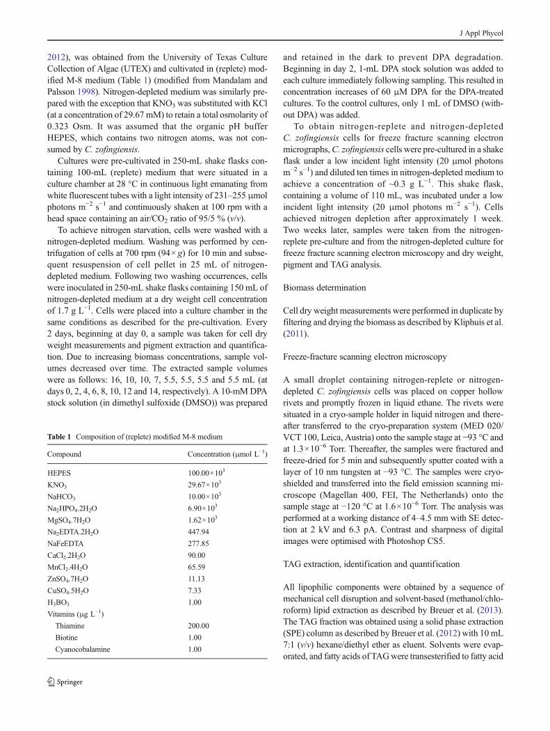
2012), was obtained from the University of Texas Culture
Collection of Algae (UTEX) and cultivated in (replete) mod-
ified M-8 medium (Table 1) (modified from Mandalam and
Palsson 1998). Nitrogen-depleted medium was similarly pre-
pared with the exception that KNO3 was substituted with KCl
(at a concentration of 29.67 mM) to retain a total osmolarity of
0.323 Osm. It was assumed that the organic pH buffer
HEPES, which contains two nitrogen atoms, was not con-
sumed by C. zofingiensis.
Cultures were pre-cultivated in 250-mL shake flasks con-
taining 100-mL (replete) medium that were situated in a
culture chamber at 28 °C in continuous light emanating from
white fluorescent tubes with a light intensity of 231–255 μmol
photons m−2 s−1 and continuously shaken at 100 rpm with a
head space containing an air/CO2 ratio of 95/5 % (v/v).
To achieve nitrogen starvation, cells were washed with a
nitrogen-depleted medium. Washing was performed by cen-
trifugation of cells at 700 rpm (94×g) for 10 min and subse-
quent resuspension of cell pellet in 25 mL of nitrogen-
depleted medium. Following two washing occurrences, cells
were inoculated in 250-mL shake flasks containing 150 mL of
nitrogen-depleted medium at a dry weight cell concentration
of 1.7 g L−1. Cells were placed into a culture chamber in the
same conditions as described for the pre-cultivation. Every
2 days, beginning at day 0, a sample was taken for cell dry
weight measurements and pigment extraction and quantifica-
tion. Due to increasing biomass concentrations, sample vol-
umes decreased over time. The extracted sample volumes
were as follows: 16, 10, 10, 7, 5.5, 5.5, 5.5 and 5.5 mL (at
days 0, 2, 4, 6, 8, 10, 12 and 14, respectively). A 10-mMDPA
stock solution (in dimethyl sulfoxide (DMSO)) was prepared
and retained in the dark to prevent DPA degradation.
Beginning in day 2, 1-mL DPA stock solution was added to
each culture immediately following sampling. This resulted in
concentration increases of 60 μM DPA for the DPA-treated
cultures. To the control cultures, only 1 mL of DMSO (with-
out DPA) was added.
To obtain nitrogen-replete and nitrogen-depleted
C. zofingiensis cells for freeze fracture scanning electron
micrographs,C. zofingiensis cells were pre-cultured in a shake
flask under a low incident light intensity (20 μmol photons
m−2 s−1) and diluted ten times in nitrogen-depleted medium to
achieve a concentration of ~0.3 g L−1. This shake flask,
containing a volume of 110 mL, was incubated under a low
incident light intensity (20 μmol photons m−2 s−1). Cells
achieved nitrogen depletion after approximately 1 week.
Two weeks later, samples were taken from the nitrogen-
replete pre-culture and from the nitrogen-depleted culture for
freeze fracture scanning electron microscopy and dry weight,
pigment and TAG analysis.
Biomass determination
Cell dry weight measurements were performed in duplicate by
filtering and drying the biomass as described by Kliphuis et al.
(2011).
Freeze-fracture scanning electron microscopy
A small droplet containing nitrogen-replete or nitrogen-
depleted C. zofingiensis cells was placed on copper hollow
rivets and promptly frozen in liquid ethane. The rivets were
situated in a cryo-sample holder in liquid nitrogen and there-
after transferred to the cryo-preparation system (MED 020/
VCT 100, Leica, Austria) onto the sample stage at −93 °C and
at 1.3×10⁻⁶ Torr. Thereafter, the samples were fractured and
freeze-dried for 5 min and subsequently sputter coated with a
layer of 10 nm tungsten at −93 °C. The samples were cryo-
shielded and transferred into the field emission scanning mi-
croscope (Magellan 400, FEI, The Netherlands) onto the
sample stage at −120 °C at 1.6×10⁻⁶ Torr. The analysis was
performed at a working distance of 4–4.5 mm with SE detec-
tion at 2 kV and 6.3 pA. Contrast and sharpness of digital
images were optimised with Photoshop CS5.
TAG extraction, identification and quantification
All lipophilic components were obtained by a sequence of
mechanical cell disruption and solvent-based (methanol/chlo-
roform) lipid extraction as described by Breuer et al. (2013).
The TAG fraction was obtained using a solid phase extraction
(SPE) column as described by Breuer et al. (2012) with 10 mL
7:1 (v/v) hexane/diethyl ether as eluent. Solvents were evap-
orated, and fatty acids of TAGwere transesterified to fatty acid
Table 1 Composition of (replete) modified M-8 medium
Compound Concentration (μmol L−1)
HEPES 100.00×10³
KNO3 29.67×10³
NaHCO3 10.00×10³
Na2HPO4.2H2O 6.90×10³
MgSO4.7H2O 1.62×10³
Na2EDTA.2H2O 447.94
NaFeEDTA 277.85
CaCl2.2H2O 90.00
MnCl2.4H2O 65.59
ZnSO4.7H2O 11.13
CuSO4.5H2O 7.33
H3BO3 1.00
Vitamins (μg L−1)
Thiamine 200.00
Biotine 1.00
Cyanocobalamine 1.00
J Appl Phycol
methyl esters (FAMEs). FAMEs were identified and quanti-
fied utilising GC-FID as described by Breuer et al. (2012).
Pigment extraction
Pigment extracts were obtained by a sequence of mechanical
cell disruption and solvent-based (methanol/chloroform) pig-
ment extraction as described by Lamers et al. (2010) with the
exception that after sampling, the cells were centrifuged for
5 min at 2,500 rpm (1,204×g). The cell pellet was transferred
to a bead-beating tube and centrifuged again for 5 min at
2,500 rpm (1,204×g). Supernatant was discarded, and cells
were stored at −80 °C. The cells were subsequently freeze-
dried. One millilitre of methanol/chloroform was added to the
bead-beating tube where after the cells were physically
disrupted by employing a Precellys® 24 bead beater
(Bertin Technology, France). To inhibit cell heating, 60 s
of disruption at 2,500 rpm (1,204×g) was followed by 5 min
of cooling on ice. For each sample, three series of
disruption/cooling cycles were performed. After extraction,
the dried chloroform fraction was resolved in acetone/
methanol 7:3 (v/v) containing 0.10 % (w/v) BHT and sup-
plemented to 75 % (v/v) acetonitrile for liquid chromatogra-
phy (LC) analysis.
Separation, identification and quantification of pigments
Separation and identification of carotenoids and porphyrins
were performed on an Accela UHPLC system (Thermo
Scientific, USA) equipped with a pump, degasser, auto sam-
pler, and photodiode array (PDA) detector, coupled in-line to a
LTQ-VelosPro dual-pressure ion trap mass spectrometer
equipped with a H-ESI probe (Thermo Scientific). Samples
(5.0 μL) were quantitatively injected on an Aquity UPLC
Shield C18 BEH column (2.1×150 mm, 1.7 μm particle size;
Waters, Milford, USA) fitted to a Vanguard pre-column (2.1×
5 mm, 1.7 μm particle size; Waters). The eluents were 50 %
(v/v) acetonitrile (A) in demineralised water, acetonitrile (B)
and ethyl acetate (C) which all contained 0.10 % (v/v) formic
acid. The flow rate was maintained at 300 μL min−1. The
program was initiated from 25 % A/75 % B and then as
follows: to 15 min—linear gradient to 100 % B, to
22.5 min—isocratic at 100 % B, to 29.5 min—linear gradient
to 87.5 % B/12.5 % C, to 31.5 min—linear gradient to 70 %
B/30 % C, to 41.5 min—linear gradient to 100 % C and to
42.5 min—isocratic at 100 % C. After 42.5 min, the eluent
composition reverted to its initial composition in 7.5 min
followed by an equilibration phase of 2.5 min. Detection
wavelengths for UV–vis were adjusted to 450 nm
(carotenoids) and 660 nm (porphyrins) with a 1-nm wave-
length step and 9-nm filter bandwidth.
All mass spectrometric data was recorded as described by
Mulders et al. (2013) with the exception that data was
recorded over the m/z range 500–620 (0.0–17.0 min), 500–
900 (17.0–30.0 min) and 1,000–1,250 (30.0–42.5 min). Data
acquisition and reprocessing were performed with Xcalibur
2.10 (Thermo Scientific). The standards employed for identi-
fication of carotenoids and their corresponding absorption
coefficients are depicted in Table 2. The criteria for compound
identification included identical retention time, UV–vis ab-
sorption and spectral fine structure, and parent mass as well as
MS2 fragmentation pattern with one of the standards.
Compounds without a matching standard were identified with
comparison to literature data (Britton 1995a; Egeland et al.
2011). Unknown compounds were not annotated in Fig. 2 and
Table 3 for the sake of clarity. Chlorophyll b, in high concen-
trations, exhibited fronting in this LC system.
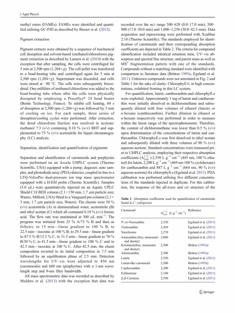
For quantification, lutein, canthaxanthin and chlorophyll a
were exploited. Approximately 1 mg of lutein and canthaxan-
thin were initially dissolved in dichloromethane and subse-
quently diluted with four volumes of ethanol (lutein) or
n-hexane (canthaxanthin). Further dilution in ethanol or
n-hexane respectively was performed in order to measure
within the linear range of the spectrophotometer. Therefore,
the content of dichloromethane was lower than 0.5 % (v/v)
upon determination of the concentrations of lutein and can-
thaxanthin. Chlorophyll a was first dissolved in ethyl acetate
and subsequently diluted with three volumes of 90 % (v/v)
aqueous acetone. Standard concentrations were measured pri-
or to UHPLC analysis, employing their respective absorption
coefficients (A1∘%1cm ):2,550 L g−1 cm−1 (445 nm, 100 % etha-
nol) for lutein, 2,200 L g−1 cm−1 (469 nm 100% cyclohexane)
for canthaxanthin and 887 L g−1 cm−1 (664 nm, 90 % (v/v)
aqueous acetone) for chlorophyll a (Egeland et al. 2011). PDA
calibration was performed utilising five different concentra-
tions of the standards injected in duplicate. For this calibra-
tion, the response of the all-trans and cis structure of the
Table 2 Absorption coefficients used for quantification of carotenoids
found in C. zofingiensis
CarotenoidA1∘%
1cm (L g−1 cm−1)Reference
9′-cis-Neoxanthin 2,330 Egeland et al. (2011)
Violaxanthin 2,450 Egeland et al. (2011)
Neochrome 2,270 Egeland et al. (2011)
Astaxanthin (free, monoester
and diester)
2,060 Egeland et al. (2011)
Ketolutein(free, monoester
and diester)
2,500 Britton (1995a)
Adonixanthin 2,500 Britton (1995a)
Lutein 2,550 Egeland et al. (2011)
Lutein-like carotenoid 2,500 Britton (1995a)
Canthaxanthin 2,200 Egeland et al. (2011)
Echinenone 2,160 Egeland et al. (2011)
β,β-Carotene 2,590 Egeland et al. (2011)
J Appl Phycol
carotenoid standards was considered equal for quantification.
The detector was ascertained as linear for lutein and cantha-
xanthin between 0.07 and 8.20 μg mL−1 with a minimum R2
of 0.998. For chlorophyll a, the detector was linear between
0.23 and 23.38 μg mL−1 with an R2 of 0.978.
Carotenoids were quantified as lutein equivalents
employing the lutein calibration curve. The responses were
corrected using the A1∘%1cm illustrated in Table 2 with the
exception of canthaxanthin. An absorption coefficient of
2,500 L g−1 cm−1 was utilised for unknown carotenoids (e.g.
lutein-like components) and carotenoids of which the absorp-
tion coefficient was not documented (e.g. ketolutein). Fatty
acid-esterified astaxanthin and ketolutein were assumed to
possess similar molar extinction coefficients as their non-
esterified analogues and were expressed as equivalents of their
respective non-esterified analogues in mole per litre culture
volume throughout this work. In Fig. 9, esterified carotenoids
were expressed as % g/g DW, without accounting for the
molecular weight of the fatty acid ester. Chlorophyll b (A1∘%1cm
514 L g−1 cm−1, 647 nm, 90 % (v/v) aqueous acetone) and
derivatives were expressed via chlorophyll a using the chlo-
rophyll a calibration curve. The detector response was
corrected for chlorophyll b using the ratio of the absorption
coefficients of chlorophylls a and b.
Results and discussion
To obtain additional insight into the pathways and regulation
of the carotenoid metabolism of C. zofingiensis, cells were
nitrogen-depleted to induce secondary carotenogenesis and
subsequently DPA-treated to inhibit ketocarotenoids biosyn-
thesis. Sixty micromolar DPA was introduced to nitrogen-
depleted cultures every 2 days beginning on day 2. Every 2
days, cell dry weights and pigment compositions were
analysed. Below, chlorophylls and carotenoids ascertained in
nitrogen-replete and nitrogen-depleted C. zofingiensis cells
are identified and the degradation and accumulation kinetics
of the overall biomass and the annotated pigments upon
nitrogen depletion and DPA treatment are discussed (Figs. 2,
3, 4, 5, 6, and 7; Fig. A1). To properly represent net
accumulation/degradation of pigments, pigment kinetics are
depicted in moles per litre culture volume. For a reference,
pigment contents on a dry weight basis are depicted in
Fig. A2. Conclusions concerning the carotenoid metabolism
are summarised graphically in Fig. 8. Finally, oil droplet
formation in nitrogen-depleted C. zofingiensis cells is elabo-
rated to support the presumed location of carotenoid
accumulation.
Pigment identification
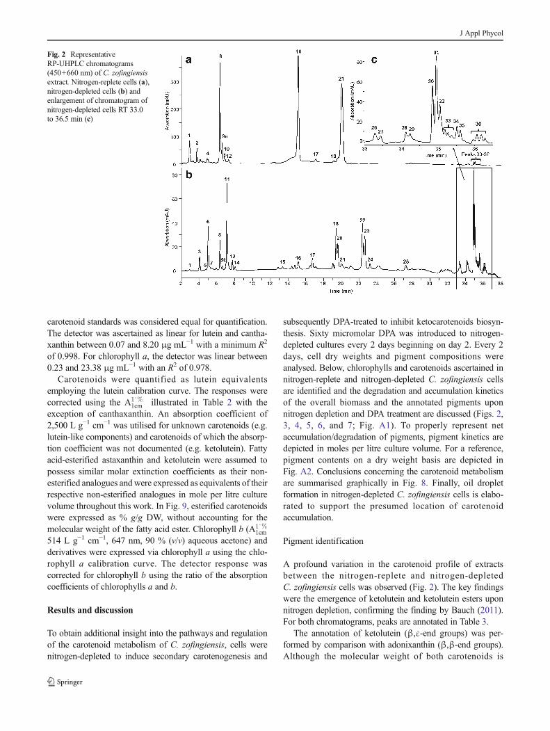
A profound variation in the carotenoid profile of extracts
between the nitrogen-replete and nitrogen-depleted
C. zofingiensis cells was observed (Fig. 2). The key findings
were the emergence of ketolutein and ketolutein esters upon
nitrogen depletion, confirming the finding by Bauch (2011).
For both chromatograms, peaks are annotated in Table 3.
The annotation of ketolutein (β,ε-end groups) was per-
formed by comparison with adonixanthin (β,β-end groups).
Although the molecular weight of both carotenoids is
Fig. 2 Representative
RP-UHPLC chromatograms
(450+660 nm) of C. zofingiensis
extract. Nitrogen-replete cells (a),
nitrogen-depleted cells (b) and
enlargement of chromatogram of
nitrogen-depleted cells RT 33.0
to 36.5 min (c)
J Appl Phycol
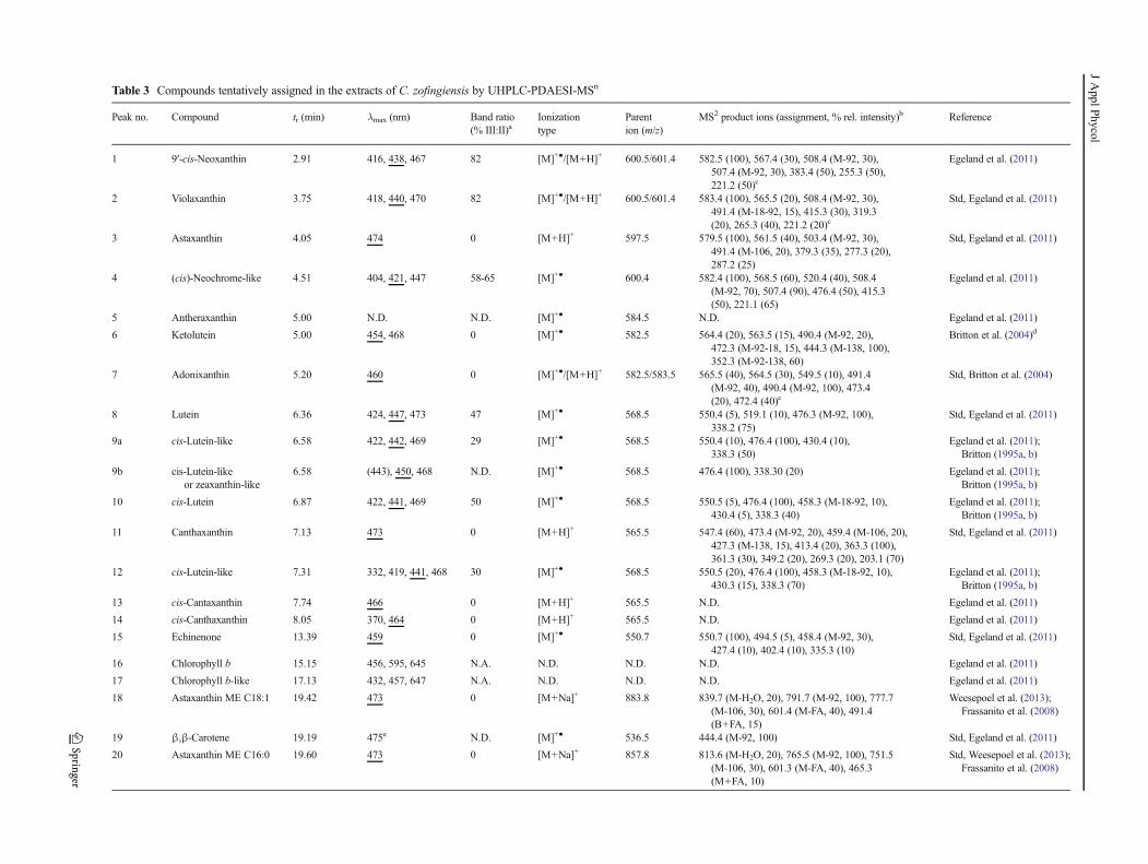
Table 3 Compounds tentatively assigned in the extracts of C. zofingiensis by UHPLC-PDAESI-MSn
Peak no. Compound tr (min) λmax (nm) Band ratio
(% III:II)aIonization
type
Parent
ion (m/z)
MS2 product ions (assignment, % rel. intensity)b Reference
1 9′-cis-Neoxanthin 2.91 416, 438, 467 82 [M]+●/[M+H]+ 600.5/601.4 582.5 (100), 567.4 (30), 508.4 (M-92, 30),
507.4 (M-92, 30), 383.4 (50), 255.3 (50),
221.2 (50)c
Egeland et al. (2011)
2 Violaxanthin 3.75 418, 440, 470 82 [M]+●/[M+H]+ 600.5/601.4 583.4 (100), 565.5 (20), 508.4 (M-92, 30),
491.4 (M-18-92, 15), 415.3 (30), 319.3
(20), 265.3 (40), 221.2 (20)c
Std, Egeland et al. (2011)
3 Astaxanthin 4.05 474 0 [M+H]+ 597.5 579.5 (100), 561.5 (40), 503.4 (M-92, 30),
491.4 (M-106, 20), 379.3 (35), 277.3 (20),
287.2 (25)
Std, Egeland et al. (2011)
4 (cis)-Neochrome-like 4.51 404, 421, 447 58-65 [M]+● 600.4 582.4 (100), 568.5 (60), 520.4 (40), 508.4
(M-92, 70), 507.4 (90), 476.4 (50), 415.3
(50), 221.1 (65)
Egeland et al. (2011)
5 Antheraxanthin 5.00 N.D. N.D. [M]+● 584.5 N.D. Egeland et al. (2011)
6 Ketolutein 5.00 454, 468 0 [M]+● 582.5 564.4 (20), 563.5 (15), 490.4 (M-92, 20),
472.3 (M-92-18, 15), 444.3 (M-138, 100),
352.3 (M-92-138, 60)
Britton et al. (2004)d
7 Adonixanthin 5.20 460 0 [M]+●/[M+H]+ 582.5/583.5 565.5 (40), 564.5 (30), 549.5 (10), 491.4
(M-92, 40), 490.4 (M-92, 100), 473.4
(20), 472.4 (40)c
Std, Britton et al. (2004)
8 Lutein 6.36 424, 447, 473 47 [M]+● 568.5 550.4 (5), 519.1 (10), 476.3 (M-92, 100),
338.2 (75)
Std, Egeland et al. (2011)
9a cis-Lutein-like 6.58 422, 442, 469 29 [M]+● 568.5 550.4 (10), 476.4 (100), 430.4 (10),
338.3 (50)
Egeland et al. (2011);
Britton (1995a, b)
9b cis-Lutein-like
or zeaxanthin-like
6.58 (443), 450, 468 N.D. [M]+● 568.5 476.4 (100), 338.30 (20) Egeland et al. (2011);
Britton (1995a, b)
10 cis-Lutein 6.87 422, 441, 469 50 [M]+● 568.5 550.5 (5), 476.4 (100), 458.3 (M-18-92, 10),
430.4 (5), 338.3 (40)
Egeland et al. (2011);
Britton (1995a, b)
11 Canthaxanthin 7.13 473 0 [M+H]+ 565.5 547.4 (60), 473.4 (M-92, 20), 459.4 (M-106, 20),
427.3 (M-138, 15), 413.4 (20), 363.3 (100),
361.3 (30), 349.2 (20), 269.3 (20), 203.1 (70)
Std, Egeland et al. (2011)
12 cis-Lutein-like 7.31 332, 419, 441, 468 30 [M]+● 568.5 550.5 (20), 476.4 (100), 458.3 (M-18-92, 10),
430.3 (15), 338.3 (70)
Egeland et al. (2011);
Britton (1995a, b)
13 cis-Cantaxanthin 7.74 466 0 [M+H]+ 565.5 N.D. Egeland et al. (2011)
14 cis-Canthaxanthin 8.05 370, 464 0 [M+H]+ 565.5 N.D. Egeland et al. (2011)
15 Echinenone 13.39 459 0 [M]+● 550.7 550.7 (100), 494.5 (5), 458.4 (M-92, 30),
427.4 (10), 402.4 (10), 335.3 (10)
Std, Egeland et al. (2011)
16 Chlorophyll b 15.15 456, 595, 645 N.A. N.D. N.D. N.D. Egeland et al. (2011)
17 Chlorophyll b-like 17.13 432, 457, 647 N.A. N.D. N.D. N.D. Egeland et al. (2011)
18 Astaxanthin ME C18:1 19.42 473 0 [M+Na]+ 883.8 839.7 (M-H2O, 20), 791.7 (M-92, 100), 777.7
(M-106, 30), 601.4 (M-FA, 40), 491.4
(B+FA, 15)
Weesepoel et al. (2013);
Frassanito et al. (2008)
19 β,β-Carotene 19.19 475e N.D. [M]+● 536.5 444.4 (M-92, 100) Std, Egeland et al. (2011)
20 Astaxanthin ME C16:0 19.60 473 0 [M+Na]+ 857.8 813.6 (M-H2O, 20), 765.5 (M-92, 100), 751.5
(M-106, 30), 601.3 (M-FA, 40), 465.3
(M+FA, 10)
Std, Weesepoel et al. (2013);
Frassanito et al. (2008)
JApplPhycol
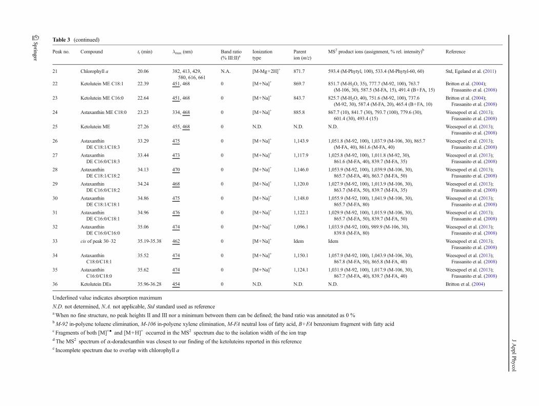
Table 3 (continued)
Peak no. Compound tr (min) λmax (nm) Band ratio
(% III:II)aIonization
type
Parent
ion (m/z)
MS2 product ions (assignment, % rel. intensity)b Reference
21 Chlorophyll a 20.06 382, 413, 429,
580, 616, 661
N.A. [M-Mg+2H]+ 871.7 593.4 (M-Phytyl, 100), 533.4 (M-Phytyl-60, 60) Std, Egeland et al. (2011)
22 Ketolutein ME C18:1 22.39 451, 468 0 [M+Na]+ 869.7 851.7 (M-H2O, 35), 777.7 (M-92, 100), 763.7
(M-106, 30), 587.5 (M-FA, 15), 491.4 (B+FA, 15)
Britton et al. (2004);
Frassanito et al. (2008)
23 Ketolutein ME C16:0 22.64 451, 468 0 [M+Na]+ 843.7 825.7 (M-H2O, 40), 751.6 (M-92, 100), 737.6
(M-92, 30), 587.4 (M-FA, 20), 465.4 (B+FA, 10)
Britton et al. (2004);
Frassanito et al. (2008)
24 Astaxanthin ME C18:0 23.23 334, 468 0 [M+Na]+ 885.8 867.7 (10), 841.7 (30), 793.7 (100), 779.6 (30),
601.4 (30), 493.4 (15)
Weesepoel et al. (2013);
Frassanito et al. (2008)
25 Ketolutein ME 27.26 455, 468 0 N.D. N.D. N.D. Weesepoel et al. (2013);
Frassanito et al. (2008)
26 Astaxanthin
DE C18:1/C18:3
33.29 475 0 [M+Na]+ 1,143.9 1,051.8 (M-92, 100), 1,037.9 (M-106, 30), 865.7
(M-FA, 40), 861.6 (M-FA, 40)
Weesepoel et al. (2013);
Frassanito et al. (2008)
27 Astaxanthin
DE C16:0/C18:3
33.44 473 0 [M+Na]+ 1,117.9 1,025.8 (M-92, 100), 1,011.8 (M-92, 30),
861.6 (M-FA, 40), 839.7 (M-FA, 35)
Weesepoel et al. (2013);
Frassanito et al. (2008)
28 Astaxanthin
DE C18:1/C18:2
34.13 470 0 [M+Na]+ 1,146.0 1,053.9 (M-92, 100), 1,039.9 (M-106, 30),
865.7 (M-FA, 40), 863.7 (M-FA, 50)
Weesepoel et al. (2013);
Frassanito et al. (2008)
29 Astaxanthin
DE C16:0/C18:2
34.24 468 0 [M+Na]+ 1,120.0 1,027.9 (M-92, 100), 1,013.9 (M-106, 30),
863.7 (M-FA, 50), 839.7 (M-FA, 35)
Weesepoel et al. (2013);
Frassanito et al. (2008)
30 Astaxanthin
DE C18:1/C18:1
34.86 475 0 [M+Na]+ 1,148.0 1,055.9 (M-92, 100), 1,041.9 (M-106, 30),
865.7 (M-FA, 80)
Weesepoel et al. (2013);
Frassanito et al. (2008)
31 Astaxanthin
DE C16:0/C18:1
34.96 476 0 [M+Na]+ 1,122.1 1,029.9 (M-92, 100), 1,015.9 (M-106, 30),
865.7 (M-FA, 50), 839.7 (M-FA, 50)
Weesepoel et al. (2013);
Frassanito et al. (2008)
32 Astaxanthin
DE C16:0/C16:0
35.06 474 0 [M+Na]+ 1,096.1 1,033.9 (M-92, 100), 989.9 (M-106, 30),
839.8 (M-FA, 80)
Weesepoel et al. (2013);
Frassanito et al. (2008)
33 cis of peak 30–32 35.19-35.38 462 0 [M+Na]+ Idem Idem Weesepoel et al. (2013);
Frassanito et al. (2008)
34 Astaxanthin
C18:0/C18:1
35.52 474 0 [M+Na]+ 1,150.1 1,057.9 (M-92, 100), 1,043.9 (M-106, 30),
867.8 (M-FA, 50), 865.8 (M-FA, 40)
Weesepoel et al. (2013);
Frassanito et al. (2008)
35 Astaxanthin
C16:0/C18:0
35.62 474 0 [M+Na]+ 1,124.1 1,031.9 (M-92, 100), 1,017.9 (M-106, 30),
867.7 (M-FA, 40), 839.7 (M-FA, 40)
Weesepoel et al. (2013);
Frassanito et al. (2008)
36 Ketolutein DEs 35.96-36.28 454 0 N.D. N.D. N.D. Britton et al. (2004)
Underlined value indicates absorption maximum
N.D. not determined, N.A. not applicable, Std standard used as referenceaWhen no fine structure, no peak heights II and III nor a minimum between them can be defined; the band ratio was annotated as 0 %bM-92 in-polyene toluene elimination, M-106 in-polyene xylene elimination, M-FA neutral loss of fatty acid, B+FA benzonium fragment with fatty acidc Fragments of both [M]+● and [M+H]+ occurred in the MS2 spectrum due to the isolation width of the ion trapdThe MS2 spectrum of α-doradexanthin was closest to our finding of the ketoluteins reported in this referencee Incomplete spectrum due to overlap with chlorophyll a
JApplPhycol
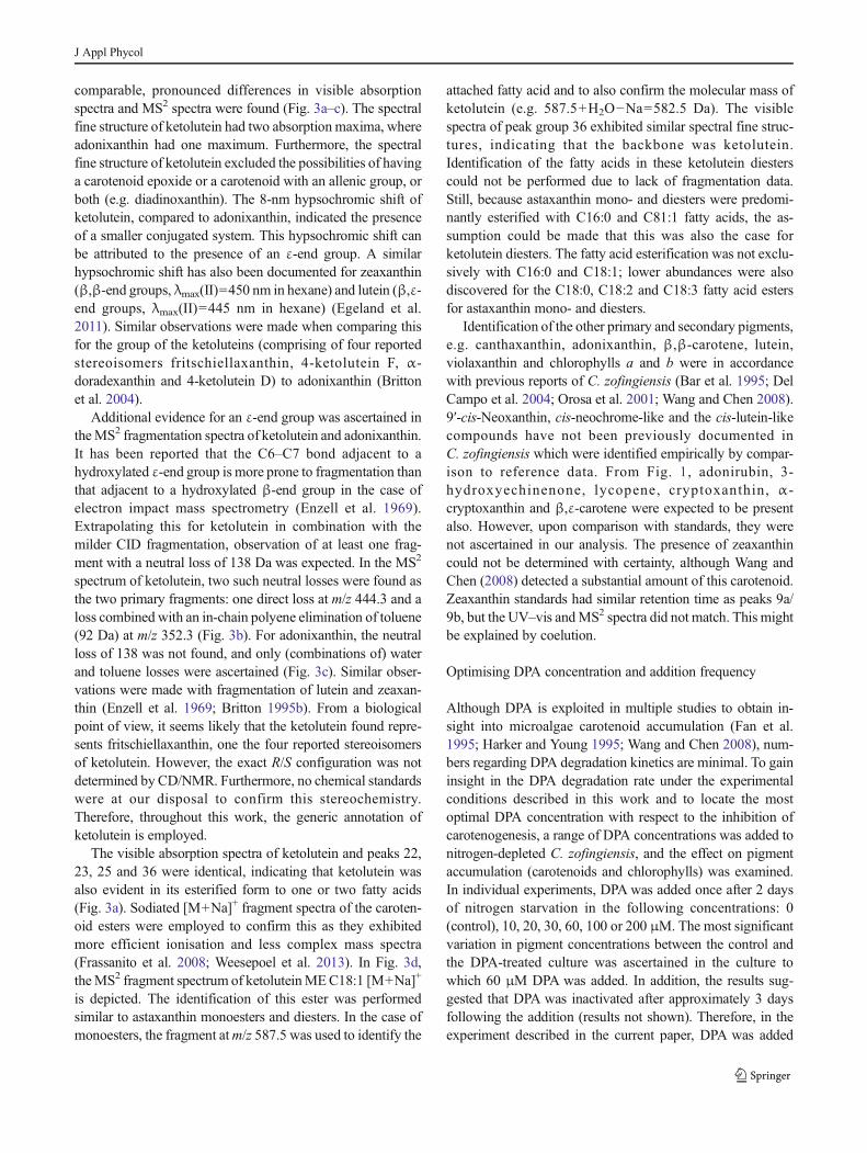
comparable, pronounced differences in visible absorption
spectra and MS2 spectra were found (Fig. 3a–c). The spectral
fine structure of ketolutein had two absorption maxima, where
adonixanthin had one maximum. Furthermore, the spectral
fine structure of ketolutein excluded the possibilities of having
a carotenoid epoxide or a carotenoid with an allenic group, or
both (e.g. diadinoxanthin). The 8-nm hypsochromic shift of
ketolutein, compared to adonixanthin, indicated the presence
of a smaller conjugated system. This hypsochromic shift can
be attributed to the presence of an ε-end group. A similar
hypsochromic shift has also been documented for zeaxanthin
(β,β-end groups, λmax(II)=450 nm in hexane) and lutein (β,ε-
end groups, λmax(II)=445 nm in hexane) (Egeland et al.
2011). Similar observations were made when comparing this
for the group of the ketoluteins (comprising of four reported
stereoisomers fritschiellaxanthin, 4-ketolutein F, α-
doradexanthin and 4-ketolutein D) to adonixanthin (Britton
et al. 2004).
Additional evidence for an ε-end group was ascertained in
theMS2 fragmentation spectra of ketolutein and adonixanthin.
It has been reported that the C6–C7 bond adjacent to a
hydroxylated ε-end group is more prone to fragmentation than
that adjacent to a hydroxylated β-end group in the case of
electron impact mass spectrometry (Enzell et al. 1969).
Extrapolating this for ketolutein in combination with the
milder CID fragmentation, observation of at least one frag-
ment with a neutral loss of 138 Da was expected. In the MS2
spectrum of ketolutein, two such neutral losses were found as
the two primary fragments: one direct loss at m/z 444.3 and a
loss combined with an in-chain polyene elimination of toluene
(92 Da) at m/z 352.3 (Fig. 3b). For adonixanthin, the neutral
loss of 138 was not found, and only (combinations of) water
and toluene losses were ascertained (Fig. 3c). Similar obser-
vations were made with fragmentation of lutein and zeaxan-
thin (Enzell et al. 1969; Britton 1995b). From a biological
point of view, it seems likely that the ketolutein found repre-
sents fritschiellaxanthin, one the four reported stereoisomers
of ketolutein. However, the exact R/S configuration was not
determined by CD/NMR. Furthermore, no chemical standards
were at our disposal to confirm this stereochemistry.
Therefore, throughout this work, the generic annotation of
ketolutein is employed.
The visible absorption spectra of ketolutein and peaks 22,
23, 25 and 36 were identical, indicating that ketolutein was
also evident in its esterified form to one or two fatty acids
(Fig. 3a). Sodiated [M+Na]+ fragment spectra of the caroten-
oid esters were employed to confirm this as they exhibited
more efficient ionisation and less complex mass spectra
(Frassanito et al. 2008; Weesepoel et al. 2013). In Fig. 3d,
theMS2 fragment spectrum of ketoluteinMEC18:1 [M+Na]+
is depicted. The identification of this ester was performed
similar to astaxanthin monoesters and diesters. In the case of
monoesters, the fragment atm/z 587.5 was used to identify the
attached fatty acid and to also confirm the molecular mass of
ketolutein (e.g. 587.5+H2O−Na=582.5 Da). The visible
spectra of peak group 36 exhibited similar spectral fine struc-
tures, indicating that the backbone was ketolutein.
Identification of the fatty acids in these ketolutein diesters
could not be performed due to lack of fragmentation data.
Still, because astaxanthin mono- and diesters were predomi-
nantly esterified with C16:0 and C81:1 fatty acids, the as-
sumption could be made that this was also the case for
ketolutein diesters. The fatty acid esterification was not exclu-
sively with C16:0 and C18:1; lower abundances were also
discovered for the C18:0, C18:2 and C18:3 fatty acid esters
for astaxanthin mono- and diesters.
Identification of the other primary and secondary pigments,
e.g. canthaxanthin, adonixanthin, β,β-carotene, lutein,
violaxanthin and chlorophylls a and b were in accordance
with previous reports of C. zofingiensis (Bar et al. 1995; Del
Campo et al. 2004; Orosa et al. 2001; Wang and Chen 2008).
9′-cis-Neoxanthin, cis-neochrome-like and the cis-lutein-like
compounds have not been previously documented in
C. zofingiensis which were identified empirically by compar-
ison to reference data. From Fig. 1, adonirubin, 3-
hydroxyechinenone, lycopene, cryptoxanthin, α-
cryptoxanthin and β,ε-carotene were expected to be present
also. However, upon comparison with standards, they were
not ascertained in our analysis. The presence of zeaxanthin
could not be determined with certainty, although Wang and
Chen (2008) detected a substantial amount of this carotenoid.
Zeaxanthin standards had similar retention time as peaks 9a/
9b, but the UV–vis andMS2 spectra did not match. This might
be explained by coelution.
Optimising DPA concentration and addition frequency
Although DPA is exploited in multiple studies to obtain in-
sight into microalgae carotenoid accumulation (Fan et al.
1995; Harker and Young 1995; Wang and Chen 2008), num-
bers regarding DPA degradation kinetics are minimal. To gain
insight in the DPA degradation rate under the experimental
conditions described in this work and to locate the most
optimal DPA concentration with respect to the inhibition of
carotenogenesis, a range of DPA concentrations was added to
nitrogen-depleted C. zofingiensis, and the effect on pigment
accumulation (carotenoids and chlorophylls) was examined.
In individual experiments, DPA was added once after 2 days
of nitrogen starvation in the following concentrations: 0
(control), 10, 20, 30, 60, 100 or 200 μM. The most significant
variation in pigment concentrations between the control and
the DPA-treated culture was ascertained in the culture to
which 60 μM DPA was added. In addition, the results sug-
gested that DPA was inactivated after approximately 3 days
following the addition (results not shown). Therefore, in the
experiment described in the current paper, DPA was added
J Appl Phycol
every 2 days, resulting each time in a concentration increase of
60 μM, in order to maintain an effective DPA concentration
for 2 weeks.
Biomass production
During the entire cultivation period, the control and the DPA-
treated cultures exhibited no significant variation in biomass
production. Following a steep initial increase, biomass pro-
duction levelled off around day 8 for all cultures. Both the
treated and the control cultures increased from 1.7 to 4.6 g L−1
in biomass concentration (Fig. 4). As the medium was
nitrogen-depleted from the beginning of cultivation, no addi-
tional nitrogen-containing cell material such as DNA and
protein could be formed. Photosynthesis and carbon fixation
still continued in the absence of nitrate. Therefore, it appears
plausible that the ascertained biomass increase is due to
carbon-rich storage compounds such as TAG. This has been
described for nitrogen-depletedC. zofingiensis byBreuer et al.
(2012) who observed a biomass increase of 580 % DW and
TAG accumulation of up to 40 % (w/w) DW. The absence of a
significant variation in biomass production between the con-
trol and the treated cultures demonstrated that DPA did not
affect the overall production of secondary storage compounds
or carbon fixation.
Degradation of chlorophylls and primary carotenoids
The chlorophylls and primary carotenoids found in the treated
culture were identical to those ascertained in the control,
specifically, chlorophylls a and b, lutein, 9′-cis-neoxanthin
and violaxanthin. The cellular contents of these pigments all
decreased over timewith no significant difference between the
control and the treated culture (Fig. 5). This demonstrates that
DPA did not affect the overall degradation of primary
pigments.
Under nitrogen-replete conditions, the primary pigments
comprised more than 99.9 % of the total pigment content of
C. zofingiensis (Figs. 5 and 6, t0). These pigments are located
in the photosystems of the chloroplast, and all are capable of
harvesting light energy (Dall’Osto et al. 2006; Falkowski and
Raven 2007; Mulders et al. 2014; Sukenik et al. 1992) which
is converted into chemical energy. Active degradation of these
individual pigments may be a strategy of the cell to manage
the oversaturating light conditions that resulted from the ni-
trogen depletion, as was, for example, observed in Isochrysis
Fig. 3 Visible absorption spectra of the three different forms of
ketolutein and adonixanthin (a). Comparison and fragment annotation
of the MS2 fragmentation of ketolutein [M]+● parent (peak 6) (b), and
adonixanthin [M]+●/[M+H]+ (peak 7) (c). Annotation of the C18:1
esterified fatty acid from ketolutein ME C18:1 (peak 22) by MS2 frag-
mentation of its [M+Na]+ parent (d). ktlut ketolutein, ktlut ME ketolutein
monoester, ktlut DE ketolutein diester, adx adonixanthin (for other ab-
breviations, see Table 3 footnote)
Fig. 4 Time courses of dry weight biomass concentration of nitrogen-
depleted C. zofingiensis exposed to no DPA (control) or exposed to
repeated additions of DPA resulting each time in a concentration increase
of 60 μM. Triangles indicate DPA additions. Absolute deviations from the
mean of duplicate dry weight measurements were all less than 4 %
J Appl Phycol

galbana (Mulders et al. 2013). However, the equiproportional
decrease of chlorophylls a and b and 9′-cis-neoxanthin lends
support to the assumption that entire photosystems were de-
graded rather than specific light harvesting pigments.
Remarkably, in both the control and the treated culture,
violaxanthin was degraded at a significantly higher rate than
the other primary pigments (Fig. 5). Violaxanthin is a compo-
nent of the violaxanthin cycle and can be converted into
zeaxanthin under oversaturating light conditions via
antheraxanthin, which is part of a photoprotective mechanism
(Goss and Jakob 2010; Jahns and Holzwarth 2012). However,
as no additional zeaxanthin or antheraxanthin was discovered
in either the control or the treated culture, it is questionable
whether this degradation cycle was genuinely operational in
our experiments. It may be that violaxanthin, antheraxanthin
and zeaxanthin were all degraded.
In addition, it is remarkable that in both the control and in
the treated culture, lutein was degraded at a lower rate com-
pared to the other primary pigments.
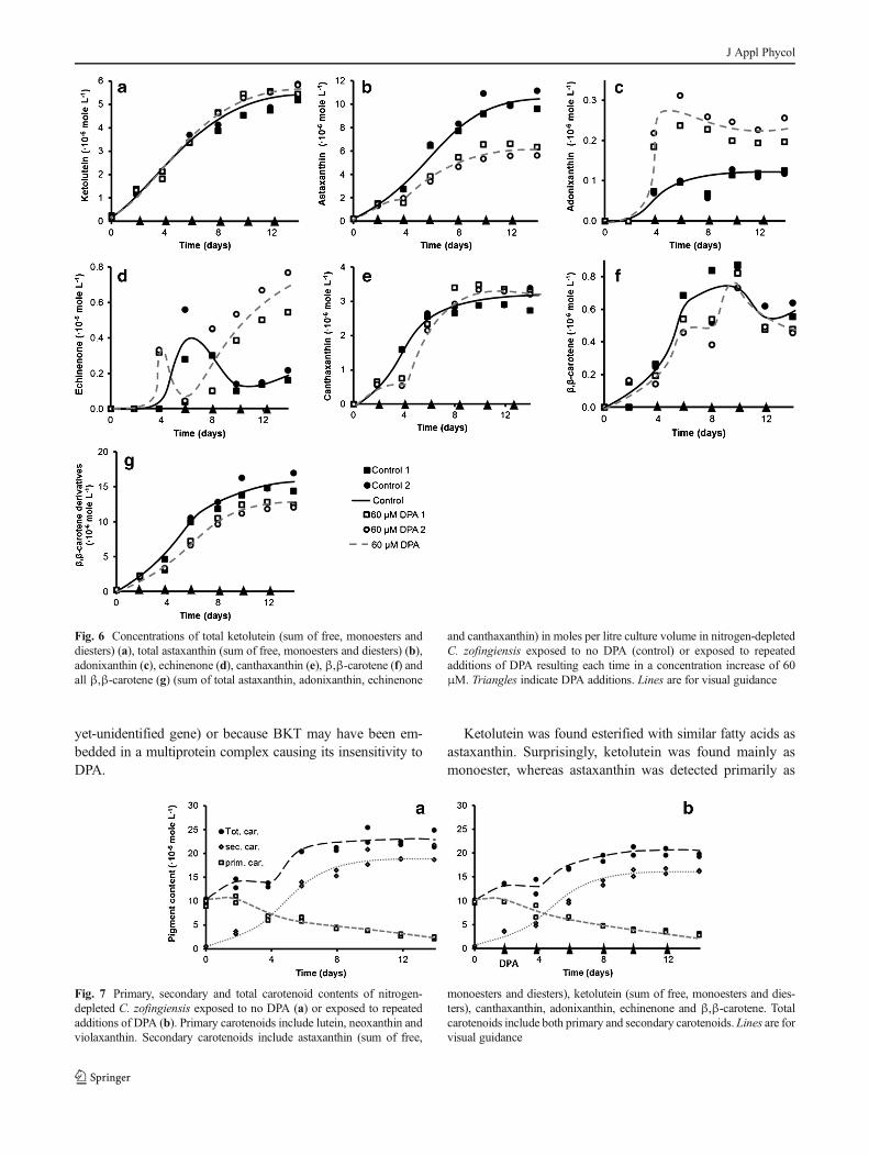
Accumulation of secondary carotenoids
The secondary carotenoids ascertained in the treated culture
were also the same as those discovered in the control, specif-
ically, astaxanthin, ketolutein, canthaxanthin, adonixanthin,
echinenone and β,β-carotene. For the majority of these carot-
enoids, the cellular content was significantly different between
the control and the treated culture (Fig. 6), indicating that
under nitrogen-deplete conditions, DPA had a significant ef-
fect on the formation of secondary carotenoids.
Ketolutein Ketolutein was the second most abundant second-
ary carotenoid in nitrogen-depleted C. zofingiensis. The cel-
lular content increased gradually from zero to the final content
with no significant variations between the control and the
treated culture (Fig. 6a). In all cultures, ketolutein was
ascertained primarily as monoester, to a more moderate extent
in the free form (unesterified) and to a very minimal extent as
diester (esterified with C16:0 and C18:1 fatty acids).
The absence of a significant difference in ketolutein con-
centration between the control and the treated culture indicat-
ed that DPA did not affect the overall formation of ketolutein.
Ketolutein most plausibly resulted from lutein oxygenation
which requires one enzymatic conversion catalysed by a
ketolase. Expression of the BKT gene (from H. pluvialis) in
Chlamydomonas reinhardtii which naturally synthesises β,β-
carotene, zeaxanthin and lutein but no β-end group
ketocarotenoids such as echinenone and adonixanthin, result-
ed in synthesis of ketolutein, whereas no β-end group
ketocarotenoids were synthesised (Vila et al. 2012). This
supports the premise that ketolutein emerged from lutein via
oxidation by BKT. It is remarkable that the lutein to ketolutein
conversion by BKT was not affected by DPA. This may be
explained by assuming that the enzyme that catalysed
ketolutein biosynthesis was insensitive to DPA, either because
it was a completely different ketolase (encoded by a different
Fig. 5 Time courses of chlorophyll a (a), chlorophyll b (b), lutein (c), 9′-
cis-neoxanthin (d) and violaxanthin (e) in moles per litre culture volume
of nitrogen-depleted C. zofingiensis exposed to no DPA (control) or
exposed to repeated additions of DPA resulting each time in a concentra-
tion increase of 60 μM. Triangles indicate DPA additions. Lines are for
visual guidance
J Appl Phycol
yet-unidentified gene) or because BKT may have been em-
bedded in a multiprotein complex causing its insensitivity to
DPA.
Ketolutein was found esterified with similar fatty acids as
astaxanthin. Surprisingly, ketolutein was found mainly as
monoester, whereas astaxanthin was detected primarily as
Fig. 6 Concentrations of total ketolutein (sum of free, monoesters and
diesters) (a), total astaxanthin (sum of free, monoesters and diesters) (b),
adonixanthin (c), echinenone (d), canthaxanthin (e), β,β-carotene (f) and
all β,β-carotene (g) (sum of total astaxanthin, adonixanthin, echinenone
and canthaxanthin) in moles per litre culture volume in nitrogen-depleted
C. zofingiensis exposed to no DPA (control) or exposed to repeated
additions of DPA resulting each time in a concentration increase of 60
μM. Triangles indicate DPA additions. Lines are for visual guidance
Fig. 7 Primary, secondary and total carotenoid contents of nitrogen-
depleted C. zofingiensis exposed to no DPA (a) or exposed to repeated
additions of DPA (b). Primary carotenoids include lutein, neoxanthin and
violaxanthin. Secondary carotenoids include astaxanthin (sum of free,
monoesters and diesters), ketolutein (sum of free, monoesters and dies-
ters), canthaxanthin, adonixanthin, echinenone and β,β-carotene. Total
carotenoids include both primary and secondary carotenoids. Lines are for
visual guidance
J Appl Phycol

diester, despite the fact that they both possess two hydroxyl
groups. This might suggest that either theβ-end group is more
easily acylated than the ε-end group or that the adjacent
carbonyl is a prerequisite for acylation.
Astaxanthin Astaxanthin was the most abundant secondary
carotenoid in nitrogen-depleted C. zofingiensis. In both the
control and the treated cultures, its content increased gradual-
ly. From the moment that DPAwas added, the increase in the
treated culture was half as high as in the control culture
(Fig. 6b), indicating that DPA significantly inhibited produc-
tion of astaxanthin.
Theoretically, adonixanthin and adonirubin may both be
precursors of astaxanthin (Fig. 1). Since adonixanthin was
detected, the pathway to this carotenoid must have been
present. Because adonixanthin was found in a very low con-
centration and only in the free form, it may be speculated that
the pathway from adonixanthin to astaxanthin was existing as
well. It remains ambiguous whether adonirubin was a precur-
sor of astaxanthin as well since this carotenoid was lacking in
nitrogen-depleted C. zofingiensis.
Adonixanthin In both the control and the treated cultures, the
cellular content of adonixanthin increased gradually. From the
moment that DPA was added, the increase in the treated
culture was approximately twice as high as in the control
culture (Fig. 6c). This finding supports the speculation that,
in nitrogen-depleted C. zofingiensis, adonixanthin was con-
verted into astaxanthin.
Adonixanthin may be formed from echinenone via 3-
hydroxyechinenone or 3′-hydroxyechinenone, as in
H. pluvialis (Lemoine and Schoefs 2010), or from zeaxanthin,
as proposed byWang and Chen (2008) andHuang et al. (2006).
This latter hypothesis was based on reduced zeaxanthin degra-
dation in carotenogenic cultures exposed to DPA. As men-
tioned previously, no substantial amounts of zeaxanthin could
be detected in any of the cultures. Additionally, we did not
discover reduced degradation of zeaxanthin precursors (i.e. all
other primary carotenoids) in the DPA-treated cultures.
Therefore, our results do not support the hypothesis that in
nitrogen-depleted C. zofingiensis, adonixanthin is formed from
zeaxanthin. In contrast, echinenone attained a cellular content
that was approximately seven times as high in the treated
cultures as in the control (Fig. 6d). This supports the hypothesis
that echinenone is a possible precursor of adonixanthin.
Canthaxanthin In both the control and the treated cultures,
canthaxanthin was ascertained in relatively high cellular con-
tents (comparable to esterified ketolutein contents). In both
cultures, the cellular content increased gradually. In the treated
cultures, canthaxanthin was detected only on day 4 in signifi-
cantly lesser amounts compared to the control culture (Fig. 6e).
This indicates that canthaxanthin formation was inhibited for
only 2 days which was compensated with increased canthaxan-
thin production 2 days thereafter. It remains unclear what
mechanism caused the recovery from DPA inhibition.
Huang et al. (2006) also discovered relatively high contents
of canthaxanthin and as stated, speculated that canthaxanthin is
an end product rather than a precursor of astaxanthin. Our data
do not provide further insights with regard to this speculation.
β,β-carotene In both the control and the treated cultures,β,β-
carotene was ascertained at low cellular contents (comparable
to echinenone, adonixanthin and free astaxanthin contents),
with no significant variation between the control and the
treated culture (Fig. 6f). The low cellularβ,β-carotene content
indicates that this carotenoid was promptly converted into
echinenone and, moreover, that a high β,β-carotene content
was not required for induction of secondary carotenogenesis.
Novel insights in the regulation of carotenogenesis
in C. zofingiensis
In both the DPA-treated culture and the control, accumulation
of secondary carotenoids occurred simultaneously with a
Fig. 8 Metabolic network model of carotenoids in nitrogen-depleted
C. zofingiensis, summarising obtained results. Cross indicates
hypothesised pathway that is not present in nitrogen-depleted
C. zofingiensis. Question marks indicate unresolved pathways. Double
arrows indicate dislocations. Jn indicate fluxes through specific path-
ways. Black carotenoids were found. Grey carotenoids were not found.
Dislocations of lutein, ketolutein and astaxanthin are not explicitly indi-
cated. Note that biosynthesis of ketolutein may as well take place in the
chloroplast (see text for other details and assumptions). 3-OHech 3-
hydroxyechinenone, 3′-OHech 3′-hydroxyechinenone, 9-cis-neo 9-cis-
neoxanthin, α-car α-carotene, adr adonirubin, adx adonixanthin, ant
antheraxanthin, ast astaxanthin, ast ME astaxanthin monoester, ast DE
astaxanthin diester, β,β-carchl β,β-carotene (chloroplast), β,β-carcyt β,β-
carotene (cytosol), cry cryptoxanthin, can canthaxanthin (cytosol), canoilcanthaxanthin (oil body), ech echinenone, ktlut ketolutein, ktlut ME
ketolutein monoester, ktlut DE ketolutein diester, lut lutein, lyc lycopene,
neo neoxanthin, phy phytoene, vio violaxanthin, zea zeaxanthin (Kanehisa
Laboratories 2012; Lemoine and Schoefs 2010; Vila et al. 2012; Wang and
Chen 2008)
J Appl Phycol
decrease in primary carotenoids (grey lines, Fig. 7). This lends
support to the supposition that under nitrogen-depleted con-
ditions, secondary carotenoids were formed from primary
carotenoids. However, the overall carotenoid content in-
creased (black lines, Fig. 7), which indicates that in the
DPA-treated culture and in the control, at least part of the
secondary carotenoids were synthesised de novo. Since the
consumed quantity of lutein equalled approximately the pro-
duced amount of ketolutein, it may be assumed that instead of
degrading into other products, lutein was completely convert-
ed into ketolutein. Since lutein constituted the major fraction
of primary carotenoids (>80 %), this implies that the remain-
ing secondary carotenoids (echinenone, canthaxanthin,
adonixanthin and astaxanthin) must have been almost
completely formed de novo in both the control and the DPA-
treated cultures.
The overall cellular content of echinenone, canthaxanthin,
adonixanthin and astaxanthin was significantly different be-
tween the DPA-treated cultures and the controls. The overall
content was reduced particularly during the first 4 days after the
initial DPA addition (between days 2 and 6; Fig. 6g), indicating
that, during this time, the flux through J1 was significantly
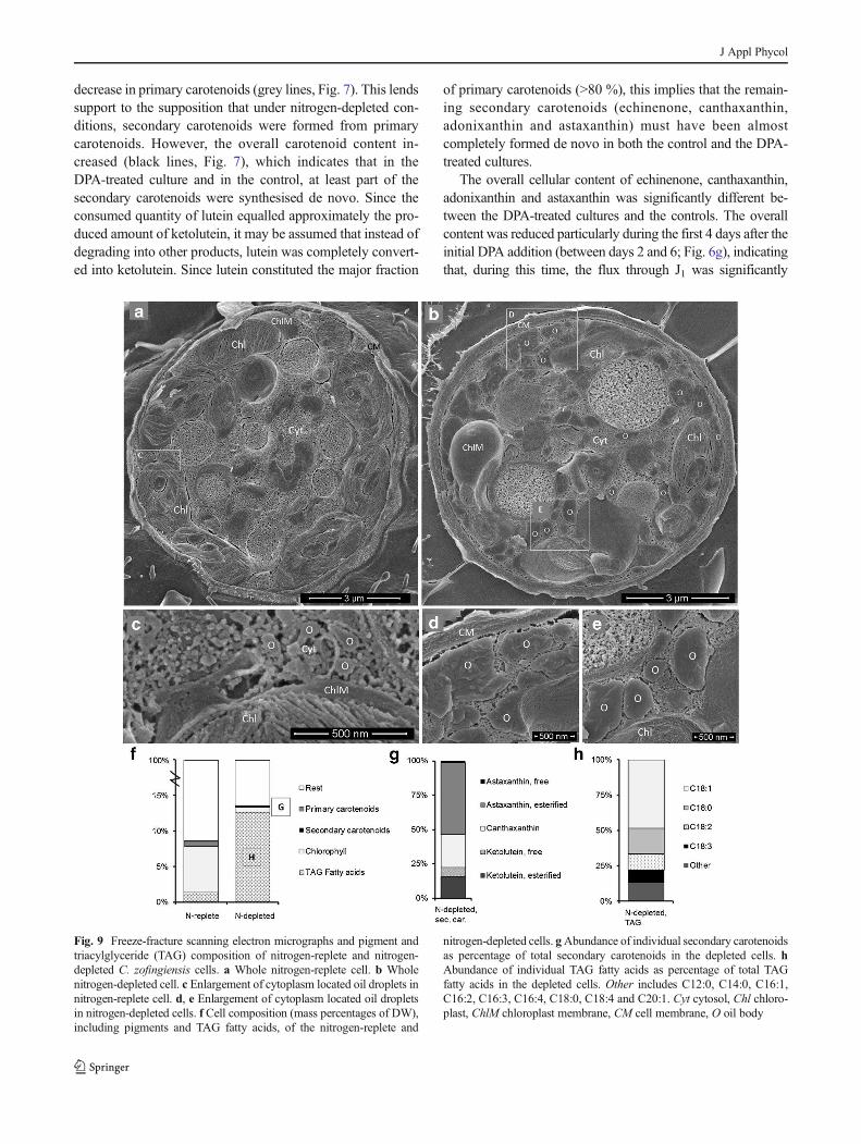
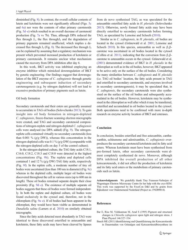
Fig. 9 Freeze-fracture scanning electron micrographs and pigment and
triacylglyceride (TAG) composition of nitrogen-replete and nitrogen-
depleted C. zofingiensis cells. a Whole nitrogen-replete cell. b Whole
nitrogen-depleted cell. c Enlargement of cytoplasm located oil droplets in
nitrogen-replete cell. d, e Enlargement of cytoplasm located oil droplets
in nitrogen-depleted cells. f Cell composition (mass percentages of DW),
including pigments and TAG fatty acids, of the nitrogen-replete and
nitrogen-depleted cells. gAbundance of individual secondary carotenoids
as percentage of total secondary carotenoids in the depleted cells. h
Abundance of individual TAG fatty acids as percentage of total TAG
fatty acids in the depleted cells. Other includes C12:0, C14:0, C16:1,
C16:2, C16:3, C16:4, C18:0, C18:4 and C20:1. Cyt cytosol, Chl chloro-
plast, ChlM chloroplast membrane, CM cell membrane, O oil body
J Appl Phycol
diminished (Fig. 8). In contrast, the overall cellular contents of
lutein and ketolutein were not significantly affected (Figs. 5c
and 6a) nor were the contents of other primary carotenoids
(Fig. 5d–e) which resulted in an overall decrease of carotenoid
production (Fig. 7a vs 7b). Thus, although DPA reduced the
flux through J1, the flux throughJ2 and the degradation of
primary pigments remained unaffected, which implies a de-
creased flux through J3 (Fig. 8). The decreased flux through J3can be explained by assuming that a regulatory mechanismwas
present which prevented increased production of ketolutein or
primary carotenoids. It remains unclear what mechanism
caused the recovery from DPA inhibition after day 6.
In this work, BKT activity was inhibited employing an
enzyme inhibitor which mimicked downregulation of BKT
by genetic engineering. Our findings suggest that downregu-
lation of the BKT enzyme of C. zofingiensis through genetic
engineering and subsequent induction of secondary
carotenogenesis (e.g. by nitrogen depletion) will not lead to
excessive production of primary pigments such as lutein.
Oil body formation
Secondary carotenoids and their esters are generally assumed
to accumulate in TAG oil bodies (Solovchenko 2013). To gain
insight into oil body formation in nitrogen-depleted
C. zofingiensis, freeze-fracture scanning electron micrographs
were created, and TAG and secondary carotenoid composi-
tions of nitrogen-replete and nitrogen-depleted C. zofingiensis
cells were analysed (no DPA added) (Fig. 9). The nitrogen-
replete cells contained virtually no secondary carotenoids (less
than 0.001 % (g/g DW)), whereas this concentration in the
nitrogen-depleted cells was 0.1 % (g/gDW) (comparable with
the nitrogen-depleted cells on day 5 of the control culture).
In the nitrogen-depleted culture, the TAG fatty acids C18:1,
C16:0, C18:2, C18:3 and C18:0 were detected in the highest
concentrations (Fig. 9h). The replete and depleted cells
contained 1 and 12 % (g/g DW) TAG fatty acids, respectively
(Fig. 9f). In the replete cells, a moderate number of tiny oil
bodies were ascertained (less than 100 nm in diameter) (Fig. 9c)
whereas in the depleted cells, multiple larger oil bodies were
discovered throughout the cell in various sizes (up to 600 nm in
length). These oil bodies remained separate despite their close
proximity (Fig. 9d–e). The existence of multiple separate oil
bodies suggests that these oil bodies were formed independent-
ly. In both the replete and depleted culture, oil bodies were
found exclusively in the cytosol and, therefore, not in the
chloroplasts (Fig. 9a–e). If oil bodies had been apparent in the
chloroplast, they would have been visible as demonstrated in
Dunaliella salina (Lamers et al. 2010) on similarly obtained
micrographs.
Since the fatty acids detected most abundantly in TAG were
identical to those discovered esterified to astaxanthin and
ketolutein, these fatty acids may have been cleaved by lipases
from de novo synthesised TAG, as was speculated for the
astaxanthin esterified fatty acids in H. pluvialis (Solovchenko
2013). Otherwise, newly formed fatty acids may have been
directly esterified to secondary carotenoids before forming
TAG, as speculated by Lemoine and Schoefs (2010).
Similar as in C. zofingiensis, in H. pluvialis, oil bodies are
located in the cytosol (Grünewald et al. 2001; Lemoine and
Schoefs 2010). In this species, astaxanthin as well as β,β-
carotene was ascertained in oil bodies located in the cytosol
(Collins et al. 2011), indicating that the conversion from β,β-
carotene to astaxanthin occurs in the cytosol. Grünewald et al.
(2001) demonstrated evidence of BKT in H. pluvialis in the
chloroplast as well as in the oil bodies, however, BKTappeared
to be active only on the surface of the oil bodies. Considering
the many similarities between C. zofingiensis and H. pluvialis
(i.e. TAG oil bodies’ location, the fatty acids present in TAG
and esterified to secondary carotenoids, and enzymes involved
in secondary carotenogenesis), it may be speculated that, in
C. zofingiensis, the secondary carotenoids were also synthe-
sised on the surface of the oil bodies and subsequently accu-
mulated in the oil bodies. However, ketolutein may be synthe-
sised in the chloroplast as well after which it may be transferred,
esterified and accumulated in oil bodies located in the cytosol.
Both speculations need to be confirmed with more in-depth
research on enzyme activity location of BKT and esterases.
Conclusion
In conclusion, besides esterified and free astaxanthin, cantha-
xanthin, echinenone and adonixanthin, C. zofingiensis over-
produces the secondary carotenoid ketolutein and its fatty acid
esters. Whereas ketolutein must have been synthesised from
pre-formed lutein, other secondary carotenoids were al-
most completely synthesised de novo. Moreover, although
DPA inhibited the overall production of all other
ketocarotenoids, it did not affect the production of ketolutein
and its fatty acid esters or the metabolism of primary caroten-
oids such as lutein.
Acknowledgments We gratefully thank Tiny Franssen-Verheijen of
Wageningen Electron Microscopy Centre for her help with the cryo-SEM.
This work was supported by the FeyeCon D&I and by grants from
Rijksdienst voor Ondernemend Nederland (Project no. FND09014).
References
Bar E, Rise M, Vishkautsan M, Arad S (1995) Pigment and structural
changes in Chlorella zofingiensis upon light and nitrogen stress. J
Plant Physiol 146:527–534
BauchME (2011) Identifizierung und Quantifizierung der Ketocarotinoide
in Dauerstadien von Grünalgen und Ketocarotinoidbiosynthese im
J Appl Phycol
Modellorganismus Chlamydomonas reinhardtii. Dissertation,
Johannes Gutenberg University, Mainz
Breuer G, Lamers PP, Martens DE, Draaisma RB, Wijffels RH (2012)
The impact of nitrogen starvation on the dynamics of triacylglycerol
accumulation in nine microalgae strains. Bioresour Technol 124:
217–226
Breuer G, Evers WAC, de Vree JH, Kleinegris DMM, Martens DE,
Wijffels RH, Lamers PP (2013) Analysis of fatty acid content and
composition in microalgae. J Vis Exp 80:e50628
Britton G (1995a) UV/visible spectroscopy. In: Britton G, Liaaen-Jensen
S, Pfander H (eds) Carotenoids, vol 1B,Spectroscopy. Birkhäuser,
Basel, pp 13–62
Britton G (1995b) Mass spectrometry. In: Britton G, Liaaen-Jensen S,
Pfander H (eds) Carotenoids, vol 1B,Spectroscopy. Birkhäuser,
Basel, pp 261–317
Britton G, Liaaen-Jensen S, Pfander H (editors) and Mercadante Z,
Egeland E (compilers) (2004) Handbook, Carotenoid series.
Birkhäuser, Basel
Collins AM, Jones HDT, Han D, Hu Q, Beechem TE, Timlin JA (2011)
Carotenoid distribution in living cells of Haematococcus pluvialis
(Chlorophyceae). PLoS ONE 6(9):e24302
Dall’Osto L, Lico C, Alric J, Giuliano G, Havaux M, Bassi R (2006)
Lutein is needed for efficient chlorophyll triplet quenching in the
major LHCII antenna complex of higher plants and effective
photoprotection in vivo under strong light. BMC Plant Biol 6:32
Del Campo JA, Rodriguez H, Moreno J, Vargas MA, Rivas J, Guerrero
MG (2004) Accumulation of astaxanthin and lutein in Chlorella
zofingiensis (Chlorophyta). Appl Microbiol Biotechnol 64:848–854
Egeland G, Garrido J, Clementson L, Andresen K, Thomas C, Zapata M,
Airs R, Llewellyn C, Newman G, Rodríguez F, Roy S (2011) Part
VI: Data sheets aiding identification of phytoplankton carotenoids
and chlorophylls. In: Roy S, Llewellyn CA, Egeland ES, Johnson G
(eds) Phytoplankton pigments: characterization, chemotaxonomy
and applications in oceanography. Cambridge University Press,
Cambridge, pp 665–822
Enzell CR, Francis GW, Liaaen-Jensen S (1969) Mass spectrometric
studies of carotenoids 2. A survey of fragmentation reactions. Acta
Chem Scand 23:727–750
Falkowski PG, Raven JA (2007) Aquatic photosynthesis, 2nd edn.
Princeton University Press, Princeton, 484 pp
Fan L, Vonshak A, Gabbay R, Hirshberg J, Cohen Z, Boussiba S (1995)
The biosynthetic pathway of astaxanthin in a green alga
Haematococcus pluvialis as indicated by inhibition with diphenyl-
amine. Plant Cell Physiol 36:1519–1524
Frassanito R, Cantonati M, Flaim G, Mancini I, Guella G (2008) A new
method for the identification and the structural characterisation of
carotenoid esters in freshwater microorganisms by liquid
chromatography/electrospray ionisation tandem mass spectrometry.
Rapid Commun Mass Spectrom 22:3531–3539
Fucíková K, Lewis LA (2012) Intersection of Chlorella, Muriella and
Bracteacoccus: resurrecting the genusChromochlorisKol et Chodat
(Chlorophyceae, Chlorophyta). Fottea 12:83–93
Goss R, Jakob T (2010) Regulation and function of xanthophyll cycle-
dependent photoprotection in algae. Photosynth Res 106:103–122
Grünewald K, Hirschberg J, Hagen C (2001) Ketocarotenoid biosynthe-
sis outside of plastids in the unicellular green alga Haematococcus
pluvialis. J Biol Chem 276:6023–6029
Harker M, Young AJ (1995) Inhibition of astaxanthin synthesis in the
green alga Haematococcus pluvialis. Eur J Phycol 30:179–187
Huang JC, Wang Y, Sandmann G, Chen F (2006) Isolation and charac-
terization of a carotenoid oxygenase gene from Chlorella
zofingiensis (Chlorophyta). Appl Microbiol Biotechnol 71:473–479
Jahns P, Holzwarth AR (2012) The role of the xanthophyll cycle and of
lutein in photoprotection of photosystem II. Biochem Biophys Acta
– Bioenerg 1817:182–193
Jin E, Lee CG, Polle JEW (2006) Secondary carotenoid accumulation in
Haematococcus (Chlorophyceae): biosynthesis, regulation, and bio-
technology. J Microbiol Biotechnol 16:821–831
Kanehisa Laboratories (2012) KEGG PATHWAYDatabase. Available at:
http://www.genome.jp/kegg/pathway.html. Accessed 25 Jul 2013
Kliphuis AMJ, Janssen M, Van Den End EJ, Martens DE, Wijffels RH
(2011) Light respiration inChlorella sorokiniana. J Appl Phycol 23:
935–947
Lamers PP, Janssen M, De Vos RCH, Bino RJ, Wijffels RH (2008)
Exploring and exploiting carotenoid accumulation in Dunaliella
salina for cell-factory applications. Trends Biotechnol 26:631–638
Lamers PP, Van De Laak CCW, Kaasenbrood PS, Lorier J, JanssenM, De
Vos RCH, Bino RJ, Wijffels RH (2010) Carotenoid and fatty acid
metabolism in light-stressed Dunaliella salina. Biotechnol Bioeng
106:638–648
Lemoine Y, Schoefs B (2010) Secondary ketocarotenoid astaxanthin
biosynthesis in algae: a multifunctional response to stress.
Photosynth Res 106:155–177
Mandalam RK, Palsson BO (1998) Elemental balancing of biomass and
medium composition enhances growth capacity in high-density
Chlorella vulgaris cultures. Biotechnol Bioeng 59:605–611
Mulders KJM, Weesepoel Y, Lamers PP, Vincken J-P, Martens DE,
Wijffels RH (2013) Growth and pigment accumulation in nutrient-
depleted Isochrysis aff. galbana T-ISO. J Appl Phycol 25:1421–
1430
Mulders KJM, Lamers PP, Martens DE,Wijffels RH (2014) Phototrophic
pigment production with microalgae: biological constraints and
opportunities. J Phycol 50:229–242
Orosa M, Torres E, Fidalgo P, Abalde J (2001) Production and analysis of
secondary carotenoids in green algae. J Appl Phycol 12:553–556
Solovchenko AE (2013) Physiology and adaptive significance of second-
ary carotenogenesis in green microalgae. Russ J Plant Physiol 60:1–
13
Spolaore P, Joannis-Cassan C, Duran E, Isambert A (2006) Commercial
applications of microalgae. J Biosci Bioeng 101:87–96
Sukenik A, Livne A, Neori A, Yacobi YZ, Katcoff D (1992) Purification
and characterization of a light-harversing chlorophyll-protein com-
plex from the marine eustigmatophyte Nannochloropsis sp. Plant
Cell Physiol 33:1041–1048
Vidhyavathi R, Venkatachalam L, Sarada R, Ravishankar GA (2008)
Regulation of carotenoid biosynthetic genes expression and carot-
enoid accumulation in the green alga Haematococcus pluvialis
under nutrient stress conditions. J Exp Bot 59:1409–1418
Vila M, Galván A, Fernández E, León R (2012) Ketocarotenoid
biosynthesis in transgenic microalgae expressing a foreign β-
C-4-carotene oxygenase gene. Methods Mol Biol 892:283–
295
Wang Y, Chen T (2008) The biosynthetic pathway of carotenoids in the
astaxanthin-producing green alga Chlorella zofingiensis. World J
Microbiol Biotechnol 24:2927–2932
Weesepoel Y, Vincken J-P, Pop R, Liu K, Gruppen H (2013) Sodiation as
a tool for enhancing the diagnostic value of MALDI-TOF/TOF-MS
spectra of astaxanthin esters in complex mixtures from
Haematococcus pluvialis. J Mass Spectrom 48:862–874
ZhekishevaM, ZarkaA, Khozin-Goldberg I, Cohen Z, Boussiba S (2005)
Inhibition of astaxanthin synthesis under high irradiance does
not abolish triacylglycerol accumulation in the green alga
Haematococcus pluvialis (Chlorophyceae). J Phycol 41:819–
826
J Appl Phycol
Related Documents