RESEARCH ARTICLE Open Access Association between levels of tumor- infiltrating lymphocytes in different subtypes of primary breast tumors and prognostic outcomes: a meta-analysis Lin He 1† , Yaling Wang 2† , Qian Wu 1 , Yuhua Song 2 , Xuezhen Ma 2 , Biyuan Zhang 3 , Haiji Wang 3* and Yong Huang 1 Abstract Background: To investigate the impact of the elevation of tumor-infiltrating lymphocytes (TILs) in different molecular subtypes of primary breast cancer, i.e. each 10% increment of TILs and high-level TILs (TILs≥50%) in tumor, on overall survival (OS) and pathological complete response (pCR) and to compare the presentation of high-level TILs across these molecular subtypes. Methods: Citation retrieval was performed in the PubMed, Cochrane Library, Embase and Web of Science databases. All statistical calculations were performed by the software of StataSE version 12.0. Results: Twenty-two eligible clinical trials including 15,676 unique patients were included for meta-analysis. Each 10% increment of TILs significantly improved OS in human epidermal growth factor receptor 2 (HER2)-overexpression (pooled Hazard ratio (HR), 0.92; 95% CI, 0.89–0.95) and triple-negative (TN) (pooled HR, 0.90; 95% CI, 0.89–0.92) breast tumors but not in luminal tumor subtype (pooled HR, 1.06; 95% CI, 0.99–1.13). It was also associated with an increased pCR rate in breast cancers (pooled Odds ratio (OR), 1.27; 95% CI, 1.19–13.5). High-level TILs were significantly related with a higher pCR rate (pooled OR, 2.73; 95% CI, 2.40–3.01) than low-level TILs. The HER2-amplified (pooled OR, 3.14; 95% CI, 1.95–5.06) and TN (pooled OR, 4.09; 95% CI, 2.71–6.19) phenotypes of breast cancers expressed significantly more high-level TILs than the luminal tumor subtype, although the presentation of those between the former two subsets was not significantly different (pooled OR, 1.30; 95%CI, 0.83–2.04). Conclusions: The elevation of TILs in breast tumors predicts favorable prognostic outcomes, particularly in the HER2- overexpression and TN subtypes. Keywords: Breast cancer, tumor-infiltrating lymphocytes, Overall survival, Pathological complete response, Meta- analysis © The Author(s). 2020 Open Access This article is licensed under a Creative Commons Attribution 4.0 International License, which permits use, sharing, adaptation, distribution and reproduction in any medium or format, as long as you give appropriate credit to the original author(s) and the source, provide a link to the Creative Commons licence, and indicate if changes were made. The images or other third party material in this article are included in the article's Creative Commons licence, unless indicated otherwise in a credit line to the material. If material is not included in the article's Creative Commons licence and your intended use is not permitted by statutory regulation or exceeds the permitted use, you will need to obtain permission directly from the copyright holder. To view a copy of this licence, visit http://creativecommons.org/licenses/by/4.0/. The Creative Commons Public Domain Dedication waiver (http://creativecommons.org/publicdomain/zero/1.0/) applies to the data made available in this article, unless otherwise stated in a credit line to the data. * Correspondence: [email protected] † Lin He and Yaling Wang contributed equally to this work. 3 Department of Radiotherapy, The Affiliated Hospital of Qingdao University, Qingdao, Shandong Province, People’s Republic of China Full list of author information is available at the end of the article He et al. BMC Women's Health (2020) 20:194 https://doi.org/10.1186/s12905-020-01038-x

Welcome message from author
This document is posted to help you gain knowledge. Please leave a comment to let me know what you think about it! Share it to your friends and learn new things together.
Transcript

RESEARCH ARTICLE Open Access
Association between levels of tumor-infiltrating lymphocytes in differentsubtypes of primary breast tumors andprognostic outcomes: a meta-analysisLin He1†, Yaling Wang 2†, Qian Wu1, Yuhua Song2, Xuezhen Ma2, Biyuan Zhang3, Haiji Wang 3* andYong Huang1
Abstract
Background: To investigate the impact of the elevation of tumor-infiltrating lymphocytes (TILs) in different molecularsubtypes of primary breast cancer, i.e. each 10% increment of TILs and high-level TILs (TILs≥50%) in tumor, on overallsurvival (OS) and pathological complete response (pCR) and to compare the presentation of high-level TILs acrossthese molecular subtypes.
Methods: Citation retrieval was performed in the PubMed, Cochrane Library, Embase and Web of Science databases.All statistical calculations were performed by the software of StataSE version 12.0.
Results: Twenty-two eligible clinical trials including 15,676 unique patients were included for meta-analysis. Each 10%increment of TILs significantly improved OS in human epidermal growth factor receptor 2 (HER2)-overexpression(pooled Hazard ratio (HR), 0.92; 95% CI, 0.89–0.95) and triple-negative (TN) (pooled HR, 0.90; 95% CI, 0.89–0.92) breasttumors but not in luminal tumor subtype (pooled HR, 1.06; 95% CI, 0.99–1.13). It was also associated with an increasedpCR rate in breast cancers (pooled Odds ratio (OR), 1.27; 95% CI, 1.19–13.5). High-level TILs were significantly relatedwith a higher pCR rate (pooled OR, 2.73; 95% CI, 2.40–3.01) than low-level TILs. The HER2-amplified (pooled OR, 3.14;95% CI, 1.95–5.06) and TN (pooled OR, 4.09; 95% CI, 2.71–6.19) phenotypes of breast cancers expressed significantlymore high-level TILs than the luminal tumor subtype, although the presentation of those between the former twosubsets was not significantly different (pooled OR, 1.30; 95%CI, 0.83–2.04).
Conclusions: The elevation of TILs in breast tumors predicts favorable prognostic outcomes, particularly in the HER2-overexpression and TN subtypes.
Keywords: Breast cancer, tumor-infiltrating lymphocytes, Overall survival, Pathological complete response, Meta-analysis
© The Author(s). 2020 Open Access This article is licensed under a Creative Commons Attribution 4.0 International License,which permits use, sharing, adaptation, distribution and reproduction in any medium or format, as long as you giveappropriate credit to the original author(s) and the source, provide a link to the Creative Commons licence, and indicate ifchanges were made. The images or other third party material in this article are included in the article's Creative Commonslicence, unless indicated otherwise in a credit line to the material. If material is not included in the article's Creative Commonslicence and your intended use is not permitted by statutory regulation or exceeds the permitted use, you will need to obtainpermission directly from the copyright holder. To view a copy of this licence, visit http://creativecommons.org/licenses/by/4.0/.The Creative Commons Public Domain Dedication waiver (http://creativecommons.org/publicdomain/zero/1.0/) applies to thedata made available in this article, unless otherwise stated in a credit line to the data.
* Correspondence: [email protected]†Lin He and Yaling Wang contributed equally to this work.3Department of Radiotherapy, The Affiliated Hospital of Qingdao University,Qingdao, Shandong Province, People’s Republic of ChinaFull list of author information is available at the end of the article
He et al. BMC Women's Health (2020) 20:194 https://doi.org/10.1186/s12905-020-01038-x
BackgroundTumor microenvironment is thought to play an importantrole in the germination, development, invasion and metas-tasis of tumors and is composed of immune cells, cyto-kines, adipocytes, and cancer-related fibroblasts, as well asthe extracellular stroma [1, 2]. The interaction of immunelymphocytes and tumor cells is cardinal in these proce-dures. In the immune system, lymphocytes can eradicatetumor cells and prevent neoplasm development throughimmune surveillance [3]; tumor-infiltrating lymphocytes(TILs) participate in the regulation of the tumor niche andthe inhibition of tumor formation and development [2].High-level TILs favor a good, long-term prognosis and
enhanced chemosensitivity in primary aggressive mo-lecular subtypes of breast cancer, including the humanepidermal growth factor receptor 2 (HER2)-positive(HER2/neu oncogene overexpressed, estrogen receptor(ER)-negative) and triple-negative (TN) subtypes. WhenTN breast cancer patients undergo chemotherapy, each10% increment of intratumoral TILs (iTILs) and stromalTILs (sTILs) leads to reductions of the recurrence riskof 17 and 15%, respectively, and to reductions of thedeath risk of 27 and 17%, respectively [4]. The presenta-tion of high-level TILs is also positively associated withthe survival benefits of anthracycline-based chemother-apy and anti-HER2 targeted therapy (trastuzumab) inHER2-positive breast tumors [5]. Of note, a pooled ana-lysis of 3371 patients who underwent neoadjuvant ther-apy had a higher concentration of TILs, which led to ashorter overall survival (OS) than lower concentrationsin the luminal phenotype of breast cancer, [6] suggestinga different biological feature of immune infiltration inthis tumor subtype.In this context, the purpose of our study is to settle
these issues, including how each 10% increment of TILsand high-level TILs in breast cancer and in three tumorphenotypes (luminal, HER2-overexpression and TN) in-fluence the OS and the pathological complete response(pCR) rate. We also compare the expression of high-level TILs across these molecular subsets.
MethodsSearch strategyElectronic retrievals were performed from thePubMed, Web of Science, Cochrane Library andEmbase databases according to the following searchstrategy: ((primary breast cancer) OR (primary breasttumor) OR (primary breast tumor)) AND ((tumor-in-filtrating lymphocytes) OR (immune cells infiltration)OR (immune cells infiltrating) OR (immune cell infil-tration) OR (immune cell infiltrating)) NOT (metasta-sis OR metastatic OR metastasize). No restrictionswere used during the retrieval process. The deadlinefor retrieval was 25 March 2019.
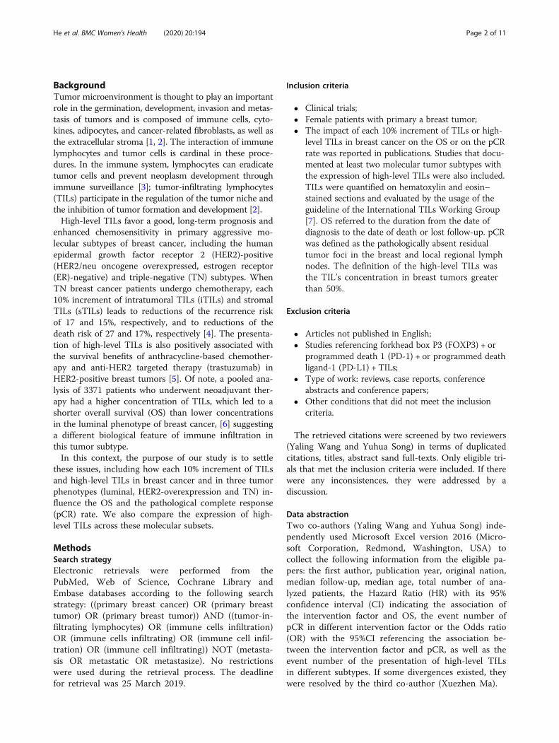
Inclusion criteria
� Clinical trials;� Female patients with primary a breast tumor;� The impact of each 10% increment of TILs or high-
level TILs in breast cancer on the OS or on the pCRrate was reported in publications. Studies that docu-mented at least two molecular tumor subtypes withthe expression of high-level TILs were also included.TILs were quantified on hematoxylin and eosin–stained sections and evaluated by the usage of theguideline of the International TILs Working Group[7]. OS referred to the duration from the date ofdiagnosis to the date of death or lost follow-up. pCRwas defined as the pathologically absent residualtumor foci in the breast and local regional lymphnodes. The definition of the high-level TILs wasthe TIL’s concentration in breast tumors greaterthan 50%.
Exclusion criteria
� Articles not published in English;� Studies referencing forkhead box P3 (FOXP3) + or
programmed death 1 (PD-1) + or programmed deathligand-1 (PD-L1) + TILs;
� Type of work: reviews, case reports, conferenceabstracts and conference papers;
� Other conditions that did not meet the inclusioncriteria.
The retrieved citations were screened by two reviewers(Yaling Wang and Yuhua Song) in terms of duplicatedcitations, titles, abstract sand full-texts. Only eligible tri-als that met the inclusion criteria were included. If therewere any inconsistences, they were addressed by adiscussion.
Data abstractionTwo co-authors (Yaling Wang and Yuhua Song) inde-pendently used Microsoft Excel version 2016 (Micro-soft Corporation, Redmond, Washington, USA) tocollect the following information from the eligible pa-pers: the first author, publication year, original nation,median follow-up, median age, total number of ana-lyzed patients, the Hazard Ratio (HR) with its 95%confidence interval (CI) indicating the association ofthe intervention factor and OS, the event number ofpCR in different intervention factor or the Odds ratio(OR) with the 95%CI referencing the association be-tween the intervention factor and pCR, as well as theevent number of the presentation of high-level TILsin different subtypes. If some divergences existed, theywere resolved by the third co-author (Xuezhen Ma).
He et al. BMC Women's Health (2020) 20:194 Page 2 of 11
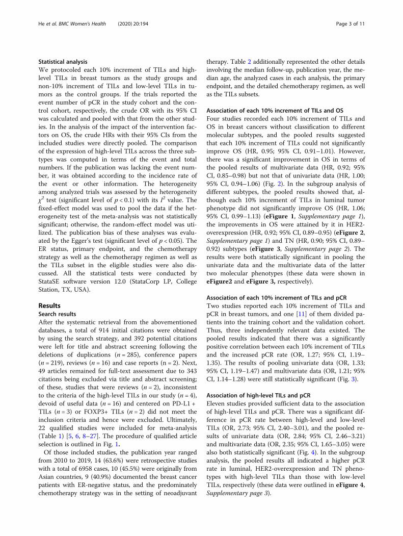
Statistical analysisWe protocoled each 10% increment of TILs and high-level TILs in breast tumors as the study groups andnon-10% increment of TILs and low-level TILs in tu-mors as the control groups. If the trials reported theevent number of pCR in the study cohort and the con-trol cohort, respectively, the crude OR with its 95% CIwas calculated and pooled with that from the other stud-ies. In the analysis of the impact of the intervention fac-tors on OS, the crude HRs with their 95% CIs from theincluded studies were directly pooled. The comparisonof the expression of high-level TILs across the three sub-types was computed in terms of the event and totalnumbers. If the publication was lacking the event num-ber, it was obtained according to the incidence rate ofthe event or other information. The heterogeneityamong analyzed trials was assessed by the heterogeneityχ2 test (significant level of p < 0.1) with its I2 value. Thefixed-effect model was used to pool the data if the het-erogeneity test of the meta-analysis was not statisticallysignificant; otherwise, the random-effect model was uti-lized. The publication bias of these analyses was evalu-ated by the Egger’s test (significant level of p < 0.05). TheER status, primary endpoint, and the chemotherapystrategy as well as the chemotherapy regimen as well asthe TILs subset in the eligible studies were also dis-cussed. All the statistical tests were conducted byStataSE software version 12.0 (StataCorp LP, CollegeStation, TX, USA).
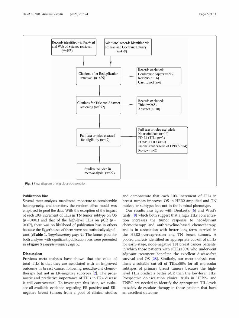
ResultsSearch resultsAfter the systematic retrieval from the abovementioneddatabases, a total of 914 initial citations were obtainedby using the search strategy, and 392 potential citationswere left for title and abstract screening following thedeletions of duplications (n = 285), conference papers(n = 219), reviews (n = 16) and case reports (n = 2). Next,49 articles remained for full-text assessment due to 343citations being excluded via title and abstract screening;of these, studies that were reviews (n = 2), inconsistentto the criteria of the high-level TILs in our study (n = 4),devoid of useful data (n = 16) and centered on PD-L1 +TILs (n = 3) or FOXP3+ TILs (n = 2) did not meet theinclusion criteria and hence were excluded. Ultimately,22 qualified studies were included for meta-analysis(Table 1) [5, 6, 8–27]. The procedure of qualified articleselection is outlined in Fig. 1.Of those included studies, the publication year ranged
from 2010 to 2019, 14 (63.6%) were retrospective studieswith a total of 6958 cases, 10 (45.5%) were originally fromAsian countries, 9 (40.9%) documented the breast cancerpatients with ER-negative status, and the predominatelychemotherapy strategy was in the setting of neoadjuvant
therapy. Table 2 additionally represented the other detailsinvolving the median follow-up, publication year, the me-dian age, the analyzed cases in each analysis, the primaryendpoint, and the detailed chemotherapy regimen, as wellas the TILs subsets.
Association of each 10% increment of TILs and OSFour studies recorded each 10% increment of TILs andOS in breast cancers without classification to differentmolecular subtypes, and the pooled results suggestedthat each 10% increment of TILs could not significantlyimprove OS (HR, 0.95; 95% CI, 0.91–1.01). However,there was a significant improvement in OS in terms ofthe pooled results of multivariate data (HR, 0.92; 95%CI, 0.85–0.98) but not that of univariate data (HR, 1.00;95% CI, 0.94–1.06) (Fig. 2). In the subgroup analysis ofdifferent subtypes, the pooled results showed that, al-though each 10% increment of TILs in luminal tumorphenotype did not significantly improve OS (HR, 1.06;95% CI, 0.99–1.13) (eFigure 1, Supplementary page 1),the improvements in OS were attained by it in HER2-overexpression (HR, 0.92; 95% CI, 0.89–0.95) (eFigure 2,Supplementary page 1) and TN (HR, 0.90; 95% CI, 0.89–0.92) subtypes (eFigure 3, Supplementary page 2). Theresults were both statistically significant in pooling theunivariate data and the multivariate data of the lattertwo molecular phenotypes (these data were shown ineFigure2 and eFigure 3, respectively).
Association of each 10% increment of TILs and pCRTwo studies reported each 10% increment of TILs andpCR in breast tumors, and one [11] of them divided pa-tients into the training cohort and the validation cohort.Thus, three independently relevant data existed. Thepooled results indicated that there was a significantlypositive correlation between each 10% increment of TILsand the increased pCR rate (OR, 1.27; 95% CI, 1.19–1.35). The results of pooling univariate data (OR, 1.33;95% CI, 1.19–1.47) and multivariate data (OR, 1.21; 95%CI, 1.14–1.28) were still statistically significant (Fig. 3).
Association of high-level TILs and pCREleven studies provided sufficient data to the associationof high-level TILs and pCR. There was a significant dif-ference in pCR rate between high-level and low-levelTILs (OR, 2.73; 95% CI, 2.40–3.01), and the pooled re-sults of univariate data (OR, 2.84; 95% CI, 2.46–3.21)and multivariate data (OR, 2.35; 95% CI, 1.65–3.05) werealso both statistically significant (Fig. 4). In the subgroupanalysis, the pooled results all indicated a higher pCRrate in luminal, HER2-overexpression and TN pheno-types with high-level TILs than those with low-levelTILs, respectively (these data were outlined in eFigure 4,Supplementary page 3).
He et al. BMC Women's Health (2020) 20:194 Page 3 of 11

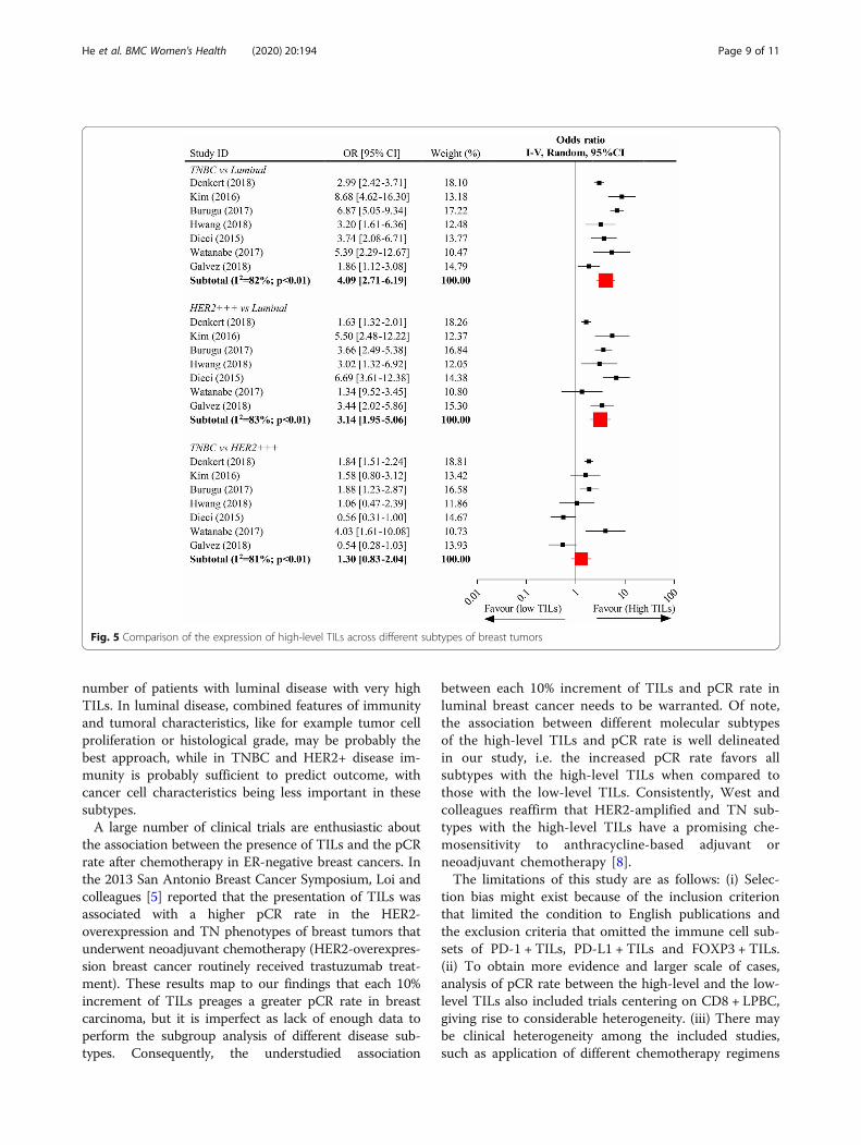
Comparison of high-level TILs expression across differentbreast cancer subsetsSeven studies were collected to perform the compari-son of expression of high-level TILs across the differ-ent subsets of breast tumors. The pooled data ofanalysis showed that the presentation of high-levelTILs between HER2-overexpression subtype and TN
subtype was not significantly different (OR, 1.30;95%CI, 0.83–2.04), whereas both subtypes experienceda significantly elevated expression of high-level TILsas compared to luminal phenotype (HER2-overexpres-sion vs. luminal, OR, 3.14; 95% CI, 1.95–5.06; andTN vs. luminal, OR, 4.09; 95% CI, 2.71–6.19; respect-ively) (Fig. 5).
Table 1 Details of the included trials
Study (Trail) PublicationYear
Study duration Originalnation
Medianfollow-up
No. ofpatient(n)
ERstatus
Primaryendpoints
Chemotherapystrategy
Regimen
West [8] 2011 Unknow Canada Unknow 111 Negative pCR NAC FEC or TET
Seo [9] 2013 2004–2011 Korea Unknow 153 Both pCR NAC AC or ACT or AD
Lee [10] 2013 2000–2009 Korea Unknow 175 Both pCR NAC AC or ACT
Denkert[11]*
2010 1999–2001 Germany Unknow 218 Both pCR NAC ACT
Denkert[11]*
2010 2002–2005 Germany Unknow 840 Both pCR NAC TAC or TAC followed byvinorelbine and capecitabine
Denkert [12] 2015 Unknow Germany Unknow 580 Negative pCR Unknow 1:1 to PM or PMCb
Watanabe[13]
2018 2008–2016 Japan 26.1 m 197 Both pCR NAC Anthracycline- or taxane- oranthracycline- plus taxane-based
Galvez [14] 2018 2003–2014 Peru Unknow 435 Both pCR NAC ACP or AC
Hida [15] 2016 2007–2014 Japan Unknow 159 Negative pCR NAC Unknow
Denkert [6] 2018 Unknow Germany Unknow 3771 Both pCR, OS NAC Docetaxel- or paclitaxel- ornab-paclitaxel-based
Hwang [16] 2019 2004–2013 Korea 60.1 m 248 Both pCR NAC Anthracycline- plus taxane-based
Kim [17] 2016 2004–2011 Korea 6.4y 688 Negative OS NAC AC or ACT or ACP
Sønderstrup[18]
2019 1997–2011 Denmark 5.8y 399 Both OS Unknow Unknow
Pruneri [19] 2016 1995–2010 Switzerland 8.2y 897 Negative OS AC CMF, CMF + AC
Pruneri [20] 2016 Unknow Italy 6.9y 647 Negative OS AC 1:1 to CM or no-CM
Tian [21] 2016 2008–2012 China 4y 372 Negative OS AC Anthracycline- or anthracycline-plus taxanes-based
Adams [22] 2014 Unknow USA 10.6y 481 Negative OS NAC ACT or ACP
Loi [5] 2014 Unknow Belgium 62m 934 Both OS NAC Docetaxel or vinorelbinefollowed by FEC
Kochi [23] 2018 Unknow Japan Unknow 40 Both OS NAC Anthracycline- plus taxane-based
Dieci [24] 2015 Unknow France 12.7y 781 Both OS AC Anthracycline-based
Luen [25] 2019 Unknow Australia 6y 375 Negative OS NAC Unknow
Luen [26] 2017 Unknow Australia 50 m 678 Negative OS AC 1:1 to trastuzumab anddocetaxel plus eitherpertuzumab or placebo
Burugu [27] 2017 1989–2002 Canada 13y 2497 Both Others AC Unknow
*This article is divided into two researches due to different regimenAbbreviations: pCR, pathological complete response; OS, overall survival; NAC, neoadjuvant chemotherapy; AC, adjuvant chemotherapy; TNBC, triple-negativebreast cancer; HER2 + BC, human epidermal growth factor receptor 2 positive breast cancerRegimen explanation: FEC: fluorouracil, epirubicin, and cyclophosphamide; TET: docetaxel followed by epirubicin plus docetaxel; AC: doxorubicin andcyclophosphamide; ACT: AC followed by docetaxel; AD: doxorubicin and docetaxel; TAC: docetaxel, doxorubicin, and cyclophosphamide; PM: Paclitaxel and non-pegylated liposomal doxorubicin; PMCb: Paclitaxel and non-pegylated liposomal doxorubicin followed by carboplatin; TP: paclitaxel plus platinum; ACP:doxorubicin and cyclophosphamide followed by paclitaxel; CAF: cyclophosphamide, adriamycin and fluorouracil; CMF: cyclophosphamide, methotrexate andfluorouracil; CM: cyclophosphamide plus methotrexate
He et al. BMC Women's Health (2020) 20:194 Page 4 of 11
Publication biasSeveral meta-analyses manifested moderate-to-considerableheterogeneity, and therefore, the random-effect model wasemployed to pool the data. With the exception of the impactof each 10% increment of TILs in TN tumor subtype on OS(p= 0.001) and that of the high-level TILs on pCR (p=0.007), there was no likelihood of publication bias in othersbecause the Egger’s tests of them were not statistically signifi-cant (eTable 1, Supplementary page 4). The funnel plots forboth analyses with significant publication bias were presentedin eFigure 5 (Supplementary page 5).
DiscussionPrevious meta-analyses have shown that the value oftotal TILs is that they are associated with an improvedoutcome in breast cancer following neoadjuvant chemo-therapy but not in ER-negative subtypes [2]. The prog-nostic and predictive importance of TILs in ER+ diseaseis still controversial. To investigate this issue, we evalu-ate all available evidence regarding ER positive and ER-negative breast tumors from a pool of clinical studies
and demonstrate that each 10% increment of TILs inbreast tumors improves OS in HER2-amplified and TNmolecular subtypes but not in the luminal phenotype.Our results also agree with Denkert’s [6] and West’s
trials, [8] which both suggest that a high TILs concentra-tion increases the tumor response to neoadjuvantchemotherapy and anthracycline-based chemotherapy,and is in association with better long-term survival inthe HER2-overexpression and TN breast tumors. Apooled analysis identified an appropriate cut-off of sTILsfor early-stage, node-negative TN breast cancer patients,in which those patients with sTILs≥30% who underwentadjuvant treatment benefited the excellent disease-freesurvival and OS [28]. Similarly, our meta-analysis con-firms a suitable cut-off of TILs≥50% for all molecularsubtypes of primary breast tumors because the high-level TILs predict a better pCR than the low-level TILs.Prospective de-escalation clinical trials in HER2+ andTNBC are needed to identify the appropriate TIL-levelsto safely de-escalate therapy in those patients that havean excellent outcome.
Fig. 1 Flow diagram of eligible article selection
He et al. BMC Women's Health (2020) 20:194 Page 5 of 11

Table 2 Summary of the characteristics of the 21 included Studies
Characteristic Studies, No. (%) (N = 22) Primary Breast Cancer Patients,No. (%) (N = 15,676)
Study type
Randomized trial 5 (22.7) 3578 (22.8)
Retrospective 14 (63.6) 6958 (44.4)
Pooled 1 (4.5) 3771 (24.1)
Prospective–retrospective 1 (4.5) 934 (6.0)
Prospective 1 (4.5) 435 (2.8)
Publication date, median (range), y 2016 (2010–2019)
Follow-up, median (range), mo* 90.6 (48.0–190.8)
Median age, median (range), y* 50.0 (46.5–54.0)
10% increment of TILs and OS,total (range), n
All subtypes 4460 (399–2346)
Luminal 1886 (463–832)
HER2-enriched 1985 (112–986)
TNBC 3847 (92–897)
10% increment of TILs and pCR,total (range), n
1638 (218–840)
LPBC and pCR, total (range), n
All subtypes 6697 (40–3771)
Luminal 1717 (91–1366)
HER2-enriched 1801 (40–1379)
TNBC 1425 (48–906)
High TILs across different subtypes,total (range), n
TNBC vs Luminal 6524 (138–2297)
HER2-enriched vs Luminal 6696 (149–2745)
TNBC vs HER2-enriched 3722 (105–2285)
Original area
Asia 10 (45.5) 3085 (19.7)
America 4 (18.2) 3524 (22.5)
Europe 8 (36.4) 9067 (57.8)
ER status
ER-positive 0 (0.0) 0 (0.0)
ER-negative 9 (40.9) 4300 (27.4)
ER-both 13 (59.1) 11,376 (72.6)
Primary endpoint
pCR 10 (45.5) 6834 (43.6)
OS 11 (50.0) 6345 (40.5)
Others 1 (4.5) 2497 (15.9)
Chemotherapy strategy
Neoadjuvant 15 (68.2) 9669 (61.7)
Adjuvant 5 (22.7) 5028 (32.1)
Unknow 2 (9.1) 979 (6.2)
Chemotherapy regimen
He et al. BMC Women's Health (2020) 20:194 Page 6 of 11

In the study by Denkert et al., [6] it is found that theincreased TILs may be an adverse factor to OS in breastcancer patients with the luminal subtype, which differsfrom our results. This difference may be explained asfollows. First, they only evaluated the OS in luminal-HER2-negative tumors, while our study also includesluminal-HER-positive of breast cancer patients. Further-more, they only center on the assessment of the impactof sTILs on OS, but we additionally assess the iTILs.
Last, the treatment strategies are not identical, as onlyneoadjuvant chemotherapy is included in their study butadjuvant chemotherapy is yet included in ours. Collect-ively, the prognostic outcome of TIL-levels in luminaldisease remains an important area of investigation. Oneof the reasons why TIL-counts are not associated withsurvival benefit in luminal disease is that the range ofTILs is not very high, with most cases having less than10% stromal TILs, so most series don’t have a significant
Table 2 Summary of the characteristics of the 21 included Studies (Continued)
Characteristic Studies, No. (%) (N = 22) Primary Breast Cancer Patients,No. (%) (N = 15,676)
Anthracycline-based 3 (13.6) 2113 (13.5)
Taxanes-based 1 (4.5) 3771 (24.1)
Anthracycline- and taxanes-based 10 (45.5) 3822 (24.4)
Methotrexate-based 3 (13.6) 1915 (12.2)
Unknow 5 (22.7) 4055 (25.9)
TILs subsets
TILs 11 (50.0) 8014 (51.1)
iTILs 3 (13.6) 4135 (26.4)
sTILs 6 (27.3) 3199 (20.4)
CD8 + TILs 1 (4.5) 175 (1.1)
CD4 + TILs 1 (4.5) 153 (1.0)
Abbreviations: TILs, tumor-infiltrating lymphocytes; OS, overall survival; HER2, human epidermal growth factor receptor 2; TNBC, triple-negative breast cancer; pCR,pathological complete response; LPBC, lymphocyte-predominant breast cancer; ER, estrogen receptor; iTILs, intratumoral tumor-infiltrating lymphocytes; sTILsstromal tumor-infiltrating lymphocytes*Median value is calculated in terms of available data
Fig. 2 Impacts of each 10% increment of tumor-infiltrating lymphocytes in breast tumor on overall survival
He et al. BMC Women's Health (2020) 20:194 Page 7 of 11

Fig. 3 Impacts of each 10% increment of tumor-infiltrating lymphocytes in breast tumor on the pathological complete response
Fig. 4 Impacts of the high-level TILs on the pathological completed response
He et al. BMC Women's Health (2020) 20:194 Page 8 of 11
number of patients with luminal disease with very highTILs. In luminal disease, combined features of immunityand tumoral characteristics, like for example tumor cellproliferation or histological grade, may be probably thebest approach, while in TNBC and HER2+ disease im-munity is probably sufficient to predict outcome, withcancer cell characteristics being less important in thesesubtypes.A large number of clinical trials are enthusiastic about
the association between the presence of TILs and the pCRrate after chemotherapy in ER-negative breast cancers. Inthe 2013 San Antonio Breast Cancer Symposium, Loi andcolleagues [5] reported that the presentation of TILs wasassociated with a higher pCR rate in the HER2-overexpression and TN phenotypes of breast tumors thatunderwent neoadjuvant chemotherapy (HER2-overexpres-sion breast cancer routinely received trastuzumab treat-ment). These results map to our findings that each 10%increment of TILs preages a greater pCR rate in breastcarcinoma, but it is imperfect as lack of enough data toperform the subgroup analysis of different disease sub-types. Consequently, the understudied association
between each 10% increment of TILs and pCR rate inluminal breast cancer needs to be warranted. Of note,the association between different molecular subtypesof the high-level TILs and pCR rate is well delineatedin our study, i.e. the increased pCR rate favors allsubtypes with the high-level TILs when compared tothose with the low-level TILs. Consistently, West andcolleagues reaffirm that HER2-amplified and TN sub-types with the high-level TILs have a promising che-mosensitivity to anthracycline-based adjuvant orneoadjuvant chemotherapy [8].The limitations of this study are as follows: (i) Selec-
tion bias might exist because of the inclusion criterionthat limited the condition to English publications andthe exclusion criteria that omitted the immune cell sub-sets of PD-1 + TILs, PD-L1 + TILs and FOXP3 + TILs.(ii) To obtain more evidence and larger scale of cases,analysis of pCR rate between the high-level and the low-level TILs also included trials centering on CD8 + LPBC,giving rise to considerable heterogeneity. (iii) There maybe clinical heterogeneity among the included studies,such as application of different chemotherapy regimens
Fig. 5 Comparison of the expression of high-level TILs across different subtypes of breast tumors
He et al. BMC Women's Health (2020) 20:194 Page 9 of 11

and treatment strategies, as well as investigation of dif-ferent TILs subtypes.Despite these limitations, this was the first meta-
analysis that systematically evaluated the influence ofeach 10% increment of TILs and the high-level TILs inbreast cancer on OS and pCR, and compared the pres-entation of high-level TILs across different molecularsubtypes. Future studies will need to supplement theunderrecognized and understudied landscapes whether ahigher pCR rate is related to each 10% increment ofTILs in the luminal subtype of breast cancer and thehigh-level TILs in clinical high-risk luminal breast can-cer patients can translate into a promising OS.
ConclusionsEach 10% increment of TILs in breast tumors predicts im-proved OS and pCR rate of patients, specifically in theHER2-overexpression and TN molecular subtypes. More-over, all subsets with the high-level TILs benefit greaterpCR rate than those with the low-level TILs. Althoughthere is no difference between the expression of high-levelTILs among HER2-overexpression and TN phenotypes ofbreast cancer, they both have greater expression than thatrelative to the luminal tumor subtype.
Supplementary informationSupplementary information accompanies this paper at https://doi.org/10.1186/s12905-020-01038-x.
Additional file 1: eFigure 1. Impacts of each 10% increment of tumor-infiltrating lymphocytes on overall survival in Luminal subtype. eFigure 2:Impacts of each 10% increment of tumor-infiltrating lymphocytes onoverall survival in HER2-overexpression subtype. eFigure 3: Impacts ofeach 10% increment of tumor-infiltrating lymphocytes on overall survivalin triple-negative subtype. eFigure 4: Impacts of the high-level TILs onthe pathological completed response in different tumor subtypes.eTable 1. Publication bias by Egg’s test in meta-analysis. eFigure 5. Funnelplots for the significant analyses of publication bias.
AbbreviationsTILs: Tumor-infiltrating lymphocytes; LPBC: Lymphocyte-predominant breastcancer; HER2: Human epidermal growth factor receptor 2; TN: Triple-negative; DFS: Disease-free survival; OS: Overall survival; pCR: Pathologiccomplete response; iTILs: Intratumoral TILs; sTILs: Stromal TILs; PD-1: Programmed death 1; PD-L1: Programmed death ligand-1;FOXP3: Forkhead box P3; HR: Hazard Ratio; CI: Confidence interval; OR: Oddsratio; ER: Estrogen receptor
AcknowledgementsWe deeply acknowledge Dr Yanan Zheng, EKG department, the SecondAffiliated Hospital of Anhui Medical University (Hefei, China), assisting usto complete the production of Table 1.
Consent to publishNot applicable.
Authors’ contributionsLH: have drafted the work or substantively revised it, interpretation of data;YW: the creation of new software used in the work; QW: acquisition, analysis;YS: acquisition, analysis; XM: acquisition, analysis; BZ: acquisition, analysis; HW:design of the work; YH: have approved the submitted version and haveagreed both to be personally accountable for the author’s own contributions
and to ensure that questions related to the accuracy or integrity of any partof the work. All authors reviewed and approved the manuscript prior tosubmission.
FundingNo funding.
Availability of data and materialsNot applicable.
Ethics approval and consent to participateThis article does not contain any studies with human participants or animalsperformed by any of the authors.
Competing interestsThe authors declare that they have no competing interests.
Author details1Breast Disease Center, The Affiliated Hospital of Qingdao University,Qingdao, Shandong Province, People’s Republic of China. 2Department ofOncology, The Second Affiliated Hospital of Medical College of QingdaoUniversity, Qingdao, Shandong Province, People’s Republic of China.3Department of Radiotherapy, The Affiliated Hospital of Qingdao University,Qingdao, Shandong Province, People’s Republic of China.
Received: 30 October 2019 Accepted: 27 July 2020
References1. Liotta LA, Kohn EC. The microenvironment of the tumour-host interface.
Nature. 2001;411(6835):375–9.2. Yu X. Zhang Z. Prognostic and predictive value of tumor-infiltrating
lymphocytes in breast cancer: a systematic review and meta-analysis. 2016;18(5):497–506.
3. Swann JB, Smyth MJ. Immune surveillance of tumors. J Clin Invest. 2007;117(5):1137–46.
4. Loi S, Sirtaine N, Piette F, Salgado R, Viale G, Van Eenoo F, et al. Prognosticand predictive value of tumor-infiltrating lymphocytes in a phase IIIrandomized adjuvant breast cancer trial in node-positive breast cancercomparing the addition of docetaxel to doxorubicin with doxorubicin-based chemotherapy: BIG 02-98. Journal of clinical oncology : official journalof the American Society of Clinical Oncology. 2013;31(7):860–7.
5. Loi S, Michiels S, Salgado R, Sirtaine N, Jose V, Fumagalli D, et al. Tumorinfiltrating lymphocytes are prognostic in triple negative breast cancer andpredictive for trastuzumab benefit in early breast cancer: results from theFinHER trial. Annals of oncology : official journal of the European Society forMedical Oncology. 2014;25(8):1544–50.
6. Denkert C, von Minckwitz G, Darb-Esfahani S, Lederer B, Heppner BI, WeberKE, et al. Tumour-infiltrating lymphocytes and prognosis in differentsubtypes of breast cancer: a pooled analysis of 3771 patients treated withneoadjuvant therapy. The Lancet Oncology. 2018;19(1):40–50.
7. Salgado R, Denkert C, Demaria S, Sirtaine N, Klauschen F, Pruneri G, et al.The evaluation of tumor-infiltrating lymphocytes (TILs) in breast cancer:recommendations by an international TILs working group 2014. Annals ofoncology : official journal of the European Society for Medical Oncology.2015;26(2):259–71.
8. West NR, Milne K, Truong PT, Macpherson N, Nelson BH, Watson PH. Tumor-infiltrating lymphocytes predict response to anthracycline-basedchemotherapy in estrogen receptor-negative breast cancer. Breast CancerResearch. 2011;13(6).
9. Seo AN, Lee HJ, Kim EJ, Kim HJ, Jang MH, Lee HE, et al. Tumour-infiltratingCD8+ lymphocytes as an independent predictive factor for pathologicalcomplete response to primary systemic therapy in breast cancer. Br JCancer. 2013;109(10):2705–13.
10. Lee HJ, Seo JY, Ahn JH, Ahn SH, Gong G. Tumor-associated lymphocytespredict response to Neoadjuvant chemotherapy in breast Cancer patients. JBreast Cancer. 2013;16(1):32–9.
11. Denkert C, Loibl S, Noske A, Roller M. M¨1ller BM, Komor M, et al. tumor-associated lymphocytes as an independent predictor of response toneoadjuvant chemotherapy in b reast cancer. Journal of clinical oncology :official journal of the American Society of Clinical Oncology. 2010;28(1):105–13.
He et al. BMC Women's Health (2020) 20:194 Page 10 of 11

12. Denkert C, von Minckwitz G, Brase JC, Sinn BV, Gade S, Kronenwett R, et al.Tumor-infiltrating lymphocytes and response to neoadjuvant chemotherapywith or without carboplatin i n human epidermal growth factor receptor 2-positive and triple-negative primary breast cancers. Journal of clinicaloncology : official journal of the American Society of Clinical Oncology.2015;33(9):983–91.
13. Watanabe T, Hida AI, Inoue N, Imamura M, Fujimoto Y, Akazawa K, et al.Abundant tumor infiltrating lymphocytes after primary systemicchemotherapy predicts poor prognosis in estrogen receptor-positive/HER2-negative breast cancers. Breast Cancer Res Treat. 2018;168(1):135–45.
14. Galvez M, Castaneda CA, Sanchez J, Castillo M, Rebaza LP, Calderon G, et al.Clinicopathological predictors of long-term benefit in breast cancer treated withneoadjuvant chemotherapy. World Journal Of Clinical Oncology. 2018;9(2):33–41.
15. Hida AI, Sagara Y, Yotsumoto D, Kanemitsu S, Kawano J, Baba S, et al.Prognostic and predictive impacts of tumor-infiltrating lymphocytes differbetween triple-negative and HER2-positive breast cancers treated withstandard systemic therapies. Breast Cancer Res Treat. 2016;158(1):1–9.
16. Hwang HW, Jung H, Hyeon J, Park YH, Ahn JS, Im YH, et al. A nomogram topredict pathologic complete response (pCR) and the value of tumor-infiltrating lymphocy tes (TILs) for prediction of response to neoadjuvantchemotherapy (NAC) in breast cancer patients. Breast Cancer Res Treat.2019;173(2):255–66.
17. Kim YA, Lee HJ, Heo SH, Park HS, Park SY, Bang WS, et al. MxA expression isassociated with tumor-infiltrating lymphocytes and is a prognostic factor intriple-negative breast cancer. Breast Cancer Res Treat. 2016;156(3):597–606.
18. Sonderstrup IMH, Jensen MB, Ejlertsen B, Eriksen JO, Gerdes AM, Kruse TA,et al. Evaluation of tumor-infiltrating lymphocytes and association withprognosis in BRCA-mutated breast cancer. Acta oncologica (Stockholm,Sweden). 2019:1–8.
19. Pruneri G, Gray KP, Vingiani A, Viale G, Curigliano G, Criscitiello C, et al.Tumor-infiltrating lymphocytes (TILs) are a powerful prognostic marker inpatients with triple-negative breast cancer enrolled in the IBCSG phase IIIrandomized clinical trial 22-00. Breast Cancer Res Treat. 2016;158(2):323–31.
20. Pruneri G, Vingiani A, Bagnardi V, Rotmensz N, De Rose A, Palazzo A, et al.Clinical validity of tumor-infiltrating lymphocytes analysis in patients withtriple-negative breast cancer. Ann Oncol. 2016;27(2):249–56.
21. Tian T, Ruan M, Yang W, Shui R. Evaluation of the prognostic value oftumor-infiltrating lymphocytes in triple-negative breast cancers. Oncotarget.2016;7(28):44395–405.
22. Adams S, Gray RJ, Demaria S, Goldstein L, Perez EA, Shulman LN, et al.Prognostic value of tumor-infiltrating lymphocytes in triple-negative breastcancers from two phase I II randomized adjuvant breast cancer trials: ECOG2197 and ECOG 1199. Journal of clinical oncology : official journal of theAmerican Society of Clinical Oncology. 2014;32(27):2959–66.
23. Kochi M, Iwamoto T, Niikura N, Bianchini G, Masuda S, Mizoo T, et al.Tumour-infiltrating lymphocytes (TILs)-related genomic signature predictschemotherapy response in breast cancer. Breast Cancer Res Treat. 2018;167(1):39–47.
24. Dieci MV, Mathieu MC, Guarneri V, Conte P, Delaloge S, Andre F, et al.Prognostic and predictive value of tumor-infiltrating lymphocytes in two phaseIII randomized adjuvant breast cancer trials. Ann Oncol. 2015;26(8):1698–704.
25. Luen SJ, Salgado R, Dieci MV, Vingiani A, Curigliano G, Gould RE, et al.Prognostic implications of residual disease tumor-infiltrating lymphocytesand residual cancer burden in triple-negative breast cancer patients afterneoadjuvant chemotherapy. Annals of oncology : official journal of theEuropean Society for Medical Oncology. 2019;30(2):236–42.
26. Luen SJ, Salgado R, Fox S, Savas P, Eng-Wong J, Clark E, et al. Tumour-infiltratinglymphocytes in advanced HER2-positive breast cancer treated with pertuzumabor placebo in addition to trastuzumab and docetaxel: a retrospective analysis ofthe CLEOPATRA study. The Lancet Oncology. 2017;18(1):52–62.
27. Burugu S, Gao D, Leung S, Chia SK, Nielsen TO. LAG-3+tumor infiltratinglymphocytes in breast cancer: clinical correlates and association with PD-1/PD-L1+tumors. Ann Oncol. 2017;28(12):2977–84.
28. Loi S, Drubay D, Adams S, Pruneri G, Francis PA, Lacroix-Triki M, et al.Tumor-infiltrating lymphocytes and prognosis: a pooled individual patientanalysis of early-stage tri ple-negative breast cancers. J Clin Oncol. 2019;37(7):559–69.
Publisher’s NoteSpringer Nature remains neutral with regard to jurisdictional claims inpublished maps and institutional affiliations.
He et al. BMC Women's Health (2020) 20:194 Page 11 of 11
Related Documents