Citation: Matyas, M.; Iancu, M.; Hasmasanu, M.; Man, A.; Zaharie, G. Association Analysis of the Cerebral Fractional Tissue Oxygen Extraction (cFTOE) and the Cerebral Oxygen Saturation (crSaO 2 ) with Perinatal Factors in Preterm Neonates: A Single Centre Study. J. Clin. Med. 2022, 11, 3546. https://doi.org/ 10.3390/jcm11123546 Academic Editor: Sylvie Girard Received: 8 May 2022 Accepted: 16 June 2022 Published: 20 June 2022 Publisher’s Note: MDPI stays neutral with regard to jurisdictional claims in published maps and institutional affil- iations. Copyright: © 2022 by the authors. Licensee MDPI, Basel, Switzerland. This article is an open access article distributed under the terms and conditions of the Creative Commons Attribution (CC BY) license (https:// creativecommons.org/licenses/by/ 4.0/). Journal of Clinical Medicine Article Association Analysis of the Cerebral Fractional Tissue Oxygen Extraction (cFTOE) and the Cerebral Oxygen Saturation (crSaO 2 ) with Perinatal Factors in Preterm Neonates: A Single Centre Study Melinda Matyas 1 , Mihaela Iancu 2, * , Monica Hasmasanu 1 , Anca Man 3 and Gabriela Zaharie 1 1 Neonatology Department, “Iuliu Hat , ieganu” University of Medicine and Pharmacy, 3, Clinicilor Street, 400012 Cluj-Napoca, Romania; [email protected] (M.M.); [email protected] (M.H.); [email protected] (G.Z.) 2 Department of Medical Informatics and Biostatistics, “Iuliu Hat , ieganu” University of Medicine and Pharmacy, 8, Victor Babes Street, 400012 Cluj-Napoca, Romania 3 Neonatology Department, County Emergency Hospital, 3, Clinicilor Street, 400347 Cluj-Napoca, Romania; [email protected] * Correspondence: [email protected]; Tel.: +40-0740-130-888 Abstract: (1) Background: Near-infrared spectroscopy (NIRS) is a non-invasive, easily performed method of monitoring brain oxygenation. The regional cerebral oxygen saturation (crSaO 2 ) and the cerebral fractional tissue oxygen extraction (cFTOE) evaluated by NIRS provide more accurate infor- mation on brain oxygenation than the blood oxygen saturation. We investigated the effect of perinatal factors on cerebral oxygenation of preterm newborns. (2) Methods: We conducted a longitudinal study with 48 preterm newborns <34 weeks of gestation who underwent NIRS registration during the first 72 h of life. crSaO 2 was measured and cFTOE was calculated foreach patient. (3) Results: One-way ANOVA showed no significant main effect of IVH severity on crSaO 2 and cFTOE (p > 0.05); there was a tendency toward statistical significance concerning the difference between the means of crSaO 2 (p = 0.083) and cFTOE (p = 0.098). Patients with intraventricular haemorrhage (IVH) had a lower mean of crSaO 2 and a higher mean of cFTOE (59.67 ± 10.37% vs. 64.92 ± 10.16% for crSaO 2 ; 0.37 ± 0.11 vs. 0.32 ± 0.11 for cFTOE) compared to those with no IVH. Significantly lower values of crSaO 2 and higher values of cFTOE were found in neonates receiving inotropic treatment (p < 0.0001). Episodes of apnoea also proved to influence the cerebral oxygen saturation of the study group (p = 0.0026). No significant association between the maternal hypertension treatment and the cerebral oxygenation of preterms was found. (4) Conclusions: This study showed a decreased cerebral oxygen saturation of preterms with IVH, inotrope support and apnoea episodes. Keywords: cerebral oxygen saturation (crSaO 2 ); near infrared spectroscopy (NIRS); premature newborns 1. Introduction The neurological evolution of preterm infants is influenced by multiple perinatal factors. Among the prenatal factors, maternal inflammation, chorioamnionitis or pre- eclampsia influence the neurological evolution [1,2]. Immediately after birth, several conditions characteristic of prematurity, and nutrition, caring and nursing have an impact on the neurological development of the premature infant. After clamping the umbilical cord, the newborn must maintain adequate oxygenation and the flow of nutrients to the brain using incompletely known self-regulatory mechanisms [1,3,4]. Monitoring the blood oxygen saturation or the heart rate does not accurately reflect the oxygenation in the brain. By using near-infrared spectroscopy (NIRS), a non-invasive technique, we can determine the regional cerebral oxygen saturation (crSaO 2 ) and the cerebral fractional tissue oxygen extraction (cFTOE) in the perinatal period. Oxygen J. Clin. Med. 2022, 11, 3546. https://doi.org/10.3390/jcm11123546 https://www.mdpi.com/journal/jcm

Welcome message from author
This document is posted to help you gain knowledge. Please leave a comment to let me know what you think about it! Share it to your friends and learn new things together.
Transcript
Citation: Matyas, M.; Iancu, M.;
Hasmasanu, M.; Man, A.; Zaharie, G.
Association Analysis of the Cerebral
Fractional Tissue Oxygen Extraction
(cFTOE) and the Cerebral Oxygen
Saturation (crSaO2) with Perinatal
Factors in Preterm Neonates: A
Single Centre Study. J. Clin. Med.
2022, 11, 3546. https://doi.org/
10.3390/jcm11123546
Academic Editor: Sylvie Girard
Received: 8 May 2022
Accepted: 16 June 2022
Published: 20 June 2022
Publisher’s Note: MDPI stays neutral
with regard to jurisdictional claims in
published maps and institutional affil-
iations.
Copyright: © 2022 by the authors.
Licensee MDPI, Basel, Switzerland.
This article is an open access article
distributed under the terms and
conditions of the Creative Commons
Attribution (CC BY) license (https://
creativecommons.org/licenses/by/
4.0/).
Journal of
Clinical Medicine
Article
Association Analysis of the Cerebral Fractional Tissue OxygenExtraction (cFTOE) and the Cerebral Oxygen Saturation(crSaO2) with Perinatal Factors in Preterm Neonates: A SingleCentre StudyMelinda Matyas 1 , Mihaela Iancu 2,* , Monica Hasmasanu 1 , Anca Man 3 and Gabriela Zaharie 1
1 Neonatology Department, “Iuliu Hat,ieganu” University of Medicine and Pharmacy, 3, Clinicilor Street,400012 Cluj-Napoca, Romania; [email protected] (M.M.); [email protected] (M.H.);[email protected] (G.Z.)
2 Department of Medical Informatics and Biostatistics, “Iuliu Hat,ieganu” University of Medicine andPharmacy, 8, Victor Babes Street, 400012 Cluj-Napoca, Romania
3 Neonatology Department, County Emergency Hospital, 3, Clinicilor Street, 400347 Cluj-Napoca, Romania;[email protected]
* Correspondence: [email protected]; Tel.: +40-0740-130-888
Abstract: (1) Background: Near-infrared spectroscopy (NIRS) is a non-invasive, easily performedmethod of monitoring brain oxygenation. The regional cerebral oxygen saturation (crSaO2) and thecerebral fractional tissue oxygen extraction (cFTOE) evaluated by NIRS provide more accurate infor-mation on brain oxygenation than the blood oxygen saturation. We investigated the effect of perinatalfactors on cerebral oxygenation of preterm newborns. (2) Methods: We conducted a longitudinalstudy with 48 preterm newborns <34 weeks of gestation who underwent NIRS registration duringthe first 72 h of life. crSaO2 was measured and cFTOE was calculated foreach patient. (3) Results:One-way ANOVA showed no significant main effect of IVH severity on crSaO2 and cFTOE (p > 0.05);there was a tendency toward statistical significance concerning the difference between the means ofcrSaO2 (p = 0.083) and cFTOE (p = 0.098). Patients with intraventricular haemorrhage (IVH) had alower mean of crSaO2 and a higher mean of cFTOE (59.67 ± 10.37% vs. 64.92 ± 10.16% for crSaO2;0.37 ± 0.11 vs. 0.32 ± 0.11 for cFTOE) compared to those with no IVH. Significantly lower values ofcrSaO2 and higher values of cFTOE were found in neonates receiving inotropic treatment (p < 0.0001).Episodes of apnoea also proved to influence the cerebral oxygen saturation of the study group(p = 0.0026). No significant association between the maternal hypertension treatment and the cerebraloxygenation of preterms was found. (4) Conclusions: This study showed a decreased cerebral oxygensaturation of preterms with IVH, inotrope support and apnoea episodes.
Keywords: cerebral oxygen saturation (crSaO2); near infrared spectroscopy (NIRS); premature newborns
1. Introduction
The neurological evolution of preterm infants is influenced by multiple perinatalfactors. Among the prenatal factors, maternal inflammation, chorioamnionitis or pre-eclampsia influence the neurological evolution [1,2]. Immediately after birth, severalconditions characteristic of prematurity, and nutrition, caring and nursing have an impacton the neurological development of the premature infant. After clamping the umbilicalcord, the newborn must maintain adequate oxygenation and the flow of nutrients to thebrain using incompletely known self-regulatory mechanisms [1,3,4].
Monitoring the blood oxygen saturation or the heart rate does not accurately reflectthe oxygenation in the brain. By using near-infrared spectroscopy (NIRS), a non-invasivetechnique, we can determine the regional cerebral oxygen saturation (crSaO2) and thecerebral fractional tissue oxygen extraction (cFTOE) in the perinatal period. Oxygen
J. Clin. Med. 2022, 11, 3546. https://doi.org/10.3390/jcm11123546 https://www.mdpi.com/journal/jcm
J. Clin. Med. 2022, 11, 3546 2 of 10
is an important substrate for brain metabolism. Hyperoxia and hypoxia are extremelyharmful to the brain, especially in premature newborns, who due to the immaturity oftheir central nervous system are much more vulnerable to oxygen saturation than full-termnewborns [2,5].
This measure of crSaO2 reflects the oxygen saturation in a mixed vascular systemdominated by venules. Fractional tissue oxygen extraction (cFTOE) is calculated based oncrSaO2 and transcutaneous arterial oxygen saturation (tcSaO2) values [1–4]. crSaO2 servesas a marker of cerebral hypoxia. cFTOE reflects the balance between the cerebral oxygendelivery (cerebral perfusion) and the cerebral oxygen consumption and thus serves as anindicator of cerebral ischemic hypoxia [2,4–6].
The NIRS is a more accurate indicator of brain oxygenation than monitoring theblood oxygen saturation, allowing the oxygen supply to be adjusted and therapeuticcorrections (which cannot be performed based on blood saturation of oxygen) to be made(e.g., initiating inotropic support) [3,6].
The aim of this study was to evaluate the brain tissue oxygen extraction (cFTOE) andthe regional brain saturation (crSaO2) in premature infants. We investigated the associationsof maternal factors with these parameters and the role of perinatal disorders that causechanges in cFTOE and crSaO2.
2. Materials and Methods2.1. Design
We conducted a longitudinal study in the neonatology department of the Clinic ofObstetrics and Gynecology I of the County Clinical Emergency Hospital, Cluj-Napoca,Romania. In the study, preterm newborns with a gestational age less than 34 weeks + 6 daysand admitted between January 2016–December 2017 in the third-level intensive care unitwere enrolled.
Brain oxygenation, oxygen saturation and pH gas values were monitored in all new-borns of the study group during the first 3 days of life. All patients included in thestudy were also evaluated by point of care ultrasound for the diagnosis and severity ofcerebral haemorrhage.
The study was approved by the Institutional Review Board of the County EmergencyHospital, Cluj-Napoca, Romania.
2.2. Analysed Parameters2.2.1. Brain Oxygenation
The brain oxygenation was monitored during the first 3 days of life with the INVOS 4100(Somanetics Corporation, Covidien, Watford, UK, Troy) using the Neonatal Oxyalert NIRSsensor to measure crSaO2 values. The optical sensor measures the quantity of reflectedlight photons as a function of 2 wavelengths (730 and 805 nm) and determines the spectralabsorption of the underlying tissue. Because the oxygenated haemoglobin and the deoxy-genated haemoglobin have distinct absorption spectra, NIRS can differentiate between thetwo. The ratio of oxygenated haemoglobin to total haemoglobin reflects the regional oxygensaturation of cerebral tissue. crSaO2 was measured in the first 72 h of life by continuousmonitoring. The optical sensor was placed on the left frontoparietal side of the infant’s headand held in place with an elastic bandage. The sensor was placed in the first hour of life ofthe newborn. We calculated cFTOE as cFTOE = (tcSaO2 − crSaO2)/tcSaO2. cFTOE reflectsthe balance between the cerebral oxygen supply and the cerebral oxygen consumption.cFTOE reflects hypoxic distress better than crSaO2 [7].
2.2.2. Oxygen Saturation and Blood Pressure
Simultaneously, we measured tcSaO2 by pulse oximetry, using a Mindray monitor. Inparallel, we monitored the blood pressure, respiratory rate and the mean arterial bloodpressure (MAP). The values were recorded every two hours. Based on the values recorded
J. Clin. Med. 2022, 11, 3546 3 of 10
for blood pressure and respiratory rate, we were able to calculate the average value ofthese parameters.
2.2.3. Head Ultrasound
A head ultrasound was performed on all newborns in the study group in the first 72 hof life. The ultrasound of the hospital ward was used with the 8 Hz probe.
2.2.4. pH Gas Value
pH gas value parameters were monitored by analysing the venous blood, accordingto the evolution of the tcSAO2, of the clinical outcomes and the changes made to theventilation parameters.
2.2.5. Clinical Variables
Prospectively, we collected details on perinatal and neonatal characteristics that mightinfluence the hemodynamic. These included the gestational age, birth weight, Apgar score,birth asphyxia, early-onset signs of circulatory failure and inotrope medication, ventilatorystatus, patency of the ductus arteriosus and medication. Maternal and pregnancy-relatedvariables such as medication, pre-eclampsia and signs of maternal intrauterine infectionwere collected. Maternal pre-eclampsia was defined as systolic blood pressure ≥160 mmHgand diastolic ≥90 mmHg associated with proteinuria [2,8].
The presence and severity of respiratory distress and the surfactant therapy wereassessed. Their correlation with brain oxygenation was examined. The diagnosis of respira-tory distress was established based on clinical criteria (chest retraction, thoraco-abdominalbalance, nasal flaring, grunting and radiological findings (namely reticulogranular patterns,air bronchograms and ground glass opacity). Depending on the clinical symptoms, 3 formsof severity of respiratory distress were established. Surfactant therapy for severe cases ofdistress was administered based on the recommendations of the European guideline [9].The presence of apnoea in the preterm infants in the study was quantified. Apnoea episodeswere defined as decreases in tcSaO2 accompanied by bradycardia, which required at leasttactile stimulation. Caffeine therapy in the first 2 h of life was initiated in all newborns inthe study group to prevent apnoea attacks.
The diagnosis of early sepsis was established based on clinical criteria associatedwith paraclinical changes such as changes in blood count, the presence of inflammatorysyndrome and possibly a positive blood culture. The persistence of ductus arteriosus wasestablished by cardiac ultrasound performed in the first 72 h. Cerebral haemorrhage wasdiagnosed by head ultrasound and was classified in 4 degrees of severity based on imagingcriteria [10]. For each patient, we measured the resistivity index (IR) at a median cerebralartery level. The ultrasound was carried out with a GE (General Electric) device, in twosections—sagittal and coronal—by a neonatologist experienced in head ultrasound usingthe 8 Hz probe. Asphyxia in the study group has been diagnosed based on an Apgar score≤ 3 for ≥10 min, pH ≤ 7, in the first hour of life or base deficit ≥ 16 mmol/L [11].
2.3. Statistical Analysis
Demographic and clinical characteristics of premature neonates were summarised byabsolute and relative frequencies (%); arithmetic mean ± standard deviation; or medianwith interquartile interval, IQR = [Q1, Q3], where Q1 = first quartile and Q3 = third quartile.
One of the studied parameters was the fractional tissue oxygen extraction (cFTOE),which was calculated based on the mean of crSaO2 and on the mean of the transcutaneousarterial oxygen saturation (tcSaO2) values, as follows: cFTOE = (mean of crSaO2 − mean oftcSaO2)/mean of tcSaO2.
The distributions of continuous characteristics (average crSaO2, cFTOE, mean ofarterial blood pressure (MAP), IR, tcSaO2, and tcSaO2_FiO2) were checked for univari-ate normality using different methods (Shapiro–Wilk test, normal Q-Q plot, skewness–kurtosis graph).
J. Clin. Med. 2022, 11, 3546 4 of 10
The association between the cerebral fractional tissue oxygen extraction (cFTOE) andthe cerebral oxygen saturation (crSaO2) with perinatal characteristics was assessed usingthe Student-t test for independent samples or one-way ANOVA.
The correlation between the cerebral fractional tissue oxygen extraction (cFTOE)and the cerebral oxygen saturation (crSaO2) with IR, MAP, tcSatO2 and tcSatO2_FiO2 wasinvestigated using Spearman’s rank correlation coefficient (ρ), considering that distributionsof continuous clinical perinatal characteristics did not follow a Gaussian distribution. Toquantify the effective size of association, a 95% confidence interval (CI) for ρ was calculatedusing the percentile method based on 1000 bootstrapped samples with replacement. Thelower and upper limits of 95% CI were the 2.5th and 97.5th percentiles.
For all two-sided statistical tests, a p-value lower than the significance level, α = 0.05,was considered as a significant result. All statistical analyses were performed using R soft-ware version 4.1.1 (R Foundation for Statistical Computing, Vienna, Austria).
3. Results3.1. Description of Preterm Neonates Sample
We analysed a sample of 48 premature neonates with the mean gestational age27.98 ± 2.37 weeks and median birth weight 1035 g (IQR 740–1270 g); ten patients wereoutborn, and two neonates died. The demographic and clinical characteristics of the studysample are presented in Table 1.
Table 1. Characteristics of the premature neonate group.
Variables Sample Statistics
Gestational Age (weeks) 27.98 ± 2.37Extremely premature (1) 19 (39.58)
Birth weight (g) 1035 [740, 1270]Extremely_low_birth weight (2) 23 (47.92)
Gender (male) 26 (54.17)RDS category
No/mild 5 (10.42)Moderate 13 (27.08)
Severe 30 (62.50)Asphyxia 17 (35.42)Anaemia 21 (43.75)
Sepsis 24 (50.00)IVH severity
Absent 24 (50.00)I–II 17 (35.42)
III–IV 7 (14.58)(1) Gestational age < 28 weeks; (2) birth weight <1000 g; RDS = respiratory distress syndrome, IVH = intraventricularhaemorrhage; data were expressed using descriptive statistics as mean ± standard deviation and absolute(relative) frequencies.
3.2. Relationship between cFTOE, crSaO2 and Maternal Pathology
Nifedipine was administered to 16 (33.33%) pregnant women. We found no significantdifference in the mean crSaO2 between the preterm neonates whose mothers receivedhypertension treatment and those whose mothers did not receive therapy (Student-t test,p = 0.263). Similar results were obtained regarding the means of cFTOE (Student-t test,p = 0.245).
3.3. Relationship between Cerebral Fractional Tissue Oxygen Extraction (cFTOE) and CerebralOxygen Saturation (crSaO2) with Demographic and Clinical Characteristics of PrematureNeonates: Bivariate Analysis
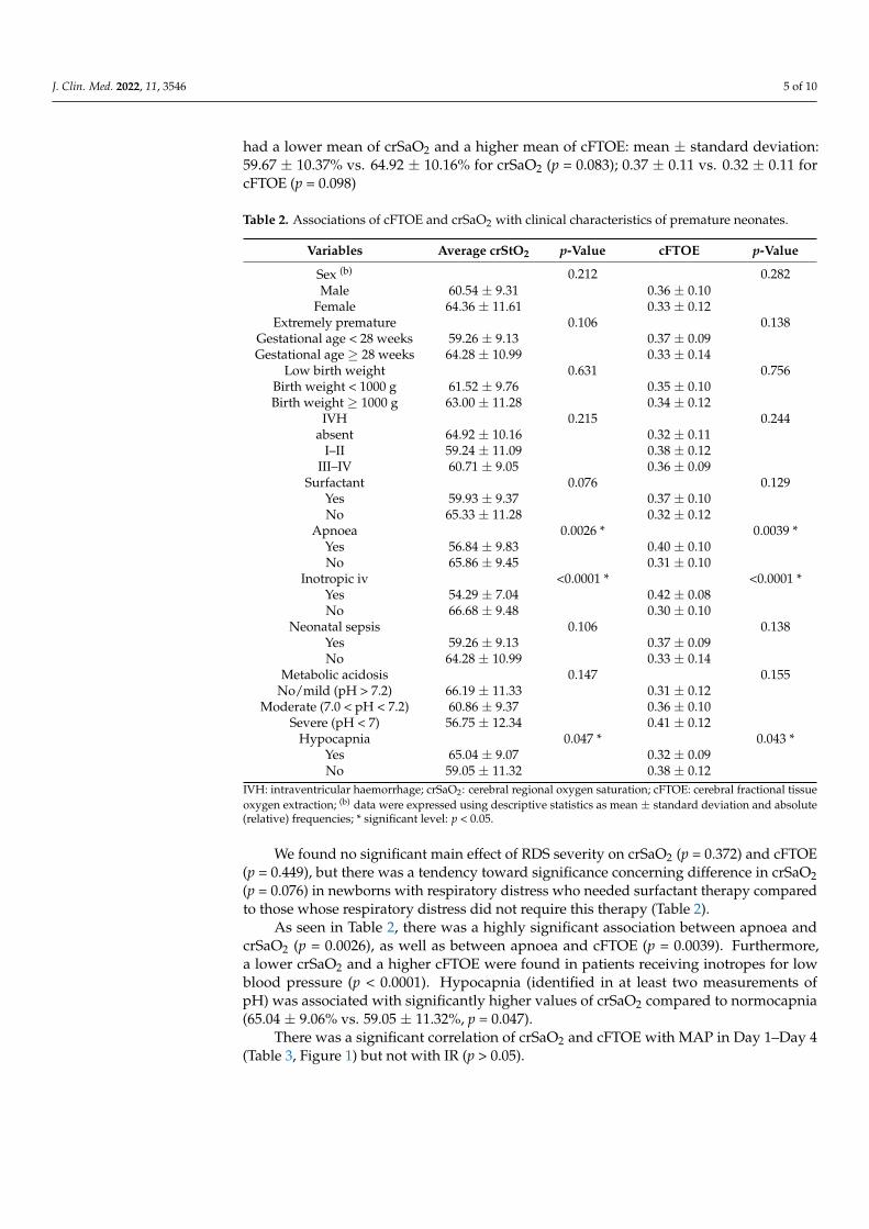
We found no significant difference in the mean values of crSaO2 (p = 0.212) or cFTOE(p = 0.282) between boys and girls (Table 2). There was no significant main effect of IVHseverity on crSaO2 and cFTOE (one-way ANOVA p > 0.05); however, patients with IVH
J. Clin. Med. 2022, 11, 3546 5 of 10
had a lower mean of crSaO2 and a higher mean of cFTOE: mean ± standard deviation:59.67 ± 10.37% vs. 64.92 ± 10.16% for crSaO2 (p = 0.083); 0.37 ± 0.11 vs. 0.32 ± 0.11 forcFTOE (p = 0.098)
Table 2. Associations of cFTOE and crSaO2 with clinical characteristics of premature neonates.
Variables Average crStO2 p-Value cFTOE p-Value
Sex (b) 0.212 0.282Male 60.54 ± 9.31 0.36 ± 0.10
Female 64.36 ± 11.61 0.33 ± 0.12Extremely premature 0.106 0.138
Gestational age < 28 weeks 59.26 ± 9.13 0.37 ± 0.09Gestational age ≥ 28 weeks 64.28 ± 10.99 0.33 ± 0.14
Low birth weight 0.631 0.756Birth weight < 1000 g 61.52 ± 9.76 0.35 ± 0.10Birth weight ≥ 1000 g 63.00 ± 11.28 0.34 ± 0.12
IVH 0.215 0.244absent 64.92 ± 10.16 0.32 ± 0.11
I–II 59.24 ± 11.09 0.38 ± 0.12III–IV 60.71 ± 9.05 0.36 ± 0.09
Surfactant 0.076 0.129Yes 59.93 ± 9.37 0.37 ± 0.10No 65.33 ± 11.28 0.32 ± 0.12
Apnoea 0.0026 * 0.0039 *Yes 56.84 ± 9.83 0.40 ± 0.10No 65.86 ± 9.45 0.31 ± 0.10
Inotropic iv <0.0001 * <0.0001 *Yes 54.29 ± 7.04 0.42 ± 0.08No 66.68 ± 9.48 0.30 ± 0.10
Neonatal sepsis 0.106 0.138Yes 59.26 ± 9.13 0.37 ± 0.09No 64.28 ± 10.99 0.33 ± 0.14
Metabolic acidosis 0.147 0.155No/mild (pH > 7.2) 66.19 ± 11.33 0.31 ± 0.12
Moderate (7.0 < pH < 7.2) 60.86 ± 9.37 0.36 ± 0.10Severe (pH < 7) 56.75 ± 12.34 0.41 ± 0.12
Hypocapnia 0.047 * 0.043 *Yes 65.04 ± 9.07 0.32 ± 0.09No 59.05 ± 11.32 0.38 ± 0.12
IVH: intraventricular haemorrhage; crSaO2: cerebral regional oxygen saturation; cFTOE: cerebral fractional tissueoxygen extraction; (b) data were expressed using descriptive statistics as mean ± standard deviation and absolute(relative) frequencies; * significant level: p < 0.05.
We found no significant main effect of RDS severity on crSaO2 (p = 0.372) and cFTOE(p = 0.449), but there was a tendency toward significance concerning difference in crSaO2(p = 0.076) in newborns with respiratory distress who needed surfactant therapy comparedto those whose respiratory distress did not require this therapy (Table 2).
As seen in Table 2, there was a highly significant association between apnoea andcrSaO2 (p = 0.0026), as well as between apnoea and cFTOE (p = 0.0039). Furthermore,a lower crSaO2 and a higher cFTOE were found in patients receiving inotropes for lowblood pressure (p < 0.0001). Hypocapnia (identified in at least two measurements ofpH) was associated with significantly higher values of crSaO2 compared to normocapnia(65.04 ± 9.06% vs. 59.05 ± 11.32%, p = 0.047).
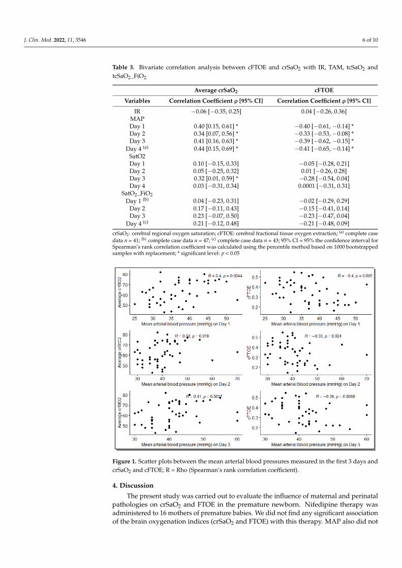
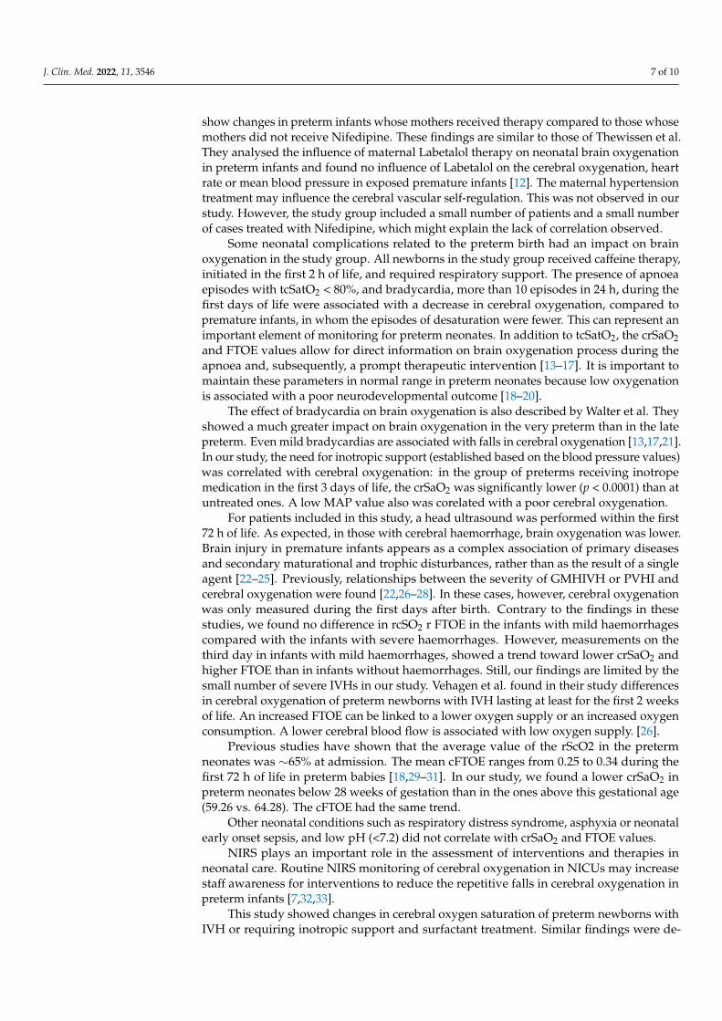
There was a significant correlation of crSaO2 and cFTOE with MAP in Day 1–Day 4(Table 3, Figure 1) but not with IR (p > 0.05).
J. Clin. Med. 2022, 11, 3546 6 of 10
Table 3. Bivariate correlation analysis between cFTOE and crSaO2 with IR, TAM, tcSaO2 andtcSaO2_FiO2.
Average crSaO2 cFTOE
Variables Correlation Coefficient ρ [95% CI] Correlation Coefficient ρ [95% CI]
IR −0.06 [−0.35, 0.25] 0.04 [−0.26, 0.36]MAPDay 1 0.40 [0.15, 0.61] * −0.40 [−0.61, −0.14] *Day 2 0.34 [0.07, 0.56] * −0.33 [−0.53, −0.08] *Day 3 0.41 [0.16, 0.63] * −0.39 [−0.62, −0.15] *
Day 4 (a) 0.44 [0.15, 0.69] * −0.41 [−0.65, −0.14] *SatO2Day 1 0.10 [−0.15, 0.33] −0.05 [−0.28, 0.21]Day 2 0.05 [−0.25, 0.32] 0.01 [−0.26, 0.28]Day 3 0.32 [0.01, 0.59] * −0.28 [−0.54, 0.04]Day 4 0.03 [−0.31, 0.34] 0.0001 [−0.31, 0.31]
SatO2_FiO2Day 1 (b) 0.04 [−0.23, 0.31] −0.02 [−0.29, 0.29]
Day 2 0.17 [−0.11, 0.43] −0.15 [−0.41, 0.14]Day 3 0.23 [−0.07, 0.50] −0.23 [−0.47, 0.04]
Day 4 (c) 0.21 [−0.12, 0.48] −0.21 [−0.48, 0.09]
crSaO2: cerebral regional oxygen saturation; cFTOE: cerebral fractional tissue oxygen extraction; (a) complete casedata n = 41; (b) complete case data n = 47; (c) complete case data n = 43; 95% CI = 95% the confidence interval forSpearman’s rank correlation coefficient was calculated using the percentile method based on 1000 bootstrappedsamples with replacement; * significant level: p < 0.05
Figure 1. Scatter plots between the mean arterial blood pressures measured in the first 3 days andcrSaO2 and cFTOE; R = Rho (Spearman’s rank correlation coefficient).
4. Discussion
The present study was carried out to evaluate the influence of maternal and perinatalpathologies on crSaO2 and FTOE in the premature newborn. Nifedipine therapy wasadministered to 16 mothers of premature babies. We did not find any significant associationof the brain oxygenation indices (crSaO2 and FTOE) with this therapy. MAP also did not
J. Clin. Med. 2022, 11, 3546 7 of 10
show changes in preterm infants whose mothers received therapy compared to those whosemothers did not receive Nifedipine. These findings are similar to those of Thewissen et al.They analysed the influence of maternal Labetalol therapy on neonatal brain oxygenationin preterm infants and found no influence of Labetalol on the cerebral oxygenation, heartrate or mean blood pressure in exposed premature infants [12]. The maternal hypertensiontreatment may influence the cerebral vascular self-regulation. This was not observed in ourstudy. However, the study group included a small number of patients and a small numberof cases treated with Nifedipine, which might explain the lack of correlation observed.
Some neonatal complications related to the preterm birth had an impact on brainoxygenation in the study group. All newborns in the study group received caffeine therapy,initiated in the first 2 h of life, and required respiratory support. The presence of apnoeaepisodes with tcSatO2 < 80%, and bradycardia, more than 10 episodes in 24 h, during thefirst days of life were associated with a decrease in cerebral oxygenation, compared topremature infants, in whom the episodes of desaturation were fewer. This can represent animportant element of monitoring for preterm neonates. In addition to tcSatO2, the crSaO2and FTOE values allow for direct information on brain oxygenation process during theapnoea and, subsequently, a prompt therapeutic intervention [13–17]. It is important tomaintain these parameters in normal range in preterm neonates because low oxygenationis associated with a poor neurodevelopmental outcome [18–20].
The effect of bradycardia on brain oxygenation is also described by Walter et al. Theyshowed a much greater impact on brain oxygenation in the very preterm than in the latepreterm. Even mild bradycardias are associated with falls in cerebral oxygenation [13,17,21].In our study, the need for inotropic support (established based on the blood pressure values)was correlated with cerebral oxygenation: in the group of preterms receiving inotropemedication in the first 3 days of life, the crSaO2 was significantly lower (p < 0.0001) than atuntreated ones. A low MAP value also was corelated with a poor cerebral oxygenation.
For patients included in this study, a head ultrasound was performed within the first72 h of life. As expected, in those with cerebral haemorrhage, brain oxygenation was lower.Brain injury in premature infants appears as a complex association of primary diseasesand secondary maturational and trophic disturbances, rather than as the result of a singleagent [22–25]. Previously, relationships between the severity of GMHIVH or PVHI andcerebral oxygenation were found [22,26–28]. In these cases, however, cerebral oxygenationwas only measured during the first days after birth. Contrary to the findings in thesestudies, we found no difference in rcSO2 r FTOE in the infants with mild haemorrhagescompared with the infants with severe haemorrhages. However, measurements on thethird day in infants with mild haemorrhages, showed a trend toward lower crSaO2 andhigher FTOE than in infants without haemorrhages. Still, our findings are limited by thesmall number of severe IVHs in our study. Vehagen et al. found in their study differencesin cerebral oxygenation of preterm newborns with IVH lasting at least for the first 2 weeksof life. An increased FTOE can be linked to a lower oxygen supply or an increased oxygenconsumption. A lower cerebral blood flow is associated with low oxygen supply. [26].
Previous studies have shown that the average value of the rScO2 in the pretermneonates was ∼65% at admission. The mean cFTOE ranges from 0.25 to 0.34 during thefirst 72 h of life in preterm babies [18,29–31]. In our study, we found a lower crSaO2 inpreterm neonates below 28 weeks of gestation than in the ones above this gestational age(59.26 vs. 64.28). The cFTOE had the same trend.
Other neonatal conditions such as respiratory distress syndrome, asphyxia or neonatalearly onset sepsis, and low pH (<7.2) did not correlate with crSaO2 and FTOE values.
NIRS plays an important role in the assessment of interventions and therapies inneonatal care. Routine NIRS monitoring of cerebral oxygenation in NICUs may increasestaff awareness for interventions to reduce the repetitive falls in cerebral oxygenation inpreterm infants [7,32,33].
This study showed changes in cerebral oxygen saturation of preterm newborns withIVH or requiring inotropic support and surfactant treatment. Similar findings were de-
J. Clin. Med. 2022, 11, 3546 8 of 10
scribed by other authors [34–36]. A greater need for mechanical ventilation and lowercerebral oximetry index was found for preterm neonates requiring dopamine treatment forhypotension [35].
The limitations of this study consist of a relatively small number of preterm infantsand the single-centre study. The present study should be regarded as exploratory researchthat investigated the associations of maternal factors and perinatal disorders with cFTOEand crSaO2. Considering the small sample size, we could not test a multivariable modelcontaining all relevant clinical factors that can explain variability of SctO2/cFTOE. A futurestudy with a higher sample size is necessary to confirm the independent maternal factors orperinatal disorders as potential predictors of SctO2/cFTOE changes. Despite the relativelysmall sample size, our findings are in accordance with other studies finding a sustainedrole and importance of the cerebral oxygen saturation monitor in the NICU patient.
5. Conclusions
In preterm newborns, the cerebral oxygenation was associated with different factors,both maternal and perinatal. Our study showed that the IVH, the need for inotrope supportand episodes of apnoea were correlated with crSaO2 and cFTOE. There was a trend ofdecreased crSaO2 and cFTOE values with younger gestational age of the neonates. Wefound no significant association between the maternal hypertension treatment and thecerebral oxygen saturation of the preterm infants.
Author Contributions: Conceptualisation, M.M. and G.Z.; methodology, M.M.; software, M.I.;validation, M.I., M.H. and A.M.; formal analysis, M.I.; investigation, A.M. and M.H.; resources,M.M. and A.M.; data curation, M.I.; writing—original draft preparation, M.M.; writing—review andediting, G.Z. and M.I.; visualisation, M.H. and M.I.; supervision, G.Z.; project administration, M.M.and G.Z.; funding acquisition, M.M. All authors have read and agreed to the published version ofthe manuscript.
Funding: This research was funded by project PDI-PFE-CDI 2021, entitled Increasing the Perfor-mance of Scientific Research, Supporting Excellence in Medical Research and Innovation, PROGRES,no. 40PFE/30.12.2021.
Institutional Review Board Statement: This study was conducted in accordance with the Declarationof Helsinki and approved by the Institutional Review Board of County Emergency Hospital Cluj-Napoca, Romania (53874/23 December 2020).
Informed Consent Statement: Informed consent was obtained from all parents of subjects involvedin the study.
Data Availability Statement: All data generated or analysed during this study are included in thispublished article.
Conflicts of Interest: The authors declare no conflict of interest.
References1. Lemmers, P.M.A.; Toet, M.; Van Schelven, L.J.; Van Bel, F. Cerebral oxygenation and cerebral oxygen extraction in the preterm
infant: The impact of respiratory distress syndrome. Exp. Brain Res. 2006, 173, 458–467. [CrossRef] [PubMed]2. Matyas, M.; Hasmasanu, M.; Silaghi, C.N.; Samasca, G.; Lupan, I.; Orsolya, K.; Zaharie, G. Early Preeclampsia Effect on Preterm
Newborns Outcome. J. Clin. Med. 2022, 11, 452. [CrossRef] [PubMed]3. Verhagen, E.A.; Keating, P.; Ter Horst, H.J.; Martijn, A.; Bos, A.F. Cerebral oxygen saturation and extraction in preterm infants
with transient periventricular echodensities. Pediatrics 2009, 124, 294–301. [CrossRef] [PubMed]4. Naulaers, G.; Meyns, B.; Miserez, M.; Leunens, V.; Van Huffel, S.; Casaer, P.; Weindling, M.; Devlieger, H. Use of tissue oxygenation
index and fractional tissue oxygen extraction as non-invasive parameters for cerebral oxygenation. A validation study in piglets.Neonatology 2007, 92, 120–126. [CrossRef] [PubMed]
5. Brown, D.W.; Hadway, J.; Lee, T.Y. Near-infrared spectroscopy measurement of oxygen extraction fraction and cerebral metabolicrate of oxygen in newborn piglets. Pediatric Res. 2003, 54, 861–867. [CrossRef]
6. Dix, L.M.L.; van Bel, F.; Lemmers, P.M.A. Monitoring cerebral oxygenation in neonates: An update. Front. Pediatrics 2017, 5, 46.[CrossRef]
J. Clin. Med. 2022, 11, 3546 9 of 10
7. Vesoulis, Z.A.; Mintzer, J.P.; Chock, V.Y. Neonatal NIRS monitoring: Recommendations for data capture and review of analytics.J. Perinatol. 2021, 41, 675–688. [CrossRef]
8. American College of Obstetricians and Gynecologists; Task Force on Hypertension in Pregnancy Hypertension in Pregnancy.Report of the American College of Obstetricians and Gynecologists’ Task Force on Hypertension in Pregnancy. Obstet. Gynecol.2013, 122, 1122–1131. [CrossRef]
9. Sweet, D.G.; Carnielli, V.; Greisen, G.; Hallman, M.; Ozek, E.; Plavka, R.; Saugstad, O.D.; Simeoni, U.; Speer, C.P.; Vento, M.; et al.European Consensus Guidelines on the Management of Respiratory Distress Syndrome—2016 Update. Neonatology 2017, 111,107–125. [CrossRef]
10. Jeffrey, M.; Perlman, J.J.V. Chapter 24—Preterm Intraventricular hemorrhage. In Volpe’s Neurology of the Newborn; Elsevier:Amsterdam, The Netherlands, 2018; pp. 637–745.e4.
11. Robertson, C.; Perlamn, M. Follow-up of the term infant after hypoxic–ischemic encephalopathy. Paediatrics Child Health 2006, 11,278–282. [CrossRef]
12. Thewissen, L.; Pistorius, L.; Baerts, W.; Naulaers, G.; Van Bel, F.; Lemmers, P. Neonatal haemodynamic effects following foetalexposure to labetalol in hypertensive disorders of pregnancy. J. Matern. Fetal Neonatal Med. 2017, 30, 1533–1538. [CrossRef]
13. Walter, L.M.; Ahmed, B.; Odoi, A.; Cooney, H.; Horne, R.S.C.; Wong, F.Y. Bradycardias are associated with more severe effects oncerebral oxygenation in very preterm infants than in late preterm infants. Early Hum. Dev. 2018, 127, 33–41. [CrossRef]
14. Pichler, G.; Urlesberger, B.; Müller, W. Impact of bradycardia on cerebral oxygenation and cerebral blood volume during apnoeain preterm infants. Physiol. Meas. 2003, 24, 671–680. [CrossRef]
15. Chock, V.Y.; Kwon, S.H.; Ambalavanan, N.; Batton, B.; Nelin, L.D.; Chalak, L.F.; Tian, L.; Van Meurs, K.P. Cerebral Oxygenationand Autoregulation in Preterm Infants (Early NIRS Study). J. Pediatrics 2020, 227, 94–100.e1. [CrossRef]
16. Lazea, C.; Sur, L.; Florea, M. ROHHAD (Rapid-onset Obesity with Hypoventilation, Hypothalamic Dysfunction, Autonomic Dys-regulation) Syndrome-What Every Pediatrician Should Know About the Etiopathogenesis, Diagnosis and Treatment: A Review.Int. J. Gen. Med. 2021, 14, 319–326. [CrossRef]
17. Liao, S.M.C.; Rao, R.; Mathur, A.M. Head position change is not associated with acute changes in bilateral cerebral oxygenation instable preterm infants during the first three days of life. Am. J. Perinatol. 2015, 32, 645–652. [CrossRef]
18. Alderliesten, T.; van Bel, F.; van der Aa, N.E.; Steendijk, P.; van Haastert, I.C.; de Vries, L.S.; Groenendaal, F.; Lemmers, P. LowCerebral Oxygenation in Preterm Infants Is Associated with Adverse Neurodevelopmental Outcome. J. Pediatrics 2019, 207,109–116.e2. [CrossRef]
19. Martini, S.; Corvaglia, L. Splanchnic NIRS monitoring in neonatal care: Rationale, current applications and future perspectives.J. Perinatol. 2018, 38, 431–443. [CrossRef]
20. Pichler, G.; Baumgartner, S.; Biermayr, M.; Dempsey, E.; Fuchs, H.; Goos, T.G.; Lista, G.; Lorenz, L.; Karpinski, L.; Mitra, S.; et al.Cerebral regional tissue Oxygen Saturation to Guide Oxygen Delivery in preterm neonates during immediate transition afterbirth (COSGOD III): An investigator-initiated, randomized, multi-center, multi-national, clinical trial on additional cerebral tissueoxygen saturation monitoring combined with defined treatment guidelines versus standard monitoring and treatment as usual inpremature infants during immediate transition: Study protocol for a randomized controlled trial. Trials 2019, 20, 178. [CrossRef]
21. Schmid, M.B.; Hopfner, R.J.; Lenhof, S.; Hummler, H.D.; Fuchs, H. Cerebral oxygenation during intermittent hypoxemia andbradycardia in preterm infants. Neonatology 2015, 107, 137–146. [CrossRef]
22. Noori, S.; McCoy, M.; Anderson, M.P.; Ramji, F.; Seri, I. Changes in cardiac function and cerebral blood flow in relation toperi/intraventricular hemorrhage in extremely preterm infants. J. Pediatrics 2014, 164, 264–270.e3. [CrossRef]
23. Zaharie, G.C.; Hăsmăsanu, M.G.; Blaga, L.; Matyas, M.; Mures, an, D.; Bolboacă, S.D. Cardiac left heart morphology and functionin newborns with intrauterine growth restriction: Relevance for long-term assessment. Med. Ultrason. 2019, 21, 62–68. [CrossRef]
24. Sortica da Costa, C.; Cardim, D.; Molnar, Z.; Kelsall, W.; Ng, I.; Czosnyka, M.; Smielewski, P.; Austin, T. Changes in hemodynamics,cerebral oxygenation and cerebrovascular reactivity during the early transitional circulation in preterm infants. Pediatric Res.2019, 86, 247–253. [CrossRef]
25. Kenosi, M.; O’Toole, J.M.; Livingston, V.; Hawkes, G.A.; Boylan, G.B.; O’Halloran, K.D.; Ryan, A.C.; Dempsey, E.M. Effects ofFractional Inspired Oxygen on Cerebral Oxygenation in Preterm Infants following Delivery. J. Pediatrics 2015, 167, 1007–1012.e1.[CrossRef]
26. Verhagen, E.A.; Ter Horst, H.J.; Keating, P.; Martijn, A.; Van Braeckel, K.N.J.A.; Bos, A.F. Cerebral oxygenation in preterm infantswith germinal matrix-intraventricular hemorrhages. Stroke 2010, 41, 2901–2907. [CrossRef]
27. Ng, I.H.X.; Da Costa, C.S.; Zeiler, F.A.; Wong, F.Y.; Smielewski, P.; Czosnyka, M.; Austin, T. Burden of hypoxia and intraventricularhaemorrhage in extremely preterm infants. Arch. Dis. Child. Fetal Neonatal Ed. 2020, 105, 242–247. [CrossRef]
28. Lin, P.Y.; Hagan, K.; Fenoglio, A.; Grant, P.E.; Franceschini, M.A. Reduced cerebral blood flow and oxygen metabolism inextremely preterm neonates with low-grade germinal matrix- intraventricular hemorrhage. Sci. Rep. 2016, 6, 25903. [CrossRef]
29. Hyttel-Sorensen, S.; Pellicer, A.; Alderliesten, T.; Austin, T.; Van Bel, F.; Benders, M.; Claris, O.; Dempsey, E.; Franz, A.R.;Fumagalli, M.; et al. Cerebral near infrared spectroscopy oximetry in extremely preterm infants: Phase II randomised clinical trial.BMJ 2015, 350, g7635. [CrossRef] [PubMed]
30. Cohen, E.; Baerts, W.; Alderliesten, T.; Derks, J.; Lemmers, P.; van Bel, F. Growth restriction and gender influence cerebraloxygenation in preterm neonates. Arch. Dis. Child. Fetal Neonatal Ed. 2016, 101, F156–F161. [CrossRef] [PubMed]
J. Clin. Med. 2022, 11, 3546 10 of 10
31. Mohamed, M.A.; Frasketi, M.J.; Aly, S.; El-Dib, M.; Hoffman, H.J.; Aly, H. Changes in cerebral tissue oxygenation and fractionaloxygen extraction with gestational age and postnatal maturation in preterm infants. J. Perinatol. 2021, 41, 836–842. [CrossRef][PubMed]
32. Alderliesten, T.; Dix, L.; Baerts, W.; Caicedo, A.; Van Huffel, S.; Naulaers, G.; Groenendaal, F.; Van Bel, F.; Lemmers, P. Referencevalues of regional cerebral oxygen saturation during the first 3 days of life in preterm neonates. Pediatric Res. 2016, 79, 55–64.[CrossRef]
33. Lorenz, L.; Marulli, A.; Dawson, J.A.; Owen, L.S.; Manley, B.J.; Donath, S.M.; Davis, P.G.; Kamlin, C.O.F. Cerebral oxygenationduring skin-to-skin care in preterm infants not receiving respiratory support. Arch. Dis. Child. Fetal Neonatal Ed. 2018, 103,F137–F142. [CrossRef]
34. Alosh, H.; Ramirez, A.; Mink, R. The correlation between brain near-infrared spectroscopy and cerebral blood flow in piglets withintracranial hypertension. J. Appl. Physiol. 2016, 121, 255–260. [CrossRef]
35. Eriksen, V.R.; Hahn, G.H.; Greisen, G. Dopamine therapy is associated with impaired cerebral autoregulation in preterm infants.Acta Paediatr. 2014, 103, 1221–1226. [CrossRef]
36. Gilmore, M.M.; Stone, B.S.; Shepard, J.A.; Czosnyka, M.; Easley, R.B.; Brady, K.M. Relationship between cerebrovasculardysautoregulation and arterial blood pressure in the premature infant. J. Perinatol. 2011, 31, 722–729. [CrossRef]
Related Documents