593 Vet. Res. 37 (2006) 593–606 © INRA, EDP Sciences, 2006 DOI: 10.1051/vetres:2006021 Original article Assessment of diagnostic tools for eradication of bovine tuberculosis in cattle co-infected with Mycobacterium bovis and M. avium subsp. paratuberculosis Alicia ARANAZ a *, Lucía DE JUAN a , Javier BEZOS a , Julio ÁLVAREZ a , Beatriz ROMERO a , Francisco LOZANO a , José L. PARAMIO b , Jesús LÓPEZ-SÁNCHEZ c , Ana MATEOS a , Lucas DOMÍNGUEZ a a Departamento de Sanidad Animal I, Facultad de Veterinaria, Universidad Complutense de Madrid, Av. Puerta de Hierro s/n, 28040 Madrid, Spain b Subdirección General de Sanidad Animal, Dirección General de Ganadería, MAPA, 28071 Madrid, Spain c Laboratorio Regional Pecuario, Consejería de Agricultura, JCCM, 02071 Albacete, Spain (Received 12 August 2005; accepted 31 January 2006) Abstract – The intradermal tuberculin (IDTB) test and the interferon-gamma (IFN-γ) assay are used worldwide for detection of bovine tuberculosis in cattle, but little is known about the effect of co-infecting agents on the performance of these diagnostic tests. This report describes a field trial conducted in a cattle herd with dual infection (bovine tuberculosis and paratuberculosis) during 3.5 years. It has been based on a strategic approach encompassing serial parallel testing (comparative IDTB test, the IFN-γ assay and serology of paratuberculosis) that was repeated 8 times over the period, and segregation of animals into two herds. The IDTB test detected 65.2% and the IFN-γ test detected 69.6% of the Mycobacterium bovis culture-positive cattle. However, the IDTB test performed better during the first part of the trial, while the IFN-γ test was the only method that detected infected animals during the following three samplings. The number of false positive reactors with the IDTB and/or the IFN-γ tests was remarkably high compared to other reports, and could be caused by cross-reactivity with M. avium subsp. paratuberculosis. Also, the M. bovis isolates from cattle and wildlife from the same property were characterised using molecular techniques to disclose an epidemiological link. The IDTB test may not be appropriate to eradicate bovine tuberculosis in herds with dual mycobacterial infections. This report highlights the need to use several diagnostic techniques for the accurate detection of M. bovis infected animals in these herds. tuberculosis / paratuberculosis / IDTB / IFN-γ / eradication 1. INTRODUCTION Eradication of bovine tuberculosis, zoon- oses caused by Mycobacterium bovis, relies on the detection of infected animals and subsequent slaughter of reactors. The intra- dermal tuberculin (IDTB) test has been used for routine field detection of infected animals since nearly a century ago [19], and it is the official test in most countries. * Corresponding author: [email protected] Article published by EDP Sciences and available at http://www.edpsciences.org/vetres or http://dx.doi.org/10.1051/vetres:2006021

Welcome message from author
This document is posted to help you gain knowledge. Please leave a comment to let me know what you think about it! Share it to your friends and learn new things together.
Transcript
593Vet. Res. 37 (2006) 593–606© INRA, EDP Sciences, 2006DOI: 10.1051/vetres:2006021
Original article
Assessment of diagnostic tools for eradication of bovine tuberculosis in cattle co-infected with
Mycobacterium bovis and M. avium subsp. paratuberculosis
Alicia ARANAZa*, Lucía DE JUANa, Javier BEZOSa, Julio ÁLVAREZa, Beatriz ROMEROa, Francisco LOZANOa, José L. PARAMIOb,
Jesús LÓPEZ-SÁNCHEZc, Ana MATEOSa, Lucas DOMÍNGUEZa
a Departamento de Sanidad Animal I, Facultad de Veterinaria, Universidad Complutense de Madrid, Av. Puerta de Hierro s/n, 28040 Madrid, Spain
b Subdirección General de Sanidad Animal, Dirección General de Ganadería, MAPA, 28071 Madrid, Spain
c Laboratorio Regional Pecuario, Consejería de Agricultura, JCCM, 02071 Albacete, Spain
(Received 12 August 2005; accepted 31 January 2006)
Abstract – The intradermal tuberculin (IDTB) test and the interferon-gamma (IFN-γ) assay areused worldwide for detection of bovine tuberculosis in cattle, but little is known about the effect ofco-infecting agents on the performance of these diagnostic tests. This report describes a field trialconducted in a cattle herd with dual infection (bovine tuberculosis and paratuberculosis) during3.5 years. It has been based on a strategic approach encompassing serial parallel testing(comparative IDTB test, the IFN-γ assay and serology of paratuberculosis) that was repeated8 times over the period, and segregation of animals into two herds. The IDTB test detected 65.2%and the IFN-γ test detected 69.6% of the Mycobacterium bovis culture-positive cattle. However, theIDTB test performed better during the first part of the trial, while the IFN-γ test was the only methodthat detected infected animals during the following three samplings. The number of false positivereactors with the IDTB and/or the IFN-γ tests was remarkably high compared to other reports, andcould be caused by cross-reactivity with M. avium subsp. paratuberculosis. Also, the M. bovisisolates from cattle and wildlife from the same property were characterised using moleculartechniques to disclose an epidemiological link. The IDTB test may not be appropriate to eradicatebovine tuberculosis in herds with dual mycobacterial infections. This report highlights the need touse several diagnostic techniques for the accurate detection of M. bovis infected animals in these herds.
tuberculosis / paratuberculosis / IDTB / IFN-γ / eradication
1. INTRODUCTION
Eradication of bovine tuberculosis, zoon-oses caused by Mycobacterium bovis, relieson the detection of infected animals and
subsequent slaughter of reactors. The intra-dermal tuberculin (IDTB) test has beenused for routine field detection of infectedanimals since nearly a century ago [19],and it is the official test in most countries.
* Corresponding author: [email protected]
Article published by EDP Sciences and available at http://www.edpsciences.org/vetres or http://dx.doi.org/10.1051/vetres:2006021
594 A. Aranaz et al.
A review estimates the sensitivity values ofthe tuberculin test (at standard interpreta-tion) to be 90% [22]. Failure to diagnoseanimals infected by M. bovis results in thepersistence of infection and may contributeto cattle to cattle spread. In Spain, the pro-gramme for the eradication of bovine tuber-culosis is based on the use of the IDTB testaccording to Council Directive 64/432/EEC1 and removal of reactors. In 2004, thistest was positive in 0.40% of animals(1.80% of herds). Despite the progressachieved over recent years, bovine tubercu-losis still poses a serious problem in spe-cific regions of the country, such as areas ofextensive management properties and co-existence of abundant wildlife, thus, the useof additional approved techniques would bedesirable to speed up the eradication.
In order to enhance sensitivity, specifi-city, and to reduce handling events, Woodet al. described the gamma-interferon (IFN-γ)test [43]. This test is an in vitro immu-noassay based on the specific release ofIFN-γ as the indicator of a response to theM. bovis antigen (bovine purified proteinderivative, bovine PPD). IFN-γ can bedetected using an enzyme immunoassay[29] which is commercially available.
Several field trials have compared theperformance of IDTB and IFN-γ tests dur-ing the last decade. In most of them, the sen-sitivity of the IFN-γ test was highercompared to the tuberculin tests [13, 18, 24,28, 32, 44, 45]. The specificity of the IFN-γ assay was 90.6 to 98.6% [3, 20, 44]. How-ever, some factors have been suggested toaffect the specificity of the IDTB test andthe IFN-γ assay, and the most frequentcould be the presence of other mycobacte-rial infections such as paratuberculosis.Paratuberculosis (Johne’s disease), causedby Mycobacterium avium subspeciesparatuberculosis, is a chronic granuloma-tous enteritis of bovids. Paratuberculosis
has been recognised to be present world-wide, though with important differences inprevalence (reviewed in [16]).
It has been reported that vaccinationagainst paratuberculosis of goats with aninactivated vaccine2, and of cattle with alive modified vaccine [17] or an inactivatedvaccine [23] could compromise the specif-icity of both the IFN-γ and SIDT tests. It issuspected that paratuberculosis itself mayhave the same effect [38]. In general terms,these immune responses generated aftervaccination or experimental infection arepunctual non-specific responses or transient(although they may last several months),and considerable variations of the cellularresponses are detected amongst animals.However, a recent report detected no sig-nificant association among cattle testingpositive for paratuberculosis and positivecaudal fold test and the IFN-γ assay [8].Some strategies have been applied to dis-criminate between a herd infected withM. bovis or M. a. paratuberculosis: (1) theuse of specific mixtures (johnin, avianPPD) or specific antigens (ESAT-6, CFP-10), or (2) selection of criteria for the inter-pretation of the results (B/A ≥ 2). It is feltthat more research should be performed toelucidate the role of co-infecting agents onthe diagnosis of bovine tuberculosis [37].To our knowledge, however, there are noreports on the use of these tests and inter-pretation of the results in cattle herds co-infected with both M. bovis and M. a.paratuberculosis. This situation is expectedto occur occasionally in Northern and Cen-tral European countries where only smalloutbreaks of tuberculosis infection haveoccurred in recent years (as cited in [9, 26,38]), but could be found more frequent inother countries such as in the UnitedKingdom, the Republic of Ireland, the
1 Council Directive 64/432/EEC, 1964, [on line](2005) http://www.eu.int/scadplus/leg/en/lvb/l12004.htm#AMENDINGACT [consulted 30January 2006].
2 Aranaz A., del Río R., Ruiz J., et al., Effect of theparatuberculosis vaccine on the IFN-γ test fortuberculosis in goats, Abstracts, 3rd Int. Confer-ence on Mycobacterium bovis, Cambridge, UK,2000, p. 11.
Diagnosis in dual mycobacterial infections 595
Mediterranean basin countries, and somestates of the USA (as cited in [8]).
This report describes a field trial con-ducted in a cattle herd with dual infection(bovine tuberculosis and paratuberculosis)with the objective of eradicating M. bovisinfection. A comprehensive approach wasperformed using a multi-test strategy toidentify M. bovis infected cattle. We havefound that the presence of dual mycobacte-rial infection confound the diagnostic testsand may result in a slowing down of theprogress of the eradication programmes.Furthermore, M. bovis isolates from domes-tic and wild animals from the property werecharacterised to determine a likely epide-miological link.
2. MATERIALS AND METHODS
2.1. Animals and design of the study
2.1.1. Herd
This study was performed in a herd ofbullfighting breed cattle of outstandinggenetic value (this specific stock has beeninbred since the 16th century). These cattleare difficult to handle because of their vig-our and temper and are bred with an exten-sive management system and occasionalfeed supplements. It is raised in a propertysituated in Sierra of Alcaraz (central-east ofSpain). The herd had suffered from theinfection of bovine tuberculosis for at least20 years before the start of this study, with2.7 to 4.8% of reactors (fluctuating percent-ages) to the IDTB test performed under offi-cial eradication campaigns. Some animalspresented diarrhoea, cachexia and death(clinical symptoms compatible with paratu-berculosis). The property is also exploitedfor game of several wildlife species.
2.1.2. Design of the study
Before the beginning of the trial, a meet-ing was held involving the owners of theproperty, the farm manager, the veterinar-
ian, the representatives of the BullfightingFarmers Association, representatives of theregional government and the Spanish Min-istry of Agriculture, Fisheries and Food,and our group to discuss the design andplanning of the study. Periodic meetingsafter every trial allowed the evaluation ofthe progress.
This study includes data from all animals(females and breeding bulls) over12 months of age that were tested in parallelby the IDTB test, the IFN-γ test and serol-ogy (ELISA test) for paratuberculosis.Since the performance of the tests wasbeing compared on the same animals, theywere tested simultaneously to avoid theinterference of one test with the other [30,41]. These tests were repeated 8 times overa 3.5-year period, avoiding the summer andthe last pregnancy seasons. In total, an aver-age of 301 (251–361) animals was tested inevery trial. All animals reacting to the IDTBtest, and a group of animals with positiveresults to the IFN-γ test and positive serol-ogy of paratuberculosis were slaughteredand true infection status was confirmed bybacteriological culture.
The rest of the cattle were allocated totwo herds depending on their results: herdA, with animals negative to all tests, andtheir offspring; and herd B, composed ofanimals with at least a positive result to theIFN-γ assay or to serology of paratubercu-losis (but not slaughtered), and their off-spring. Herds A and B were kept in differentareas of the property, with independent pas-tures and water resources, with no contact.
2.2. Diagnostic tests
2.2.1. IDTB test
The cervical comparative IDTB test wascarried out according to the RD 2611/1996(direct transposition of Council Directive64/432/EEC) by the Official VeterinaryServices using the official bovine and avianPPDs (CZ Veterinaria, Porriño, Spain). Theanimals were simultaneously inoculated0.1 mL (0.1 mg, 2500 CTU) bovine PPD on
596 A. Aranaz et al.
the left side of the neck, and 0.1 mL(2500 IU) avian PPD on the right side. Theincrease of skin-fold thickness was re-measured 72 h later. A positive bovine reac-tion, which was more than 4 mm greaterthan the avian reaction, or that producedclinical signs (diffuse or extensive oedema,exudation, necrosis, pain or inflammationof the lymphatic ducts in that region or ofthe lymph nodes), was interpreted as a pos-itive result to bovine PPD.
2.2.2. IFN-γ test
Blood samples were collected in tubeswith lithium heparin at the time of the IDTBtesting and shipped to the laboratory atroom temperature. Stimulation of wholeblood was performed within 8 h of collec-tion, and 2 or 3 days were required to testthe whole herd. The IFN-γ test was carriedout as described [18, 29]. In the first trial, afourth well with 2 µg/mL of poke-weedmitogen (Sigma Aldrich Gmbh, Steinheim,Germany) was included as the positive control.
Detection of IFN-γ in supernatant wasperformed in duplicate (in two differentplates) with the ELISA kit BovigamTM
(CSL Ltd, Parkville, Australia) accordingto the manufacturer’s instructions. An ani-mal was considered positive for bovinetuberculosis when the mean OD of the sam-ple stimulated with bovine PPD minus themean OD of nil antigen was greater that0.050 [44, 45]. Other criteria previouslyused by other authors have been appliedonly to allow the comparison of the resultsobtained in this field trial with other scien-tific publications.
2.2.3. Serology of paratuberculosis
All animals were assayed in duplicate (intwo different plates) using the absorbedELISA kit ParachekTM (CSL Ltd, Parkville,Australia) according to the manufacturer’sinstructions. An animal was consideredpositive when its mean OD was greater thanthe plate negative control value plus 0.100.
2.3. Bacteriology and molecular characterisation
2.3.1. Selection of animals and tissue sample collection
Reactors to the IDTB test and a group ofother selected animals were slaughtered inabattoirs. Animals were recommended forculling if they gave positive reactions to theIFN-γ assay after whole blood stimulationwith bovine PPD and/or strong positiveresults (OD > 0.300) in the serology forparatuberculosis. The final decision toremove animals from the herd and to seg-regate the animals with doubtful results wasmade by veterinarians in agreement withthe owners taking into consideration theresults of the tests and the history in previ-ous tests (if available). Samples from anumber of cattle that had not been testedwere also cultured: heifers (younger than12 months) (n = 41), and bullfighting bulls,approximately 4 years old (n = 12), that hadbeen killed at bullfighting arenas in theyears 2001 and 2003.
Samples were taken at the post morteminspection by members of our group withthe kind help of the abattoir staff. They con-sisted of retropharyngeal, mediastinal andbronchial lymph nodes, and lungs for detec-tion of tuberculosis, and the ileocecal valveand mesenteric lymph node for detection ofparatuberculosis (although only the ileoce-cal valve was collected at the first trial). Allsamples were stored at –20 °C until analy-sis.
2.3.2. Bacteriology (M. bovis)
Bacteriological culture was used as thegold standard for the determination of thetrue status of the animals. An animal wasconsidered infected with bovine tuberculo-sis only when M. bovis was isolated by bac-teriological culture from its tissue samples.
Tissue samples selected for bacteriolog-ical analysis were the tuberculosis compat-ible lesions and the adjacent area, or a pool
Diagnosis in dual mycobacterial infections 597
of the collected tissues if no macroscopiclesion was present. Smears were stainedwith phenolated auramine [33] and observedby fluorescence microscopy. Bacteriologi-cal culture and identification of isolates wasperformed as previously described [4, 42].
2.3.3. Molecular characterisation of M. bovis isolates
Thirty-six M. bovis isolates were char-acterised by spoligotyping and MIRU-VNTR: the 23 M. bovis isolates from theanimals slaughtered in the field trial, thethree isolates from bulls, and ten isolatesavailable from wild animals.
The spacer oligonucleotide typing (spo-ligotyping) method [15] was performedwith heat-treated cell suspensions. Thebiotin-labelled amplified product washybridised onto a membrane (Isogen Bio-science BV, Maarssen, The Netherlands)and detected with the streptavidin-peroxi-dase conjugate (Roche Diagnostics GmbH,Penzberg, Germany) and the ECL system(Amersham Biosciences UK Ltd., Buck-inghamshire, UK). A part of these resultshave been published previously [1].
The M. bovis strains were also analysedby mycobacterial interspersed repetitiveunits (MIRU) – variable-number tandemrepeats (VNTR) aimed at loci ETR-A [10]and loci 4 [36].
2.3.4. Bacteriology (M. a. paratuberculosis)
Smears of the ileocaecal valve andmesenteric lymph nodes were stained byZiehl-Neelsen for a presumptive detectionof M. a. paratuberculosis. Tissue sampleswere decontaminated following a recom-mended protocol [14], cultured and the isolateswere identified as M. a. paratuberculosisby mycobactin-dependency, colony mor-phology and IS900-PCR as described [6].
2.3.5. Wildlife sampling
Samples from wild species were alsostudied, including wild red deer (Cervuselaphus) (n = 2), wild boar (Sus scrofa) (n =21), and hare (Lepus europaeus) (n = 10),all collected during the hunting season2001–2002. Samples were cultured for M.bovis and M. a. paratuberculosis and char-acterised as described. The decision to sepa-rate the cattle herd from wildlife by fencingwas adopted at the first meeting.
3. RESULTS
3.1. Mycobacterial isolation
After the trials, all reactors to the IDTBtest and a selection of animals were slaugh-tered in an abattoir. In total, 143 animalswere necropsied. The results from cultureof 131 animals are available (Tab. I), sincewe could not get samples from seven ani-mals and the culture media was contami-nated in five other animals. M. bovis wasisolated from respiratory lymph nodes and/or lungs from 23 cattle included in the fieldtrial, 19 of the 23 showed lesions compati-ble with tuberculous infection.
Regarding the group of animals thatwere not tested, all heifers were negative,and samples from three of the 12 bullfight-ing bulls yielded M. bovis.
Paratuberculosis infection in the herdwas confirmed by culture. Very slow-grow-ing M. a. paratuberculosis isolates wereobtained from 20 cattle. As an approach tothe detection of this agent we considered ananimal as likely infected with M. a. paratu-berculosis also when acid-fast bacilli weredetected in the intestinal tissue samplesafter direct Ziehl-Neelsen staining.
3.2. Detection of M. bovis infected cattle by the diagnosis techniques
The comparative IDTB test detected 15(65.2%) and the IFN-γ test (bovine PPD ≥ nil

598 A. Aranaz et al.
+ 0.050) detected 16 (69.6%) of the M. bovisculture-positive cattle. As expected, thenumber of M. bovis culture-positive ani-mals detected by the IFN-γ assay decreased
when less strict criteria of interpretation ofthe test were applied (Tab. II). The IDTBtest detected infected animals during thefirst three trials (12 during the first trial, one
Table I. Classification of the slaughtered cattle according to their response to the diagnostic tests andbacteriological culture for M. bovis.
No. of animals of each status No. M. bovis culturea
Positive Negative
44 IDTBb + IFN-γ Bc Parachek + 1 0 1IFN-γ B Parachek – 22 8 14
IFN-γ avianB d Parachek + 1 1 0IFN-γ avianB Parachek – – – –
IFN-γ - Parachek + 3 1 2IFN-γ - Parachek – 17 5 12
Total 15 2987 IDTB – IFN-γ B Parachek + 5 2 3
IFN-γ B Parachek – 14 4 10IFN-γ avianB Parachek + 34 1 33IFN-γ avianB Parachek – 3 0 3
IFN-γ - Parachek + 28 1 27IFN-γ - Parachek – 3 0 3
Total 8 79a Results of culture are available for 131 cattle, culture not available for the other 12 animals.b Comparative IDTB test performed in the neck, interpretation according to the RD 2611/1996 (directtransposition of EU directive 64/432) by the Official Veterinary Services.c Bovine PPD ≥ nil + 0.050, and bovine PPD ≥ avian PPD.d AvianB, bovine PPD ≥ nil + 0.050, and also avian PPD ≥ nil + 0.100 and avian PPD ≥ bovine PPD.
Table II. Apparent sensitivity and false positive reactors (%) of the IDTB test and the IFN-γ assay(performed as described in the text) depending on the criteria used for the interpretation of the results.
Criteria Sensitivitya False positive reactorsb
Comparative IDTB test 65.2 26.8IFN-γ assay
Bovine PPD ≥ nil + 0.050 69.6 59.3Bovine PPD ≥ nil + 0.050, bovine PPD ≥ avian PPD 60.9 25.9Bovine PPD ≥ nil + 0.100 43.5 31.5Bovine PPD ≥ nil + 0.100, bovine PPD ≥ avian PPD 39.1 8.3Bovine PPD / nil ≥ 1.5 60.9 54.6Bovine PPD / nil ≥ 2 34.8 33.3
a Sensitivity determined on the 23 M. bovis-culture positive animals.b % of false positive reactors determined on the 108 culture-negative animals (true negatives + falsepositives).
Diagnosis in dual mycobacterial infections 599
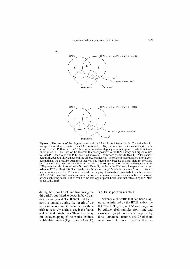
during the second trial, and two during thethird trial), but failed to detect infected cat-tle after that period. The IFN-γ test detectedpositive animals during the length of thestudy (nine, one and three in the first threetrials respectively, and also one in the fourth,and two in the sixth trial). There was a verylimited overlapping of the results obtainedwith both techniques (Fig. 1, panels A and B).
3.3. False positive reactors
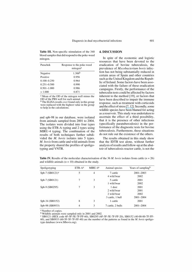
Seventy-eight cattle that had been diag-nosed as infected by the IDTB and/or theIFN-γ tests (Fig. 2, panel A) were negativeby culture, their samples from lung andassociated lymph nodes were negative bydirect auramine staining, and 76 of themwere no-visible lesions reactors. If a less
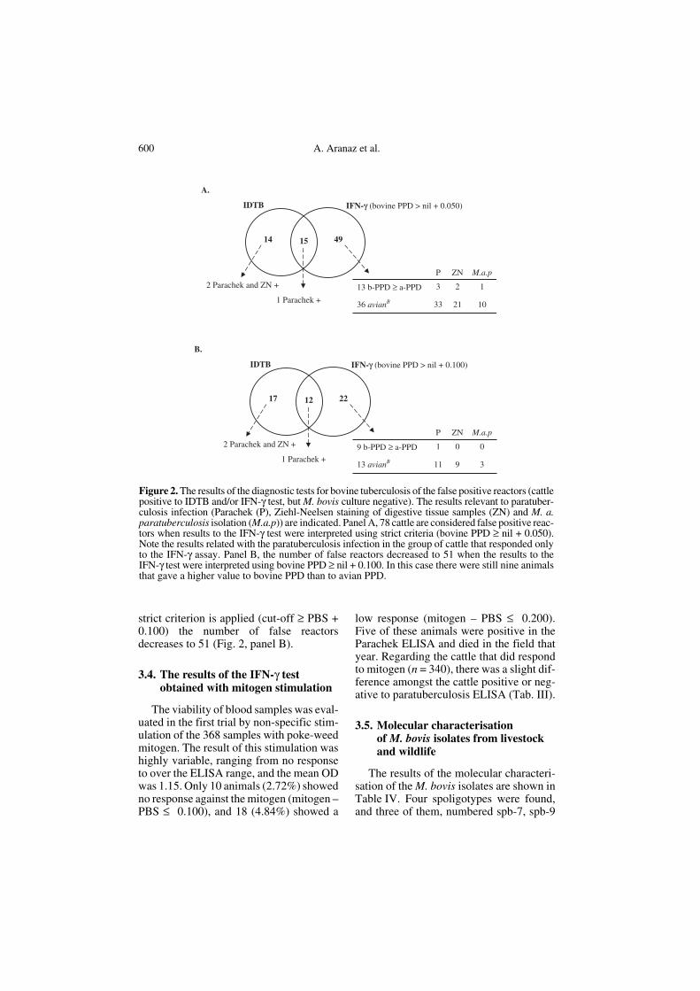
Figure 1. The results of the diagnostic tests of the 23 M. bovis infected cattle. The animals withunexpected results are marked. Panel A, results to the IFN-γ test were interpreted using the strict cri-terion (bovine PPD ≥ nil + 0.050). There was a limited overlapping of animals positive to both methods(9 out of 22, 40.9%). Two of the 16 cows that were positive to the IFN-γ assay had higher valuesto avian PPD than to bovine PPD (designed as avianB), both were positive to the ELISA for paratu-berculosis, but both showed generalised tuberculosis lesions (one of them was classified as total con-demnation at the abattoir). An animal that was slaughtered only because of its result to the serologyof paratuberculosis (it was a weak avian reactor to the comparative IDTB test and negative to theIFN-γ test) was also infected with M. bovis. Panel B, results to the IFN-γ test interpreted accordingto bovine PPD ≥ nil + 0.100. Note that this panel contained only 22 cattle because one M. bovis infectedanimal went undetected. There is a reduced overlapping of animals positive to both methods (5 outof 20, 25%). The avianB reactors are also indicated. In this case, two infected animals were detectedafter slaughtering because of its result to the serology of paratuberculosis (not detected by IFN-γ testor the IDTB test).
600 A. Aranaz et al.
strict criterion is applied (cut-off ≥ PBS +0.100) the number of false reactorsdecreases to 51 (Fig. 2, panel B).
3.4. The results of the IFN-γ test obtained with mitogen stimulation
The viability of blood samples was eval-uated in the first trial by non-specific stim-ulation of the 368 samples with poke-weedmitogen. The result of this stimulation washighly variable, ranging from no responseto over the ELISA range, and the mean ODwas 1.15. Only 10 animals (2.72%) showedno response against the mitogen (mitogen –PBS ≤ 0.100), and 18 (4.84%) showed a
low response (mitogen – PBS ≤ 0.200).Five of these animals were positive in theParachek ELISA and died in the field thatyear. Regarding the cattle that did respondto mitogen (n = 340), there was a slight dif-ference amongst the cattle positive or neg-ative to paratuberculosis ELISA (Tab. III).
3.5. Molecular characterisation of M. bovis isolates from livestock and wildlife
The results of the molecular characteri-sation of the M. bovis isolates are shown inTable IV. Four spoligotypes were found,and three of them, numbered spb-7, spb-9
Figure 2. The results of the diagnostic tests for bovine tuberculosis of the false positive reactors (cattlepositive to IDTB and/or IFN-γ test, but M. bovis culture negative). The results relevant to paratuber-culosis infection (Parachek (P), Ziehl-Neelsen staining of digestive tissue samples (ZN) and M. a.paratuberculosis isolation (M.a.p)) are indicated. Panel A, 78 cattle are considered false positive reac-tors when results to the IFN-γ test were interpreted using strict criteria (bovine PPD ≥ nil + 0.050).Note the results related with the paratuberculosis infection in the group of cattle that responded onlyto the IFN-γ assay. Panel B, the number of false reactors decreased to 51 when the results to theIFN-γ test were interpreted using bovine PPD ≥ nil + 0.100. In this case there were still nine animalsthat gave a higher value to bovine PPD than to avian PPD.
Diagnosis in dual mycobacterial infections 601
and spb-98 in our database, were isolatedfrom animals sampled from 2001 to 2004.The isolates were divided into four typesusing the ETR-A typing and 2 types usingMIRU-4 typing. The combination of theresults of both techniques further subdi-vided the M. bovis isolates into 5 types.M. bovis from cattle and wild animals fromthe property shared the profiles of spoligo-typing and VNTR.
4. DISCUSSION
In spite of the economic and logisticresources that have been devoted to theeradication of bovine tuberculosis, theprevalence of Mycobacterium bovis infec-tion has not being substantially reduced incertain areas of Spain and other countriessuch as the United Kingdom and the Repub-lic of Ireland. Some factors have been asso-ciated with the failure of these eradicationcampaigns. Firstly, the performance of thetuberculin tests could be affected by factorsinherent to the method [19], or factors thathave been described to impair the immuneresponse, such as treatment with corticoidsand the effect of stress [7, 12]. Secondly, somewildlife species have been blamed for actingas a reservoir. This study was undertaken toascertain the effect of a third possibility,that it is the presence of other infections(specifically paratuberculosis) in the per-formance of the diagnostic tests for bovinetuberculosis. Furthermore, these situationsdo not rule out the existence of the others.
The results obtained in this study showthat the IDTB test alone, without furtheranalysis of results and follow-up at the abat-toir of tuberculosis reactor cattle, is not the
Table III. Non-specific stimulation of the 340blood samples that did respond to the poke-weedmitogen.
Parachek Response to the poke-weed mitogena
Negative 1.306b
Positive 0.9560.100–0.250 0.9640.251–0.500 0.9900.501–1.000 0.986> 1.000 0.871a Mean of the OD of the mitogen well minus theOD of the PBS well for each animal.b The ELISA results over found only in this groupwere replaced with the highest value in the groupto help in the calculations.
Table IV. Results of the molecular characterisation of the 36 M. bovis isolates from cattle (n = 26)and wildlife animals (n = 10) obtained in the study.
Spoligotyping ETR-Aa MIRU-4a Animal species Years of samplingb
Spb-7 (SB0121)c 5 4 7 cattle4 wild boar
2001–20032002
Spb-7 (SB0121) 7 3 5 cattle1 wild boar
20012002
Spb-9 (SB0295) 7 3 1 deer2 wild boar2 wild boar
3 cattle, 1 bull
200120012002
2001–2004Spb-34 (SB0152) 8 3 1 cattle 2001
Spb-98 (SB0933) 4 3 7 cattle, 2 bulls 2001–2004
a Number of copies.b Wildlife animals were sampled only in 2001 and 2002. c SB0121 (HEX code 6F-5F-5E-7F-FF-60), SB0295 (6F-5F-5E-7F-FF-20), SB0152 (40-00-00-7F-FF-60), and SB0933 (60-5F-5F-7F-FF-40) are the number of the patterns as found in the M. bovis spoligo-type database (www.Mbovis.org).
602 A. Aranaz et al.
optimum strategy for the eradication cam-paign if both mycobacterial infections arepresent. It presents two main drawbacks inthe eradication campaign; first, the unsatis-factory level of sensitivity to clear the infec-tion, and secondly, the finding of falsetuberculosis reactors, which is detrimentalto farmers, results in serious financial lossand may lead to non-compliance with erad-ication schemes.
The development of the IFN-γ test hasbeen a major advancement in the diagnosisof bovine tuberculosis and it has been offi-cially approved in many countries. How-ever, there is little information about theperformance of this test and the criteria thatshould be used in the different epidemio-logical contexts. Since our main target wasthe eradication of tuberculosis in the herd,the cut-off value of the ELISA for IFN-γwas adjusted to obtain a maximum sensi-tivity (bovine PPD ≥ nil + 0.050).
Although it is difficult to compare theresults obtained in the published field trialsbecause of the use of different methodology(sampling time, different PPD and ELISAkit), source of cattle, and criteria of inter-pretation, the sensitivity values obtained inthis study for both the IDTB and the IFN-γtests have been only moderate, and in gen-eral terms, they were lower than thosefound in other reports [13, 18, 28, 31, 32,45]. This low performance is more obviousregarding the IFN-γ assay since it is gener-ally accepted to have a higher sensitivity. Inthis study, the apparent sensitivity of bothIDTB and IFN-γ tests was low and similar,65.2% and 69.6% respectively. The sensi-tivity of the tests may be overestimatedbecause only a moderate number of non-reacting cattle were necropsied. Asreported in other studies, there was an over-lapping population of infected animals thatwas detected by both the IDTB test and theIFN-γ assay, and a population positive oneither the skin test or the IFN-γ assay. In thiscase, this common population was alsosmall (40.9%). Therefore, and in agreementwith other authors [13, 28, 32, 39, 45], the
detection of the maximum number ofinfected animals is achieved by the combi-nation of both tests. Both tests were com-plementary also in the way that the IDTBtest performed better during the first part ofthe trial, while the IFN-γ test was the onlymethod that detected the infected animals atthe second part of the study (it detected theM. bovis culture positive cattle that failed toreact to the IDTB test). The practical effectin the eradication of this infection is that byusing the comparative IDTB test alone, theherd would have recovered the officiallytuberculosis-free herd status that enable formovement and trade of animals althoughsome residual M. bovis infection remainedat that moment. Cattle movement and cattlepurchase is a potential for the transmissionof the disease [27]. In fact, a recent paperhighlights that the movement of cattle fromareas where bovine tuberculosis is reportedoutperforms other variables as the predictorof disease occurrence in Great Britain [11].
Two of the M. bovis culture positive ani-mals were avianB reactors. The identifica-tion of avian reactors further complicatesthe interpretation of the results. These ani-mals are usually considered negative totuberculous infection, and it has beenreported that cows with a positive IFN-γresponse to avian PPD generally also hadpositive lower responses to bovine PPD[35]. However, the isolation of M. bovisfrom avianB reactors was described in agoat flock [18]. We have found that it canalso occur in cattle.
Serial testing combining two techniques(IDTB and IFN-γ assay) has been previ-ously used for the eradication of tuberculo-sis in goats3 [18] and cattle [13] resulting in
3 Vidal D., Domingo M., Aranaz A., et al., Eradi-cation of tuberculosis from goat herds by means ofthe interferon-γ assay and the single intradermalcomparative skin test, in: Griffin F., De Lisle G.(Eds.), Tuberculosis in wildlife and domestic ani-mals, Proc. 2nd Int. Conference on Mycobacte-rium bovis, Otago Conference Series, Universityof Otago Press, Dunedin, New Zealand, 1995,328–330.

Diagnosis in dual mycobacterial infections 603
gradual reduction in the prevalence of theinfection and elimination after two to fourcycles. The fact that more cycles wereneeded in this herd could be due to the typeof cattle, the presence of paratuberculosis inthe same herd, and a possibility of infectionfrom wildlife species.
Although the use of a mitogen as thepositive control has not been consideredabsolutely necessary [37], we includedstimulation with poke-weed mitogen as apositive control, and the general immuneresponse obtained was satisfactory. Theresults indicate that the response of cattlenegative to the Parachek test is slightlyhigher than those positive to the Parachektest. Secretion of greater quantities ofIFN-γ in response to mitogens in cows withsubclinical paratuberculosis than in cowswith clinical paratuberculosis has beenreported [34]. The finding that five of thecattle which did not respond to mitogenwere at the last stage of paratuberculosisinfection may suggest that clinicallyadvanced paratuberculosis hampers thedevelopment of cell responses, resulting infalse negative reactions.
Although our data should be interpretedas approximate because the slaughteredanimals were not randomly selected, thenumber of false reactors (IDTB test positiveand/or IFN-γ test positive, but M. bovis cul-ture negative) found in this study wasremarkably high compared to other reports,with the exception of another Spanish study[13]. Our results were obtained on a singlefarm, but we think that they could be extrap-olated to many other farms in the same sin-gular epidemiological situation.
In our experience, the number of falsepositive reactors is too high to be explainedonly as a failure to detect M. bovis because(1) lesions were in unusual location andtherefore not sampled at the abattoirbecause we examined the tissues with ahigher probability to contain bacilli (medi-astinal, retropharyngeal, bronchial andmesenteric lymph nodes, and lung) [5, 40],or (2) as failure of the bacteriological cul-
ture to detect low-viability bacteria thatcould be damaged after freezing or decon-tamination procedures [5]. In total, 39 of the78 (50%) false reactors were positive inthe serology of paratuberculosis using theParachek kit, 24 were positive in the Ziehn-Neelsen staining, and M. a. paratuberculo-sis was isolated from 11 cattle; most of themwere from the group detected by the IFN-γassay. Therefore, we think that this can beexplained by the cross-reactivity due tobacteria other than M. bovis, likely cross-reactivity due to M. a. paratuberculosis.
The animals were separated into twoherds according to their results to the tests.The advantages of this systems of segrega-tion (apart from the study of the evolutionof the results) are that it avoids unnecessaryculling and helps to maintain a number ofcattle in the property to keep its economicviability, while limiting the spread of theinfections. Only the offspring of herd Awere used for replacement, and as the con-trol progressed, the trend was the elimina-tion of herd B. The obvious drawback is thatit can be carried out only when no limitationof land and other resources is present.
Another aspect of the epidemiology ofthe M. bovis infection that was taken intoaccount in this study was the potential roleof wildlife as reservoirs of the infection.M. bovis can also infect a wide range ofdomestic and wild animals [21, 25] andsome of them are considered as reservoirsof infection for livestock. The risk these res-ervoirs constitute for domestic animalsdepends on the specific epidemiologicalsituation of the species and the environment[21] and has been demonstrated under asimilar situation in Spain [1].
Sustainable control policies can only beachieved through a better understanding ofthe epidemiology of tuberculosis in both cat-tle and wildlife reservoirs [2]. The M. bovisisolates obtained in this study were charac-terised by spoligotyping and VNTR typing,and the results indicate that three of the fivepatterns are shared by domestic and wildlifespecies in the property. The awareness of
604 A. Aranaz et al.
this potential risk resulted in a more precisetargeting of control measures; the ownerswere advised to take measures to limit con-tact between cattle and wild species, andwildlife was fenced in a separate area.
For the time being, the strategy describedin this report has been applied for 3.5 yearsin the herd, and we have not found any M.bovis culture-positive cattle since the sixthtrial. All animals in herd A were negativeto the IFN-γ assay in the last trial to date.The herd has achieved the officially tuber-culosis-free status, but continuous surveil-lance will be maintained.
In summary, we found that the perform-ance of the IDTB and the IFN-γ tests isimpaired in cattle with dual mycobacterialinfections; and this drop is more evident inthe latter. In this context, it is not possibleto accurately forecast the infection status ofan animal with a single test. These resultsindicate that a different mentality isrequired in approaching the eradicationcampaign in herds where both diseases arepresent. The combined use of diagnostictechniques allied to improved farm man-agement practices have been useful for theobjective of this field trial. The strategyimplemented in the property (serial com-bined use of diagnosis techniques and seg-regation of animals depending on theresults, together with separation of domes-tic and wild animals) has resulted in theelimination of M. bovis infection from theherd and a significant control of the paratu-berculosis infection. However, in especialwhen a herd is dually infected, these proce-dures require a substantial technical andeconomic effort both in farm managementand in the laboratory, because a detailed fol-low-up of the animals is needed. Thesestrategies should be understood as amedium-long term task, and the trusted col-laboration of all implied parties is essential.
ACKNOWLEDGEMENTS
A. Aranaz has a fellowship “Ramon y CajalProgram” from the Spanish Ministry of Science
and Technology and the Universidad Com-plutense de Madrid. This study was supported bythe Spanish Ministry of Agriculture, Food andFisheries. We are grateful to N. Montero fortechnical help; to veterinary practitioners F.Moneo-López (deceased during this study) andI. Larrauri, and to the Official Veterinary Serv-ices, in especial to C. Gil-Gonzalez de Ubieta,for the IDTB testing and collection of bloodsamples; and to J.M. Rubio-Ruiz, A.J.Domínguez-Sánchez and M. Fernandez-Sánchez(abattoir staff of Frimancha, Valdepeñas, Ciu-dad Real) for assistance in collection of tissuesamples. We also thank J. Fernández-Sanz, rep-resentative of the Union de Criadores de Toro deLidia (Bullfighting Cattle Breeders Union). Weare grateful to Matthew Gilmour for careful revi-sion of the manuscript. The willingness andcooperation of landowners and farm managersof the property throughout the study is greatlyappreciated. We would like to thank the Nationaland Regional Animal Health authorities, inespecial A. Cabello, for their continuous encour-agement.
REFERENCES
[1] Aranaz A., de Juan L., Montero N., SánchezC., Galka M., Delso C., Álvarez J., Romero B.,Bezos J., Vela A.I., Briones V., Mateos A.,Domínguez L., Bovine tuberculosis (Myco-bacterium bovis) in wildlife in Spain, J. Clin.Microbiol. 42 (2004) 2602–2608.
[2] Bourne J., Donnelly C.A., Cox D.R., GettinbyG.D., McInerney J.P., Morrison I., WoodroffeR., Bovine tuberculosis: towards a future con-trol strategy, Vet. Rec. 146 (2000) 207–210.
[3] Cagiola M., Feliziani F., Severi G., PasqualiP., Rutili D., Analysis of possible factorsaffecting the specificity of the gamma inter-feron test in tuberculosis-free cattle herds,Clin. Diagn. Lab. Immunol. 11 (2004) 952–956.
[4] Corner L.A., Trajstman A.C., An evaluationof 1-hexadecyl-pyridinium chloride as adecontaminant in the primary isolation ofMycobacterium bovis from bovine lesions,Vet. Microbiol. 18 (1988) 127–134.
[5] Corner L.A., Post mortem diagnosis of Myco-bacterium bovis infection in cattle, Vet.Microbiol. 40 (1994) 53–63.
[6] De Juan L., Mateos A., Domminguez L.,Sharp J.M., Stevenson K., Genetic diversity ofMycobacterium avium subspecies paratuber-culosis isolates from goats detected by pulsed-field gel electrophoresis, Vet. Microbiol. 106(2005) 249–257.
Diagnosis in dual mycobacterial infections 605
[7] Doherty M.L., Basset H.F., Quinn P.J., DavisW.C., Monaghan M.L., Effects of dexameth-asone on cell-mediated immune responses incattle sensitized to Mycobacterium bovis, Am.J. Vet. Res. 56 (1995) 1300–1306.
[8] Dunn J.R., Kaneene J.B., Grooms D.L., BolinS.R., Bolin C.A., Brunning-Fann C.S., Effectsof positive results for Mycobacterium aviumsubsp. paratuberculosis as determined bymicrobial culture of feces or antibody ELISAon results of caudal fold tuberculin test andinterferon-γ assay for tuberculosis in cattle, J.Am. Vet. Med. Assoc. 226 (2005) 429–435.
[9] Fischer E.A.J., van Roermund H.J.W.,Hemerik L., van Asseldonk M.A.P.M., deJong M.C.M., Evaluation of surveillancestrategies for bovine tuberculosis (Mycobac-terium bovis) using an individual based epi-demiological model, Prev. Vet. Med. 67(2005) 283–301.
[10] Frothingham R., Meeker-O’Connell W.A.,Genetic diversity in the Mycobacteriumtuberculosis complex based on variable num-bers of tandem repeats, Microbiology 144(1998) 1189–1196.
[11] Gilbert M., Mitchell A., Bourn D., MawdsleyJ., Clifton-Hadley R., Wint W., Cattle move-ments and bovine tuberculosis in Great-Britain,Nature 435 (2005) 491–496.
[12] Goff B.S.L., Effect of dexamethasone treat-ment of tuberculous cattle on results of thegamma-interferon test for Mycobacteriumbovis, Vet. Immunol. Immunopathol. 53(1996) 39–47.
[13] González-Llamazares O.R., Gutiérrez-MartínC.B., Álvarez-Nistal D., de la Puente-Redondo V.A., Domínguez-Rodríguez L.,Rodríguez-Ferri E.F., Field evaluation of thesingle intradermal cervical tuberculin test andthe interferon-γ assay for detection anderadication of bovine tuberculosis in Spain,Vet. Microbiol. 70 (1999) 55–66.
[14] Greig A., Stevenson K., Pérez V., Pirie A.A.,Grant J.M., Sharp J.M., Paratuberculosis inwild rabbits (Oryctolagus cuniculus), Vet.Rec. 140 (1997) 141–143.
[15] Kamerbeek J., Schouls L., Kolk A., VanAgterveld M., van Soolingen D., Kuijper S.,Bunschoten A., Molhuizen H., Shaw R.,Goyal M., van Embden J.D.A., Simultaneousdetection and strain differentiation of Myco-bacterium tuberculosis for diagnosis and epi-demiology, J. Clin. Microbiol. 35 (1997)907– 914.
[16] Kennedy D.J., Benedictus G., Control ofMycobacterium avium subsp. paratuberculo-sis infection in agricultural species, Rev. sci.tech. Off. Int. Epizoot. 20 (2001) 151–179.
[17] Köhler H., Gyra H., Zimmer K., Dräger K.G.,Burkert B., Lemser B., Hausleithner D., CublerK., Klawonn W., Hess R.G., Immune reactionsin cattle after immunization with a Mycobac-terium paratuberculosis vaccine and implica-tions for the diagnosis of M. paratuberculosisand M. bovis infections, J. Vet. Med. B Infect.Dis. Vet. Public Health 48 (2001) 185–195.
[18] Liébana E., Aranaz A., Urquía J.J., Mateos A.,Domínguez L., Evaluation of the gamma-interferon assay for eradication of tuberculosisin a goat herd, Aust. Vet. J. 76 (1998) 50–53.
[19] Monaghan M.L., Doherty M.L., Collins J.D.,Kazda J.F., Quinn P.J., The tuberculin test,Vet. Microbiol. 40 (1994) 111–124.
[20] Monaghan M., Quinn P.J., Kelly A.P., McGillK., McMurray C., O’Crowley K., BassettH.F., Costello E., Quigley F., Rothel J.S.,Wood P.R., Collins J.D., A pilot trial to eval-uate the γ-interferon assay for the detection ofMycobacterium bovis infected cattle underIrish conditions, Irish Vet. J. 50 (1997) 229–232.
[21] Morris R.S., Pfeiffer D.U., Jackson R., Theepidemiology of Mycobacterium bovis infec-tions, Vet. Microbiol. 40 (1994) 153–157.
[22] Morrison I., Bourne J., Cox D.R., DonnellyC.A., Gettinby G., McInerney J.P.,Woodroffe R., Pathogenesis and diagnosis ofinfections with Mycobacterium bovis in cattle,Vet. Rec. 146 (2000) 236–242.
[23] Muskens J., van Zijderveld F., Eger A.,Bakker D., Evaluation of the long-termimmune response in cattle after vaccinationagainst paratuberculosis in two Dutch dairyherds, Vet. Microbiol. 86 (2002) 269–278.
[24] Neill S.D., Cassidy J., Hanna J., Mackie D.P.,Pollock J.M., Clements A., Walton E., BrysonD.G., Detection of Mycobacterium bovisinfection in skin test-negative cattle with anassay for bovine interferon-gamma, Vet. Rec.135 (1994) 134–135.
[25] O’Reilly L.M., Daborn C.J., The epidemiol-ogy of Mycobacterium bovis infections in ani-mals and man: a review, Tuber. Lung Dis. 76(1995) 1–46.
[26] Pavlik I., Ayele W.Y., Parmova I., MelicharekI., Hanzilikova M., Körmendy B., Nagy G.,Cvetnic Z., Ocepek M., Fejzic N., Lipiec M.,Incidence of bovine tuberculosis in cattle inseven Central European countries during theyears 1990–1999, Vet. Med. Czech 47 (2002)45–51.
[27] Phillips C.J.C., Foster C.R.W., Morris P.A.,Teverson R., The transmission of Mycobacte-rium bovis infection to cattle, Res. Vet. Sci. 74(2003) 1–15.
[28] Pollock J.M., Girvin R.M., Lightbody K.A.,Clements R.A., Neill S.D., Buddle B.M.,

606 A. Aranaz et al.
Andersen P., Assessment of defined antigensfor the diagnosis of bovine tuberculosis in skintest-reactor cattle, Vet. Rec. 146 (2000) 659–665.
[29] Rothel J.S., Jones S.L., Corner L.A., Cox J.C.,Wood P.R., A sandwich enzyme immu-noassay for bovine interferon-γ and its use forthe detection of tuberculosis in cattle, Aust.Vet. J. 67 (1990) 134–137.
[30] Rothel J.S., Jones S.L., Corner L.A., Cox J.C.,Wood P.R., The gamma-interferon assay fordiagnosis of bovine tuberculosis in cattle: con-ditions affecting the production of gamma-interferon in whole blood culture, Aust. Vet.J. 69 (1992) 1–4.
[31] Ryan T.J., Buddle B.M., de Lisle G.W., Anevaluation of the gamma-interferon test fordetecting bovine tuberculosis in cattle 8 to28 days after tuberculin skin testing, Res. Vet.Sci. 69 (2000) 57–61.
[32] Scacchia M., Lelli R., Petrini A., Prencipe V.,Calistri P., Giovannini A., Use of innovativemethods in the eradication of bovine tubercu-losis, J. Vet. Med. B 47 (2000) 321–327.
[33] Smithwick R.W., Laboratory manual for acid-fast microscopy, second edition, US Depart-ment of Health, Education and Welfare, Cent-ers for Disease Control, Georgia, USA, 1976.
[34] Stabel J.R., Production of γ-interferon byperipheral blood mononuclear cells: animportant diagnostic tool for detection of sub-clinical paratuberculosis, J. Vet. Diagn.Invest. 8 (1996) 345–350.
[35] Stabel J.R., Whitlock R.H., An evaluation ofa modified interferon-γ assay for the detectionof paratuberculosis in dairy herds, Vet. Immu-nol. Immunopathol. 79 (2001) 69–81.
[36] Supply P., Lesjean S., Savine E., Kremer K.,van Soolingen D., Locht C., Automated high-throughput genotyping for study of global epi-demiology of Mycobacterium tuberculosisbased on mycobacterial interspersed repetitiveunits, J. Clin. Microbiol. 39 (2001) 3563–3571.
[37] Vordermeier M., Aranaz A., Pollock J.M.,Immunodiagnosis of bovine tuberculosis,Summary of a satellite workshop of the Myco-bacterium bovis 2000 Conference, Cam-
bridge, UK, 17 August 2000, Tuberculosis 81(2001) 177–180.
[38] Walravens K., Marché S., Rosseels V.,Wellemans V., Boelaert F., Huygen K., God-froid J., IFN-γ diagnostic tests in the contextof bovine mycobacterial infections in Bel-gium, Vet. Immunol. Immunopathol. 87(2002) 401–406.
[39] Whipple D.L., Bolin C.A., Davies A.J.,Jarnagin J.L., Johnson D.C., Nabors R.S.,Payeur J.B., Saari D.A., Wilson A.J., WolfM.M., Comparison of the sensitivity of thecaudal fold skin test and a commercialgamma-interferon assay for diagnosis ofbovine tuberculosis, Am. J. Vet. Res. 56(1995) 415–419.
[40] Whipple D.L., Bolin C.A., Miller J.M., Dis-tribution of lesions in cattle infected withMycobacterium bovis, J. Vet. Diagn. Invest. 8(1996) 351–354.
[41] Whipple D.L., Palmer M.V., Slaughter R.E.,Jones S.L., Comparison of purified proteinderivatives and effect of skin testing on resultsof a commercial gamma interferon assay fordiagnosis of tuberculosis in cattle, J. Vet.Diagn. Invest. 13 (2001) 117–122.
[42] Wilton S., Cousins D., Detection and identi-fication of multiple mycobacterial pathogensby DNA amplification in a single tube, PCRMethods Appl. 1 (1992) 269–273.
[43] Wood P.R., Corner L.A., Plackett P., Devel-opment of a simple, rapid in vitro cellularassay for bovine tuberculosis based on theproduction of gamma interferon, Res. Vet.Sci. 49 (1990) 46–49.
[44] Wood P.R., Corner L.A., Rothel J.S., BaldockC., Jones S.L., Cousins D.B., McCormickB.S., Francis B.R., Creeper J., Tweddle N.E.,Field comparison of the interferon-gammaassay and the intradermal tuberculin test forthe diagnosis of bovine tuberculosis, Aust.Vet. J. 68 (1991) 286–290.
[45] Wood P.R., Corner L.A., Rothel J.S., RipperJ.L., Fifis T., McCormick B.S., Francis B.,Melville L., Small K., De Witte K., Tolson J.,Ryan T.J., de Lisle G.W., Cox J.C., Jones S.L.,A field evaluation of serological and cellulardiagnostic tests for bovine tuberculosis, Vet.Microbiol. 31 (1992) 71–79.
Related Documents