Review Are retinoids potential therapeutic agents in disorders of social cognition including autism? Richard P. Ebstein a,b,⇑ , David Mankuta c , Nurit Yirmiya b , Fabio Malavasi d a Department of Psychology, National University of Singapore, Singapore b Psychology Department, Hebrew University, Jerusalem 91095, Israel c Department of Obstretics and Gynecology, Hadassah Medical Center, Hebrew University, Jerusalem 91120, Israel d Genetics Department, University of Torino, Torino 10126, Italy article info Article history: Received 26 March 2011 Revised 3 May 2011 Accepted 3 May 2011 Available online 6 May 2011 Edited by Sergio Papa, Gianfranco Gilardi and Wilhelm Just Keywords: Autism spectrum disorder (ASD) All-trans retinoic acid (ATRA) CD38 Oxytocin Polymorphism abstract Increasing evidence suggests that the nonapeptide, oxytocin (OT), helps shape social and affiliative behaviors not only in lower mammals but also in humans. Recently, an essential mediator of brain OT release has been discovered, ADP-ribosyl cyclase and/or CD38. We have subsequently shown that polymorphisms across the CD38 gene are associated with autism spectrum disorders (ASD). Notably, CD38 expression in lymphoblastoid cells (LBC) is reduced in cell lines derived from ASD subjects compared to parental cell lines. Intriguingly, a correlation was observed between CD38 expression and measures of social function in ASD. Finally, we have shown that all-trans retinoic acid (ATRA), a known inducer of CD38 transcription, can rescue low CD38 expressing LBC lines derived from ASD subjects and restore normal levels of transcription of this ectoenzyme providing ‘proof of principle’ in a peripheral model that retinoids are potential therapeutic agents in ASD. Ó 2011 Federation of European Biochemical Societies. Published by Elsevier B.V. All rights reserved. 1. Oxytocin Classically, the nonapeptide oxytocin (OT) has been viewed as a hypothalamic neuropeptide that is released into the general circu- lation from the neural lobe of the pituitary, inducing uterine con- tractions during parturition and milk ejection during lactation. OT is derived from a pre-prohormone precursor that is synthesized in the hypothalamus and stored in vesicles at the posterior pitui- tary for storage and subsequent release into the bloodstream (see [1] for comprehensive review of the oxytocin receptor system). 2. OT and social behavior Beyond the long-known peripheral effects of OT, a wealth of animal studies have elaborated the role of OT, or their analogues such as isotocin and vasotocin [2], in molding social behavior from fish to mammals [3]. In the past few years the role of OT has also been examined in our own species, and similar to what has been learned from animal studies, it appears that this nonapeptides also influence social behaviors in humans [4,5]. Indeed, OT has been suggested as the ‘great facilitator of life’ in a recent review [6]. In humans, intranasal administration of OT has been shown to increase trust [7], facilitate mind-reading [8], enhance human memory for social identity [9], increase positive communication between couples [10], increase gaze to the eye region [11] and in- crease generosity [12]. Intriguingly, OT plasma levels have been linked to individual patterns of maternal-fetal attachment [13] and salivary OT levels were associated with bonding to own par- ents and inversely related to psychological distress, particularly depressive symptoms [14]. Social anxiety symptom severity, ad- justed for age and gender in a healthy group of subjects, was asso- ciated with higher plasma oxytocin levels [15]. Imaging studies reinforce the role of OT in influencing human social behavior with evidence demonstrating that OT modulates the amygdala and other brain regions [16]. 3. Oxytocin receptor gene (OXTR) The OT receptor gene is present in single copy in the haploid hu- man genome and was mapped to the gene locus 3p25–3p26.2 [1]. The gene spans 17 kb and contains 3 introns and 4 exons. Exons 1 and 2 correspond to the 5 0 non-coding region. Exons 3 and 4 encode the amino acids of the OT receptor. Intron 3, which is the largest at 0014-5793/$36.00 Ó 2011 Federation of European Biochemical Societies. Published by Elsevier B.V. All rights reserved. doi:10.1016/j.febslet.2011.05.004 ⇑ Corresponding author. Address: Psychology Department, Faculty of Arts and Sciences, Block AS4, 9 Arts Link, National University of Singapore, Singapore 117570, Singapore. Fax: +65 6773 1843. E-mail address: [email protected] (R.P. Ebstein). FEBS Letters 585 (2011) 1529–1536 journal homepage: www.FEBSLetters.org

Welcome message from author
This document is posted to help you gain knowledge. Please leave a comment to let me know what you think about it! Share it to your friends and learn new things together.
Transcript
FEBS Letters 585 (2011) 1529–1536
journal homepage: www.FEBSLetters .org
Review
Are retinoids potential therapeutic agents in disorders of social cognitionincluding autism?
Richard P. Ebstein a,b,⇑, David Mankuta c, Nurit Yirmiya b, Fabio Malavasi d
a Department of Psychology, National University of Singapore, Singaporeb Psychology Department, Hebrew University, Jerusalem 91095, Israelc Department of Obstretics and Gynecology, Hadassah Medical Center, Hebrew University, Jerusalem 91120, Israeld Genetics Department, University of Torino, Torino 10126, Italy
a r t i c l e i n f o a b s t r a c t
Article history:Received 26 March 2011Revised 3 May 2011Accepted 3 May 2011Available online 6 May 2011
Edited by Sergio Papa, Gianfranco Gilardiand Wilhelm Just
Keywords:Autism spectrum disorder (ASD)All-trans retinoic acid (ATRA)CD38OxytocinPolymorphism
0014-5793/$36.00 � 2011 Federation of European Biodoi:10.1016/j.febslet.2011.05.004
⇑ Corresponding author. Address: Psychology DepSciences, Block AS4, 9 Arts Link, National Univers117570, Singapore. Fax: +65 6773 1843.
E-mail address: [email protected] (R.P. Ebstein
Increasing evidence suggests that the nonapeptide, oxytocin (OT), helps shape social and affiliativebehaviors not only in lower mammals but also in humans. Recently, an essential mediator of brainOT release has been discovered, ADP-ribosyl cyclase and/or CD38. We have subsequently shown thatpolymorphisms across the CD38 gene are associated with autism spectrum disorders (ASD). Notably,CD38 expression in lymphoblastoid cells (LBC) is reduced in cell lines derived from ASD subjectscompared to parental cell lines. Intriguingly, a correlation was observed between CD38 expressionand measures of social function in ASD. Finally, we have shown that all-trans retinoic acid (ATRA),a known inducer of CD38 transcription, can rescue low CD38 expressing LBC lines derived from ASDsubjects and restore normal levels of transcription of this ectoenzyme providing ‘proof of principle’in a peripheral model that retinoids are potential therapeutic agents in ASD.� 2011 Federation of European Biochemical Societies. Published by Elsevier B.V. All rights reserved.
1. Oxytocin
Classically, the nonapeptide oxytocin (OT) has been viewed as ahypothalamic neuropeptide that is released into the general circu-lation from the neural lobe of the pituitary, inducing uterine con-tractions during parturition and milk ejection during lactation.OT is derived from a pre-prohormone precursor that is synthesizedin the hypothalamus and stored in vesicles at the posterior pitui-tary for storage and subsequent release into the bloodstream (see[1] for comprehensive review of the oxytocin receptor system).
2. OT and social behavior
Beyond the long-known peripheral effects of OT, a wealth ofanimal studies have elaborated the role of OT, or their analoguessuch as isotocin and vasotocin [2], in molding social behavior fromfish to mammals [3]. In the past few years the role of OT has alsobeen examined in our own species, and similar to what has beenlearned from animal studies, it appears that this nonapeptides also
chemical Societies. Published by E
artment, Faculty of Arts andity of Singapore, Singapore
).
influence social behaviors in humans [4,5]. Indeed, OT has beensuggested as the ‘great facilitator of life’ in a recent review [6].
In humans, intranasal administration of OT has been shown toincrease trust [7], facilitate mind-reading [8], enhance humanmemory for social identity [9], increase positive communicationbetween couples [10], increase gaze to the eye region [11] and in-crease generosity [12]. Intriguingly, OT plasma levels have beenlinked to individual patterns of maternal-fetal attachment [13]and salivary OT levels were associated with bonding to own par-ents and inversely related to psychological distress, particularlydepressive symptoms [14]. Social anxiety symptom severity, ad-justed for age and gender in a healthy group of subjects, was asso-ciated with higher plasma oxytocin levels [15]. Imaging studiesreinforce the role of OT in influencing human social behavior withevidence demonstrating that OT modulates the amygdala andother brain regions [16].
3. Oxytocin receptor gene (OXTR)
The OT receptor gene is present in single copy in the haploid hu-man genome and was mapped to the gene locus 3p25–3p26.2 [1].The gene spans 17 kb and contains 3 introns and 4 exons. Exons 1and 2 correspond to the 50 non-coding region. Exons 3 and 4 encodethe amino acids of the OT receptor. Intron 3, which is the largest at
lsevier B.V. All rights reserved.
1530 R.P. Ebstein et al. / FEBS Letters 585 (2011) 1529–1536
12 kb, separates the coding region immediately after the putativetransmembrane domain 6. Exon 4 contains the sequence encodingthe seventh transmembrane domain, the COOH terminus, and theentire 3’-non-coding region, including the polyadenylation signals.The OT receptor protein is a typical member of the rhodopsin-type(class I) G protein class of receptors (GPCR) family [1]. The seventransmembrane-helices are most highly conserved among the GPCRfamily members. Conserved residues among the GPCRs may be in-volved in a common mechanism for activation and signal transduc-tion to the G protein. The OT receptor signals via the Gq/11 and Giclass of guanosine triphosphate (GTP) binding proteins that stimu-late, together with Gbx, the phospholipase C-b isoforms [17]. Theselectivity of the OT receptor for G-proteins appears to be more com-plex than the selectivity of the vasopressin receptors. Coexpressionof different combinations of intracellular domains demonstratesthat all intracellular loops may contribute to the selectivity of theOT receptor to some extent. In addition to the intracellular domains,a highly conserved region of the C-terminal domain is involved in theselectivity of coupling to Gq/11 but not Gi.
4. Oxytocin and autism spectrum disorders (ASD)
Autistic disorders (AD) are a group of disorders characterized bythe three core deficits: qualitative impairment in social interactionand communication, and restricted repetitive and stereotyped pat-terns of behavior, interests, and activities [18]. The three disordersautism, Asperger syndrome (AS) and pervasive developmental dis-order-not otherwise specified (PDD-NOS) are currently thought ofby most researchers as a continuum of the same disorder withvarying degrees of severity, associated intellectual functioningand medical conditions. Recent studies estimated the prevalenceof AD to around 0.5–1% [19]. Genetic factors play an important rolein ASD [20].
A number of molecular genetic findings support the involve-ment of oxytocin neurotransmission in some of the social deficitsand symptomatology of ASD. Firstly, significant association be-tween OXTR SNPs [21–26] and risk for autism has been provision-ally shown (but also see [27,28]) and, notably, was mediated bysocialization skills in our study [23]. Additionally, SNPs associatedwith ASD are also associated with amygdala volume [29], depres-sion/anxiety [29], negative emotionality [30], attention deficit[31], adult attachment styles [32,33] (but see[34]), affectivity,emotional loneliness and IQ [35], empathy and stress reactivity[36], observed parenting [37], with sexual phenotypes [38] andin our own studies with prosocial behavior [39]. Interestingly,the AVPR1a receptor of the closely related nonapeptide vasopres-sin has also been associated with ASD [40–42]. This provisionalrole of OT in ASD, is further supported by two linkage studies[43,44], clinical evidence by several groups demonstrating clinicalimprovement in ASD following OT administration [45–51] (also see[52,53]) and studies showing that ASD is also associated with alter-ations in OT plasma levels [54].
5. Dendritic release of oxytocin
An interesting feature of OT action in the brain is that althoughOT has profound effects on social bonding that are exerted at sitesthat richly express oxytocin receptors, such brain regions areinnervated by few, if any, oxytocin-containing projections [55].How then does OT exert its effect in limbic and other ‘socially sen-sitive’ brain regions in the absence of apparent direct OT contain-ing neurons? Oxytocin synthesis takes place in hypothalamicnuclei, including the supraoptic nucleus (SON) and paraventricularnucleus (PVN). Specifically, magnocellular neurons in these twonuclei contain most of the OT and AVP in the CNS and are
characterized by dendritic release, which is regulated indepen-dently of secretion into the blood. Indeed, dendrites are apparentlythe major source of peptides released in the brain [55]. Importantlydendritic release does not parallel axonal release and both pro-cesses are regulated independently. Although release of OT andAVP from axons is linked to electrical activity resulting fromCa2+ entry through voltage-gated ion channels following depolar-ization of the terminals by invading action potentials [56], OT andAVP themselves [57], can elicit dendritic peptide release withoutincreasing electrical activity. In OT neurons, OT itself mobilizesCa2+ from thapsigargin-sensitive intracellular stores [58]. Activa-tion of peptide receptors on the dendrites or soma elevates intra-cellular Ca2+ concentrations and triggers exocytosis of largedense-core vesicles (LDCVs), and once dendritic peptide release istriggered, because of the peptide feedback, dendritic release canbe self-sustaining and, therefore, long-lasting.
6. CD38
A seminal paper by Higashida and his group led to the discoverythat OT release in the brain is mediated by ADP-ribosyl cyclaseand/or CD38 [59]. They used CD38 gene knockout mice (Cd38�/�), and discovered that CD38-dependent cyclic ADP ribose (cAD-PR)- and NAADP-sensitive intracellular Ca2+ mobilization plays akey role in OT release from soma and axon terminals of hypotha-lamic neurons, with marked effects on social behavior. In particu-lar, maternal behavior was dependent on OT, and social amnesia inmales was evident in the absence of this hormone. Activation ofCD38 with its substrate ligand NAD+ results in hydrolysis ofNAD+ and cyclic ADP-ribose (cADPR) to ADP-ribose, or in cycliza-tion of NAD+ to cADPR. In addition to production of cADPR, the en-zyme can use NADP+ as a substrate and catalyze the exchange ofits nicotinamide group with nicotinic acid to produce NAADP+.cADPR mobilizes Ca2+ from ryanodine-sensitive intracellularCa2+ stores in the endoplasmic reticulum and NAADP liberates itfrom other pools located in lysosomes or secretory granules. Thetwo molecules act as second messengers independent of inositol1,4,5-trisphosphate (IP3) [60,61]. To summarize, CD38 is a multi-functional molecule (ecto-enzyme) combining enzymatic andreceptor properties and playing a key role in various physiologicalprocesses in the tissues (proliferation, differentiation, migration,adhesion, and secretion). In the brain, CD38 is found in neuronsand glial cells, shows intracellular or plasma membrane location,and is enriched in neuronal perikarya and dendrites [62,63].CD38 is critical for OT but not AVP release.
7. CD38 and autism spectrum disorders
The accumulating evidence discussed above, that OT plays animportant role in both normal as well as dysfunctional social rela-tionships/cognition [64,65], ipso facto targets CD38, a key mediatorof OT brain release, as a potential focus of interest in normal hu-man social behaviors as well as disorders of social cognition espe-cially autism [66,67]. In the past year, two research groups haveindependently addressed the role of CD38 in autism in humansubjects. Higashida and his colleagues [68] analyzed 10 singlenucleotide polymorphisms (SNPs) and mutations of CD38 by re-sequencing DNAs mainly from a case–control study in Japan, andCaucasian cases mainly recruited to the Autism Genetic ResourceExchange (AGRE). CD38 SNPs, rs6449197 and rs3796863 showedsignificant associations with a subset of ASD subjects (IQ > 70; des-ignated as high functioning autism/HFA) in 104 AGRE family trios,but not with Japanese 188 HFA subjects. Interestingly, a mutation/rare polymorphism that caused tryptophan to replace arginine atamino acid residue 140 (R140W; (rs1800561, 4693C > T)) was
R.P. Ebstein et al. / FEBS Letters 585 (2011) 1529–1536 1531
found in 0.6–4.6% of the Japanese population and was associatedwith ASD in the smaller case–control study. The SNP was clusteredin pedigrees in which the fathers and brothers of T-allele-carrierprobands had ASD or ASD traits. In this cohort [68] OT plasma lev-els were lower in subjects with the T allele than in those without.
In our first study of CD38 [69], we examined all tagging SNPsacross the CD38 gene region in 170 subjects diagnosed with ASDfrom 149 families (see [23] for description of the subjects). Indi-vidual SNPs and haplotypes were tested for association withASD. Additionally, the relationship between diabetes, autismand CD38 [70], as well as the use of CD38 as a disease marker[71], suggests that it would also be worthwhile to exploreCD38 expression in immune cell lines derived from ASD patients.These considerations prompted us to measure CD38 geneexpression in lymphoblastoid cell lines (LBC) derived from bothASD subjects and unaffected parents. We also include in thegene expression and family-based association analysis the SNP(rs3796863), which proved significantly associated with ASD inthe Munesue et al. [72] study.
8. Molecular genetic association [71]
We first examined association between CD38 tagging SNPs andDSM IV ASD. ASD subjects were evenly grouped into high and lowfunctioning based on an IQ cutoff of 70. This subject stratificationwas aimed at reducing phenotypic heterogeneity in the autismsample. Significant association was observed between low func-tioning ASD and three – seven haplotypes (Table 1). The resultsshown in the Table are significant (p < 0.05) following permutationtesting. Importantly, the SNP (and the ‘C’ allele) identified in theMunesue et al study [68] (rs3796863), which they found
Table 1Haplotype association of CD38 SNPs with low functioning ASD subjects.
HAP Freq. Transmitted Untransmitted Chi Square p value
rs3796863-rs1803404-rs1130169Likelihood ratio chi square = 12.14; df = 4; global p-value = 0.016 (0.019)CTC 0.501 85.87 64.62 4.78 0.028ATT 0.201 35.87 38.81 0.939 0.332CTT 0.194 17.13 36.38 7.47 0.006rs10805347–rs3796863–rs1803404–rs1130169Likelihood ratio chi square = 20.69; df = 9; global p-value = 0.014 (0.01)ACTC 0.316 39.97 39.24 0.005 0.938GCTC 0.187 42.58 20.25 9.419 0.002GATT 0.186 31.53 24.23 1.101 0.294GCTT 0.178 16.44 31.45 4.796 0.028rs2286553–rs10805347–rs3796863–rs1803404–rs1130169Likelihood ratio chi square = 25.46; df = 11; global p-value = 0.008 (0.019)GACTC 0.317 39.99 39.72 3.25e�05 0.999GGCTC 0.186 42.25 19.76 9.768 0.001GGCTT 0.177 16.76 31.8 4.762 0.029GGATT 0.173 28.97 24.45 0.493 0.482rs3796864–rs2286553–rs10805347–rs3796863–rs1803404–rs1130169Likelihood ratio chi square = 41.52; df = 13; global p-value = 7.81e�05 (0.009)CGACTC 0.313 39.99 37.76 0.075 0.783CGGCTC 0.179 41.19 18.85 9.528 0.002CGGCTT 0.167 15.77 29.15 3.525 0.061AGGATT 0.125 16.95 19 0.152 0.695rs4516711–rs3796864–rs2286553–rs10805347–rs3796863–rs1803404–
rs1130169Likelihood ratio chi square = 34.78; df = 12; global p-value = 0.0005 (0.0049)GCGACTC 0.297 38.99 31.84 1.006 0.315GCGGCTC 0.180 38.16 18.97 8.064 0.004GCGGCTT 0.162 14.84 27.03 3.808 0.051CAGGATT 0.123 16.99 18 0.396 0.842
Global p-values are presented for each haplotype window followed by the p-valuesfor the individual common haplotypes (>10%). p-values in parentheses are afterpermutation test. The IQ cutoff <70 was used to define the low functioning group.
significantly associated with ASD, is located in all except one ofthe significant haplotypes in our study.
9. Is CD38 expression in peripheral cells a hallmark for ASD[69,72]?
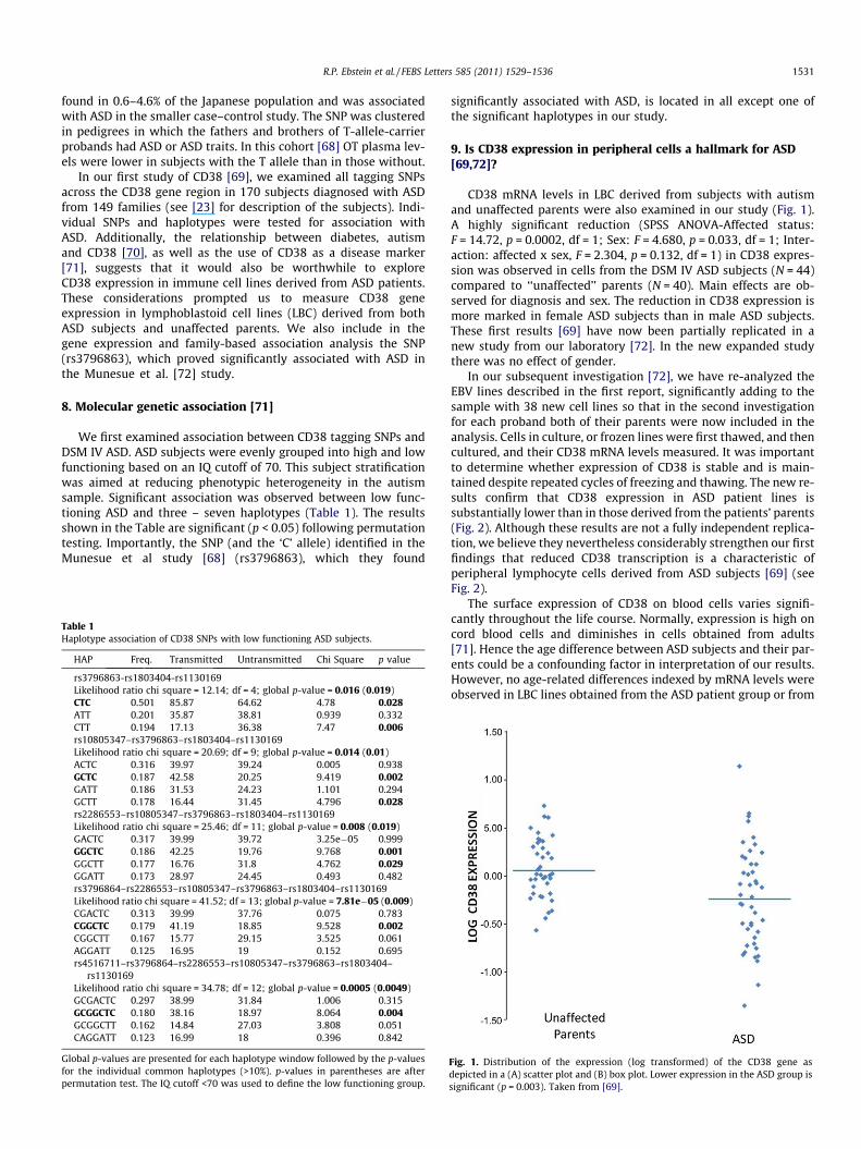
CD38 mRNA levels in LBC derived from subjects with autismand unaffected parents were also examined in our study (Fig. 1).A highly significant reduction (SPSS ANOVA-Affected status:F = 14.72, p = 0.0002, df = 1; Sex: F = 4.680, p = 0.033, df = 1; Inter-action: affected x sex, F = 2.304, p = 0.132, df = 1) in CD38 expres-sion was observed in cells from the DSM IV ASD subjects (N = 44)compared to ‘‘unaffected’’ parents (N = 40). Main effects are ob-served for diagnosis and sex. The reduction in CD38 expression ismore marked in female ASD subjects than in male ASD subjects.These first results [69] have now been partially replicated in anew study from our laboratory [72]. In the new expanded studythere was no effect of gender.
In our subsequent investigation [72], we have re-analyzed theEBV lines described in the first report, significantly adding to thesample with 38 new cell lines so that in the second investigationfor each proband both of their parents were now included in theanalysis. Cells in culture, or frozen lines were first thawed, and thencultured, and their CD38 mRNA levels measured. It was importantto determine whether expression of CD38 is stable and is main-tained despite repeated cycles of freezing and thawing. The new re-sults confirm that CD38 expression in ASD patient lines issubstantially lower than in those derived from the patients’ parents(Fig. 2). Although these results are not a fully independent replica-tion, we believe they nevertheless considerably strengthen our firstfindings that reduced CD38 transcription is a characteristic ofperipheral lymphocyte cells derived from ASD subjects [69] (seeFig. 2).
The surface expression of CD38 on blood cells varies signifi-cantly throughout the life course. Normally, expression is high oncord blood cells and diminishes in cells obtained from adults[71]. Hence the age difference between ASD subjects and their par-ents could be a confounding factor in interpretation of our results.However, no age-related differences indexed by mRNA levels wereobserved in LBC lines obtained from the ASD patient group or from
Fig. 1. Distribution of the expression (log transformed) of the CD38 gene asdepicted in a (A) scatter plot and (B) box plot. Lower expression in the ASD group issignificant (p = 0.003). Taken from [69].
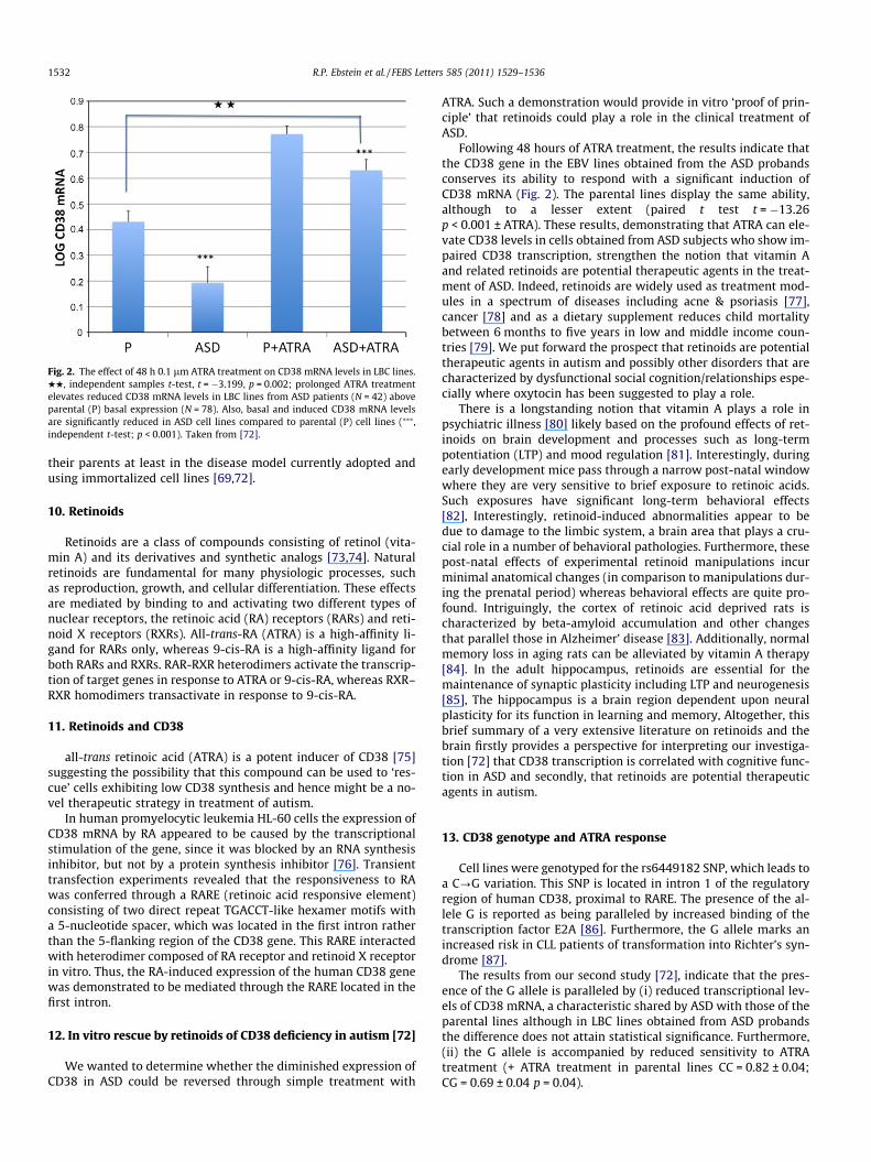
Fig. 2. The effect of 48 h 0.1 lm ATRA treatment on CD38 mRNA levels in LBC lines.HH, independent samples t-test, t = �3.199, p = 0.002; prolonged ATRA treatmentelevates reduced CD38 mRNA levels in LBC lines from ASD patients (N = 42) aboveparental (P) basal expression (N = 78). Also, basal and induced CD38 mRNA levelsare significantly reduced in ASD cell lines compared to parental (P) cell lines (⁄⁄⁄,independent t-test; p < 0.001). Taken from [72].
1532 R.P. Ebstein et al. / FEBS Letters 585 (2011) 1529–1536
their parents at least in the disease model currently adopted andusing immortalized cell lines [69,72].
10. Retinoids
Retinoids are a class of compounds consisting of retinol (vita-min A) and its derivatives and synthetic analogs [73,74]. Naturalretinoids are fundamental for many physiologic processes, suchas reproduction, growth, and cellular differentiation. These effectsare mediated by binding to and activating two different types ofnuclear receptors, the retinoic acid (RA) receptors (RARs) and reti-noid X receptors (RXRs). All-trans-RA (ATRA) is a high-affinity li-gand for RARs only, whereas 9-cis-RA is a high-affinity ligand forboth RARs and RXRs. RAR-RXR heterodimers activate the transcrip-tion of target genes in response to ATRA or 9-cis-RA, whereas RXR–RXR homodimers transactivate in response to 9-cis-RA.
11. Retinoids and CD38
all-trans retinoic acid (ATRA) is a potent inducer of CD38 [75]suggesting the possibility that this compound can be used to ‘res-cue’ cells exhibiting low CD38 synthesis and hence might be a no-vel therapeutic strategy in treatment of autism.
In human promyelocytic leukemia HL-60 cells the expression ofCD38 mRNA by RA appeared to be caused by the transcriptionalstimulation of the gene, since it was blocked by an RNA synthesisinhibitor, but not by a protein synthesis inhibitor [76]. Transienttransfection experiments revealed that the responsiveness to RAwas conferred through a RARE (retinoic acid responsive element)consisting of two direct repeat TGACCT-like hexamer motifs witha 5-nucleotide spacer, which was located in the first intron ratherthan the 5-flanking region of the CD38 gene. This RARE interactedwith heterodimer composed of RA receptor and retinoid X receptorin vitro. Thus, the RA-induced expression of the human CD38 genewas demonstrated to be mediated through the RARE located in thefirst intron.
12. In vitro rescue by retinoids of CD38 deficiency in autism [72]
We wanted to determine whether the diminished expression ofCD38 in ASD could be reversed through simple treatment with
ATRA. Such a demonstration would provide in vitro ‘proof of prin-ciple’ that retinoids could play a role in the clinical treatment ofASD.
Following 48 hours of ATRA treatment, the results indicate thatthe CD38 gene in the EBV lines obtained from the ASD probandsconserves its ability to respond with a significant induction ofCD38 mRNA (Fig. 2). The parental lines display the same ability,although to a lesser extent (paired t test t = �13.26p < 0.001 ± ATRA). These results, demonstrating that ATRA can ele-vate CD38 levels in cells obtained from ASD subjects who show im-paired CD38 transcription, strengthen the notion that vitamin Aand related retinoids are potential therapeutic agents in the treat-ment of ASD. Indeed, retinoids are widely used as treatment mod-ules in a spectrum of diseases including acne & psoriasis [77],cancer [78] and as a dietary supplement reduces child mortalitybetween 6 months to five years in low and middle income coun-tries [79]. We put forward the prospect that retinoids are potentialtherapeutic agents in autism and possibly other disorders that arecharacterized by dysfunctional social cognition/relationships espe-cially where oxytocin has been suggested to play a role.
There is a longstanding notion that vitamin A plays a role inpsychiatric illness [80] likely based on the profound effects of ret-inoids on brain development and processes such as long-termpotentiation (LTP) and mood regulation [81]. Interestingly, duringearly development mice pass through a narrow post-natal windowwhere they are very sensitive to brief exposure to retinoic acids.Such exposures have significant long-term behavioral effects[82], Interestingly, retinoid-induced abnormalities appear to bedue to damage to the limbic system, a brain area that plays a cru-cial role in a number of behavioral pathologies. Furthermore, thesepost-natal effects of experimental retinoid manipulations incurminimal anatomical changes (in comparison to manipulations dur-ing the prenatal period) whereas behavioral effects are quite pro-found. Intriguingly, the cortex of retinoic acid deprived rats ischaracterized by beta-amyloid accumulation and other changesthat parallel those in Alzheimer’ disease [83]. Additionally, normalmemory loss in aging rats can be alleviated by vitamin A therapy[84]. In the adult hippocampus, retinoids are essential for themaintenance of synaptic plasticity including LTP and neurogenesis[85], The hippocampus is a brain region dependent upon neuralplasticity for its function in learning and memory, Altogether, thisbrief summary of a very extensive literature on retinoids and thebrain firstly provides a perspective for interpreting our investiga-tion [72] that CD38 transcription is correlated with cognitive func-tion in ASD and secondly, that retinoids are potential therapeuticagents in autism.
13. CD38 genotype and ATRA response
Cell lines were genotyped for the rs6449182 SNP, which leads toa C?G variation. This SNP is located in intron 1 of the regulatoryregion of human CD38, proximal to RARE. The presence of the al-lele G is reported as being paralleled by increased binding of thetranscription factor E2A [86]. Furthermore, the G allele marks anincreased risk in CLL patients of transformation into Richter’s syn-drome [87].
The results from our second study [72], indicate that the pres-ence of the G allele is paralleled by (i) reduced transcriptional lev-els of CD38 mRNA, a characteristic shared by ASD with those of theparental lines although in LBC lines obtained from ASD probandsthe difference does not attain statistical significance. Furthermore,(ii) the G allele is accompanied by reduced sensitivity to ATRAtreatment (+ ATRA treatment in parental lines CC = 0.82 ± 0.04;CG = 0.69 ± 0.04 p = 0.04).
R.P. Ebstein et al. / FEBS Letters 585 (2011) 1529–1536 1533
14. Link between CD38 expression and clinical characteristics inthe ASD sample
Our results showing that CD38 expression is reduced in ASDprompted us to examine whether its expression levels might alsoreflect phenotypical characteristics of ASD further enhancing thevalue of this ectoenzyme as a potential biomarker. We looked atsocial functioning measures that were available for these probandssince such deficits are a core clinical characteristic of autism. Theresults obtained clearly show a significant correlation betweentranscriptional levels of CD38 mRNA and IQ and Vineland AdaptiveBehavioral Scores (VABS) scores [88], except for VABS socialization.Nonetheless, the correlation with the VABS total scores does provesignificant (r = 0.431, p = 0.008, N = 42).
CD38 mediates oxytocin brain release [59] and importantly,oxytocin itself enhances social learning and memory in the lim-bic system [89]. If retinoids modulate CD38 transcription in thebrain, which in turn mediates oxytocin release, then the relation-ship we have shown [72] between cognitive function and CD38mRNA levels in LBC cells may be reflecting common state char-acteristics of OT-CD38-RA pathways in different tissues. Indeed,various studies have employed LBC lines to model brain dysfunc-tions in autism [90–92] and other neuropsychiatric disorders[93–97].
Finally, it needs to be noted that the potential use of retinoids inany therapeutic intervention must be tempered with the evidencethat too high exposure to retinoids is as harmful as too little.
15. Biomarkers for autism spectrum disorders
Despite the considerable part that heredity plays in the etiologyof ASD the identification of which specific genes, and how manygenes, contribute to ASD remains a challenge. For example, a recentGWAS study [98] genotyped 1558 families and only a single markerattained genome-wide significance. Exploratory analysis of pheno-type subtypes yielded some promising candidate genes, which didnot survive correction for multiple testing. To date, only rare de novomutations are validated genetic risk factors for ASD and most otherfindings from GWAS and association studies need to be consideredprovisional. However, such rare variants, although they might offerimportant clues to the underlying pathophysiology and brain bio-chemistry in autism, only account for a small proportion of the totalgenetic risk. Some chromosomal rearrangements appear causal,with the most common being maternal duplicationof 15q11–q13[99]. Rare de novo mutations of high penetrancefor ASD have beenidentified in synaptic genes, including NLGN3, NLGN4X and SHANK3[100–102]. Similar to other neuropsychiatric disorders copy numbervariations (CNV) also contribute overall risk to ASD [103] includingrare deletion CNVs of SHANK3 and the surrounding 22q13.33 regionfound in individuals with ASD [103].
The current observation that CD38 expression is reduced inlymphoblastoid cells derived from ASD subjects suggests theprospect that CD38 might be an early hallmark for this disorder.As noted by Yirmiya and Charman [104] ‘‘the primary motivationfor identifying the earliest signs of emerging ASDs is the desire todevelop and test early or even ‘preventative’ interventions tolessen morbidity by changing the course of early emergingdevelopmental perturbation, thus preventing ‘secondary’ neuro-developmental disturbances.’’ Towards evaluating the potentialof CD38 as a hallmark in ASD, reduced CD38 expression needsfirstly to be verified in circulating lymphocytes. Secondly, it mustbe stressed that CD38 transcription is a marker for other diseases.It is prognostic for HIV infected subjects [105], chronic lymphoidleukemia [106] and for diabetic patients with nephropathy [107].Hence reduced CD38 transcription cannot be pathognomonic for
ASD but nevertheless might prove of salient clinical value in adisorder diagnosed solely using behavioral assessments reliablycarried out only at the age of three [104]. Moreover, CD38 expres-sion changes throughout the lifespan [71] and age-dependentCD38 expression in circulating lymphocytes are potential con-founds in its use as a diagnostic indicator in ASD. However, thecritical need in ASD is for very early (prenatal or perinatal) diag-nostic tools and, hence from this perspective, CD38 mRNA levelsin cord blood or amniotic fluid might be of substantial value not-withstanding subsequent age-related changes in lymphocyteCD38 expression.
One group of biomarkers for neuropsychiatric disorders thatshows considerable promise is blood gene expression profiling[108–113]. Most of the studies to date have focused on humanlymphocytes gene expression profiling, comparison between ill-ness groups and normal controls, and cross-matching with humanpostmortem brain gene expression data. A number of studies havespecifically examined gene expression patterns in ASD [90–92,114–123]. It should be noted that predating this gush of expres-sion studies in ASD, hyperserotonemia was observed in one third ofpatients and platelet serotonin was suggested as a marker for thisdisorder [124–127].
To summarize, the difficulty to validate specific gene variantsfor most cases of autism likely reflects a variety of causes includ-ing gene x gene interactions, the heterogeneous nature of thisdisorder, and epigenetic modifications due to diverse environ-mental challenges. The challenge of finding specific genes con-tributing to ASD, except in those rare cases and their familiesshowing Mendelian inheritance, suggests that complementarystrategies to supplement more standard molecular genetic asso-ciation studies would be worthwhile. Such an approach has beenproposed by Le-Niculescu and his colleagues [128]. Their ap-proach, termed convergent functional genomics (CFG), whichtranslationally cross-matches animal model gene expression datawith human genetic data and human tissue data (blood, post-mortem brain), as a Bayesian strategy of cross-validating find-ings, reducing the false positives and false negatives inherentin each individual approach, and helping identify true candidategenes, pathways and mechanisms for neuropsychiatric disorders.
16. Conclusion
The current study adds to the growing list of potential biomark-ers in ASD and moreover, uniquely observes a correlation betweenexpression levels of CD38 in LBC derived from these subjects andsocial and communication skills that are core deficits in this disor-der. Notably, the potential of CD38 expression as a diagnostic indi-cator for ASD was a hypothesis driven idea catalyzed by theseminal study of Higashida and his colleagues [59] in the CD38knockout mouse and reinforced by two independent molecular ge-netic studies showing association between SNPs in the CD38 geneand ASD [68,69].
In addition to the potential of CD38 as a hallmark that mayprove useful in early diagnosis of illness, the study of CD38 expres-sion in peripheral lymphocytes has allowed us to model the poten-tial of retinoic acids as a therapeutic agent in ASD. Indeed, we haveshown that LBC derived from ASD subjects and characterized by re-duced CD38 transcription can be ‘rescued’ by simple treatmentwith all-trans retinoic acid. We believe these results provide thefirst ‘proof of principle’ for a novel therapeutic strategy in treat-ment of ASD by enhancing OT secretion in the brain indirectly byATRA induction of CD38 followed by mobilization of ryanodine-sensitive intracellular Ca2+ stores from the endoplasmic reticulumwhich in turn release OT.
1534 R.P. Ebstein et al. / FEBS Letters 585 (2011) 1529–1536
17. Disclosure
We declare that the authors have no competing interests asdefined by Molecular Medicine, or other interests that might beperceived to influence the results and discussion reported in thispaper.
Acknowledgements
We thank Autism Speaks for partial support of this research(R.P.E.) as well as support (F.M.) by AIRC (Special ProgramMolecular and Clinical Oncology 5x1000) and by the FondazioneInternazionale Ricerca Medicina Sperimentale (FIRMS).
References
[1] Gimpl, G. and Fahrenholz, F. (2001) The oxytocin receptor system: structure,function, and regulation. Physiol. Rev. 81, 629–683.
[2] Goodson, J.L. (2008) Nonapeptides and the evolutionary patterning ofsociality. Prog. Brain Res. 170, 3–15.
[3] Insel, T.R. (2010) The challenge of translation in social neuroscience: a reviewof oxytocin, vasopressin, and affiliative behavior. Neuron 65,768–779.
[4] Heinrichs, M., von Dawans, B. and Domes, G. (2009) Oxytocin, vasopressin,and human social behavior. Front. Neuroendocrinol. 30, 548–557.
[5] Donaldson, Z.R. and Young, L.J. (2008) Oxytocin, vasopressin, and theneurogenetics of sociality. Science 322, 900–904.
[6] Lee, H.J., Macbeth, A.H., Pagani, J.H. and Young 3rd., W.S. (2009) Oxytocin: thegreat facilitator of life. Prog. Neurobiol. 88, 127–151.
[7] Kosfeld, M., Heinrichs, M., Zak, P.J., Fischbacher, U. and Fehr, E. (2005)Oxytocin increases trust in humans. Nature 435, 673–676.
[8] Domes, G., Heinrichs, M., Michel, A., Berger, C. and Herpertz, S.C. (2007)Oxytocin improves ‘‘mind-reading’’ in humans. Biol. Psychiatry 61, 731–733.
[9] Savaskan, E., Ehrhardt, R., Schulz, A., Walter, M. and Schachinger, H. (2008)Post-learning intranasal oxytocin modulates human memory for facialidentity. Psychoneuroendocrinology.
[10] Ditzen, B., Schaer, M., Gabriel, B., Bodenmann, G., Ehlert, U. and Heinrichs, M.(2008) Intranasal oxytocin increases positive communication and reducescortisol levels during couple conflict. Biol. Psychiatry 1.
[11] Guastella, A.J., Mitchell, P.B. and Dadds, M.R. (2007) Oxytocin increases gazeto the eye region of human faces. Biol. Psychiatry.
[12] Zak, P.J., Stanton, A.A. and Ahmadi, S. (2007) Oxytocin increases generosity inhumans. PLoS ONE 2, e1128.
[13] Levine, A., Zagoory-Sharon, O., Feldman, R. and Weller, A. (2007) Oxytocinduring pregnancy and early postpartum: individual patterns and maternal-fetal attachment. Peptides 28, 1162–1169.
[14] Gordon, I., Zagoory-Sharon, O., Schneiderman, I., Leckman, J.F., Weller, A. andFeldman, R. (2008) Oxytocin and cortisol in romantically unattached youngadults: associations with bonding and psychological distress.Psychophysiology.
[15] Hoge, E.A., Pollack, M.H., Kaufman, R.E., Zak, P.J. and Simon, N.M. (2008)Oxytocin levels in social anxiety disorder. CNS Neurosci. Ther. 14, 165–170.
[16] Baumgartner, T., Heinrichs, M., Vonlanthen, A., Fischbacher, U. and Fehr, E.(2008) Oxytocin shapes the neural circuitry of trust and trust adaptation inhumans. Neuron 58, 639–650.
[17] Blanks, A.M., Shmygol, A. and Thornton, S. (2007) Regulation of oxytocinreceptors and oxytocin receptor signaling. Semin. Reprod. Med. 25, 52–59.
[18] Rutter, M. (1989) Child psychiatric disorders in ICD 10. J. Child Psychol.Psychiatry 30, 499–513.
[19] Chakrabarti, S. and Fombonne, E. (2005) Pervasive developmental disordersin preschool children: confirmation of high prevalence. Am. J. Psychiatry 162,1133–1141.
[20] Freitag, C.M., Staal, W., Klauck, S.M., Duketis, E. and Waltes, R. (2010)Genetics of autistic disorders: review and clinical implications. Eur. ChildAdolesc. Psychiatry 19, 169–178.
[21] Jacob, S., Brune, C.W., Carter, C.S., Leventhal, B.L., Lord, C. and Cook Jr., E.H.(2007) Association of the oxytocin receptor gene (OXTR) in Caucasianchildren and adolescents with autism. Neurosci. Lett. 417, 6–9.
[22] Yrigollen, C.M., Han, S.S., Kochetkova, A., Babitz, T., Chang, J.T., Volkmar, F.R.,Leckman, J.F. and Grigorenko, E.L. (2008) Genes controlling affiliativebehavior as candidate genes for autism. Biol. Psychiatry 63, 911–916.
[23] Lerer, E., Levi, S., Salomon, S., Darvasi, A., Yirmiya, N. and Ebstein, R.P. (2008)Association between the oxytocin receptor (OXTR) gene and autism:relationship to Vineland Adaptive Behavior Scales and cognition. Mol.Psychiatry 13, 980–988.
[24] Wu, S. et al. (2005) Positive association of the oxytocin receptor gene (OXTR)with autism in the Chinese Han population. Biol. Psychiatry 58, 74–77.
[25] Liu, X. et al. (2010) J. Hum. Genet.
[26] Wermter, A.K., Kamp-Becker, I., Hesse, P., Schulte-Korne, G., Strauch, K. andRemschmidt, H. (2010) Evidence for the involvement of genetic variation in theoxytocin receptor gene (OXTR) in the etiology of autistic disorders on high-functioning level. Am. J. Med. Genet. B Neuropsychiatr. Genet. 153B, 629–639.
[27] Apicella, C.L., Cesarini, D., Johannesson, M., Dawes, C.T., Lichtenstein, P.,Wallace, B., Beauchamp, J. and Westberg, L. (2010) No association betweenoxytocin receptor (OXTR) gene polymorphisms and experimentally elicitedsocial preferences. PLoS ONE 5, e11153.
[28] Kelemenova, S., Schmidtova, E., Ficek, A., Celec, P., Kubranska, A. andOstatnikova, D. (2010) Polymorphisms of candidate genes in Slovak autisticpatients. Psychiatr. Genet. 20, 137–139.
[29] Thompson, R.J., Parker, K.J., Hallmayer, J.F., Waugh, C.E. and Gotlib, I.H. (2011)Oxytocin receptor gene polymorphism (rs2254298) interacts with familialrisk for psychopathology to predict symptoms of depression and anxiety inadolescent girls. Psychoneuroendocrinology 36, 144–147.
[30] Montag, C., Fiebach, C.J., Kirsch, P. and Reuter, M. (2010) Interaction of 5-HTTLPR and a variation on the oxytocin receptor gene influences negativeemotionality. Biol. Psychiatry.
[31] Park, J. et al. (2010) Evidence that genetic variation in the oxytocin receptor(OXTR) gene influences social cognition in ADHD. Prog. Neuropsychopharmacol.Biol. Psychiatry.
[32] Costa, B. et al. (2009) Oxytocin receptor polymorphisms and adultattachment style in patients with depression. Psychoneuroendocrinology.
[33] Costa, B. et al. (2009) Mutation analysis of oxytocin gene in individuals withadult separation anxiety. Psychiatry Res. 168, 87–93.
[34] Gillath, O., Shaver, P.R., Baek, J.M. and Chun, D.S. (2008) Genetic correlates ofadult attachment style. Pers. Soc. Psychol Bull.
[35] Lucht, M.J. et al. (2009) Associations between the oxytocin receptor gene(OXTR) and affect, loneliness and intelligence in normal subjects. Prog.Neuropsychopharmacol. Biol. Psychiatry 33, 860–866.
[36] Rodrigues, S.M., Saslow, L.R., Garcia, N., John, O.P. and Keltner, D. (2009)Oxytocin receptor genetic variation relates to empathy and stress reactivityin humans. Proc. Natl. Acad. Sci. USA.
[37] Bakermans-Kranenburg, M.J. and van Ijzendoorn, M.H. (2008) Oxytocinreceptor (OXTR) and serotonin transporter (5-HTT) genes associated withobserved parenting. Soc. Cogn. Affect Neurosci. 3, 128–134.
[38] Prichard, Z.M., Mackinnon, A.J., Jorm, A.F. and Easteal, S. (2007) AVPR1A andOXTR polymorphisms are associated with sexual and reproductivebehavioral phenotypes in humans Mutation in brief No. 981. Online. Hum.Mutat. 28, 1150.
[39] Israel, S. et al. (2009) The oxytocin receptor (OXTR) contributes to prosocialfund allocations in the dictator game and the social value orientations task.PLoS ONE 4, e5535.
[40] Kim, S.J. et al. (2002) Transmission disequilibrium testing of argininevasopressin receptor 1A (AVPR1A) polymorphisms in autism. Mol.Psychiatry 7, 503–507.
[41] Wassink, T.H., Piven, J., Vieland, V.J., Pietila, J., Goedken, R.J., Folstein, S.E. andSheffield, V.C. (2004) Examination of AVPR1a as an autism susceptibilitygene. Mol. Psychiatry 9, 968–972.
[42] Yirmiya, N., Rosenberg, C., Levi, S., Salomon, S., Shulman, C., Nemanov, L.,Dina, C. and Ebstein, R.P. (2006) Association between the argininevasopressin 1a receptor (AVPR1a) gene and autism in a family-basedstudy: mediation by socialization skills. Mol. Psychiatry 11, 488–494.
[43] McCauley, J.L. et al. (2005) Genome-wide and ordered-subset linkageanalyses provide support for autism loci on 17q and 19p with evidence ofphenotypic and interlocus genetic correlates. BMC Med. Genet. 6, 1.
[44] Lauritsen, M.B. et al. (2006) A genome-wide search for alleles and haplotypesassociated with autism and related pervasive developmental disorders on theFaroe Islands. Mol. Psychiatry 11, 37–46.
[45] Hollander, E., Bartz, J., Chaplin, W., Phillips, A., Sumner, J., Soorya, L.,Anagnostou, E. and Wasserman, S. (2007) Oxytocin increases retention ofsocial cognition in autism. Biol. Psychiatry 61, 498–503.
[46] Andari, E., Duhamel, J.R., Zalla, T., Herbrecht, E., Leboyer, M. and Sirigu, A.(2010) Promoting social behavior with oxytocin in high-functioning autismspectrum disorders. Proc. Natl. Acad. Sci. USA 107, 4389–4394.
[47] Guastella, A.J., Einfeld, S.L., Gray, K.M., Rinehart, N.J., Tonge, B.J., Lambert, T.J.and Hickie, I.B. (2010) Intranasal oxytocin improves emotion recognition foryouth with autism spectrum disorders. Biol. Psychiatry 67, 692–694.
[48] Guastella, A.J., Einfeld, S.L., Gray, K.M., Rinehart, N.J., Tonge, B.J., Lambert, T.J.and Hickie, I.B. (2009) Intranasal oxytocin improves emotion recognition foryouth with autism spectrum disorders. Biol. Psychiatry.
[49] Hollander, E., Novotny, S., Hanratty, M., Yaffe, R., DeCaria, C.M., Aronowitz,B.R. and Mosovich, S. (2003) Oxytocin infusion reduces repetitive behaviorsin adults with autistic and Asperger’s disorders. Neuropsychopharmacology28, 193–198.
[50] Feifel, D. et al. (2010) Adjunctive intranasal oxytocin reduces symptoms inschizophrenia patients. Biol. Psychiatry 68, 678–680.
[51] Guastella, A.J., Howard, A.L., Dadds, M.R., Mitchell, P. and Carson, D.S. (2009)A randomized controlled trial of intranasal oxytocin as an adjunct toexposure therapy for social anxiety disorder. Psychoneuroendocrinology.
[52] Kuehn, B.M. (2011) Scientists probe oxytocin therapy for social deficits inautism, schizophrenia. JAMA 305, 659–661.
[53] (2010). Extending trust. A report of a potential therapy for some of the socialbehavior patterns associated with autism highlights the need for a societaldialog to discuss the ethical issues raised by these treatments. Nat. Neurosci.13, 905.
R.P. Ebstein et al. / FEBS Letters 585 (2011) 1529–1536 1535
[54] Modahl, C., Green, L., Fein, D., Morris, M., Waterhouse, L., Feinstein, C. andLevin, H. (1998) Plasma oxytocin levels in autistic children. Biol. Psychiatry43, 270–277.
[55] Ludwig, M. and Leng, G. (2006) Dendritic peptide release and peptide-dependent behaviours. Nat. Rev. Neurosci. 7, 126–136.
[56] Leng, G., Brown, C.H. and Russell, J.A. (1999) Physiological pathwaysregulating the activity of magnocellular neurosecretory cells. Prog.Neurobiol. 57, 625–655.
[57] Moos, F., Poulain, D.A., Rodriguez, F., Guerne, Y., Vincent, J.D. and Richard, P.(1989) Release of oxytocin within the supraoptic nucleus during the milkejection reflex in rats. Exp. Brain Res. 76, 593–602.
[58] Lambert, R.C., Dayanithi, G., Moos, F.C. and Richard, P. (1994) A rise in theintracellular Ca2+ concentration of isolated rat supraoptic cells in response tooxytocin. J. Physiol. 478 (Pt 2), 275–287.
[59] Jin, D. et al. (2007) CD38 is critical for social behaviour by regulating oxytocinsecretion. Nature 446, 41–45.
[60] Higashida, H., Hashii, M., Yokoyama, S., Hoshi, N., Chen, X.L., Egorova, A.,Noda, M. and Zhang, J.S. (2001) Cyclic ADP-ribose as a second messengerrevisited from a new aspect of signal transduction from receptors to ADP-ribosyl cyclase. Pharmacol. Ther. 90, 283–296.
[61] Salmina, A.B., Lopatina, O., Ekimova, M.V., Mikhutkina, S.V. and Higashida, H.(2010) CD38/cADPR-SYSTEM: a new player for oxytocin secretion andregulation of social behaviour. J. Neuroendocrinol.
[62] Ceni, C. et al. (2006) The CD38-independent ADP-ribosyl cyclase from mousebrain synaptosomes: a comparative study of neonate and adult brain.Biochem. J. 395, 417–426.
[63] Mizuguchi, M. et al. (1995) Neuronal localization of CD38 antigen in thehuman brain. Brain Res. 697, 235–240.
[64] Ebstein, R.P. et al. (2009) Arginine vasopressin and oxytocin modulate humansocial behavior. Ann. N. Y. Acad. Sci. 1167, 87–102.
[65] Israel, S. et al. (2008) Molecular genetic studies of the arginine vasopressin 1areceptor (AVPR1a) and the oxytocin receptor (OXTR) in human behaviour:from autism to altruism with some notes in between. Prog. Brain Res. 170,435–449.
[66] Bartz, J.A. and McInnes, L.A. (2007) CD38 regulates oxytocin secretion andcomplex social behavior. BioEssays 29, 837–841.
[67] Young, L.J. (2007) Regulating the social brain: a new role for CD38. Neuron54, 353–356.
[68] Munesue, T. et al. (2010) Two genetic variants of CD38 in subjects withautism spectrum disorder and controls. Neurosci. Res. 67, 181–191.
[69] Lerer, E., Levi, S., Israel, S., Yaari, M., Nemanov, L., Mankuta, D., Nurit, Y. andEbstein, R.P. (2010) Low CD38 expression in lymphoblastoid cells andhaplotypes are both associated with autism in a family-based study. AutismRes. 3, 293–302.
[70] Atladottir, H.O., Pedersen, M.G., Thorsen, P., Mortensen, P.B., Deleuran, B., Eaton,W.W. and Parner, E.T. (2009) Association of family history of autoimmunediseases and autism spectrum disorders. Pediatrics 124, 687–694.
[71] Malavasi, F., Deaglio, S., Funaro, A., Ferrero, E., Horenstein, A.L., Ortolan, E.,Vaisitti, T. and Aydin, S. (2008) Evolution and function of the ADP ribosylcyclase/CD38 gene family in physiology and pathology. Physiol. Rev. 88, 841–886.
[72] Riebold, M. et al. (in press). All-trans-retinoic-acid (ATRA) upregulatesreduced CD38 transcription in lymphoblastoid cell lines from autismspectrum disorder. Mol. Med.
[73] Alvarez, S., Bourguet, W., Gronemeyer, H. and de Lera, A.R. (2011) Retinoicacid receptor modulators: a perspective on recent advances and promises.Expert Opin. Ther. Pat. 21, 55–63.
[74] Theodosiou, M., Laudet, V. and Schubert, M. (2010) From carrot to clinic: anoverview of the retinoic acid signaling pathway. Cell. Mol. Life Sci. 67, 1423–1445.
[75] Ferrero, E. and Malavasi, F. (2002) A Natural History of the Human CD38Gene, Kluwer Academic Publishers. pp. 81–99.
[76] Kishimoto, H., Hoshino, S., Ohori, M., Kontani, K., Nishina, H., Suzawa, M.,Kato, S. and Katada, T. (1998) Molecular mechanism of human CD38 geneexpression by retinoic acid. Identification of retinoic acid response element inthe first intron. J. Biol. Chem. 273, 15429–15434.
[77] Trapasso, E., Cosco, D., Celia, C., Fresta, M. and Paolino, D. (2009) Retinoids:new use by innovative drug-delivery systems. Expert Opin. Drug Deliv. 6,465–483.
[78] Siddikuzzaman, Guruvayoorappan, C. and Berlin Grace, V.M. (2010). All TransRetinoic Acid and Cancer. Immunopharmacol. Immunotoxicol.
[79] Imdad, A., Herzer, K., Mayo-Wilson, E., Yakoob, M.Y. and Bhutta, Z.A. (2010).Vitamin A supplementation for preventing morbidity and mortality inchildren from 6 months to 5 years of age. Cochrane Database Syst. Rev. 12,CD008524.
[80] Luo, T., Wagner, E. and Drager, U.C. (2009) Integrating retinoic acid signalingwith brain function. Dev. Psychol. 45, 139–150.
[81] Chiang, M.Y. et al. (1998) An essential role for retinoid receptors RARbeta andRXRgamma in long-term potentiation and depression. Neuron 21, 1353–1361.
[82] Luo, T., Wagner, E., Crandall, J.E. and Drager, U.C. (2004) A retinoic-acidcritical period in the early postnatal mouse brain. Biol. Psychiatry 56, 971–980.
[83] Corcoran, J.P., So, P.L. and Maden, M. (2004) Disruption of the retinoidsignalling pathway causes a deposition of amyloid beta in the adult rat brain.Eur. J. NeuroSci. 20, 896–902.
[84] Etchamendy, N., Enderlin, V., Marighetto, A., Vouimba, R.M., Pallet, V., Jaffard,R. and Higueret, P. (2001) Alleviation of a selective age-related relationalmemory deficit in mice by pharmacologically induced normalization of brainretinoid signaling. J. Neurosci. 21, 6423–6429.
[85] McCaffery, P., Zhang, J. and Crandall, J.E. (2006) Retinoic acid signaling andfunction in the adult hippocampus. J. Neurobiol. 66, 780–791.
[86] Saborit-Villarroya, I., Vaisitti, T., Rossi, D., D’Arena, G., Gaidano, G., Malavasi,F. and Deaglio, S. (2011) E2A is a transcriptional regulator of CD38 expressionin chronic lymphocytic leukemia. Leukemia.
[87] Aydin, S. et al. (2008) CD38 gene polymorphism and chronic lymphocyticleukemia: a role in transformation to Richter syndrome? Blood 111, 5646–5653.
[88] Sparrow, S.S., Balla, D.A. and Cicchetti, D.V. (1984) Vineland AdaptiveBehavior Scales, American Guidance Services, Minneapolis, Minnesota.
[89] Ferguson, J.N., Young, L.J. and Insel, T.R. (2002) The neuroendocrine basis ofsocial recognition. Front. Neuroendocrinol. 23, 200–224.
[90] Hu, V.W. et al. (2009) Gene expression profiling of lymphoblasts from autisticand nonaffected sib pairs: altered pathways in neuronal development andsteroid biosynthesis. PLoS ONE 4, e5775.
[91] Nishimura, Y. et al. (2007) Genome-wide expression profiling oflymphoblastoid cell lines distinguishes different forms of autism andreveals shared pathways. Hum. Mol. Genet. 16, 1682–1698.
[92] Walker, S.J., Segal, J. and Aschner, M. (2006) Cultured lymphocytes fromautistic children and non-autistic siblings up-regulate heat shock proteinRNA in response to thimerosal challenge. Neurotoxicology 27, 685–692.
[93] Chagnon, Y.C., Roy, M.A., Bureau, A., Merette, C. and Maziade, M. (2008)Differential RNA expression between schizophrenic patients and controls ofthe dystrobrevin binding protein 1 and neuregulin 1 genes in immortalizedlymphocytes. Schizophr. Res. 100, 281–290.
[94] De Luca, V., Strauss, J., Semeralul, M., Huang, S., Li, P.P., Warsh, J.J., Kennedy,J.L. and Wong, A.H. (2008) Analysis of BDNF Val66Met allele-specific mRNAlevels in bipolar disorder. Neurosci. Lett. 441, 229–232.
[95] Kuratomi, G., Iwamoto, K., Bundo, M., Kusumi, I., Kato, N., Iwata, N., Ozaki, N.and Kato, T. (2008) Aberrant DNA methylation associated with bipolardisorder identified from discordant monozygotic twins. Mol. Psychiatry 13,429–441.
[96] Pandey, G.N., Rizavi, H.S., Dwivedi, Y. and Pavuluri, M.N. (2008) Brain-derivedneurotrophic factor gene expression in pediatric bipolar disorder: effects oftreatment and clinical response. J. Am. Acad. Child Adolesc. Psychiatry 47,1077–1085.
[97] Tseng, M. et al. (2008) BDNF protein levels are decreased in transformedlymphoblasts from lithium-responsive patients with bipolar disorder. J.Psychiatry Neurosci. 33, 449–453.
[98] Anney, R. et al. (2010) A genome-wide scan for common alleles affecting riskfor autism. Hum. Mol. Genet.
[99] Shao, Y. et al. (2003) Fine mapping of autistic disorder to chromosome15q11–q13 by use of phenotypic subtypes. Am. J. Hum. Genet. 72, 539–548.
[100] Jamain, S. et al. (2003) Mutations of the X-linked genes encoding neuroliginsNLGN3 and NLGN4 are associated with autism. Nat. Genet. 34, 27–29.
[101] Jamain, S. et al. (2002) Linkage and association of the glutamate receptor 6gene with autism. Mol. Psychiatry 7, 302–310.
[102] Durand, C.M. et al. (2007) Mutations in the gene encoding the synapticscaffolding protein SHANK3 are associated with autism spectrum disorders.Nat. Genet. 39, 25–27.
[103] Sebat, J. et al. (2007) Strong association of de novo copy number mutationswith autism. Science 316, 445–449.
[104] Yirmiya, N. and Charman, T. (2010) The prodrome of autism: early behavioraland biological signs, regression, peri- and post-natal development andgenetics. J. Child Psychol. Psychiatry 51, 432–458.
[105] Liu, Z., Cumberland, W.G., Hultin, L.E., Prince, H.E., Detels, R. and Giorgi, J.V.(1997) Elevated CD38 antigen expression on CD8+ T cells is a strongermarker for the risk of chronic HIV disease progression to AIDS and death inthe Multicenter AIDS Cohort Study than CD4+ cell count, soluble immuneactivation markers, or combinations of HLA-DR and CD38 expression. J.Acquir. Immune Defic. Syndr. Hum. Retrovirol. 16, 83–92.
[106] Deaglio, S., Aydin, S., Vaisitti, T., Bergui, L. and Malavasi, F. (2008) CD38 at thejunction between prognostic marker and therapeutic target. Trends Mol.Med. 14, 210–218.
[107] Ohtsuji, M. et al. (2008) Decreased ADP-ribosyl cyclase activity in peripheralblood mononuclear cells from diabetic patients with nephropathy. Exp.Diabetes Res. 2008, 897508.
[108] Vawter, M.P., Ferran, E., Galke, B., Cooper, K., Bunney, W.E. and Byerley, W.(2004) Microarray screening of lymphocyte gene expression differences in amultiplex schizophrenia pedigree. Schizophr. Res. 67, 41–52.
[109] Tsuang, M.T., Nossova, N., Yager, T., Tsuang, M.M., Guo, S.C., Shyu, K.G., Glatt,S.J. and Liew, C.C. (2005) Assessing the validity of blood-based gene expressionprofiles for the classification of schizophrenia and bipolar disorder: apreliminary report. Am. J. Med. Genet. B Neuropsychiatr. Genet. 133B, 1–5.
[110] Segman, R.H., Shefi, N., Goltser-Dubner, T., Friedman, N., Kaminski, N. andShalev, A.Y. (2005) Peripheral blood mononuclear cell gene expressionprofiles identify emergent post-traumatic stress disorder among traumasurvivors. Mol. Psychiatry 10 (500–513), 425.
[111] Middleton, F.A. et al. (2005) Gene expression analysis of peripheral bloodleukocytes from discordant sib-pairs with schizophrenia and bipolar disorderreveals points of convergence between genetic and functional genomicapproaches. Am. J. Med. Genet. B Neuropsychiatr. Genet. 136B, 12–25.
1536 R.P. Ebstein et al. / FEBS Letters 585 (2011) 1529–1536
[112] Glatt, S.J. et al. (2005) Comparative gene expression analysis of blood andbrain provides concurrent validation of SELENBP1 up-regulation inschizophrenia. Proc. Natl Acad. Sci. USA 102, 15533–15538.
[113] Sullivan, P.F., Fan, C. and Perou, C.M. (2006) Evaluating the comparability ofgene expression in blood and brain. Am. J. Med. Genet. B Neuropsychiatr.Genet. 141B, 261–268.
[114] Alter, M.D. et al. (2011) Autism and increased paternal age related changes inglobal levels of gene expression regulation. PLoS ONE 6, e16715.
[115] Nguyen, A., Rauch, T.A., Pfeifer, G.P. and Hu, V.W. (2010) Global methylationprofiling of lymphoblastoid cell lines reveals epigenetic contributions toautism spectrum disorders and a novel autism candidate gene, RORA, whoseprotein product is reduced in autistic brain. FASEB J. 24, 3036–3051.
[116] Saresella, M. et al. (2009) An autistic endophenotype results in compleximmune dysfunction in healthy siblings of autistic children. Biol. Psychiatry66, 978–984.
[117] Nakamura, K. et al. (2008) Genetic and expression analyses reveal elevatedexpression of syntaxin 1A (STX1A) in high functioning autism. Int. J.Neuropsychopharmacol. 11, 1073–1084.
[118] Nishimura, K. et al. (2007) Genetic analyses of the brain-derivedneurotrophic factor (BDNF) gene in autism. Biochem. Biophys. Res.Commun. 356, 200–206.
[119] Baron, C.A., Liu, S.Y., Hicks, C. and Gregg, J.P. (2006) Utilization oflymphoblastoid cell lines as a system for the molecular modeling ofautism. J. Autism Dev. Disord. 36, 973–982.
[120] Hu, V.W., Frank, B.C., Heine, S., Lee, N.H. and Quackenbush, J. (2006) Geneexpression profiling of lymphoblastoid cell lines from monozygotic twins
discordant in severity of autism reveals differential regulation ofneurologically relevant genes. BMC Genomics 7, 118.
[121] Melin, M., Carlsson, B., Anckarsater, H., Rastam, M., Betancur, C., Isaksson, A.,Gillberg, C. and Dahl, N. (2006) Constitutional downregulation of SEMA5Aexpression in autism. Neuropsychobiology 54, 64–69.
[122] Huang, C.H., Chen, M.L., Tsai, Y.L., Tsai, M.T. and Chen, C.H. (2004) Elevatedadrenomedullin mRNA in lymphoblastoid cells from schizophrenic patients.Neuroreport 15, 1443–1446.
[123] Hollander, E. et al. (1999) B lymphocyte antigen D8/17 and repetitivebehaviors in autism. Am. J. Psychiatry 156, 317–320.
[124] Boullin, D.J., Coleman, M. and O’Brien, R.A. (1970) Abnormalities in platelet 5-hydroxytryptamine efflux in patients with infantile autism. Nature 226, 371–372.
[125] Piven, J., Tsai, G.C., Nehme, E., Coyle, J.T., Chase, G.A. and Folstein, S.E. (1991)Platelet serotonin, a possible marker for familial autism. J. Autism Dev.Disord. 21, 51–59.
[126] Hranilovic, D., Bujas-Petkovic, Z., Tomicic, M., Bordukalo-Niksic, T., Blazevic,S. and Cicin-Sain, L. (2009) Hyperserotonemia in autism: activity of 5HT-associated platelet proteins. J. Neural. Transm.
[127] Hranilovic, D., Novak, R., Babic, M., Novokmet, M., Bujas-Petkovic, Z. andJernej, B. (2008) Hyperserotonemia in autism: the potential role of 5HT-related gene variants. Coll. Antropol. 32 (Suppl. 1), 75–80.
[128] Le-Niculescu, H. et al. (2009) Identifying blood biomarkers for mooddisorders using convergent functional genomics. Mol. Psychiatry 14, 156–174.
Related Documents