Approccio al bambino con TRAUMA CRANICO MINORE Prof.ssa Liviana Da Dalt Dipartimento Materno-Infantile Azienda ULSS 9 - Treviso Università di Padova Roma 9 novembre 2011

Welcome message from author
This document is posted to help you gain knowledge. Please leave a comment to let me know what you think about it! Share it to your friends and learn new things together.
Transcript

Approccio al bambino con TRAUMA CRANICO
MINORE
Prof.ssa Liviana Da Dalt Dipartimento Materno-Infantile
Azienda ULSS 9 - Treviso
Università di Padova
Roma 9 novembre 2011
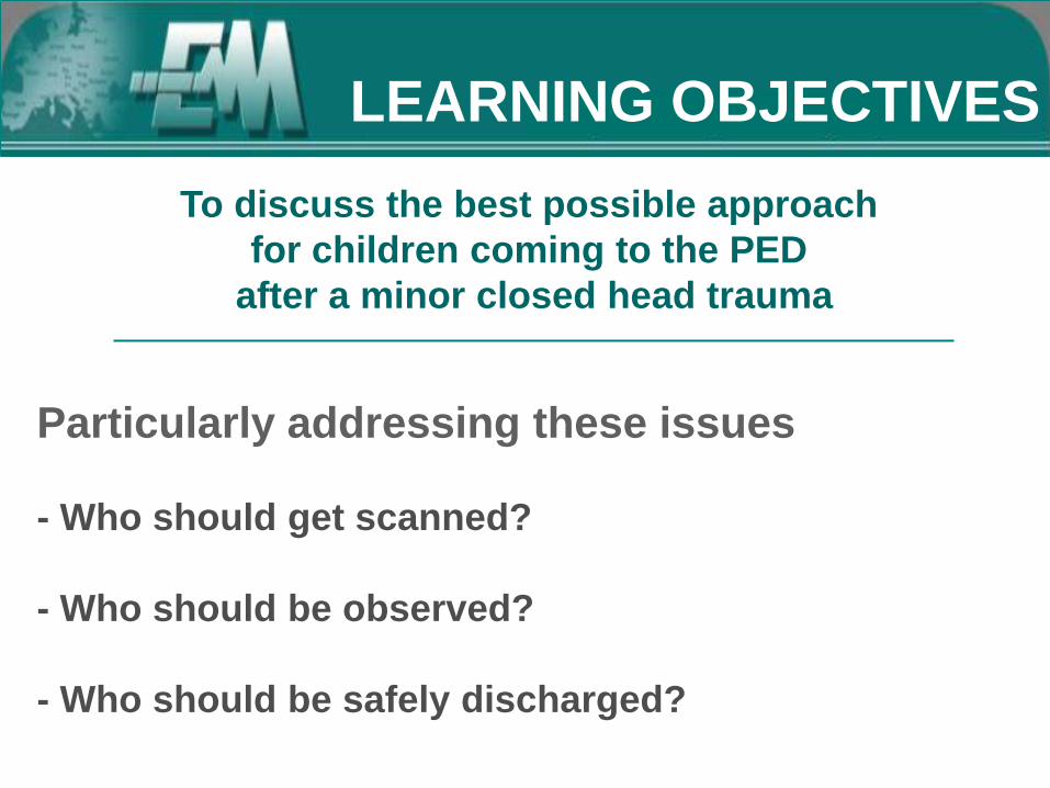
Particularly addressing these issues - Who should get scanned? - Who should be observed? - Who should be safely discharged?
LEARNING OBJECTIVES To discuss the best possible approach
for children coming to the PED after a minor closed head trauma
Head trauma is one of the most common reasons for acute visit in PED
BACKGROUND

Head trauma is one of the most common reasons for acute visit in PED
Traumatic brain injuries are the leading cause of death
and disability in children, accounting for about 30% of deaths in the paediatric population
BACKGROUND
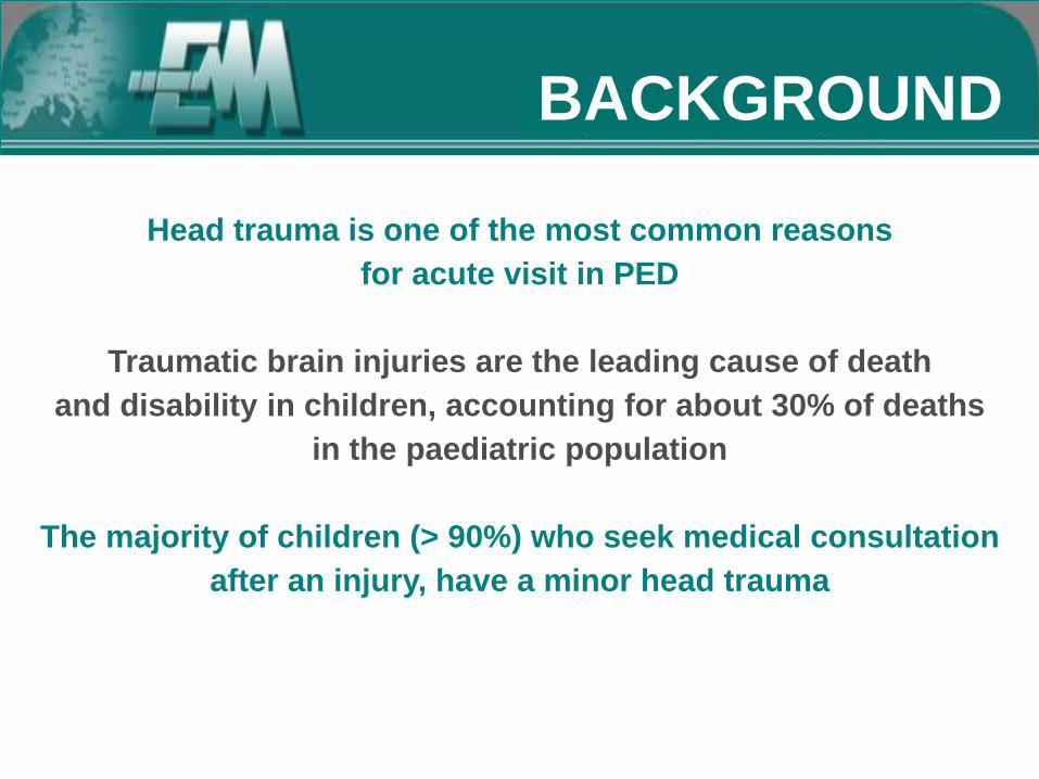
Head trauma is one of the most common reasons for acute visit in PED
Traumatic brain injuries are the leading cause of death
and disability in children, accounting for about 30% of deaths in the paediatric population
The majority of children (> 90%) who seek medical consultation
after an injury, have a minor head trauma
BACKGROUND
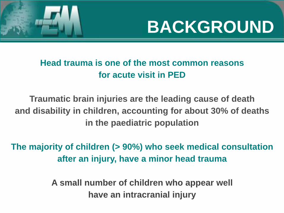
Head trauma is one of the most common reasons for acute visit in PED
Traumatic brain injuries are the leading cause of death
and disability in children, accounting for about 30% of deaths in the paediatric population
The majority of children (> 90%) who seek medical consultation
after an injury, have a minor head trauma
A small number of children who appear well have an intracranial injury
BACKGROUND

BACKGROUND
Holmes JF, Acad Emerg Med, 2005
Minor head trauma: definition
When reviewing the literature it should be noted that there is no standard definition for Minor Head Trauma. This definition has been based mostly on the Glasgow Coma Scale, usually 14-15 (according to some Authors > 13)


normal mental status at the initial examination (within 24 hours of the trauma)
no abnormal findings on neurologic examination
no physical evidence of complicate skull fracture (such as palpable bone depression, Battle's sign, hemotympanum etc.)
They may, or may not, have had: - temporary loss of consciousness - lethargy - headache - vomiting - seizure immediately after injury
BACKGROUND
AAP. Pediatrics, 1999
Minor head trauma: AAP definition
BACKGROUND The great majority of children who sustained
a minor head trauma have no sequelae and most of them can be discharged
after a short observation

BACKGROUND
The great majority of children who sustained a minor head trauma have no sequelae and most of them can be discharged
after a short observation
HOWEVER
A small number of children who appear well at the arrival develop an intracranial injury
(~1% in unselected populations admitted to an ED with head trauma)
BACKGROUND
Computed tomography (CT) is the gold standard for the detection traumatic brain injuries

BACKGROUND
Computed tomography (CT) is the gold standard for the detection of traumatic brain injuries
HOWEVER
The decision to obtain neuroimaging for children with minor head trauma
must be balanced with the risk of CT in terms of radiation exposure and need for sedation

BACKGROUND
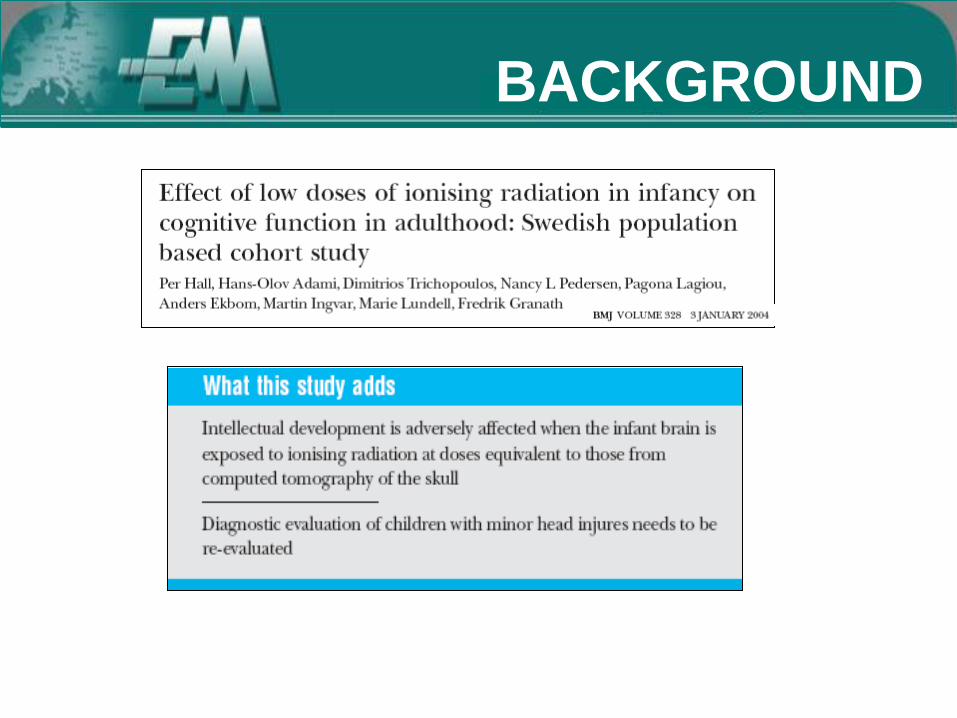
CONCLUSION The best available risk estimates suggest that paediatric CT will result in significantly lifetime radiation risk over adult CT. The lifetimes attributable risk of mortality for leukaemia or solid organ malignancy from a single pediatric head CT ranges from approximately 1:2000 for infants to 1:5000 for older children


BACKGROUND
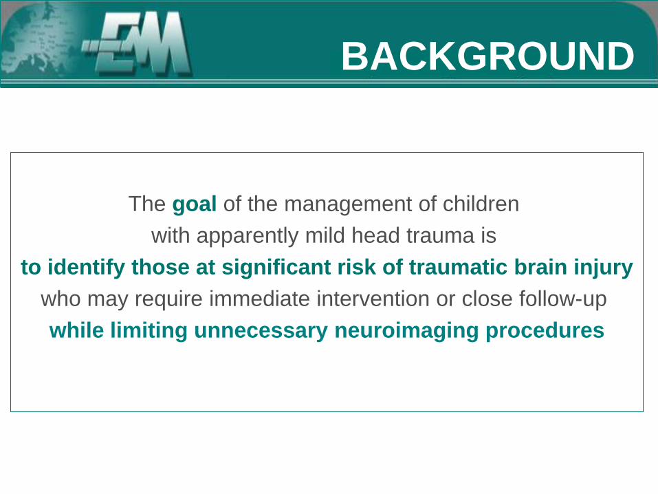
The goal of the management of children
with apparently mild head trauma is to identify those at significant risk of traumatic brain injury
who may require immediate intervention or close follow-up while limiting unnecessary neuroimaging procedures
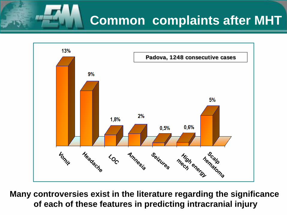
Padova, 1248 consecutive cases
Many controversies exist in the literature regarding the significance of each of these features in predicting intracranial injury
Common complaints after MHT

Padova, 1248 consecutive cases
…and no single feature has been demostrated to predict TBI with sufficient sensitivity
Common complaints after MHT

Considering that no single clinical feature reliably predicts the presence of TBI with sufficient sensitivity
more recently investigators have sought to derive “clinical prediction rules”, that use a combination of
clinical variables obtained from history or clinical examination in order to improve the accuracy in
identifying children with TBI
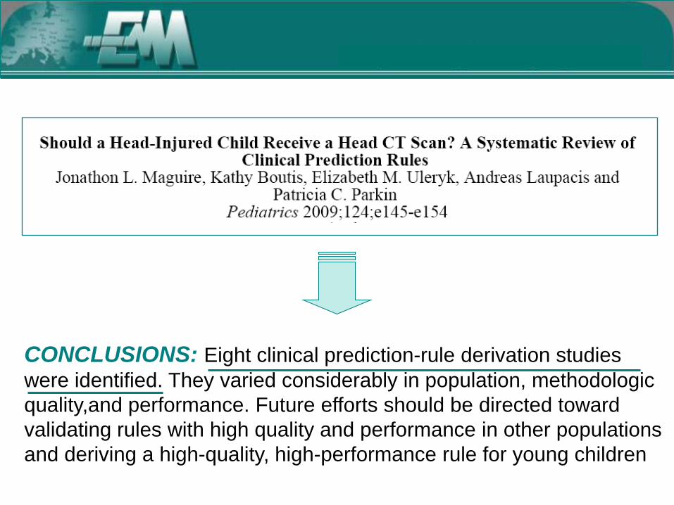
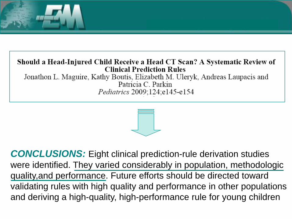
CONCLUSIONS: Eight clinical prediction-rule derivation studies were identified. They varied considerably in population, methodologic quality,and performance. Future efforts should be directed toward validating rules with high quality and performance in other populations and deriving a high-quality, high-performance rule for young children
CONCLUSIONS: Eight clinical prediction-rule derivation studies were identified. They varied considerably in population, methodologic quality,and performance. Future efforts should be directed toward validating rules with high quality and performance in other populations and deriving a high-quality, high-performance rule for young children
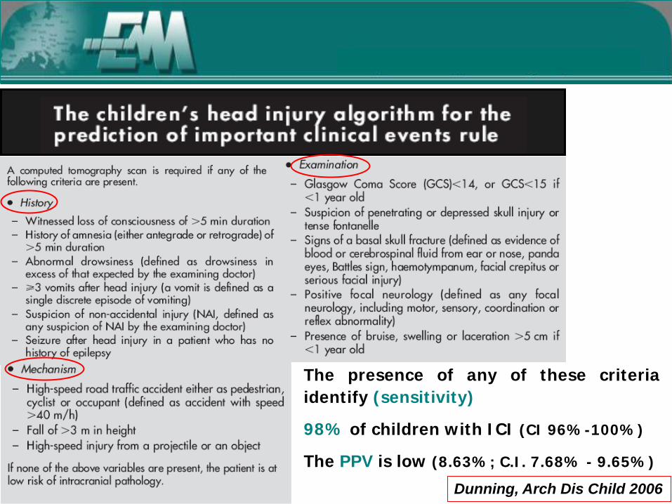
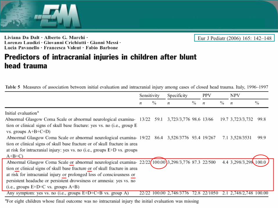
The presence of any of these criteria identify (sensitivity)
98% of children with ICI (CI 96%-100%)
The PPV is low (8.63%; C.I. 7.68% - 9.65%)
Dunning, Arch Dis Child 2006

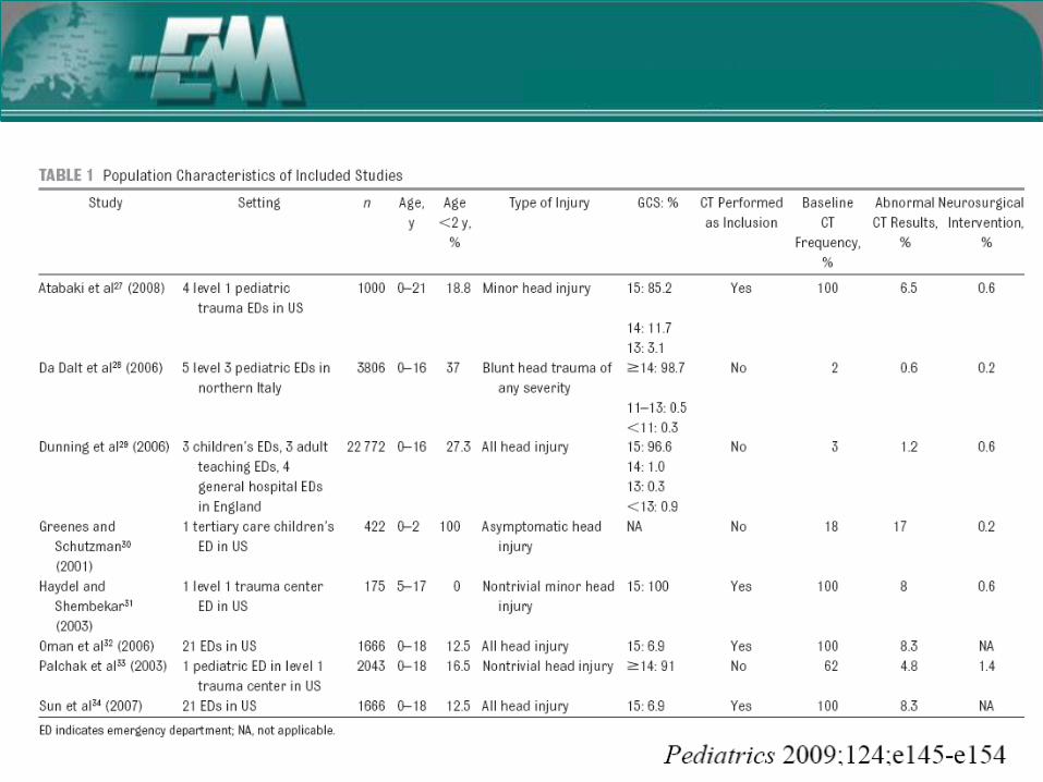
Atabaki 2008
Da Dalt, 2006
Dunning, 2006*
Greenes 2001
Heydell
2003
Oman, 2006
PaltchacK2003
Sun 2007
Altered Mental Status + + + + + +
Focal neurological signs + + + +
Evidence of basal skull fracture + + + + + +
Scalp hematoma (<2y) or any evidence of skull fracture
+ + + + + + +
Prolonged LOC/amnesia + + + +
Persistent vomiting + (> 3) + + + +
Abnormal behaviour + + +
Headache + + +
Seizures + +
Suspected inflicted injury +
Coagulopathy +
Significant mechanism of injury + +/-
Da sistemare
* 0-2 years, asyntomatic
Prediction Rules

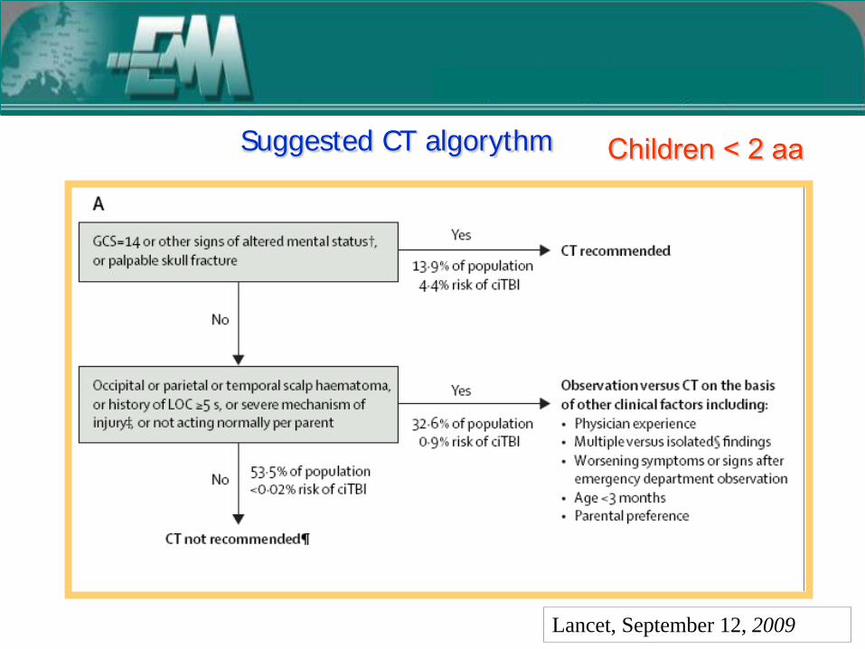
Lancet, September 12, 2009
• 42412 patient 0-18 y, no trivial trauma GCS 14-15
• Identify low-risk group that does not need CT
• Outcomes: clinically important Brain Injury (Death, Intubation, Neurosurgical procedure, Intracranial Injury (ICI) and 2 days in hospital)

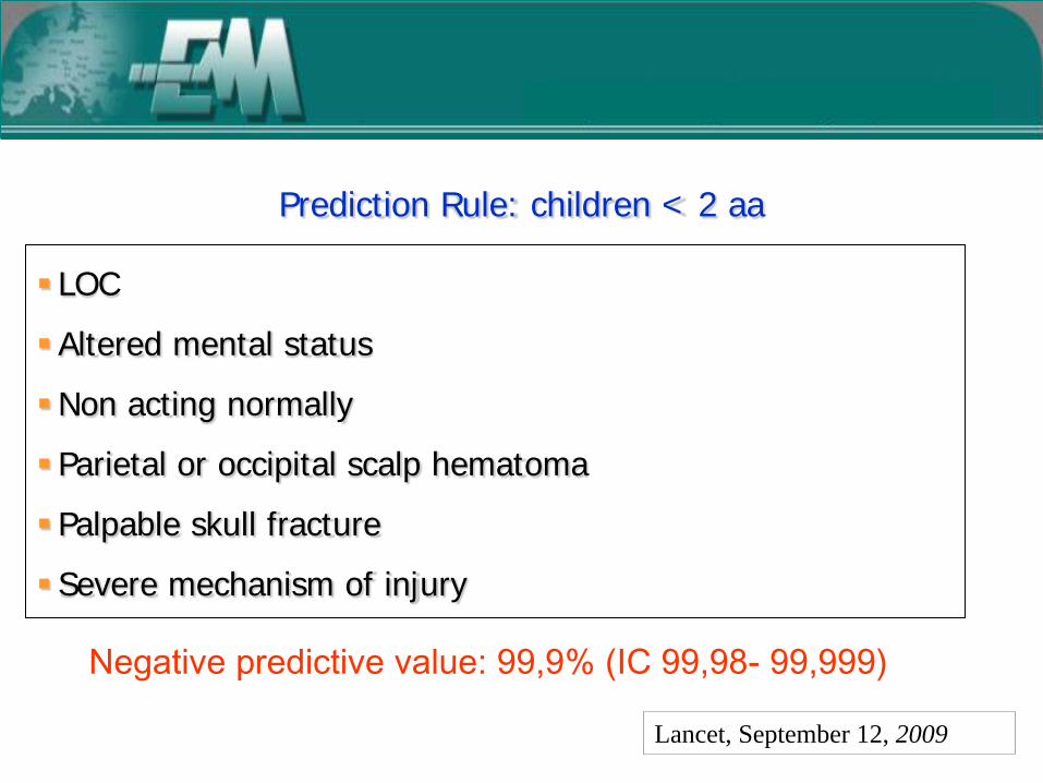
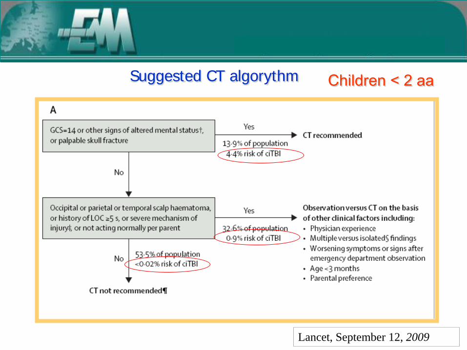
Prediction Rule: children < 2 aa
LOC
Altered mental status
Non acting normally
Parietal or occipital scalp hematoma
Palpable skull fracture
Severe mechanism of injury
Negative predictive value: 99,9% (IC 99,98- 99,999)
Lancet, September 12, 2009
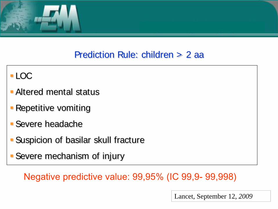
Prediction Rule: children > 2 aa
LOC
Altered mental status
Repetitive vomiting
Severe headache
Suspicion of basilar skull fracture
Severe mechanism of injury
Negative predictive value: 99,95% (IC 99,9- 99,998)
Lancet, September 12, 2009
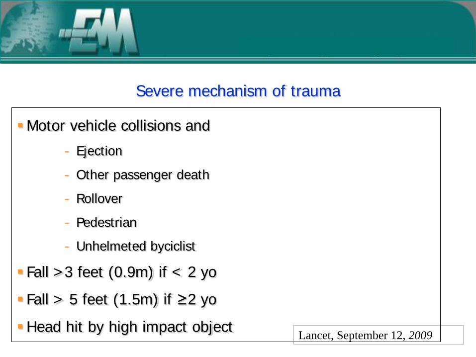
Severe mechanism of trauma
Motor vehicle collisions and
- Ejection
- Other passenger death
- Rollover
- Pedestrian
- Unhelmeted byciclist
Fall >3 feet (0.9m) if < 2 yo
Fall > 5 feet (1.5m) if ≥2 yo
Head hit by high impact object Lancet, September 12, 2009

Case scenario 1 A 7 m/o infant is brought to the PED after a fall on a marble floor, from the changing table almost 4 feet high(1,2 mt). No problems in the following 12 hours. He is brought to the PED the day after for the onset of a scalp hematoma noticed by his parents when he woke up. Still appearing well. PE: normal mental status, no neurological abnormalities. Left large parietal scalp hematoma (7 x 8 cm), without skull depression. No other pathologic findings on the remaining PE.
Lancet, September 12, 2009
Suggested CT algorythm Children < 2 aa
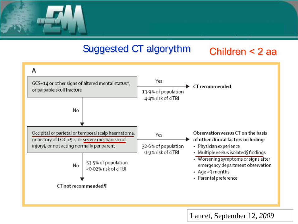
Lancet, September 12, 2009
Suggested CT algorythm Children < 2 aa

High-risk signs or symptoms include the following: • Focal neurologic findings • Acute skull fracture, including depressed or basilar fracture • Depressed mental status • Irritability • Bulging fontanel • Persistent vomiting • Seizure • Definite loss of consciousness (especially more than a few seconds and associated with a high-risk mechanism of injury) • Suspicion of child abuse • Underlying condition predisposing to intracranial injury
The risk of clinically important traumatic brain injury is 4 percent or higher for patients with one or more of these findings.
Performing imaging Children < 2 aa
19.2 May 2011

Case scenario 1 - Head CT scan Left temporo-parietal extradural hematoma, 1 cm thick; no other abnormalities

Case scenario 1 - Head CT scan Left temporo-parietal extradural hematoma, 1 cm thick; no other abnormalities
He underwent neurosugical intervention
with good outcome

Case scenario 2 A 3 m/o infant is brought to the PED after a fall from his parents bed almost 3 feet high. No LOC, he cried immediately after falls and is still appearing well. PE: alert, no neurological abnormalities. Left temporo-parietal scalp hematoma (4 x 5 cm), No other pathologic findings on the remaining PE.
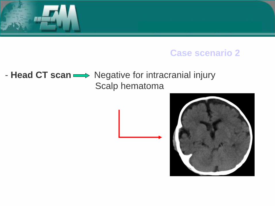
Case scenario 2 - Head CT scan Negative for intracranial injury
Scalp hematoma

Case scenario 2 - Head CT scan Negative for intracranial injury
Scalp hematoma Temporal skull fracture
What is the role of skull X-ray in young children with suspected skull fracture?
Skull fractures are not uncommon following minor head trauma in children, particularly in those younger than two years of age The vast majority of skull fractures are linear. Among children with linear skull fractures, 15 to 30 percent have associated intracranial injuries Most children with skull fractures will have overlying scalp hematomas
Skull fractures in children < 2 years 19.2 May 2011
Skull fractures in children < 2 years and skull x-rays
Skull radiographs may occasionally be useful to screen for fracture and avoid the risk of radiation and sedation from CT in selected asymptomatic patients 3 to 24 months of age with concerning scalp hematomas However, skull radiographs should only be performed if a radiologist with pediatric expertise is available to provide an interpretation because physicians with pediatric emergency expertise may have limited accuracy in correctly identifying skull fractures in young children If a screening skull radiograph shows a fracture, then a head CT should be performed.
19.2 May 2011

Case scenario 3 A 6 y/o child is brought to the PED for head trauma consequent to a motor vehicle collision (reported speed around 60 Km/hr, no rollover of the car, no ejection,reported use of restraint, mother ok) Brief LOC. After that keeping well, alert and oriented. She vomited twice prior to arrival and has been complaining of headache. PMHx: unremarkable
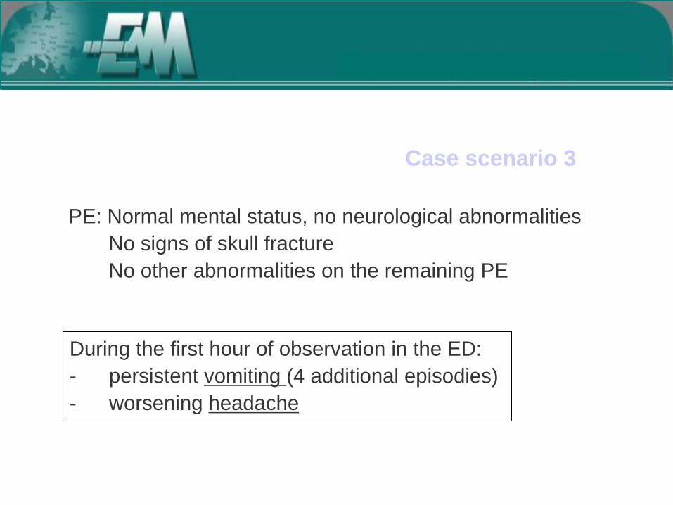
Case scenario 3 PE: Normal mental status, no neurological abnormalities No signs of skull fracture No other abnormalities on the remaining PE
During the first hour of observation in the ED: - persistent vomiting (4 additional episodies) - worsening headache


Lancet, September 12, 2009
Children > 2 aa Suggested CT algorythm

Lancet, September 12, 2009
Children > 2 aa Suggested CT algorythm
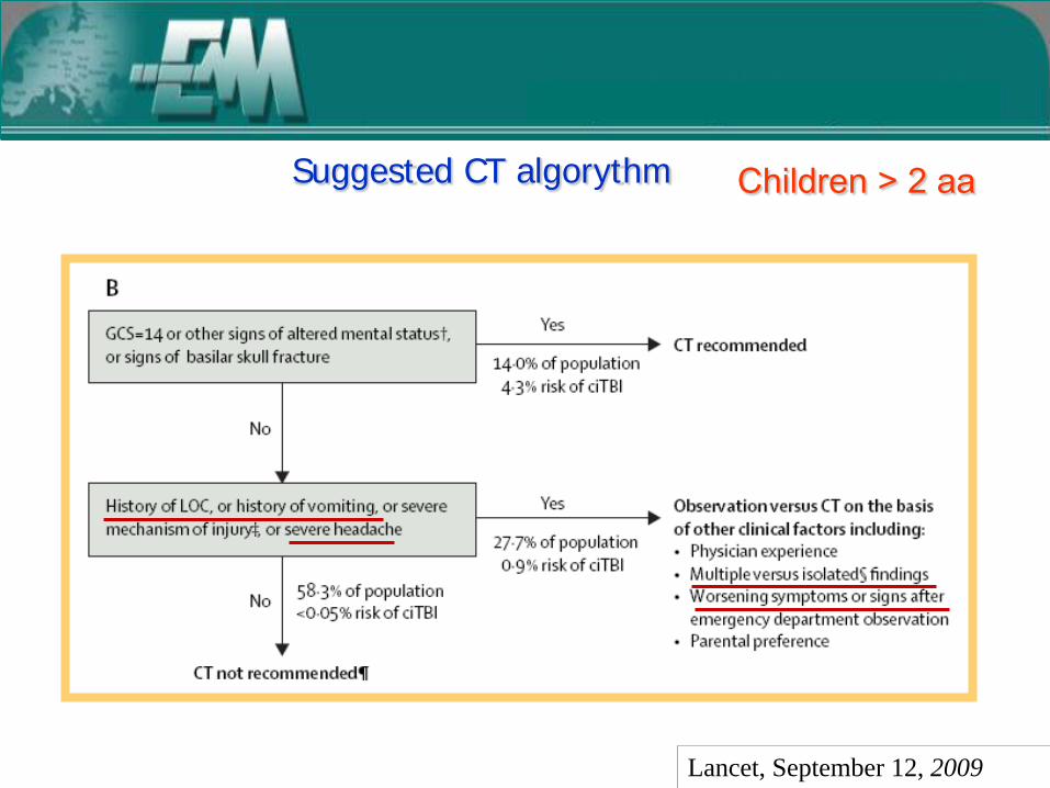
Lancet, September 12, 2009
Children > 2 aa Suggested CT algorythm

High-risk signs or symptoms include the following: • Focal neurologic findings • Skull fracture, especially findings of basilar skull fracture • Altered mental status (eg agitation, lethargy, repetitive questioning or slow response to verbal questioning • Irritability • Prolonged loss of consciousness
Perform imaging Children > 2 aa
19.2 May 2011
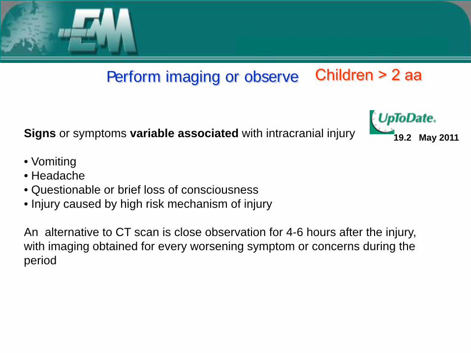
Signs or symptoms variable associated with intracranial injury • Vomiting • Headache • Questionable or brief loss of consciousness • Injury caused by high risk mechanism of injury
An alternative to CT scan is close observation for 4-6 hours after the injury, with imaging obtained for every worsening symptom or concerns during the period
Perform imaging or observe Children > 2 aa
19.2 May 2011
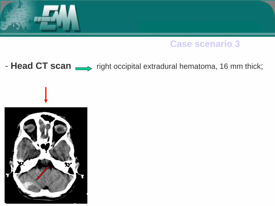
Case scenario 3 - Head CT scan right occipital extradural hematoma, 16 mm thick;
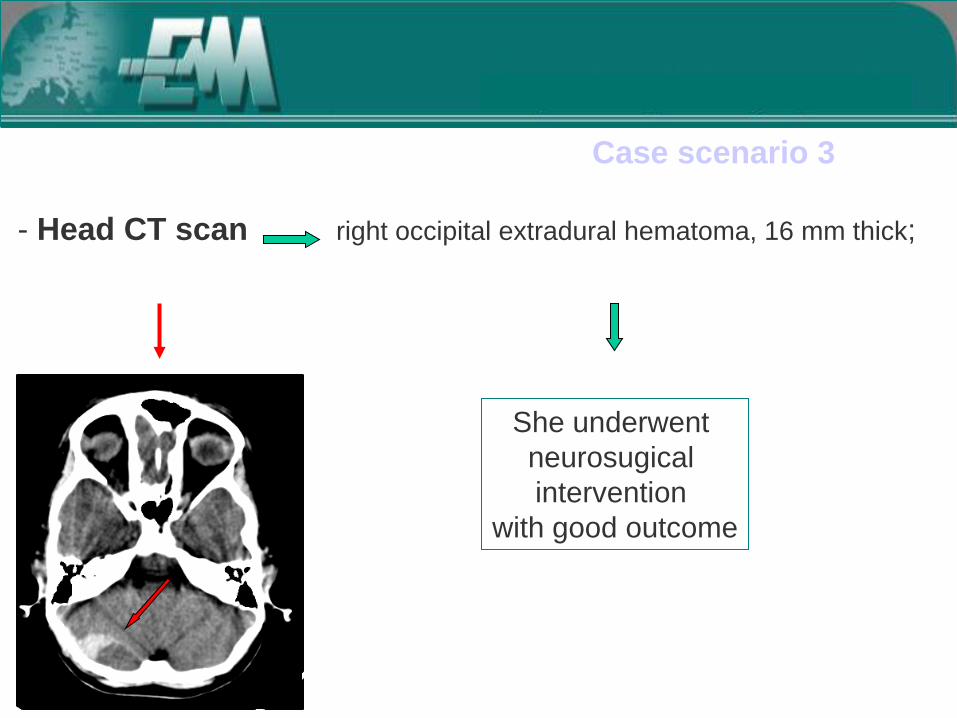
Case scenario 3 - Head CT scan right occipital extradural hematoma, 16 mm thick;
She underwent neurosugical intervention
with good outcome

Case scenario 4 A 2 y/o boy is brought to the PED after a fall on the ground from an amusement park ride 1.5 m high, 4 hrs before. No LOC, nor other signs or symptoms reported except for vomiting twice in the first hour following the trauma. PMHx: unremarkable apart from recurrent vomiting, especially during infections, and motion sickness. PE: no abnormalities

During observation in the PED the child vomited another 4 times
Case scenario 4 Observation in the O.U.

Case scenario 4 - Head CT scan normal
After complete awaking from sedation the child is alert,
no vomiting, no neurological abnormalities

Case scenario 4 - Head CT scan normal
After complete awaking from sedation the child is alert,
no vomiting, no neurological abnormalities
Discharged to home


Conclusion: Post-traumatic vomiting is significantly related to personal or familiar predisposition to vomit rather than to the presence of intracranial lesions.
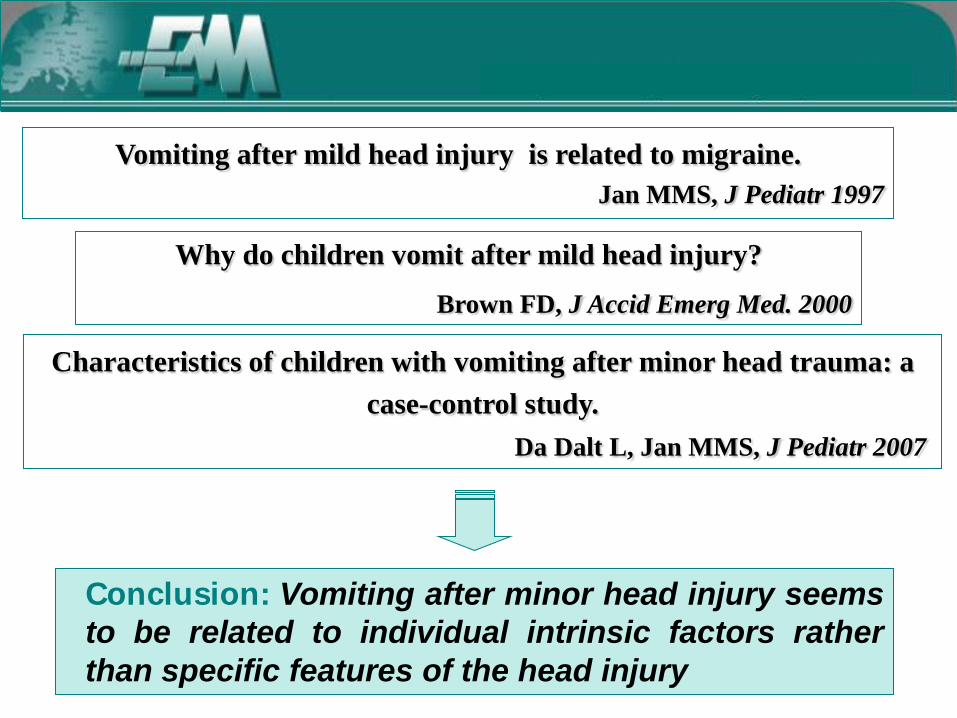
Why do children vomit after mild head injury?
Brown FD, J Accid Emerg Med. 2000
Vomiting after mild head injury is related to migraine. Jan MMS, J Pediatr 1997
Conclusion: Vomiting after minor head injury seems to be related to individual intrinsic factors rather than specific features of the head injury
Characteristics of children with vomiting after minor head trauma: a case-control study.
Da Dalt L, Jan MMS, J Pediatr 2007

Holmes et al, Ann Emerg Med 2011
Normal CT: no intracranial hemorrage, no cerebral edema, no pneumocephalus, no any skull fracture


Holmes et al, Ann Emerg Med 2011
Conclusions. Children with blunt head trauma and initial ED GCS scores of 14 or 15 and normal cranial CT scan results are at very low risk for subsequent traumatic findings on neuroimaging and extremely low risk of needing neurosurgical intervention. Hospitalization of children with minor head trauma after normal CT scan results for neurologic observation is generally unnecessary
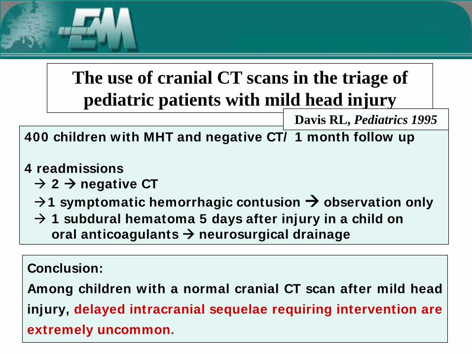
400 children with MHT and negative CT/ 1 month follow up 4 readmissions 2 negative CT 1 symptomatic hemorrhagic contusion observation only 1 subdural hematoma 5 days after injury in a child on
oral anticoagulants neurosurgical drainage
The use of cranial CT scans in the triage of pediatric patients with mild head injury
Davis RL, Pediatrics 1995
Conclusion: Among children with a normal cranial CT scan after mild head injury, delayed intracranial sequelae requiring intervention are extremely uncommon.
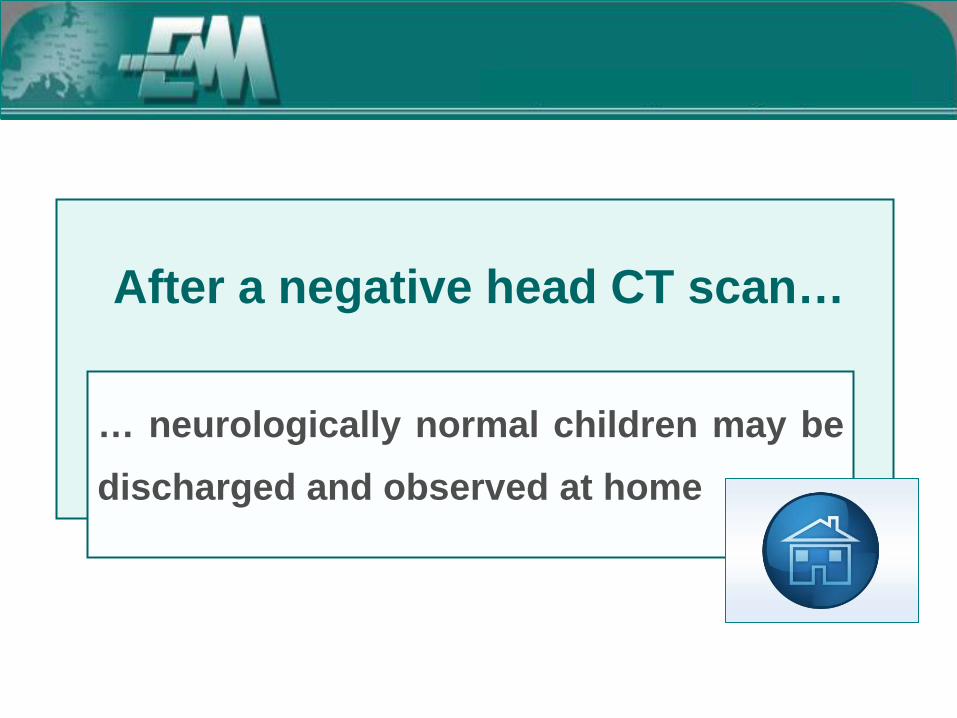
After a negative head CT scan…
… neurologically normal children may be discharged and observed at home


Among children who sustain a minor head injury - Who should get scanned?
- Who should be observed?
- Who should be safely discharged?
ISSUES TO BE ADDRESSED

… an acceptable management option
may be In- Hospital Observation
For intermediate risk patients as an alternative to immediate CT…
Lancet, September 12, 2009
Suggested CT algorythm Children < 2 aa

Lancet, September 12, 2009
Children > 2 aa Suggested CT algorythm

IN-HOSPITAL OBSERVATION
ADVANTAGES No radiation cost No sedation No risk of false
positive results More care to the
patient
DISADVANTAGES Cost of admission Risk of missing
clinically silent lesions

Comparable outcomes in terms of Recovery Late complications Patients’ satisfaction
2602 pts > 6 years/ 920 children 6-15 years Randomization immediate CT /discharge vs admission
Lower costs for immediate CT vs admission
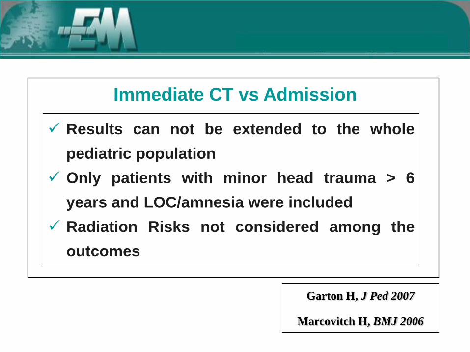
Immediate CT vs Admission
Results can not be extended to the whole pediatric population
Only patients with minor head trauma > 6 years and LOC/amnesia were included
Radiation Risks not considered among the outcomes
Garton H, J Ped 2007
Marcovitch H, BMJ 2006
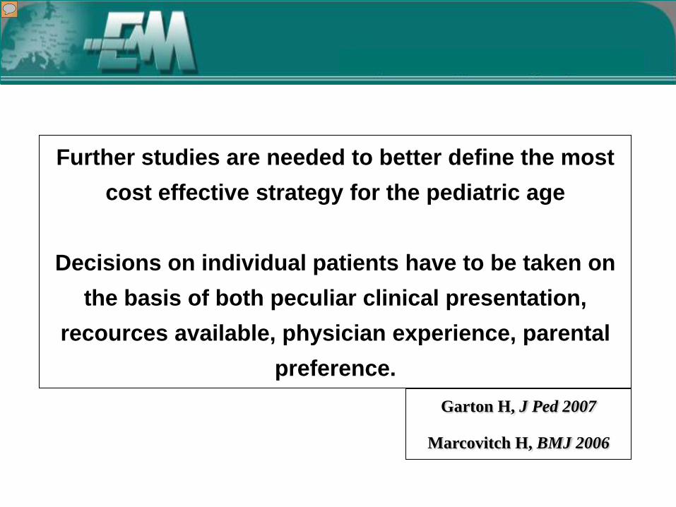
Further studies are needed to better define the most cost effective strategy for the pediatric age
Decisions on individual patients have to be taken on
the basis of both peculiar clinical presentation, recources available, physician experience, parental
preference.
Garton H, J Ped 2007
Marcovitch H, BMJ 2006
Relatore
Note di presentazione
E cosa fare una volta che la TAC cerebrale è negativa: su questo punto la letteratura è concorde nel dire che i bambini asintomatici e con esame neurologico negativo, dopo una TAC cerebrale negativa possono; più studi hanno infatti dimostrato nel bambino con Trauma cranicominore e TAC cerebrale normale, il rischio di deterioramento neurologico tardivo è pressoché irrilevante.


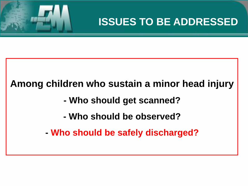
Among children who sustain a minor head injury - Who should get scanned?
- Who should be observed?
- Who should be safely discharged?
ISSUES TO BE ADDRESSED

SAFE DISCHARGE CRITERIA
No suspicion of inflicted injury
The child is easily aroused with light touch and has a normal neurologic examination
The child has returned to baseline level of function and tolerated oral fluids, if there has been vomiting
Caretakers are capable of reliably observing the child and can return for care if indicated
Specific instructions have been given regarding the level of observation required, indications for seeking care, and follow-up
There are no extracranial injuries requiring admission Schutzman S, UpToDate, 2010
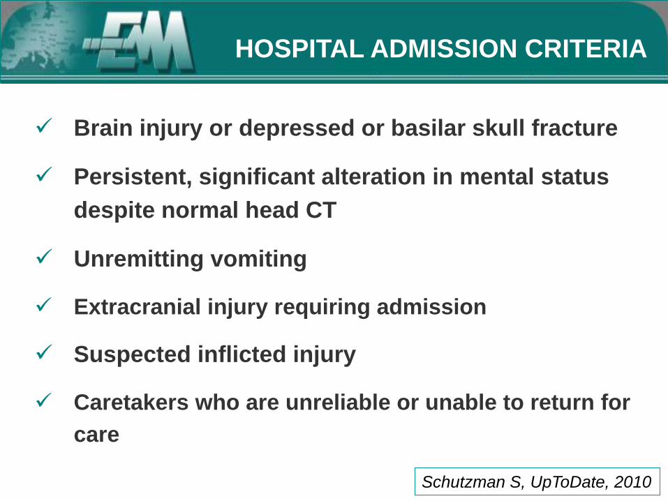
HOSPITAL ADMISSION CRITERIA
Brain injury or depressed or basilar skull fracture
Persistent, significant alteration in mental status despite normal head CT
Unremitting vomiting
Extracranial injury requiring admission
Suspected inflicted injury
Caretakers who are unreliable or unable to return for care
Schutzman S, UpToDate, 2010

Suggested CT algorythm
The algorithmic approach proposed in the study is likely to gain wide acceptance for management of head-injured children given its scientific rigor and easy to use
The rules my might not be perfect, but represent the best current scientific evidence
Kupperman N,, Lancet, September 12, 2009
Klig JE, Kaplan CP, Curr Opin Ped 2010
Parkin PC, Maguire JR, Lancet 2009

Grazie per l’attenzione
Prof.ssa Liviana Da Dalt Dipartimento Materno-Infantile
Azienda ULSS 9 - Treviso
Università di Padova
Roma 9 novembre 2011
Related Documents


