International Journal of Science and Research (IJSR) ISSN (Online): 2319-7064 Impact Factor (2012): 3.358 Volume 3 Issue 10, October 2014 www.ijsr.net Licensed Under Creative Commons Attribution CC BY Application of Texture Analysis Algorithm for Data Extraction in Dental X-Ray Images Reham I. Abdelwahab 1 , Yousif Mohamed Y. Abdallah 2 1 Sudan University of Science and Technology, College of Medical Radiological Science, 2 Elbaladi Street, Khartoum 1908, Sudan Abstract: This paper presented an appropriate approach for the robust estimation of noise statistic in dental x-rays images. To achieve maximum image quality after denoising, a new, low order, local adaptive Gaussian Scale Mixture model is presented, which accomplishes nonlinearities from scattering. State of art methods use multi scale filtering of images to reduce the irrelevant part of information, based on generic estimation of noise. The usual assumption of a distribution of Gaussian and Poisson statistics only lead to overestimation of the noise variance in regions of low intensity (small photon counts), but to underestimation in regions of high intensity and therefore to non-optional results. In this paper, prominent constraints are firstly preservation of image's overall look; secondly preservation of the diagnostic content in the image and thirdly detection of small low contrast details in diagnostic content of the image. As shown in previously, state of the art methods provide non-convincing results. The new approach is funded on an attempt to interpret the problem from the view of blind source separation (BSS), thus to see the panoramic image as a simple mixture of (unwanted) background information, diagnostic information and noise. Keywords: Dental x-rays, Matlab, Texture analysis 1. Introduction The intraoral radiograph examination of the teeth is relatively comfortable for the patient, and the overall radiation dose is generally acceptable; however, it is a time-consuming endeavor, and in some cases provides a less-than-optimum depiction of individual teeth. Nevertheless, it offers valuable information as a basis for treatment planning, is far superior to purely clinical observations, and often gives impetus for more extensive examination strategies. Finally, the 18- film“full series” exam provides a sense of security for both dentist and patient, and enhances confidence through communication. On the other hand, clear radiographic depiction of not only the teeth, but also the jaws, the temporomandibular joints, and the alveolar lobes of the maxillary sinuses will reduce the risk of an incomplete and possibly incorrect examination, which in the worst-case scenario could lead to malpractice. In this regard, the panoramic radiograph always leads to a broadening of horizons because it improves the dentist’s knowledge of radiographic anatomy and thus improves her/his skill in distinguishing between and among normal and pathologic conditions. This, in conjunction with a better understanding of the interrelationships of systemic medical problems and dental/oral problems can open new avenues for treatment planning [1-4]. The question of whether the treating dentist her/himself actually takes the radiographs or delegates this task to auxiliary personnel is really a question of responsibility for the radiographic quality that is achieved, and the radiation dose that this requires. If, for whatever reason, this assignment is delegated, the person who bears the primary responsibility must see to it that the auxiliary personnel are well-trained and have received legal certification. They must not only become expert in the production of high-quality radiographs, but must also be knowledgeable regarding the dental indications and the procedures for protecting patients and staff from excessive radiation exposure. Auxiliary dental personnel who are given the responsibility of taking radiographs must remain current in all continuing education standards in order to insure high radiographic quality and the lowest possible radiation dose for every patient. This is critical. An image defined as it may be applied to a picture such as a photograph, a painting or a sketch which has a real physical existence. But it may be also being applied to an idea or concept which has a mental rather than physical existence. If asked to imagine an object such as apple, a mental image of an apple comes to mind. The existence of this visual image helps us to grasp the concept of an apple. Of course, a photograph of an apple represents only one aspect of an apple: its visual appearance. It provides no representation of the taste, smell or feel of the characteristics of an apple which we remember from past experiences [5-7]. There are two components are superimposed in the image. The presence of noise limits the amount of information which can be extended from image. In particular, the finer details of structure may be lost by being swamped by the effect of noise. Noise may see an unlikely term apply to a visual image which is silent. The use of the term probably originates from radio engineering, where the quality reception of radio signals is often impaired by background noise (i.e. hisses and whistles), particularly when the origin signal is rather weak and the radio receiver is being used with maximum amplification. The effect of such noise on the information communicated via a radio link is very similar to the effect of noise on the information carried by an image. Under optimum conditions the magnitude of the signal is very much greater than the magnitude of the noise. The signal-to-noise ratio is said to be high. Under adverse conditions the signal-to-noise ratio is law, and much information is lost. It is not uncommon for a radiographic image to be fogged, perhaps due to an accidental exposure to scattered radiation in X-ray room, or to poor conditions of storage. When such a radiograph is examined its image appears as viewed through a mist or fog. The information content of the image is reduced. Some information is lost Paper ID: OCT14660 1934

Welcome message from author
This document is posted to help you gain knowledge. Please leave a comment to let me know what you think about it! Share it to your friends and learn new things together.
Transcript
International Journal of Science and Research (IJSR) ISSN (Online): 2319-7064
Impact Factor (2012): 3.358
Volume 3 Issue 10, October 2014 www.ijsr.net
Licensed Under Creative Commons Attribution CC BY
Application of Texture Analysis Algorithm for Data Extraction in Dental X-Ray Images
Reham I. Abdelwahab1, Yousif Mohamed Y. Abdallah2
1Sudan University of Science and Technology, College of Medical Radiological Science,
2Elbaladi Street, Khartoum 1908, Sudan Abstract: This paper presented an appropriate approach for the robust estimation of noise statistic in dental x-rays images. To achieve maximum image quality after denoising, a new, low order, local adaptive Gaussian Scale Mixture model is presented, which accomplishes nonlinearities from scattering. State of art methods use multi scale filtering of images to reduce the irrelevant part of information, based on generic estimation of noise. The usual assumption of a distribution of Gaussian and Poisson statistics only lead to overestimation of the noise variance in regions of low intensity (small photon counts), but to underestimation in regions of high intensity and therefore to non-optional results. In this paper, prominent constraints are firstly preservation of image's overall look; secondly preservation of the diagnostic content in the image and thirdly detection of small low contrast details in diagnostic content of the image. As shown in previously, state of the art methods provide non-convincing results. The new approach is funded on an attempt to interpret the problem from the view of blind source separation (BSS), thus to see the panoramic image as a simple mixture of (unwanted) background information, diagnostic information and noise. Keywords: Dental x-rays, Matlab, Texture analysis 1. Introduction The intraoral radiograph examination of the teeth is relatively comfortable for the patient, and the overall radiation dose is generally acceptable; however, it is a time-consuming endeavor, and in some cases provides a less-than-optimum depiction of individual teeth. Nevertheless, it offers valuable information as a basis for treatment planning, is far superior to purely clinical observations, and often gives impetus for more extensive examination strategies. Finally, the 18-film“full series” exam provides a sense of security for both dentist and patient, and enhances confidence through communication. On the other hand, clear radiographic depiction of not only the teeth, but also the jaws, the temporomandibular joints, and the alveolar lobes of the maxillary sinuses will reduce the risk of an incomplete and possibly incorrect examination, which in the worst-case scenario could lead to malpractice. In this regard, the panoramic radiograph always leads to a broadening of horizons because it improves the dentist’s knowledge of radiographic anatomy and thus improves her/his skill in distinguishing between and among normal and pathologic conditions. This, in conjunction with a better understanding of the interrelationships of systemic medical problems and dental/oral problems can open new avenues for treatment planning [1-4]. The question of whether the treating dentist her/himself actually takes the radiographs or delegates this task to auxiliary personnel is really a question of responsibility for the radiographic quality that is achieved, and the radiation dose that this requires. If, for whatever reason, this assignment is delegated, the person who bears the primary responsibility must see to it that the auxiliary personnel are well-trained and have received legal certification. They must not only become expert in the production of high-quality radiographs, but must also be knowledgeable regarding the dental indications and the procedures for protecting patients and staff from excessive radiation exposure. Auxiliary dental personnel who are given
the responsibility of taking radiographs must remain current in all continuing education standards in order to insure high radiographic quality and the lowest possible radiation dose for every patient. This is critical. An image defined as it may be applied to a picture such as a photograph, a painting or a sketch which has a real physical existence. But it may be also being applied to an idea or concept which has a mental rather than physical existence. If asked to imagine an object such as apple, a mental image of an apple comes to mind. The existence of this visual image helps us to grasp the concept of an apple. Of course, a photograph of an apple represents only one aspect of an apple: its visual appearance. It provides no representation of the taste, smell or feel of the characteristics of an apple which we remember from past experiences [5-7]. There are two components are superimposed in the image. The presence of noise limits the amount of information which can be extended from image. In particular, the finer details of structure may be lost by being swamped by the effect of noise. Noise may see an unlikely term apply to a visual image which is silent. The use of the term probably originates from radio engineering, where the quality reception of radio signals is often impaired by background noise (i.e. hisses and whistles), particularly when the origin signal is rather weak and the radio receiver is being used with maximum amplification. The effect of such noise on the information communicated via a radio link is very similar to the effect of noise on the information carried by an image. Under optimum conditions the magnitude of the signal is very much greater than the magnitude of the noise. The signal-to-noise ratio is said to be high. Under adverse conditions the signal-to-noise ratio is law, and much information is lost. It is not uncommon for a radiographic image to be fogged, perhaps due to an accidental exposure to scattered radiation in X-ray room, or to poor conditions of storage. When such a radiograph is examined its image appears as viewed through a mist or fog. The information content of the image is reduced. Some information is lost
Paper ID: OCT14660 1934
International Journal of Science and Research (IJSR) ISSN (Online): 2319-7064
Impact Factor (2012): 3.358
Volume 3 Issue 10, October 2014 www.ijsr.net
Licensed Under Creative Commons Attribution CC BY
entirely, whilst that which remains is more difficult to see. The fogging contains no information about the subject of radiograph. Such fogging is an example of one form of image noise. Another type of noise may be seen on a television image. In this case, the image appears as if viewed through a snowstorm. The screen display a very large number of small white specks which appear superimposed on the image. Again, the effect is to reduce the information content of the image. Such as appearance may be due to inferior design of the electronic circuitry employed, causing electronic noise. However, in many cases the basic is that the signal being displayed is too weak, and those speaks represent the absence of information, rather like the spaces left by missing pieces in a jigsaw puzzle. It is possible for a similar effect to occur in the image on a radiograph, expect that in this case the image appears to have a large number of very small black specks superimposed on it, giving it a grainy or mottled appearance commonly known as quantum mottle or quantum noise. To be able to identify a feature on an image, it must appear different its surroundings. On a radiograph, a structure must be of a different optical density (shade of grey) from adjacent structures. On television image, the structure must be of a different luminance (brightness). The term contrast is used to describe these differences are small; it is difficult to identify the structure will stand out well from its surroundings and it is said the contrast is high (good). If the differences are small, it is difficult to identify the structure against its background. The contrast is said to be low (poor) [8-9]. 2. Methods and Materials For panoramic images each film scanned using digitizer scanner then treat by using image processing program (MatLab), where the enhancement and contrast of the image was determined. The scanned image was saved in a TIFF file format to preserve the quality of the image. The data analyzed used to enhance the contrast within the soft tissues, the gray levels which can be redistributed both linearly and nonlinearly using the gray level frequencies and noise estimation of the original panoramic images. In Figure 2-1 explains the block diagram about the flow proposed research paper. Researchers use in this study a method for image preprocessing of X-ray radiographic images step by step process. This flow diagram explains the flow of work. Collecting the X-ray images from Govt. Hospitals and converting this image RGB into Gray color. Panoramic images were filtered by two different filtering algorithms. The filtering algorithms are used for anisotropic filtering and median filtering algorithm. The output results were compared in PSNR and MSE values. The output of filtering image applied for Image enhancement. That given image improves the subjective quality of contrast, noise reduction and edge sharpening. The targets for image enhancing are better contrast, sharpness of detail and visibility of features. Several algorithms are Histogram Equalization (CLAHE). Here researchers applied contrast limited adaptive histogram equalization algorithm. The best PSNR value of X-ray image output is given into input of Contrast Limited Adaptive Histogram Equalization (CLAHE).
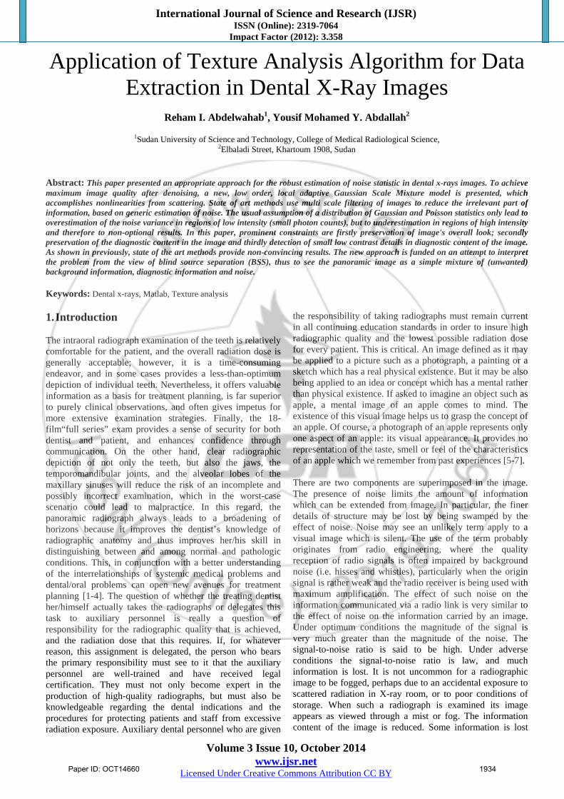
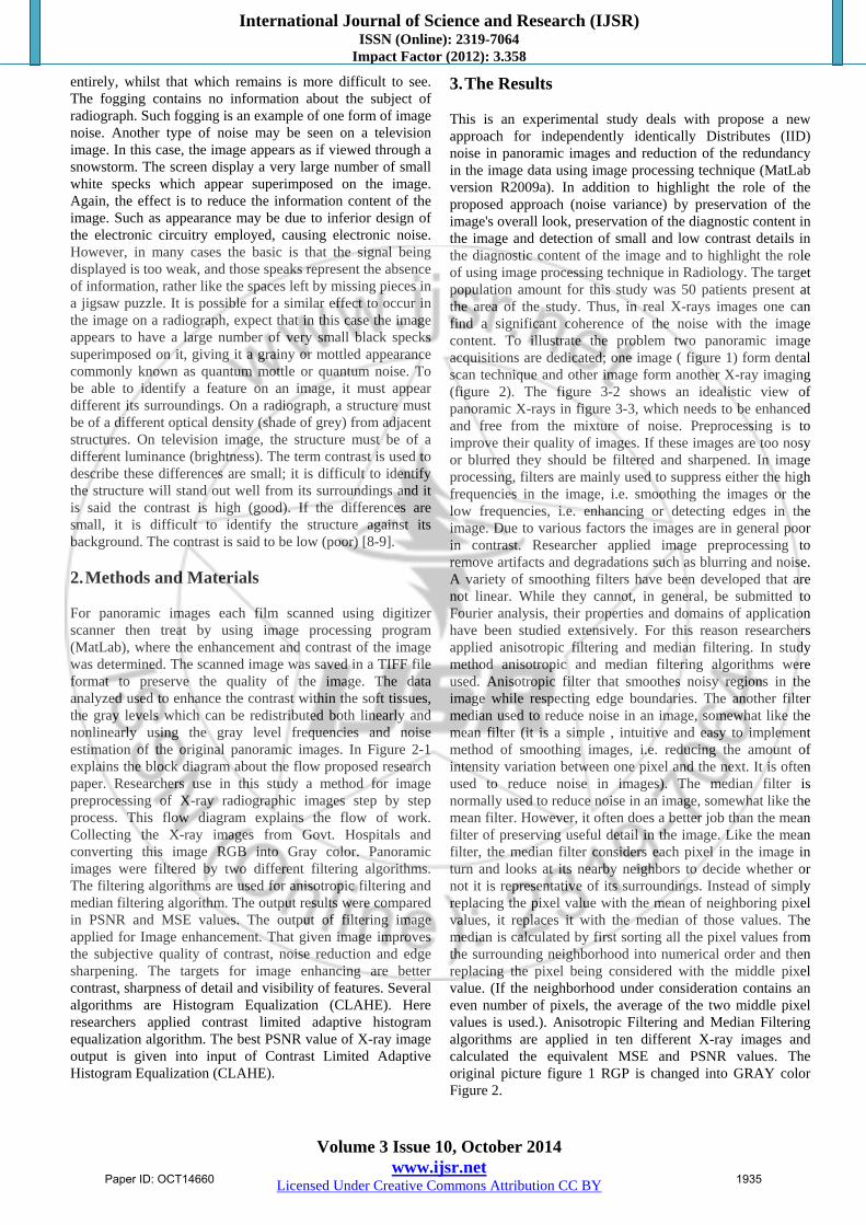
3. The Results This is an experimental study deals with propose a new approach for independently identically Distributes (IID) noise in panoramic images and reduction of the redundancy in the image data using image processing technique (MatLab version R2009a). In addition to highlight the role of the proposed approach (noise variance) by preservation of the image's overall look, preservation of the diagnostic content in the image and detection of small and low contrast details in the diagnostic content of the image and to highlight the role of using image processing technique in Radiology. The target population amount for this study was 50 patients present at the area of the study. Thus, in real X-rays images one can find a significant coherence of the noise with the image content. To illustrate the problem two panoramic image acquisitions are dedicated; one image ( figure 1) form dental scan technique and other image form another X-ray imaging (figure 2). The figure 3-2 shows an idealistic view of panoramic X-rays in figure 3-3, which needs to be enhanced and free from the mixture of noise. Preprocessing is to improve their quality of images. If these images are too nosy or blurred they should be filtered and sharpened. In image processing, filters are mainly used to suppress either the high frequencies in the image, i.e. smoothing the images or the low frequencies, i.e. enhancing or detecting edges in the image. Due to various factors the images are in general poor in contrast. Researcher applied image preprocessing to remove artifacts and degradations such as blurring and noise. A variety of smoothing filters have been developed that are not linear. While they cannot, in general, be submitted to Fourier analysis, their properties and domains of application have been studied extensively. For this reason researchers applied anisotropic filtering and median filtering. In study method anisotropic and median filtering algorithms were used. Anisotropic filter that smoothes noisy regions in the image while respecting edge boundaries. The another filter median used to reduce noise in an image, somewhat like the mean filter (it is a simple , intuitive and easy to implement method of smoothing images, i.e. reducing the amount of intensity variation between one pixel and the next. It is often used to reduce noise in images). The median filter is normally used to reduce noise in an image, somewhat like the mean filter. However, it often does a better job than the mean filter of preserving useful detail in the image. Like the mean filter, the median filter considers each pixel in the image in turn and looks at its nearby neighbors to decide whether or not it is representative of its surroundings. Instead of simply replacing the pixel value with the mean of neighboring pixel values, it replaces it with the median of those values. The median is calculated by first sorting all the pixel values from the surrounding neighborhood into numerical order and then replacing the pixel being considered with the middle pixel value. (If the neighborhood under consideration contains an even number of pixels, the average of the two middle pixel values is used.). Anisotropic Filtering and Median Filtering algorithms are applied in ten different X-ray images and calculated the equivalent MSE and PSNR values. The original picture figure 1 RGP is changed into GRAY color Figure 2.
Paper ID: OCT14660 1935
International Journal of Science and Research (IJSR) ISSN (Online): 2319-7064
Impact Factor (2012): 3.358
Volume 3 Issue 10, October 2014 www.ijsr.net
Licensed Under Creative Commons Attribution CC BY
Figure 1: Shows the original image
Figure 2: Shows gray scale image
This gray color image applied into anisotropic filtering method the output is in Figure 2 and also applied in median filtering algorithms the output is in Figure 3 (a, b, c & d).
Figure 3 (a, b, c & d): Shows median filter
Figure 4 (a, b, c & d): Shows histogram of the median
filtered images Edge Detection Technique: When working with large images, normal image processing techniques can sometimes break down. The images can either be too large to load into memory, or else they can be loaded
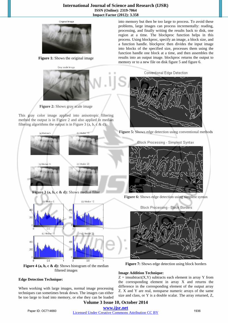
into memory but then be too large to process. To avoid these problems, large images can process incrementally: reading, processing, and finally writing the results back to disk, one region at a time. The blockproc function helps in this process. Using blockproc, specify an image, a block size, and a function handle. blockproc then divides the input image into blocks of the specified size, processes them using the function handle one block at a time, and then assembles the results into an output image. blockproc returns the output to memory or to a new file on disk figure 5 and figure 6.
Figure 5: Shows edge detection using conventional methods
Figure 6: Shows edge detection using simplest syntax
Figure 7: Shows edge detection using block borders
Image Addition Technique: Z = imsubtract(X,Y) subtracts each element in array Y from the corresponding element in array X and returns the difference in the corresponding element of the output array Z. X and Y are real, nonsparse numeric arrays of the same size and class, or Y is a double scalar. The array returned, Z,
Paper ID: OCT14660 1936

International Journal of Science and Research (IJSR) ISSN (Online): 2319-7064
Impact Factor (2012): 3.358
Volume 3 Issue 10, October 2014 www.ijsr.net
Licensed Under Creative Commons Attribution CC BY
has the same size and class as X unless X is logical, in which case Z is double. If X is an integer array, elements of the output that exceed the range of the integer type are truncated, and fractional values are rounded.
Figure 7: Shows the original image
Figure 8: Shows image subtraction technique
4. Conclusion This was an experimental study proposed a new approach for independently identically Distributes (IID) noise in dental x-rays images and reduction of the redundancy in the image data using image processing technique (MatLab version R2009a). In addition it highlighted the role of the proposed approach (noise variance) by preservation of the image's overall look, persevered of the diagnostic content in the image and detected of small and low contrast details in the diagnostic content of the image and highlighted the role of using image processing technique in Radiology. The result of edge detection now closely matches the original in-memory result. Some additional artifacts along the boundaries could be noticed. These were due to the different methods of padding used by the Canny edge detector. Currently, blockproc only supports zero-padding along the image boundaries. So conclusion of this paper that the new approach is funded on an attempt to interpret the problem from the view of blind source separation (BSS), edge detection and image subtraction technique thus to see the panoramic image as a simple mixture of (unwanted) background information, diagnostic information and noise and filtered it. The detection of the noise is a complex procedure which is difficult to detect by naked eye so that image analysis should be performed by using powerful image processing.
References [1] Adam MJ, Wilbur DS (2005) Radiohalogens for imaging
and therapy. Chem Soc Rev 34:153–63 [2] Adelson, E.H., Bergen, J.R. 1991. "The plenoptic
function and the elements of early vision", In Computation Models of Visual Processing, M. Landyand J.A. Movshon, eds., MIT Press, Cambridge, 1991, pp. 3-20.
[3] Arvo, J., 1994, The Irradiance Jacobian for Partially Occluded Polyhedral Sources, Proc. ACM SIGGRAPH, ACM Press, pp. 335-342.
[4] Ball, J., Moore, A., 1997, Essential physics for radiographers, 3rd edition, Blackwell Scientific, Oxford.
[5] Ball, J., Price, T., 1995, Chesney's radiographic imaging, 6th edition, Blackwell Scientific, Oxford.
[6] Buehler, C., Bosse, M., McMillan, L., Gortler, S., Cohen, M., 2001, Unstructured Lumigraph rendering, Proc. ACM SIGGRAPH, ACM Press.
[7] Farr, R., Allisy-Roberts, P., 1997, Physics for medical imaging, W.B. Saunders, London.
[8] Fritsch D.S.; Chaney E.L.; McAuliffe M.J.; Raghavan S.; BoxwalaA.; Earnhart J.R.D., 1995,International Journal of Radiation Oncology, Biology, Physics, Volume 32, Number 971, , pp. 217-217.
[9] Georgiev, T., Zheng, C., Nayar, S., Curless, B., Salesin, D., Intwala, C., 2006, Spatio-angular Resolution Trade-offs in Integral Photography, Proc. EGSR
Author Profile Reham I. Abdelwahab received the B.Sc. Scientific Laboratory Physics technology, in nuclear medicine from College of Science, Sudan University of Science and Technology in 2005 and 2011, respectively. She is M.Sc. medical physics student.
Yousif Mohamed Yousif Abdallah received the B.S., M.Sc. and PhD degrees and M.Sc. in nuclear medicine and Radiotherapy Technology from College of Medical radiological Science, Sudan
University of Science and Technology in 2005, 2009 and 2013, 2014, respectively. During 2006 up to date, he is staying in College of Medical radiological Science, Sudan University of Science and Technology. He is now assistant professor, college registrar and Consultant Radiation Therapist.
Paper ID: OCT14660 1937
Related Documents