Chapter 3 Application of Optical Coherence Tomography and Macular Holes in Ophthalmology Robert J. Lowe and Ronald C. Gentile Additional information is available at the end of the chapter http://dx.doi.org/10.5772/53535 1. Introduction Optical coherence tomography (OCT) has revolutionized how we understand macular holes. The purpose of this chapter is to describe the integration of OCT in the diagnosis, classification, and management of macular holes. 2. Definitions of macular holes A macular hole is a full thickness defect, or hole, in the neurosensory retina located within, or just eccentric to the center of the fovea. An impending macular hole, also known as a stage 1 macular hole, is considered the precur‐ sor to a full thickness idiopathic macular hole. Impending macular holes have a splitting of the inner retina with the clinical appearance of a foveolar cyst or pseudocyst. In some cases, this inner splitting can be associated with a defect in the underlying outer retina (Lee, Kang et al. 2011). An impending macular hole with a defect in the outer retina can have a very thin intact inner retina referred to as the roof of the impending macular hole. These impend‐ ing macular holes can progress to become full thickness macular hole once a break in the inner retina or roof occurs. Most macular holes, unless otherwise specified, refer to idiopathic macular holes. Idiopathic macular holes occur from tractional forces on the foveola at the vitreoretinal interface not associated with other causes. Other types of macular holes include those associated with © 2013 Lowe and Gentile; licensee InTech. This is an open access article distributed under the terms of the Creative Commons Attribution License (http://creativecommons.org/licenses/by/3.0), which permits unrestricted use, distribution, and reproduction in any medium, provided the original work is properly cited.

Welcome message from author
This document is posted to help you gain knowledge. Please leave a comment to let me know what you think about it! Share it to your friends and learn new things together.
Transcript
Chapter 3
Application of Optical Coherence Tomography andMacular Holes in Ophthalmology
Robert J. Lowe and Ronald C. Gentile
Additional information is available at the end of the chapter
http://dx.doi.org/10.5772/53535
1. Introduction
Optical coherence tomography (OCT) has revolutionized how we understand macularholes. The purpose of this chapter is to describe the integration of OCT in the diagnosis,classification, and management of macular holes.
2. Definitions of macular holes
A macular hole is a full thickness defect, or hole, in the neurosensory retina located within,or just eccentric to the center of the fovea.
An impending macular hole, also known as a stage 1 macular hole, is considered the precur‐sor to a full thickness idiopathic macular hole. Impending macular holes have a splitting ofthe inner retina with the clinical appearance of a foveolar cyst or pseudocyst. In some cases,this inner splitting can be associated with a defect in the underlying outer retina (Lee, Kanget al. 2011). An impending macular hole with a defect in the outer retina can have a verythin intact inner retina referred to as the roof of the impending macular hole. These impend‐ing macular holes can progress to become full thickness macular hole once a break in theinner retina or roof occurs.
Most macular holes, unless otherwise specified, refer to idiopathic macular holes. Idiopathicmacular holes occur from tractional forces on the foveola at the vitreoretinal interface notassociated with other causes. Other types of macular holes include those associated with
© 2013 Lowe and Gentile; licensee InTech. This is an open access article distributed under the terms of theCreative Commons Attribution License (http://creativecommons.org/licenses/by/3.0), which permitsunrestricted use, distribution, and reproduction in any medium, provided the original work is properly cited.
trauma, high myopia, retinal detachments, lasers accidents, lightning strikes, diabetic retin‐opathy, and epiretinal membranes.
3. Types of macular holes
3.1. Idiopathic macular holes
Idiopathic macular holes are the most common type of macular hole. A population basedchart review of patients with macular holes reported that 92% of macular holes were idio‐pathic. Mean age in this study was 68.6 (range 47.5–89.6) years. The prevalence of idiopathicmacular holes ranges from 0.02% to 0.8%. The incidence of idiopathic macular holes wasfound to be 8.5 persons per 100,000 population per year (McCannel, Ensminger et al. 2009).
3.2. Traumatic macular holes
Macular holes can be associated with ocular trauma and are often referred to as traumaticmacular holes. In the same population based study mentioned above, only 2% of all macularholes were traumatic macular holes (McCannel, Ensminger et al. 2009).
The formation of a traumatic macular hole is believed to be related to the rapid changesat the vitreofoveal interface that occur during the traumatic event. In a study from theWalter Reed Army Medical Center, 3% of soldiers who sustained combat related oculartrauma were found to have full thickness macular holes (Weichel and Colyer 2009). Incontrast to the formation of idiopathic macular holes that may occur over the course ofweeks to months, the formation of traumatic macular holes is quicker. (Johnson, McDo‐nald et al. 2001).
Other associated findings seen in traumatic macular holes, not present in idiopathic macu‐lar holes, include retinal pigment epithelium (RPE) mottling with damaged to the RPE.Damage to the RPE appears to be directly related to the trauma. Despite the presence ofRPE damage, visual recovery is still possible (Chow, Williams et al. 1999; Johnson, McDo‐nald et al. 2001). Fortunately, the surgical closure rate (96%) and visual improvement intraumatic macular holes is similar to that found with idiopathic macular hole closure(Johnson, McDonald et al. 2001).
3.3. Myopic macular holes
Macular holes can be associated with high myopia and are referred to as myopic macularholes. High myopia is most commonly defined as a refractive error equal to or greaterthan −6.00 diopters of axial myopia with an axial length of greater than 26 mm (Wu andKung 2011).
Optical Coherence Tomography50
Highly myopic patients seem to be at higher risk for developing macular holes that haveunique features and associations. Myopic macular holes develop as the refractive error in‐creases (Kobayashi, Kobayashi et al. 2002) and can be associated with retinal detachmentsand myopic schisis. The association with retinal detachment appears greatest in the presenceof a posterior staphaloma and longer axial length (greater than or equal to 30 mm).
The success rate of surgical repair of myopic macular holes is not as high as the surgical clo‐sure rate of macular holes in non-myopic eyes. Some studies have reported success rates aslow at 60% and 62.5% (Patel, Loo et al. 2001; Wu and Kung 2011) compared to the 90% suc‐cess rates seen in idiopathic macular hole repair. The lower success rate is believed to be re‐lated to a foreshortened retina within the staphaloma that can create residual tractionalforces on the retina despite surgery (Ikuno, Sayanagi et al. 2003; Wu and Kung 2011).
3.4. Macular holes following retinal detachment surgery
Rarely, macular holes can form after retinal detachment surgery. The prevalence of macularhole formation after any type of retinal detachment surgery has been reported to be 0.9%(Benzerroug, Genevois et al. 2008). In eyes that underwent retinal detachment surgery withpars plana vitrectomy, the prevalence has ranged between 0.2% to 1.1% (Lee, Park et al.2010; Fabian, Moisseiev et al. 2011). For reasons not well understood, macular holes formingafter retinal detachment surgery tend to occur more often in macula-off retinal detachments(Benzerroug, Genevois et al. 2008). In one study, the prevalence of macular holes after reti‐nal detachment surgery is more than 3 times greater than the prevalence of macular holesdue to idiopathic causes (0.3%) (Fabian, Moisseiev et al. 2011). Fortunately, the surgical clo‐sure rate in eyes with macular holes after retinal detachment surgery is similar to the successrate of idiopathic macular hole closure, around 90% success (Benzerroug, Genevois et al.2008; Lee, Park et al. 2010).
4. Histopathology of idiopathic macular holes
Histopathological analysis of macular holes in the 1980s helped elucidate how macular holesform. Idiopathic macular holes were found to be associated with an epiretinal membrane(ERM) leading to the wrinkling of the internal limiting membrane of the inner retina. Fur‐thermore, cystoid macular edema within the inner nuclear and outer plexiform layers of theretina, located around these idiopathic macular holes, were also discovered. An operculumof glial cells have been found over the macular holes (Frangieh, Green et al. 1981).
From these histopathological studies, the ILM, ELM formation, and cystoid macular edemawere believed to play a role in the pathogenesis of idiopathic macular holes. Many felt thatvitreous traction may be implicated in the pathogenesis of macular holes, especially in cases
Application of Optical Coherence Tomography and Macular Holes in Ophthalmologyhttp://dx.doi.org/10.5772/53535
51
where an operculum of glial tissue was seen on the posterior hyloid face of the vitreous(Frangieh, Green et al. 1981). Anterior-posterior traction from a posterior vitreous detach‐ment can also convert a macular cyst to a macular hole (McDonnell, Fine et al. 1982).
At the time of these histological studies, surgical repair of macular holes was not an option.Macular holes that have closed were due to spontaneous closure. Histopathology of sponta‐neously closed macular holes have revealed proliferation of fibroglial cells filling and bridg‐ing the macular hole (Frangieh, Green et al. 1981). This finding may be seen in surgicallyclosed macular holes as well.
5. Gass classification of idiopathic macular holes
Donald Gass and Robert Johnson in the late 1980s and early 1990s created a classificationsystem for idiopathic macular holes. They divided idiopathic macular holes into 4 stagesbased on clinical findings using contact lens biomicroscopy. Remarkably, this was done be‐fore the clinical use of optical coherence tomography (OCT).
5.1. Stage 1
Stage 1 macular holes, also known as impending macular holes, are considered precursorsof full thickness macular holes. Stage 1 macular holes are divided into 2 substages, 1A and1B (Gass 1988). Stage 1A appears as a yellow spot in the center of the fovea (Gass 1995). Thefoveal yellow spot ranges between 100 to 200 µm in diameter and has flattening of the nor‐mal foveal depression (Gass 1988). Stage 1B appears as a yellow ring that measures roughly200 to 350 µm in diameter. The yellow ring is believed to be due to the centrifugal displace‐ment of the xanthophylls and foveal retinal tissue (Gass 1995). Visual acuity in patients witha stage 1 (impending) macular holes are usually good, but can range from 20/25 to 20/70.Retinal angiography using fluorescein dye (fluorescein angiography, or FA) of stage 1 macu‐lar holes is usually normal or may show early hyperfluorescece without late staining orleakage (Gass 1988).
5.2. Stage 2
Progression from stage 1 to a stage 2 can occur over weeks to months. Stage 2 macular holes(early hole formation) are full thickness holes of less than 400 µm in size. An early stage 2macular hole can be located within, or just eccentric to the center of the fovea. When locatedcentrally the holes are usually round. When the holes are located eccentric, they can be ovalor crescent shaped (Gass 1995). Gass hypothesized that stage 2 macular holes start as a breaklocated either eccentrically at one end of the yellow ring or in 2 peripheral locations. Thisbreak could enlarge along the ring and may lead to a release of an operculum of neuronaltissue into the vitreous cavity. However, if the break started centrally and enlarged, an op‐erculum may not be found. Stage 2 macular holes are consistently hyperfluorescent early on
Optical Coherence Tomography52

FA without late staining or leakage (Gass 1988). Vision is usually between 20/70 and 20/200(Gass 1988).
5.3. Stage 3 and 4
Progression from a stage 2 to a stage 3 hole can occur over the course of several months withvarying degrees of vitreofoveal separation. Stage 3 macular holes are full thickness holes of≥ 400 µm in size without a complete posterior vitreous detachment. A stage 3 macular holebecomes a stage 4 macular hole once a complete posterior vitreous detachment occurs withdetachment of the posterior vitreous hyaloid from the entire macula and optic disc (Gass1995). Progression from a stage 3 to a stage 4 macular hole may take several years (Gass1988). Vision usually deteriorates to about 20/200.
6. Introduction of optical coherence tomography
Optical coherence tomography (OCT) is considered the gold standard in diagnosing andclassifying macular holes (Dayani, Maldonado et al. 2009). OCT scans display cross section‐al, in vivo, representations of the retina. Remarkably, early histological work and Gass’ clini‐cal observations on macular holes correlate very well with what is seen on OCT. Before theadvent of OCT, physicians diagnosed and characterized macular holes based on biomicro‐scopy and used visual acuity, amsler grid, Watzke-Allen sign, and FA testing to help con‐firm the diagnosis (McDonnell, Fine et al. 1982).
The advent of OCT made diagnosing macular holes easier and can distinguish macularholes from other macular pathology that, prior to OCT, was difficult. For example, OCT candifferentiate macular holes from lamellar holes and pseudoholes. Also, OCT is superior tobiomicoscopy in eyes with limited macular pigmentation or depigmentation. Decreasedcontrast between the retina, RPE and choroid (i.e. chorioretinal atrophy) when there is lesspigmentation makes the biomicoscopic evaluation and diagnosis of macular holes difficult.This is especially true in the setting of high myopia and a posterior staphaloma (Coppe, Ri‐pandelli et al. 2005; Wu and Kung 2011). In addition, the detection of an operculum has beenfacilitated and made more reliable with OCT. Without OCT, finding an operculum in thevitreous cavity can be difficult using biomicoscopy alone (Yuzawa, Watanabe et al. 1994).
OCTs have evolved with improvements in both resolution and acquisition times. Due tothese improvements, commercially available time-domain (TD) OCT has been replaced withthe newer spectral-domain (SD) OCT. Resolution of the OCT system, dependent on thebandwidth of the light source used, have increased the axial resolution of the OCT imagesfrom 10 - 20 µm in the TD OCTs to 5 - 6 µm in the SD OCTs (Sano, Shimoda et al. 2009). Inaddition, since SD OCTs measure the interferometric signal detected as a function of opticalfrequencies, it has imaging speeds 50 times faster than TD OCT and can provide a greaternumber of images per unit area. With SD OCT, 512 x 170 scans (horizontal x vertical) in a 6 x6 mm2 area can be made to ensure that all dimensions of the macular hole are not missed(Masuyama, Yamakiri et al. 2009).
Application of Optical Coherence Tomography and Macular Holes in Ophthalmologyhttp://dx.doi.org/10.5772/53535
53
7. Mechanism of macular hole formation
7.1. Idiopathic macular hole formation
Based on clinical observations, Gass proposed a mechanism of macular hole formation thatinvolved vitreous traction on the fovea. Vitreous traction would start anteriorly from the ret‐ina and was followed by a tangential traction as the vitreous cortex above the fovea contract‐ed (Gass 1988). The traction would create the stage 1A macular hole with continued tractioncausing the macular hole to progress from stage 1A to stage 4.
The high resolution imaging from OCT has lead to a better understanding of the relation‐ship between the vitreous and retina during the formation of a macular hole. Using OCT thepathophysiology of a macular hole can be divided into three phases and one pivotal event.The first phase, or formation phase, is the initiating event. The first phase is followed by thepivotal event that determines if the macular hole enters the second phase, or the progressionphase. The third phase, or closure phase, most often occurs surgically. In some cases, the clo‐sure phase can occur spontaneously.
Anterior –posterior contraction of the cortical vitreous from the fovea in time-lapsed OCTmorphing videos appears to be the initiating event, or phase 1, in idiopathic macular holeformation. In phase 1 the anterior-posterior traction comes from the detachment of the pos‐terior hyloid from the fovea. The anterior-posterior traction may cause a break in the ILMand/or ELM. If a break occurs in both the ILM and ELM, the foveal integrity can becomedestabilized, and the pivotal event occurs.
After the pivotal event, fluid may enter the breaks in the ILM and ELM and cause hydrationof the neurosensory retina with cystic formation at the edges of the macular hole. This accu‐mulation of cystic fluid, or phase 2, leads to progression and enlargement of the macularhole. This cystic hydration leads to further elevation of the edges of the macular hole off theRPE with progressive enlargement of the macular hole. In phase 3, or closure phase, migra‐tion of glial cells over the macular hole leads to closure of the macular hole and subsequentreabsorb the cystic and subretinal fluid by the RPE pump (Gentile, Landa et al. 2010).
7.2. Traumatic macular hole formation
It was hypothesized that formation of traumatic macular holes occur when the blunt ocu‐lar trauma causes a rapid compression of the cornea and expansion of the globe. Thisforce leads to expansion of the equator, flattening of the posterior pole, and subsequentposterior expansion of the posterior pole. The tractional force on the thin fovea is thoughtto lead to the formation of a macular hole (Johnson, McDonald et al. 2001). This mecha‐nism can occur without a posterior vitreous detachment. OCT of traumatic macular holeshave shown a full thickness macular hole without a posterior vitreous detachment in pa‐tients who report immediate vision loss after the trauma (Yamashita, Uemara et al. 2002).If there is a delay in vision loss and a delay in the formation of the macular hole aftertrauma, this might occur if there is persistent vitreous adhesion to the fovea after the
Optical Coherence Tomography54
trauma. The persistent vitreous adhesion, as demonstrated on OCT, can cause traction onthe fovea and lead to delayed dehiscence with later onset of vision loss. Once the persis‐tent vitreous adhesion resolves, traumatic macular holes have been shown to spontane‐ously close (Yamashita, Uemara et al. 2002).
7.3. Macular hole formation in high myopia
OCT has been utilized to helped understand how macular holes form in highly myopic eyes.Macular hole formation in highly myopic eyes can arise from macular retinoschisis (Sun, Liuet al. 2010). Retinoschisis is a splitting of the retinal layers and was first described in highlymyopic eyes with posterior staphalomas using OCT (Takano and Kishi 1999). It is hypothe‐sized that macular holes develop in highly myopic eyes from macular retinoschisis andoverlying vitreofoveal traction (Benhamou, Massin et al. 2002; Shimada, Ohno-Matsui et al.2006; Sun, Liu et al. 2010). Two patterns of macular hole formation from myopic macular ret‐inoschisis have been described and are differentiated by the location of the initial lamellardefect. Pattern 1 starts with an outer retinal lamellar defect that unroofs and leads to a fullthickness macular hole. Pattern 2 starts with an inner retinal lamellar defect with the defectprogressing posteriorly to the RPE to become a full thickness macular hole. Time to progres‐sion of pattern 1 and 2 to become a full thickness macular hole can be 11 and 9 months, re‐spectively (Sun, Liu et al. 2010).
7.4. Macular hole formation after pars plana vitrectomy
The mechanism of macular hole formation after pars plana vitrectomy is unclear. This posesinteresting challenges to the belief that the inciting mechanism of idiopathic macular holeformation is from anterior-posterior vitreous traction. Vitreous traction is not present inthese eyes because they have already undergone vitrectomy and in most cases a posteriorvitreous detachment has already occurred.
Furthermore, macular holes seen after retinal detachment repair tend to occur in macula-offretinal detachments. It is unclear if having subretinal fluid under the fovea plays a role inmacular hole formation in these cases (Benzerroug, Genevois et al. 2008).
Various mechanisms have been proposed to help explain how macular holes form after parsplana vitrectomy for retinal detachment repair. These mechanisms include traction duringthe surgical repair or traction from the ILM, ERM or residual vitreous cortex (Fabian, Mois‐seiev et al. 2011).
7.5. Formation of aborted macular holes
In addition to fully understanding how macular holes develop, it is also important to vis‐ualize how macular holes can abort. The mechanism of aborted macular holes depends onthe occurrence of a posterior vitreous foveal detachment during stage 1 of the macularhole. If the posterior vitreous detaches from the fovea after formation of a stage 1 macu‐lar hole, progression to stage 2 macular hole is aborted about 50% of the time (Gass 1995).
Application of Optical Coherence Tomography and Macular Holes in Ophthalmologyhttp://dx.doi.org/10.5772/53535
55
In these cases, there may be improvement or stabilization of visual acuity to 20/30 or bet‐ter. Clinically there is also improvement in the yellow ring seen on biomicroscopy, and insome cases resolution of the early hyperfluorescece seen on FA (Gass 1988). However, de‐spite resolution of the foveal elevation seen in stage 1, a lamellar defect of the inner reti‐nal layers can still occur. In these cases, an operculum over a lamellar defect or a vitreousopacity overlying a normal fovea may be present in 1/3 of cases (Johnson and Gass 1988;Gass 1995).
8. Optical coherence tomography characterization of stage 1 through 4macular holes
8.1. Stage 1A
The use of OCT has given us an in vivo visualization of macular holes that we could neverhave achieved clinically. New descriptions of findings have been characterized based onOCT. For example, stage 1A macular holes were found to have a triangular detachment ofthe foveola on OCT. The mean width of the foveolar detachment was further characterizedas 167.7 µm with a range between 146 and 205 µm. The triangular detachment of the foveolawas felt to be a detachment of the posterior tips of the cone outer segments (COST) (Takaha‐shi, Nagaoka et al. 2011).
The COST and its relationship to macular holes were further described using OCT. The an‐terior-posterior vitreal traction onto the fovea may extend through the Müller cells of theretina onto the photoreceptors to cause a localized photoreceptor detachment or COST de‐tachment (Takahashi, Nagaoka et al. 2010). The COST detachment can enlarge horizontallyand anteriorly to involve the inner segment/outer segment junction (Takahashi, Nagaoka etal. 2011).
After complete posterior vitreous detachment, a stage 1A macular hole can abort and be‐come an aborted macular hole. The triangular detachment of the COST and yellow spot mayresolve. Also in some cases a residual defect in the outer retina, specifically the junction be‐tween the inner segments and outer segments, can be seen underneath the fovea in theaborted macular holes (Takahashi, Nagaoka et al. 2011).
8.2. Stage 1B
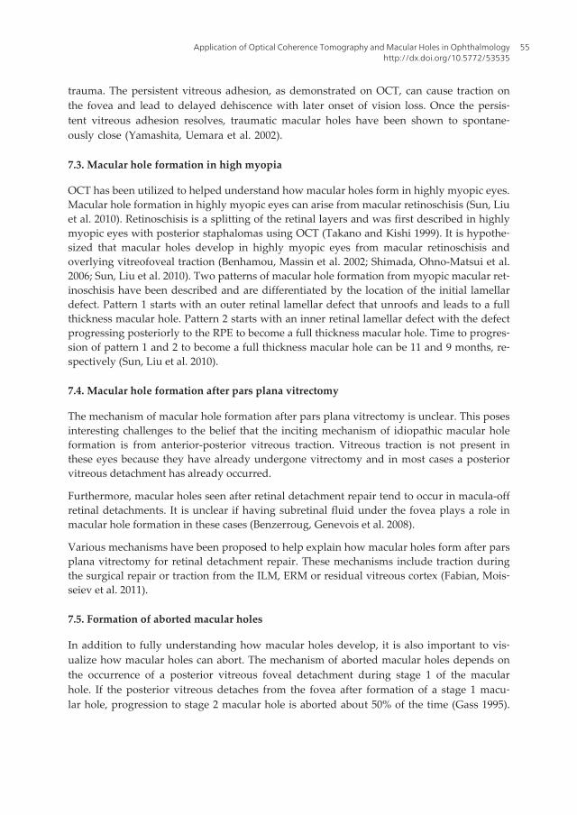
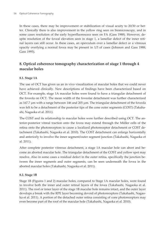
Stage 1B (Figures 1 and 2) macular holes, compared to Stage 1A macular holes, were foundto involve both the inner and outer retinal layers of the fovea (Takahashi, Nagaoka et al.2011). The roof or inner layer of the stage 1B macular hole remains intact, and the outer layerdevelops a break with the RPE layer becoming devoid of photoreceptors (Takahashi, Nagao‐ka et al. 2011). A portion of the detached outer retina consisting of cone photoreceptors mayeven become part of the roof of the macular hole (Takahashi, Nagaoka et al. 2010).
Optical Coherence Tomography56
Figure 1. Fundus photo of a stage 1B macular hole
*All OCT images in the book chapter were obtained using the Cirrus HD-OCT Model 4000, Carl Zeiss Meditec, Inc. orSpectral OCT SLO, Opko/OTI, Inc.
Figure 2. OCT* of the same eye from figure 1 showing a stage 1B macular hole. There is a defect in the outer layers ofthe retina.
8.3. Stage 2
OCT can show a break in the roof of a stage 2 full thickness macular hole (Figure 3). Thepatient, a 69 year old woman, had a decrease in visual acuity (VA) to 20/100 (Figure 3). After
Application of Optical Coherence Tomography and Macular Holes in Ophthalmologyhttp://dx.doi.org/10.5772/53535
57
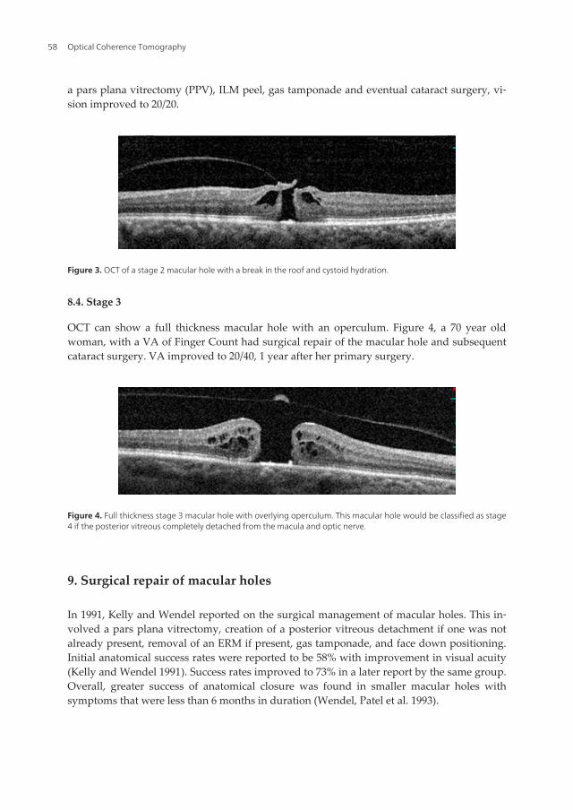
a pars plana vitrectomy (PPV), ILM peel, gas tamponade and eventual cataract surgery, vi‐sion improved to 20/20.
Figure 3. OCT of a stage 2 macular hole with a break in the roof and cystoid hydration.
8.4. Stage 3
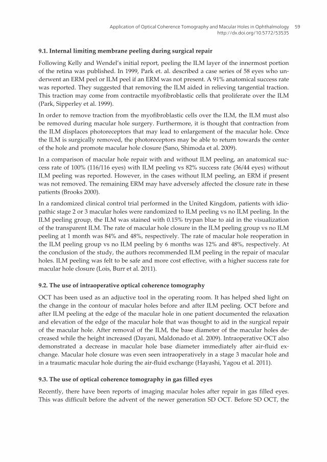
OCT can show a full thickness macular hole with an operculum. Figure 4, a 70 year oldwoman, with a VA of Finger Count had surgical repair of the macular hole and subsequentcataract surgery. VA improved to 20/40, 1 year after her primary surgery.
Figure 4. Full thickness stage 3 macular hole with overlying operculum. This macular hole would be classified as stage4 if the posterior vitreous completely detached from the macula and optic nerve.
9. Surgical repair of macular holes
In 1991, Kelly and Wendel reported on the surgical management of macular holes. This in‐volved a pars plana vitrectomy, creation of a posterior vitreous detachment if one was notalready present, removal of an ERM if present, gas tamponade, and face down positioning.Initial anatomical success rates were reported to be 58% with improvement in visual acuity(Kelly and Wendel 1991). Success rates improved to 73% in a later report by the same group.Overall, greater success of anatomical closure was found in smaller macular holes withsymptoms that were less than 6 months in duration (Wendel, Patel et al. 1993).
Optical Coherence Tomography58
9.1. Internal limiting membrane peeling during surgical repair
Following Kelly and Wendel’s initial report, peeling the ILM layer of the innermost portionof the retina was published. In 1999, Park et. al. described a case series of 58 eyes who un‐derwent an ERM peel or ILM peel if an ERM was not present. A 91% anatomical success ratewas reported. They suggested that removing the ILM aided in relieving tangential traction.This traction may come from contractile myofibroblastic cells that proliferate over the ILM(Park, Sipperley et al. 1999).
In order to remove traction from the myofibroblastic cells over the ILM, the ILM must alsobe removed during macular hole surgery. Furthermore, it is thought that contraction fromthe ILM displaces photoreceptors that may lead to enlargement of the macular hole. Oncethe ILM is surgically removed, the photoreceptors may be able to return towards the centerof the hole and promote macular hole closure (Sano, Shimoda et al. 2009).
In a comparison of macular hole repair with and without ILM peeling, an anatomical suc‐cess rate of 100% (116/116 eyes) with ILM peeling vs 82% success rate (36/44 eyes) withoutILM peeling was reported. However, in the cases without ILM peeling, an ERM if presentwas not removed. The remaining ERM may have adversely affected the closure rate in thesepatients (Brooks 2000).
In a randomized clinical control trial performed in the United Kingdom, patients with idio‐pathic stage 2 or 3 macular holes were randomized to ILM peeling vs no ILM peeling. In theILM peeling group, the ILM was stained with 0.15% trypan blue to aid in the visualizationof the transparent ILM. The rate of macular hole closure in the ILM peeling group vs no ILMpeeling at 1 month was 84% and 48%, respectively. The rate of macular hole reoperation inthe ILM peeling group vs no ILM peeling by 6 months was 12% and 48%, respectively. Atthe conclusion of the study, the authors recommended ILM peeling in the repair of macularholes. ILM peeling was felt to be safe and more cost effective, with a higher success rate formacular hole closure (Lois, Burr et al. 2011).
9.2. The use of intraoperative optical coherence tomography
OCT has been used as an adjuctive tool in the operating room. It has helped shed light onthe change in the contour of macular holes before and after ILM peeling. OCT before andafter ILM peeling at the edge of the macular hole in one patient documented the relaxationand elevation of the edge of the macular hole that was thought to aid in the surgical repairof the macular hole. After removal of the ILM, the base diameter of the macular holes de‐creased while the height increased (Dayani, Maldonado et al. 2009). Intraoperative OCT alsodemonstrated a decrease in macular hole base diameter immediately after air-fluid ex‐change. Macular hole closure was even seen intraoperatively in a stage 3 macular hole andin a traumatic macular hole during the air-fluid exchange (Hayashi, Yagou et al. 2011).
9.3. The use of optical coherence tomography in gas filled eyes
Recently, there have been reports of imaging macular holes after repair in gas filled eyes.This was difficult before the advent of the newer generation SD OCT. Before SD OCT, the
Application of Optical Coherence Tomography and Macular Holes in Ophthalmologyhttp://dx.doi.org/10.5772/53535
59
older TD OCTs were unable to image eyes under gas tamponade due to the excessive lightreflectivity of the gas-retinal interface and difficulty in imaging the correct area, easilymissed with TD OCT (Masuyama, Yamakiri et al. 2009).
Imaging the macular hole area is accomplished more easily using SD OCT. SD OCT canquickly perform serial scans over the macular area. Using SD OCT, it was found that imag‐ing though gas was easier with a complete gas fill. Once the gas level decreased to a 70% fill,the light reflex from the lower meniscus of the gas made imaging of the macular hole moredifficult. Imaging became possible again once the gas fill reached 50% (Masuyama, Yamakiriet al. 2009).
A recent study examining how quickly macular holes can close postoperatively using OCTfound closed macular holes in 10 of 13 eyes (76.9%) on postoperative day 1 and an addition‐al 2 eyes that closed by postoperative day 2 (Masuyama, Yamakiri et al. 2009). Sano et. al.had similar experiences with SD OCT imaging of macular holes through gas. Of the over90% of eyes they were able to image on postoperative day 1, over 90% were found to beclosed (Sano, Inoue et al. 2011).
After the vitrectomy and ILM peeling for the surgical repair of macular holes, patients wereinstructed to remain in a face down position so that the gas will be juxtaposed against themacular hole. The use of SD OCT in the immediate postoperative period may help in limit‐ing the amount of face down positioning needed. Patients may consider stopping their facedown positioning once the macular hole appears closed on OCT, which may be as early aspostoperative day 1 (Masuyama, Yamakiri et al. 2009; Sano, Inoue et al. 2011).
9.4. Silicone oil tamponade
For some patients, face down positioning may be impossible. In such patients, silicone oiltamponade can be a viable option. Oster et. al. demonstrated using SD OCT that there wasno difference in silicone oil tamponade with face forward or face down positioning in 75%of the study eyes (Oster, Mojana et al. 2010).
The use of silicone oil for myopic macular hole repair without face down positioning hasbeen show to be successful in the closure of myopic macular holes with subsequent im‐provement of vision. Myopic macular holes were successfully closed in 22 of 24 eyes (92%)using silicone oil after ILM peeling. These eyes had an average axial length of 29.6 ± 1.8 mmand an average of 23.3 ± 14.0 months of tamponade (Nishimura, Kimura et al. 2011).
Furthermore, Jumper et. al. found similar OCT findings of early post-surgical macular holeclosure through silicone oil tamponade as compared to gas tamponade. As early as the firstpostoperative day in eyes with silicone oil tamponade, flattening of the edges of the macularhole has been shown using OCT. This flattening of the edges was accompanied by a de‐crease in both the size of the intraretinal cystic spaces and in the subretinal fluid located atthe cuff of the macular hole (Jumper, Gallemore et al. 2000).
However, Oster et. al. has demonstrated that silicone oil tamponade may have some limita‐tions when compared with gas tamonade. Supine positioning may cause the silicone oil to
Optical Coherence Tomography60
rise anteriorly and a fluid pocket to form between the macular hole and silicone oil interface.In 7 of 10 eyes, the silicone oil bridged the macular hole while in the remaining 3 eyes, thesilicone oil filled the macular hole and was in contact with the RPE. If the silicone oil fills themacular hole, it may prevent the complete close of the macular hole. Furthermore, siliconeoil contact with the RPE may be toxic to the subfoveal RPE. This potential damage to theRPE does not occur with gas tamponade (Oster, Mojana et al. 2010).
10. Two OCT classifications to define macular hole closure
OCT has helps characterize macular hole closure. Two groups have described their ownclassification for macular hole closures.
Kang et. al. described macular hole closure as either type 1 or type 2 as imaged on OCT.Type 1 closure is defined as the presence of a continuous layer of retinal tissue bridging themacular hole. Normal foveal contour is seen in type 1 macular hole closures. Type 2 closureis defined as an interruption of retinal tissue between the edges of the macular hole. OnOCT, the edges of the macular hole were attached to the underlying RPE without subretinalfluid. According to Kang et al. type 1 and 2 closures represent successful anatomical closureof macular holes (Kang, Ahn et al. 2003). As expected, type 1 closure was associated withbetter visual outcomes than type 2 closure. Larger macular holes (mean diameter: 674 µm)were associated with type 2 closures, while smaller holes (mean diameter: 469 µm) weremore associated with type 1 closures (Kang, Ahn et al. 2003).
Tornambe et. al. provided a similar OCT classification system divided also into type 1 andtype 2 macular hole closures. Type 1 represented a flat and closed macular hole configura‐tion. Type 2 closure is an open macular hole with flat edges. Both types were consideredsuccessful macular hole closure. In comparison, a failed macular hole closure was seen as anopen macular hole with the edges being elevated by the subretinal fluid underneath (Tor‐nambe, Poliner et al. 1998).
11. Cases of surgical repair of macular holes
11.1. Idiopathic macular hole
A 69 year old woman presented with an idiopathic macular hole. The macular hole wasstage 2 with a break in the overlying roof with cystoid hydration at the edges of the macularhole (Figure 5). Visual acuity was Finger Count. The patient underwent a PPV, indocyaninegreen (ICG) staining of the ILM, ILM peeling and gas tamponade. After face down position‐ing, the macular hole closed (Figure 6). After subsequent cataract surgery, 6 months after in‐itial macular hole surgical repair, vision improved to 20/60.
Application of Optical Coherence Tomography and Macular Holes in Ophthalmologyhttp://dx.doi.org/10.5772/53535
61

Figure 5. Preoperative OCT of an eccentric stage 2 macular hole with vitreous adhesion to the roof of the macularhole
Figure 6. Postoperative OCT of the closed macular hole one month after surgical repair. Note the residual gas bubbleobscuring view of the superior retinal blood vessels at the top photo.
11.2. Traumatic macular hole
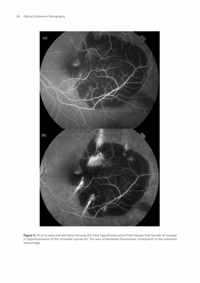
A 31 year old man presented with a traumatic macular hole following an airbag injury (Fig‐ure 7). Vision initially was 20/100 with OCT (Figure 8) revealing a full thickness macularhole with subretinal hemorrhage. There was an associated choroidal rupture (Figure 9). De‐spite no change in visual acuity, repeat OCT two weeks later revealed progressive cystoidhydration at the edges of the macular hole with subretinal fluid (Figure 10). The patient un‐derwent repair of the macular hole with PPV, ILM peeling, and gas tamponade with closureof the macular hole (Figure 11) and improvement in vision to 20/30.
Optical Coherence Tomography62
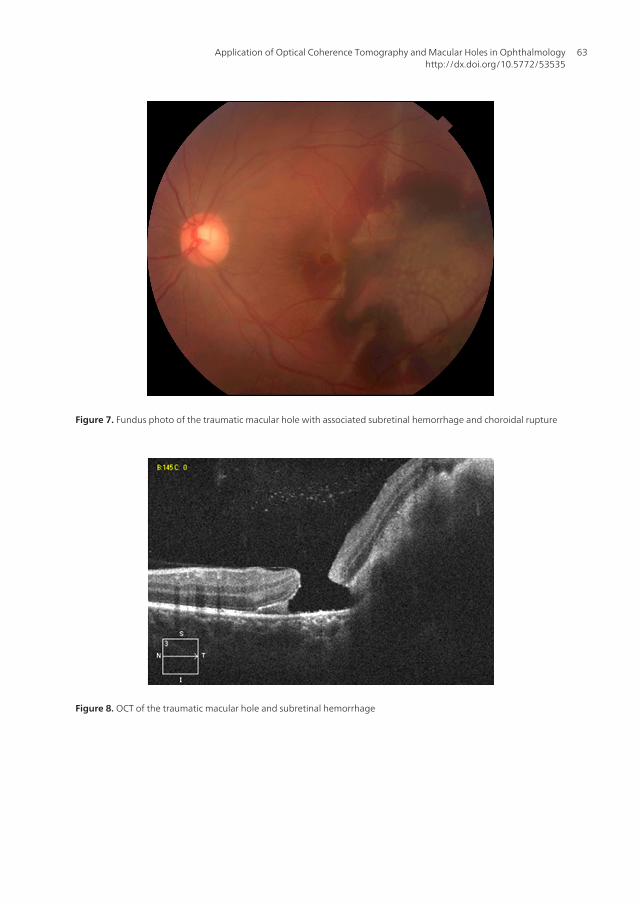
Figure 7. Fundus photo of the traumatic macular hole with associated subretinal hemorrhage and choroidal rupture
Figure 8. OCT of the traumatic macular hole and subretinal hemorrhage
Application of Optical Coherence Tomography and Macular Holes in Ophthalmologyhttp://dx.doi.org/10.5772/53535
63
(a)
(b)
Figure 9. FA of an early and late frame showing the initial hyperflourescence of the macular hole (a) with an increasein hyperfluoresence of the choroidal rupture (b). The area of decreased fluorescence corresponds to the subretinalhemorrhage.
Optical Coherence Tomography64
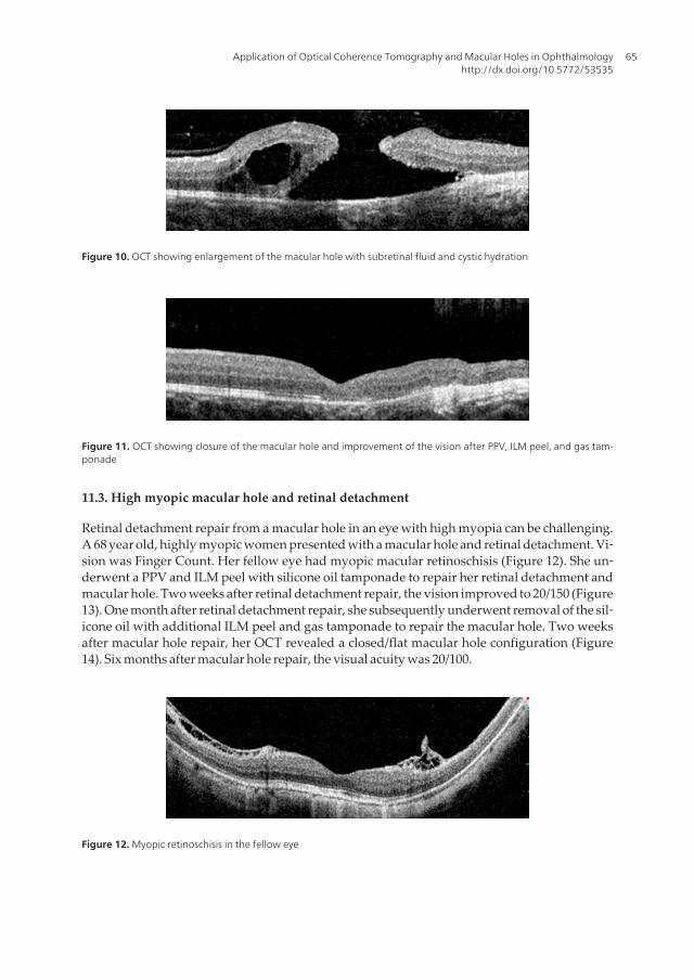
Figure 10. OCT showing enlargement of the macular hole with subretinal fluid and cystic hydration
Figure 11. OCT showing closure of the macular hole and improvement of the vision after PPV, ILM peel, and gas tam‐ponade
11.3. High myopic macular hole and retinal detachment
Retinal detachment repair from a macular hole in an eye with high myopia can be challenging.A 68 year old, highly myopic women presented with a macular hole and retinal detachment. Vi‐sion was Finger Count. Her fellow eye had myopic macular retinoschisis (Figure 12). She un‐derwent a PPV and ILM peel with silicone oil tamponade to repair her retinal detachment andmacular hole. Two weeks after retinal detachment repair, the vision improved to 20/150 (Figure13). One month after retinal detachment repair, she subsequently underwent removal of the sil‐icone oil with additional ILM peel and gas tamponade to repair the macular hole. Two weeksafter macular hole repair, her OCT revealed a closed/flat macular hole configuration (Figure14). Six months after macular hole repair, the visual acuity was 20/100.
Figure 12. Myopic retinoschisis in the fellow eye
Application of Optical Coherence Tomography and Macular Holes in Ophthalmologyhttp://dx.doi.org/10.5772/53535
65
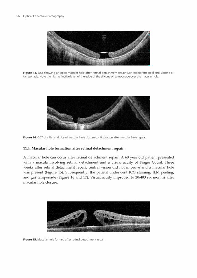
Figure 13. OCT showing an open macular hole after retinal detachment repair with membrane peel and silicone oiltamponade. Note the high reflective layer of the edge of the silicone oil tamponade over the macular hole.
Figure 14. OCT of a flat and closed macular hole closure configuration after macular hole repair.
11.4. Macular hole formation after retinal detachment repair
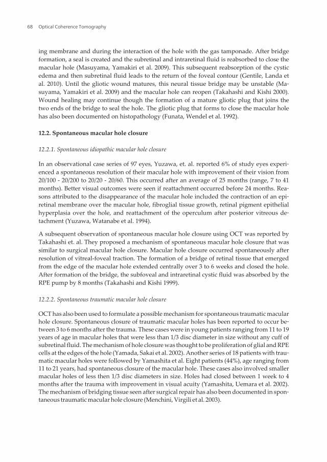
A macular hole can occur after retinal detachment repair. A 60 year old patient presentedwith a macula involving retinal detachment and a visual acuity of Finger Count. Threeweeks after retinal detachment repair, central vision did not improve and a macular holewas present (Figure 15). Subsequently, the patient underwent ICG staining, ILM peeling,and gas tamponade (Figure 16 and 17). Visual acuity improved to 20/400 six months aftermacular hole closure.
Figure 15. Macular hole formed after retinal detachment repair.
Optical Coherence Tomography66
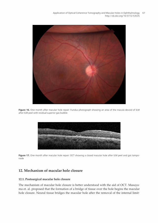
Figure 16. One month after macular hole repair: Fundus photograph showing an area of the macula devoid of ILMafter ILM peel with residual superior gas bubble
Figure 17. One month after macular hole repair: OCT showing a closed macular hole after ILM peel and gas tampo‐nade
12. Mechanism of macular hole closure
12.1. Postsurgical macular hole closure
The mechanism of macular hole closure is better understood with the aid of OCT. Masuya‐ma et. al. proposed that the formation of a bridge of tissue over the hole begins the macularhole closure. Neural tissue bridges the macular hole after the removal of the internal limit‐
Application of Optical Coherence Tomography and Macular Holes in Ophthalmologyhttp://dx.doi.org/10.5772/53535
67
ing membrane and during the interaction of the hole with the gas tamponade. After bridgeformation, a seal is created and the subretinal and intraretinal fluid is reabsorbed to close themacular hole (Masuyama, Yamakiri et al. 2009). This subsequent reabsorption of the cysticedema and then subretinal fluid leads to the return of the foveal contour (Gentile, Landa etal. 2010). Until the gliotic wound matures, this neural tissue bridge may be unstable (Ma‐suyama, Yamakiri et al. 2009) and the macular hole can reopen (Takahashi and Kishi 2000).Wound healing may continue though the formation of a mature gliotic plug that joins thetwo ends of the bridge to seal the hole. The gliotic plug that forms to close the macular holehas also been documented on histopathology (Funata, Wendel et al. 1992).
12.2. Spontaneous macular hole closure
12.2.1. Spontaneous idiopathic macular hole closure
In an observational case series of 97 eyes, Yuzawa, et. al. reported 6% of study eyes experi‐enced a spontaneous resolution of their macular hole with improvement of their vision from20/100 - 20/200 to 20/20 - 20/60. This occurred after an average of 25 months (range, 7 to 41months). Better visual outcomes were seen if reattachment occurred before 24 months. Rea‐sons attributed to the disappearance of the macular hole included the contraction of an epi‐retinal membrane over the macular hole, fibroglial tissue growth, retinal pigment epithelialhyperplasia over the hole, and reattachment of the operculum after posterior vitreous de‐tachment (Yuzawa, Watanabe et al. 1994).
A subsequent observation of spontaneous macular hole closure using OCT was reported byTakahashi et. al. They proposed a mechanism of spontaneous macular hole closure that wassimilar to surgical macular hole closure. Macular hole closure occurred spontaneously afterresolution of vitreal-foveal traction. The formation of a bridge of retinal tissue that emergedfrom the edge of the macular hole extended centrally over 3 to 6 weeks and closed the hole.After formation of the bridge, the subfoveal and intraretinal cystic fluid was absorbed by theRPE pump by 8 months (Takahashi and Kishi 1999).
12.2.2. Spontaneous traumatic macular hole closure
OCT has also been used to formulate a possible mechanism for spontaneous traumatic macularhole closure. Spontaneous closure of traumatic macular holes has been reported to occur be‐tween 3 to 6 months after the trauma. These cases were in young patients ranging from 11 to 19years of age in macular holes that were less than 1/3 disc diameter in size without any cuff ofsubretinal fluid. The mechanism of hole closure was thought to be proliferation of glial and RPEcells at the edges of the hole (Yamada, Sakai et al. 2002). Another series of 18 patients with trau‐matic macular holes were followed by Yamashita et al. Eight patients (44%), age ranging from11 to 21 years, had spontaneous closure of the macular hole. These cases also involved smallermacular holes of less then 1/3 disc diameters in size. Holes had closed between 1 week to 4months after the trauma with improvement in visual acuity (Yamashita, Uemara et al. 2002).The mechanism of bridging tissue seen after surgical repair has also been documented in spon‐taneous traumatic macular hole closure (Menchini, Virgili et al. 2003).
Optical Coherence Tomography68
13. Morphological changes after macular hole closure
OCT imaging of surgically closed macular holes has been showed to aid in predicting visualoutcome. The preservation of the external limiting membrane (ELM) layer and photorecep‐tor inner and outer segment (IS/OS) junction seems to predict visual acuity and photorecep‐tor cell survival (Shimozono, Oishi et al. 2011). This is believed to be because the ELMrepresents the junction between the Müller cells and photoreceptor cells (Bottoni, De An‐gelis et al. 2011) and the IS/OS junction represents the integrity of photoreceptor alignment.Furthermore, the outer foveal thickness measured between the ELM and inner layer of theRPE may be a surrogate to outer segment regeneration (Shimozono, Oishi et al. 2011). Anincrease in outer foveal thickness 6 months after surgery is associated with better visual out‐comes. In addition, ELM and IS/OS defects are associated with worse visual acuity aftermacular hole surgical repair (Shimozono, Oishi et al. 2011).
Visual recovery after macular hole closure is thought to occur after the detached photorecep‐tors at the edges of the macular hole reattach to the RPE (Yuzawa, Watanabe et al. 1994). Over‐all, vision improves when the outer segments elongate (increase in outer foveal thickness) withsubsequent realignment of the photoreceptors (return of the IS/OS junction) as seen on OCT.An intact ELM (viable photoreceptor cells) needs to be present before the process of visual re‐covery begins (Shimozono, Oishi et al. 2011). After macular hole closure, the ELM appears be‐fore the IS/OS layer returns. In a study by Bottoni, et. al., there were no cases seen where therewas a continuous IS/OS junction and a discontinuous ELM after macular hole closure (Bottoni,De Angelis et al. 2011). Early disruption of the ELM during the postoperative period may be in‐dicative of a poor visual outcome (Shimozono, Oishi et al. 2011).
Despite the successful reapproximation of the photoreceptors during macular hole closure,the status of the IS/OS layer on OCT can still vary (Sano, Shimoda et al. 2009). When com‐paring the continuity or discontinuity of the IS/OS layer, a continuous IS/OS in the foveawas associated with better visual outcomes at 6 months. A discontinuous IS/OS layer ap‐pears to represent damaged and/or missing photoreceptor outer segments (Sano, Shimodaet al. 2009).
14. Masquerades and differential diagnosis
OCT has helped in differentiating macular holes from clinically similar diagnoses. Makingthe correct diagnosis is important and will determine the medical and surgical managementof these conditions.
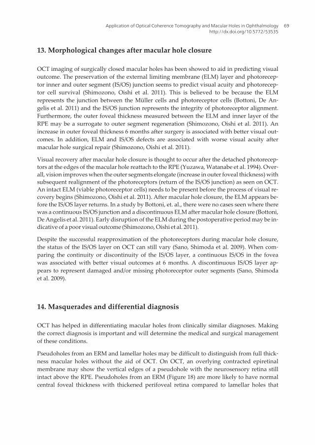
Pseudoholes from an ERM and lamellar holes may be difficult to distinguish from full thick‐ness macular holes without the aid of OCT. On OCT, an overlying contracted epiretinalmembrane may show the vertical edges of a pseudohole with the neurosensory retina stillintact above the RPE. Pseudoholes from an ERM (Figure 18) are more likely to have normalcentral foveal thickness with thickened perifoveal retina compared to lamellar holes that
Application of Optical Coherence Tomography and Macular Holes in Ophthalmologyhttp://dx.doi.org/10.5772/53535
69
usually have a thinner and irregular center with normal perifoval thickness (Haouchine,Massin et al. 2004).
Figure 18. 60 year old woman with an ERM and pseudohole and 20/50 vision.
Lamellar macular holes are not true holes. Instead, they consist of defects in the inner retinawith varying degrees of foveal thinning (Yeh, Chen et al. 2010). OCT has greatly enhanced ourability to diagnose lamellar holes, especially in highly myopic eyes. (Tanaka, Shimada et al.2011). Findings on OCT include a discontinuous inner retinal layer, splitting of the inner retinallayer, and an irregular foveal contour. Lamellar macular holes are believed in some cases to bethe result of aborted macular holes. Inner retinal defects may be due to the rupture and subse‐quent loss of the roof of an inner cyst of a stage 1 macular hole (Yeh, Chen et al. 2010).
However, lamellar macular holes progressing to full thickness holes have occurred (Takaha‐shi and Kishi 2000; Tanaka, Shimada et al. 2011). These lamellar macular holes tend to havea horizontal split between the inner and outer retina that emanate from the center (Haou‐chine, Massin et al. 2004).
In addition, OCT can quantify the extent of vitreofoveal traction and differentiate vitreomac‐ular traction syndrome from stage 1A or stage 1B macular holes. Vitreomacular traction syn‐dromes have a much broader based of vitreal-retinal adhesions compared to the focal areasof vitreal-foveal adhesions seen in stage 1A or 1B macular holes (Comander, Gardiner et al.2011). Furthermore, OCT has helped in differentiating the yellow spot seen on biomicrosco‐py in stage 1A macular holes from the yellow spots seen in central serous chroidopathy,ERM, or vitelliform dystrophy (Gass 1995).
Solar retinopathy can also have an appearance similar to a small full thickness macular hole.When solar retinopathy initially presents after solar exposure, it has the appearance of a yel‐low spot similar to what is seen in a stage 1A macular hole. Late findings in solar retinop‐athy involve a rectangular shaped outer retinal defect with straight edges that encompass anarea from the RPE to the external limiting membrane. This outer retinal defect may be diffi‐cult to distinguish from a closed macular hole with a persistent similarly shaped outer reti‐nal defect (Comander, Gardiner et al. 2011).
Optical Coherence Tomography70
15. Conclusion
Our understanding of macular holes has come a long way from the initial descriptions byGass to our current imaging with OCT. OCT has provided an in vivo ability to see changesin the retina that we were not able to obtain without histopathology. Correlating clinicalfindings, understanding the mechanism of macular hole formation and closure, and differ‐entiating masquerades have been made possible with the aid of OCT technology.
Author details
Robert J. Lowe1,2 and Ronald C. Gentile1
1 The New York Eye and Ear Infirmary, New York, USA and New York Medical College,Valhalla, NY, USA
2 Kaiser Permanente Medical Group, Walnut Creek, California, USA
References
[1] Benhamou, N., P. Massin, et al. (2002). "Macular retinoschisis in highly myopic eyes."Am J Ophthalmol 133(6): 794-800.
[2] Benzerroug, M., O. Genevois, et al. (2008). "Results of surgery on macular holes thatdevelop after rhegmatogenous retinal detachment." Br J Ophthalmol 92(2): 217-219.
[3] Bottoni, F., S. De Angelis, et al. (2011). "The dynamic healing process of idiopathicmacular holes after surgical repair: a spectral-domain optical coherence tomographystudy." Invest Ophthalmol Vis Sci 52(7): 4439-4446.
[4] Brooks, H. L., Jr. (2000). "Macular hole surgery with and without internal limitingmembrane peeling." Ophthalmology 107(10): 1939-1948; discussion 1948-1939.
[5] Chow, D. R., G. A. Williams, et al. (1999). "Successful closure of traumatic macularholes." Retina 19(5): 405-409.
[6] Comander, J., M. Gardiner, et al. (2011). "High-Resolution Optical Coherence Tomog‐raphy Findings in Solar Maculopathy and the Differential Diagnosis of Outer RetinalHoles." Am J Ophthalmol.
[7] Coppe, A. M., G. Ripandelli, et al. (2005). "Prevalence of asymptomatic macular holesin highly myopic eyes." Ophthalmology 112(12): 2103-2109.
[8] Dayani, P. N., R. Maldonado, et al. (2009). "Intraoperative use of handheld spectraldomain optical coherence tomography imaging in macular surgery." Retina 29(10):1457-1468.
Application of Optical Coherence Tomography and Macular Holes in Ophthalmologyhttp://dx.doi.org/10.5772/53535
71
[9] Fabian, I. D., E. Moisseiev, et al. (2011). "Macular Hole After Vitrectomy for PrimaryRhegmatogenous Retinal Detachment." Retina.
[10] Frangieh, G. T., W. R. Green, et al. (1981). "A histopathologic study of macular cystsand holes." Retina 1(4): 311-336.
[11] Funata, M., R. T. Wendel, et al. (1992). "Clinicopathologic study of bilateral macularholes treated with pars plana vitrectomy and gas tamponade." Retina 12(4): 289-298.
[12] Gass, J. D. (1988). "Idiopathic senile macular hole. Its early stages and pathogenesis."Arch Ophthalmol 106(5): 629-639.
[13] Gass, J. D. (1995). "Reappraisal of biomicroscopic classification of stages of develop‐ment of a macular hole." Am J Ophthalmol 119(6): 752-759.
[14] Gentile, R. C., G. Landa, et al. (2010). "Macular hole formation, progression, and sur‐gical repair: case series of serial optical coherence tomography and time lapsemorphing video study." BMC Ophthalmol 10: 24.
[15] Haouchine, B., P. Massin, et al. (2004). "Diagnosis of macular pseudoholes and lamel‐lar macular holes by optical coherence tomography." Am J Ophthalmol 138(5):732-739.
[16] Hayashi, A., T. Yagou, et al. (2011). "Intraoperative changes in idiopathic macularholes by spectral-domain optical coherence tomography." Case Report Ophthalmol2(2): 149-154.
[17] Ikuno, Y., K. Sayanagi, et al. (2003). "Optical coherence tomographic findings of mac‐ular holes and retinal detachment after vitrectomy in highly myopic eyes." Am JOphthalmol 136(3): 477-481.
[18] Johnson, R. N. and J. D. Gass (1988). "Idiopathic macular holes. Observations, stagesof formation, and implications for surgical intervention." Ophthalmology 95(7):917-924.
[19] Johnson, R. N., H. R. McDonald, et al. (2001). "Traumatic macular hole: observations,pathogenesis, and results of vitrectomy surgery." Ophthalmology 108(5): 853-857.
[20] Jumper, J. M., R. P. Gallemore, et al. (2000). "Features of macular hole closure in theearly postoperative period using optical coherence tomography." Retina 20(3):232-237.
[21] Kang, S. W., K. Ahn, et al. (2003). "Types of macular hole closure and their clinicalimplications." Br J Ophthalmol 87(8): 1015-1019.
[22] Kelly, N. E. and R. T. Wendel (1991). "Vitreous surgery for idiopathic macular holes.Results of a pilot study." Arch Ophthalmol 109(5): 654-659.
[23] Kobayashi, H., K. Kobayashi, et al. (2002). "Macular hole and myopic refraction." Br JOphthalmol 86(11): 1269-1273.
Optical Coherence Tomography72
[24] Lee, S. H., K. H. Park, et al. (2010). "Secondary macular hole formation after vitrecto‐my." Retina 30(7): 1072-1077.
[25] Lee, S. W., S. W. Kang, et al. (2011). "Vitreous surgery for impending macular hole."Retina 31(5): 909-914.
[26] Lois, N., J. Burr, et al. (2011). "Internal limiting membrane peeling versus no peelingfor idiopathic full thickness macular hole: a pragmatic randomized controlled trial."Invest Ophthalmol Vis Sci 52(3): 1586-1592.
[27] Masuyama, K., K. Yamakiri, et al. (2009). "Posturing time after macular hole surgerymodified by optical coherence tomography images: a pilot study." Am J Ophthalmol147(3): 481-488 e482.
[28] McCannel, C. A., J. L. Ensminger, et al. (2009). "Population-based incidence of macu‐lar holes." Ophthalmology 116(7): 1366-1369.
[29] McDonnell, P. J., S. L. Fine, et al. (1982). "Clinical features of idiopathic macular cystsand holes." Am J Ophthalmol 93(6): 777-786.
[30] Menchini, U., G. Virgili, et al. (2003). "Mechanism of spontaneous closure of traumat‐ic macular hole: OCT study of one case." Retina 23(1): 104-106.
[31] Nishimura, A., M. Kimura, et al. (2011). "Efficacy of primary silicone oil tamponadefor the treatment of retinal detachment caused by macular hole in high myopia." AmJ Ophthalmol 151(1): 148-155.
[32] Oster, S. F., F. Mojana, et al. (2010). "Dynamics of the macular hole-silicone oil tampo‐nade interface with patient positioning as imaged by spectral domain-optical coher‐ence tomography." Retina 30(6): 924-929.
[33] Park, D. W., J. O. Sipperley, et al. (1999). "Macular hole surgery with internal-limitingmembrane peeling and intravitreous air." Ophthalmology 106(7): 1392-1397; discus‐sion 1397-1398.
[34] Patel, S. C., R. H. Loo, et al. (2001). "Macular hole surgery in high myopia." Ophthal‐mology 108(2): 377-380.
[35] Sano, M., M. Inoue, et al. (2011). "Ability to determine postoperative status of macu‐lar hole in gas-filled eyes by spectral domain-optical coherence tomography." ClinExperiment Ophthalmol.
[36] Sano, M., Y. Shimoda, et al. (2009). "Restored photoreceptor outer segment and visualrecovery after macular hole closure." Am J Ophthalmol 147(2): 313-318 e311.
[37] Shimada, N., K. Ohno-Matsui, et al. (2006). "Natural course of macular retinoschisisin highly myopic eyes without macular hole or retinal detachment." Am J Ophthal‐mol 142(3): 497-500.
Application of Optical Coherence Tomography and Macular Holes in Ophthalmologyhttp://dx.doi.org/10.5772/53535
73
[38] Shimozono, M., A. Oishi, et al. (2011). "Restoration of the photoreceptor outer seg‐ment and visual outcomes after macular hole closure: spectral-domain optical coher‐ence tomography analysis." Graefes Arch Clin Exp Ophthalmol.
[39] Sun, C. B., Z. Liu, et al. (2010). "Natural evolution from macular retinoschisis to fullthickness macular hole in highly myopic eyes." Eye (Lond) 24(12): 1787-1791.
[40] Takahashi, A., T. Nagaoka, et al. (2010). "Foveal anatomic changes in a progressingstage 1 macular hole documented by spectral-domain optical coherence tomogra‐phy." Ophthalmology 117(4): 806-810.
[41] Takahashi, A., T. Nagaoka, et al. (2011). "Stage 1-A macular hole: a prospective spec‐tral-domain optical coherence tomography study." Retina 31(1): 127-147.
[42] Takahashi, H. and S. Kishi (1999). "Optical coherence tomography images of sponta‐neous macular hole closure." Am J Ophthalmol 128(4): 519-520.
[43] Takahashi, H. and S. Kishi (2000). "Tomographic features of a lamellar macular holeformation and a lamellar hole that progressed to a full thickness macular hole." Am JOphthalmol 130(5): 677-679.
[44] Takahashi, H. and S. Kishi (2000). "Tomographic features of early macular hole clo‐sure after vitreous surgery." Am J Ophthalmol 130(2): 192-196.
[45] Takano, M. and S. Kishi (1999). "Foveal retinoschisis and retinal detachment in se‐verely myopic eyes with posterior staphyloma." Am J Ophthalmol 128(4): 472-476.
[46] Tanaka, Y., N. Shimada, et al. (2011). "Natural history of lamellar macular holes inhighly myopic eyes." Am J Ophthalmol 152(1): 96-99 e91.
[47] Tornambe, P. E., L. S. Poliner, et al. (1998). "Definition of macular hole surgery endpoints: elevated/open, flat/open, flat/closed." Retina 18(3): 286-287.
[48] Weichel, E. D. and M. H. Colyer (2009). "Traumatic macular holes secondary to com‐bat ocular trauma." Retina 29(3): 349-354.
[49] Wendel, R. T., A. C. Patel, et al. (1993). "Vitreous surgery for macular holes." Oph‐thalmology 100(11): 1671-1676.
[50] Wu, T. T. and Y. H. Kung (2011). "Comparison of anatomical and visual outcomes ofmacular hole surgery in patients with high myopia vs. non-high myopia: a case-con‐trol study using optical coherence tomography." Graefes Arch Clin Exp Ophthalmol.
[51] Yamada, H., A. Sakai, et al. (2002). "Spontaneous closure of traumatic macular hole."Am J Ophthalmol 134(3): 340-347.
[52] Yamashita, T., A. Uemara, et al. (2002). "Spontaneous closure of traumatic macularhole." Am J Ophthalmol 133(2): 230-235.
[53] Yeh, P. T., T. C. Chen, et al. (2010). "Formation of idiopathic macular hole-reapprais‐al." Graefes Arch Clin Exp Ophthalmol 248(6): 793-798.
Optical Coherence Tomography74

[54] Yuzawa, M., A. Watanabe, et al. (1994). "Observation of idiopathic full thickness mac‐
ular holes. Follow-up observation." Arch Ophthalmol 112(8): 1051-1056.
Application of Optical Coherence Tomography and Macular Holes in Ophthalmologyhttp://dx.doi.org/10.5772/53535
75

Related Documents