Apoptosis-associated proteins in oral lymphomas from HIV- positive patients Joseph A. Regezi, DDS, MS, a Alex McMillan, PhD, b Nusi Dekker, MA, a Troy E. Daniels, DDS, MS, a Sol Silverman, Jr., DDS, a Michael Schoelch, DMD, a,c and Barry L. Ziober, PhD, a San Francisco, Calif. UNIVERSITYOF CALIFORNIA AT SAN FRANCISCO Extranodal oral lymphomas, seen with increasing frequency in HIV infection, may have dysfunctional apoptotic mech- anisms that favor tumor progression. The purpose of this study was to evaluate extranodal lymphomas from HlV-positive patients for expression of apoptosis-associated proteins. Correlations were made with 10 histologically comparable extranodal lymphomas from HlV-negative patients and 6 hyperplastic lymph nodes from otherwise healthy young adults. Formalin-fixed tissue sections were immunohistochemically stained for apoptosis-associated proteins (Bcl-2, Bcl-x, Bax, Bak, p53, MDM2, BHRF). In situ hybridization was also done on deparaffinized sections for Epstein-Barr virus EBER mRNA. Eighteen consecutive oral lymphomas were studied in HIV/AIDS-positive patients. Four of 5 intermediate-grade lymphomas expressed Bcl-2 to a greater degree than did high-grade lymphomas (4 of 13). Most lymphomas were positive for Bcl-x and Bax, and few expressed Bak. The staining patterns for these proteins were similar to those seen in HIV-negative patients. Staining patterns were relatively consistent in the hyperplastic lymph nodes, whereas such patterns were irregular in lymphomas. Positive p53 staining was seen in 11 of 18 HIV-positive cases; 9 of these were also MDM2-positive. Double stains suggested that both p53 and MDM2 proteins were expressed in the same cells in these nine cases. Epstein-Barr virus-EBER mRNA was detected in 14 of 18 cases and in 3 of 10 cases from HIV-negative patients. BHRF staining was evident in onlya few cells of three HIV-positive lymphomas. The irreg- ular expression of Bcl-2, Bcl-x, Bax, and Bak in oral lymphomas indicates dysfunctional apoptotic mechanisms in these tumors. Bc[-2 staining differs with tumor grade. Positive staining for p53 and MDM2 proteins is a notable feature of lymphomas in HIV- positive patients and may relate to binding of MDM2 to wild-type p53. Epstein-Barr virus is more commonly associated with oral lymphomas in HlV-positive patients, although the Epstein-Barr virus-produced protein BHRF, which has Bcl-2-1ike activity, is minimally expressed. (Oral Surg Oral Med Oral Pathol Oral Radiol Endod 1998;86:196-202) Non-Hodgkin's lymphomas are relatively common neoplasms (53,600 new cases estimated for 1997) 1 and often occur in extranodal head and neck sites, espe- cially in cases of HIV infection (AIDS). 2-6 Until recently, extranodal oral lymphomas, appearing as either primary or secondary oral disease, were uncommon.7, 8 With the emergence of HIV infection, oral HIV-associated lymphomas are now seen with increasing frequency and are accompanied by a distinct shift to predominately high-grade tumors associated with Epstein-Barr virus (EBV) infection. 9-14 Apoptosis is a genetically controlled cell death mechanism that can be activated by a number of stimuli and detected by morphologic alterations15,16 and biochemical changes (endonuclease-associated nuclear fragmentation).17 Apoptosis-associated gene products have either proapoptotic or antiapoptotic functions. 18-20Loss of apoptosis control is a potentially important aspect of tumor biology. This study was supportedin part by NIH grant PO1-DE-07946. aDepartmeutof Stomatology, UCSE bUCSF Cancer Center. CVisitingInvestigator from the US Navy. Receivedfor publicationJan. 15, 1998; returnedfor revisionMar. 23, 1998; acceptedfor publicationApr. 7, 1998. Copyright © 1998 by Mosby,Inc. 1079-2104/98/$5.00 + 0 7/14/90964 Important apoptosis-associated genes are members of the Bcl-2 family (Bcl-2, Bcl-x, Bax, Bak, and others). Bcl-2 protein is antiapoptotic. 21,22 Bcl-x represents two proteins (Bcl-x L and Bcl-xs) that have opposing func- tions, 23-25 whereas Bax 26,27 and Bak 28,29 are proapop- totic. The many competing proteins in the Bcl-2 family regulate apoptosis by their relative quantities and formation of competing dimers.26, 30 Overexpression of Bcl-2 has been described in follicular lymphomas31,32 and other lymphomas, including HIV-associated and oral lymphomas. 33-35 Other Bcl-2-associated proteins are also involved in the pathogenesis of lymphomas (non-HIV and HIV-associated), inasmuch as both Bcl-x and Bax have been detected in these tumors. The protein designated p53, which is a tumor suppressor gene product, can arrest the cell cycle in the G 1phase to mediate DNA repair, or it can induce apop- tosis through regulation of apoptosis-associated genes (Bcl-2 suppression and increased Bax expression). 36,~7 p53 mutations resulting in defective protein contribute to cell proliferation. Although wild-type and mutated p53 overexpression is frequently seen in lymphomas, many cases are not associated with gene mutation. 38 p53-associated cell proliferation can also occur when wild-type protein is bound and inactivated by other cellular proteins (MDM2 protein) and viral proteins (human papillomavirus E6 protein, EBV EBNA5, and 196

Welcome message from author
This document is posted to help you gain knowledge. Please leave a comment to let me know what you think about it! Share it to your friends and learn new things together.
Transcript
Apoptosis-associated proteins in oral lymphomas from HIV- positive patients Joseph A. Regezi, DDS, MS, a Alex McMillan, PhD, b Nusi Dekker, MA, a Troy E. Daniels, DDS, MS, a Sol Silverman, Jr., DDS, a Michael Schoelch, DMD, a,c and Barry L. Ziober, PhD, a San Francisco, Calif. UNIVERSITY OF CALIFORNIA AT SAN FRANCISCO
Extranodal oral lymphomas, seen with increasing frequency in HIV infection, may have dysfunctional apoptotic mech- anisms that favor tumor progression. The purpose of this study was to evaluate extranodal lymphomas from HlV-positive patients for expression of apoptosis-associated proteins. Correlations were made with 10 histologically comparable extranodal lymphomas from HlV-negative patients and 6 hyperplastic lymph nodes from otherwise healthy young adults. Formalin-fixed tissue sections were immunohistochemically stained for apoptosis-associated proteins (Bcl-2, Bcl-x, Bax, Bak, p53, MDM2, BHRF). In situ hybridization was also done on deparaffinized sections for Epstein-Barr virus EBER mRNA. Eighteen consecutive oral lymphomas were studied in HIV/AIDS-positive patients. Four of 5 intermediate-grade lymphomas expressed Bcl-2 to a greater degree than did high-grade lymphomas (4 of 13). Most lymphomas were positive for Bcl-x and Bax, and few expressed Bak. The staining patterns for these proteins were similar to those seen in HIV-negative patients. Staining patterns were relatively consistent in the hyperplastic lymph nodes, whereas such patterns were irregular in lymphomas. Positive p53 staining was seen in 11 of 18 HIV-positive cases; 9 of these were also MDM2-positive. Double stains suggested that both p53 and MDM2 proteins were expressed in the same cells in these nine cases. Epstein-Barr virus-EBER mRNA was detected in 14 of 18 cases and in 3 of 10 cases from HIV-negative patients. BHRF staining was evident in onlya few cells of three HIV-positive lymphomas. The irreg- ular expression of Bcl-2, Bcl-x, Bax, and Bak in oral lymphomas indicates dysfunctional apoptotic mechanisms in these tumors. Bc[-2 staining differs with tumor grade. Positive staining for p53 and MDM2 proteins is a notable feature of lymphomas in HIV- positive patients and may relate to binding of MDM2 to wild-type p53. Epstein-Barr virus is more commonly associated with oral lymphomas in HlV-positive patients, although the Epstein-Barr virus-produced protein BHRF, which has Bcl-2-1ike activity, is minimally expressed. (Oral Surg Oral Med Oral Pathol Oral Radiol Endod 1998;86:196-202)
Non-Hodgkin 's lymphomas are relatively common neoplasms (53,600 new cases estimated for 1997) 1 and often occur in extranodal head and neck sites, espe- cially in cases of HIV infection (AIDS). 2-6 Until recently, extranodal oral lymphomas, appearing as either primary or secondary oral disease, were uncommon.7, 8 With the emergence of HIV infection, oral HIV-associated lymphomas are now seen with increasing frequency and are accompanied by a distinct shift to predominately high-grade tumors associated with Epstein-Barr virus (EBV) infection. 9-14
Apoptosis is a genetically controlled cell death mechanism that can be activated by a number of stimuli and detected by morphologic alterations15,16 and biochemical changes (endonuclease-associated nuclear fragmentation).17 Apoptosis-associated gene products have either proapoptotic or antiapoptotic functions. 18-20 Loss of apoptosis control is a potentially important aspect of tumor biology.
This study was supported in part by NIH grant PO1-DE-07946. aDepartmeut of Stomatology, UCSE bUCSF Cancer Center. CVisiting Investigator from the US Navy. Received for publication Jan. 15, 1998; returned for revision Mar. 23, 1998; accepted for publication Apr. 7, 1998. Copyright © 1998 by Mosby, Inc. 1079-2104/98/$5.00 + 0 7/14/90964
Important apoptosis-associated genes are members of the Bcl-2 family (Bcl-2, Bcl-x, Bax, Bak, and others). Bcl-2 protein is antiapoptotic. 21,22 Bcl-x represents two proteins (Bcl-x L and Bcl-xs) that have opposing func- tions, 23-25 whereas Bax 26,27 and Bak 28,29 are proapop- totic. The many competing proteins in the Bcl-2 family regulate apoptosis by their relative quantities and formation of competing dimers.26, 30 Overexpression of Bcl-2 has been described in follicular lymphomas31, 32 and other lymphomas, including HIV-associated and oral lymphomas. 33-35 Other Bcl-2-associated proteins are also involved in the pathogenesis of lymphomas (non-HIV and HIV-associated), inasmuch as both Bcl-x and Bax have been detected in these tumors.
The protein designated p53, which is a tumor suppressor gene product, can arrest the cell cycle in the G 1 phase to mediate DNA repair, or it can induce apop- tosis through regulation of apoptosis-associated genes (Bcl-2 suppression and increased Bax expression). 36,~7 p53 mutations resulting in defective protein contribute to cell proliferation. Although wild-type and mutated p53 overexpression is frequently seen in lymphomas, many cases are not associated with gene mutation. 38 p53-associated cell proliferation can also occur when wild-type protein is bound and inactivated by other cellular proteins (MDM2 protein) and viral proteins (human papillomavirus E6 protein, EBV EBNA5, and
196
ORAL SURGERY ORAL MEDICINE ORAL PATHOLOGY Regezi etal. 197 Volume 86, Number 2
Table I. Antibodies used to identify apoptosis-associated proteins
Antigen Type Clone~type Source Dilution
Bcl-2 Antiapoptosis 124 (IgGlk) Dako, Carpinteria, Calif. 1:100 Bcl-Xs/l* Apoptosis (Xs) and Polyclonal Santa Cruz Biotechnology, 1:1000
antiapoptosis (X1) Santa Cruz, Calif. Apoptosis Coulter/Immunotech, Westbrook, Me. Apoptosis Oncogene Science, Cambridge, Mass. Proliferation Oncogene Science Apoptosis and Novocastra0 Burlingame, Calif. antiproliferation Antiapoptosis
Bax 4F11 (IgG2b) 1:200 Bak TC-100 (IgG2k) 1:25 MDM2 1F2 (IgG2bk) 1:25 p537 DO-7 (IgG2b) 1:200
BHRF1 5B 17 (IgG1) Advanced Biotech 1:600
*Antibody to Bcl-x does not discriminate between long and short isoforms of Bcl-x. ?Antibody to p53 identifies both wild-type and mutant proteins.
Table II. Summary of results of immunohistochemical and in situ hybridization reactions in HIV-positive oral lymphomas, HIV-negative oral lymphomas, and lymphoid hyperplasias
Bcl-2 Bcl-x Bax Bak MDM2 p53 EBER BHRF
HIV-positive 8/18* 16/18 17/18 7/18 9/18 11/18 14/18 3/18~ HIV-negative 5/10 9/10 9/10 9/10 1/10 4/10 3/10 0/10 Hyperplastic nodes 6/6:~ 5/6§ 6/6 0/6 0/6 0/6 0/6 0/6
*Positive/total specimens. ?Few cells positive in these 3 cases. $Staining seen predominantly in small lymphocytes. §Staining seen predominantly in large lymphoid cells.
BZLF proteins). 39,4° MDM2 protein is a negative regu- lator of p53 protein, and when overexpressed it has a tumor-promoting effect similar to that of mutated p53. This mechanism is active in some sarcomas and lymphomas.38,41-45
EBV association with lymphomas is well known. In addition to producing p53-binding proteins, EBV may also contribute to tumorigenesis through other proteins, such as LMP1, which blocks p53-triggered apoptosis through up-regulation of Bcl-2 gene46, 47 and BHRF1 that functions similarly to Bcl-2 protein. BHRF1 protects the cell from apoptosis even in the presence of some anticancer chemotherapeutic agents. 48-50
The purpose of this investigation was to evaluate oral lymphomas from HIV-positive patients for expression of apoptosis-regulating proteins (Bcl-2, Bcl-x, Bax, Bak, p53, MDM2). Tumors were also evaluated for EBV-associated BHRF protein and EBER mRNA tran- scripts by in situ hybridization.
MATERIAL AND METHODS Study subjects
Tissue sections of formalin-fixed oral lymphomas were reviewed independently by two pathologists for classification purposes. Eighteen consecutive acces- sions from patients known to be HIV-positive or to have been diagnosed with AIDS were identified. Classified according to the National Institutes of Health Working Formulation, there were 5 interme-
diate-grade lymphomas (diffuse large cell) and 13 high-grade lymphomas (3 large cell immunoblastic, 7 large cell anaplastic, and 3 small noncleaved). Classified according to the Revised European- American Lymphoma (REAL) scheme, there were 15 diffuse large B-cell lymphomas and 3 Burkitt's or Burkitt-like lymphomas. For comparison, 10 compa- rably diagnosed oral lymphomas were included from patients presumed to be HIV-negative (biopsy before AIDS epidemic or no HIV risk factors). These included eight diffuse large cell lymphomas, one large cell immunoblastic lymphoma, and one small noncleaved lymphoma; according to the REAL classification system, these were 9 diffuse large B-cell lymphomas and one Burkitt's or Burkitt-like lymphoma. Formalin- fixed hyperplastic lymph nodes, removed for diag- nostic purposes from the buccal mucosa of six patients (mean age, 16 years; age range, 9-27 years; gender distribution, 2 male and 4 female) between 1970 and 1980 served as lymphoid "controls."
The clinical profile for the 18 HIV-positive patients with lymphoma was as follows: mean age, 46 years; age range, 35 to 65 years; gender distribution, 18 male and 0 female; location, 5 palate, 4 buccal mucosa and vestibule, 4 gingiva, 4 jaw, and 1 lip; presenting sign, 10 ulcerated mass and 8 mass. For all patients in this group, oral lesions represented the first sign of their lymphomas. The profile for the 10 patients in the HIV- negative group was as follows: mean age, 61 years; age
198 Regezi et aL ORAL SURGERY ORAL MEDICINE ORAL PATHOLOGY August 1998
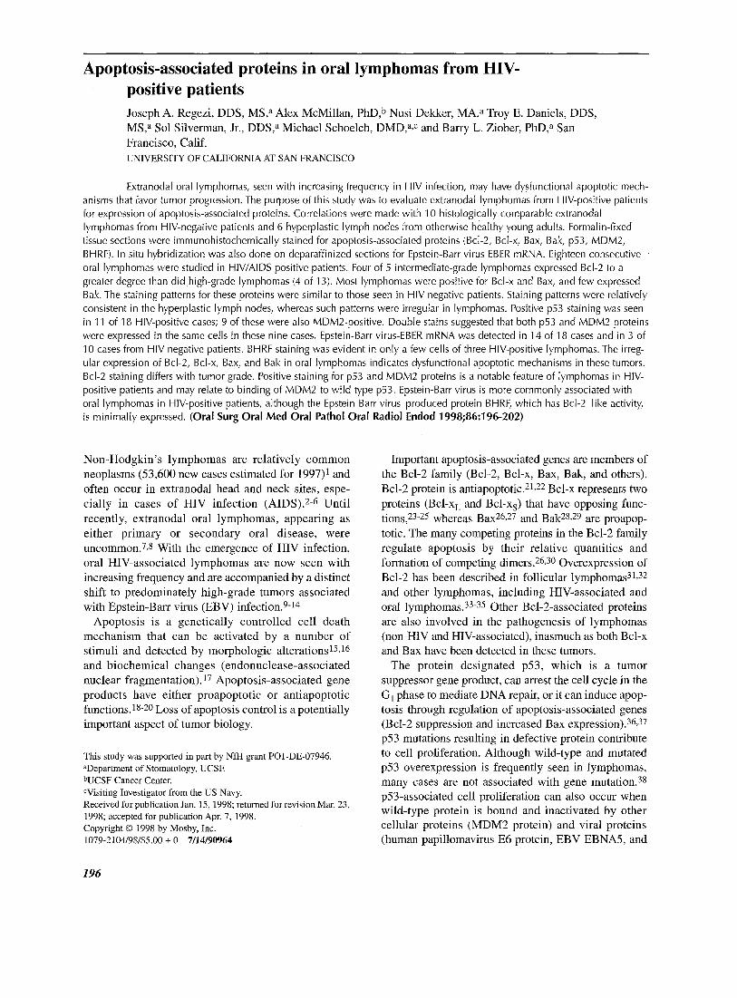
Fig. 1. Immunohistochemical patterns for Bcl-2 family of proteins in oral lymphomas: Bcl-2 (top left; HIV-negative patient); Bcl-x (top right; HIV-positive patient); Bax (bottom left; HIV-positive patient); Bak (bottom right; HI-V-negative patient; original magnification xl000).
range 41 to 80 years; gender distribution, 5 male and 5 female; location, 5 gingiva, 2 buccal mucosa, 1 palate, 1 jaw, and 1 lip; presenting sign, 4 ulcer and 6 mass. Oral lesions were the first sign of lymphoma in eight patients in this group, and two had known systemic disease.
Immunohistochemistry Paraffin-embedded, formalin-fixed tissue sections
were used in a standard immunohistochemistry protocol. Sections were microwaved in 0.1 mol/L citrate buffer for 7 minutes. They were incubated for 1 hour with antibodies listed in Table I and then with a biotinylated supersensitive multilink peroxidase agent (BioGenex, San Ramon, Calif.). Antigen-antibody
reactions were visualized with the chromogen aminoethylcarbazole. Normal mouse (or rabbit) serum containing mixed immunoglobulins at a concentration approximating that in primary antibodies was used as a negative control. Immunohistochemically stained sections were evaluated through use of a semiquantita- tive grading scale: <25% cells positive, 1+; 25% to <50% cells positive, 2+; 50% to <75% cells positive, 3+; >75% cells positive, 4+.
To determine whether p53 (wild-type and mutant proteins) and MDM2 were coexpressed in tumor cells, double immunohistochemical stains were done on 10 cases (9 HIV-positive, 1 HIV-negative) in which single stains for both of these antigens were positive. A modi- fied Vector Laboratories (Burlingame, Calif.) protocol
ORAL SURGERY ORAL MEDICINE ORAL PATHOLOGY Regezi et al. 199 Volume 86, Number 2
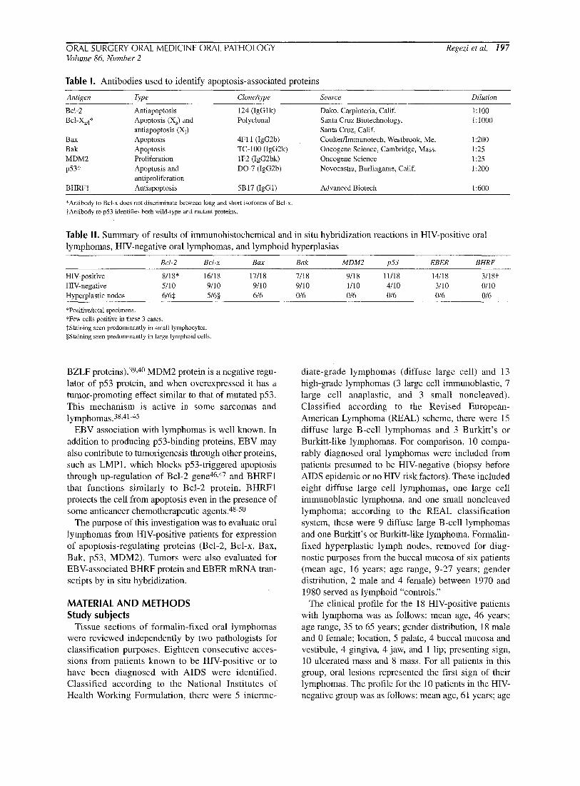
Fig. 2. Immunohistochemical stains show positive nuclear reaction for p53 protein (left) and MDM2 protein (right) in oral lymphoma (HIV-positive patient; original magnification xl000).
was used to identify two nuclear antigens with two different mouse monoclonal antibodies. After sections were deparaffinized, antigen enhancement was achieved by microwaving in citrate buffer. After hydrogen peroxide block and BSA/PBS bath, sections were incubated with the first primary antibody for 1 hour and then with BioGenex Multilink avidin/biotin peroxidase and substrate Vector SG (blue). Sections were washed, treated again with BSA/PBS, and incu- bated with the second primary antibody for 1 hour. BioGenex Multilink avidin/biotin peroxidase and substrate aminoethylcarbazole (red) were applied. No counter stain was used. In additional sections, the sequence of primary antibodies was reversed but the order of the detection systems was the same.
In situ hybridization Paraffin sections were stained for EBV-EBER mRNA
transcripts through use of an in situ hybridization kit (BioGenex).5 ~-53 Detection of EBER mRNA in formalin- fixed, paraffin-embedded tissue is possible when sensi- tive in situ hybridization methods are used. A 0 to 4+ scoring system was used, as described previously.
RESULTS Generally, expression of apoptosis-associated
proteins in oral lymphomas from HIV-positive patients was unpredictable. Cytoplasmic expression of Bcl-2 was seen in 8 of 18 cases (Table II), ranging from 1+ to 4+. In intermediate-grade lymphomas (Working Formulation) 4 of 5 cases were positive, and in high- grade lymphomas 4 of 13 cases were positive. Bcl-x and Bax proteins were detected in the cytoplasm of most tumor cells of most lymphomas. Staining was generally intense (3+ to 4+) for these proteins. Positive
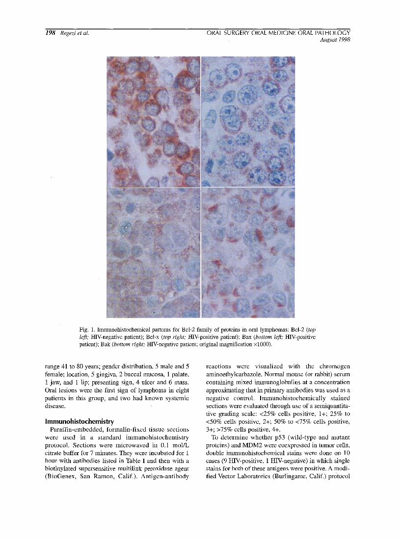
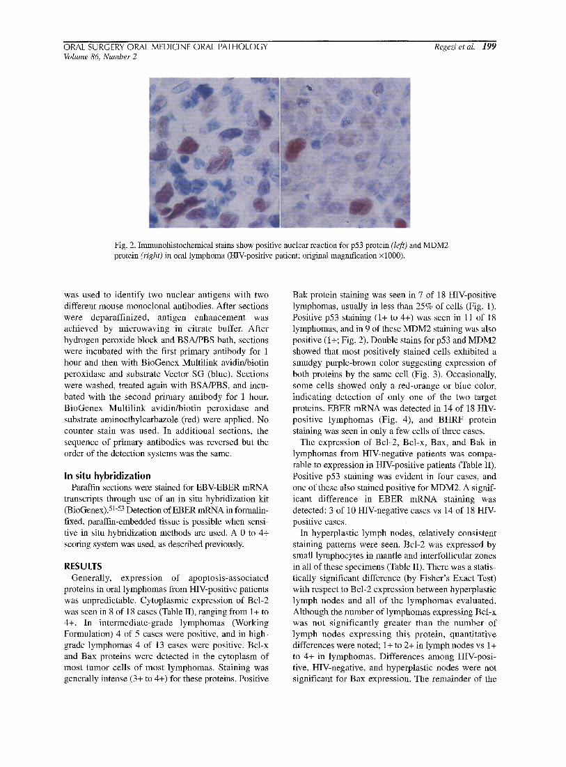
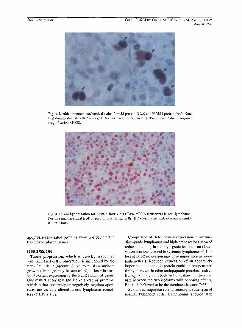
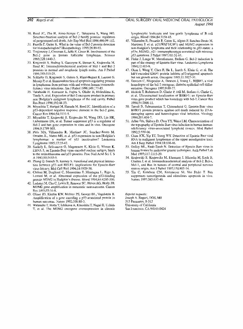
Bak protein staining was seen in 7 of 18 HIV-positive lymphomas, usually in less than 25% of cells (Fig. 1). Positive p53 staining (1+ to 4+) was seen in 11 of 18 lymphomas, and in 9 of these MDM2 staining was also positive (1+; Fig. 2). Double stains for p53 and MDM2 showed that most positively stained cells exhibited a smudgy purple-brown color suggesting expression of both proteins by the same cell (Fig. 3). Occasionally, some cells showed only a red-orange or blue color, indicating detection of only one of the two target proteins. EBER mRNA was detected in 14 of 18 HIV- positive lymphomas (Fig. 4), and BHRF protein staining was seen in only a few cells of three cases.
The expression of Bcl-2, Bcl-x, Bax, and Bak in lymphomas from HIV-negative patients was compa- rable to expression in HIV-positive patients (Table II). Positive p53 staining was evident in four cases, and one of these also stained positive for MDM2. A signif- icant difference in EBER mRNA staining was detected: 3 of 10 HIV-negative cases vs 14 of 18 HtV- positive cases.
In hyperplastic lymph nodes, relatively consistent staining patterns were seen. Bcl-2 was expressed by small lymphocytes in mantle and interfollicular zones in all of these specimens (Table II). There was a statis- tically significant difference (by Fisher's Exact Test) with respect to Bcl-2 expression between hyperplastic lymph nodes and all of the lymphomas evaluated. Although the number of lymphomas expressing Bcl-x was not significantly greater than the number of lymph nodes expressing this protein, quantitative differences were noted; 1+ to 2+ in lymph nodes vs 1+ to 4+ in lymphomas. Differences among HIV-posi- tive, HIV-negative, and hyperplastic nodes were not significant for Bax expression. The remainder of the
200 Regezi et al. ORAL SURGERY ORAL MEDICINE ORAL PATHOLOGY August 1998
Fig. 3. Double immunohistochemical stains for p53 protein (blue) and MDM2 protein (red). Note that double-stained cells (arrows) appear as dark purple nuclei (HIV-positive patient; original magnification x 1000).
Fig. 4. In situ hybridization for Epstein-Barr virus EBER mRNA transcripts in oral lymphoma. Positive nuclear signal (red) is seen in most tumor cells (HIV-positive patient; original magnifi- cation x400).
apoptosis-associated proteins were not detected in these hyperplastic tissues.
DISCUSSION Tumor progression, which is directly associated
with increased cell proliferation, is influenced by the rate of cell death (apoptosis). An apoptosis-associated growth advantage may be controlled, at least in part, by abnormal expression of the Bcl-2 family of genes. Our results show that the Bcl-2 group of proteins, which either positively or negatively regulate apop- tosis, are variably altered in oral lymphomas regard- less of HIV status.
Comparison of Bcl-2 protein expression in interme- diate-grade lymphomas and high-grade lesions showed reduced staining in the high-grade lesions--an obser- vation previously noted in systemic lymphomas. 34 This loss of Bcl-2 expression may have importance in tumor pathogenesis. Reduced expression of an apparently important antiapoptotic protein could be compensated for by increases in other antiapoptotic proteins, such as Bcl-x L. Although antibody to Bcl-x does not discrimi- nate between the two isoforms with opposing effects, Bcl-x L is believed to be the dominant isoform. 24,54
Bax has an important role in limiting the life span of normal lymphoid cells. Lymphomas showed Bax

ORAL SURGERY ORAL MEDICINE ORAL PATHOLOGY Regezi et al. 201 Volume 86, Number 2
staining that was similar to that of hyperplastic nodes.
Almost all tumors that expressed Bax also expressed
Bcl-2 or Bcl-x. The apoptotic effects of Bax, and in a few cases Bak, could be counteracted by Bcl-x or Bcl-
2 and contribute to tumor cell longevity. The ratio of
apoptosis- inducer proteins to apoptosis- inhibitor
proteins could not be determined.
It has been shown recently that Bax may act as a
tumor suppressor gene, being transcriptionally acti-
vated by p53 protein. 55 This being the case, Bax levels
would be expected to be reduced in cases in which
mutant p53 protein is produced. However, there were
few tumors that were Bax-negative and p53-positive.
This suggests the possibility that in the cases that were
both p53-positive and Bax-positive, p53 may not be mutated. 38 MDM2 protein binding to wild-type p53
protein may account for positive staining in many of these cases. 44,45 The double staining (p53 and MDM2)
of tumor cells adds support to this notion.
As expected, there was evidence for the presence of
EBV in oral lymphomas from HIV-positive patients.
This indicat ion of latent EBV infection (an in situ
hybridization signal for EBER mRNA) is consistent
with reports of other HIV-associated lymphomas and
supports the belief that EBV may play a role in the pathogenesis of HIV-associated lymphomas.51, 52 The
EBV-produced protein BHRF, which has Bcl-2-1ike
antiapoptotic activity, was absent in all but one case,
which suggests a minor role for this protein in the pathogenesis of oral lymphomas.
We conclude that Bcl-2, Bcl-x, and Bax are altered
and prominently expressed in oral lymphomas regard-
less of HIV status, indicating dysfunctional apoptotic
mechanisms in these tumors. Bak expression, less
prominent in lymphomas and not apparent in hyper-
plastic nodes, may also be involved in few cases.
Positive staining for p53 and MDM2 proteins appears
to be a feature of lymphomas from HIV-positive
patients. EBV also is more commonly associated with
oral lymphomas in HIV-positive patients, although the
EBV-produced protein BHRF, which has Bcl-2-1ike
activity, is minimally expressed.
REFERENCES l. Parker SL, Tong T, Bolden S, Wingo PA. Cancer statistics, 1997.
CA Cancer J Clin 1997;47:5-27. 2. Economopoulos T, Asprou N, Stathakis N, Papageorgio E,
Dervenoulas J, Xanthaki K, et al. Primary extranodal non- Hodgkin's lymphoma in adults: clinicopathological and survival characteristics. Leukemia Lymphoma 1996;21:131-6.
3. Hamilton-Dutoit S J, Pallesen G, Franzmann MB, et al. AIDS- related lymphoma. Am J Pathol 1991; 138:149-63.
4. Ioachim HL, Dorsett B, Cronin W, Maya M, Wahl S. Acquired immunodeficiency syndrome-associated lymphomas: clinical, pathologic, immunologic, and viral characteristics of 111 cases. Hum Pathol 1991;22:659-73.
5. Serraino D, Pezzotti P, Dorrucci M, Alliegro MB, Sinicco A,
Rezza G. Cancer incidence in a cohort of human immunodefi- ciency virus seroconverters. Cancer 1997;79:1004-8.
6. Ziegler JL, Beckstead JA, Volberding PA, Abrams DI, Levine AM, Lukes R J, et al. Non-Hodgkin's lymphoma in 90 homosexual men. N Engl J Meal 1984;311:565-70.
7. Fukuda Y, Ishida T, Fujimoto M, Ueda T, Aozasa K. Malignant lymphoma of the oral cavity: clinicopathologic analysis of 20 cases. J Oral Patbol 1987;16:8-12.
8. Soderholm AL, Lindqvist C, Heikinheimo K, Forssell K, Happonen RE Non-Hodgkin's lymphomas presenting through oral symptoms. Int J Oral Maxillofac Surg 1990; 19:13 l-4.
9. Carbone A, Vaccher E, Barzan L, Gloghini A, Volpe R, De Re V, et al. Head and neck lymphomas associated with human immun- odeficiency virus infection. Arch Otolaryngol Head Neck Surg 1995;121:210-8.
10. Green TL, Eversole LR. Oral lymphomas in HIV-infected patients: association with Epstein-Barr virus. Oral Surg Oral Med Oral Pathol 1989;67:437-42.
11. Handlers JR Howell RE, Abrams AM, Melrose RJ. Extranodal oral lymphoma, I: a morphologic and immunoperoxidase study of 34 cases. Oral Surg Oral Med Oral Pathol 1986;61:362-7.
12. Lozada-Nur F, De Sanz S, Silverman S, Miranda C, Regezi J. lntraoral non-Hodgkin's lymphoma in seven patients with acquired immunodeficiency syndrome. Oral Surg Oral Med Oral Pathol Oral Radiol Endod 1996;82:173-8.
13. Regezi JA, Zarbo RJ, Stewart JCB. Extranodal oral lymphomas: histologic subtypes and immunophenotypes (in routinely processed tissue). Oral Surg Oral Med Oral Pathol 1991;72:702-8.
14. Raphael M, Gentilhomme O, Tuillez M, Byron PA, Diebold J. Histopathologic features of high-grade non-Hodgkin's lymphomas in acquired immunodeficiency syndrome. Arch Pathol Lab Med 1991;115:15-20.
15. Majno G, Joris I. Apoptosis, oncosis, and necrosis: an overview of cell death. Am J Pathol 1995; 146:3-15.
16. Payne CM, Bernstein C, Bernstein H. Apoptosis overview empha- sizing the role of oxidative stress, DNA damage and signal-trans- duction pathways. Leukemia Lymphoma 1995; t9:43-93.
17. Gavrieli Y, Sherman Y, Ben-Sasson SA. Identification of programmed cell death in situ via specific labeling of nuclear DNA fragmentation. J Cell Biol 1992;119:493-501.
18. Farber E. Programmed cell death: necrosis versus apoptosis. Mod Pathol 1994;7:605-9.
19. Stellar H. Mechanisms and genes of cellular suicide. Science 1995;267:1445-8.
20. Thompson CB. Apoptosis in the pathogenesis and treatment of disease. Science 1995;267:1456-62.
21. Cory S. Regulation of lymphocyte survival by the Bcl-2 gene family. Annu Rev Immunol 1995;13:513-43.
22. Reed JC. Bcl-2 and the regulation of programmed cell death. J Cell Biol 1994;124:1-6.
23. Boise LH, Gonzalez M, Postema CE, Ding L, Lindsten T, Turka LA, et al. Bcl-X, a Bcl-2-related gene that functions as a domi- nant regulator of apoptotic cell death. Cell 1993;74:597-608.
24. Gonzalez-Garcia M, Perez-Ballestero R, Ding L, Duan L, Boise LH, Thompson CB, et al. Bcl-x L is the major Bcl-x mRNA form expressed during murine development and its product localizes to mitochondria. Development 1994;120:3033-42.
25. Nunez G, Merino R, Grillot D, Gonzalez-Garcia M. Bcl-2 and Bcl-x: regulatory switches for lymphoid death and survival. Immunol Today 1994;15:582-8.
26. Oltval ZN, Korsmeyer SJ. Checkpoints of dueling dimers foil death wishes. Cell 1994;79:189-92.
27. Oltvai ZN, Milliman C1, Korsmeyer SJ. Bcl-2 heterodimerizes in vivo with a conserved homolog, Bax, that accelerates programmed cell death. Cell 1993;74:609-19.
28. Chittenden T, Harrington EA, O'Connor R, Flemington C, Lutz RJ, Evan GI, et al. Induction of apoptosis by the Bcl-2 homo- logue Bak. Nature 1995;374:733-6.
29. Keifer MC, Brauer MJ, Powers VC, Wu JJ, Umanski SR, Tomei LD, et al. Modulation of apoptosis by the widely distributed Bcl- 2 homologue Bak. Nature 1995;374:736-9.
202 Regezi et al. ORAL SURGERY ORAL MEDICINE ORAL PATHOLOGY August 1998
30. Reed JC, Zha H, Aime-Sempe C, Takayama S, Wang HG. Structure-function analysis of Bcl-2 family proteins: regulators of programmed cell death. Adv Exp Med Biol 1996;406:99-112.
31. Pezella K Garter K. What is the value of Bcl-2 protein detection for histopathologists? Histopathology 1995;26:89-93.
32. Tsujimotoy J, Cossman E, Jaffe E, Croce H. Involvement of the Bcl-2 gene in human follicular lymphoma. Science 1985;228:1440-3.
33. Krajewski S, Bodrug S, Gascoyne R, Berean K, Krajewska M, Reed JC. Immunohistochemical analysis of Mcl-I and Bcl-2 proteins in normal and neoplastic lymph nodes. Am J Pathol 1994; 145:515-25.
34. Schlaifer D, Krajewski S, Galoin S, Rigal-Huguet E Laurent G, Massip R et al. Immunodetection of apoptosis-regulating proteins in lymphomas from patients with and without human immunode- ficiency virus infections. Am J Pathol 1996;149:177-85.
35. Takahashi H, Kawazoe K, Fujita S, Okabe H, Hideshima K, Tsnda N, et al. Expression of Bcl-2 oncogene product in primary non-Hodgkin's malignant lymphoma of the oral cavity. Pathol Res Pract 1996;192:44-53.
36. Miyashita T, Harigai M, Hanada M, Reed JC. Identification of a p53-dependent negative response element in the bcl-2 gene. Cancer Res 1994;54:3131-5.
37. Miyashita T, Krajewski S, Krajewska M, Wang HG, Lin HK, Liebermann DA, et al. Tumor suppressor p53 is a regulator of Bcl-2 and bax gene expression in vitro and in vivo. Oncogene 1994;9:1799-805.
38. Piris MA, Villuendas R, Martinez JC, Sanchez-Beato M, Orradre JL, Mateo MS, et al. p53 expression in non-Hodgkin's lymphomas: a marker of p53 inactivation? Leukemia Lymphoma 1995;17:35-42.
39. Szekely L, Selivanova G, Magnusson K, Klein G, Wiman K. EBNA-5, an Epstein-Barr virus-encoded nuclear antigen, binds to the retinoblastoma and p53 proteins. Proc Natl Acad Sci U S A 1993;90:5455-9.
40. Zhang Q, Gutsch D, Kenney S. Functional and physical interac- tion between p53 and BZLFI: implications for Epstein-Barr virus latency. Mol Cell Biol 1994; 14:1929-38.
41. Chilosi M, Doglioni C, Menestrina F, Montagna L, Rigo A, Lestani M, et al. Abnormal expression of the p53-binding protein MDM2 in Hodgkin's disease. Blood 1994;84:4295-300.
42. Ladanyi M, Cha C, Lewis R, Jhanwar SC, Huvos AG, Healy JH. MDM2 gene amplification in metastatic osteosarcoma. Cancer Res 1993;53:16-8.
43. Oliner JD, Klnzler KW, Meltzer PS, George DL, Vogelstein B. Amplification of a gene encoding a p53-associated protein in human sarcomas. Nature 1992;358:80-3.
44. Watanabe T, Hotta T, Ichikawa A, Kiuoshita T, Nagai H, Uchida T, et al. The MDM2 oncogene overexpression in chronic
lymphocytic leukemia and low-grade lymphoma of B-cell origin, Blood 1994;84:3158-65.
45. Villuendas R, Pezzella E Gatter K, Algara P, Sanchez-Beato M, Martinez P, et al. p2I(WAFI/CIP1) and MDM2 expression in non-Hodgkin's lymphoma and their relationship to p53 status: a p53+, MDM2-, p21- immunophenotype associated with missense p53 mutations. J Pathol 1997;181:51-61.
46. Finke J, Lange W, Mertelsmann, Dolken G. Bcl-2 induction is part of the strategy of Epstein-Barr virus. Leukemia Lymphoma 1994;12:413-9.
47. Okan I, Wang Y, Chen F, Hu L, Irareh S, Klein G, et al. The EBV-encoded LMP1 protein inhibits p53-triggered apoptosis but not growth arrest. Oncogene 1995; 11:1027-31.
48. Dawson C, Eliopoulos A, Dawson J, Young L. BHRF1, a viral homologue of the bcl-2 oncogene, disturbs epithelial cell differ- entiation. Oncogene 1995;9:69-77.
49. Hickish T, Robertson D, Clarke P, Hill M, Stefano F, Clarke C, et al. Ultrastructual localization of BHRFI: an Epstein-Barr virus gene product which has homology with bcl-2. Cancer Res 1994;54:2808-11.
50. Tarodi B, Subramanian T, Chinnadurai G. Epstein-Barr virus BHRF1 protein protects against cell death induced by DNA- damaging agents and heterologous viral infection. Virology 1994;201:404-7.
51. Arber DA, Shibata D, Chen YY, Weiss LM. Characterization of the topography of Epstein-Barr virus infection in human immun- odeficiency virus-associated lymphoid tissues. Mod Pathol 1992;5:559-66.
52. Chan JCK, Yip TT, Tsang WY. Detection of Epstein-Barr viral RNA in malignant lymphomas of the upper aerodigestive tract. Am J Surg Pathol 1994;18:938-46.
53. Gulley ML, Raab-Traub N. Detection of Epstein-Burr virus in human tissues by molecular genetic techniques. Arch Pathol Lab Med 1993;117:1115-20.
54. Krajewski S, Krajewska M, Ehrmann J, Sikorska M, Lach B, Chatten J, et al. Immnnohistochemical analysis of Bcl-2, Bcl-x, Mcl-1, and Bax in tumors of central and peripheral nervous system origin. Am J Pathol 1997;150:805-14.
55. Yin C, Knudson CM, Korsmeyer SJ, Van Dyke T. Bax suppresses tumorigenesis and stimulates apoptosis in vivo. Nature 1997;385:637-40.
Reprint requests: Joseph A. Regezi, DDS, MS 513 Parnassus, S-512 University of California San Francisco, CA 94143-0424
Related Documents