CHAPTER *Corresponding Author: Afshin Samali—NCBES, National University of Ireland, Galway, University Road, Galway city, Ireland, E-mail: [email protected] Heat Shock Proteins in Neural Cells, edited by Christiane Richter-Landsberg. ©2005 Eurekah.com. Heat Shock Proteins and the Regulation of Apoptosis Una FitzGerald, Adrienne M. Gorman and Afshin Samali* Summary S ince the elucidation of their functions in protein folding and translocation, heat shock protein chaperones have been a target of research in all spheres of biomedicine. Within the last five years, research efforts have intensified, following the discovery of raised levels of heat shock protein (Hsp) expression in the brains of patients suffering from many neurodegenerative disorders, including Alzheimer’s, Parkinson’s and Huntington Disease and cerebral ischaemia. Expression of Hsps in the brains of patients is thought to form part of a general protective stress response. The stress in question, however, varies, depending on the particular disease. For example, accumulation of α-synuclein aggregates in Parkinson’s Disease causes stress to the protein folding machinery of the cells, with consequent up-regulation of stress proteins including Hsps. When markers indicative of the occurrence of apoptosis were also found in degenerating brain tissue, the question of how heat shock proteins might impact on apoptotic neural cells was raised. However, their particular function under diseased condi- tions remains unclear. This chapter highlights the involvement of Hsps in the regulation of neural apoptosis, from the original reports of Hsp expression during neurological disorders, to evidence of their neuroprotective properties and their potential as therapeutic molecules. Introduction to Apoptosis Within tissues cell death can occur via either necrosis or apoptosis. Necrosis is a passive form of cell death occurring mainly under pathological conditions, where a rapid loss of ion-flux control leads to the swelling and rupture of the cell and its organelles. In contrast, apoptosis is a controlled, energy-dependent form of cell death involving an elaborate network of signal transduction pathways in both its initiation and execution. Characteristic hallmarks of apoptosis include membrane blebbing, condensation of nuclear chromatin, cytoplasmic shrinkage, nuclear fragmentation and formation of apoptotic bodies. Apoptosis is induced in response to a large variety of stimuli including cytokines, cytotoxic drugs, oxidative stress and ionising radiation. 1 These diverse stimuli trigger apoptosis by activating one or more signal transduction pathways, which then converge to activate a conserved family of aspartic-acid specific cysteine proteases, referred to as caspases. 2 Caspases are constitutively expressed within cells as inactive precursor zymogens and are activated in response to apoptotic stimuli by changes in the three dimen- sional structure of the protein or by specific proteolytic cleavage. 3-5 Once activated they or- chestrate the demise of the cell through the cleavage of a specific subset of cellular substrates, 6 resulting in the characteristic biochemical and morphological changes associated with apoptosis. 7 Richter(Samali) 9/14/05, 3:12 PM 1

Welcome message from author
This document is posted to help you gain knowledge. Please leave a comment to let me know what you think about it! Share it to your friends and learn new things together.
Transcript

CHAPTER
*Corresponding Author: Afshin Samali—NCBES, National University of Ireland, Galway,University Road, Galway city, Ireland, E-mail: [email protected]
Heat Shock Proteins in Neural Cells, edited by Christiane Richter-Landsberg.©2005 Eurekah.com.
Heat Shock Proteins and the Regulationof ApoptosisUna FitzGerald, Adrienne M. Gorman and Afshin Samali*
Summary
Since the elucidation of their functions in protein folding and translocation, heat shockprotein chaperones have been a target of research in all spheres of biomedicine. Withinthe last five years, research efforts have intensified, following the discovery of raised levels
of heat shock protein (Hsp) expression in the brains of patients suffering from manyneurodegenerative disorders, including Alzheimer’s, Parkinson’s and Huntington Disease andcerebral ischaemia. Expression of Hsps in the brains of patients is thought to form part of ageneral protective stress response. The stress in question, however, varies, depending on theparticular disease. For example, accumulation of α-synuclein aggregates in Parkinson’s Diseasecauses stress to the protein folding machinery of the cells, with consequent up-regulation ofstress proteins including Hsps. When markers indicative of the occurrence of apoptosis werealso found in degenerating brain tissue, the question of how heat shock proteins might impacton apoptotic neural cells was raised. However, their particular function under diseased condi-tions remains unclear. This chapter highlights the involvement of Hsps in the regulation ofneural apoptosis, from the original reports of Hsp expression during neurological disorders, toevidence of their neuroprotective properties and their potential as therapeutic molecules.
Introduction to ApoptosisWithin tissues cell death can occur via either necrosis or apoptosis. Necrosis is a passive
form of cell death occurring mainly under pathological conditions, where a rapid loss of ion-fluxcontrol leads to the swelling and rupture of the cell and its organelles. In contrast, apoptosis isa controlled, energy-dependent form of cell death involving an elaborate network of signaltransduction pathways in both its initiation and execution. Characteristic hallmarks of apoptosisinclude membrane blebbing, condensation of nuclear chromatin, cytoplasmic shrinkage, nuclearfragmentation and formation of apoptotic bodies. Apoptosis is induced in response to a largevariety of stimuli including cytokines, cytotoxic drugs, oxidative stress and ionising radiation.1
These diverse stimuli trigger apoptosis by activating one or more signal transduction pathways,which then converge to activate a conserved family of aspartic-acid specific cysteine proteases,referred to as caspases.2 Caspases are constitutively expressed within cells as inactive precursorzymogens and are activated in response to apoptotic stimuli by changes in the three dimen-sional structure of the protein or by specific proteolytic cleavage.3-5 Once activated they or-chestrate the demise of the cell through the cleavage of a specific subset of cellular substrates,6
resulting in the characteristic biochemical and morphological changes associated with apoptosis.7
Richter(Samali) 9/14/05, 3:12 PM1

Heat Shock Proteins in Neural Cells2
Caspase activation during apoptosis can proceeds via a number of different pathways (Fig.1). The extrinsic pathway involves the binding of death ligands to cell surface receptors (e.g.,Fas/CD95/Apo-1 or TNF receptor) resulting in the recruitment of the adaptor molecules FasAssociated Death Domain (FADD) or TNF Receptor Associated Death Domain (TRADD) tothe cytosolic end of the receptor leading to the formation of the Death Inducing SignallingComplex (DISC) at the plasma membrane and resultant activation of pro-caspase-8 and therebypro-caspase-3.8-10 In addition to FADD, the DAXX adaptor molecule may also transduce deathsignals to JNK, via apoptosis signal-regulating kinase 1 (ASK1) recruitment to the DISC. AsJNK is a substrate of ASK1, this broadens the effect of Fas ligand binding to includeJNK-mediated apoptosis (Fig. 1A).11
The intrinsic pathway is initiated through the release of cytochrome c from the intermem-brane space of mitochondria (Fig. 1B).12,13 Cytochrome c translocates across the outer mito-chondrial membrane, by a number of possible pathways14 to the cytosol, where it binds toApoptosis Protease Activating Factor 1 (Apaf-1) and in the presence of dATP (or ATP) cyto-chrome c/dATP facilitates Apaf-1 oligomerization and the recruitment of pro-caspase-9.15 Theformation of this caspase-activating complex, termed the apoptosome, results in the activationof pro-caspase-9, which further amplifies the caspase cascade by its ability to process effectorcaspases including pro-caspase-3.16
The endoplasmic reticulum (ER) stress-induced pathway is a more recent and controversialapoptotic program, originally identified when ER-associated caspase-12 was cleaved in responseto chemical inducers of ER stress.17-20 Although the origin of the particular ER disturbancemay differ, it activates a common signalling pathway termed the unfolded protein response(UPR).21,22 Three ER trans-membrane proteins initiate the UPR and these are: an ER-associatedtype I trans-membrane protein kinase (Ire1), activated transcription factor 6 (ATF6) andPKR-like endoplasmic reticulum kinase (PERK). In resting cells, the 78 kDa glucose-regulatedprotein (Grp78 or Bip) binds the ER-luminal ends of these molecules but in the presence ofER stress, it detaches from Ire1, PERK and ATF6, thus, enabling their activation. PERK acti-vation leads to a temporary halt in protein translation and under certain conditions, the activa-tion of the pro-apoptotic protein Gadd153 or CHOP. Active Ire1 causes induction of theX-box binding 1 (XBP1) transcription factor, thought to induce expression of itself and asubset of other proteins involved in the UPR.23 ATF6, activated through cleavage, activatestranscription of chaperone proteins and transcription factors, e.g., Grp78 and XBP1. Contro-versy surrounding ER stress-induced apoptosis has centred on the lack of expression of caspase-12in human cells.24,25 However, the search for the functional homologues of human caspase-12has indicated a possible role for caspase-4 in mediating apoptosis triggered by the ER (Fig.1C).26
The mitochondrion has been identified as a central control point for the integration ofdiverse death signals during apoptosis. Many of the key molecules involved in apoptosis arelocated within or associated with the mitochondrion.13 These include cytochrome c,27 AIF,28
Smac/Diablo,29 EndoG,30 Puma31 and pro-caspase-3.2,32 When released into the cytosol, thesemolecules function both in the initiation and execution of the apoptotic program. It is there-fore unsurprising to find that the mitochondrion serves as a control point for cross-talk be-tween the intrinsic, extrinsic and ER stress pathways of apoptosis. In this respect it is wellestablished that the activation of pro-caspase-8 by death receptors can result in the cleavage ofan endogenous cellular protein, Bid, generating a truncated pro-apoptotic fragment that trans-locates to the mitochondrion where it induces cytochrome c release.33,34 This mitochondrialstep is found to be critical in certain cell types where the engagement of death receptors aloneis not sufficient to induce caspase activation without cytochrome c release.35 Further reportsalso suggest that stimuli that induce DNA damage and subsequently apoptosis do so via trans-location from the nucleus to the mitochondrion of molecules that stimulates the release ofcytochrome c.36,37 Naturally, since the mitochondrion has such a key role during apoptosismany of the endogenous cellular proteins that function as crucial inhibitors of cell death ex-
Richter(Samali) 9/14/05, 3:12 PM2

3Heat Shock Proteins and the Regulation of Apoptosis
ecute their anti-apoptotic capabilities by acting on mitochondria and preventing the release ofcrucial pro-apoptotic proteins.
To date the best-characterised endogenous protein modulators of apoptosis are the mem-bers of the Bcl-2 family of proteins.38 Both pro- and anti-apoptotic members of this familyhave been identified and studied within mammalian cells, where they are seen to function askey determinants of cell fate. However, the recent correlation between the expression of Hspsand increased cell survival, has pointed to Hsps as playing a critical role in the regulation of theapoptotic machinery.
Figure 1. Caspases can be activated by at least three different pathways leading to apoptosis. A. Mitochon-drial pathway is initiated due to release of cytochrome c from the mitochondrial intermembrane space intothe cytosol, leading to the formation of the apoptososme which culminates to the activation of caspase-9activation. B. The death receptor pathway, activated by trimerization of death receptors at the plasmamembrane (e.g., Fas receptor), which leads to autoactivation of caspase-8. Daxx, a nuclear protein, trans-locates to the membrane during Fas-mediated apoptosis. Daxx binds at one end to the Fas receptor and atthe other with Ask1, thus mediating a caspase-independent cell death. There is a cross-talk between deathreceptor pathway and the mitochondria, through cleavage of the protein Bid, by caspase-8. C. The ER stresspathway is activated when unfolded proteins accumulate in the ER or when there is disruption in the ERCa2+ homeostasis, which leads to activation of caspase-12, possibly by a Ca2+-mediated process involvingcalpain. Ca2+ may also amplify the pathway by acting on mitochondria.
Richter(Samali) 9/14/05, 3:12 PM3

Heat Shock Proteins in Neural Cells4
Hsp27 and Hsp70, the Anti-apoptotic Heat Shock ProteinsMembers of the heat shock family of proteins can be divided into two camps. The first
contains chaperones, that are constitutively expressed in whose normal function is aiding pro-tein folding and maturation (e.g., Hsc70), whose expression does not seem to readily change inresponse to stress. The second consists of Hsps which may, albeit at low levels, or may not beconstitutively expressed in cells, but whose expression is significantly induced in response tostress. These latter proteins have been shown to have cytoprotective properties and exert theireffect by inhibiting apoptosis. Hsp27 and Hsp70 are the main chaperones associated with ananti-apoptotic effect in vivo. The mechanisms by which they are thought to impinge on apoptosis,are described below and summarised in Figure 2.
Hsp70 and the Regulation of ApoptosisThere are many ways in which Hsp70 can inhibit the execution of caspase-dependent
apoptosis and caspase-independent cell death. Most are centred on mitochondria-mediatedevents. For example, direct binding of the Apaf-1 CARD domain by Hsp70 was shown toprevent apoptosome formation and caspase-9 activation in cell-free systems.39,40 However, re-cent findings suggest that inhibition of caspase activation by Hsp70 is due to interference with mito-chondrial release of cytochrome c.41 Through direct binding of another mitochondria-associated protein,AIF, Hsp70 can broaden its protective role by inhibiting caspase-independent nuclear fragmentation.42,43
Although Hsp70 has been shown to be protective in a TNFα-induced cell death model, recentstudies indicate that whether or not Hsp70 is protective in a given pathway, depends on whetheror not activation of the mitochondrial pathway is required.44 Hsp70 may also employ indirectmechanisms to inhibit amplification of death inducing signals. For example, Hsp70, by inhib-iting JNK activity, prevents or down-regulates Bid-, p53- and/or c-myc-mediated release ofcytochrome c and Smac/Diablo in response to stress.45-48 However, this notion was contra-dicted in a different model system by Jaattela and colleagues who showed that cells over-expressingHsp70 did not prevent activation of stress kinases including JNK, but rather inhibited thedownstream effects of caspase activity.49 Alternatively, Hsp70 may interfere withpost-mitochondrial signalling, through direct binding of pro-caspases -3 and -7.50
In addition to the stress-activated JNK, Hsp70 has also been shown to directly bind ASK1,preventing its homo-oligomerization, with consequent protection of cells from oxidative stressand death through inhibition of ASK1-mediated cytochrome c release.51
The range of protective effects associated with Hsp70 may be altered, depending on whetheror not Hsp70 is in complex with cochaperones including Hsp40 or BAG-1. Under normalconditions, Hsp70 exists as a monomer, executing house-keeping chaperone functions. How-ever, under stressed conditions, it may be found in complex with Hsp40, thus, broadening itssubstrate specificity.52,53 This is especially important when such a complex can alter the ex-pected anti-apoptotic effect, to a pro-apoptotic one. For example, in complex with Hsp40,Hsp70 had been shown to enhance caspase-activated DNase, thus augmenting T-cellreceptor-mediated apoptosis.54 However, in line with general observations for Hsp70, while incomplex with other cochaperones, e.g., DNaJ, Hsp70 protects cells by inhibiting Bax translo-cation to the mitochondria.55
Hsp27 and the Regulation of ApoptosisIn many ways, the protective effects of Hsp27 supplement those described for Hsp70. For
example, while Hsp70 may prevent release of cytochrome c from mitochondria, phosphory-lated Hsp27, through direct cytochrome c binding, inhibits apoptosome formation and subse-quent activation of caspase-9.56-59 In addition, Hsp27 has been shown to inhibit caspase-3activity, by interacting with pro-caspase-3, preventing its cleavage by caspase-9.57,60 At the levelof mitochondria, Hsp27 may also exert its protective effect by inhibiting the release of SMAC/Diablo.61
Richter(Samali) 9/14/05, 3:12 PM4

5Heat Shock Proteins and the Regulation of Apoptosis
Unlike Hsp70, Hsp27 has the potential to interfere directly with death-receptordeath-inducing signals, without the need for the involvement of mitochondrial factors. Thiseffect is achieved by phosphorylated dimers of Hsp27 which are capable of interacting withand inhibiting DAXX, which links the Fas receptor to Ask 1 and downstream JNK pathway.62
Another kinase linked to Hsp27’s protective role, is Akt, a pro-survival molecule, which targetsand inactivates Ask1.63 Hsp27 interacts with Akt directly and has been proposed to enhance itsactivation through its chaperone function.64,65 Hsp27 is itself a target of active Akt and oncephosphorylated, it is released from the Akt complex and so becomes available to participate inother anti-apoptotic functions. A similar indirect protective activity has been proposed forHsp27, through participation in a complex between ubiquitin and IkBα. IkBα functions to
Figure 2. Heat shock protein regulation of apoptotic pathways. A mechanism by which apoptosis is initiatedis due to changes in the intracellular redox balance and production of reactive oxygen species. This resultsin changes in the mitochondria and release of pro-apoptotic factors. Hsp27 and Hsp70 can maintain bothredox homeostasis and mitochondrial stability in the cell. Also Hsp27 can bind to cytochrome c, after itsrelease from mitochondria, and pro-caspase-3, thus, preventing apoptosome formation and events down-stream of mitochondrial damage. In cell-free systems, Hsp70 has also been demonstrated to inhibit apoptosomeformation. Apart from the caspase-dependent apoptosis, Hsp27 is reported to block Daxx-mediated celldeath. Hsp27 can prevent the translocation of Daxx to membrane and its interaction with Fas. Currentlythere is no information regarding the role of Hsp27 or Hsp70 in the regulation of ER stress-mediatedapoptosis.
Richter(Samali) 9/14/05, 3:12 PM5
Heat Shock Proteins in Neural Cells6
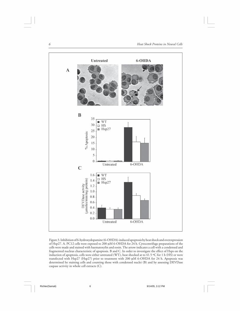
Figure 3. Inhibition of 6-hydroxydopamine (6-OHDA)-induced apoptosis by heat shock and overexpressionof Hsp27. A. PC12 cells were exposed to 200 µM 6-OHDA for 24 h. Cytocentrifuge preparations of thecells were made and stained with haematoxylin and eosin. The arrow indicates a cell with a condensed andfragmented nucleus characteristic of apoptosis. B and C. In order to investigate the effect of Hsps on theinduction of apoptosis, cells were either untreated (WT), heat shocked at to 41.5 ºC for 1 h (HS) or weretransfected with Hsp27 (Hsp27) prior to treatment with 200 µM 6-OHDA for 24 h. Apoptosis wasdetermined by staining cells and counting those with condensed nuclei (B) and by assessing DEVDasecaspase activity in whole cell extracts (C).
Richter(Samali) 9/14/05, 3:12 PM6

7Heat Shock Proteins and the Regulation of Apoptosis
sequester the pro-survival transcription factor NF-κB. Phosphorylation of IkBα leads to itsubiquitination and subsequent degradation by the 26S proteasome. By direct binding of theproteasome, the ubiquitin chains and phosphorylated IκBα, Hsp27 facilitates the degradationof IkBα.66 This releases NF-κB, enabling its translocation to the nucleus where it transactivatesthe expression of pro-survival genes.
Studies of Hsp27 function under conditions of oxidative stress-induced cell death, haveindicated that Hsp27 achieves its protective effect by increasing glutathione levels,67-69 reduc-ing cytosolic reactive oxygen species (ROS),68,70 raising glucose-6-phosphate dehydrogenaseactivity71 and/or lowering cytosolic iron.69
The potential for Hsp27 to protect cells through preservation of the architecture of thecytoplasm has been the subject of much recent discussion. Based on studies of post-translationalmodification of Hsp27 in heat-shocked or stressed cells, stabilisation of actin filaments by thephosphorylated nonoligomeric form of Hsp27 has been implicated in its protective function.59,72-
74 Hsp27 has a known role in actin reorganisation, a phenomenon which is required for theformation of membrane blebs and apoptotic bodies that are generated during the course ofapoptosis.75,76 This role appears to contradict the suggested actin-protecting functions describedabove, raising the possibility that the particular actin-associated function of Hsp27 duringapoptosis may be signal- and cell type-dependent. However, given that many structural pro-teins are cleaved by caspase-3 during apoptosis,6 it is conceivable that Hsp27’s ability to inhibitcaspase-3 may be sufficient to maintain the cytoskeleton intact. In neurons, the cytoskeletalprotective function of Hsp27 has been linked to its ability to decrease levels of phosphorylatedtau and enhance nonubiquitin-dependent degradation of hyperphosphorylated tau.77,78 Fur-ther details of the effects of Hsp27 on tau are provided in chapters 2 and 6.
Apoptosis, Hsps and Neurodegenerative DisordersThat apoptosis occurs during the course of the neurodegenerative disease process, is well
documented. Caution is, however, required when interpreting data derived from human tissue,as artefactual occurrence of DNA degradation during tissue processing may lead to false con-clusions. In most cases, detection of nuclear fragmentation or cleaved forms of caspases or theirsubstrates is used to detect apoptosis. For example, in neurons and glia, terminaltransferase-mediated biotinylated-UTP nick end-labelling (TUNEL) detected raised level offragmented DNA in brain tissue derived from Alzheimer’s79-82 and Huntington’s Disease pa-tients.81,83 More recently, TUNEL positivity was detected in addition to raised levels of Bax,Bcl-2, cleaved caspase-3 and cleaved poly(ADP-ribose) polymerase (PARP) in Huntington’sDisease brain tissue.84 A very useful discussion of some of the issues surrounding the use ofDNA fragmentation as an apoptosis marker is presented by Tatton et al (2003),85 who alsoconfirmed the occurrence of DNA fragmentation, Bax up-regulation and caspase-3 cleavageduring Parkinson’s Disease (PD).86 Additional studies of PD tissue have shown the raised levelsof apoptosis signalling molecules including the Fas receptor87 and caspases -3, -8 and -9.88,89
Neurodegenerative diseases, whether acute or chronic, are a major medical problem in theaging population. Therapies are rare and applied usually during the late stages of the disease,when a vast number of cells are already lost. Many efforts have been made to develop newstrategies to treat these disorders, but so far, there has been no breakthrough. A characteristicshared by some experimental neuroprotective substances is the induction of the Hsps and inparticular Hsp70 and Hsp27. In response to many metabolic disturbances and injuries cellsmount a stress response with induction of a variety of proteins, most notably inducible Hsps.There is evidence that upon injury to the brain Hsp70 is induced to high levels in brain regionsthat are relatively resistant to injury. With the availability of transgenic animals and gene trans-fer, it has become increasingly clear that selective over-expression of Hsps leads to protection inseveral different models of nervous system injury that involve neurodegeneration and apoptosis,including ischemia/excitotoxicity and models of Parkinson’s and Alzheimer’s diseases. Some ofthese will be discussed.
Richter(Samali) 9/14/05, 3:12 PM7

Heat Shock Proteins in Neural Cells8
Ischemia/ExcitotoxicityAbout 700,000 Americans suffer ischemic stroke each year. That is, one every 45 seconds
(American Stroke Society statistics). The brain is particularly vulnerable to ischemic damageand even transient interruption to blood flow can cause significant neuronal cell death. Con-siderable evidence supports a role for apoptosis in cerebral ischemia. While damaged neuronsoften die from necrosis, apoptosis contributes significantly to cell death subsequent to cerebralischemia, with apoptosis being predominant when the insult is relatively mild. Hippocampalneurons are particularly susceptible to cell damage and this is largely mediated throughover-excitation of glutamate receptors as a result of uncontrolled glutamate release. This latterphenomenon is termed excitotoxicity. Oxygen-glucose deprivation and exposure of neurons toglutamate are both used to mimic neuronal cell death during ischemia.
Overexpression of Hsp70 has been shown to provide protection from cerebral ischaemiaboth in animal models of stroke and in cell culture models and has been shown to involveanti-apoptotic, anti-necrotic and anti-protein aggregation mechanisms.90 For example, com-parison of the effect of an ischemic insult on Hsp70 transgenic mice versus their wild-typelitter-mates demonstrated significant neuroprotection in transgenic animals.91 Similarly, Hsp70over-expression protects hippocampal neurons from global cerebral ischemia, and this protec-tion may be mediated in part by increased Bcl-2 expression.92 In another example ofHsp70-mediated protection it was shown that primary cultures of dorsal root ganglia (DRG)sensory neurons can be protected against subsequent severe thermal or ischaemic stress by mildthermal or ischaemic preconditioning or by over-expression of Hsp70.93 In an excitotoxic modelof retinal ischaemia, intravitreal injection of NMDA, a glutamate receptor agonist, was used toinduce apoptosis in retinal ganglion cells. Again, hyperthermic preconditioning leading to el-evated Hsp70 reduced the number of TUNEL-positive cells in the RGCL.94
Astrocytes perform many functions that protect neurons during stress, including transmit-ter uptake, metabolic support, and protection from oxidative stress. However, their protectiverole can be enhanced by over-expressing Hsp70 in astrocytes. This was elegantly demonstratedby Gifford and colleagues, when murine cortical astrocytes transfected with Hsp70 werecocultured with neurons and shown to protect them from the effects of combined oxygen-glucosedeprivation, or glucose deprivation.95
Protection from death caused by ischaemia can also be achieved through over-expression ofHsp27, as demonstrated when rat retinal ganglion cells transfected with Hsp27.96
Parkinson’s DiseaseMitochondrial dysfunction and oxidative stress have been implicated in Parkinson PD. In
addition, genetic evidence points to an important role for protein misfolding, aggregation, andfailure in the proteasomal degradation of specific neuronal proteins in the pathogenesis of PD.Recently, Hsp70 gene transfer to dopaminergic neurons by a recombinant adeno-associatedvirus (AAV) was found to significantly protect the mouse dopaminergic system againstMPTP-induced neuron loss and the associated decline in striatal dopamine levels and tyrosinehydroxylase-positive fibers.97
A variety of in vitro model systems have also been used to investigate molecular mecha-nisms of PD, including cell lines such as PC12, MN9D and primary cultures of mesencephalicneurons. For example, over-expression of Hsp70 protected MES (mesencephalic/neuroblas-toma) cells from rotenone-mediated cytotoxicity and decreased soluble α-synuclein aggrega-tion.98 In this study, the protection afforded by Hsp70 transfection was related to suppressionof rotenone-induced oxidative stress as well as mitochondrial and proteasomal dysfunction.Heat shock, leading to the induction of both Hsp70 and Hsp25, has also been shown toprotect PC12 cells against cell death by the Parkinson mimetics, MPP+ and 6-hydroxydopamine(Fig. 3).99,100 Moreover, the induction of Hsp25 in PC12 cells following exposure to 6-OHDAwas associated with cell survival and over-expression of human Hsp27 in these cells attenuated6-OHDA-induced apoptosis.100
Richter(Samali) 9/14/05, 3:12 PM8

9Heat Shock Proteins and the Regulation of Apoptosis
Table 1. Neuroprotective strategies exploiting Hsp70
Model Disorder Mode of Comment Refe-System targeted over- rence
expression
HT22 General Retroviral Partial protection from 106
mouse neurodege- transduct- glutamate toxicity achiev-hippoc- neration ion ed.ampalneuronsPrimary Ischaemia HSV-medi- Geldanamycin treatment dup- 90
astrocy- ated inf- licated effect of infectiontes ection of cellsPrimary Ischaemia Transgene Primary astrocytes and hip- 107
astrocy- expression pocampal but not corticaltes and neurons were protected fromneurons ischaemia. Whole animal notfrom protected.Hsp70transge-nic mouseDorsal Ischaemia Thermal Survival of DRG cells 93
Root Pre-condi- following lethal thermalGanglion tioning shock or ischaemia wasneurons and trans- increased by Hsp70 over-
fection expression.Transge- Ischaemia Transgene Reduced mortality and brain 91
nic Mouse over-expr- damage in Hsp70 transgenicession mice. Suggested role for
HSP interaction with AIF.Animal Ischaemia HSV-media- Hippocampal CA1 neurons 90
subjected ted infec- protected in vivo fromto ischa- tion affects of ischaemia.emiaTransge- Ischaemia Transgene Infarction volume reduced 108
nic mouse expression following MCAO blockadeTransge- Ischaemia Transgene Hippocampal neurons showed 109
nic mouse expression no pyknosis after MCAOocclusion.
Intravi- Ischaemia Thermal Reduced number of TUNEL- 94
treal and exci- Pre-condi- positive retinal ganglioninjection totoxicity tioning cellsof NMDAMPTP Parkins- Adenoviral Neuronal loss in substantia 97
mouse on’s infection nigra reduceddisease
Table continued on the next page
Richter(Samali) 9/14/05, 3:13 PM9
Heat Shock Proteins in Neural Cells10
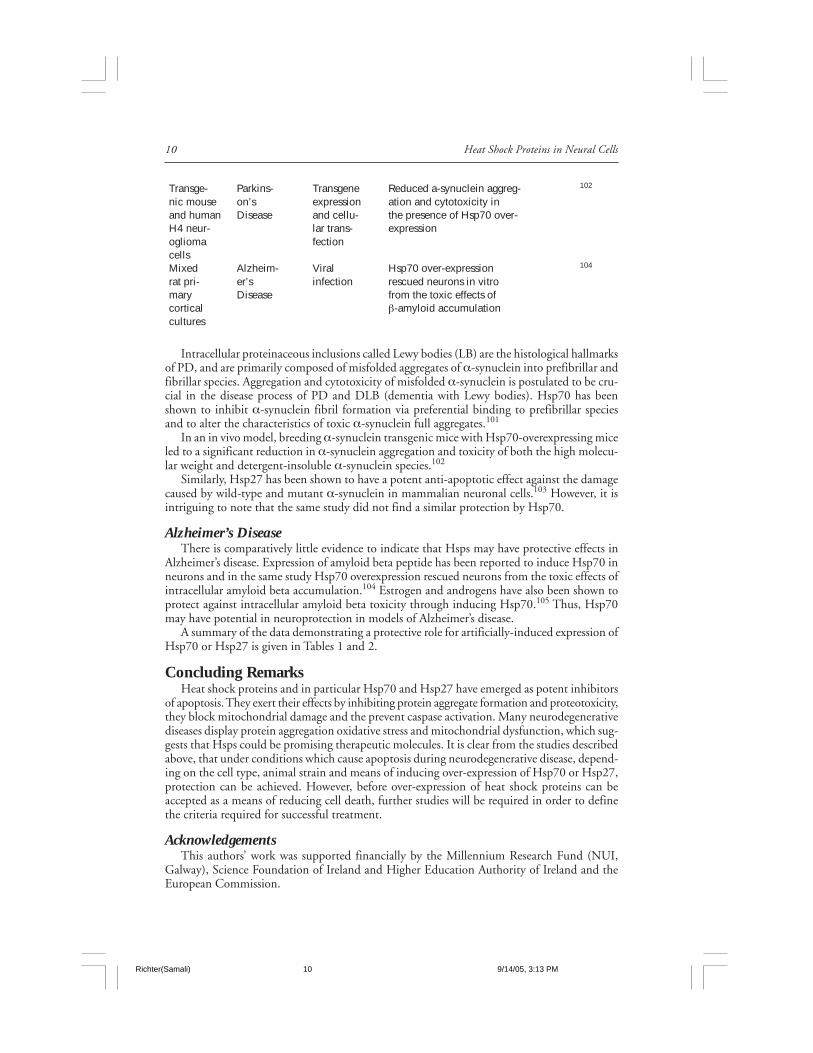
Intracellular proteinaceous inclusions called Lewy bodies (LB) are the histological hallmarksof PD, and are primarily composed of misfolded aggregates of α-synuclein into prefibrillar andfibrillar species. Aggregation and cytotoxicity of misfolded α-synuclein is postulated to be cru-cial in the disease process of PD and DLB (dementia with Lewy bodies). Hsp70 has beenshown to inhibit α-synuclein fibril formation via preferential binding to prefibrillar speciesand to alter the characteristics of toxic α-synuclein full aggregates.101
In an in vivo model, breeding α-synuclein transgenic mice with Hsp70-overexpressing miceled to a significant reduction in α-synuclein aggregation and toxicity of both the high molecu-lar weight and detergent-insoluble α-synuclein species.102
Similarly, Hsp27 has been shown to have a potent anti-apoptotic effect against the damagecaused by wild-type and mutant α-synuclein in mammalian neuronal cells.103 However, it isintriguing to note that the same study did not find a similar protection by Hsp70.
Alzheimer’s DiseaseThere is comparatively little evidence to indicate that Hsps may have protective effects in
Alzheimer’s disease. Expression of amyloid beta peptide has been reported to induce Hsp70 inneurons and in the same study Hsp70 overexpression rescued neurons from the toxic effects ofintracellular amyloid beta accumulation.104 Estrogen and androgens have also been shown toprotect against intracellular amyloid beta toxicity through inducing Hsp70.105 Thus, Hsp70may have potential in neuroprotection in models of Alzheimer’s disease.
A summary of the data demonstrating a protective role for artificially-induced expression ofHsp70 or Hsp27 is given in Tables 1 and 2.
Concluding RemarksHeat shock proteins and in particular Hsp70 and Hsp27 have emerged as potent inhibitors
of apoptosis. They exert their effects by inhibiting protein aggregate formation and proteotoxicity,they block mitochondrial damage and the prevent caspase activation. Many neurodegenerativediseases display protein aggregation oxidative stress and mitochondrial dysfunction, which sug-gests that Hsps could be promising therapeutic molecules. It is clear from the studies describedabove, that under conditions which cause apoptosis during neurodegenerative disease, depend-ing on the cell type, animal strain and means of inducing over-expression of Hsp70 or Hsp27,protection can be achieved. However, before over-expression of heat shock proteins can beaccepted as a means of reducing cell death, further studies will be required in order to definethe criteria required for successful treatment.
AcknowledgementsThis authors’ work was supported financially by the Millennium Research Fund (NUI,
Galway), Science Foundation of Ireland and Higher Education Authority of Ireland and theEuropean Commission.
Transge- Parkins- Transgene Reduced a-synuclein aggreg- 102
nic mouse on’s expression ation and cytotoxicity inand human Disease and cellu- the presence of Hsp70 over-H4 neur- lar trans- expressionoglioma fectioncellsMixed Alzheim- Viral Hsp70 over-expression 104
rat pri- er’s infection rescued neurons in vitromary Disease from the toxic effects ofcortical β-amyloid accumulationcultures
Richter(Samali) 9/14/05, 3:13 PM10

11Heat Shock Proteins and the Regulation of Apoptosis
References1. Hengartner MO. The biochemistry of apoptosis. Nature 2000; 407:770-776.2. Samali A, Cai J, Zhivotovsky B et al. Presence of a preapoptotic complex of pro-caspase-3, Hsp60
and Hsp10 in the mitochondrial fraction of jurkat cells. EMBO J 1999; 18:2040-2048.3. Fuentes-Prior P, Salvesen GS. The protein structures that shape caspase activity, specificity, activa-
tion and inhibition. Biochem J 2004; 384:201-232.4. Philchenkov A. Caspases: Potential targets for regulating cell death. J Cell Mol Med 2004; 8:432-
444.5. Riedl SJ, Shi Y. Molecular mechanisms of caspase regulation during apoptosis. Nat Rev Mol Cell
Biol 2004; 5:897-907.6. Fischer U, Janicke RU, Schulze-Osthoff K. Many cuts to ruin: A comprehensive update of caspase
substrates. Cell Death Differ 2003; 10:76-100.7. Samali A, Gorman AM, Cotter TG. Apoptosis - The story so far. Experientia 1996; 52:933-941.8. Thorburn A. Death receptor-induced cell killing. Cell Signal 2004; 16:139-144.9. Wang S, El-Deiry WS. TRAIL and apoptosis induction by TNF-family death receptors. Oncogene
2003; 22:8628-8633.10. Curtin JF, Cotter TG. Live and let die: Regulatory mechanisms in Fas-mediated apoptosis. Cell
Signal 2003; 15:983-992.11. Chang HY, Nishitoh H, Yang X et al. Activation of apoptosis signal-regulating kinase 1 (ASK1) by
the adapter protein Daxx. Science 1998; 281:1860-1863.12. Green DR, Reed JC. Mitochondria and apoptosis. Science 1998; 281:1309-1312.13. Mishra NC, Kumar S. Apoptosis: A mitochondrial perspective on cell death. Indian J Exp Biol
2005; 43:25-34.14. Polster BM, Fiskum G. Mitochondrial mechanisms of neural cell apoptosis. J Neurochem 2004;
90:1281-1289.
Table 2. Neuroprotective strategies exploiting Hsp27
Model Disorder Mode of over- Comment Refe-System targeted expression rence
RGC-5 rat Ischaemia Transfection Cells protected 96
retinal gan- from serum,glion cell oxygen andline glucose reduction
and calciumoverload.
HCN2A neuro- Alzheimer’s Chemical Hsp27 reduced 77
nal cells disease delivery levels ofhyperphosphorylat-ed tau and enhanc-ed its degradation.
ND7 DRG Parkinson’s HSV-mediated Reduced caspase 103
dorsal root disease infection activity detected.ganglion/ Hsp70 was not pro-neuroblastoma tective againstfusion cells same insults, with
exception of isch-aemia.
PC12 rat Parkinson’s Transfection Increased survival 100
pheochromo- disease of 6-OHDA-treatedcytoma cells cells over-express-
ing Hsp27
Richter(Samali) 9/14/05, 3:13 PM11
Heat Shock Proteins in Neural Cells12
15. Zhou P, Chou J, Olea RS et al. Solution structure of Apaf-1 CARD and its interaction withcaspase-9 CARD: A structural basis for specific adaptor/caspase interaction. Proc Natl Acad SciUSA 1999; 96:11265-11270.
16. Hill MM, Adrain C, Martin SJ. Portrait of a killer: The mitochondrial apoptosome emerges fromthe shadows. Mol Interv 2003; 3:19-26.
17. Nakagawa T, Zhu H, Morishima N et al. Caspase-12 mediates endoplasmic-reticulum-specificapoptosis and cytotoxicity by amyloid-beta. Nature 2000; 403:98-103.
18. Yoneda T, Imaizumi K, Oono K et al. Activation of caspase-12, an endoplastic reticulum (ER)resident caspase, through tumor necrosis factor receptor-associated factor 2-dependent mechanismin response to the ER stress. J Biol Chem 2001; 276:13935-13940.
19. Rao RV, Hermel E, Castro-Obregon S et al. Coupling endoplasmic reticulum stress to the celldeath program. Mechanism of caspase activation. J Biol Chem 2001; 276:33869-33874.
20. Morishima N, Nakanishi K, Takenouchi H et al. An endoplasmic reticulum stress-specific caspasecascade in apoptosis. Cytochrome c-independent activation of caspase-9 by caspase-12. J Biol Chem2002; 277:34287-34294.
21. Rutkowski DT, Kaufman RJ. A trip to the ER: Coping with stress. Trends Cell Biol 2004; 14:20-28.
22. Ma Y, Hendershot LM. The mammalian endoplasmic reticulum as a sensor for cellular stress. CellStress Chaperones 2002; 7:222-229.
23. Lee AH, Iwakoshi NN, Glimcher LH. XBP-1 regulates a subset of endoplasmic reticulum residentchaperone genes in the unfolded protein response. Mol Cell Biol 2003; 23:7448-7459.
24. Fischer H, Koenig U, Eckhart L et al. Human caspase 12 has acquired deleterious mutations.Biochem Biophys Res Commun 2002; 293:722-726.
25. Saleh M, Vaillancourt JP, Graham RK et al. Differential modulation of endotoxin responsivenessby human caspase-12 polymorphisms. Nature 2004; 429:75-79.
26. Hitomi J, Katayama T, Eguchi Y et al. Involvement of caspase-4 in endoplasmic reticulumstress-induced apoptosis and Abeta-induced cell death. J Cell Biol 2004; 165:347-356.
27. Liu X, Kim CN, Yang J et al. Induction of apoptotic program in cell-free extracts: Requirementfor dATP and cytochrome c. Cell 1996; 86:147-157.
28. Lorenzo HK, Susin SA, Penninger J et al. Apoptosis inducing factor (AIF): A phylogenetically old,caspase-independent effector of cell death. Cell Death Differ 1999; 6:516-524.
29. Verhagen AM, Ekert PG, Pakusch M et al. Identification of DIABLO, a mammalian protein thatpromotes apoptosis by binding to and antagonizing IAP proteins. Cell 2000; 102:43-53.
30. Schafer P, Scholz SR, Gimadutdinow O et al. Structural and functional characterization of mito-chondrial EndoG, a sugar nonspecific nuclease which plays an important role during apoptosis. JMol Biol 2004; 338:217-228.
31. Melino G, Bernassola F, Ranalli M et al. p73 Induces apoptosis via PUMA transactivation and Baxmitochondrial translocation. J Biol Chem 2004; 279:8076-8083.
32. Xanthoudakis S, Roy S, Rasper D et al. Hsp60 accelerates the maturation of pro-caspase-3 byupstream activator proteases during apoptosis. EMBO J 1999; 18:2049-2056.
33. Li H, Zhu H, Xu CJ et al. Cleavage of BID by caspase 8 mediates the mitochondrial damage inthe Fas pathway of apoptosis. Cell 1998; 94:491-501.
34. Luo X, Budihardjo I, Zou H et al. Bid, a Bcl2 interacting protein, mediates cytochrome c releasefrom mitochondria in response to activation of cell surface death receptors. Cell 1998; 94:481-490.
35. Scaffidi C, Fulda S, Srinivasan A et al. Two CD95 (APO-1/Fas) signaling pathways. EMBO J1998; 17:1675-1687.
36. Robertson JD, Gogvadze V, Zhivotovsky B et al. Distinct pathways for stimulation of cytochromec release by etoposide. J Biol Chem 2000; 275:32438-32443.
37. Rich T, Allen RL, Wyllie AH. Defying death after DNA damage. Nature 2000; 407:777-783.38. Willis S, Day CL, Hinds MG et al. The Bcl-2-regulated apoptotic pathway. J Cell Sci 2003;
116:4053-4056.39. Saleh A, Srinivasula SM, Balkir L et al. Negative regulation of the Apaf-1 apoptosome by Hsp70.
Nat Cell Biol 2000; 2:476-483.40. Beere HM, Wolf BB, Cain K et al. Heat-shock protein 70 inhibits apoptosis by preventing recruit-
ment of procaspase-9 to the Apaf-1 apoptosome. Nat Cell Biol 2000; 2:469-475.41. Steel R, Doherty JP, Buzzard K et al. Hsp72 inhibits apoptosis upstream of the mitochondria and
not through interactions with Apaf-1. J Biol Chem 2004; 279:51490-51499.42. Creagh EM, Carmody RJ, Cotter TG. Heat shock protein 70 inhibits caspase-dependent and -in-
dependent apoptosis in Jurkat T cells. Exp Cell Res 2000; 257:58-66.
Richter(Samali) 9/14/05, 3:13 PM12

13Heat Shock Proteins and the Regulation of Apoptosis
43. Ravagnan L, Gurbuxani S, Susin SA et al. Heat-shock protein 70 antagonizes apoptosis-inducingfactor. Nat Cell Biol 2001; 3:839-843.
44. Clemens MJ. Translational control in virus-infected cells: Models for cellular stress responses. SeminCell Dev Biol 2005; 16:13-20.
45. Gabai VL, Yaglom JA, Volloch V et al. Hsp72-mediated suppression of c-Jun N-terminal kinase isimplicated in development of tolerance to caspase-independent cell death. Mol Cell Biol 2000;20:6826-6836.
46. Mosser DD, Caron AW, Bourget L et al. Role of the human heat shock protein hsp70 in protec-tion against stress-induced apoptosis. Mol Cell Biol 1997; 17:5317-5327.
47. Park J, Liu AY. JNK phosphorylates the HSF1 transcriptional activation domain: Role of JNK inthe regulation of the heat shock response. J Cell Biochem 2001; 82:326-338.
48. Beere HM. “The stress of dying”: The role of heat shock proteins in the regulation of apoptosis. JCell Sci 2004; 117:2641-2651.
49. Jaattela M, Wissing D, Kokholm K et al. Hsp70 exerts its anti-apoptotic function downstream ofcaspase-3-like proteases. EMBO J 1998; 17:6124-6134.
50. Komarova EY, Afanasyeva EA, Bulatova MM et al. Downstream caspases are novel targets for theantiapoptotic activity of the molecular chaperone hsp70. Cell Stress Chaperones 2004; 9:265-275.
51. Park HS, Cho SG, Kim CK et al. Heat shock protein hsp72 is a negative regulator of apoptosissignal-regulating kinase 1. Mol Cell Biol 2002; 22:7721-7730.
52. Michels AA, Kanon B, Konings AW et al. Hsp70 and Hsp40 chaperone activities in the cytoplasmand the nucleus of mammalian cells. J Biol Chem 1997; 272:33283-33289.
53. Lu Z, Cyr DM. Protein folding activity of Hsp70 is modified differentially by the hsp40cochaperones Sis1 and Ydj1. J Biol Chem 1998; 273:27824-27830.
54. Liu QL, Kishi H, Ohtsuka K et al. Heat shock protein 70 binds caspase-activated DNase andenhances its activity in TCR-stimulated T cells. Blood 2003; 102:1788-1796.
55. Gotoh T, Terada K, Oyadomari S et al. hsp70-DnaJ chaperone pair prevents nitric oxide- andCHOP-induced apoptosis by inhibiting translocation of Bax to mitochondria. Cell Death Differ2004; 11:390-402.
56. Bruey JM, Ducasse C, Bonniaud P et al. Hsp27 negatively regulates cell death by interacting withcytochrome c. Nat Cell Biol 2000; 2:645-652.
57. Concannon CG, Orrenius S, Samali A. Hsp27 inhibits cytochrome c-mediated caspase activationby sequestering both pro-caspase-3 and cytochrome c. Gene Expr 2001; 9:195-201.
58. Garrido C, Bruey JM, Fromentin A et al. HSP27 inhibits cytochrome c-dependent activation ofprocaspase-9. Faseb J 1999; 13:2061-2070.
59. Paul C, Manero F, Gonin S et al. Hsp27 as a negative regulator of cytochrome C release. Mol CellBiol 2002; 22:816-834.
60. Pandey P, Farber R, Nakazawa A et al. Hsp27 functions as a negative regulator of cytochromec-dependent activation of procaspase-3. Oncogene 2000; 19:1975-1981.
61. Chauhan D, Li G, Hideshima T et al. Hsp27 inhibits release of mitochondrial protein Smac inmultiple myeloma cells and confers dexamethasone resistance. Blood 2003; 102:3379-3386.
62. Charette SJ, Landry J. The interaction of HSP27 with Daxx identifies a potential regulatory roleof HSP27 in Fas-induced apoptosis. Ann NY Acad Sci 2000; 926:126-131.
63. Thompson JE, Thompson CB. Putting the rap on Akt. J Clin Oncol 2004; 22:4217-4226.64. Mearow KM, Dodge ME, Rahimtula M et al. Stress-mediated signaling in PC12 cells - The role
of the small heat shock protein, Hsp27, and Akt in protecting cells from heat stress and nervegrowth factor withdrawal. J Neurochem 2002; 83:452-462.
65. Rane MJ, Pan Y, Singh S et al. Heat shock protein 27 controls apoptosis by regulating Akt activa-tion. J Biol Chem 2003; 278:27828-27835.
66. Parcellier A, Schmitt E, Gurbuxani S et al. HSP27 is a ubiquitin-binding protein involved inI-kappaBalpha proteasomal degradation. Mol Cell Biol 2003; 23:5790-5802.
67. Mehlen P, Schulze-Osthoff K, Arrigo AP. Small stress proteins as novel regulators of apoptosis.Heat shock protein 27 blocks Fas/APO-1- and staurosporine-induced cell death. J Biol Chem 1996;271:16510-16514.
68. Mehlen P, Coronas V, Ljubic-Thibal V et al. Small stress protein Hsp27 accumulation duringdopamine-mediated differentiation of rat olfactory neurons counteracts apoptosis. Cell Death Dif-fer 1999; 6:227-233.
69. Arrigo AP, Virot S, Chaufour S et al. Hsp27 consolidates intracellular redox homeostasis by up-holding glutathione in its reduced form and by decreasing iron intracellular levels. Antioxid RedoxSignal 2005; 7:414-422.
Richter(Samali) 9/14/05, 3:13 PM13
Heat Shock Proteins in Neural Cells14
70. Wyttenbach A, Sauvageot O, Carmichael J et al. Heat shock protein 27 prevents cellularpolyglutamine toxicity and suppresses the increase of reactive oxygen species caused by huntingtin.Hum Mol Genet 2002; 11:1137-1151.
71. Preville X, Salvemini F, Giraud S et al. Mammalian small stress proteins protect against oxidativestress through their ability to increase glucose-6-phosphate dehydrogenase activity and by main-taining optimal cellular detoxifying machinery. Exp Cell Res 1999; 247:61-78.
72. Lavoie JN, Lambert H, Hickey E et al. Modulation of cellular thermoresistance and actin filamentstability accompanies phosphorylation-induced changes in the oligomeric structure of heat shockprotein 27. Mol Cell Biol 1995; 15:505-516.
73. Geum D, Son GH, Kim K. Phosphorylation-dependent cellular localization and thermoprotectiverole of heat shock protein 25 in hippocampal progenitor cells. J Biol Chem 2002; 277:19913-19921.
74. Van Why SK, Mann AS, Ardito T et al. Hsp27 associates with actin and limits injury in energydepleted renal epithelia. J Am Soc Nephrol 2003; 14:98-106.
75. Guay J, Lambert H, Gingras-Breton G et al. Regulation of actin filament dynamics by p38 mapkinase-mediated phosphorylation of heat shock protein 27. J Cell Sci 1997; 110(Pt 3):357-368.
76. Huot J, Houle F, Rousseau S et al. SAPK2/p38-dependent F-actin reorganization regulates earlymembrane blebbing during stress-induced apoptosis. J Cell Biol 1998; 143:1361-1373.
77. Shimura H, Miura-Shimura Y, Kosik KS. Binding of tau to heat shock protein 27 leads to de-creased concentration of hyperphosphorylated tau and enhanced cell survival. J Biol Chem 2004;279:17957-17962.
78. Kosik KS, Shimura H. Phosphorylated tau and the neurodegenerative foldopathies. Biochim BiophysActa 2005; 1739:298-310.
79. Lassmann H, Bancher C, Breitschopf H et al. Cell death in Alzheimer’s disease evaluated by DNAfragmentation in situ. Acta Neuropathol (Berl) 1995; 89:35-41.
80. Smale G, Nichols NR, Brady DR et al. Evidence for apoptotic cell death in Alzheimer’s disease.Exp Neurol 1995; 133:225-230.
81. Dragunow M, Preston K, Dodd J et al. Clusterin accumulates in dying neurons following statusepilepticus. Brain Res Mol Brain Res 1995; 32:279-290.
82. Sugaya K, Reeves M, McKinney M. Topographic associations between DNA fragmentation andAlzheimer’s disease neuropathology in the hippocampus. Neurochem Int 1997; 31:275-281.
83. Thomas LB, Gates DJ, Richfield EK et al. DNA end labeling (TUNEL) in Huntington’s diseaseand other neuropathological conditions. Exp Neurol 1995; 133:265-272.
84. Vis JC, Schipper E, de Boer-van Huizen RT et al. Expression pattern of apoptosis-related markersin Huntington’s disease. Acta Neuropathol (Berl) 2005.
85. Tatton WG, Chalmers-Redman R, Brown D et al. Apoptosis in Parkinson’s disease: Signals forneuronal degradation. Ann Neurol 2003; 53(Suppl 3):S61-70, (discussion S70-62).
86. Tatton NA. Increased caspase 3 and Bax immunoreactivity accompany nuclear GAPDH transloca-tion and neuronal apoptosis in Parkinson’s disease. Exp Neurol 2000; 166:29-43.
87. de la Monte SM, Sohn YK, Ganju N et al. P53- and CD95-associated apoptosis in neurodegenerativediseases. Lab Invest 1998; 78:401-411.
88. Hartmann A, Hunot S, Michel PP et al. Caspase-3: A vulnerability factor and final effector inapoptotic death of dopaminergic neurons in Parkinson’s disease. Proc Natl Acad Sci USA 2000;97:2875-2880.
89. Hartmann A, Troadec JD, Hunot S et al. Caspase-8 is an effector in apoptotic death of dopamin-ergic neurons in Parkinson’s disease, but pathway inhibition results in neuronal necrosis. J Neurosci2001; 21:2247-2255.
90. Giffard RG, Yenari MA. Many mechanisms for hsp70 protection from cerebral ischemia. J NeurosurgAnesthesiol 2004; 16:53-61.
91. Matsumori Y, Hong SM, Aoyama K et al. Hsp70 overexpression sequesters AIF and reduces neo-natal hypoxic/ischemic brain injury. J Cereb Blood Flow Metab 2005.
92. Kelly S, Yenari MA. Neuroprotection: Heat shock proteins. Curr Med Res Opin 2002; 18(Suppl2):s55-60.
93. Amin V, Cumming DV, Latchman DS. Over-expression of heat shock protein 70 protects neu-ronal cells against both thermal and ischaemic stress but with different efficiencies. Neurosci Lett1996; 206:45-48.
94. Kwong JM, Lam TT, Caprioli J. Hyperthermic preconditioning protects retinal neurons fromN-methyl-D-aspartate (NMDA)-induced apoptosis in rat. Brain Res 2003; 970:119-130.
95. Xu L, Lee JE, Giffard RG. Overexpression of bcl-2, bcl-XL or hsp70 in murine cortical astrocytesreduces injury of cocultured neurons. Neurosci Lett 1999; 277:193-197.
Richter(Samali) 9/14/05, 3:13 PM14

15Heat Shock Proteins and the Regulation of Apoptosis
96. Whitlock NA, Lindsey K, Agarwal N et al. Heat shock protein 27 delays Ca2+-induced cell deathin a caspase-dependent and -independent manner in rat retinal ganglion cells. Invest OphthalmolVis Sci 2005; 46:1085-1091.
97. Dong Z, Wolfer DP, Lipp HP et al. Hsp70 gene transfer by adeno-associated virus inhibitsMPTP-induced nigrostriatal degeneration in the mouse model of Parkinson disease. Mol Ther 2005;11:80-88.
98. Zhou Y, Gu G, Goodlett DR et al. Analysis of alpha-synuclein-associated proteins by quantitativeproteomics. J Biol Chem 2004; 279:39155-39164.
99. Quigney DJ, Gorman AM, Samali A. Heat shock protects PC12 cells against MPP+ toxicity. BrainRes 2003; 993:133-139.
100. Gorman AM, Szegezdi E, Quigney DJ et al. Hsp27 inhibits 6-hydroxydopamine-induced cyto-chrome c release and apoptosis in PC12 cells. Biochem Biophys Res Commun 2005; 327:801-810.
101. Dedmon MM, Christodoulou J, Wilson MR et al. Heat shock protein 70 inhibits {alpha}-synucleinfibril formation via preferential binding to prefibrillar species. J Biol Chem 2005; 280:14733-14740.
102. Klucken J, Shin Y, Masliah E et al. Hsp70 Reduces alpha-synuclein aggregation and toxicity. JBiol Chem 2004; 279:25497-25502.
103. Zourlidou A, Payne Smith MD, Latchman DS. HSP27 but not HSP70 has a potent protectiveeffect against alpha-synuclein-induced cell death in mammalian neuronal cells. J Neurochem 2004;88:1439-1448.
104. Magrane J, Smith RC, Walsh K et al. Heat shock protein 70 participates in the neuroprotectiveresponse to intracellularly expressed beta-amyloid in neurons. J Neurosci 2004; 24:1700-1706.
105. Zhang Y, Champagne N, Beitel LK et al. Estrogen and androgen protection of human neuronsagainst intracellular amyloid beta1-42 toxicity through heat shock protein 70. J Neurosci 2004;24:5315-5321.
106. Rossler OG, Bauer I, Chung HY et al. Glutamate-induced cell death of immortalized murine hip-pocampal neurons: Neuroprotective activity of heme oxygenase-1, heat shock protein 70, and so-dium selenite. Neurosci Lett 2004; 362:253-257.
107. Lee JE, Yenari MA, Sun GH et al. Differential neuroprotection from human heat shock protein 70overexpression in vitro and in vivo models of ischemia and ischemia-like conditions. Exp Neurol2001; 170:129-139.
108. Rajdev S, Hara K, Kokubo Y et al. Mice overexpressing rat heat shock protein 70 are protectedagainst cerebral infarction. Ann Neurol 2000; 47:782-791.
109. Plumier JC, Krueger AM, Currie RW et al. Transgenic mice expressing the human inducible Hsp70have hippocampal neurons resistant to ischemic injury. Cell Stress Chaperones 1997; 2:162-167.
Richter(Samali) 9/14/05, 3:13 PM15
Related Documents