From the Neonatal Unit, Department of Woman and Child Health, Karolinska Institutet and Astrid Lindgren Children´s Hospital, Stockholm, Sweden Apnea and infection in neonates: Mediatory role of interleukin-1β and prostaglandin E 2 Annika Olsson Hofstetter Stockholm 2006

Welcome message from author
This document is posted to help you gain knowledge. Please leave a comment to let me know what you think about it! Share it to your friends and learn new things together.
Transcript
From the Neonatal Unit, Department of Woman and Child Health,
Karolinska Institutet and Astrid Lindgren Children´s Hospital, Stockholm, Sweden
Apnea and infection in neonates:
Mediatory role of interleukin-1β and prostaglandin E2
Annika Olsson Hofstetter
Stockholm 2006


Dedicated to Christoph and my parents
4
ABSTRACT
The breathing pattern of infants, particularly preterm infants, is often irregular or periodic and is frequently interrupted by apnea. The latter represents a major concern in neonatology, yet much remains unknown about its incidence, appearance, and pathophysiology. This thesis further characterizes cardiorespiratory activity in preterm infants during postnatal development and investigates the association between infection and apnea in neonates, focusing on the mediatory role of interleukin-1β and prostaglandin E2 in depressing central respiration.
Cardiorespiratory activity was evaluated in extremely preterm infants between birth and term-equivalent age using impedance pneumography, electrocardiography, and pulse oximetry. The incidence of apnea, bradycardia, and hypoxemia diminished with advancing age, although these events often persisted at term-equivalent age and after hospital discharge. Infection was clearly associated with an increased apnea and hypoxemia incidence.
To further elucidate the association between infection and apnea, respiration was examined in neonatal rodents using whole-body plethysmography after administration of the cytokine interleukin-1β (IL-1β) or the bacterial endotoxin lipopolysaccharide (LPS). Animals given IL-1β or LPS exhibited a lower basal respiratory frequency, depressed anoxic gasping, and a reduced ability to autoresuscitate following hypoxic apnea compared to control animals. Hyperoxic challenge revealed functioning peripheral chemoreceptors in all animals, suggesting a central mechanism underlying the ventilatory effects of these immunomodulators. However, IL-1β did not affect the respiration-related activity in neonatal rat brainstem-spinal cord preparations, indicating that it may communicate indirectly with this central respiratory network.
Prostaglandin E2 (PGE2) may serve as a critical mediator of ventilatory changes induced by IL-1β. In newborn infants, the infectious marker C-reactive protein was correlated with an elevated PGE2 concentration, which in turn was associated with an increased apnea frequency. In newborn rodents, PGE2 reversibly inhibited respiratory neurons in vitro and induced apnea and irregular breathing patterns in vivo. Moreover, IL-1β rapidly induced brainstem microsomal prostaglandin E synthase-1 (mPGES-1), an enzyme crucial for PGE2 biosynthesis. Pretreatment with indomethacin, a prostaglandin synthesis inhibitor, clearly attenuated the adverse effects of IL-1β and LPS on basal respiration and anoxic ventilatory response in neonatal rats. Additionally, mPGES-1 knockout mice did not exhibit IL-1β-induced respiratory depression during hyperoxia and anoxia. Similarly, mice lacking the EP3 receptor for PGE2 had fewer PGE2-induced apneas in vivo and less PGE2-induced inhibition of brainstem respiratory activity in vitro compared to wildtype mice. These findings strongly suggest that IL-1β alters breathing and hypoxic defense via central mPGES-1 activation and subsequent PGE2 synthesis and binding to brainstem EP3 receptors. These studies have important clinical implications for the diagnosis, surveillance, and treatment of neonatal apnea associated with infection.
5
LIST OF ORIGINAL PAPERS
This thesis is based upon the following papers, which will be referred to by their Roman
numerals:
I. Hofstetter AO, Legnevall L, Herlenius E, Katz-Salamon M. Cardiorespiratory function
in extremely preterm infants during early postnatal development. Manuscript.
II. Hofstetter AO, Herlenius E. Interleukin-1β depresses hypoxic gasping and
autoresuscitation in neonatal DBA/1lacJ mice. Respiratory Physiology and
Neurobiology, 146 (2-3): 135-146, 2005.
III. Olsson A, Kayhan G, Lagercrantz H, Herlenius E. Interleukin-1β depresses respiration
and anoxic survival via a prostaglandin-dependent pathway in neonatal rats. Pediatric
Research, 54 (3): 326-331, 2003.
IV. Hofstetter AO, Saha S, Siljehav V, Jakobsson PJ, Herlenius E. The induced
prostaglandin E2 pathway – a key regulator of the respiratory response to infection and
hypoxia in neonates. Manuscript submitted.

6
ABBREVIATIONS
AI apnea/hypopnea index
BI bradycardia index
COX-2 cyclooxygenase-2
CRP C-reactive protein
CSF cerebrospinal fluid
EP3R EP3 receptor
fR respiratory frequency
GA gestational age
HI hypoxemia index
HR heart rate
IL-1β interleukin-1β
LPS lipopolysaccharide
mPGES-1 microsomal prostaglandin E synthase-1
NTS nucleus tractus solitarius
PCA post-conceptional age
PGE2 prostaglandin E2
PNA postnatal age
preBötC pre-Bötzinger complex
RR respiratory rate
RVLM rostral ventrolateral medulla
SIDS Sudden Infant Death Syndrome
VT tidal volume
VE minute ventilation
7
CONTENTS
Abstract 4
List of original papers 5
Abbreviations 6
1. Introduction 9
1.1. Respiratory rhythm generation 9
1.2. Fetal breathing and transition at birth 10
1.3. Neonatal respiration 11
1.4. Pathophysiology of neonatal apnea 12
1.5. Apnea characteristics and treatment 13
1.6. Infection, apnea, and SIDS 15
1.7. Interleukin-1β 16
1.8. Prostaglandin E2 17
2. Aims 19
3. Methodology 20
3.1. Human subjects 20
3.2. Animal models 20
3.3. Drugs 21
3.4. Cardiorespiratory monitoring 21
3.5. Whole-body plethysmography 22
3.6. Brainstem-spinal cord preparation 25
3.7. Enzymatic assay 26
3.8. Enzyme immunoassay 26
3.9. Data analysis 27
4. Results and discussion 29
4.1. Cardiorespiratory development in preterm infants 29
8
4.2. Cardiorespiratory events during early postnatal life 29
4.3. Infection increases cardiorespiratory events in infants 31
4.4. Respiratory behavior in neonatal DBA/1lacJ mice 31
4.5. IL-1β depresses respiration via central actions 32
4.6. Endogenous PGE2 exerts tonic respiratory effects 33
4.7. PGE2 inhibits respiratory activity via EP3R 33
4.8. IL-1β and hypoxia activate mPGES-1 34
4.9. PGE2 mediates the respiratory effects of IL-1β 34
5. Conclusions 36
6. Acknowledgements 37
7. References 38
Introduction ___________________________________________________________________________
9
1. Introduction This thesis explores the incidence, appearance, and pathophysiology of apnea, or the
cessation of breathing, in newborns. It examines the association between infection and
apnea, specifically the role of two immunomodulators, interleukin-1β and prostaglandin E2,
in altering respiration.
1.1. Respiratory rhythm generation
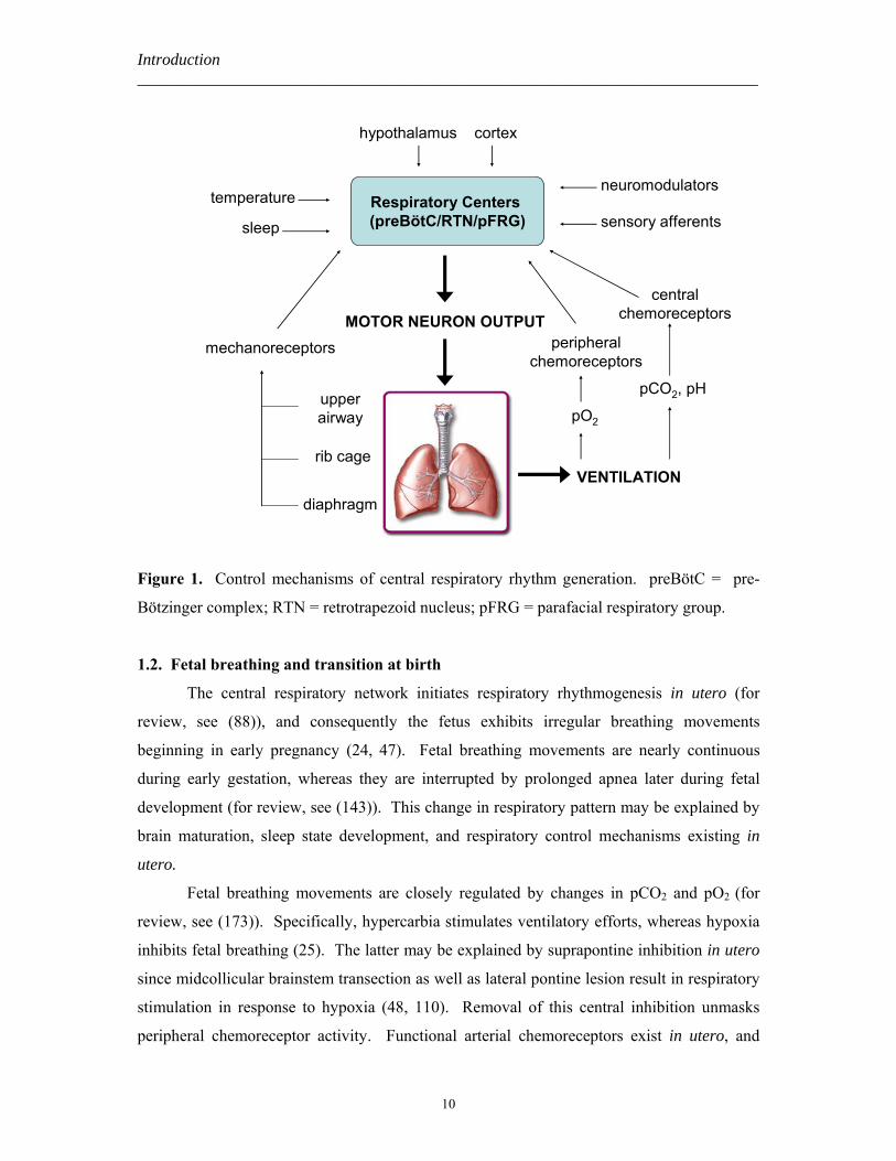
Respiratory efforts are generated and regulated via a complex integrative system
consisting of a central respiratory network and multiple feedback mechanisms (Figure 1).
The Greek physician Galen (ca 130 – 201 A.D.) was one of the first to purpose that the
brainstem was crucial for respiratory rhythmogenesis based upon his observations of injured
animals in the arena and criminals on the scaffold (81). This hypothesis has been confirmed
and refined since ancient times (76, 128, 216), and recent investigations indicate that the
central respiratory network is formed by neurons in three distinct regions of the brainstem: 1)
the ventral respiratory group (VRG) in the ventrolateral medulla (VLM); 2) the dorsal
respiratory group (DRG) in the nucleus tractus solitarius (NTS); and 3) the pontine
respiratory group (PRG) in the dorsolateral pons (for review, see (143)). The exact site and
mechanism of rhythm generation have been much debated. The pre-Bötzinger complex (pre-
BötC) as well as the retrotrapezoid nucleus (RTN) and parafacial respiratory group (pFRG)
have been proposed as crucial centers for respiratory rhythmogenesis (71, 155, 190, 191).
Recently, Feldman et al hypothesized that the pre-BötC and RTN/pFRG create a coupled
oscillator system, whereby the former serves as the dominant inspiratory rhythm generator
and the latter functions as the main expiratory rhythm generator (71). Three main respiratory
rhythms are generated: eupnea (e.g., normal resting respiration); sighing (e.g., large
inspiratory efforts overlying and interspersed within eupnea); and gasping (e.g., short
inspiratory efforts of high amplitude preceding long expiratory pauses). These ventilatory
patterns may be modulated by input from suprapontine structures within the central nervous
system (CNS), chemoreceptors, mechanoreceptors, and other sensory afferents. Ultimately,
this complex respiratory system regulates oxygenation, CO2 removal, and acid-base
homeostasis in the body.
Introduction ___________________________________________________________________________
10
Respiratory Centers (preBötC/RTN/pFRG)sleep
temperature
mechanoreceptors peripheral chemoreceptors
VENTILATION
diaphragm
pCO2, pHupper airway
neuromodulators
pO2
central chemoreceptors
cortexhypothalamus
MOTOR NEURON OUTPUT
sensory afferents
rib cage
Figure 1. Control mechanisms of central respiratory rhythm generation. preBötC = pre-
Bötzinger complex; RTN = retrotrapezoid nucleus; pFRG = parafacial respiratory group.
1.2. Fetal breathing and transition at birth
The central respiratory network initiates respiratory rhythmogenesis in utero (for
review, see (88)), and consequently the fetus exhibits irregular breathing movements
beginning in early pregnancy (24, 47). Fetal breathing movements are nearly continuous
during early gestation, whereas they are interrupted by prolonged apnea later during fetal
development (for review, see (143)). This change in respiratory pattern may be explained by
brain maturation, sleep state development, and respiratory control mechanisms existing in
utero.
Fetal breathing movements are closely regulated by changes in pCO2 and pO2 (for
review, see (173)). Specifically, hypercarbia stimulates ventilatory efforts, whereas hypoxia
inhibits fetal breathing (25). The latter may be explained by suprapontine inhibition in utero
since midcollicular brainstem transection as well as lateral pontine lesion result in respiratory
stimulation in response to hypoxia (48, 110). Removal of this central inhibition unmasks
peripheral chemoreceptor activity. Functional arterial chemoreceptors exist in utero, and
Introduction ___________________________________________________________________________
11
their activity is increased in response to low pO2 and high pCO2 (19). However, their
sensitivity is set at a lower pO2 than after birth, probably due to the lower fetal pO2.
Neuromodulators such as adenosine, prostaglandin, and endorphins also play an
active role in the regulation of fetal breathing. Adenosine inhibits central respiration-related
neurons in fetal rats (99), and its release during hypoxia may make it more abundant in utero
due to the lower pO2 (for review, see (101)). Similarly, prostaglandin and endorphins inhibit
fetal breathing movements (118, 141, 181). Conversely, fetal breathing is stimulated by
administration of their antagonists, e.g., methylxanthines, indomethacin, and naloxone,
respectively (39, 99, 117, 119, 148, 182).
At birth, there is a transition from irregular breathing movements to continuous
respiration. The exact mechanism underlying this transition remains unclear. Central cooling
of the newborn infant is an important trigger of continuous breathing at birth (22, 121, 122).
Moreover, several genes encoding for respiratory-stimulating neurotransmitters are switched
on during the perinatal period, and a surge in these excitatory neurotransmitters may play a
key role in the respiratory transition and general arousal after birth (123-126, 176, 192, 223).
Perinatal changes in inhibitory neurotransmitter expression and activity have also been
implicated in the maintenance of continuous respiration, e.g. lower adenosine concentrations
and less adenosine A1-receptor inhibition (99) and removal of placental inhibitory factors
such as prostaglandin (5, 6). Changes in suprapontine stimuli, chemosensitivity, and other
reflex responses during the early postnatal period play a crucial role in the stabilization of
neonatal respiration and will be discussed in greater detail below.
1.3. Neonatal respiration
Development of the intrinsic properties and functional organization of the central
respiratory network continues after birth. Not only is there a change in the motor pattern and
neurotransmitter sensitivity of respiration-related neurons with advancing postnatal age (99,
160, 169), but there is a maturation of dendritic morphology and increase in synaptic
connections and myelination after birth (167). Collectively, these processes help stabilize
respiratory activity during the postnatal period.
Central chemosensitivity plays an important role in modulating neonatal respiration.
The ventilatory response to CO2 in healthy term neonates is similar to that in adults (8),
indicating that the central chemoreceptors are functional immediately after birth. This is
supported by evidence that the primary central chemoreceptor area at the ventral medullary
Introduction ___________________________________________________________________________
12
surface exhibits c-fos mRNA expression directly after birth, and this expression is further
enhanced by hypercarbia at one day after birth (221).
While arterial chemoreceptors are functional in utero, they become quiescent
immediately after birth. However, within the first few days of postnatal life, peripheral
chemoreceptors increase their responsiveness towards adult levels (19, 103, 104). This
resetting process most likely results from a rise in pO2 concentrations at birth (21).
Peripheral chemosensitivity continues to develop during the postnatal period and plays an
important role in respiratory regulation (29, 41, 174). Differential expression of
neuromodulators within the carotid body may alter chemosensitivity. For example, a
decreased release of dopamine, an inhibitory neuromodulator in the carotid body, coincides
with the enhanced chemosensitivity after birth (103).
The ventilatory response to hypoxia also changes after birth. In newborn mammals,
hypoxia induces a biphasic respiratory response that is comprised of an increase in ventilation
followed by a decrease in respiratory efforts (for review, see (137)). This biphasic ventilatory
response persists until at least 8 weeks postnatal age in preterm infants (138). The initial
hyperventilation, lasting 1 – 2 min, results from activation of peripheral chemoreceptors. The
hypoxic ventilatory depression, characterized by primary apnea, gasping, and secondary or
terminal apnea (32), may result from the persistence of descending inhibitory tracts involved
in the fetal response to hypoxia (20, 48, 58, 203). Additionally, modulation of central
respiration-related neurons may contribute to the hypoxic ventilatory depression (159).
Inhibitory neurotransmitters such as adenosine (100, 127, 180), endorphins, and GABA have
also been implicated (for review, see (188)).
Control of neonatal respiration is also influenced by a variety of reflex responses from
the lungs, respiratory muscles, and airways. The Hering-Breuer reflex and laryngeal
chemoreflex are more profound in neonates than adults (44, 154, 204). Additionally, a
hypotonic upper airway, increased chest wall compliance, lower functional residual capacity,
and decreased coordination between respiratory muscles contribute to instability of
ventilation during the immediate postnatal period (for review, see (143)). Development of
these reflex responses and respiratory mechanics is a key factor involved in the maintenance
of adequate respiration after birth.
1.4. Pathophysiology of neonatal apnea
Apnea, or the cessation of breathing, occurs frequently in the neonatal population, and
immaturity of the central neuronal network plays a crucial role in its pathogenesis. Preterm
Introduction ___________________________________________________________________________
13
infants with frequent apnea exhibit prolonged auditory evoked responses, indicating that they
may have decreased neuronal diameter, less myelination, or slower synapse transmission time
(97). This immaturity increases their vulnerability to postnatal events such as infection,
intracranial hemorrhage, and thermal instability, particularly if they occur during critical
periods of respiratory plasticity (37, 142).
Immature chemosensitivity to CO2 may also contribute to apnea. Preterm infants in
general and those with apnea in particular exhibit an impaired ventilatory response to
hypercarbia (77, 84, 115, 174). This is further potentiated by a narrow window between
baseline CO2 levels and the apneic CO2 threshold (116) as well as a higher CO2 threshold for
upper airway muscle tone (36). In these infants, CO2 sensitivity increases with advancing
postnatal age (77, 174), which may be due to activation of additional chemosensitive regions
in the brainstem (222). It may also reflect maturation of respiratory mechanics.
Premature infants with apnea also exhibit abnormal O2 responsiveness. They
demonstrate enhanced peripheral chemoreceptor activity as evidenced by a greater immediate
increase in ventilation in response to hypoxia and respiratory depression in response to
hyperoxia (3, 153). They also have a more pronounced hypoxic ventilatory depression (4,
175) that may be due to immaturity of the central respiratory network as well as significant
suprapontine inhibition, which plays a crucial role in the fetal response to hypoxia.
Neuromodulator expression is also pronounced during early postnatal life, and developmental
changes in expression may explain alterations in the hypoxic responsiveness (99, 101).
Chemoreceptor dysfunction as well as central respiratory depression may impair the infant’s
ability to autoresuscitate following an apnea event (115, 138).
Apnea in preterm infants may also result from the marked excitability of pulmonary
stretch receptors as well as mechano- and chemoreceptors in the laryngeal mucosa,
particularly during hypoxia (220). Furthermore, apnea may be secondary to conditions such
as infection, which is one of the most frequent problems encountered in preterm infants
(170). The role of infection in altering neonatal respiration is the focus of the present thesis
and will be discussed in greater detail below.
1.5. Apnea characteristics and treatment
Conventionally, apnea has been defined as a respiratory pause greater than 20 seconds
or a pause of shorter duration accompanied by bradycardia or hypoxemia (1). Apnea has
been classified into subtypes, e.g, central, obstructive, or mixed events (for review, see
(137)). Central apnea occurs when there is a lack of inspiratory effort, but no apparent
Introduction ___________________________________________________________________________
14
airway obstruction. Obstructive apnea occurs when the infant initiates a respiratory effort
against an obstructed upper airway. Mixed apnea occurs when there is a lack of respiratory
effort in the setting of airway obstruction. The distribution of apnea subtypes has been
frequently described (30, 55, 74, 129, 150). Findings are variable (i.e., 40 – 93% of apnea in
preterm infants is of central origin), which likely reflects different methodological approaches
and infant populations (74, 129).
While apnea commonly occurs in preterm infants, its incidence in this population
remains unclear. The largest investigation of apnea was conducted between 1974 and 1979,
and it showed an increased frequency and prolonged duration of recurrent apnea in infants
born at earlier gestational ages (GA) (95). However, this study included few very preterm
infants (less than 8% infants were born before 28 weeks GA). Since it was published, infant
demographics have changed dramatically with greater survival of patients born at a younger
gestational age. Their immature cardiorespiratory function puts them at greater risk for
apnea, bradycardia, and hypoxemia events, which seem to persist beyond term gestation (42,
59, 168). The appearance of cardiorespiratory events in preterm infants has been described in
multiple cross-sectional studies (13, 35, 96, 165, 217). The majority of apnea events are not
accompanied by clinically significant changes in heart rate (HR) or oxygen saturation (SpO2)
(168), although apnea may occasionally occur concomitantly with bradycardia and/or
hypoxemia (35, 96, 165, 210, 217). Prolonged apnea in particular is associated with a greater
incidence, duration, and severity of bradycardia and hypoxemia (35, 74, 96, 163, 165, 210).
Methylxanthine derivatives such as caffeine and theophylline are the preferred
treatment worldwide for neonatal apnea. In newborn mammals, methylxanthines stimulate
respiration and reduce hypoxia-induced respiratory depression by inhibiting brainstem
adenosine receptors (78). They consequently reduce apnea frequency and mechanical
ventilation use in infants (98). However, deleterious effects have been described in animal
models (57, 89, 207), which may result from blocking the neuroprotective effects of
adenosine during ischemia. A large multi-center investigation, including infants from the
neonatal intensive care unit at Karolinska University Hospital, is underway to examine the
long-term neurodevelopmental outcome of preterm infants who received caffeine to treat
apnea (184). In addition to methylxanthines, ventilatory support or therapies targeting
secondary causes of apnea may be utilized.
Introduction ___________________________________________________________________________
15
1.6. Infection, apnea, and SIDS
Apnea is a common presenting symptom in infants with infection. Approximately
20% of newborns hospitalized with a respiratory syncytial virus (RSV) infection and 55% of
infants with late-onset sepsis suffer from apnea (27, 70). Infection and hypoxia have been
linked to Sudden Infant Death Syndrome with the majority of SIDS victims exhibiting minor
signs of infection (e.g., intermittent cough, congestion) or evidence of hypoxia (e.g., hypoxic
gasping, elevated hypoxanthine and vascular endothelial growth factor concentrations) prior
to death (18, 111, 156, 166, 170, 178, 205). The SIDS incidence is greatest between two and
four months when infants exhibit reduced maternal antibodies and an immature immune
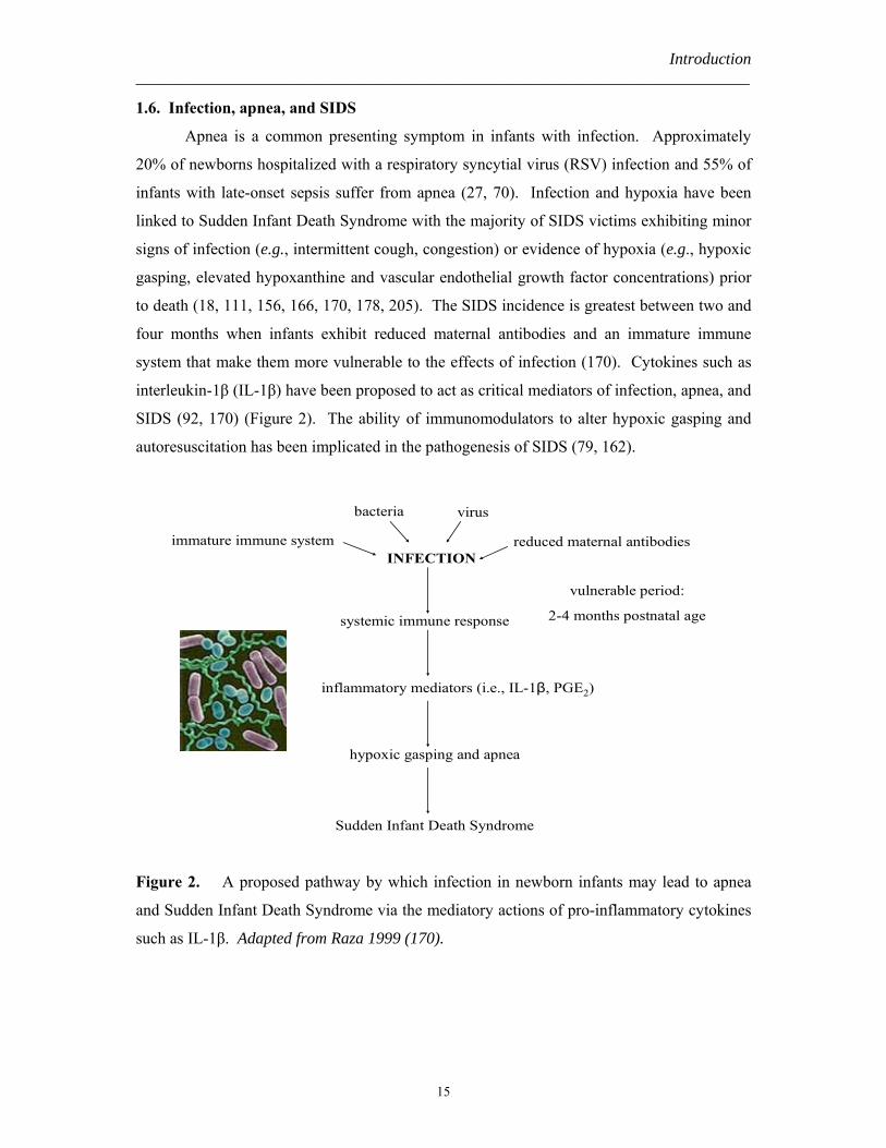
system that make them more vulnerable to the effects of infection (170). Cytokines such as
interleukin-1β (IL-1β) have been proposed to act as critical mediators of infection, apnea, and
SIDS (92, 170) (Figure 2). The ability of immunomodulators to alter hypoxic gasping and
autoresuscitation has been implicated in the pathogenesis of SIDS (79, 162).
INFECTION
bacteria virus
reduced maternal antibodiesimmature immune system
vulnerable period:
2-4 months postnatal agesystemic immune response
inflammatory mediators (i.e., IL-1β, PGE2)
hypoxic gasping and apnea
Sudden Infant Death Syndrome
Figure 2. A proposed pathway by which infection in newborn infants may lead to apnea
and Sudden Infant Death Syndrome via the mediatory actions of pro-inflammatory cytokines
such as IL-1β. Adapted from Raza 1999 (170).
Introduction ___________________________________________________________________________
16
1.7. Interleukin-1β
Interleukin-1β (IL-1β) is a pro-inflammatory cytokine that is synthesized and released
from activated monocytes and macrophages, neutrophils, and brain glial cells during an acute
phase immune response to infection and inflammation (for review, see (46)). IL-1β induces a
variety of sickness behaviors, including fever (54), hypersomnia (120), hypophagia (161),
and neuroendocrine changes (15). These physiological changes are highly organized and
strategically implemented during the body’s fight against infectious pathogens. While these
responses have distinct benefits, they can have adverse effects as well. In a newborn infant at
a critical stage of development, the simultaneous occurrence of an infectious process and a
hypoxic event may have a deleterious outcome. Infection with respiratory syncytial virus
(RSV) in newborn lambs prolongs the duration of apnea induced by laryngeal stimulation
(133). IL-1β may contribute to this finding as it similarly increases the duration of reflex
apnea in piglets (80, 195). Additionally, IL-1β concentrations in pharyngeal secretions of
human infants with RSV infection are positively correlated to the clinical severity of apnea
(132). IL-1β may also have detrimental effects on autoresuscitation as it has been shown to
reduce the respiratory frequency following apnea in piglets (80, 195). Furthermore, increased
levels of IL-1β have been found in the cerebrospinal fluid of SIDS victims (215).
As IL-1β induces behavioral and physiological changes of central origin, many
studies have investigated the presence and activation of IL-1 receptors (IL-1Rs) within the
CNS. In the rat, Type 1 IL-1R (IL-1R1) mRNA has been localized primarily to elements
associated with the blood-brain barrier such as the vascular endothelium, leptomeninges,
ependyma, and choroid plexus (69). In the mouse, IL-1R1 expression has been observed
predominantly upon endothelial cells, the choroid plexus, and the meninges as well as within
the dentate gyrus and over the midline raphe system (11, 45). Previous studies illustrate that
systemic administration of IL-1β and lipopolysaccharide (LPS), an endotoxin that increases
IL-1 bioactivity, immunoreactivity, and mRNA expression (for review, see (152)), induces
time- and dose-dependent expression of the immediate-early gene c-fos in respiratory regions
of the brainstem such as the NTS and RVLM (26, 49, 63, 68, 69). Interestingly, these
specific areas of IL-1β-induced Fos immunoreactivity do not appear to express IL-1R mRNA
(69).
There are several routes by which systemic IL-1β may relay immune signals to
autonomic regulatory centers in the brain. Although IL-1β is a large, lipophobic protein, it
may enter the CNS via carrier-mediated transport across the blood-brain barrier or by passage
Introduction ___________________________________________________________________________
17
through circumventricular organs (12, 23, 113). However, active transport systems have a
low capacity and are rapidly saturated (64). Moreover, barrier cells may prevent IL-1β
diffusion through circumventricular organs (219). Thus, IL-1β may communicate with the
central respiratory network via an indirect mechanism.
IL-1β may alter central behavior by inducing the synthesis and release of
prostaglandin E2 (PGE2) at the blood-brain barrier. Circulating IL-1β has been shown to bind
to and subsequently activate vascular endothelial cells expressing IL-1R mRNA at the blood-
brain interface (69, 213, 224). Within an hour of intravenous administration of IL-1β, an
increased expression of cyclooxygenase-2 (COX-2) and microsomal prostaglandin E
synthase-1 (mPGES-1) mRNA is observed at these endothelial cells (61). This results in
enhanced prostaglandin immunoreactivity and a dose-dependent rise in PGE2 production
(213). Similarly, peripheral LPS induces COX-2 and mPGES-1 co-localized to brain
endothelial cells (225), which in turn evokes a time-dependent increase in PGE
immunoreactivity and production in the choroid plexus and brain microvasculature (16, 214).
After synthesis, PGE2 may diffuse throughout the brain parenchyma and bind to its receptors
within the CNS. A high density of prostaglandin binding sites exist near respiratory-related
regions in the brainstem, including the NTS, nucleus ambiguus, and nucleus parabrachialis
(139, 197). Furthermore, systemic IL-1β administration results in colocalization of mRNA
expression of the PGE2 receptor subtypes EP3 and EP4 with c-fos activation in the NTS and
VLM (60, 227).
1.8. Prostaglandin E2
Prostaglandin E2 (PGE2) is a critical component of the immune response to infection
and inflammation. Prostaglandin H2 (PGH2) is synthesized from arachidonic acid (AA) by
cyclooxygenase-2 (COX-2). PGE2 is then synthesized from PGH2 by microsomal
prostaglandin E synthase-1 (mPGES-1). Prostaglandins mediate many of the central effects
of IL-1β, including the induction of fever (40), behavioral responses (43), and
neuroendocrine changes (67, 114, 151). Prostaglandin also appears to play an important role
in respiratory control. In vivo animal studies demonstrate that PGE2 depresses fetal and
neonatal respiration by decreasing respiratory frequency, tidal volume, and central CO2
sensitivity (90, 118, 181, 198). PGE2 also increases the frequency and duration of apneas in
newborn animals (90) and has been correlated to a higher apnea frequency in human
neonates, particularly those weighing less than 2000 g (105, 130, 189). In vitro studies reveal

Introduction ___________________________________________________________________________
18
that PGE1 inhibits Pre-BötC neurons involved in both eupnea and gasping (10). Given these
findings, we hypothesize that IL-1β alters respiratory mechanisms within the brainstem via a
central PGE2-dependent pathway.
Aims ___________________________________________________________________________
19
2. Aims
The general aim of this thesis was to examine the incidence, appearance, and
pathophysiology of neonatal apnea. This can be divided into the following goals:
• To characterize cardiorespiratory activity in neonates during the early postnatal period
(Studies I, II)
• To explore the association between infection and cardiorespiratory events in human
neonates (Studies I, IV)
• To examine the role of interleukin-1β in altering respiratory control in newborn rodents
and humans (Studies II – IV)
• To investigate a potential mechanism by which interleukin-1β may exert such effects, i.e.,
via a prostaglandin E2-mediated pathway (Studies III, IV)
Methods ___________________________________________________________________________
20
3. Methodology 3.1. Human subjects
In Study I, the main objective was to examine cardiorespiratory maturation in
extremely preterm infants with a focus on longitudinal changes in apnea incidence and
characteristics. Infants were eligible for the study if they were born before 29 weeks
gestational age (GA) and were not on mechanical ventilation at the time of recruitment.
Infants suffering from certain conditions that cause secondary apnea such as intraventricular
hemorrhage (grade > 2) and white matter disease were excluded from the study.
In Study IV, we investigated the correlation between the infectious marker C-reactive
protein, central PGE2 concentrations, and apnea events in human neonates. Infants were
eligible for inclusion if they underwent a lumbar puncture for routine clinical indications such
as suspected infection or neurological changes. While we were particularly interested in
those infants with infection, infants with other medical conditions served as valuable controls.
For both studies, informed written consent was obtained from infant guardians.
Pertinent medical information was documented for each infant, including neonatal delivery
data, medical conditions, infection status, respiratory therapy, and medications. The studies
were performed in accordance with European Community guidelines and approved by the
regional hospital research ethics committee (Dnr: 00-328, 03-174).
3.2. Animal models
Transgenic mouse models play an important role in the investigation of respiratory
control mechanisms (83, 199). In Studies II and IV, male and female inbred DBA/1lacJ mice
at postnatal age 9 days were used. The microsomal prostaglandin E synthase 1 (mPGES-1)
gene was selectively deleted in knockout mice as described previously (208). There is a great
variability in the development and sensitivity to hypoxia and hypercarbia between mouse
strains (200). The strain DBA/1lacJ was chosen since used previously to evaluate
inflammatory processes (65, 208) and mice of a related strain (DBA/2J) are highly sensitive
to hypoxia (201) suggesting that DBA/1lacJ mice might also respond strongly to pO2
changes. Thus, this strain seemed particularly appropriate for examining the effects of
immune system mediators on the hypoxic ventilatory response. In Study IV, male and female
inbred C57BL/6 mice at postnatal age 9 days were also used, and the EP3 receptor (EP3R)
gene was selectively deleted in knockout mice as described previously (75). These mice
Methods ___________________________________________________________________________
21
enabled us to further eludicate the particular mechanism by which PGE2 may alter
respiration-related neurons within the brainstem, i.e., via the EP3R.
In Study III, male and female rats of postnatal age 0-4 days (in vitro) and 7 days (in
vivo) were used. This investigation was performed in rats for two primary reasons: 1) the rat
is a well-established model system for the evaluation of respiration using in vitro and in vivo
techniques (71); and 2) our hypothesis and protocol were based upon previous investigations
using the rat as the model species (60, 68, 213, 214, 226).
All newborn rodents were born and reared by their mothers under standardized
conditions with food and water provided ad libitum. The studies were performed in
accordance with European Community guidelines and approved by the regional animal
research ethics committee (N141/99; N126/03; N305/03; N354/03).
3.3. Drugs
IL-1β or LPS was administered (i.p.) in newborn rodents in order to induce an
immune response resembling that which occurs during an infectious or inflammatory process
(46). PGE2 was administered (i.c.v.) in order to examine its effects on respiration in vivo in
wildtype mice and mice lacking mPGES-1 and EP3R. Recombinant mouse IL-1β,
recombinant rat IL-1β, and PGE2 were also applied to the en bloc brainstem-spinal cord
preparations of neonatal rodents in order to determine their direct effect on respiration-related
neurons. Pretreatment with indomethacin crystalline, a nonspecific cyclooxygenase inhibitor,
was performed in select rats to prevent the subsequent induction of prostaglandin synthesis
by IL-1β or LPS. Concentrations of IL-1β, LPS, PGE2 and indomethacin were chosen based
upon concentrations used in similar rodent studies (38, 62, 65, 82, 94, 136, 179).
3.4. Cardiorespiratory monitoring
Impedance pneumography and electrocardiography (ECG) recorded baseline
respiratory rate (RR) and heart rate (HR) as well as apnea/hypopnea and bradycardia events.
Pulse oximetry continuously monitored changes in pulse rate and oxygen saturation.
Impedance pneumography translates changes in alternating current between surface
electrodes into a waveform corresponding to thoracic movements (53). While it is a well-
established method for cardiorespiratory surveillance, there are potential disadvantages to its
use. Since impedance pneumography monitors chest wall movements and is dependent on
posture, it may be difficult to quantitatively assess respiratory tidal volume. Thus, qualitative
comparisons must be employed. Additionally, cardiogenic artefacts may occur since this
Methods ___________________________________________________________________________
22
technique detects electrical currents through other conducting materials (211). Furthermore,
it may be difficult to ascertain apnea types (i.e., central, obstructive, or mixed) using this
system. While respiratory inductance plethysmography is a more accurate system for
monitoring cardiorespiratory activity, its current use is limited in the clinical setting and in
home monitoring systems (9).
Study I Protocol: Overnight cardiorespiratory recordings were performed weekly between
birth and term-equivalent age. Home recordings were done following hospital discharge and
after parental training of monitor use.
Study IV Protocol: Cerebrospinal fluid (CSF) was collected in infants with a clinical
indication for lumbar puncture (i.e., infection), and a cardiorespiratory recording was
performed as soon as possible thereafter. Early evaluation was crucial given that the
infection-induced synthesis and central effects of PGE2 are time-dependent and that common
treatments (e.g., antibiotics, anti-pyretics, respiratory therapies) may alter the intrinsic
immune response and cardiorespiratory function.
3.5. Whole-body plethysmography
Evaluation of respiration in vivo can be carried out using several techniques.
Unrestrained whole-body plethysmography was implemented in our investigations as it is a
non-invasive alternative to methods such as spirometry and pneumotachography in smaller
animals (146). Barometric plethysmography, which was first described in 1955, is based
upon the principle that warming and humidification of inspired air results in an increased
pressure within the plethysmograph chamber (56). Tidal volume can then be calculated from
the temperature, humidity, and pressure values (66). Flow plethysmography is based upon
the principle that fluctuations in airflow superimposed upon the baseline flow through the
plethysmograph chamber are the result of the animal’s respiratory efforts. While there are
advantages to using plethysmography for monitoring respiration, there are also
disadvantages. With the barometric method, alterations in pressure and temperature within
the chamber (i.e., due to changes in gas composition or heat production) can profoundly
influence VT measurements (146). The introduction of an open flow plethysmography system
reduces the effects of pressure and temperature gradients within the chamber. Nonetheless, it
has been suggested that VT should be examined qualitatively, not quantitatively (66).
Similarly, as VE depends upon VT, it may also be important to emphasize relative changes in
VE rather than focus on absolute measurements.
Methods ___________________________________________________________________________
23
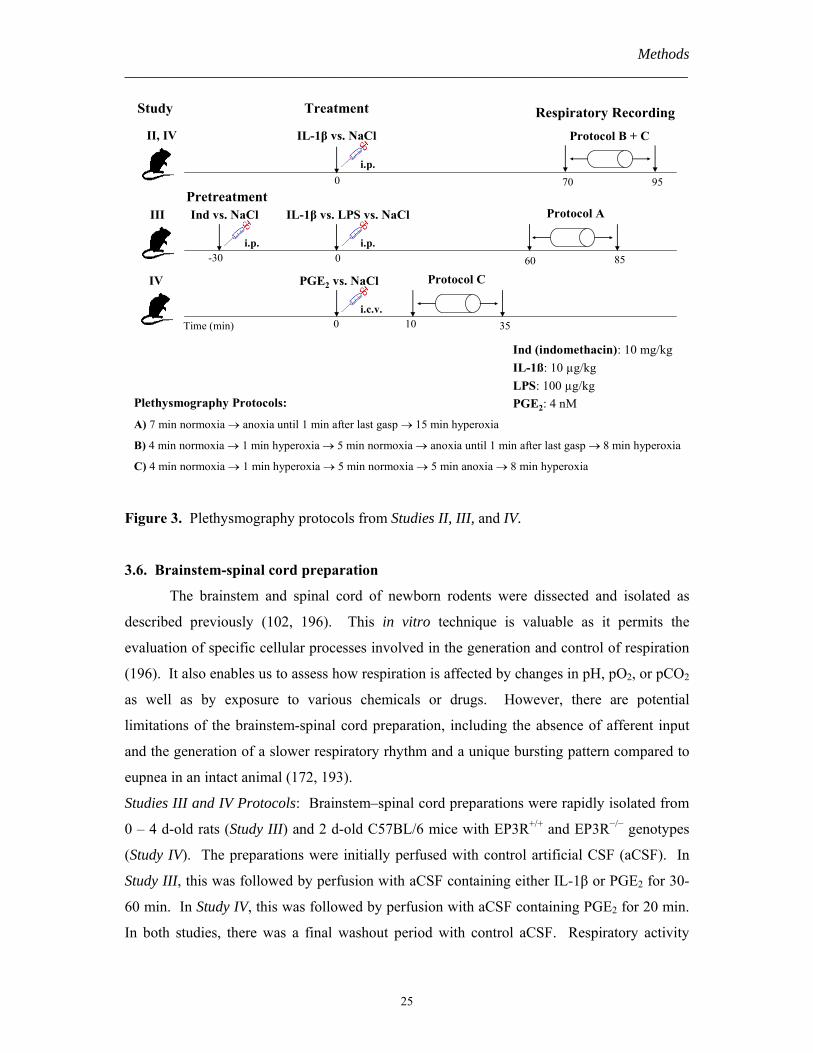
Studies II and IV Protocol: At 70 min after intraperitoneal injection (i.p.) of IL-1β or NaCl,
mice were placed in the plethysmograph chamber and their respiratory activity was measured
using the following protocol: a) 4 min normoxia (21% O2); b) 1 min hyperoxia (100% O2); c)
5 min normoxia; d) anoxia (100% N2) until 1 min after the animal’s last gasp (Study II) or for
5 min (Studies II and IV); e) 8 min hyperoxia (Figure 3). In all animals, skin temperature was
measured at baseline as well as immediately before and after experimentation.
Study III Protocol: Each rat received an initial i.p. injection of NaCl or indomethacin 30 min
prior to a second i.p. injection of NaCl, IL-1β, or LPS. The animal was placed in the
plethysmograph chamber at 60 min after the second injection and exposed to 7 min
normoxia, anoxia until 1 min after its last gasp, and then 100% O2 for 15 min or until
autoresuscitation (Figure 3). In a sample population, skin temperature was measured during
experimentation.
Supplemental Protocols: In order to characterize the respiratory behavior of neonatal
DBA/1lacJ mice, the ventilatory response to varying concentrations of O2 and CO2 was
examined in six neonatal DBA/1lacJ mice in Study II. Each animal was exposed sequentially
to normoxia (21% O2), mild hypercapnia (3% CO2 in synthetic air), moderate hypoxia with
mild hypercapnia (10%O2, 3% CO2), severe hypercapnia (8%CO2, 21%O2), severe hypoxia
with mild hypercapnia (5% O2, 3% CO2), and hyperoxia (100% O2). Normoxia was
administered between periods. In Study IV, the respiratory response to central PGE2 was also
investigated in neonatal mice using flow plethysmography. Immediately after anesthesia
administration and i.c.v. injection of PGE2 or vehicle, the mouse was placed into the
plethysmograph chamber. After a 10 min recovery period in normoxia, the mouse was
exposed to the same gas protocol described in Study IV above (Figure 3).
General considerations: Several important factors were considered in the design and
implementation of these studies. First, ambient temperature can strongly influence the
respiratory response to anoxia (31, 186). Thus, in all plethysmography experiments, chamber
temperature was maintained at approximately 30°C in accordance with the documented
thermoneutral range for rats and mice of similar age (109, 145). Second, gases were chosen
with specific objectives. Normoxia was used to establish baseline respiratory characteristics
within the control population and to determine how IL-1β, LPS, and PGE2 alter basal
respiration. The mice were exposed to a brief hyperoxic challenge in order to blunt
peripheral chemoreceptor activity and unmask central respiratory drive (51). This enabled us
to better assess whether the ventilatory effects of IL-1β and PGE2 occur via peripheral or
Methods ___________________________________________________________________________
24
central actions. Anoxia was used to induce hypoxic gasping, while chamber reoxygenation
permitted the examination of autoresuscitation.
Drug administration protocols were based on careful evaluation of previous
investigations using these drugs. Indomethacin was given 30 min prior to IL-1β or LPS in
order to allow sufficient time to block cyclooxygenase before these immunomodulators could
induce enzymatic activity (28, 86). IL-1β and LPS have been shown to increase COX-2
mRNA expression at 1 hr after intraperitoneal (i.p.) injection (34, 61). Thus, respiratory
recordings were performed between 60 – 95 min after i.p. administration of IL-1β or LPS in
order to allow sufficient time for respiratory effects to occur while attempting to minimize
confounding systemic effects.
IL-1β, LPS, and PGE2 evoke a broad array of centrally mediated adaptive responses,
which themselves may contribute to alterations in respiratory control. For example, IL-1β
has been shown to increase metabolic rate (14, 209), and an increased metabolism has been
associated with a shorter gasping duration (108). However, animals exhibited similar skin
temperatures at baseline, post-anesthesia in the i.c.v. experiments, and 60 – 70 min after i.p.
injection of IL-1β or LPS. These temperature measurements corresponded to previously
reported values for mice and rats of similar age (85, 185). Previous studies in rodents
indicate that IL-1β and LPS do not induce significant temperature increases until at least 90
min after i.p. injection (33, 34, 38, 73, 82, 179) and that PGE2 does not induce maximum
fever until 20 – 25 min after i.c.v. administration (212). Consequently, respiratory recordings
were performed within these time frames. Moreover, fever induced by IL-1β does not affect
the duration of hypoxia gasping nor does it hinder autoresuscitation following repeated
hypoxic exposure in newborn rats (73). Lastly, gross motor activity was similar between
animals. This is consistent with previous studies demonstrating that IL-1β does not evoke
sleep or hyperalgesia in rats until 2 or 4 hours after peripheral administration, respectively
(144, 157).
Methods ___________________________________________________________________________
25
Plethysmography Protocols:
A) 7 min normoxia → anoxia until 1 min after last gasp → 15 min hyperoxia
B) 4 min normoxia → 1 min hyperoxia → 5 min normoxia → anoxia until 1 min after last gasp → 8 min hyperoxia
C) 4 min normoxia → 1 min hyperoxia → 5 min normoxia → 5 min anoxia → 8 min hyperoxia
Time (min)
0 70
IL-1β vs. NaCl
95
0 10
PGE2 vs. NaCl
35
Ind (indomethacin): 10 mg/kgIL-1ß: 10 µg/kgLPS: 100 µg/kgPGE2: 4 nM
Pretreatment
Treatment Respiratory RecordingProtocol B + C
Protocol C
i.c.v.
i.p.
Study
II, IV
-30 0 8560
Ind vs. NaCl IL-1β vs. LPS vs. NaCl Protocol A
i.p. i.p.
III
IV
Figure 3. Plethysmography protocols from Studies II, III, and IV.
3.6. Brainstem-spinal cord preparation
The brainstem and spinal cord of newborn rodents were dissected and isolated as
described previously (102, 196). This in vitro technique is valuable as it permits the
evaluation of specific cellular processes involved in the generation and control of respiration
(196). It also enables us to assess how respiration is affected by changes in pH, pO2, or pCO2
as well as by exposure to various chemicals or drugs. However, there are potential
limitations of the brainstem-spinal cord preparation, including the absence of afferent input
and the generation of a slower respiratory rhythm and a unique bursting pattern compared to
eupnea in an intact animal (172, 193).
Studies III and IV Protocols: Brainstem–spinal cord preparations were rapidly isolated from
0 – 4 d-old rats (Study III) and 2 d-old C57BL/6 mice with EP3R+/+ and EP3R−/− genotypes
(Study IV). The preparations were initially perfused with control artificial CSF (aCSF). In
Study III, this was followed by perfusion with aCSF containing either IL-1β or PGE2 for 30-
60 min. In Study IV, this was followed by perfusion with aCSF containing PGE2 for 20 min.
In both studies, there was a final washout period with control aCSF. Respiratory activity
Methods ___________________________________________________________________________
26
corresponding to the inspiratory rhythm was recorded using glass suction electrodes applied
to the proximal end of the cut C4 ventral root.
3.7. Enzymatic assay
In Study IV, microsomal prostaglandin E synthase-1 (mPGES-1) activity was assessed
in the cortex and brainstem of neonatal wildtype mice as well as mPGES-1 knockout mice
using a quantitative enzymatic assay first described by Thorén and Jakobsson in 2000 (206).
This assay has been shown to recover 85 ± 11% of PGE2 (206). Our study objective was to
evaluate endogenous PGE2 production as well as the ability of IL-1β and hypoxia to induce
mPGES-1 activity. It also enabled us to determine the location of greatest mPGES-1 activity,
i.e., cortex vs. brainstem.
Protocol: Newborn mouse brains were homogenized in 0.1M KPi buffer containing 0.25M
sucrose, 1X complete protease inhibitor, and 1mM reduced glutathione. This was followed
by sonication. Membrane fraction was isolated by sub-cellular fractionation. Protein
concentration was determined by the Bradford method. mPGES-1 activity was assayed
by incubating the membrane fraction with 10uM PGH2 followed by termination of the
reaction using an acidified FeCl2 solution. Solid phase extraction of the reaction product was
then performed using C18 chromabond columns. PGE2 was eluted with acetone, evaporated
under nitrogen flow, and dissolved in 33% acetonitrile. An aliquot was analyzed by RP-
HPLC combined with UV detection at 195 nm. Enzymatic formation of PGE2 was calculated
after subtracting the non-enzymatic PGE2 formation in the buffer.
3.8. Enzyme immunoassay
In Study IV, PGE2 concentrations in infant cerebrospinal fluid (CSF) were measured
using enzyme immunoassay (EIA). CSF bathes the central nervous system, and thus CSF
concentrations may provide an estimate of levels within the brain parenchyma (52). EIA
allows enzyme detection using small sample volumes, which is crucial given the small CSF
volume in neonates. Since PGE2 is rapidly metabolized to 13,14-dihydro-5-keto PGE2 in
vivo, concentrations of 13,14-dihydro-15-keto PGA2, a non-enzymatically formed stable
metabolite of 13,14-dihydro-5 keto PGE2, were also measured using EIA.
Protocol: In study patients, a small volume of cerebrospinal fluid (0.75 – 1.5 ml) was
collected for research purposes. PGE2 and PGE2 metabolite concentrations were then
determined using a standardized EIA protocol. In order to maximize sample integrity as well
Methods ___________________________________________________________________________
27
as increase compliance amongst study collaborators, all samples were immediately stored at –
18°C and transferred as soon as possible to –80°C.
3.9. Data analysis
In vivo plethysmography experiments: Since the animals were placed unrestrained in the
plethymograph chamber, we used visual observations during experimentation as well as two
different analysis methods to select the best periods for analysis during normoxia, hyperoxia,
hypercapnia, and hypoxia (i.e., calm respiration without movement artefact). Respiratory
frequency (fR, breaths/min) was calculated manually. Tidal volume (VT, µL/breath), minute
ventilation (VE, mL/min), time of inspiration (Ti, s), and time of expiration (Te, s) were also
measured for flow plethysmograph data. In response to severe hypoxia and anoxia, the
duration of hyperpnea, primary apnea, gasping phases, and secondary apnea was determined.
The fR during hyperpnea was calculated manually, and the VT was calculated in mice that
were calm during the analysis period. The number, frequency, and appearance of gasps were
determined. Survival was recorded for all animals. The duration of secondary apnea and
time required to autoresuscitate following O2 administration were calculated in survivors.
The fR following autoresuscitation was also calculated. Apnea was defined as cessation of
breathing for ≥ three respiratory cycles. Regularity of breathing was quantified in some i.c.v.
experiments using the coefficient of variation (C.V.) (i.e., SD of ∆ fR / mean of ∆ fR during 60
s period). In Study III, similar findings were obtained with both flow and barometric
plethysmography; thus, data were normalized to facilitate statistical comparisons. In Studies
II and IV, we attempted to perform all recordings at age P9 since there is a variable response
to anoxia based upon age (72); however, some mice may have been evaluated at P9 ± 1 d.
Thus, we attempted to minimize confounding age-related effects by using weight as a
correlate of age and excluding those mice weighing > 1 SD of the mean population weight in
the anoxia and survival analyses.
In vitro brainstem-spinal cord preparation experiments: Respiratory frequency (fR,
burst/min) was calculated from the mean C4 burst interval during consecutive 2-5 min
periods. Baseline fR and changes in fR in response to IL-1β and PGE2 were assessed.
Infant cardiorespiratory data analyses: The monitor software was used to calculate baseline
respiratory rate, heart rate, pulse rate, and SpO2 values and to visualize cardiorespiratory
events for each recording. Apnea/hypopnea was defined as a ≥ 10 sec reduction of the
impedance signal amplitude to < 16% of the mean amplitude. It was described by
Methods ___________________________________________________________________________
28
apnea/hypopnea index (AI = # apneas/hypopneas per hour recording), duration, and
morphology. The latter was characterized by a predominant reduction in either RR or Vt.
Bradycardia was defined as a HR < 80 bpm for > 1 sec and expressed as bradycardia index
(BI = # bradycardias/ hour recording), duration, and HR nadir. Oxygen desaturation was
defined as a SpO2 value ≤ 90% and characterized by hypoxemia index (HI = # hypoxemias/
hour recording), duration, and nadir SpO2 values. Periodic breathing was defined as an
episode of three or more successive apnea pauses of > 3 breath duration separated by < 20 sec
of normal respiration. The occurrence of periodic breathing in the 60 sec following an apnea
event was examined. Mean RR, HR, and SpO2 immediately prior to an event were recorded.
All movement artefacts as well as recordings < 2 h duration were excluded from analysis.
Results and discussion ___________________________________________________________________________
29
4. Results and discussion 4.1. Cardiorespiratory development in preterm infants
There is a paucity of longitudinal data describing maturational changes in baseline
cardiorespiratory function in extremely preterm infants during early postnatal development.
In Study I, we reveal that the resting respiratory frequency did not change significantly
between birth and term-equivalent age in extremely preterm infants, whereas a reduction in
baseline respiratory rate during postnatal development has been shown previously in older
preterm infants (106, 112). The lack of age-dependent changes in respiratory rate may reflect
delayed maturation of respiratory control mechanisms in this very preterm infant population.
Conversely, we show a diminishing heart rate during the postnatal period, which may
indicate comparatively earlier cardiovascular development. The lowering of HR may be a
consequence of increased parasympathetic tone, which also influences baroreceptor reflex
sensitivity and contributes to HR variability during the postnatal period (7, 135). Our infants,
as well as those born at later gestation (163), also exhibited an improvement in oxygen
saturation with advancing post-conceptional age. This finding may reflect complex
mechanisms such as changes in ventilation to perfusion matching, chemosensitivity, and lung
mechanics. Ventilatory management may also influence baseline saturation in premature
infants, e.g., a target saturation of 88 – 92% SpO2 is frequently used in the neonatal unit.
Since our study was performed in a relatively small cohort of extremely preterm
infants, it would be beneficial to expand this investigation to include more infants who may
be further stratified according to pertinent demographic and clinical variables. Nonetheless,
our study provides useful reference data for baseline cardiorespiratory function in this
population.
4.2. Cardiorespiratory events during early postnatal life
Age-dependent changes in the incidence and appearance of cardiorespiratory events
have not been thoroughly described in extremely preterm infants during early postnatal life.
In Study I, we reveal that all infants born between 23 and 28 wk GA experienced recurrent
apnea, and 67% of infants continued to exhibit apnea/hypopnea events beyond 36 wk PCA.
Our findings confirm retrospective data describing an age-dependent reduction in apnea
incidence during the hospitalization period in a similar patient population using nursing
records (59), which is a less reliable method of apnea detection (150). Our findings differ
from those shown in older preterm infants, where a more pronounced decline in apnea
Results and discussion ___________________________________________________________________________
30
incidence results in fewer apnea events beyond term-equivalent age (95, 168). In general,
this discrepancy may reflect maturational changes in the central respiratory network,
chemoreceptor activity, reflex responses, and respiratory mechanics, but it may also mirror
differences in methodological approach (e.g., apnea definition, monitoring technique, data
collection).
In our study, the appearance of apnea/hypopnea changed during the postnatal period.
We reveal a dynamic age-dependent alteration in apnea/hypopnea frequency, i.e., an initial
rise followed by gradual decline in frequency, resembling that shown in older preterm infants
at a later PCA (129, 158, 194). Additionally, there was a predominant RR reduction during
apnea/hypopnea events occurring at an earlier PCA. This change may mirror the
development of hypoxic responsiveness in preterm infants since hypoxic ventilatory
depression is initially characterized by a marked reduction in RR than Vt (171), but
subsequently the neonate is better able to sustain RR during hypoxia (218). Unlike these
postnatal changes, the duration of apnea/hypopnea events remained constant with advancing
age. Prolonged apnea/hypopnea occurred beyond term-equivalent age and was associated
with a prolonged HR depression. This finding emphasizes the importance of close
surveillance of this infant population after discharge from the well-controlled hospital
environment. Furthermore, the long-term consequences of such events in extremely preterm
infants must be ascertained in future investigations.
A small percentage of apnea/hypopnea events occurred concomitantly with
bradycardia and/or hypoxemia events. There was a strong correlation between AI and BI,
which may be secondary to peripheral chemoreceptor activation following apnea-induced
hypoxemia (96) as well as a reflex response to cessation of lung inflation or superior
laryngeal and trigeminal nerve stimulation (142). It may also indicate that infants with more
immature respiratory control frequently exhibit cardiovascular dysregulation. Bradycardia
incidence and duration declined rapidly with maturation. This finding, along with age-
dependent changes in baseline HR, suggests that the cardiovascular system may undergo
developmental changes more rapidly than the respiratory system. The hypoxemia index also
diminished during postnatal development, which has also been shown in older preterm
infants (163). Cardiorespiratory events may cause hypoxemia via reduced alveolar
ventilation or arterial perfusion, although transient hypoxemia may also occur without
simultaneous apnea or bradycardia episodes (164). Age-dependent changes may also be due
to improved chemoreceptor function, which enables infants to better maintain their O2
saturation.
Results and discussion ___________________________________________________________________________
31
4.3. Infection increases cardiorespiratory events in infants
In Study I, infection was clearly correlated with a higher incidence of apnea/hypopnea
and hypoxemia events in preterm infants less than 31 wk PCA, but not in infants greater than
31 wk PCA. This indicates that younger preterm infants are particularly vulnerable to
postnatal insults such as infection. These findings are consistent with a large multi-center
study demonstrating that an increased apnea frequency occurs in the majority of infants with
sepsis (70) and in 20% of infants hospitalized with respiratory syncytial virus (RSV) (27). In
human neonates with RSV, IL-1β has been positively correlated to the clinical severity of
apnea (132). Thus, we propose that IL-1β plays a crucial mediatory role in this association
between infection and apnea in the newborn population. This hypothesis was explored
further in Studies II – IV.
4.4. Respiratory behavior in neonatal DBA/1lacJ mice
In Study II, we characterized the respiratory behavior of newborn DBA/1lacJ mice
since it was crucial to determine baseline function in these mice, which had not been done
previously, prior to examining the effect of immunomodulators on ventilation in wildtype
mice as well as mPGES-1 knockout mice. We describe respiratory frequency (fR), tidal
volume (VT), and minute ventilation (VE) during normoxia in wildtype mice, and these values
resembled those reported previously in Swiss CD-1 and C57BL/6 mice of similar age (17,
177). The DBA/1lacJ mice exhibited a characteristic reduction in fR in response to hyperoxia,
although fR decreased to a greater extent than described in other newborn mice (147). The
DBA/1lacJ mice also demonstrated a more pronounced response to hypoxia and anoxia
compared to that shown previously in other mouse strains (85, 108). Conversely, mild
hypercapnia did not alter ventilation compared to normoxia, and severe hypoxia without
hypocapnia (3% CO2) produced a hypoxic ventilatory depression similar to that observed
during anoxia with hyperpnea-induced hypocapnia. These findings indicate that DBA/1lacJ
mice have a heightened sensitivity to O2 concentration, but are less sensitive to pCO2
changes. This is important to consider when comparing our findings in DBA/1lacJ mice with
those of C57BL/6 mice. For example, whereas all DBA/1lacJ mice in Study II and IV
terminated their gasping response during the five minutes of anoxic exposure, all neonatal
wildtype C57BL/6 mice were able to sustain their gasping response beyond the five-minute
anoxic period. This suggests that the DBA/1lacJ mice have a greater sensitivity to pO2
changes than the C57BL/6 mice. Interestingly, hypoxic ventilatory depression is absent in
Results and discussion ___________________________________________________________________________
32
P1-P3 mice of the C57BL/6 strain, and it does not appear until P7 (17). This finding may
explain why our C57BL/6 mice demonstrate a less robust hypoxic ventilatory depression at
age P9 compared to the DBA/1lacJ mice.
4.5. IL-1β depresses respiration via central actions
In Studies II – IV, the ventilatory effects of IL-1β were investigated in wildtype
rodents. We show that IL-1β lowered fR during normoxia in newborn rats and reduced basal
VT, VE, and a weight-normalized fR in neonatal wildtype mice. LPS also tended to depress
basal respiration in newborn rats. These findings are in accordance with data showing that
IL-1β, given together with TNF-α, decreases normoxic fR in rabbits and that LPS reduces fR
and VT during normoxia in BALB/c mice (107, 209).
We hypothesize that IL-1β alters the central respiratory network rather than peripheral
chemosensitivity. In Studies II and IV, mice were subjected to hyperoxic challenge in order
to induce a physiological denervation of peripheral chemoreceptors and consequently unmask
central respiratory drive. All mice exhibited an appropriate peripheral chemoreceptor
response, and IL-1β induced a more pronounced respiratory depression during hyperoxia.
These findings suggest that a compensatory activation of peripheral chemoreceptors occurs
during normoxia in IL-1β-treated mice in order to balance the IL-1β-induced depression of
central respiration-related neurons.
In our studies, IL-1β depressed the anoxic ventilatory response by lowering the
gasping frequency in newborn rats and by reducing the number of gasps and the ability to
sustain respiratory efforts during anoxia in newborn DBA/1lacJ wildtype mice. IL-1β also
markedly reduced the ability of neonatal rodents to autoresuscitate following hypoxic apnea.
Prolonged anoxia causes a gradual loss of afferent inputs, central accumulation of inhibitory
neurotransmitters, and reconfiguration of neurons in the brainstem responsible for respiratory
rhythm and pattern generation (131, 218). IL-1β may selectively modulate these processes
responsible for hypoxic ventilation depression. However, we reveal that IL-1β was unable to
directly alter bursting activity of central respiratory neurons in vitro, and a previously
published study shows that Type 1 IL-1 receptor mRNA are not localized to respiration-
related regions of the brainstem (69). These findings indicate that IL-1β communicates with
the central respiratory network via an indirect mechanism. We suggest that these actions
occur via a prostaglandin-mediated pathway.
Results and discussion ___________________________________________________________________________
33
4.6. Endogenous PGE2 exerts tonic respiratory effects
In Study IV, we clearly demonstrate an endogenous expression of mPGES-1 activity
in wildtype mice, particularly in the brainstem. Additionally, respiratory depression was
greater in wildtype mice than in mPGES-1 knockout mice when central respiratory drive was
unmasked during hyperoxia. These findings indicate that endogenous PGE2 has a tonic effect
on respiratory rhythm generation during the perinatal period. These results are consistent
with data showing that prostaglandin synthesis inhibitors increase fetal breathing movements
as well as central respiratory activity the early neonatal period (91, 117, 134), although
indomethacin failed to stimulate respiration beyond basal levels in the newborn rats of Study
III. This may be explained in part by developmental changes in the modulatory effects of
prostaglandin with an initial inhibition of ventilation (90, 118) followed by little or no
alteration in central respiration beyond the perinatal period (198). These changes may be
secondary to a reduction of brainstem PGE2 receptor expression beyond the perinatal period
(197). An investigation of the ontogenesis of EP3R in respiration-related regions of the
brainstem would be valuable.
4.7. PGE2 inhibits respiratory activity via EP3R
In Study IV, the infectious marker C-reactive protein was positively correlated with
PGE2 levels in the cerebrospinal fluid of human infants, and the latter was associated with a
higher apnea frequency. Although our study cohort was small and relatively heterogeneous,
these findings support our hypothesis that infection induces neonatal apnea via a PGE2-
mediated mechanism. They are in accordance with previous investigations showing an
independent association between CRP levels and apnea/hypopnea index in children with
sleep apnea (202) as well as a positive correlation between urine PGE metabolite and central
apneas in newborn infants (105). Moreover, human neonates treated with prostaglandin often
display an increased apnea frequency (130, 189). Further evaluation in a larger patient
population is warranted.
We hypothesize that PGE2 depresses respiration centrally by binding to EP3 receptors
in the brainstem. In Study IV, all PGE2-treated mice demonstrated an appropriate peripheral
chemoreceptor response to hyperoxic challenge, indicating a central mechanism underlying the
ventilatory effects of PGE2. This corroborates data showing that PGE2 inhibits fetal breathing
movements in sheep after denervation of the carotid sinus and vagus nerve (149).
Furthermore, in our investigation, central administration of PGE2 induced apnea events and
Results and discussion ___________________________________________________________________________
34
irregular breathing patterns in neonatal wildtype mice, but not in EP3R knockout mice. PGE2
also depressed central respiratory-related bursting activity in vitro in newborn rats and EP3R
wildtype mice, but not in mice lacking the EP3 receptor. Thus, this study provides clear
evidence that the central respiratory effects of PGE2 occur via EP3R, which is consistent with
evidence that EP3 receptors are located within the NTS and RVLM (60). We are currently
investigating the co-localization of EP3R with neurokinin-1 receptors found specifically
within the preBötC and pFRG (87) using double immunohistochemistry.
4.8. IL-1β and hypoxia activate mPGES-1
In Study IV, IL-1β and brief anoxic exposure increased mPGES-1 activity in the
mouse brainstem. Previous investigations have shown that anoxia increases PGE2 production
in the mouse cortex ex vivo (187) and transient asphyxia increases PGE2 concentrations in the
newborn guinea pig brain (2). The exact mechanism of mPGES-1 upregulation in our study
remains unclear. Induced gene expression and mPGES-1 activation are less likely to occur
during such a brief hypoxic event. Potential etiologies include post-transcriptional regulation
or stabilization of mPGES-1 mRNA, which has been previously shown in neonatal mouse
cardiomyocytes (50).
4.9. PGE2 mediates the respiratory effects of IL-1β
In Studies III and IV, we further explored the mediatory role of prostaglandin in IL-
1β-induced respiratory changes in vivo. First, indomethacin pretreatment attenuated the basal
respiratory depression induced by IL-1β in neonatal rats. Indomethacin also markedly
improved the ability of IL-1β-treated rats to survive anoxic challenge. Similarly, IL-1β was
unable to alter basal respiration or the ventilatory response to hyperoxia in mice lacking
mPGES-1 or EP3R. Additionally, it had no effect on anoxic gasping or the ability to
autoresucitate following hypoxic apnea in mPGES-1 and EP3R knockout mice. Collectively,
these findings suggest that IL-1β inhibits central respiratory mechanisms indirectly via
activation of mPGES-1 and PGE2 binding to EP3 receptors (Figure 4).
We hypothesize that IL-1β- and hypoxia-induced PGE2 selectively modulates
respiration-related neurons in the preBötC. There is persuasive evidence that preBötC
neurons are crucial for the neurogenesis of gasping and subsequent autoresuscitation from
hypoxia (159). Lesions within the preBötC have been shown to disrupt anoxic gasping and
evoke central apneas and ataxic breathing (71, 140). Other neuromodulators inhibit these
Results and discussion ___________________________________________________________________________
35
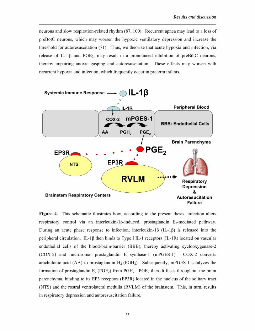
neurons and slow respiration-related rhythm (87, 100). Recurrent apnea may lead to a loss of
preBötC neurons, which may worsen the hypoxic ventilatory depression and increase the
threshold for autoresuscitation (71). Thus, we theorize that acute hypoxia and infection, via
release of IL-1β and PGE2, may result in a pronounced inhibition of preBötC neurons,
thereby impairing anoxic gasping and autoresuscitation. These effects may worsen with
recurrent hypoxia and infection, which frequently occur in preterm infants.
IL-1βPeripheral Blood
BBB: Endothelial Cells
IL-1R
COX-2 mPGES-1
PGE2
RVLM
NTS
EP3REP3R
AA PGH2 PGE2
Systemic Immune Response
Brainstem Respiratory Centers
RespiratoryDepression
&Autoresucitation
Failure
Brain Parenchyma
Figure 4. This schematic illustrates how, according to the present thesis, infection alters
respiratory control via an interleukin-1β-induced, prostaglandin E2-mediated pathway.
During an acute phase response to infection, interleukin-1β (IL-1β) is released into the
peripheral circulation. IL-1β then binds to Type I IL-1 receptors (IL-1R) located on vascular
endothelial cells of the blood-brain-barrier (BBB), thereby activating cyclooxygenase-2
(COX-2) and microsomal prostaglandin E synthase-1 (mPGES-1). COX-2 converts
arachidonic acid (AA) to prostaglandin H2 (PGH2). Subsequently, mPGES-1 catalyzes the
formation of prostaglandin E2 (PGE2) from PGH2. PGE2 then diffuses throughout the brain
parenchyma, binding to its EP3 receptors (EP3R) located in the nucleus of the solitary tract
(NTS) and the rostral ventrolateral medulla (RVLM) of the brainstem. This, in turn, results
in respiratory depression and autoresuscitation failure.
Conclusions ___________________________________________________________________________
36
5. Conclusions This thesis describes age-dependent changes in the incidence and characteristics of
cardiorespiratory events during the early postnatal period in extremely preterm infants.
Importantly, we identified a high incidence of cardiorespiratory events beyond term-
equivalent age and an increased vulnerability to postnatal insults such as infection in this
population. These findings are of particular concern and emphasize the importance of careful
surveillance and management outside of the hospital environment.
This thesis also identifies a novel mechanism linking systemic infectious response
with respiratory control disturbances in neonates. We show that IL-1β alters basal respiration
and hypoxic ventilation via central mPGES-1 activation and PGE2 binding to brainstem EP3
receptors. Moreover, PGE2 appears to play an important role in the respiratory response to
anoxia. These findings have important implications for the clinical management of neonates.
The rapid synthesis of PGE2 in response to cytokine and transient anoxia may make it
particularly useful in the diagnosis and monitoring of infants with increased apneas due to
suspected infection or hypoxia. Our studies may also influence treatment strategies for
neonatal apnea related to infection or hypoxia by selectively targeting mPGES-1 or EP3R.
In conclusion, the cytokine-induced, PGE2-dependent pathway described in the present thesis
could potentially explain the association between infection, apnea, and Sudden Infant Death
Syndrome.
Acknowledgments ___________________________________________________________________________
37
6. Acknowledgments I would like to offer my deepest thanks and appreciation to all of you who supported and encouraged me during my doctoral education: Eric Herlenius, my main supervisor, for his tremendous devotion to our research projects, incredible flexibility in working cross-continentally, exciting conversations over burned laboratory coffee, and introduction to the wonders of Harry Potter. Hugo Lagercrantz, my co-supervisor, for graciously welcoming me into his laboratory group, introducing me to the field of respiratory physiology, and providing guidance throughout my research education. Miriam Katz-Salamon, my co-supervisor, for her introduction to clinical investigations, positive encouragement, and inspiring me to become a better researcher. Lena Legnevall, for her incredible spirit and dedication to our clinical projects, which would not have been possible without her perseverance, and bringing a smile to my face in even the darkest of moments. Eva Lundberg, Viveca Karlsson, Ann-Christine Eklöf, and Astrid Häggblad for their kind assistance in logistical arrangements. My “roommates” Jonas Berner, Yuri Shvarev, Linda Danielsson, Ruth Detlofsson, Johan Jäderstad, Jeo Park, Marco Bartocci, Zoltan Nagy, Maria Shariatmadari, and Panos Papachristou, for filling the Brainstem and Lab rooms with laughter, keeping me sane, and sharing your wisdom both in scientific and personal matters. I would also like to thank the following people for their collaboration during my research studies and for creating an inspiring lab environment: Sipra Saha, Veronica Siljehav, Per-Johan Jakobsson, Gulcin Kayhan, Kristin Leifsdottir, Maneck Bhiladvala, Birgitta Böhm, Ronny Wickström, Jean-Christophe Roux, Julie Peyronnet, Yuji Yamamoto, Ulrika Ådén, Thomas Ringstedt, Hans Holgert, Lena Bergquist, and Eva Horemuzova. My pediatric residency program at Columbia University for their support in completing my doctoral education. Christoph Hofstetter, my husband, for his incredible patience, understanding, and eagerness to embark on new adventures together. My family, for teaching me the importance of exploration and for their unconditional support during this educational journey.
References ___________________________________________________________________________
38
7. References 1. American Academy of Pediatrics. Task Force on Prolonged Apnea. Prolonged apnea. Pediatrics 61: 651-
652, 1978. 2. Allen LG, Louis TM, and Kopelman AE. Brain prostaglandins E2 and F2 alpha following neonatal
asphyxia in the guinea pig. Biol Neonate 42: 8-14, 1982. 3. Al-Matary A, Kutbi I, Qurashi M, Khalil M, Alvaro R, Kwiatkowski K, Cates D, and Rigatto H.
Increased peripheral chemoreceptor activity may be critical in destabilizing breathing in neonates. Semin Perinatol 28: 264-272, 2004.
4. Alvaro R, Alvarez J, Kwiatkowski K, Cates D, and Rigatto H. Small preterm infants (less than or equal to 1500 g) have only a sustained decrease in ventilation in response to hypoxia. Pediatr Res 32: 403-406, 1992.
5. Alvaro R, de Almeida V, al-Alaiyan S, Robertson M, Nowaczyk B, Cates D, and Rigatto H. A placental extract inhibits breathing induced by umbilical cord occlusion in fetal sheep. J Dev Physiol 19: 23-28, 1993.
6. Alvaro RE, Hasan SU, Chemtob S, Qurashi M, Al-Saif S, and Rigatto H. Prostaglandins are responsible for the inhibition of breathing observed with a placental extract in fetal sheep. Respir Physiol Neurobiol 144: 35-44, 2004.
7. Andriessen P, Oetomo SB, Peters C, Vermeulen B, Wijn PF, and Blanco CE. Baroreceptor reflex sensitivity in human neonates: the effect of postmenstrual age. J Physiol 568: 333-341, 2005.
8. Avery ME, Chernick V, Dutton RE, and Permutt S. Ventilatory Response to Inspired Carbon Dioxide in Infants and Adults. J Appl Physiol 18: 895-903, 1963.
9. Baird TM. Clinical correlates, natural history and outcome of neonatal apnoea. Semin Neonatol 9: 205-211, 2004.
10. Ballanyi K, Lalley PM, Hoch B, and Richter DW. cAMP-dependent reversal of opioid- and prostaglandin-mediated depression of the isolated respiratory network in newborn rats. J Physiol 504: 127-134, 1997.
11. Ban E, Milon G, Prudhomme N, Fillion G, and Haour F. Receptors for interleukin-1 (alpha and beta) in mouse brain: mapping and neuronal localization in hippocampus. Neuroscience 43: 21-30, 1991.
12. Banks WA and Kastin AJ. Blood to brain transport of interleukin links the immune and central nervous systems. Life Sci 48: PL117-121, 1991.
13. Barrington KJ and Finer NN. Periodic breathing and apnea in preterm infants. Pediatr Res 27: 118-121, 1990.
14. Berg S, Sappington PL, Guzik LJ, Delude RL, and Fink MP. Proinflammatory cytokines increase the rate of glycolysis and adenosine-5'-triphosphate turnover in cultured rat enterocytes. Crit Care Med 31: 1203-1212, 2003.
15. Besedovsky H, del Rey A, Sorkin E, and Dinarello CA. Immunoregulatory feedback between interleukin-1 and glucocorticoid hormones. Science 233: 652-654, 1986.
16. Bishai I, Dinarello CA, and Coceani F. Prostaglandin formation in feline cerebral microvessels: effect of endotoxin and interleukin-1. Can J Physiol Pharmacol 65: 2225-2230, 1987.
17. Bissonnette JM and Knopp SJ. Developmental changes in the hypoxic ventilatory response in C57BL/6 mice. Respir Physiol 128: 179-186, 2001.
18. Blackwell CC and Weir DM. The role of infection in sudden infant death syndrome. FEMS Immunol Med Microbiol 25: 1-6, 1999.
19. Blanco CE, Dawes GS, Hanson MA, and McCooke HB. The response to hypoxia of arterial chemoreceptors in fetal sheep and new-born lambs. J Physiol 351: 25-37, 1984.
20. Blanco CE, Hanson MA, Johnson P, and Rigatto H. Breathing pattern of kittens during hypoxia. J Appl Physiol 56: 12-17, 1984.
21. Blanco CE, Hanson MA, and McCooke HB. Effects on carotid chemoreceptor resetting of pulmonary ventilation in the fetal lamb in utero. J Dev Physiol 10: 167-174, 1988.
22. Blanco CE, Martin CB, Jr., Hanson MA, and McCooke HB. Determinants of the onset of continuous air breathing at birth. Eur J Obstet Gynecol Reprod Biol 26: 183-192, 1987.
23. Blatteis CM. Role of the OVLT in the febrile response to circulating pyrogens. Prog Brain Res 91: 409-412, 1992.
24. Boddy K and Dawes GS. Fetal breathing. Br Med Bull 31: 3-7, 1975. 25. Boddy K, Dawes GS, Fisher R, Pinter S, and Robinson JS. Foetal respiratory movements, electrocortical
and cardiovascular responses to hypoxaemia and hypercapnia in sheep. J Physiol 243: 599-618, 1974. 26. Brady L, Lynn, AB, Herkenham, M, Gottesfeld, Z. Systemic interleukin-1 induces early and late patterns
of c-fos mRNA expression in brain. J Neurosci 14: 4951-4964, 1994.
References ___________________________________________________________________________
39
27. Bruhn FW, Mokrohisky ST, and McIntosh K. Apnea associated with respiratory syncytial virus infection in young infants. J Pediatr 90: 382-386, 1977.
28. Bucinskaite V, Kurosawa M, and Lundeberg T. Effect of interleukin-1beta on subdiaphragmatic vagal efferents in the rat. Auton Neurosci 85: 93-97, 2000.
29. Bureau MA, Lamarche J, Foulon P, and Dalle D. Postnatal maturation of respiration in intact and carotid body-chemodenervated lambs. J Appl Physiol 59: 869-874, 1985.
30. Butcher-Puech MC, Henderson-Smart DJ, Holley D, Lacey JL, and Edwards DA. Relation between apnoea duration and type and neurological status of preterm infants. Arch Dis Child 60: 953-958, 1985.
31. Cameron YL, Merazzi D, and Mortola JP. Variability of the breathing pattern in newborn rats: effects of ambient temperature in normoxia or hypoxia. Pediatr Res 47: 813-818, 2000.
32. Campbell A, Cross, KW, Dawes, GS, Hyman, AI. A comparison of air and O2 in the hyperbaric chamber or by positive pressure ventilation in the resuscitation of newborn rabbits. J Pediatr 68: 153-163, 1966.
33. Cao C, Matsumura K, Yamagata K, and Watanabe Y. Endothelial cells of the rat brain vasculature express cyclooxygenase-2 mRNA in response to systemic interleukin-1 beta: a possible site of prostaglandin synthesis responsible for fever. Brain Res 733: 263-272, 1996.
34. Cao C, Matsumura K, Yamagata K, and Watanabe Y. Induction by lipopolysaccharide of cyclooxygenase-2 mRNA in rat brain; its possible role in the febrile response. Brain Res 697: 187-196, 1995.
35. Carbone T, Marrero LC, Weiss J, Hiatt M, and Hegyi T. Heart rate and oxygen saturation correlates of infant apnea. J Perinatol 19: 44-47, 1999.
36. Carlo WA and DiFiore JM. Respiratory muscle responses to changes in chemoreceptor drive in infants. J Appl Physiol 68: 1041-1047, 1990.
37. Carroll JL. Developmental plasticity in respiratory control. J Appl Physiol 94: 375-389, 2003. 38. Chai Z, Gatti S, Toniatti C, Poli V, and Bartfai T. Interleukin (IL)-6 gene expression in the central
nervous system is necessary for fever response to lipopolysaccharide or IL-1 beta: a study on IL-6-deficient mice. J Exp Med 183: 311-316, 1996.
39. Chernick V and Craig RJ. Naloxone reverses neonatal depression caused by fetal asphyxia. Science 216: 1252-1253, 1982.
40. Coceani F and Akarsu ES. Prostaglandin E2 in the pathogenesis of fever. An update. Ann N Y Acad Sci 856: 76-82, 1998.
41. Cohen G and Katz-Salamon M. Development of chemoreceptor responses in infants. Respir Physiol Neurobiol 149: 233-242, 2005.
42. Cote A, Hum C, Brouillette RT, and Themens M. Frequency and timing of recurrent events in infants using home cardiorespiratory monitors. J Pediatr 132: 783-789, 1998.
43. Crestani F, Seguy, F, Dantzer, R. Behavioural effects of peripherally injected interleukin-1: role of prostaglandin. Brain Res 542: 330-335, 1991.
44. Cross KW, Klaus M, Tooley WH, and Weisser K. The response of the new-born baby to inflation of the lungs. J Physiol 151: 551-565, 1960.
45. Cunningham ET, Jr., Wada E, Carter DB, Tracey DE, Battey JF, and De Souza EB. In situ histochemical localization of type I interleukin-1 receptor messenger RNA in the central nervous system, pituitary, and adrenal gland of the mouse. J Neurosci 12: 1101-1114, 1992.
46. Dantzer R. Cytokine-induced sickness behavior: mechanisms and implications. Ann N Y Acad Sci 933: 222-234, 2001.
47. Dawes GS, Fox HE, Leduc BM, Liggins GC, and Richards RT. Respiratory movements and paradoxical sleep in the foetal lamb. J Physiol 210: 47P-48P, 1970.
48. Dawes GS, Gardner WN, Johnston BM, and Walker DW. Breathing in fetal lambs: the effect of brain stem section. J Physiol 335: 535-553, 1983.
49. Day H, Akil, H. Differential pattern of c-fos mRNA in rat brain following central and systemic administration of interleukin-1-beta: Implications for mechanism of action. Neuroendocrinology 63: 207-218, 1996.
50. Degousee N, Angoulvant D, Fazel S, Stefanski E, Saha S, Iliescu K, Lindsay TF, Fish JE, Marsden PA, Li RK, Audoly LP, Jakobsson PJ, and Rubin BB. c-Jun N-terminal kinase-mediated stabilization of microsomal prostaglandin E2 synthase-1 mRNA regulates delayed microsomal prostaglandin E2 synthase-1 expression and prostaglandin E2 biosynthesis by cardiomyocytes. J Biol Chem 281: 16443-16452, 2006.
51. Dejours P. Chemoreflexes in breathing. Planta Med 42: 335-358, 1962. 52. Dembo G, Park SB, and Kharasch ED. Central nervous system concentrations of cyclooxygenase-2
inhibitors in humans. Anesthesiology 102: 409-415, 2005. 53. Di Fiore JM. Neonatal cardiorespiratory monitoring techniques. Semin Neonatol 9: 195-203, 2004. 54. Dinarello CA. Interleukin-1. Rev Infect Dis 6: 51-95, 1984.
References ___________________________________________________________________________
40
55. Dransfield DA, Spitzer AR, and Fox WW. Episodic airway obstruction in premature infants. Am J Dis Child 137: 441-443, 1983.
56. Drorbaugh JE and Fenn WO. A barometric method for measuring ventilation in newborn infants. Pediatrics 16: 81-87, 1955.
57. Dux E, Fastbom, J, Ungerstedt, U, Rudolphi, K, Fredholm, BB. Protective effect of adenosine and a novel xanthine derivative propentofylline on the cell damage after bilateral carotid occlusion in the gerbil hippocampus. Brain Research 516: 248-256, 1990.
58. Eden GJ and Hanson MA. Maturation of the respiratory response to acute hypoxia in the newborn rat. J Physiol 392: 1-9, 1987.
59. Eichenwald EC, Aina A, and Stark AR. Apnea frequently persists beyond term gestation in infants delivered at 24 to 28 weeks. Pediatrics 100: 354-359, 1997.
60. Ek M, Arias C, Sawchenko P, and Ericsson-Dahlstrand A. Distribution of the EP3 prostaglandin E(2) receptor subtype in the rat brain: relationship to sites of interleukin-1-induced cellular responsiveness. J Comp Neurol 428: 5-20, 2000.
61. Ek M, Engblom D, Saha S, Blomqvist A, Jakobsson PJ, and Ericsson-Dahlstrand A. Inflammatory response: pathway across the blood-brain barrier. Nature 410: 430-431, 2001.
62. Ek M, Kurosawa, M, Lundeberg, T, Ericsson, A. Activation of vagal afferents after intravenous injection of interleukin-1beta: Role of endogenous prostaglandins. Journal of Neuroscience 18: 9471-9479, 1998.
63. Elmquist J, Scammell, TE, Jacobson, CD, Saper, CB. Distribution of Fos-like immunoreactivity in the rat brain following intravenous lipopolysaccharide administration. J Comp Neurol 371: 85-103, 1996.
64. Engblom D, Ek, M, Saha, S, Ericsson-Dahlstrand, A, Jakobsson, PJ, Blomqvist, A. Prostaglandins as inflammatory messengers across the blood-brain barrier. J Mol Med 80: 5-15, 2002.
65. Engblom D, Saha S, Engstrom L, Westman M, Audoly LP, Jakobsson PJ, and Blomqvist A. Microsomal prostaglandin E synthase-1 is the central switch during immune-induced pyresis. Nat Neurosci 6: 1137-1138, 2003.
66. Enhorning G, van Schaik S, Lundgren C, and Vargas I. Whole-body plethysmography, does it measure tidal volume of small animals? Can J Physiol Pharmacol 76: 945-951, 1998.
67. Ericsson A, Arias, C, Sawchenko, PE. Evidence for an intramedullary prostaglandin-dependent mechanism in the activation of stress-related neuroendocrine circuitry by intravenous interleukin-1. Journal of Neuroscience 18: 7166-7179, 1997.
68. Ericsson A, Kovacs, KJ, Sawchenko, PE. A functional anatomical analysis of central pathways subserving the effects of interleukin-1 on stress-related neuroendocrine neurons. Journal of Neuroscience 14: 897-913, 1994.
69. Ericsson A, Liu, C, Hart, RP, Sawchenko, PE. Type 1 interleukin-1 receptor in the rat brain: distribution, regulation, and relationship to sites of IL-1-induced cellular activation. J Comp Neurol 361: 681-698, 1995.
70. Fanaroff AA, Korones SB, Wright LL, Verter J, Poland RL, Bauer CR, Tyson JE, Philips JB, Edwards W, Lucey JF, Catz CS, Shankaran S, and Oh W. Incidence, presenting features, risk factors and significance of late onset septicemia in very low birth weight infants. The National Institute of Child Health and Human Development Neonatal Research Network. The Pediatric Infectious Disease Journal 17: 593-598, 1998.
71. Feldman JL and Del Negro CA. Looking for inspiration: new perspectives on respiratory rhythm. 7: 232-241, 2006.
72. Fewell JE, Smith FG, Ng VK, Wong VH, and Wang Y. Postnatal age influences the ability of rats to autoresuscitate from hypoxic-induced apnea. Am J Physiol Regul Integr Comp Physiol 279: R39-46, 2000.
73. Fewell JE and Wong VH. Interleukin-1beta-induced fever does not alter the ability of 5- to 6-day-old rat pups to autoresuscitate from hypoxia-induced apnoea. Exp Physiol 87: 17-24, 2002.
74. Finer NN, Barrington KJ, Hayes BJ, and Hugh A. Obstructive, mixed, and central apnea in the neonate: physiologic correlates. J Pediatr 121: 943-950, 1992.
75. Fleming EF, Athirakul K, Oliverio MI, Key M, Goulet J, Koller BH, and Coffman TM. Urinary concentrating function in mice lacking EP3 receptors for prostaglandin E2. Am J Physiol 275: F955-961, 1998.
76. Flourens P. Reserches expérimentales sur les propriétés et les fonctions du système nerveus, dan les animaux vertébrés. Paris: Crevot, 1824.
77. Frantz ID, 3rd, Adler SM, Thach BT, and Taeusch HW, Jr. Maturational effects on respiratory responses to carbon dioxide in premature infants. J Appl Physiol 41: 41-45, 1976.
78. Fredholm B. Adenosine, adenosine receptors, and the actions of caffeine. Pharmacology and Toxocology 76: 93-101, 1995.
References ___________________________________________________________________________
41
79. French JW, Morgan BC, and Guntheroth WG. Infant monkeys--a model for crib death. Am J Dis Child 123: 480-484, 1972.
80. Froen J, Akre, H, Stray-Pedersen, B, Saugstad, OD. Adverse effects of nicotine and interleukin-1beta on autoresuscitation after apnea in piglets: Implications for Sudden Infant Death Syndrome. Pediatrics 105: E52, 2000.
81. Galen. Galen on the usefulness of the parts of the body (De usu partium). Ithaca, NY: Cornell University Press, 1968.
82. Gatti S, Beck J, Fantuzzi G, Bartfai T, and Dinarello CA. Effect of interleukin-18 on mouse core body temperature. Am J Physiol Regul Integr Comp Physiol 282: R702-709, 2002.
83. Gaultier C, Matrot B, and Gallego J. Transgenic models to study disorders of respiratory control in newborn mice. Ilar Journal 47: 15-21, 2006.
84. Gerhardt T and Bancalari E. Apnea of prematurity: I. Lung function and regulation of breathing. Pediatrics 74: 58-62, 1984.
85. Gershan WM, Jacobi MS, and Thach BT. Maturation of cardiorespiratory interactions in spontaneous recovery from hypoxic apnea (autoresuscitation). Pediatr Res 28: 87-93, 1990.
86. Graff GR and Gozal D. Cardiorespiratory responses to interleukin-1beta in adult rats: role of nitric oxide, eicosanoids and glucocorticoids. Arch Physiol Biochem 107: 97-112, 1999.
87. Gray PA, Rekling JC, Bocchiaro CM, and Feldman JL. Modulation of respiratory frequency by peptidergic input to rhythmogenic neurons in the preBotzinger complex. Science 286: 1566-1568, 1999.
88. Greer JJ, Funk GD, and Ballanyi K. Preparing for the first breath: prenatal maturation of respiratory neural control. J Physiol 570: 437-444, 2006.
89. Grosfeld J, Dalsing, MC, Hull, M, Weber, TR. Neonatal apnea, xanthines, and necrotizing enterocolitis. Journal of Pediatric Surgery 18: 80-84, 1983.
90. Guerra F, Savich, RD, Wallen, LD, Lee, CH, Clyman, RI, Mauray, FE, Kitterman, JA. Prostaglandin E2 causes hypoventilation and apnea in newborn lambs. Journal of Applied Physiology 64: 2160-2166, 1988.
91. Guerra FA, Savich RD, Clyman RI, and Kitterman JA. Meclofenamate increases ventilation in lambs. J Dev Physiol 11: 1-6, 1989.
92. Guntheroth W. Interleukin-1 as intermediary causing prolonged sleep apnea and SIDS during respiratory infections. Medical Hypotheses 28: 121-123, 1989.
93. Hammerman C and Zangen D. Indomethacin and apnea of prematurity. Crit Care Med 21: 154-155, 1993.
94. Hansen MK, Nguyen KT, Goehler LE, Gaykema RP, Fleshner M, Maier SF, and Watkins LR. Effects of vagotomy on lipopolysaccharide-induced brain interleukin-1beta protein in rats. Auton Neurosci 85: 119-126, 2000.
95. Henderson-Smart DJ. The effect of gestational age on the incidence and duration of recurrent apnoea in newborn babies. Australian Pediatric Journal 17: 273-276, 1981.
96. Henderson-Smart DJ, Butcher-Puech MC, and Edwards DA. Incidence and mechanism of bradycardia during apnoea in preterm infants. Arch Dis Child 61: 227-232, 1986.
97. Henderson-Smart DJ, Pettigrew AG, and Campbell DJ. Clinical apnea and brain-stem neural function in preterm infants. N Engl J Med 308: 353-357, 1983.
98. Henderson-Smart DJ and Steer P. Methylxanthine treatment for apnea in preterm infants. Cochrane Database Syst Rev: CD000140, 2001.
99. Herlenius E, Aden U, Tang LQ, and Lagercrantz H. Perinatal respiratory control and its modulation by adenosine and caffeine in the rat. Pediatr Res 51: 4-12, 2002.
100. Herlenius E and Lagercrantz H. Adenosinergic modulation of respiratory neurones in the neonatal rat brainstem in vitro. J Physiol 518 ( Pt 1): 159-172, 1999.
101. Herlenius E and Lagercrantz H. Development of neurotransmitter systems during critical periods. Exp Neurol 190 Suppl 1: S8-21, 2004.
102. Herlenius E, Lagercrantz, H, Yamamoto, Y. Adenosine modulates inspiratory neurons and the respiratory pattern in the brainstem of neonatal rats. Pediatric Research 42: 46-53, 1997.
103. Hertzberg T, Hellstrom S, Lagercrantz H, and Pequignot JM. Development of the arterial chemoreflex and turnover of carotid body catecholamines in the newborn rat. J Physiol 425: 211-225, 1990.
104. Hertzberg T and Lagercrantz H. Postnatal sensitivity of the peripheral chemoreceptors in newborn infants. Arch Dis Child 62: 1238-1241, 1987.
105. Hoch B and Bernhard M. Central apnoea and endogenous prostaglandins in neonates. Acta Paediatr 89: 1364-1368, 2000.
106. Holditch-Davis D, Scher M, and Schwartz T. Respiratory development in preterm infants. J Perinatol 24: 631-639, 2004.
107. Holub M and Lawrence DA. Influence of endotoxin-induced acute lung injury on pulmonary innate and adaptive immunity. Apmis 111: 571-580, 2003.
References ___________________________________________________________________________
42
108. Jacobi MS, Gershan WM, and Thach BT. Mechanism of failure of recovery from hypoxic apnea by gasping in 17- to 23-day-old mice. J Appl Physiol 71: 1098-1105, 1991.
109. Jacobi MS and Thach BT. Effect of maturation on spontaneous recovery from hypoxic apnea by gasping. J Appl Physiol 66: 2384-2390, 1989.
110. Johnston BM and Gluckman PD. Peripheral chemoreceptors respond to hypoxia in pontine-lesioned fetal lambs in utero. J Appl Physiol 75: 1027-1034, 1993.
111. Jones KL, Krous HF, Nadeau J, Blackbourne B, Zielke HR, and Gozal D. Vascular endothelial growth factor in the cerebrospinal fluid of infants who died of sudden infant death syndrome: evidence for antecedent hypoxia. Pediatrics 111: 358-363, 2003.
112. Katona PG, Frasz A, and Egbert J. Maturation of cardiac control in full-term and preterm infants during sleep. Early Hum Dev 4: 145-159, 1980.
113. Katsuura G, Arimura, A, Koves, K, Gottschall, PE. Involvement of organum vasculosum of lamina terminalis and preoptic area in interleukin 1 beta-induced ACTH release. American Journal of Physiology 258: E163-171, 1990.
114. Katsuura G, Gottschall, PE, Dahl, RR, Arimura, A. Adrenocorticotropin release induced by intracerebroventricular injection of recombinant human interleukin-1 in rats: possible involvement of prostaglandin. Endocrinology 122: 1773-1779, 1988.
115. Katz-Salamon M. Delayed chemoreceptor responses in infants with apnoea. Arch Dis Child 89: 261-266, 2004.
116. Khan A, Qurashi M, Kwiatkowski K, Cates D, and Rigatto H. Measurement of the CO2 apneic threshold in newborn infants: possible relevance for periodic breathing and apnea. J Appl Physiol 98: 1171-1176, 2005.
117. Kitterman J, Liggins, GC, Clements, JA, Tooley, WH. Stimulation of breathing movements in fetal sheep by inhibitors of prostaglandin synthesis. J Dev Physiol 1: 453-466, 1979.
118. Kitterman J, Liggins, GC, Fewell, JE, Tooley, WH. Inhibition of breathing movements in fetal sheep by prostaglandins. J Appl Physiol 54: 687-692, 1983.
119. Koos B. Central stimulation of breathing movements in fetal lambs by prostaglandin synthetase inhibitors. Journal of Physiology 362: 455-466, 1985.
120. Krueger JM, Walter J, Dinarello CA, Wolff SM, and Chedid L. Sleep-promoting effects of endogenous pyrogen (interleukin-1). Am J Physiol 246: R994-999, 1984.
121. Kuipers IM, Maertzdorf WJ, De Jong DS, Hanson MA, and Blanco CE. The effect of hypercapnia and hypercapnia associated with central cooling on breathing in unanesthetized fetal lambs. Pediatr Res 41: 90-95, 1997.
122. Kuipers IM, Maertzdorf WJ, De Jong DS, Hanson MA, and Blanco CE. Initiation and maintenance of continuous breathing at birth. Pediatr Res 42: 163-168, 1997.
123. Lagercrantz H, Holgert H, Pequignot JM, and Srinivasan M. Expression and release of neuroregulators during development: monoamines and neuropeptides. Dev Pharmacol Ther 18: 136-138, 1992.
124. Lagercrantz H, Pequignot J, Pequignot JM, and Peyrin L. The first breaths of air stimulate noradrenaline turnover in the brain of the newborn rat. Acta Physiol Scand 144: 433-438, 1992.
125. Lagercrantz H, Pequignot JM, Hertzberg T, Holgert H, and Ringstedt T. Birth-related changes of expression and turnover of some neuroactive agents and respiratory control. Biol Neonate 65: 145-148, 1994.
126. Lagercrantz H and Slotkin TA. The "stress" of being born. Sci Am 254: 100-107, 1986. 127. Lagercrantz H, Yamamoto Y, Fredholm BB, Prabhakar NR, and von Euler C. Adenosine analogues
depress ventilation in rabbit neonates. Theophylline stimulation of respiration via adenosine receptors? Pediatr Res 18: 387-390, 1984.
128. Le Gallois J. Expériences sur le principe de la vie. Paris: D´Hautel, 1812. 129. Lee D, Caces R, Kwiatkowski K, Cates D, and Rigatto H. A developmental study on types and frequency
distribution of short apneas (3 to 15 seconds) in term and preterm infants. Pediatr Res 22: 344-349, 1987. 130. Lewis AB, Freed MD, Heymann MA, Roehl SL, and Kensey RC. Side effects of therapy with
prostaglandin E1 in infants with critical congenital heart disease. Circulation 64: 893-898, 1981. 131. Lieske SP, Thoby-Brisson M, Telgkamp P, and Ramirez JM. Reconfiguration of the neural network
controlling multiple breathing patterns: eupnea, sighs and gasps [see comment]. Nat Neurosci 3: 600-607, 2000.
132. Lindgren C, Grogaard, J. Reflex apnoea response and inflammatory mediators in infants with respiratory tract infection. Acta Paediatrica 85: 798-803, 1996.
133. Lindgren C, Jing L, Graham B, Grogaard J, and Sundell H. Respiratory syncytial virus infection reinforces reflex apnea in young lambs. Pediatr Res 31: 381-385, 1992.
134. Long W. Prostaglandins and control of breathing in newborn piglets. J Appl Physiol 64: 409-418, 1988.
References ___________________________________________________________________________
43
135. Longin E, Gerstner T, Schaible T, Lenz T, and Konig S. Maturation of the autonomic nervous system: differences in heart rate variability in premature vs. term infants. J Perinat Med 34: 303-308, 2006.
136. Luheshi GN, Bluthe RM, Rushforth D, Mulcahy N, Konsman JP, Goldbach M, and Dantzer R. Vagotomy attenuates the behavioural but not the pyrogenic effects of interleukin-1 in rats. Auton Neurosci 85: 127-132, 2000.
137. Martin RJ and Abu-Shaweesh JM. Control of breathing and neonatal apnea. Biol Neonate 87: 288-295, 2005.
138. Martin RJ, DiFiore JM, Jana L, Davis RL, Miller MJ, Coles SK, and Dick TE. Persistence of the biphasic ventilatory response to hypoxia in preterm infants. J Pediatr 132: 960-964, 1998.
139. Matsumura K, Watanabe, Y, Imai-Matsumura, K, Connolly, M, Koyama, Y, Onoe, H, Watanabe, Y. Mapping of prostaglandin E2 binding sites in rat brain using quantitative autoradiography. Brain Res 581: 292-298, 1992.
140. McKay LC, Janczewski WA, and Feldman JL. Sleep-disordered breathing after targeted ablation of preBotzinger complex neurons. Nature Neuroscience 8: 1142-1144, 2005.
141. McMillen IC and Walker DW. Effect of beta-endorphin on fetal breathing movements in sheep. J Appl Physiol 61: 1005-1011, 1986.
142. Miller MJ and Martin RJ. Pathophysiology of apnea of prematurity. In: Fetal and Neonatal Physiology, edited by Polin RA, Fox WW and Abman S. Philadelphia: WB Saunders Company, 2004.
143. Milner AD, Lagercrantz H, and Wickstrom HR. Control of breathing. In: Neonatal respiratory disorders (2nd ed.), edited by Greenough A and Milner AD. London: Oxford University Press, 2003.
144. Morgan MM, Clayton CC, and Heinricher MM. Simultaneous analysis of the time course for changes in core body temperature, activity, and nociception following systemic administration of interleukin-1beta in the rat. Brain Res 996: 187-192, 2004.
145. Mortola JP. Respiratory physiology of newborn mammals: a comparative perspective. Baltimore: The Johns Hopkins University Press, 2001.
146. Mortola JP and Frappell PB. On the barometric method for measurements of ventilation, and its use in small animals. Can J Physiol Pharmacol 76: 937-944, 1998.
147. Mortola JP and Tenney SM. Effects of hyperoxia on ventilatory and metabolic rates of newborn mice. Respir Physiol 63: 267-274, 1986.
148. Moss IR and Scarpelli EM. CO2 and naloxone modify sleep/wake state and activate breathing in the acute fetal lamb preparation. Respir Physiol 55: 325-340, 1984.
149. Murai DT, Wallen LD, Lee CC, Clyman RI, Mauray F, and Kitterman JA. Effects of prostaglandins on fetal breathing do not involve peripheral chemoreceptors. J Appl Physiol 62: 271-277, 1987.
150. Muttitt SC, Finer NN, Tierney AJ, and Rossmann J. Neonatal apnea: diagnosis by nurse versus computer. Pediatrics 82: 713-720, 1988.
151. Navarra P, Tsagarakis, S, Faria, MS, Rees, LH, Besser, GM, Grossman, AB. Interleukins-1 and -6 stimulate the release of corticotropin-releasing hormone-41 from rat hypothalamus in vitro via the eicosanoid cyclooxygenase pathway. Endocrinology 128: 37-44, 1991.
152. Nguyen K, Deak, T, Owens, SM, Kohno, T, Fleshner, M, Watkins, LR, Maier, SF. Exposure to acute stress induces brain interleukin-1beta protein in the rat. J Neurosci 18: 2239-2246, 1998.
153. Nock ML, Difiore JM, Arko MK, and Martin RJ. Relationship of the ventilatory response to hypoxia with neonatal apnea in preterm infants. J Pediatr 144: 291-295, 2004.
154. Olinsky A, Bryan MH, and Bryan AC. Influence of lung inflation on respiratory control in neonates. J Appl Physiol 36: 426-429, 1974.
155. Onimaru H and Homma I. A novel functional neuron group for respiratory rhythm generation in the ventral medulla. J Neurosci 23: 1478-1486, 2003.
156. Opdal SH, Rognum TO, Vege A, and Saugstad OD. Hypoxanthine levels in vitreous humor: a study of influencing factors in sudden infant death syndrome. Pediatr Res 44: 192-196., 1998.
157. Opp MR and Toth LA. Somnogenic and pyrogenic effects of interleukin-1beta and lipopolysaccharide in intact and vagotomized rats. Life Sci 62: 923-936, 1998.
158. Parmelee AH, Stern E, and Harris MA. Maturation of respiration in prematures and young infants. Neuropadiatrie 3: 294-304, 1972.
159. Paton JF, Abdala AP, Koizumi H, Smith JC, and St-John WM. Respiratory rhythm generation during gasping depends on persistent sodium current. Nat Neurosci 9: 311-313, 2006.
160. Paton JF, Ramirez JM, and Richter DW. Mechanisms of respiratory rhythm generation change profoundly during early life in mice and rats. Neurosci Lett 170: 167-170, 1994.
161. Plata-Salaman CR, Oomura Y, and Kai Y. Tumor necrosis factor and interleukin-1 beta: suppression of food intake by direct action in the central nervous system. Brain Res 448: 106-114, 1988.
162. Poets CF, Meny RG, Chobanian MR, and Bonofiglo RE. Gasping and other cardiorespiratory patterns during sudden infant deaths. Pediatr Res 45: 350-354, 1999.
References ___________________________________________________________________________
44
163. Poets CF, Stebbens VA, Alexander JR, Arrowsmith WA, Salfield SA, and Southall DP. Arterial oxygen saturation in preterm infants at discharge from the hospital and six weeks later. J Pediatr 120: 447-454, 1992.
164. Poets CF, Stebbens VA, Richard D, and Southall DP. Prolonged episodes of hypoxemia in preterm infants undetectable by cardiorespiratory monitors. Pediatrics 95: 860-863, 1995.
165. Poets CF, Stebbens VA, Samuels MP, and Southall DP. The relationship between bradycardia, apnea, and hypoxemia in preterm infants. Pediatr Res 34: 144-147, 1993.
166. Prandota J. Possible pathomechanisms of sudden infant death syndrome: key role of chronic hypoxia, infection/inflammation states, cytokine irregularities, and metabolic trauma in genetically predisposed infants. Am J Ther 11: 517-546, 2004.
167. Purpura DP. Normal and aberrant neuronal development in the cerebral cortex of human fetus and young infant. UCLA Forum Med Sci: 141-169, 1975.
168. Ramanathan R, Corwin MJ, Hunt CE, Lister G, Tinsley LR, Baird T, Silvestri JM, Crowell DH, Hufford D, Martin RJ, Neuman MR, Weese-Mayer DE, Cupples LA, Peucker M, Willinger M, and Keens TG. Cardiorespiratory events recorded on home monitors: Comparison of healthy infants with those at increased risk for SIDS. Jama 285: 2199-2207, 2001.
169. Ramirez JM, Quellmalz UJ, and Richter DW. Postnatal changes in the mammalian respiratory network as revealed by the transverse brainstem slice of mice. J Physiol 491 ( Pt 3): 799-812, 1996.
170. Raza M, Blackwell, CC. Sudden Infant Death Syndrome, virus infections and cytokines. FEMS Immunology and Medical Microbiology 25: 85-96, 1999.
171. Rehan V, Haider AZ, Alvaro RE, Nowaczyk B, Cates DB, Kwiatkowski K, and Rigatto H. The biphasic ventilatory response to hypoxia in preterm infants is not due to a decrease in metabolism. Pediatr Pulmonol 22: 287-294, 1996.
172. Rekling JC and Feldman JL. PreBotzinger complex and pacemaker neurons: hypothesized site and kernel for respiratory rhythm generation. Annu Rev Physiol 60: 385-405, 1998.
173. Rigatto H. Control of breathing in fetal life and onset and control of breathing in the neonate. In: Fetal and Neonatal Physiology, edited by Polin RA, Fox WW and Abman S. Philadelphia: WB Saunders Company, 2004.
174. Rigatto H, Brady JP, and de la Torre Verduzco R. Chemoreceptor reflexes in preterm infants: II. The effect of gestational and postnatal age on the ventilatory response to inhaled carbon dioxide. Pediatrics 55: 614-620, 1975.
175. Rigatto H, Brady, JP. Periodic breathing and apnea in preterm infants. II. Hypoxia as a primary event. Pediatrics 50: 219-228, 1972.
176. Ringstedt T, Tang LQ, Persson H, Lendahl U, and Lagercrantz H. Expression of c-fos, tyrosine hydroxylase, and neuropeptide mRNA in the rat brain around birth: effects of hypoxia and hypothermia. Pediatr Res 37: 15-20, 1995.
177. Robinson DM, Kwok H, Adams BM, Peebles KC, and Funk GD. Development of the ventilatory response to hypoxia in Swiss CD-1 mice. J Appl Physiol 88: 1907-1914, 2000.
178. Rognum TO and Saugstad OD. Hypoxanthine levels in vitreous humor: evidence of hypoxia in most infants who died of sudden infant death syndrome. Pediatrics 87: 306-310., 1991.
179. Rothwell NJ. Sixteenth Gaddum Memorial Lecture December 1996. Neuroimmune interactions: the role of cytokines. Br J Pharmacol 121: 841-847, 1997.
180. Runold M, Lagercrantz, H, Prabhakar, NR, Fredholm, BB. Role of adenosine in hypoxic ventilatory depression. Journal of Applied Physiology 67: 541-546, 1989.
181. Savich R, Guerra, FA, Lee, CC, Kitterman, JA. Prostaglandin E2 decreases fetal breathing movements, but not pulmonary blood flow, in fetal sheep. Journal of Applied Physiology 78: 1477-1484, 1995.
182. Savich RD, Guerra FA, Lee CC, and Kitterman JA. Effect of inhibition of prostaglandin synthesis on breathing movements and pulmonary blood flow in fetal sheep. J Appl Physiol 78: 531-538, 1995.
183. Schmidt B, Davis P, Moddemann D, Ohlsson A, Roberts RS, Saigal S, Solimano A, Vincer M, and Wright LL. Long-term effects of indomethacin prophylaxis in extremely-low-birth-weight infants. N Engl J Med 344: 1966-1972, 2001.
184. Schmidt B, Roberts RS, Davis P, Doyle LW, Barrington KJ, Ohlsson A, Solimano A, and Tin W. Caffeine therapy for apnea of prematurity. N Engl J Med 354: 2112-2121, 2006.
185. Seifert EL and Mortola JP. Light-dark differences in the effects of ambient temperature on gaseous metabolism in newborn rats. J Appl Physiol 88: 1853-1858, 2000.
186. Serdarevich C and Fewell JE. Influence of core temperature on autoresuscitation during repeated exposure to hypoxia in normal rat pups. J Appl Physiol 87: 1346-1353, 1999.
187. Shohami E and Gross J. Effects of hypoxia and anoxia on the ex vivo release of prostaglandins from mouse cortical slices. J Neurochem 47: 1678-1681, 1986.
References ___________________________________________________________________________
45
188. Simakajornboon N and Kuptanon T. Maturational changes in neuromodulation of central pathways underlying hypoxic ventilatory response. Respir Physiol Neurobiol 149: 273-286, 2005.
189. Singh G, Fong, LV, Salmon, AP, Keeton, BR. Study of low dosage prostaglandin--usages and complications. Eur Heart J 15: 377-381, 1994.
190. Smith JC, Ellenberger HH, Ballanyi K, Richter DW, and Feldman JL. Pre-Botzinger complex: a brainstem region that may generate respiratory rhythm in mammals. Science 254: 726-729, 1991.
191. Smith JC, Morrison DE, Ellenberger HH, Otto MR, and Feldman JL. Brainstem projections to the major respiratory neuron populations in the medulla of the cat. J Comp Neurol 281: 69-96, 1989.
192. Srinivasan M, Yamamoto Y, Persson H, and Lagercrantz H. Birth-related activation of preprotachykinin-A mRNA in the respiratory neural structures of the rabbit. Pediatr Res 29: 369-371, 1991.
193. St John WM. Medullary regions for neurogenesis of gasping: noeud vital or noeuds vitals? J Appl Physiol 81: 1865-1877., 1996.
194. Stefano JL, Anday EK, Davis JM, Fox WW, and Spitzer AR. Pneumograms in premature infants: a study of longitudinal data. Am J Perinatol 8: 170-173, 1991.
195. Stoltenberg L, Sunder, T, Almaas, R, Storm, H, Rognum, TO, Saugstad, OD. Changes in apnea and autoresuscitation in piglets after intravenous and intrathecal interleukin-1B injection. Journal of Perinatal Medicine 22: 421-432, 1994.
196. Suzue T. Respiratory rhythm generation in the in vitro brain stem-spinal cord preparation of the neonatal rat. J Physiol 354: 173-183, 1984.
197. Tai T, MacLusky, NJ, Adamson, SL. Ontogenesis of prostaglandin E2 binding sites in the brainstem of the sheep. Brain Res 652: 28-39, 1994.
198. Tai TC and Adamson SL. Developmental changes in respiratory, febrile, and cardiovascular responses to PGE(2) in newborn lambs. Am J Physiol 278: 1460-1473, 2000.
199. Tankersley CG. Genetic control of ventilation: what are we learning from murine models? Curr Opin Pulm Med 5: 344-348, 1999.
200. Tankersley CG, Elston RC, and Schnell AH. Genetic determinants of acute hypoxic ventilation: patterns of inheritance in mice. J Appl Physiol 88: 2310-2318, 2000.
201. Tankersley CG, Fitzgerald RS, and Kleeberger SR. Differential control of ventilation among inbred strains of mice. Am J Physiol 267: R1371-1377, 1994.
202. Tauman R, Ivanenko A, O'Brien LM, and Gozal D. Plasma C-reactive protein levels among children with sleep-disordered breathing. Pediatrics 113: e564-569, 2004.
203. Tenney SM and Ou LC. Ventilatory response of decorticate and decerebrate cats to hypoxia and CO2. Respir Physiol 29: 81-92, 1977.
204. Thach BT. Maturation and transformation of reflexes that protect the laryngeal airway from liquid aspiration from fetal to adult life. Am J Med 111 Suppl 8A: 69S-77S, 2001.
205. Thach BT. The role of respiratory control disorders in SIDS. Respir Physiol Neurobiol 149: 343-353, 2005.
206. Thoren S and Jakobsson PJ. Coordinate up- and down-regulation of glutathione-dependent prostaglandin E synthase and cyclooxygenase-2 in A549 cells. Inhibition by NS-398 and leukotriene C4. Eur J Biochem 267: 6428-6434, 2000.
207. Thurston J, Hauhart, RE, Dirgo, JA. Aminophylline increases cerebral metabolic rate and decreases anoxic survival in young mice. Science 201: 649-651, 1978.
208. Trebino CE, Stock JL, Gibbons CP, Naiman BM, Wachtmann TS, Umland JP, Pandher K, Lapointe JM, Saha S, Roach ML, Carter D, Thomas NA, Durtschi BA, McNeish JD, Hambor JE, Jakobsson PJ, Carty TJ, Perez JR, and Audoly LP. Impaired inflammatory and pain responses in mice lacking an inducible prostaglandin E synthase. Proc Natl Acad Sci U S A 100: 9044-9049, 2003.
209. Tredget EE, Yu YM, Zhong S, Burini R, Okusawa S, Gelfand JA, Dinarello CA, Young VR, and Burke JF. Role of interleukin 1 and tumor necrosis factor on energy metabolism in rabbits. Am J Physiol 255: E760-768, 1988.
210. Upton CJ, Milner AD, and Stokes GM. Apnoea, bradycardia, and oxygen saturation in preterm infants. Arch Dis Child 66: 381-385, 1991.
211. Upton CJ, Milner AD, and Stokes GM. Combined impedance and inductance for the detection of apnoea of prematurity. Early Hum Dev 24: 55-63, 1990.
212. Ushikubi F, Segi, E, Sugimoto, Y, Murata, T, Matsuoka, T, Kobayashi, T, Hizaki, H, Tuboi, K, Katsuyama, M, Ichikawa, A, Tanaka, T, Yoshida, N, Narumiya, S. Impaired febrile response in mice lacking the prostaglandin E receptor subtype EP3. Nature 395: 281-284, 1998.
213. Van Dam A, De Vries, HE, Kuiper, J, Zijlstra, FJ, De Boer, AG, Tilders, FJ, Berkenbosch, F. Interleukin-1 receptors on rat brain endothelial cells: A role in neuroimmune interaction? FASEB Journal 10: 351-356, 1996.
References ___________________________________________________________________________
46
214. Van Dam AM, Brouns M, Man AHW, and Berkenbosch F. Immunocytochemical detection of prostaglandin E2 in microvasculature and in neurons of rat brain after administration of bacterial endotoxin. Brain Res 613: 331-336., 1993.
215. Vege A, Rognum, TO, Aasen, AO, Saugstad, OD. Are elevated cerebrospinal fluid levels of IL-6 in sudden unexplained deaths, infectious deaths and deaths due to heart/lung disease in infants and children due to hypoxia? Acta Paediatrica 87: 819-824, 1998.
216. Von Euler C. On the origin and pattern control of breathing rhythmicity in mammals. Symp Soc Exp Biol 37: 469-485, 1983.
217. Vyas H, Milner AD, and Hopkin IE. Relationship between apnoea and bradycardia in preterm infants. Acta Paediatr Scand 70: 785-790, 1981.
218. Waters KA and Gozal D. Responses to hypoxia during early development. Respir Physiol Neurobiol 136: 115-129, 2003.
219. Watkins LR, Maier SF, and Goehler LE. Cytokine-to-brain communication: a review & analysis of alternative mechanisms. Life Sci 57: 1011-1026, 1995.
220. Wennergren G, Hertzberg T, Milerad J, Bjure J, and Lagercrantz H. Hypoxia reinforces laryngeal reflex bradycardia in infants. Acta Paediatr Scand 78: 11-17, 1989.
221. Wickstrom HR, Holgert H, Hokfelt T, and Lagercrantz H. Birth-related expression of c-fos, c-jun and substance P mRNAs in the rat brainstem and pia mater: possible relationship to changes in central chemosensitivity. Brain Res Dev Brain Res 112: 255-266, 1999.
222. Wickstrom R, Hokfelt T, and Lagercrantz H. Development of CO(2)-response in the early newborn period in rat. Respir Physiol Neurobiol 132: 145-158, 2002.
223. Wickstrom R, Holgert H, Lagercrantz H, and Hokfelt T. Perinatal distribution of galanin and galanin receptor-1 mRNA in the rat hindbrain. Brain Res Dev Brain Res 123: 53-65, 2000.
224. Yabuuchi K, Minami, M, Katsumata, S, Satoh, M. Localization of type I interleukin-1 receptor mRNA in the rat brain. Brain Res Mol Brain Res 27: 27-36, 1994.
225. Yamagata K, Matsumura K, Inoue W, Shiraki T, Suzuki K, Yasuda S, Sugiura H, Cao C, Watanabe Y, and Kobayashi S. Coexpression of microsomal-type prostaglandin E synthase with cyclooxygenase-2 in brain endothelial cells of rats during endotoxin- induced fever. J Neurosci 21: 2669-2677, 2001.
226. Yuan S, Runold, M, Lagercrantz, H. Adrenalectomy reduces the ability of newborn rats to gasp and survive anoxia. Acta Physiol Scand 159: 285-292, 1997.
227. Zhang J, Rivest, S. Distribution, regulation and colocalization of the genes encoding the EP2- and EP4-PGE2 receptors in the rat brain and neuronal responses to systemic inflammation. Eur J Neurosci 11: 2651-2668, 1999.
Related Documents