Ankle Orthopedic Exams

Ankle Orthopedic Exams. Medial Aspect Medial Tendons.
Dec 23, 2015
Welcome message from author
This document is posted to help you gain knowledge. Please leave a comment to let me know what you think about it! Share it to your friends and learn new things together.
Transcript
Ankle Orthopedic Exams

Medial Aspect
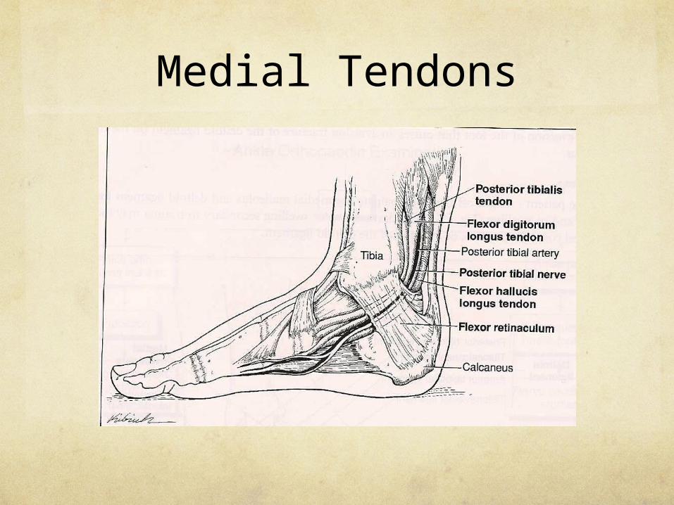
Medial Tendons

Posterior Tibial Artery, Tibial Nerve

Lateral Malleolus & Attached Ligaments
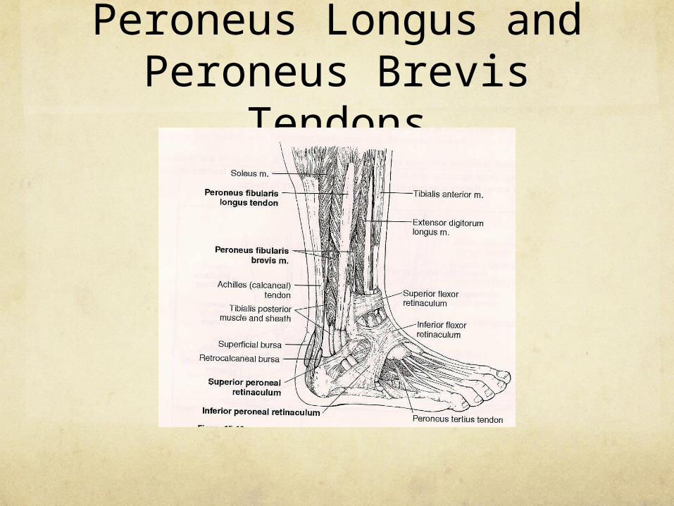
Peroneus Longus and Peroneus Brevis
Tendons
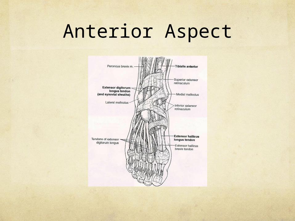
Anterior Aspect

Posterior Aspect
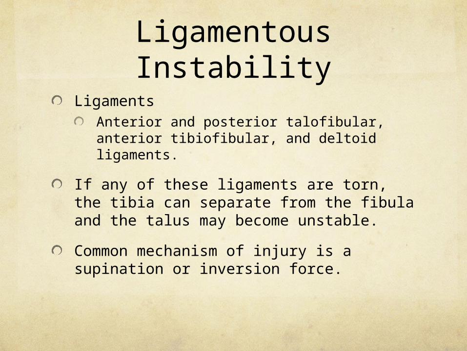
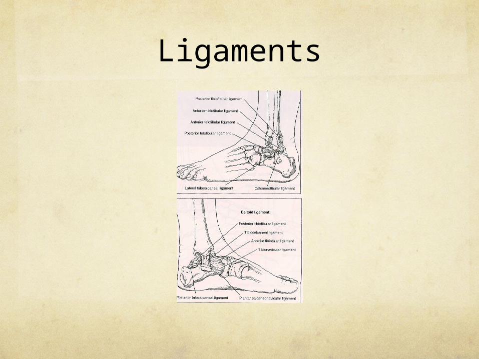
Ligamentous Instability
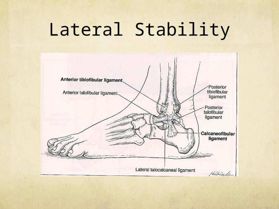
LigamentsAnterior and posterior talofibular, anterior tibiofibular, and deltoid ligaments.
If any of these ligaments are torn, the tibia can separate from the fibula and the talus may become unstable.
Common mechanism of injury is a supination or inversion force.

Ligamentous Instability
The foot turns under the ankle after walking or running on uneven surfaces or when landing on an inverted foot after a jump.
The most common injured ligament is the anterior talofibular ligament.
Ligament laxity can lead to chronic ankle sprains.
Ligamentous Instability
Clinical Signs and SymptomsAnkle swelling
Static ankle pain
Pain on passive motion
Tenderness over affected ligament
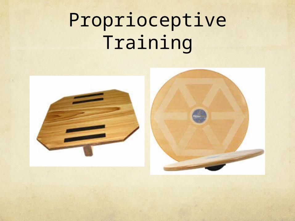
Proprioceptive Training
Ligaments

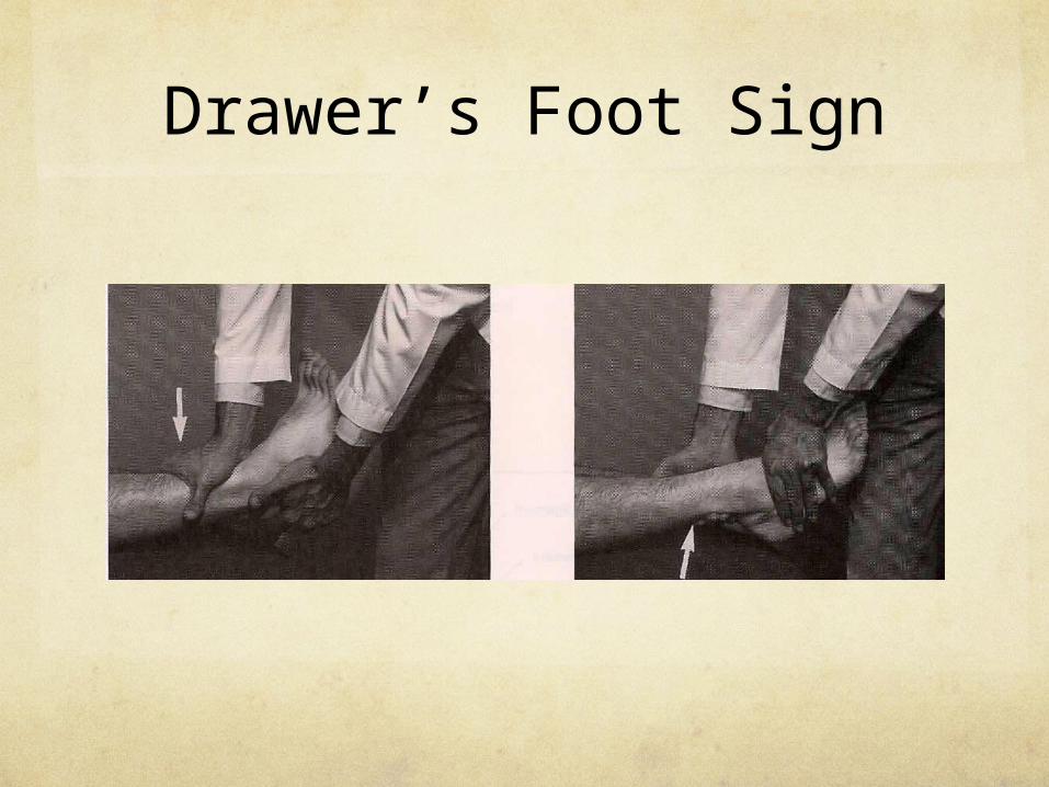
Drawer’s Foot Sign
Procedure: Patient supine. Stabilize ankle with one hand. Press posterior on tibia with the other hand. Next, grasp anterior aspect of the foot with one hand and the posterior aspect of the tibia with the other. Pull anterior.
Rationale:Gapping with posterior push – tear anterior talofibular
Gapping with anterior pull – tear posterior talofibular
Drawer’s Foot Sign

Drawer’s Foot Sign
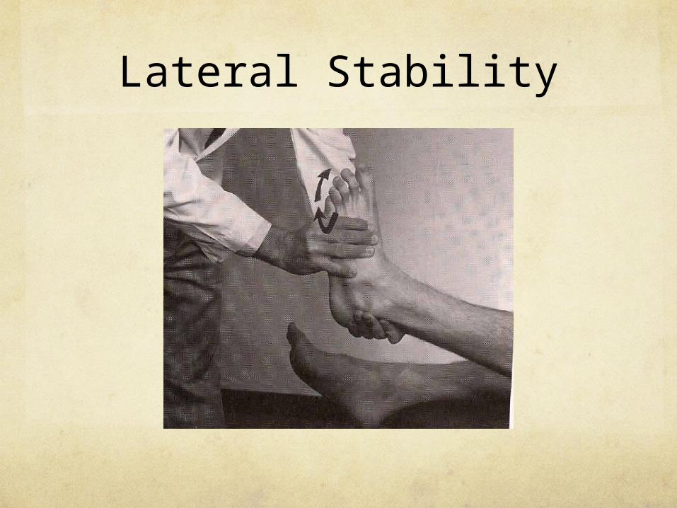
Lateral Stability
Procedure: Patient supine. Passively invert foot.
Rationale: Gapping secondary to trauma. Suspect tear of anterior talofibular ligament or calcaneofibular ligament.
Lateral Stability
Lateral Stability
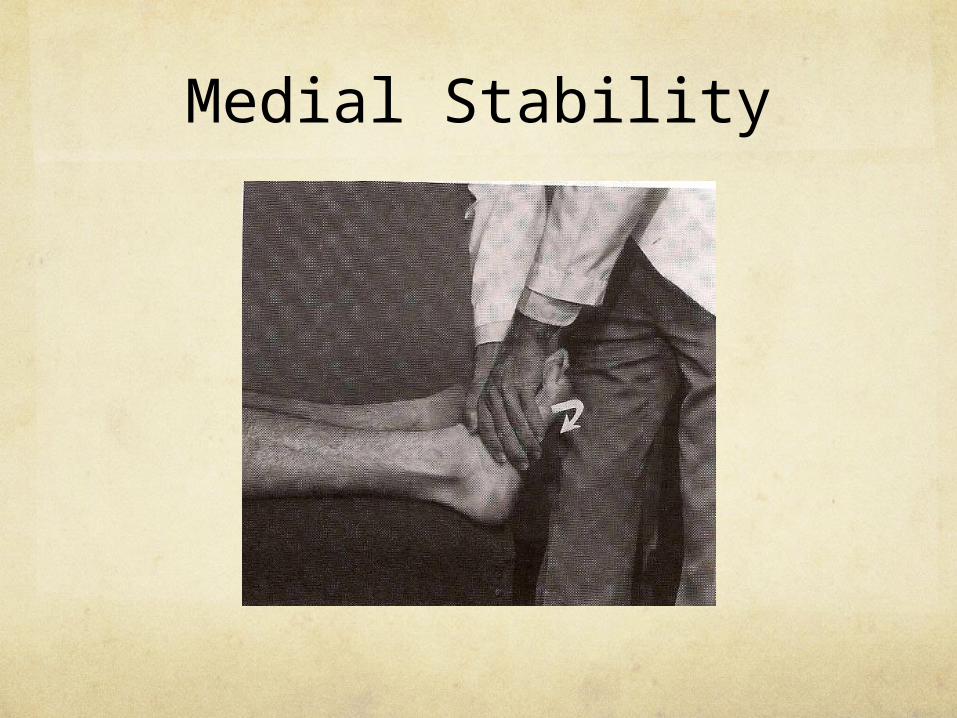
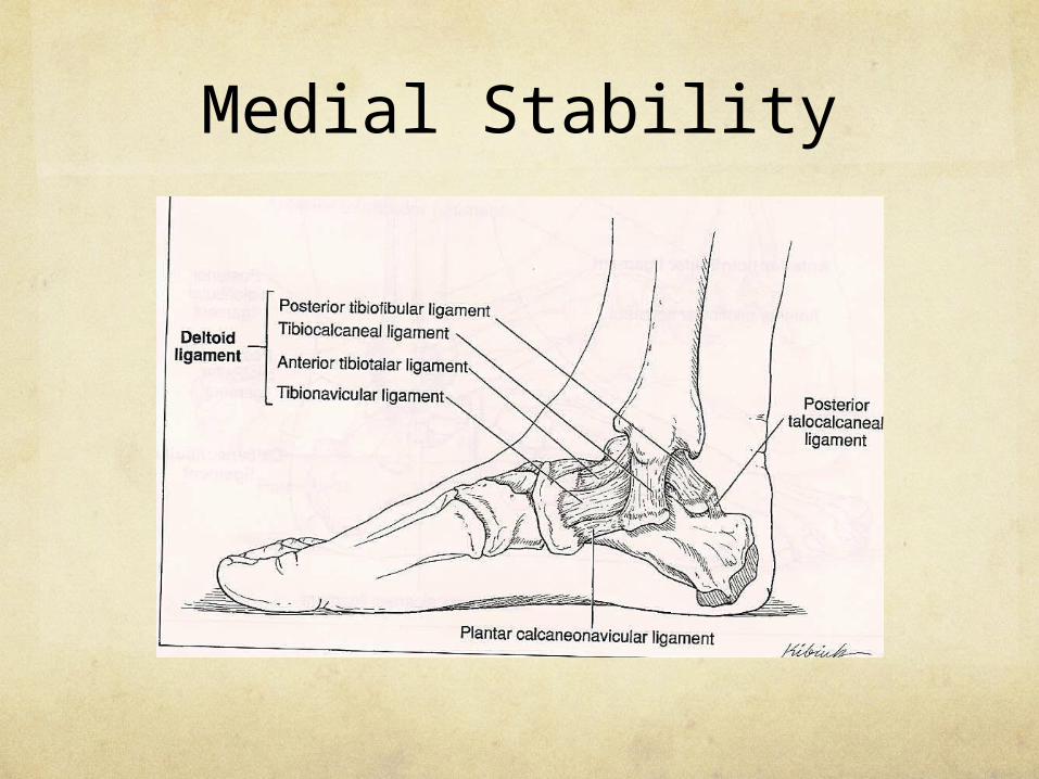
Medial Stability
Procedure: Patient supine. Passively evert foot.
Rationale: Gapping secondary to trauma. Suspect tear of deltoid ligament.
Medial Stability
Medial Stability
Tarsal Tunnel Syndrome
Tarsal tunnel syndrome occurs when the posterior tibial nerve becomes entrapped in its tunnel as it passes behind the medial malleolus to enter the foot.
The tunnel can be compressed either intrinsically or extrinsically.
Space-occupying lesions account for 50% of the cases.
Tarsal Tunnel Syndrome
Direct trauma and repetitive dorsiflexion account for a significant portion of the remaining cases.
A severe flat foot can unduly stretch the posterior tibial nerve.
Other possible causes include: fracture callus, ganglion of the tendon sheath, lipoma, engorged venus plexus, and excessive pronation of the hind foot.
Tarsal Tunnel Syndrome
Clinical Signs and SymptomsIntermittent paresthesia of plantar aspect of foot
Pain on foot inversion and / or eversion of the foot
Pain radiating to medial aspect of the leg
Pain made worse by activity and improved by rest

Tarsal Tunnel
Tinel’s Foot Sign
Procedure: Tap over the posterior tibial nerve with a neurological reflex hammer.
Rationale: Paresthesias radiating to the foot indicate irritation of the posterior tibial nerve that may be caused by constriction at the tarsal tunnel.
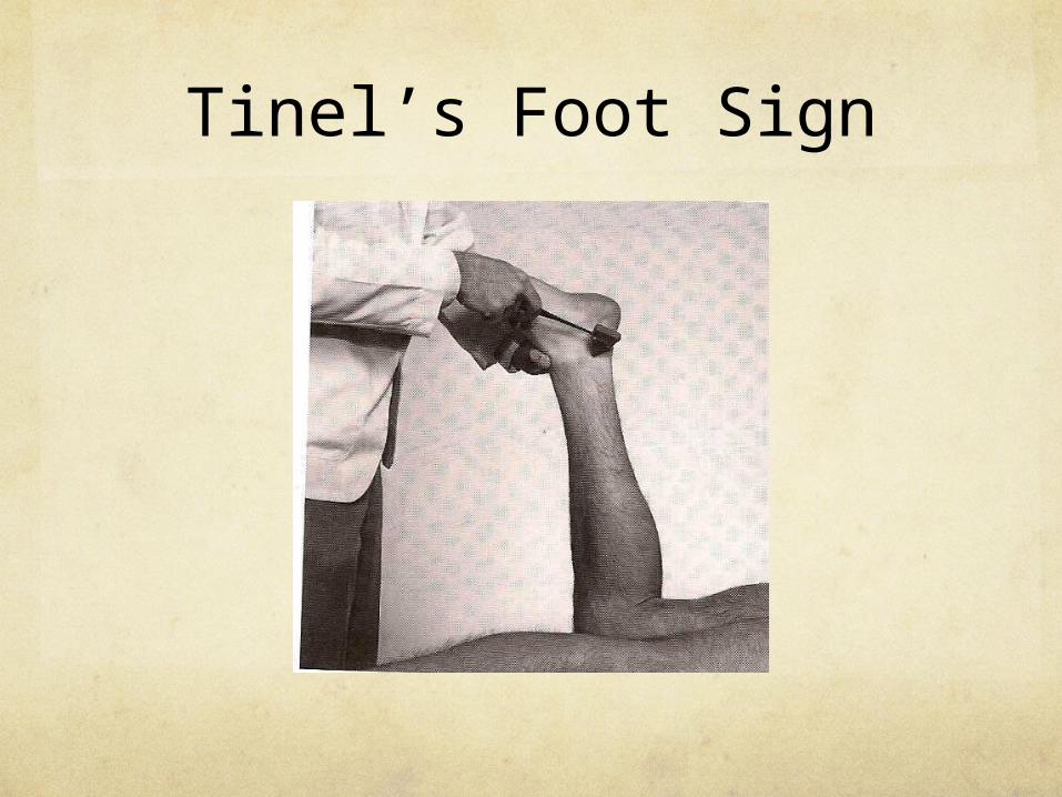
Tinel’s Foot Sign
Achilles Tendon Rupture
Achilles tendon rupture generally occurs in adults aged 30 to 50.
It is usually spontaneous in athletes who account for most of these injuries.
Decreased vascularity of the Achilles tendon as the patient ages may contribute.

Achilles Tendon Rupture
Mechanism of injury - forced dorsiflexion of the foot as the soleus and gastrocnemius contract.
Rupture occurs 2 to 6 cm from the insertion of the Achilles tendon into the calcaneus.
As the proximal aspect of the tendon retracts, there is usually a palpable defect of the tendon.

Achilles Tendon Rupture
Clinical Signs and SymptomsSevere posterior ankle pain
Inability to stand on toes
Posterior leg and heel swelling
Posterior leg and heel ecchymosis
Thompson’s Test
Procedure: Patient prone. Flex knee. Squeeze the calf muscles against the tibia and fibula.
Rationale: The the gastrocnemius and soleus are squeezed, they mechanically contract. They are attached to the Achilles tendon, which plantar-flexes the foot. If the tendon is ruptured, contraction of the gastrocnemius and soleus muscles will NOT plantar-flex the foot.

Thompson’s Test
Related Documents