ELSEVIER PI1 SO730-725X( 96) 00378-5 l Original Contribution Magnetic Resonance Imaging, Vol. 15, No. 4, pp. 433-440, 1997 Copyright 0 1997 Elsevier Science Inc. Printed in the USA. All rights reserved 0730-725X/97 $17.00 + .OO ANISOTROPIC WATER DIFFUSION IN WHITE AND GRAY MATTER OF THE NEONATAL PIGLET BRAIN BEFORE AND AFTER TRANSIENT HYPOXIA-ISCHAEMIA J.S. THORNTON, * R.J. ORDIDGE, * J. PENRICE,? E.B. CADY, * P.N. AMESS,~ S. PUNWANI, * M. CLEMENCE* AND J.S. WYATT? Departments of *Medical Physics and Bioengineering, and TPaediatrics, University College London, London, WClE 6JA, UK Measurements of tissue water apparent diffusion coefficient (ADC) performed with diffusion sensitization applied separately along the x, y, and z axes revealed significant diffusion anisotropy in both cerebral white and gray matter in six newborn (<24 h old) piglets. Mean baseline white matter ADC for a particular region of interest was 125.8% (SD 32.0%; p < .OOl) greater when the diffusion gradients were applied along the y axis as compared to along the x. For the cortical gray matter region considered, the situation was reversed, the mean ADC value measured along x exceeding that along y by 15.2% (SD 6.1%; p < .Ol). Forty-three hours subsequent to a transient cerebra1 hypoxic-ischaemic insult, phosphorous MRS measurements indicated that the animals had suffered severe secondary cerebral energy failure. This was accompanied by a significant (p < .Ol) decrease in the white matter anisotropy, such that the mean y direction ADC now exceeded that along the x by only 70.9% (SD 29.4%; p < .03). There was no change in the gray matter anisotropy. The average of the ADC values measured in the x, y, and z directions had decreased by 35.3% (SD 18.5%; p < .Ol) in white matter and 31.4% (SD 21.9%; p < .05) in cortical gray matter. Diffusion anisotropy measurements may provide additional information useful in the characterisa- tion of hypoxic-ischaemic injury in the neonatal brain, and must be consideredif tissuewater ADC values are to be unambiguously interpreted in this context. 0 1997Elsevier ScienceInc. Keywords: hypoxia-ischaemia; diffusion; anisotropy; DWI; neonatal brain. INTRODUCTION MR measurements of the apparent diffusion coefficient (ADC) of brain tissue water have proved uniquely sensitive in the delineation of ischaemic injury. ADC values have been shown to decline significantly within minutes of the onset of an ischaemic episode.‘.’ In addition, the (scalar) ADC of tissue water has been shown to be markedly anisotropic in certain tissues, and this has been exploited to delineate white matter fibre tracts in the brain.3-5 Diffusion-weighted imaging (DWI) shows promise in several clinical applications, notably in the assessment of stroke patients,6 and, ger- mane to the subject of this paper, in the study of perina- tally asphyxiated infants.’ Although the reasonsfor a decline in ADC values during and subsequent to cere- bra1 ischaemic damage have not been unambiguously established, reduced values are thought to be related to cell swelling and a concomitant increase in tortuos- ity of water movement. We hypothesise that the direc- tional freedom of cells to swell in post-ischaemic white matter is influenced by the structural alignment of nerve fibres, and that this should be reflected in relative changes to anisotropy following ischaemia. The aim of this work was to investigate cerebral ADC values and diffusion anisotropy in an established porcine model of perinatal-asphyxia,8 in which phos- phorous (3’P-) MRS spectra acquired from the same animals were employed as an index of cerebral energy failure. It has been shown that this model faithfully reproduces the sequenceof alterations in cerebral en- RECEIVED 719196; ACCEPTED 10127196. Addresscorrespondence to John Thornton, Department of Medical Physicsand Bioengineering, University College London, 1st Floor, Shropshire House, 1 l-20 Capper St., Lon- don, WClE 6JA, UK. 433

Welcome message from author
This document is posted to help you gain knowledge. Please leave a comment to let me know what you think about it! Share it to your friends and learn new things together.
Transcript
ELSEVIER PI1 SO730-725X( 96) 00378-5
l Original Contribution
Magnetic Resonance Imaging, Vol. 15, No. 4, pp. 433-440, 1997 Copyright 0 1997 Elsevier Science Inc.
Printed in the USA. All rights reserved 0730-725X/97 $17.00 + .OO
ANISOTROPIC WATER DIFFUSION IN WHITE AND GRAY MATTER OF THE NEONATAL PIGLET BRAIN BEFORE AND AFTER
TRANSIENT HYPOXIA-ISCHAEMIA
J.S. THORNTON, * R.J. ORDIDGE, * J. PENRICE,? E.B. CADY, * P.N. AMESS,~ S. PUNWANI, *
M. CLEMENCE* AND J.S. WYATT?
Departments of *Medical Physics and Bioengineering, and TPaediatrics, University College London, London, WClE 6JA, UK
Measurements of tissue water apparent diffusion coefficient (ADC) performed with diffusion sensitization applied separately along the x, y, and z axes revealed significant diffusion anisotropy in both cerebral white and gray matter in six newborn (<24 h old) piglets. Mean baseline white matter ADC for a particular region of interest was 125.8% (SD 32.0%; p < .OOl) greater when the diffusion gradients were applied along the y axis as compared to along the x. For the cortical gray matter region considered, the situation was reversed, the mean ADC value measured along x exceeding that along y by 15.2% (SD 6.1%; p < .Ol). Forty-three hours subsequent to a transient cerebra1 hypoxic-ischaemic insult, phosphorous MRS measurements indicated that the animals had suffered severe secondary cerebral energy failure. This was accompanied by a significant (p < .Ol) decrease in the white matter anisotropy, such that the mean y direction ADC now exceeded that along the x by only 70.9% (SD 29.4%; p < .03). There was no change in the gray matter anisotropy. The average of the ADC values measured in the x, y, and z directions had decreased by 35.3% (SD 18.5% ; p < .Ol) in white matter and 31.4% (SD 21.9% ; p < .05) in cortical gray matter. Diffusion anisotropy measurements may provide additional information useful in the characterisa- tion of hypoxic-ischaemic injury in the neonatal brain, and must be considered if tissue water ADC values are to be unambiguously interpreted in this context. 0 1997 Elsevier Science Inc.
Keywords: hypoxia-ischaemia; diffusion; anisotropy; DWI; neonatal brain.
INTRODUCTION
MR measurements of the apparent diffusion coefficient (ADC) of brain tissue water have proved uniquely sensitive in the delineation of ischaemic injury. ADC values have been shown to decline significantly within minutes of the onset of an ischaemic episode.‘.’ In addition, the (scalar) ADC of tissue water has been shown to be markedly anisotropic in certain tissues, and this has been exploited to delineate white matter fibre tracts in the brain.3-5 Diffusion-weighted imaging (DWI) shows promise in several clinical applications, notably in the assessment of stroke patients,6 and, ger- mane to the subject of this paper, in the study of perina- tally asphyxiated infants.’ Although the reasons for a decline in ADC values during and subsequent to cere-
bra1 ischaemic damage have not been unambiguously established, reduced values are thought to be related to cell swelling and a concomitant increase in tortuos- ity of water movement. We hypothesise that the direc- tional freedom of cells to swell in post-ischaemic white matter is influenced by the structural alignment of nerve fibres, and that this should be reflected in relative changes to anisotropy following ischaemia.
The aim of this work was to investigate cerebral ADC values and diffusion anisotropy in an established porcine model of perinatal-asphyxia,8 in which phos- phorous (3’P-) MRS spectra acquired from the same animals were employed as an index of cerebral energy failure. It has been shown that this model faithfully reproduces the sequence of alterations in cerebral en-
RECEIVED 719196; ACCEPTED 10127196. Address correspondence to John Thornton, Department
of Medical Physics and Bioengineering, University College
London, 1st Floor, Shropshire House, 1 l-20 Capper St., Lon- don, WClE 6JA, UK.
433
434 Magnetic Resonance Imaging 0 Volume 15, Number 4, 1997
ergy metabolism that are observed in birth-asphyxiated human infants.’ During hypoxia-ischaemia 31P MRS shows falls in the levels of the high-energy metabolites phosphocreatine (PCr) and adenosine triphosphate ( ATP) concomitant with a rise in inorganic phosphate (Pi). Initial recovery immediately post-resuscitation is then followed by gradual declines in [ PCr] and [ ATP] and a large rise in [Pi] over the next 48 h: the latter changes constitute “secondary” or delayed energy failure. [ PCr] /[ Pi] is proportional to the “phosphory- lation” potential and following birth asphyxia low val- ues convey an unfavourable prognosis.‘*
In the present study, ADC anisotropy data were obtained from cortical gray matter and subcortical white matter in piglet brain by measuring ADC values along three orthogonal axes corresponding to the x, y, and z gradient directions. Baseline data were obtained, and changes in anisotropy investigated at 43 h follow- ing a defined transient hypoxic-ischaemic episode. These measurements are of clinical relevance to MR examinations of perinatally asphyxiated infants who are routinely scanned in our hospital within a few days of birth.9-‘2
MATERIALS AND METHODS
Animal Model Experiments were performed on six healthy term
Large White piglets, aged less than 24 h. Anaesthesia was induced by inhalation of 5% isoflurane followed by tracheostomy and ventilation with a mixture of nitrous oxide, oxygen, and isoflurane ( < 1.5% ) . The oxygen concentration was adjusted such that the mean baseline arterial oxygen partial pressure (Paoa) was 13.8 (SD 2.8) kPa. Inflatable cuffs were positioned around both common carotid arteries. The anaesthe- tized animal was then positioned prone on a tempera- ture-regulated water-filled mattress in a cylindrical perspex pod, designed to fit snugly into the bore of the magnet. The head was constrained using a stereotactic system, with care being taken to ensure precise posi- tioning of both the animals within the pod, and the pod within the magnet bore. Heart rate, blood pres- sure, rectal and tympanic temperatures were moni- tored continuously.
Transient cerebral hypoxia-ischaemia was induced as previously described.8 Briefly, the inspired oxygen concentration was reduced to 12%, producing a mean Pao, of 3.9 (SD 0.8) kPa, and both common carotid arteries were temporarily occluded until severe deple- tion of high energy phosphates was detected by 31P MRS. The animals were then resuscitated by increas- ing the inspired oxygen to normalize Paoz and releasing the carotid occluders. The animals were maintained with full intensive care inside the magnet bore as ob- servations continued throughout the subsequent 48 h.
MRI and MRS MR measurements were performed using a 7T
Bruker Biospec imaging spectrometer system (Bruker Medzin Technik GmBH., Karlsruhe, Germany) em- ploying standard (non-shielded) gradient coils (Bruker G152). Data were acquired using a double tuned (31P and proton (‘H)) 25 mm diameter transmit/receive surface coil incorporating an inductively balanced matching arrangement. The coil was positioned on the intact scalp over the parietal lobes of the brain. Prior to each measurement, shimming was performed until a water line-width of better than 30 Hz was obtained.
Fully relaxed 31P pulse-acquire spectra were ob- tained, as previously reported, 8 with a relaxation delay (TR) of 10 s. Resonance peak areas were measured by Lorentzian curve fitting via x ’ minimisation.13 Spectra were obtained from 192 summed free induction decays (FIDs) at baseline, 36 FIDs during the insult, and 384 summed FIDs post-resuscitation. Values of [ PCr]/ [ Pi] obtained at baseline, at the nadir of the insult, and at 2 and 46 h post-resuscitation are reported.
ADC values were measured in a coronal plane using a diffusion-weighted stimulated echo acquisition (STEAM) sequence.14 ECG and respiratory gating were not found necessary. The sequence timing param- eters were as follows: TR = 2 s, echo time (TE) = 34 ms, mixing time (TM) = 200 ms, acquisition time = 2.2 min per image. The slice thickness was 2.5 mm, the field of view 3.0 cm and the image matrix of 128 x 64 was zero-filled to 128 X 128 to give an in-plane pixel resolution of 0.23 mm.
Rectangular diffusion-sensitizing gradient pulses were applied between the first and second radiofre- quency (RF) pulses, and after the third RF pulse of the STEAM sequence. The duration of each diffusion- sensitizing gradient pulse was 10 ms and the separation between the rising edges of the two gradient pulses was 214 ms. Gradient strengths of 0, 17, 25, and 30 mT * m-* were employed giving ‘b factors’ of 0, 440, 900, and 1350 X 109 m* * s-’ for diffusion sensitization along the x and y axes (corresponding respectively to horizontal and vertical directions in the images). The equivalent ‘b-factors’ for diffusion sensitization along the z axis were 0, 724, 1270, and 1780 X lo9 rn*. s-l after taking into account cross-terms arising from the slice selection (z) gradients. For measurements with diffusion-sensitization along x and y axes, the readout gradient was applied along an orthogonal axis (e.g., y and x, respectively), thereby minimising errors due to additional cross-terms in the calculation of ‘b-factors.’ Careful adjustment of the gradient system preemphasis was required to minimise artifacts arising from gradi- ent-induced eddy currents.
ADC maps were calculated using a pixel by pixel
Anisotropic water diffusion in white and gray matter 0 J.S. THORNTON ETAL. 435
log-linear fitting algorithm. A numerical index of an- isotropy, ANIS, was calculated using the formula
ANIS,,, = ADC, - ADCb
ADC, x 100% (1)
where ADC, and ADCb represent ADC values mea- sured with the diffusion weighting gradients applied along separate orthogonal directions. Maps of ANIS were used to visually demonstrate the spatial distribu- tion of anisotropy. A value of ANIS of zero, displayed as a mid-gray level on a linear gray scale, indicates equal propensity to diffuse along both directions. Tak- ing ANIS,, as an example, positive and negative values indicate preferential diffusion along the x and y direc- tion, respectively, and are shown as regions of hyper- intensity and hypo-intensity relative to the midgray level.
ANIS, is not an absolute quantity; it is dependent upon the relative orientation of the animal’s head within the Cartesian coordinate system. However, we believe that the positioning of the animals within the magnet bore was sufficiently consistent to allow a quantitative assessment of anisotropy in this manner, particularly because individual animals were not moved between successive ADC measurements.
The results presented below consist of control data collected from each animal prior to the induction of transient hypoxia-ischaemia and further data collected at 43 h post-resuscitation. (The approximately 3-h de- lay between the final ADC and the final 3’P measure- ments occurred while measurement of other MR pa- rameters, as will be reported elsewhere, was per- formed.) The statistical significance of ANI&, values was assessed by performing paired t-tests for mean ADC, vs. mean ADCb. Validation of ADC accuracy was performed using a spherical distilled-water phan- tom imaged using the same protocol.
RESULTS
Phantom Measurements Uniform, artifact-free ADC maps were obtained for
each diffusion-sensitizing direction. Values of ADC obtained from a common region of interest (ROI) for the x, y, and z directions were 2.22 + 0.06 X lo-’ m2.s-’ at room temperature in reasonable agreement with literature values.
In Vivo Results
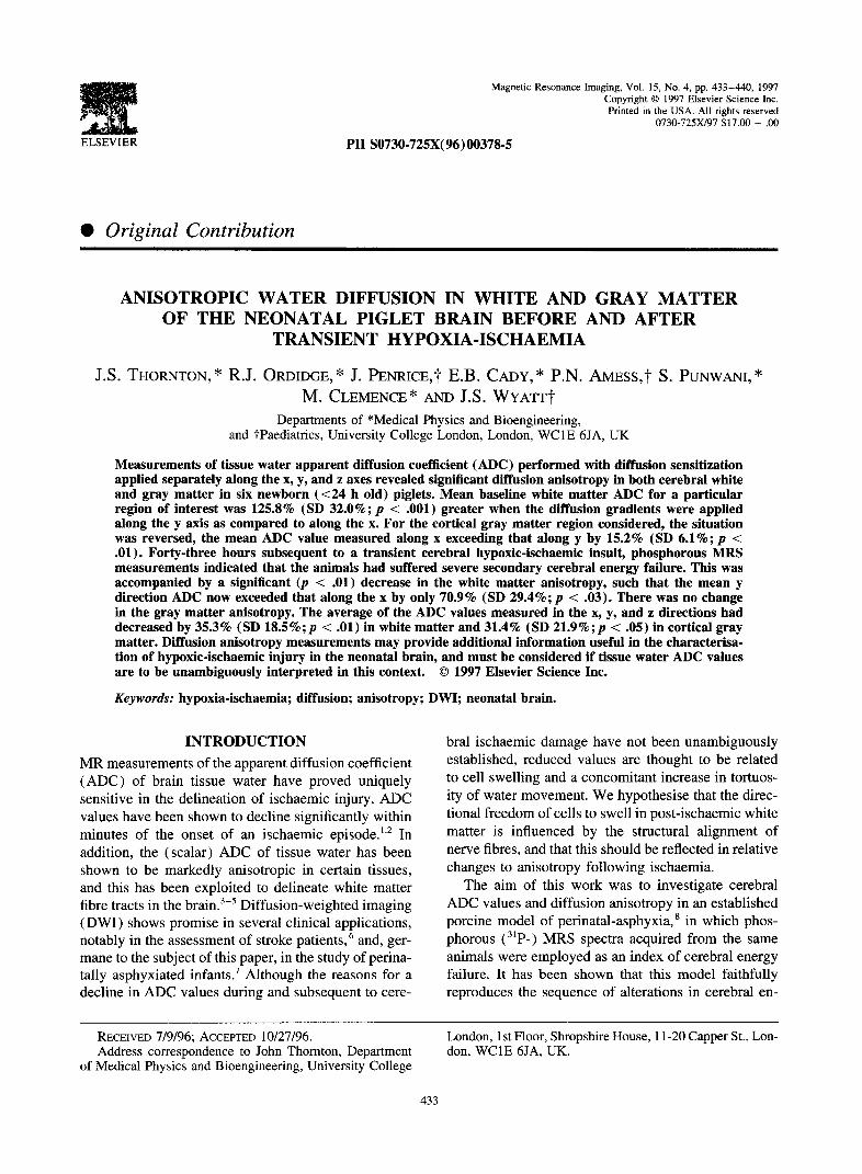
Baseline Values. Figure 1 shows typical baseline diffusion-weighted images obtained with diffusion sensitization applied along each of the x, y, and z axes together with corresponding maps of absolute ADC
(a) W
Cd) (6 m
Fig. 1. (a-c) Heavily diffusion-weighted images of a coro- nal section through the parietal lobes of a newborn piglet, with diffusion sensitizing gradients applied along the x and y (b = 1350 X 10’ m2*s-‘); and z axes (b = 1780 x lo9 m* * s -’ ), respectively. (d-f) Corresponding maps of ADC,, ADC,, and ADC,. Arrows indicate regions of white matter (A) and cortical gray matter (B).
values. The cortical gray matter and underlying white matter tracts are clearly visible. The appearance of cerebrospinal fluid in the sulci differs for the x-direc- tion (for which the phase encode direction was left- right) compared to the y- and z-direction DWI images (for which the phase encode direction was up-down in the image plane). This was likely due to artifacts arising from the rectangular (64 X 128) acquisition matrix, or to slow flow.
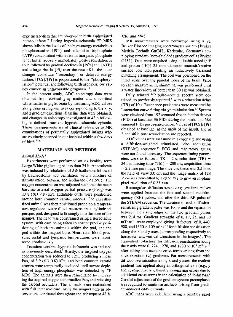
Comparing DWI images obtained with x- and y- diffusion sensitization, a marked anisotropy in tissue water diffusion was observed in the white matter tracts central to each gyrus (region A). This anisotropy also appeared as a distinct difference in the ADC, and ADC, maps. Closer inspection revealed that the ADC value measured in cortical gray matter was also slightly anisotropic (region B) . The major axis of diffusional freedom in gray matter along the main section of the gyri appears to be perpendicular to that of white matter. However, at the tip of the gyri, the gray matter diffu- sion anisotropy appears to be parallel with white mat- ter, causing a characteristic “arrowhead” feature ob- served in Fig. la. In the anisotropy index maps of Fig. 2, the brighter appearance of gray matter along the inner fold of the gyri contrasted against the dark ap- pearance of white matter demonstrates the perpendicu- lar diffusional anisotropy characteristics of tissues in these locations. However, this contrast becomes diffuse at the tip of the gyri as gray matter folds around the terminating strip of white matter. For this animal, val- ues of ANIS,, at 8 pixel ROIs centred on positions A and B were -114.0% (SD 20.0%) and 24.0% (SD 6.0%), respectively. The value at the tip of the gyrus indicated was -47.3% (SD 8.1%).
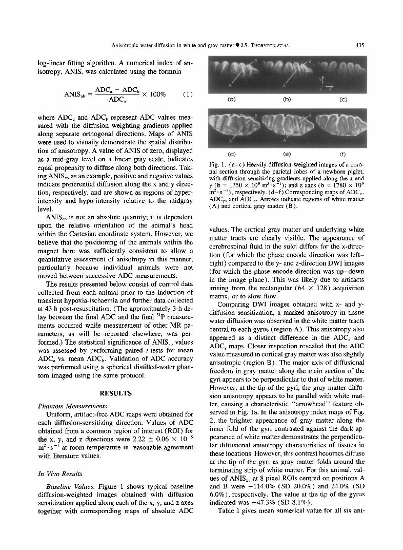
Table 1 gives mean numerical value for all six ani-
436 Magnetic Resonance Imaging l Volume 15. Number 4, 1997
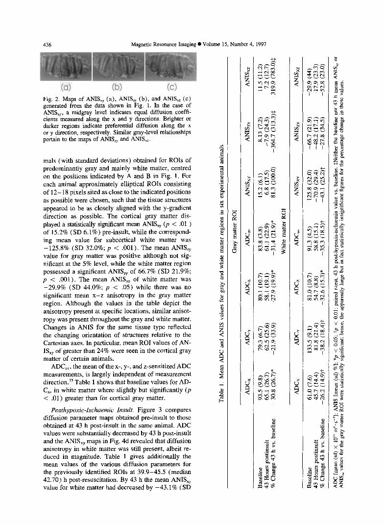
(a> (bl cc> Fig. 2. Maps of ANIS,, (a), ANIS,, (b), and ANIS,, (c) generated from the data shown in Fig. 1. In the case of ANIS,,, a midgray level indicates equal diffusion coeffi- cients measured along the x and y directions. Brighter or darker regions indicate preferential diffusion along the x or y direction, respectively. Similar gray-level relationships pertain to the maps of ANIS,, and ANIS,,.
mals (with standard deviations) obtained for ROIs of predominantly gray and mainly white matter, centred on the positions indicated by A and B in Fig. 1. For each animal approximately elliptical ROIs consisting of 12- 18 pixels sited as close to the indicated positions as possible were chosen, such that the tissue structures appeared to be as closely aligned with the y-gradient direction as possible. The cortical gray matter dis- played a statistically significant mean ANIS, (p < .Ol ) of 15.2% (SD 6.1%) pre-insult, while the correspond- ing mean value for subcortical white matter was - 125.8% (SD 32.0%; p < .OOl). The mean ANIS, value for gray matter was positive although not sig- nificant at the 5% level, while the white matter region possessed a significant ANIS,, of 66.7% (SD 21.9%; p < .OOl). The mean ANIS,, of white matter was -29.9% (SD 44.0%; p < .05) while there was no significant mean x-z anisotropy in the gray matter region. Although the values in the table depict the anisotropy present at specific locations, similar anisot- ropy was present throughout the gray and white matter. Changes in ANIS for the same tissue type reflected the changing orientation of structures relative to the Cartesian axes. In particular, mean ROI values of AN- IS,, of greater than 24% were seen in the cortical gray matter of certain animals.
ADC, , the mean of the x-, y-, and z-sensitized ADC measurements, is largely independent of measurement direction.15 Table 1 shows that baseline values for AD- C,, in white matter where slightly but significantly (p < .Ol) greater than for cortical gray matter.
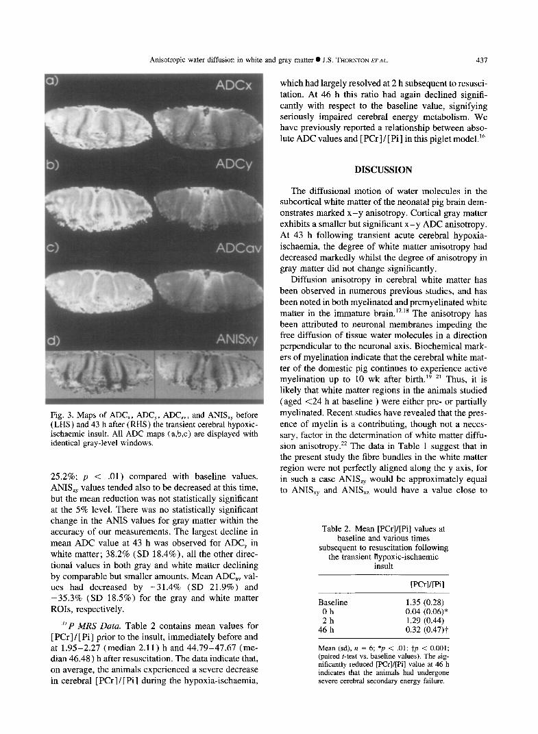
Posthypoxic-Ischaemic Insult. Figure 3 compares diffusion parameter maps obtained pre-insult to those obtained at 43 h post-insult in the same animal. ADC values were substantially decreased by 43 h post-insult and the ANIS., maps in Fig. 4d revealed that diffusion anisotropy in white matter was still present, albeit re- duced in magnitude. Table 1 gives additionally the mean values of the various diffusion parameters for the previously identified ROIs at 39.9-45.5 (median 42.70) h post-resuscitation. By 43 h the mean ANIS,, value for white matter had decreased by -43.1% (SD
3
Anisotropic water diffusion in white and gray matter 0 J.S. THORNTON ETAL. 437
Fig. 3. Maps of ADC,, ADC,, ADC,,, and ANIS,, before (LHS) and 43 h after (FWS) the transient cerebral hypoxic- ischaemic insult. All ADC maps (a,b,c) are displayed with identical gray-level windows.
25.2%; p < .Ol) compared with baseline values. ANIS,, values tended also to be decreased at this time, but the mean reduction was not statistically significant at the 5% level. There was no statistically significant change in the ANIS values for gray matter within the accuracy of our measurements. The largest decline in mean ADC value at 43 h was observed for ADC, in white matter; 38.2% (SD 18.4%), all the other direc- tional values in both gray and white matter declining by comparable but smaller amounts. Mean ADC,, val- ues had decreased by -31.4% (SD 21.9%) and -35.3% (SD 18.5%) for the gray and white matter ROIs, respectively.
3’ P MRS Data. Table 2 contains mean values for [ PCr]/[ Pi] prior to the insult, immediately before and at 1.95-2.27 (median 2.11) h and 44.79-47.67 (me- dian 46.48) h after resuscitation. The data indicate that, on average, the animals experienced a severe decrease in cerebral [ PCr] / [ Pi] during the hypoxia-ischaemia,
which had largely resolved at 2 h subsequent to resusci- tation. At 46 h this ratio had again declined signifi- cantly with respect to the baseline value, signifying seriously impaired cerebral energy metabolism. We have previously reported a relationship between abso- lute ADC values and [ PCr] / [ Pi] in this piglet model.16
DISCUSSION
The diffusional motion of water molecules in the subcortical white matter of the neonatal pig brain dem- onstrates marked x-y anisotropy. Cortical gray matter exhibits a smaller but significant x-y ADC anisotropy. At 43 h following transient acute cerebral hypoxia- ischaemia, the degree of white matter anisotropy had decreased markedly whilst the degree of anisotropy in gray matter did not change significantly.
Diffusion anisotropy in cerebral white matter has been observed in numerous previous studies, and has been noted in both myelinated and premyelinated white matter in the immature brain.17,” The anisotropy has been attributed to neuronal membranes impeding the free diffusion of tissue water molecules in a direction perpendicular to the neuronal axis. Biochemical mark- ers of myelination indicate that the cerebral white mat- ter of the domestic pig continues to experience active myelination up to 10 wk after birth.“-” Thus, it is likely that white matter regions in the animals studied (aged <24 h at baseline ) were either pre- or partially myelinated. Recent studies have revealed that the pres- ence of myelin is a contributing, though not a neces- sary, factor in the determination of white matter diffu- sion anisotropy. The data in Table 1 suggest that in the present study the fibre bundles in the white matter region were not perfectly aligned along the y axis, for in such a case ANIS,, would be approximately equal to ANIS,, and ANIS,, would have a value close to
Table 2. Mean [PCr]/[Pi] values at baseline and various times
subsequent to resuscitation following the transient llypoxic-ischaemic
insult
[PCr]/[Pi]
Baseline Oh 2h
46 h
1.35 (0.28) 0.04 (0.06)” 1.29 (0.44) 0.32 (0.47)t
Mean (sd), n = 6; *p < .Ol; tp < 0.001: (paired r-test vs. baseline values). The sig- nificantly reduced [PCr]/[Pi] value at 46 h indicates that the animals had undergone severe cerebral secondary energy failure.
438 Magnetic Resonance Imaging l Volume 1.5, Number 4, 1997
zero. The reduced ANIS,,, compared with ANIS,,, together with the smaller, but substantial, ANIS,, may be a consequence of the white matter fibres being partly aligned with the z axis in the region investigated.
The anisotropy effect in cortical gray matter is, to the authors’ knowledge, a new observation in neonatal brain, although gray matter anisotropy in the cortex of mature rat brain has recently been reported,23 with a corresponding ANIS,, value of 22%. The observed anisotropy presumably reflects a degree of microscopic geometrical order within the cortical tissue. Histologi- cally stained sections of adult human cerebral cortex demonstrate bands of neural processes orientated par- allel or orthogonal to the brain surface in different cortical areas.” Such structures, if present in the devel- oping porcine brain, might account for the observed corticai ADC anisotropy.
Because DWI data was not available from control animals, it was not possible to completely rule out the contribution of any potential deleterious effects of prolonged anaesthesia to the observed ADC varia- tions. However, previous MRS studies’ have shown no significant changes in cerebral high-energy phos- phate metabolite levels in sham-operated control pig- lets maintained within the magnet for an equivalent period of 4X h. This, together with the observation that all systemic physiological parameters monitored remained within normal limits, leads us to believe that the changes in ADC reported here were genuine sequelae of hypoxia-ischaemia.
Surface coil in vivo 31P MRS evaluates cerebral energy status in a volume including both gray and white matter, the spatial selectivity of the technique being such as to preclude knowledge of the relative contribution of each tissue type to changes in 31P metabolite levels. In all of the animals included in this study the ratio [ PCr]/[ Pi] was significantly re- duced at 46 h post-resuscitation. The reduced absolute ADC,, and ANIS,, observed in white matter at ~43 h post-resuscitation were, therefore, presumed to reflect the impaired cerebral energy metabolism indicated by reduced [ PCr] / [ Pi]. The larger standard deviations of the late time point ADC- and [ PCr]/[ Pi] values, as compared with the baseline results, may reflect variations in insult severity, which correlates with the extent of cerebral energy impairment 24-48 h post- resuscitation.’
It has been suggested that the reduced ADC values following ischaemia may be caused by the constraint of more tissue water within the intra-cellular compartment resulting in cellular swelling, and the remaining extracel- lular fraction being subject to a less voluminous and more tortuous environment.26-30 In white matter, the effect of cell swelling would be expected to be more complicated than in gray matter. Along an axis perpendicular to fibre
orientation, the magnitude of ADC reduction would be expected to be similar to gray matter post-insult, because the distribution of cell membrane barriers would presum- ably change in the same relative manner in response to ischaemia. Our results presented in Table 1 suggest that ADC, values for white matter tracts, which are presum- ably more aligned with fibre orientation due to elevated control values, are reduced more markedly than ADC, post-insult. The resulting significant reduction in white matter anisotropy implies that the ability of water to move along the fibre axis has been more markedly reduced. Various explanations might account for this observation, which may reflect a squeezing of the extracelluar space between myelin sheaths, or possibly reduced efficacy of axonal active transport mechanisms. Neuropathological data was not available for this group of animals. How- ever, in a previous study2* histologic examination of the brains of a similar group of experimental animals re- vealed microglial reaction and astrocytic proliferation in the white matter of severely injured brains, which might also act to impede the movement of water molecules along the fibre axes. The net effect is a large, significant, reduction in mean x-y anisotropy index post-insult in white matter.
These observations have significant implications as DWI techniques are appended to clinical MR scan pro- tocols of human neonates. If ADC values are to be useful in interpreting the severity of ischaemic damage, it is important that tissue anisotropy in the regions of interest is well understood. Also, for anisotropy effects to be correctly interpreted, it is essential that measure- ments be performed with sufficiently high spatial reso- lution to prevent partial volume effects.
It is concluded that ADC anisotropy must be taken into account to allow unambiguous interpretation of DWI results, and to provide meaningful inter-subject and inter-site result comparisons. A DWI measure- ment with a single diffusion sensitizing gradient di- rection is of limited value in this context. More re- cently, it has been recognized that measurements of the three-dimensional apparent diffusion tensor allow more complete characterisation of such anisotropic diffusion behaviour, including the explicit determina- tion of flbre orientation. l5 Such studies minimize pos- sible errors introduced in the assessment of ADC an- isotropy caused by inconsistent positioning of sub- jects relative to the measurement coordinate system or cross-terms generated between imaging and diffusion sensitizing gradients. Unfortunately such comprehen- sive measurements may not be practical in the time available for clinical examinations using currently available standard scanner technology. A more practi- cal approach may be to obtain values of the trace of the diffusion tensor, to which our measurements of ADC,, are a good approximation, yielding values in-
Anisotropic water diffusion in white and gray matter 0 J.S. THORNTON ETAL.
dependent of the relative orientation of the fibre and measurement coordinate axes. l5
A sequential study of the full time course of changes in absolute ADC values during this early period (up to 48 h following injury) is currently being performed, and the relationship to the time course of changes in cerebral energy metabolism is the subject of ongoing research.
Acknowledgments-The authors thank the Medical Research Coun- cil, University College Hospitals NHS Trust, and the Wellcome Trust for financial support.
REFERENCES
1. Knight, R.A.; Ordidge, R.J.; Helpern, J.A.; Chopp, M.; Rodolsi, L.C.; Peck, D. Temporal evaluation of ischaemic damage in rat brain measured by proton nuclear magnetic resonance imaging. Stroke 23:576- 582; 1992.
2. Mintorovich, J.; Mosely, M.E.; Chileuitt, L.; Shim& H.; Cohen, Y.; Weinstein, P.R. Comparison of diffusion- and T,-weighted MRI for the early detection of cerebral &hernia and reperfusion in rats. Magn. Res. Med. 18:39-50; 1991.
3. Doran, M.; Hajnal, J.V.; Van Bruggen, N.; King, M.D.; Young, I.R.; Bydder, G.M. Normal and abnormal white matter tracts shown by MR imaging using directional diffusion weighted sequences. J. Comput. Assist. To- mogr. 14:865-873; 1990.
4. Douek, P.; Turner, R.; Pekar, J.; Patronas, N.; Le Bihan, D. MR colour mapping of myelin fibre orientation. J. Comput. Assist. Tomogr. 15:923-929; 1991.
5. Coremans, J.; Luypaert, R.; Verhelle, F.; Stadnik, T.; Osteaux, M. A method for myelin fibre orientation map- ping using diffusion-weighted MR images. Magn. Res. Imaging 12443-454; 1994.
6. Warach, S.; Gaa, J.; Siewart, B.; Wielopolski, P.; Edel- man, R.R. Acute human stroke studied by whole brain echo-planar diffusion-weighted magnetic resonance im- aging. Ann. Neurol. 37:231-241; 1995.
7. Cowan, F.M.; Pennock, J.M.; Hamahan, J.D.; Manji, K.P.; Edwards, A.D. Early detection of cerebral in- farction and hypoxic ischemic encephalopathy in neo- nates using diffusion-weighted magnetic resonance im- aging. Neuropediatrics 25:172- 191; 1994.
8. Lorek, A.; Takei, Y.; Cady, E.B.; et al. Delayed ( “sec- ondary”) cerebral energy failure after acute hypoxia- ischaemia in the newborn piglet: Continuous 48-hour studies by phosphorous magnetic resonance spectros- copy. Pediatr. Res. 36:699-706; 1994.
9. Hope, P.L.; Costello, A.M.; Cady, E.B.; Delpy, D.T.; Tofts, P.S.; Chu, A.; Hamilton, P.A.; Reynolds, E.O.R. Cerebral energy metabolism studied with phosphorous NMR spectroscopy in normal and birth-asphyxiated in- fants. Lancet 2:336-370; 1984.
10. Azzopardi, D.; Wyatt, J.S.; Cady, E.B.; Delpy, D.T.; Baudin, J.; Stewart, A.L.; Hope, P.L.; Hamilton, P.A.; Reynolds, E.O.R. Prognosis of newborn infants with hypoxiclischaemic brain injury assessed by phospho-
11.
12.
13.
14.
15.
16.
17.
18.
19.
20.
21.
22.
23.
24.
439
rous magnetic resonance spectroscopy. Pediatr. Res. 24:445-451; 1989. Cady, E.B. Quantitative combined phosphorous and pro- ton PRESS of the b?ains of newborn human infants. Magn. Res. Med. 33:557-563; 1995. Penrice, J.; Cady, E.B.; Lorek, A.; Wylezinska, M.; Amess, P.N.; Aldridge, R.F.; Stewart, A.L.; Wyatt, J.S., Reynolds, E.O.R. Proton magnetic resonance spectros- copy of the brain in normal preterm and term infants, and early changes following perinatal hypoxia-isch- aemia. Pediatr. Res. (in press). Cady, E.B. A reappraisal of the absolute concentrations of phosphorylated metabolites in the human neonatal cerebral cortex obtained by fitting Lorentzian curves to the 31P NMR spectrum. J. Magn. Reson. 87:433-446; 1990. Merboldt, K.D.; Hanicke, W.; Frahm, J. Diffusion im- aging using stimulated echoes. Magn. Res. Med. 19: 233-239; 1991. Basser, P.J.; Mattiello, J.; Le Bihan, D. MR diffusion tensor spectroscopy and imaging. Biophys. J. 66:259- 267; 1994. Thornton, J.S.; Ordidge, R.J.; Pemice, J.; Cady, E.B.; Punwani, S.; Wyatt, J.S. Reduced apparent diffusion coefficients in the newborn brain during and after acute hypoxia-ischaemia correlate with impaired cerebral en- ergy generation. In: Book of abstracts: Third annual meeting of the Society of Magnetic Resonance in Medi- cine. Berkeley, CA: SMRM; 1995: p. 1374. Rutherford, M.A.; Cowan, F.M.; Manzur, A.Y.; et al. MR imaging of anisotropically restricted diffusion in the brain of neonates and infants. J. Comput. Assist. Tomogr. 15:188-198. 1991. Wimberger, D.M.; Roberts, T.P.; Barkovich, A.J.; Prayer, L.M.; Moseley, M.E.; Kucharczyk, J. Identifica- tion of “premyelination” by diffusion-weighted MRI. J. Comput. Assist. Tomogr. 19:28-33; 1995. Dickerson, J.W.T.; Dobbing, J. Prenatal and postnatal growth and development of the central nervous system of the pig. Proc. R. Sot. Lond. Ser. B 166:384-395; 1966. Sweasey, D.; Patterson, D.S.P.; Glancy, E.M. Biphasic myelination and the fatty acid composition of cerebro- sides and cholesterol esters in the developing central nervous system of the domestic pig. J. Neurochem. 27:375-380; 1976. Sheng, H.Z.; de Rosbo, N.K.; Carnegie, P.R.; Bernard C.C.A. Developmental study of myelin basic protein variants in various regions of pig nervous system. J. Neurochem. 52:736-740; 1989. Bealieu, C.; Allen, P.S. Determinants of anisotropic wa- ter diffusion in nerves. Magn. Res. Med. 31:394-400; 1994. Hoehn-Berlage, M.; Eis, M.; Back, T.; Kohno, K.; Ya- mashita, K. Changes of relaxation times ( T1,T2) and apparent diffusion coefficient after permanent middle cerebral artery occlusion in the rat: Temporal evolution, regional extent, and comparison with histology. Magn. Res. Med. 341824-834; 1995. Nolte, J. The Human Brain, 2nd ed. St. Louis, MO: C.V. Mosby; 1988.
440 Magnetic Resonance Imaging l Volume 15, Number 4, 1997
25. Thoreson, M.; Pemice, J.; Lorek, A.; Cady, E.B.; et al. Mild hypothermia after severe transient hypoxia-isch- emia ameliorates delayed cerebral energy failure in the newborn piglet. Pediatr. Res. 37:667-670; 1995.
26. Beneviste, H.; Hedlund, L.W.; Johnson, G.A. Mecha- nism of detection of acute cerebral ischaemia in rats by diffusion-weighted magnetic resonance microscopy. Stroke 23:746-754; 1992.
28. Latour, L.L.; Svoboda K.; Mitra, P.P.; Sotak, C.H. Time-dependent diffusion of water in a biological model system. Proc. Natl. Acad. Sci. USA 91:1229- 1233; 1994.
29. Anderson, A.W.; Zhong, J.; Petroff, AC.; Szafer, A.; et al. Effects of osmotically driven cell volume changes on diffusion-weighted imaging of the rat optic nerve. Magn. Reson. Med. 35:162-167; 1996.
27. Norris, D.G.; Niendorf, T.; Leibfritz, D. A theory of 30. van der Tom, A.; Sykova, E.; Dijkhuizen, R.M.; et al. diffusion contrast in healthy and infarcted tissue. In: Dynamic changes in water ADC, energy metabolism, Book of Abstracts: Twelfth annual meeting of the Soci- extracellular space volume, and tortuosity in neonatal ety of Magnetic Resonance in Medicine. Berkeley, CA: rat brain during global ischaemia. Magn. Reson. Med. SMRM; 1993: p. 579. 36:52-60; 1996.
Related Documents


