Anesthesia management in endovascular procedure Dr. Firoozabadi Cardiac Anesthetist

Welcome message from author
This document is posted to help you gain knowledge. Please leave a comment to let me know what you think about it! Share it to your friends and learn new things together.
Transcript

Anesthesia management in
endovascular procedure
Dr. Firoozabadi
Cardiac Anesthetist
This techniqe was first sugested by dotter in 1969 and
clinical in1990.
The adventage:
Less invasive procedure
Aortic occlusion insignificant
Less hemodynamic and metabolic stress
Ambulate and discharge earlier
Overall cost

preoperative
Cardiac,pulmonary,antiseizure medication
ACE inhibitor,ARblocker(day)
Oral hypoglycemic agent(night) exept
metformin(day),Insuline(up to 50%)
Warfarine
Plt receptor inh(clopidogrel),abciximab
Aspirine
Cougulopathy, increase the risk for hemorrage
associated with neuraxial techniqe such as lumbar
CSF drainage, epidural analgesia
Cervical spine, Esophagial dis prohibit the use of
intraop.TEE

Anesthetic Management
Thoracic Endovascular Aortic Repair(TEVAR)
Descending thoracic and TAAA
Balanced general anesthetic,neuroaxial,local.
Invasive blood pressure preferred right redial art.
CVP monitoring,PAC may be helpful
TEE may assist in hemodynamic monitoring,
procedural guidance and leak det
SSEP,MEP for spinal cord moitoring
The risk factors for spinal cord
ischemia after TEVAR
Perioperative hypotention(decrease scpp)
Prior abd/desending thoracic aortic proced
(compromised spinal collateral art network)
Coverage of the entire des tho aorta
(loss of intercostal arteries).

The risk factor for stroke after TEVAR
History of prior stroke
Mobile aortic arch athroma
proxmal des .thoracic art.
(detection of mobile athroma in arch by TEE is
important for predicts a greater stroke risk)

Indication for CSF drainage in TEVAR
Extensive coverage of the des. Tho. aorta.
History of prior abd/ des. tho. aortic procedure.
Postoperative paraparesis/ paraplagia despite
relative HTN.

SCPP:MAP –CSF PRESSURE
The scpp should be maintained greater than 70mmHg
after TAAA repair,that is,a MAP of 80 to 100 mmHg

Tube grafts reinforced by a wire frame that
collapsed within a catheter for delivery of aortic lumen.
TEVAR requires a landing zone for each end of the
tubular graft.
Two major option for endovascular TAAA
repair: Total and Hybrid TEVAR.
Total repair preserve major aortic branches
With fenestration or side branches (high risk).
Hybrid repaire need aortic debranching for landig
zone (lt subclaivian art,renal and mesenteric arteries)
TEVAR as compare with open aortic repair reduced
perioperative mortality,paraplegia,pneumonia,cardiac
complication,renal failure,bleeding and hospital stay.
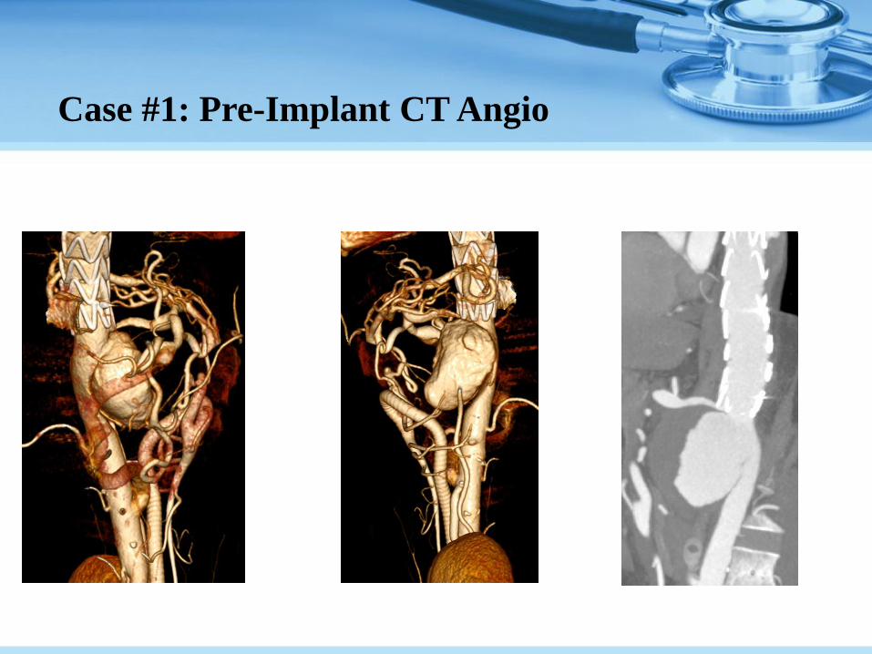
Case #1: Pre-Implant CT Angio

Case #1: Proposed Treatment Solution
A fenestrated stent-graft solution was developed to
maintain celiac trunk perfusion, and exclude pseudo-
aneurysm endovascularly
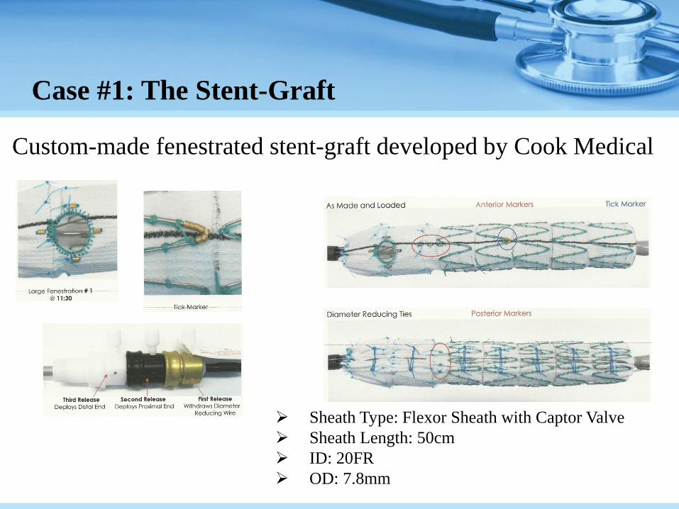
Case #1: The Stent-Graft
Custom-made fenestrated stent-graft developed by Cook Medical
Sheath Type: Flexor Sheath with Captor Valve
Sheath Length: 50cm
ID: 20FR
OD: 7.8mm
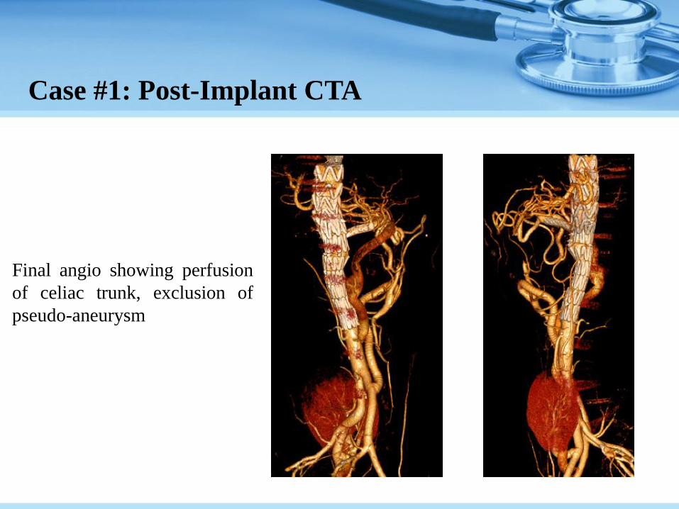
Case #1: Post-Implant CTA
Final angio showing perfusion
of celiac trunk, exclusion of
pseudo-aneurysm
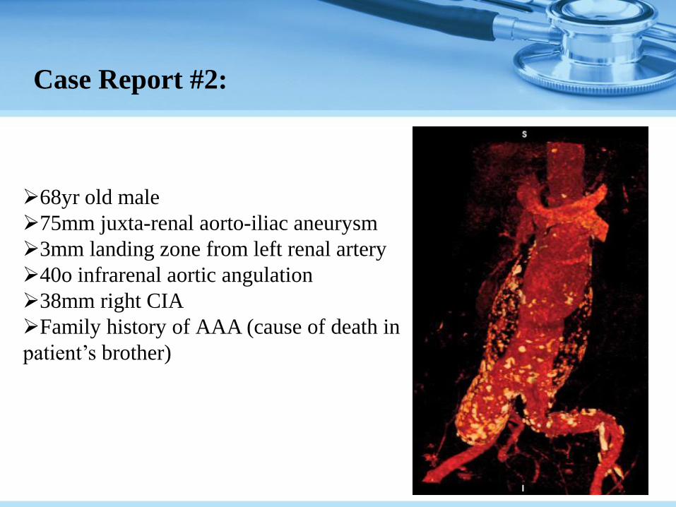
Case Report #2:
68yr old male
75mm juxta-renal aorto-iliac aneurysm
3mm landing zone from left renal artery
40o infrarenal aortic angulation
38mm right CIA
Family history of AAA (cause of death in
patient’s brother)

Case Report #2:

Case #2: Plan
Fenestrated Tube-graft for juxta-renal
aorta
Two V12 covered stents for renal arts.
Iliac branch device for right CIA
One V12 covered stent for right IIA
Bifurcated main body stent graft for
abdominal aorta
Connecting stent between bifurcated
main body and Iliac branch device

Case #2: Cook Custom Made Device

Case #4
75yr old female
54mm aneurysm in aortic arch
(no proximal landing zone to
brachiocephalic artery)
Another 32mm saccular
pseudo-anerysm which was 4cm
distal to arch
Very large innonimate artery
(16mm in diameter)

Case #4: Plan
RCA to LCA to LSA bypass
Ligation of LCA and LSA
Two V12 Chimney stents from
right axillary
One Zenith TX2 tube-graft
placed in Zone 0 (ascending
aorta) to Zone 4 (T5 of
descending aorta)

Case #4: Implant Angio
First angio showing
bypass grafts

Case #5
72yr old male
56mm thoraco -abdominal-aortic-
aneurysm (TAAA)
Symptomatic
History of CABG
On Dialysis
Hypertensive
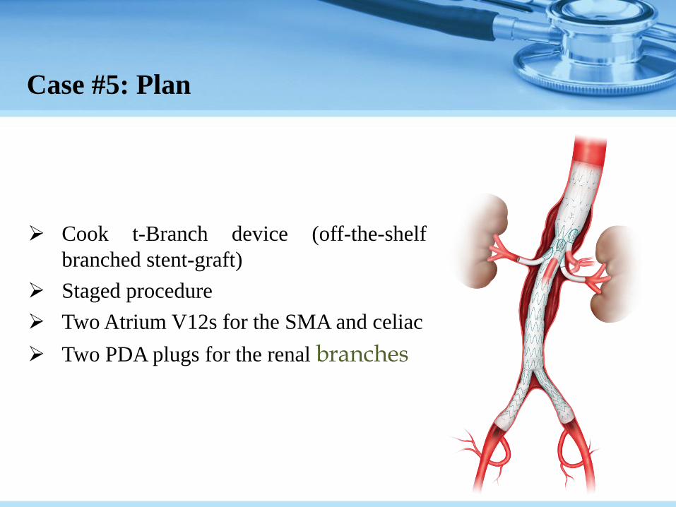
Case #5: Plan
Cook t-Branch device (off-the-shelf
branched stent-graft)
Staged procedure
Two Atrium V12s for the SMA and celiac
Two PDA plugs for the renal branches
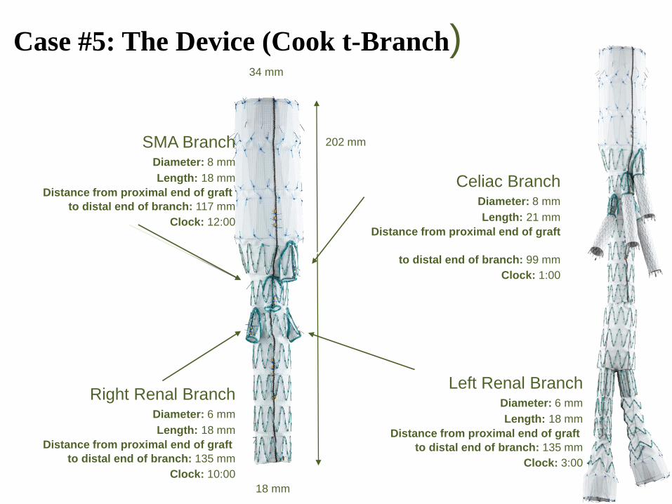
Case #5: The Device (Cook t-Branch) 34 mm
202 mm
18 mm
Celiac Branch Diameter: 8 mm
Length: 21 mm
Distance from proximal end of graft
to distal end of branch: 99 mm
Clock: 1:00
SMA Branch Diameter: 8 mm
Length: 18 mm
Distance from proximal end of graft
to distal end of branch: 117 mm
Clock: 12:00
Left Renal Branch Diameter: 6 mm
Length: 18 mm
Distance from proximal end of graft
to distal end of branch: 135 mm
Clock: 3:00
Right Renal Branch Diameter: 6 mm
Length: 18 mm
Distance from proximal end of graft
to distal end of branch: 135 mm
Clock: 10:00

Case #5: The Steps
Step 1 Step 2 Step 3 Step 4
Insertion and
deployment of the t-
Branch component
(retrograde)
Cannulation and
stenting of the
visceral branche
one-by-one
(antegrade)
Insertion &
Deployment of
distal bifurcated
main body
(retrograde)
Extension into the
common iliacs with
iliac extension
devices (retrograde)

Anesthetic management in
TAVI or proximal graft
Induced hypotension during device deployment may
reduce the magnitude of migration duo to forward
aortic blood flow(reduce occlusion of major art.
banches or incomplete aneurysm exclusion)
Minimal induced hypotension are usually
adequate(fast-acing venous or art. vasodilator

Adenosine induced asystol increase the accuracy of
device positioning.
High dose adenosine induce temporary high degree AV
block and asystol during device deployment.
Temporary external T.T pacing and defibrilation pads
for manage. prolong Av block.
General ,neuroaxial or local anesthesia Rt radial
art,Rt int juglar, PAC.
Sedate with etomidate 0.1-0.2 mg/kg or propofol 1-
mg/kg during angioplasty baloon inflation or aden.
induced asystol
Permanent pacemaker reprogram to prevent capture
during induced asystol.
Adenosine 24mg(48,60,90)
Temorary pace. if asystol exceed 15-20 sec

Venticular fibrilation
Alternative techniqe to induce temporary asystol , higly
invasive technique.A10 sec V.F was induced byA/C
transformer and defib. with 200j shock via the ext.pad
V.F, flat intra art press tracing(several times)
After deflation of angioplasty baloon the pts was
defibril 2ooj

T.E.E
Perioperative dynamic views of the cv sys
Diagnostic aortic pathology.
Location of the guidwires and endografts prior to
deployment.
Exclusion of flow from the aorta into aneurysm by
doppler color flow imaging
TEE probe interfered with fluoroscopic imagin
Color-flow doppler exclude any persistant leaks from
the aorta into the aneurysm or pseudoaneurysem at
the endograft margins.
Confirmed patency of the intercostal artery.
Visualization of the distal ascend aorta and proximal
aortic arc may be limited.

First human implantation: Alain Cribier
April 16, 2002 ( France)
Bovine pericardium valve
23mm in diameter
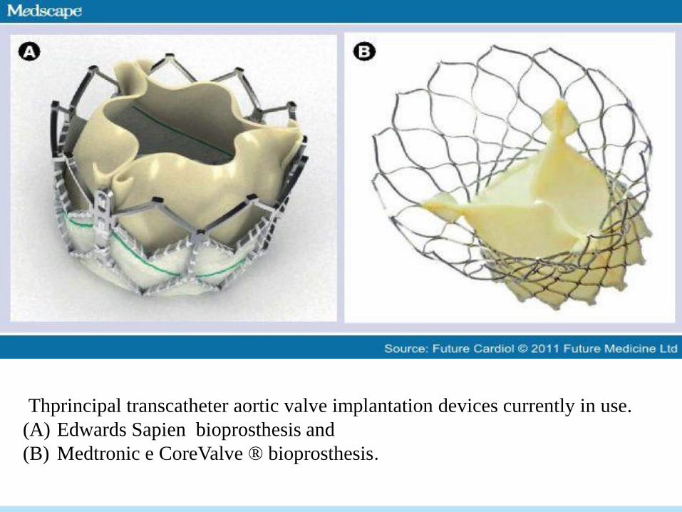
Thprincipal transcatheter aortic valve implantation devices currently in use.
(A) Edwards Sapien bioprosthesis and
(B) Medtronic e CoreValve ® bioprosthesis.


TAVI patient`s Work up
Pre-anesthetic work up.
Cardiothoracic Surgical Evaluation [access, AVR, risk assessment]
Imaging (Pre-procedure) (Echo; CT-Angio ; U/S):
TTE: AS severity, valve morphology , calcification
TEE: Annular size and shape (TEE [2D / 3D]; CT Angio; CMR).
~ LV function (LVEF > 20%).
~ Other valvular disease, sub aortic obstruction; PASP.
CT-Angio Aortic root:
~ annulus to coronary ostia (>8mm), Aortic Atheroma, ca
~ Vascular anatomy from access site to annulus (Ilio-femoral ).
Imaging (post procedure) (Echo … MRI):
~ Degree of AR; MV assessment; new LV RWMA.
~ Cerebrovascular embolism.

TRANSFEMORAL TRANSAPICAL TRANSAORTIC
TRANS SUBCLAVIAN TRANSAXILLARY
TRANSSEPTAL
RETROPERITONEAL
TRANSCAROTID
Are the latest access sites applicable for
TAVI
Vascular access sites


Complications of
endovascular aortic repaire
HYPOTENTION
SPINAL CORD ISCHEMIA
POST IMPLANTATION SYNDROM
Hypotention
Intraoperative aortic rupture (the anesthe team must
be prepared for resusitation)
IV,CVP,ART Press, Inotrope,Vasodilator.
DDX
Sympa. nerve blockade by regional anes.
Acute aortic rupture, endoleak
Allergic reaction
Adenosine(peripheral dilation, Avblock)

Spinal Cord Ischemia During
E.V Taa Repair
The typical level ischemia after TAA is midthoracic
(high perioperative mortality)
Thoracolumbar has multiple art. sourse and
vulnerable to ischemia
Adamkiewics (intercostal arteries t9-t12 in 75%)as
watershed region.
Ischemia after TAAA repair is
variable,asymetric,sensory or motor function

Paraplegia & paraparesis
Immediate onset: lower ext weaknes on emergence from
anestesia within 24 hrs
Delayed onset: follows a normal post op neurrologic
exam after emergence from anes.
Incidence rate:3-4% immediate:63%
Immediate :irreversible
Immediate:infarction, delay:ischemic
Cosequently,strategies to prevent immediate parapl. are
directed toward intra op protection.

Intraoperative S.C monitoring is to detect S.C
ischemia for immediate intervention to improve S.C
perfusion.(SSEP,MEP)
Sterategies to minimise delayed onset:
Prevention of periop.hypotention
Early anest.emergence for early and subsequent serial
neurilogic assessment.
Lumbar CSFdrainage.

Prevention & treatment of delayed
onset S. C. ischemia
MAP>85
Augment SCPP
Increase MAP with vasopressor therapy
CSF drainage
Prevent hypotension
Lumbar CSF drainage
Spinal cord protective strategy for TAAA repair.
Reduction of CSF pressure improves SCPP
Silicon elastomer ventriculostomy catheter,14 gauge
needie at the L3-L4.
Advanced 10-15 cm in to the s. a. space.
CSF is drained when CSF pressure exeeds 10mmhg.

Pressure transduser zero-referenced to the midline of
the brain.
Inserted before or at the time of surgery up to the 24
hrs after surgery.
SCPP:MAP-CSF pressure.
The scpp should be maitained greater
than70mmHg,MAP about 80-100mmHg in TAA
Vasopressor therapy in spinal vasodilatory
shock(significant sympathctomy)

Complication of csf drainage
Neuraxial hematoma
Catheter fracture
Meningitis
Intracranial hypotension
Spinal headache

Decrease the risk for intraop.SC ischemia
Mild systemic hypotermia
Lumbar csf drainage
Epidural cooling(cold saline)
Pharmacologic neuroprotection
Intraop. SSEP, MEP monitoring
Arterial pressure augmentation
Pharmacologic protection
Systemi glucorticoids,manitol,gabapentin
Esmolol,anti infl.agents,intrathecal
(papaverine,mgso4,naloxan)
Erythropoitin(promote the recruitment of bone
marrow mese.stem cells to the site of injured spinal
cord and repair of neurons ,glial cells)
Xenon,inert gas,neuroprotective propert

POST IMPLANTATION SYN
After endovas. Aortic rapair
Fever, elevated CRP level
Leukocytosis in the absence of an infectious agent
Mild , self limited,2-10 days postop
(hyperpyrexia, hypotension, coag).
Occasionally, excessive capillary permeability,
Leakage lead to life threatening intravas.hypov
Res.failure, DIC
(due to sig. inflam. response ,endothe. activation
From intra aneurysmal device manipulation)
Conclusion
Endovascular aortic repair is a minimally
Invasive procedure which may offer many adventage
over open aortic repair.
Succes at least 80-90%
The perioperative mortality is most likely
Less than conventional surgical repaire .

Thank you
Related Documents