The Flexor Compartment Of Forearm

Anatomy of flexor compartment of forearm
Jul 15, 2015
Welcome message from author
This document is posted to help you gain knowledge. Please leave a comment to let me know what you think about it! Share it to your friends and learn new things together.
Transcript

The Flexor Compartment Of Forearm

Neurovascular Pattern of Forearm 3 nerves supply of the forearm and each nerve passes between 2 heads of
muscle Nerve supply of the flexor compartment
The median nerve passes between the 2 heads of pronator teres.
The ulnar nerve passes between the 2 heads of flexor carpi ulnaris. Nerve supply of the extensor compartment
The posterior interosseous nerve (a branch from the radial nerve) passes between the 2 layers of the supinator.
The arterial supply of the forearm comes from the common interosseous branch of the ulnar artery .This divides into the anterior and posterior interosseous arteries.
The anterior (from the median nerve) and posterior (from the radial nerve) interosseous nerves remain in their own compartment and supply muscles, They do not reach skin.
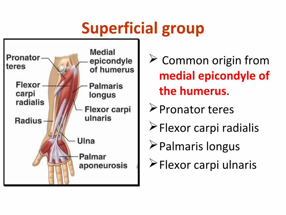
Superficial group
Common origin from medial epicondyle of the humerus.
Pronator teres Flexor carpi radialisPalmaris longus Flexor carpi ulnaris

Pronator Teres Origin by two headsSuperficial head: Medial
epicondyle.Deep Head : Coronoid process of
ulna Insertion: middle of lateral side
of shaft of radius. Innervated by median nerve. Action: Pronates and flexes
forearm.Note : Median nerve lies between its
two heads and the ulnar artery passes deep to the deep head
It forms medial border of cubital fossa.
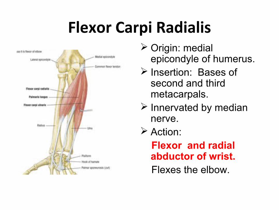
Flexor Carpi Radialis Origin: medial
epicondyle of humerus. Insertion: Bases of
second and third metacarpals.
Innervated by median nerve.
Action: Flexor and radial
abductor of wrist. Flexes the elbow.
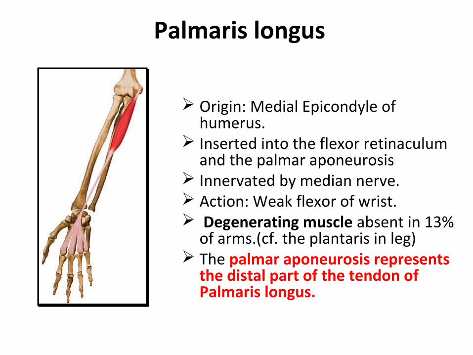
Palmaris longus
Origin: Medial Epicondyle of humerus.
Inserted into the flexor retinaculum and the palmar aponeurosis
Innervated by median nerve. Action: Weak flexor of wrist. Degenerating muscle absent in 13%
of arms.(cf. the plantaris in leg) The palmar aponeurosis represents
the distal part of the tendon of Palmaris longus.
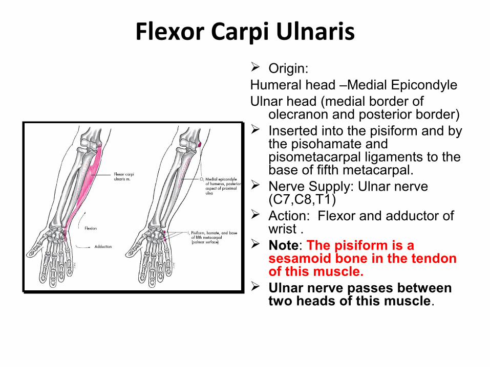
Flexor Carpi Ulnaris Origin:Humeral head –Medial EpicondyleUlnar head (medial border of
olecranon and posterior border) Inserted into the pisiform and by
the pisohamate and pisometacarpal ligaments to the base of fifth metacarpal.
Nerve Supply: Ulnar nerve (C7,C8,T1)
Action: Flexor and adductor of wrist .
Note: The pisiform is a sesamoid bone in the tendon of this muscle.
Ulnar nerve passes between two heads of this muscle.
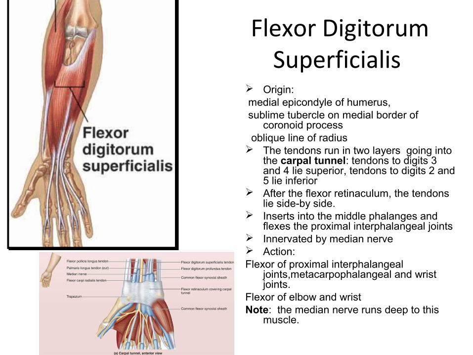
Flexor Digitorum Superficialis
Origin: medial epicondyle of humerus, sublime tubercle on medial border of
coronoid process oblique line of radius The tendons run in two layers going into
the carpal tunnel: tendons to digits 3 and 4 lie superior, tendons to digits 2 and 5 lie inferior
After the flexor retinaculum, the tendons lie side-by side.
Inserts into the middle phalanges and flexes the proximal interphalangeal joints
Innervated by median nerve Action: Flexor of proximal interphalangeal
joints,metacarpophalangeal and wrist joints.
Flexor of elbow and wristNote: the median nerve runs deep to this
muscle.
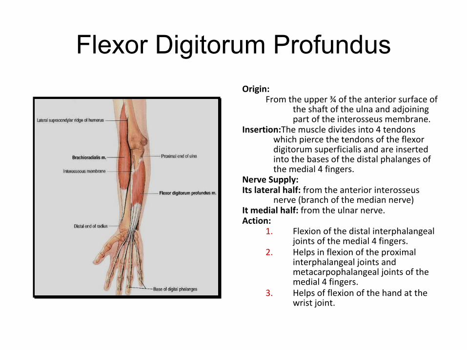
Flexor Digitorum Profundus
Origin:From the upper ¾ of the anterior surface of
the shaft of the ulna and adjoining part of the interosseus membrane.
Insertion:The muscle divides into 4 tendons which pierce the tendons of the flexor digitorum superficialis and are inserted into the bases of the distal phalanges of the medial 4 fingers.
Nerve Supply:Its lateral half: from the anterior interosseus
nerve (branch of the median nerve)It medial half: from the ulnar nerve.Action:
1. Flexion of the distal interphalangeal joints of the medial 4 fingers.
2. Helps in flexion of the proximal interphalangeal joints and metacarpophalangeal joints of the medial 4 fingers.
3. Helps of flexion of the hand at the wrist joint.
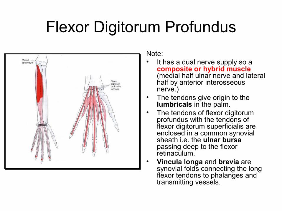
Flexor Digitorum Profundus
Note: • It has a dual nerve supply so a
composite or hybrid muscle (medial half ulnar nerve and lateral half by anterior interosseous nerve.)
• The tendons give origin to the lumbricals in the palm.
• The tendons of flexor digitorum profundus with the tendons of flexor digitorum superficialis are enclosed in a common synovial sheath i.e. the ulnar bursa passing deep to the flexor retinaculum.
• Vincula longa and brevia are synovial folds connecting the long flexor tendons to phalanges and transmitting vessels.

Pronator Quadratus
• Arises from anterior surface of distal ulna.
• Inserted into anterior surface of distal radius.
• Innervated by anterior interosseous nerve
• Action: Pronates the forearm and helps to hold lower ends of radius and ulna together, especially when hand is weight bearing.
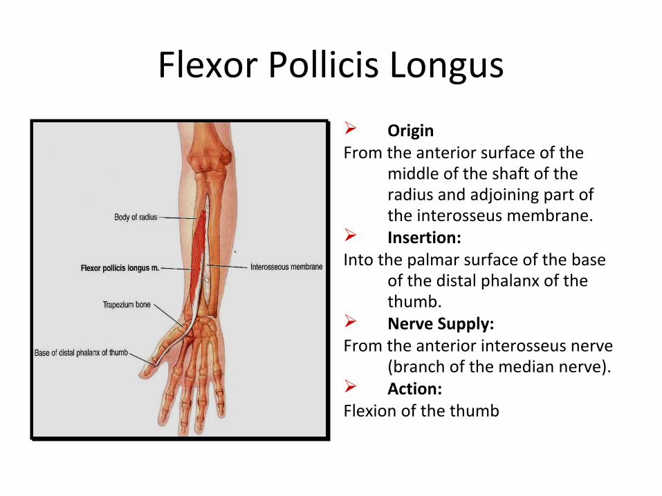
Flexor Pollicis Longus OriginFrom the anterior surface of the
middle of the shaft of the radius and adjoining part of the interosseus membrane.
Insertion:Into the palmar surface of the base
of the distal phalanx of the thumb.
Nerve Supply:From the anterior interosseus nerve
(branch of the median nerve). Action:Flexion of the thumb.

Radial arterySmaller lateral branch of brachial artery in cubital fossa.Runs through the anatomical snuffbox on the surface of scaphoid and trapezium enters palm by passing between two heads of first dorsal interosseous muscle divides into princeps pollicis and deep palmar branch.Radial recurrent artery Superficial palmar branch Anastomoses with superficial branch of ulnar artery to complete the superficial palmar arterial arch.Princeps pollicis arteryDivides into two proper digital arteries for each side of thumb.Radialis indicis arteryDeep palmar ArchIs formed by main termination of radial artery and is usually completed by deep palmar branch of ulnar artery..
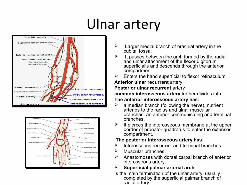
Ulnar artery Larger medial branch of brachial artery in the
cubital fossa. It passes between the arch formed by the radial
and ulnar attachment of the flexor digitorum superficialis and descends through the anterior compartment
Enters the hand superficial to flexor retinaculum.Anterior ulnar recurrent artery Posterior ulnar recurrent arterycommon interosseous artery further divides into The anterior interosseous artery has: a median branch (following the nerve), nutrient
arteries to the radius and ulna, muscular branches, an anterior communicating and terminal branches.
It pierces the interosseous membrane at the upper border of pronator quadratus to enter the extensor compartment.
The posterior interosseous artery has: Interosseous recurrent and terminal branches Muscular branches Anastomoses with dorsal carpal branch of anterior
interosseous artery. Superficial palmar arterial archIs the main termination of the ulnar artery, usually
completed by the superficial palmar branch of radial artery.
The Median Nerve
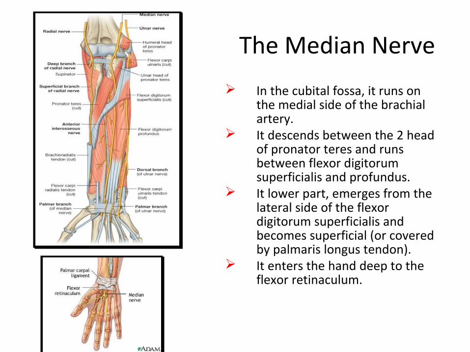
In the cubital fossa, it runs on the medial side of the brachial artery.
It descends between the 2 head of pronator teres and runs between flexor digitorum superficialis and profundus.
It lower part, emerges from the lateral side of the flexor digitorum superficialis and becomes superficial (or covered by palmaris longus tendon).
It enters the hand deep to the flexor retinaculum.

Median Nerve-Branches1. Muscular branches: to
pronator teres, flexor carpi radialis, palmaris longus and flexor digitorum superficialis.
2. Articular branches: to the elbow joint.
3. Anterior interosseus nerve.4. Palmar cutaneous branches:
passes superficial to the flexor retinaculum and supplies skin of the lateral 2/3 of the palm.
5. Enters the palm of hand through the carpal tunnel , gives off a muscular branch (recurrent branch )to the thenar muscles and terminates by dividing into three common palmar digital nerves.

Median Nerve
Ape Hand deformity
Injury to median nerve is cause due to supracondylar fracture of humerus or by compression in the carpal tunnel.It results in loss of pronation, opposion of thumb, flexion of lateral two interphalangeal joints.The thumb is adducted and laterally rotated.It produces characteristic flattening of thenar eminence referred to as Ape thumb.The median nerve enables the hand to grip as it supplies most of thenar muscles. So it is called Labourer’s nerve.
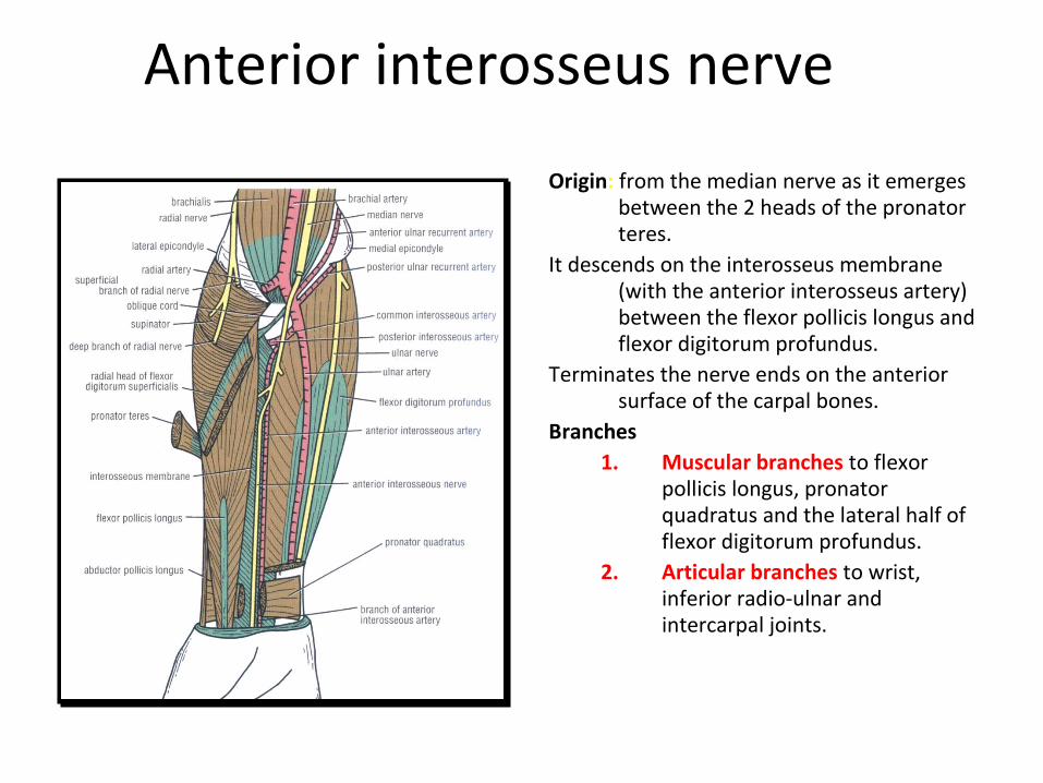
Anterior interosseus nerve
Origin: from the median nerve as it emerges between the 2 heads of the pronator teres.
It descends on the interosseus membrane (with the anterior interosseus artery) between the flexor pollicis longus and flexor digitorum profundus.
Terminates the nerve ends on the anterior surface of the carpal bones.
Branches1. Muscular branches to flexor
pollicis longus, pronator quadratus and the lateral half of flexor digitorum profundus.
2. Articular branches to wrist, inferior radio-ulnar and intercarpal joints.
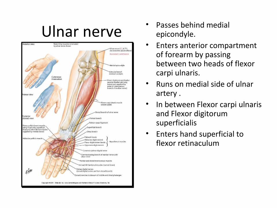
Ulnar nerve • Passes behind medial epicondyle.
• Enters anterior compartment of forearm by passing between two heads of flexor carpi ulnaris.
• Runs on medial side of ulnar artery .
• In between Flexor carpi ulnaris and Flexor digitorum superficialis
• Enters hand superficial to flexor retinaculum
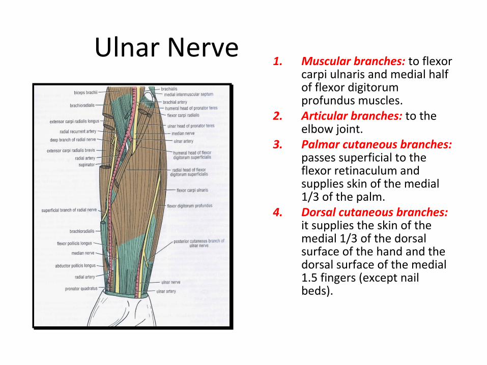
Ulnar Nerve1. Muscular branches: to flexor
carpi ulnaris and medial half of flexor digitorum profundus muscles.
2. Articular branches: to the elbow joint.
3. Palmar cutaneous branches: passes superficial to the flexor retinaculum and supplies skin of the medial 1/3 of the palm.
4. Dorsal cutaneous branches: it supplies the skin of the medial 1/3 of the dorsal surface of the hand and the dorsal surface of the medial 1.5 fingers (except nail beds).

Ulnar Nerve Injury • Injury to ulnar nerve at elbow caused by a
fracture dislocation of elbow results in a Claw Hand in which ring and little fingers are hyperextended at metacarpophalangeal joints and flexed at interphalangeal joints.
• It results in loss of abduction and adduction of fingers and flexion of metacarpophalangeal joints (due to paralysis of palmar and dorsal interossei and medial two lumbricals)
• It produces a wasted hypothenar eminence and loss of adduction of thumb.
• Injury to ulnar nerve at wrist due to superficial cuts or injuries at wrist causes paralysis of hypothenar muscles, all interossei, adductor pollicis and medial two lumbricals. The ulnar claw hand is more marked than at the level of elbow, since flexor digitorum profundus is spared.
• The ulnar nerve is responsible for intricate movements of the hand as it supplies most of the small intrinsic muscles of hand. So it is called Musician’s nerve.
Related Documents