Spring 2016 Dr. Maher Hadidi, University of Jordan 1 Upper Limb Lecture 6 Anterior compartment of the Forearm

Welcome message from author
This document is posted to help you gain knowledge. Please leave a comment to let me know what you think about it! Share it to your friends and learn new things together.
Transcript
Spring 2016 Dr. Maher Hadidi, University of Jordan 1
Upper LimbLecture 6
Anterior compartment of the Forearm
Spring 2016 Dr. Maher Hadidi, University of Jordan 2
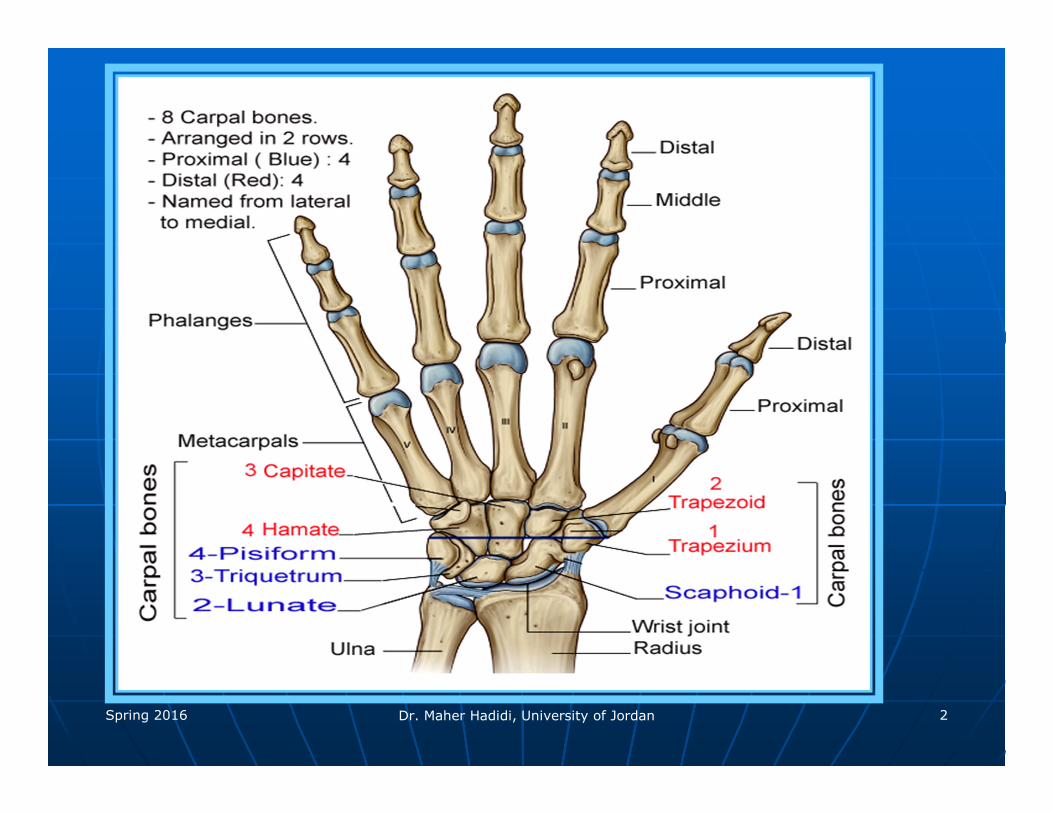
Compartments of the forearm
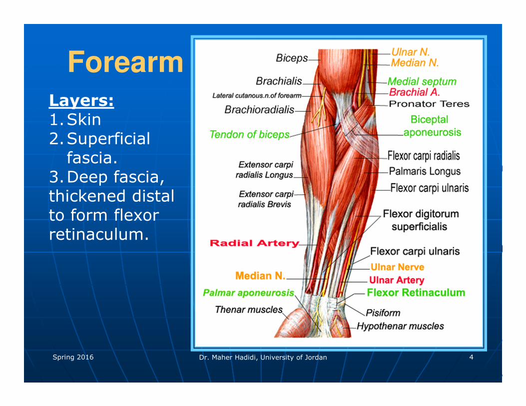
Spring 2016 Dr. Maher Hadidi, University of Jordan 4
ForearmLayers:1.Skin2.Superficial
fascia.3.Deep fascia,thickened distalto form flexorretinaculum.
Spring 2016 Dr. Maher Hadidi, University of Jordan 5
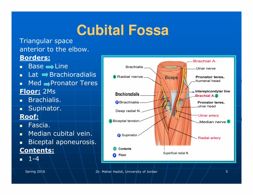
Cubital FossaTriangular space
anterior to the elbow.
Borders:
� Base Line
� Lat Brachioradialis
� Med Pronator Teres
Floor: 2Ms
� Brachialis.
� Supinator.
Roof:
� Fascia.
� Median cubital vein.
� Biceptal aponeurosis.
Contents:
� 1-4

Spring 2016 Dr. Maher Hadidi, University of Jordan 6
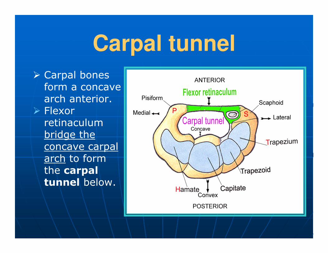
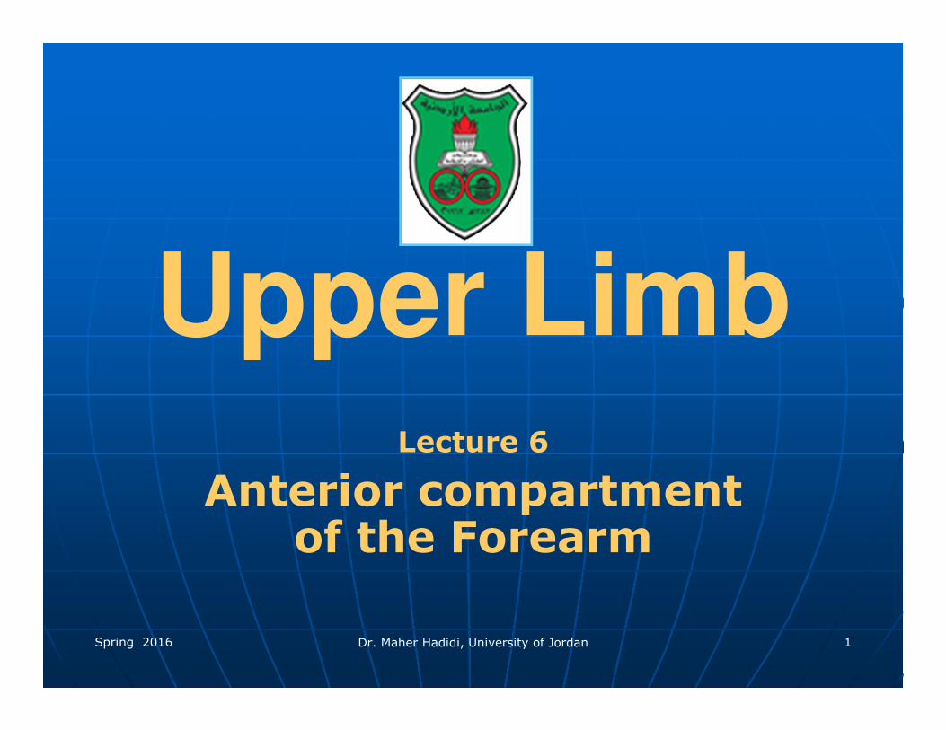
Carpal tunnel
� Carpal bones form a concave arch anterior.
� Flexor retinaculum bridge the concave carpal arch to form the carpal tunnel below.
Spring 2016 Dr. Maher Hadidi, University of Jordan 8
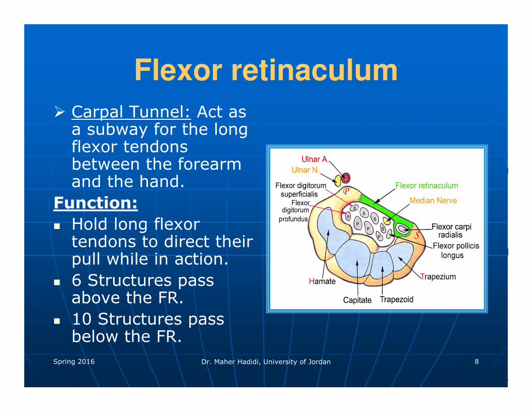
Flexor retinaculum
� Carpal Tunnel: Act as a subway for the long flexor tendons between the forearm and the hand.
Function:
� Hold long flexor tendons to direct their pull while in action.
� 6 Structures pass above the FR.
� 10 Structures pass below the FR.
Spring 2016 Dr. Maher Hadidi, University of Jordan 9
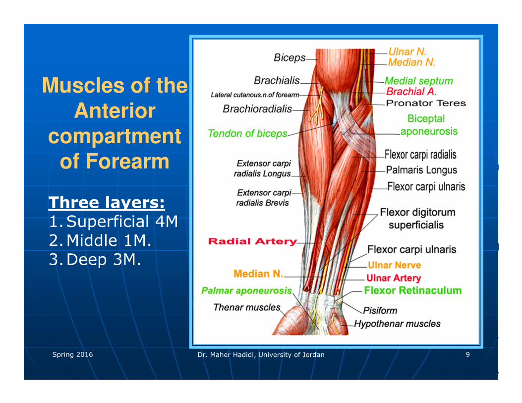
Muscles of the Anterior
compartment of Forearm
Three layers:1.Superficial 4M2.Middle 1M. 3.Deep 3M.
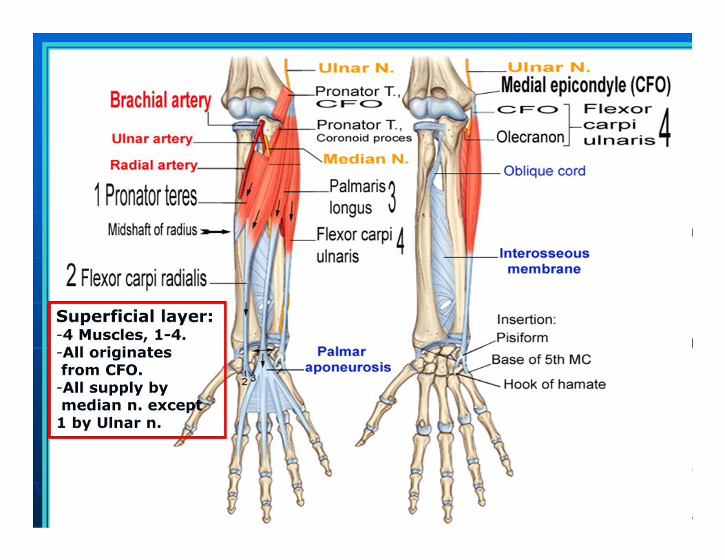
Superficial layer:-4 Muscles, 1-4.-All originates from CFO.-All supply bymedian n. except
1 by Ulnar n.
Spring 2016 Dr. Maher Hadidi, University of Jordan 11
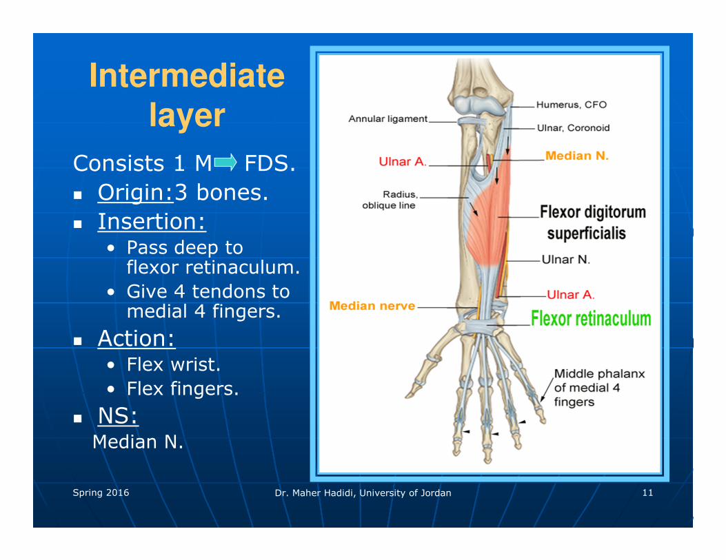
Intermediate
layer
Consists 1 M FDS.
� Origin:3 bones.
� Insertion:• Pass deep to
flexor retinaculum.
• Give 4 tendons to medial 4 fingers.
� Action:• Flex wrist.
• Flex fingers.
� NS:Median N.

Spring 2016 Dr. Maher Hadidi, University of Jordan 12
Spring 2016 Dr. Maher Hadidi, University of Jordan 13
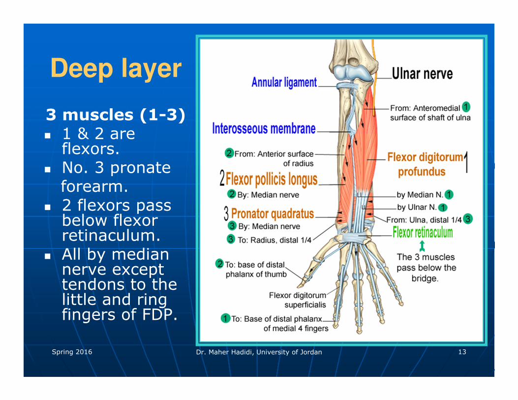
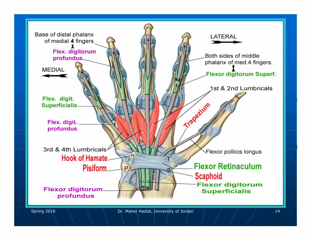
Deep layer
3 muscles (1-3)� 1 & 2 are
flexors.� No. 3 pronate
forearm.� 2 flexors pass
below flexor retinaculum.
� All by median nerve except tendons to the little and ring fingers of FDP.
Spring 2016 Dr. Maher Hadidi, University of Jordan 14
Spring 2016 Dr. Maher Hadidi, University of Jordan 15
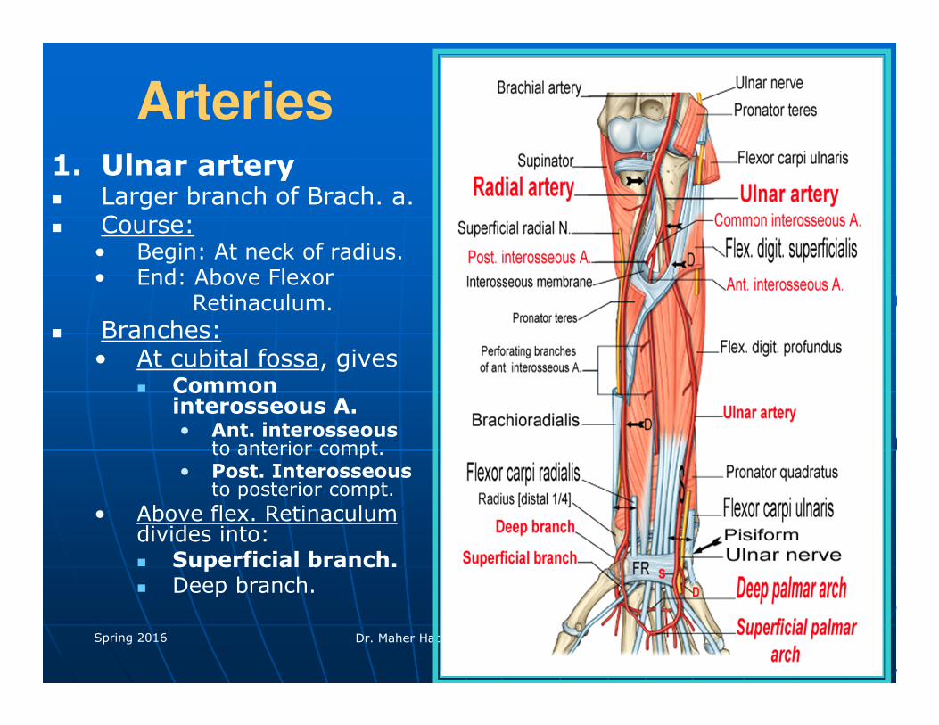
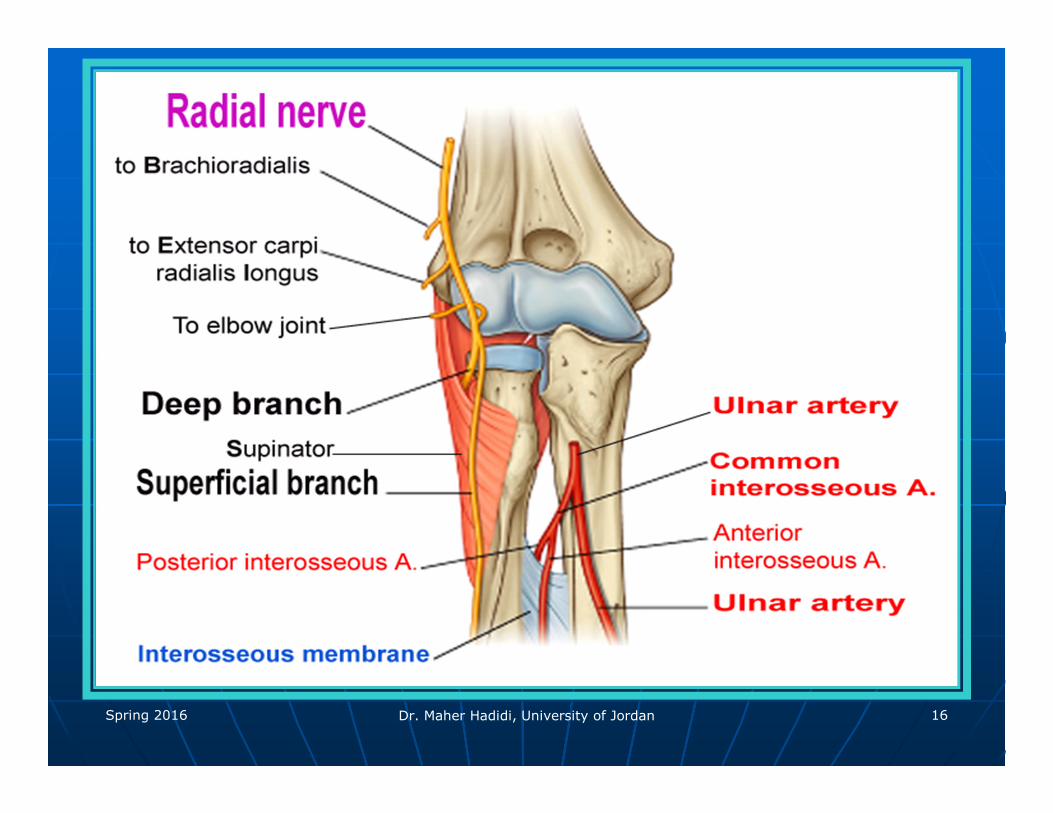
Arteries1. Ulnar artery� Larger branch of Brach. a.� Course:
• Begin: At neck of radius.• End: Above Flexor
Retinaculum.
� Branches:• At cubital fossa, gives
� Common interosseous A.• Ant. interosseous
to anterior compt.• Post. Interosseous
to posterior compt.
• Above flex. Retinaculumdivides into:� Superficial branch. � Deep branch.
Spring 2016 Dr. Maher Hadidi, University of Jordan 16
Spring 2016 Dr. Maher Hadidi, University of Jordan 17
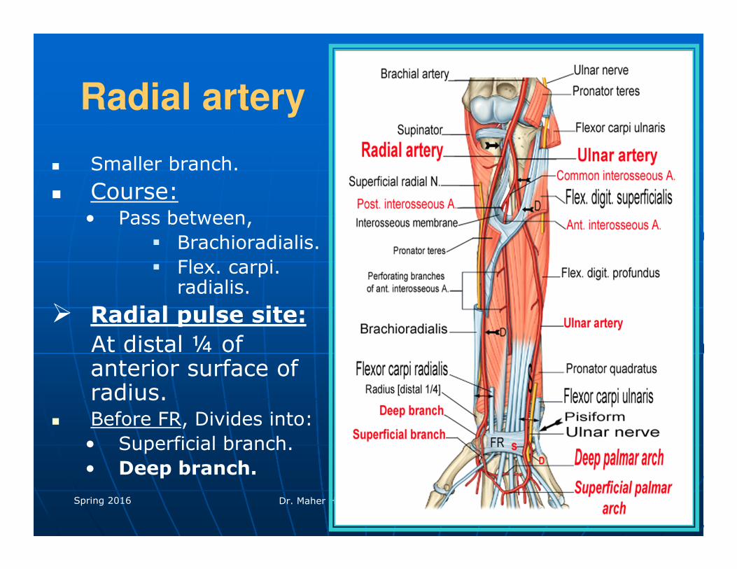
Radial artery
� Smaller branch.
� Course:• Pass between,
� Brachioradialis.
� Flex. carpi. radialis.
� Radial pulse site:
At distal ¼ of anterior surface of radius.
� Before FR, Divides into:
• Superficial branch.
• Deep branch.
Spring 2016 Dr. Maher Hadidi, University of Jordan 18
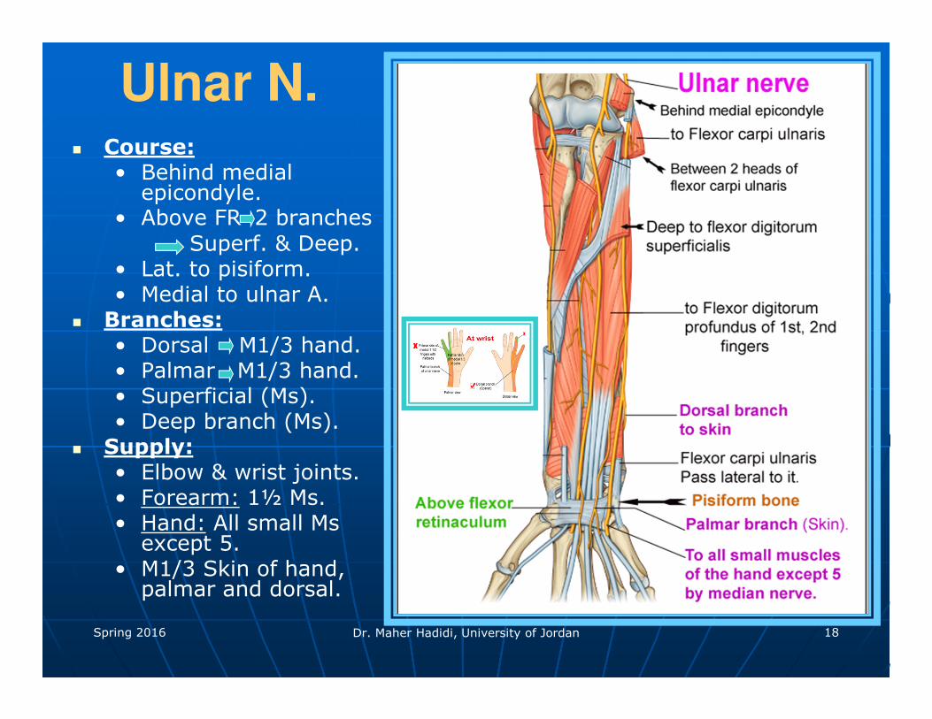
Ulnar N.� Course:
• Behind medial epicondyle.
• Above FR 2 branches
Superf. & Deep.• Lat. to pisiform.• Medial to ulnar A.
� Branches:• Dorsal M1/3 hand.• Palmar M1/3 hand.• Superficial (Ms).• Deep branch (Ms).
� Supply:• Elbow & wrist joints. • Forearm: 1½ Ms.• Hand: All small Ms
except 5.• M1/3 Skin of hand,
palmar and dorsal.
Spring 2016 Dr. Maher Hadidi, University of Jordan 19
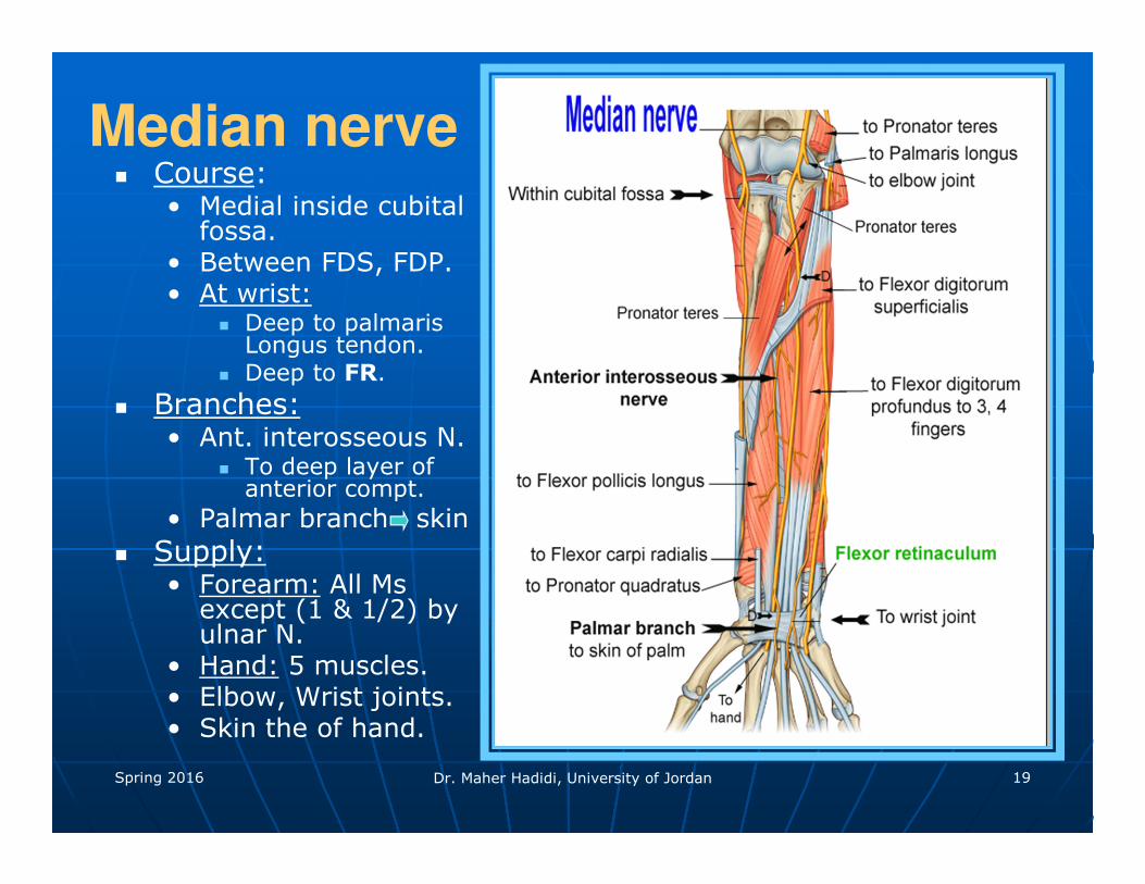
Median nerve� Course:
• Medial inside cubital fossa.
• Between FDS, FDP.• At wrist:
� Deep to palmaris Longus tendon.
� Deep to FR.
� Branches:• Ant. interosseous N.
� To deep layer of anterior compt.
• Palmar branch skin
� Supply:• Forearm: All Ms
except (1 & 1/2) by ulnar N.
• Hand: 5 muscles.• Elbow, Wrist joints.• Skin the of hand.
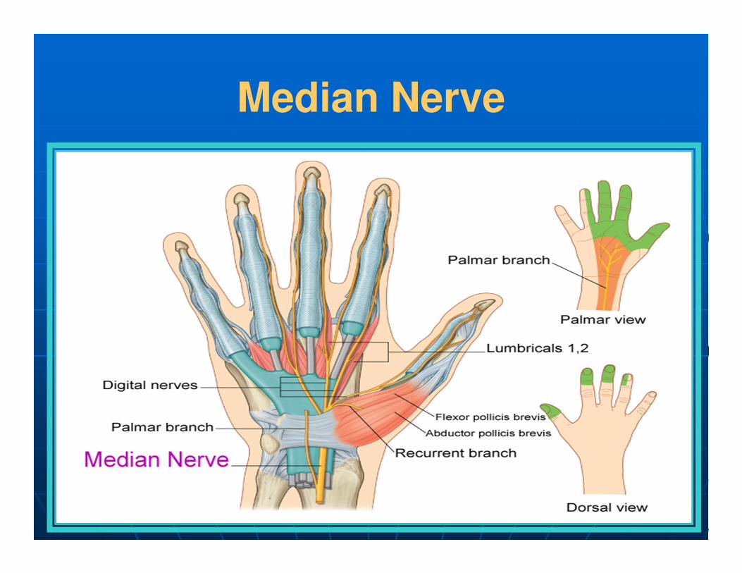
Median Nerve
Related Documents