Analysis of mTOR signaling by the small G-proteins, Rheb and RhebL1 Andrew R. Tee a, * , John Blenis b , Christopher G. Proud a a University of Dundee, Medical Sciences Institute/Wellcome Building Complex, Dow Street, Dundee DD1 5EH, UK b Department of Cell Biology, Harvard Medical School, 240 Longwood Avenue, Boston, MA 02115, USA Received 10 March 2005; revised 28 June 2005; accepted 20 July 2005 Available online 11 August 2005 Edited by Berend Wieringa Abstract The small G protein Rheb (Ras homologue enriched in brain) is known to promote mammalian target of rapamycin (mTOR) signaling. In this study, we show that Rheb like-1 protein (RhebL1) rescues mTOR signaling during nutrient withdrawal and that tuberous sclerosis complex-1 (TSC) and TSC2 impairs RhebL1-mediated signaling through mTOR. We identify critical residues within the switch I region (N41) and ÔconstitutiveÕ effector (Ec) region (Y/F54 and L56) of Rheb and RhebL1, which are required for their efficient activation of mTOR signaling. Mutation of Rheb and RhebL1 at N41 impaired their interaction with mTOR, which identifies mTOR as a common downstream target of both Rheb and RhebL1. Ó 2005 Federation of European Biochemical Societies. Published by Elsevier B.V. All rights reserved. Keywords: Rheb; mTOR; TSC2; S6K1; 4E-BP1; RhebL1 1. Introduction Rheb (Ras homologue enriched in brain) is a small G-pro- tein that is related to the Ras, Rap, and Ral subfamilies (for review see [1]). Rheb acts as a molecular switch within cells that promotes signal transduction through mammalian target of rapamycin (mTOR) (see review [2]). Tuberous sclerosis complex-2 (TSC2) functions as a GTPase activating protein (GAP) that enhances the intrinsic GTPase activity of Rheb and inhibits mTOR signaling. This suggests that Rheb only activates mTOR when bound to GTP. When TSC2 is bound to TSC1 this RhebGAP activity is significantly increased [3]. Loss of function mutations of either TSC1 or TSC2 results in a human disorder termed the tuberous sclerosis complex. Tuberous sclerosis patients characteristically develop benign hamartomatous tumors in the brain, kidneys, heart, lungs, skin or eyes. The severity of the clinical symptoms is dependent on the size and location of the tumor. Brain hamartomas can re- sult in neurological disorders such as seizures, mental retarda- tion and autism (for review see [4]). Rheb is highly homologous to H-Ras (37% identical and 60% similar). There are two regions (referred to as switch I and switch II) within Ras whose structures change markedly when Ras switches from a GTP to a GDP-bound state (re- viewed in [5]). The downstream effectors of Ras, i.e., Raf-ki- nase, Ral-GDS and phosphoinositide-3-kinase (PI3K), bind to the switch I region when Ras is bound to GTP. The Ôconsti- tutiveÕ effector (Ec) region is also required for the biological function of Ras and lies between the switch I and II regions [6]. To further characterize Rheb and Rheb like 1 (RhebL1) proteins, we introduced mutations into their switch I and Ec regions. 2. Materials and methods 2.1. Chemicals and materials Wortmannin and rapamycin were purchased from Biomol Research Laboratories, Inc. and Merck Biosciences Ltd., respectively. Insulin, anti-Flag antibodies (M2), and MG132 were bought from Sigma while radio-labeled reagents and m 7 GTP-Sepharose were purchased from Amersham BioSciences. Anti-4E-BP1 and -Akt antibodies were sup- plied by Cell Signaling Technology. Anti-HA antibodies were provided by M. Chou (University of Pennsylvania, Philadelphia, PA). Anti-AU1 antibody was purchased from Covance. Anti-mTAB1 antibodies were kindly provided by Professor Dick Denton (University of Bristol, UK). All other reagents were obtained from VWR Scientific. 2.2. Molecular biology and plasmids Human RhebL1 cDNA (bought from ATCC, I.M.A.G.E. Clone ID:5933910) was subcloned into pRK7 and pEBG-2T with an N-ter- minal Flag-tag and GST-tag, respectively. Flag-tagged Rheb was sub- cloned into pcDNA3.1 from pRK7 using the BamHI and EcoRI sites. The Rheb point mutants were generated by site-directed mutagenesis (QuikChange; Stratagene). pACTAG2 expressing human HA-tagged 4E-BP1 was kindly provided by N. Sonenberg (McGill University, Montreal, Canada). HA-tagged S6K1/pRK7 was generated as previ- ously described [7]. 2.3. Tissue culture and analysis of cell lysates Cell transfection, generation of cell lysates and Western blot analysis were carried out as previously described [8]. eIF4E was purified using affinity chromatography on m 7 GTP-Sepharose as described [9]. S6K1 kinase activity assays were determined in vitro by using recombinant GST-S6 as a substrate, as previously described [9]. To reduce the basal activity of S6K1 and to increase the sensitivity by which Rheb and RhebL1 induced S6K1 activation, HEK293 cells were nutrient de- prived for 1 h in D-PBS. 2.4. Rheb nucleotide binding To analyze GTP and GDP loading of Rheb in vivo, cells (grown on 10 cm 2 plates) were incubated in 5 ml of phosphate-free medium con- taining 0.5 mCi [ 32 P]orthophosphate for 3 h. These cells were then har- vested in buffer A (50 mM HEPES (pH 7.4), 100 mM NaCl, 10 mM MgCl 2 , 1 mg/ml BSA, 1 mM DTT, 1% Trition). Flag-tagged Rheb Abbreviations: Ec, constitutive effector; DMEM, DulbeccoÕs modified Eagle medium; 4E-BP1, eIF4E-binding protein 1; GAP, GTPase act- ivating protein; HEK, human embryonic kidney; mTOR, mammalian target of rapamycin; PI3K, phosphoinositide-3-kinase; Rheb, Ras homologue enriched in brain; RhebL1, Rheb like-1 protein; S6K1, ribosomal protein S6 kinase 1; TSC, tuberous sclerosis complex * Corresponding author. Fax: +44 1382 34 5507. E-mail address: [email protected] (A.R. Tee). 0014-5793/$30.00 Ó 2005 Federation of European Biochemical Societies. Published by Elsevier B.V. All rights reserved. doi:10.1016/j.febslet.2005.07.054 FEBS 29851 FEBS Letters 579 (2005) 4763–4768

Welcome message from author
This document is posted to help you gain knowledge. Please leave a comment to let me know what you think about it! Share it to your friends and learn new things together.
Transcript

FEBS 29851 FEBS Letters 579 (2005) 4763–4768
Analysis of mTOR signaling by the small G-proteins, Rheb and RhebL1
Andrew R. Teea,*, John Blenisb, Christopher G. Prouda
a University of Dundee, Medical Sciences Institute/Wellcome Building Complex, Dow Street, Dundee DD1 5EH, UKb Department of Cell Biology, Harvard Medical School, 240 Longwood Avenue, Boston, MA 02115, USA
Received 10 March 2005; revised 28 June 2005; accepted 20 July 2005
Available online 11 August 2005
Edited by Berend Wieringa
Abstract The small G protein Rheb (Ras homologue enrichedin brain) is known to promote mammalian target of rapamycin(mTOR) signaling. In this study, we show that Rheb like-1protein (RhebL1) rescues mTOR signaling during nutrientwithdrawal and that tuberous sclerosis complex-1 (TSC) andTSC2 impairs RhebL1-mediated signaling through mTOR.We identify critical residues within the switch I region (N41)and �constitutive� effector (Ec) region (Y/F54 and L56) of Rheband RhebL1, which are required for their efficient activationof mTOR signaling. Mutation of Rheb and RhebL1 at N41impaired their interaction with mTOR, which identifies mTORas a common downstream target of both Rheb and RhebL1.� 2005 Federation of European Biochemical Societies. Publishedby Elsevier B.V. All rights reserved.
Keywords: Rheb; mTOR; TSC2; S6K1; 4E-BP1; RhebL1
1. Introduction
Rheb (Ras homologue enriched in brain) is a small G-pro-
tein that is related to the Ras, Rap, and Ral subfamilies (for
review see [1]). Rheb acts as a molecular switch within cells
that promotes signal transduction through mammalian target
of rapamycin (mTOR) (see review [2]). Tuberous sclerosis
complex-2 (TSC2) functions as a GTPase activating protein
(GAP) that enhances the intrinsic GTPase activity of Rheb
and inhibits mTOR signaling. This suggests that Rheb only
activates mTOR when bound to GTP. When TSC2 is bound
to TSC1 this RhebGAP activity is significantly increased [3].
Loss of function mutations of either TSC1 or TSC2 results
in a human disorder termed the tuberous sclerosis complex.
Tuberous sclerosis patients characteristically develop benign
hamartomatous tumors in the brain, kidneys, heart, lungs, skin
or eyes. The severity of the clinical symptoms is dependent on
the size and location of the tumor. Brain hamartomas can re-
sult in neurological disorders such as seizures, mental retarda-
tion and autism (for review see [4]).
Abbreviations: Ec, constitutive effector; DMEM, Dulbecco�s modifiedEagle medium; 4E-BP1, eIF4E-binding protein 1; GAP, GTPase act-ivating protein; HEK, human embryonic kidney; mTOR, mammaliantarget of rapamycin; PI3K, phosphoinositide-3-kinase; Rheb, Rashomologue enriched in brain; RhebL1, Rheb like-1 protein;S6K1, ribosomal protein S6 kinase 1; TSC, tuberous sclerosis complex
*Corresponding author. Fax: +44 1382 34 5507.E-mail address: [email protected] (A.R. Tee).
0014-5793/$30.00 � 2005 Federation of European Biochemical Societies. Pu
doi:10.1016/j.febslet.2005.07.054
Rheb is highly homologous to H-Ras (37% identical and
60% similar). There are two regions (referred to as switch I
and switch II) within Ras whose structures change markedly
when Ras switches from a GTP to a GDP-bound state (re-
viewed in [5]). The downstream effectors of Ras, i.e., Raf-ki-
nase, Ral-GDS and phosphoinositide-3-kinase (PI3K), bind
to the switch I region when Ras is bound to GTP. The �consti-tutive� effector (Ec) region is also required for the biological
function of Ras and lies between the switch I and II regions
[6]. To further characterize Rheb and Rheb like 1 (RhebL1)
proteins, we introduced mutations into their switch I and Ec
regions.
2. Materials and methods
2.1. Chemicals and materialsWortmannin and rapamycin were purchased from Biomol Research
Laboratories, Inc. and Merck Biosciences Ltd., respectively. Insulin,anti-Flag antibodies (M2), and MG132 were bought from Sigma whileradio-labeled reagents and m7GTP-Sepharose were purchased fromAmersham BioSciences. Anti-4E-BP1 and -Akt antibodies were sup-plied by Cell Signaling Technology. Anti-HA antibodies were providedby M. Chou (University of Pennsylvania, Philadelphia, PA). Anti-AU1antibody was purchased from Covance. Anti-mTAB1 antibodies werekindly provided by Professor Dick Denton (University of Bristol, UK).All other reagents were obtained from VWR Scientific.
2.2. Molecular biology and plasmidsHuman RhebL1 cDNA (bought from ATCC, I.M.A.G.E. Clone
ID:5933910) was subcloned into pRK7 and pEBG-2T with an N-ter-minal Flag-tag and GST-tag, respectively. Flag-tagged Rheb was sub-cloned into pcDNA3.1 from pRK7 using the BamHI and EcoRI sites.The Rheb point mutants were generated by site-directed mutagenesis(QuikChange; Stratagene). pACTAG2 expressing human HA-tagged4E-BP1 was kindly provided by N. Sonenberg (McGill University,Montreal, Canada). HA-tagged S6K1/pRK7 was generated as previ-ously described [7].
2.3. Tissue culture and analysis of cell lysatesCell transfection, generation of cell lysates and Western blot analysis
were carried out as previously described [8]. eIF4E was purified usingaffinity chromatography on m7GTP-Sepharose as described [9]. S6K1kinase activity assays were determined in vitro by using recombinantGST-S6 as a substrate, as previously described [9]. To reduce the basalactivity of S6K1 and to increase the sensitivity by which Rheb andRhebL1 induced S6K1 activation, HEK293 cells were nutrient de-prived for 1 h in D-PBS.
2.4. Rheb nucleotide bindingTo analyze GTP and GDP loading of Rheb in vivo, cells (grown on
10 cm2 plates) were incubated in 5 ml of phosphate-free medium con-taining 0.5 mCi [32P]orthophosphate for 3 h. These cells were then har-vested in buffer A (50 mM HEPES (pH 7.4), 100 mM NaCl, 10 mMMgCl2, 1 mg/ml BSA, 1 mM DTT, 1% Trition). Flag-tagged Rheb
blished by Elsevier B.V. All rights reserved.

4764 A.R. Tee et al. / FEBS Letters 579 (2005) 4763–4768
was immunoprecipitated for 1 h with anti-Flag antibodies bound toprotein G-Sepharose. Immunoprecipitates were washed twice eachwith both buffer A and buffer B (50 mM HEPES (pH 7.4), 100 mMNaCl, 10 mM MgCl2, 0.1% Triton) in the presence of protease inhib-itors. [32P]-labeled GTP and GDP were eluted from Rheb using 20 llRheb elution buffer (0.5 mM GDP, 0.5 mM GTP, 5 mM DTT,5 mM EDTA, 0.2% SDS) at 68 �C for 20 min and then resolved by thinlayer chromatography on polyethyleneimine cellulose (Sigma) withKH2PO4. [
32P]GTP and [32P]GDP levels were quantified with a phos-phoimager.
2.5. Analysis of mTOR association to Rheb and RhebL1HEK293 cells over-expressing either Flag-Rheb or Flag-RhebL1
with or without AU1-mTOR were harvested in 20 mM Tris–HCl(pH 7.5), 5 mM EGTA, 10 mM MgCl2, 25 mM NaCl, 25 mM b-glyc-erophosphate, 0.2% CHAPS (Calbiochem). Rheb and RhebL1 wereimmunoprecipitated for 2 h with anti-Flag antibodies bound to proteinG-Sepharose. Immunoprecipitates were washed three times in extrac-tion buffer and then subjected to SDS–PAGE and Western blot anal-ysis for detection of mTOR, Rheb and RhebL1. mTOR was detectedusing either anti-AU1 and mTAB1 antibodies.
2.6. ReproducibilityImage/J software (available at rsb.info.nih.gov/ij/) was used for
quantification where indicated. These ratios are arbitrary and do notindicate true stoichiometries. All experiments were repeated at leastthree times, with similar outcomes. In the case of Western blots, datafrom a typical experiment is shown.
Fig. 1. Characterization of RhebL1. (A) Serum starved HEK293 cells expreand stimulated with 100 nM insulin for 30 min, where indicated. S6K1 kinas4E-BP1 with RhebL1 were serum-starved. Cells were pre-treated with 25 nM100 nM insulin for 30 min, where indicated. Phosphorylation of 4E-BP1 at Sewere subjected to affinity chromatography on m7GTP-Sepharose and prodetermined. (C) Cells expressing two different levels of Rheb or RhebL1 (lowFlag-TSC1 and Flag-TSC2 were nutrient deprived. The total levels of TSC1,measured. (D) Serum-starved HEK293 cells expressing GST-RhebL1 in thein vivo radiolabeling and the level of bound guanine nucleotide was determin1 h prior to being harvested. (E) Sequence alignment of the switch I and II, a(identical (black) and conserved (shadowed)). Rheb mutations used in this s
3. Results
3.1. RhebL1 activates mTOR-dependant signaling
To determine whether RhebL1 promotes mTOR signaling,
we over-expressed RhebL1 in HEK293 cells. Indeed, RhebL1
significantly increased the basal and insulin-stimulated activity
of S6K1, even in the absence of nutrients (Fig. 1A). Further-
more, RhebL1 potently enhanced the phosphorylation of
4E-BP1 in serum-starved cells, as indicated by increased phos-
phorylation of Ser65 (Fig. 1B). Wortmannin did not block
RhebL1-induced 4E-BP1 phosphorylation (Fig. 1B), which
suggests that PI3K signaling is not involved. In contrast, inhi-
bition of mTOR with rapamycin blocked RhebL1-induced
4E-BP1 phosphorylation. Furthermore, RhebL1 expression
promoted the release of 4E-BP1 from eIF4E, which was
blocked by rapamycin (Fig. 1B). These data imply that
RhebL1 functions upstream of mTOR. To compare the po-
tency by which Rheb and RhebL1 induces mTOR signaling,
we expressed these proteins at high and low levels. We ob-
served that RhebL1 did not activate S6K1 as potently as Rheb
(compare lane 8 with lane 4 (Fig. 1C)). Over-expression of
TSC1 and TSC2 was sufficient to block RhebL1-induced
S6K1 activation. Interestingly, TSC1/2 expression significantly
reduced the protein levels of RhebL1 suggesting that TSC1/2
ssing S6K1 and RhebL1 (�DMEM�) where nutrient deprived (�D-PBS�)e assays were carried out. (B) HEK293 cells co-expressing HA-taggedrapamycin or 100 nMWortmannin for 30 min and then stimulated withr65 and Akt phosphorylation on Ser473 where determined. Cell extractstein levels of both endogenous eIF4E and HA-tagged 4E-BP1 were�+� and high �++�) with HA-tagged S6K1 in the presence or absence ofTSC2, Rheb, RhebL1 and S6K1 are shown and the S6K1 activity waspresence or absence of Flag-TSC1 and Flag-TSC2 were subjected toed as described in Section 2. Cells were treated with 50 lM MG132 fornd constitutive effector regions of Rheb, RhebL1 and H-Ras is showntudy are shown �\� (alanine substitutions are indicated with a ���).

A.R. Tee et al. / FEBS Letters 579 (2005) 4763–4768 4765
heterodimers may reduce the protein stability of RhebL1. To
examine whether TSC1/2 could function as a GAP towards
RhebL1, we over-expressed GST-RhebL1 with or without
TSC1/2 (Fig. 1D). The proteosomal inhibitor, MG132, was
supplemented to the media to prevent proteosomal-mediated
degradation of RhebL1. We observed that over-expression of
TSC1/2 increased the amount of RhebL1 bound to GDP
suggesting that TSC1/2 functioned as a RhebL1GAP.
Since Rheb and RhebL1 both promote mTOR signaling in a
nutrient and rapamycin sensitive manner, they likely interact
with a common downstream effector. The switch I region of
Ras interacts with an array of downstream effectors, that in-
cludes Raf, RalGDS, and PI3K (discussed in [10]). The amino
acid sequence alignment of Rheb, RhebL1 and H-Ras revealed
that their switch I region are highly conserved (Fig. 1E). It is
likely that residues within the switch I region of Rheb and
RhebL1 that are not conserved in H-Ras may mediate their
signaling through mTOR.
3.2. Rheb signaling through mTOR is impaired by the T38M
mutation within its Switch I region
Ras binding toRaf, RalGDS, and PI3Kdownstream effectors
can be individually blocked by switch I T35M, E37G, andY40C
mutations, respectively (discussed in [10]). Based on the close
similarity between the switch I regions of Ras and Rheb (see
Fig. 1E), we generated analogous Rheb switch I mutants
(T38M, E40G, and F43C). The switch I Rheb mutants were still
able to promote 4E-BP1 phosphorylation (Fig. 2A), and S6K1
activation (Fig. 2B) under amino acid starved conditions. Inter-
estingly, theRheb(T38M)mutantwas less efficient in promoting
mTOR signaling when compared wild-type Rheb.
Fig. 2. Characterization of switch I region mutants of Rheb. HEK293cells co-expressing HA-4E-BP1 (A) or HA-S6K1 (B) with pcDNA3.1,Rheb, or Rheb switch I mutants (�T38M�, �E40G�, and �F43C�) whereindicated, were serum-starved and nutrient deprived as described inSection 2. The phosphorylation of exogenous 4E-BP1 was determinedfrom the lysates with anti-HA antibodies and phospho-specificantibodies for Ser65 as indicated. The a-, b-, and c-species of 4E-BP1 are labeled accordingly. The total levels of Rheb and S6K1 areshown and S6K1 activity was measured.
3.3. Alanine-scanning mutagenesis of the �constitutive� effectorregion of Rheb
The Ec region of Ras is known to be necessary for Ras-med-
iated MAP kinase activation [6]. The Ec region of Rheb is con-
served in RhebL1 but not in Ras (see Fig. 1E). We, therefore,
used an alanine-scanning mutagenesis approach (shown in
Fig. 3A) and analyzed the mutants generated to promote
mTOR signaling. The T48A, V49A, and E53A point muta-
tions subtly reduced the Rheb-induced phosphorylation of
4E-BP1 on Ser65, and the L46A point mutation caused a slight
reduction in S6K1 activation. Unlike these weak Ec region
inhibitory mutants of Rheb, Y54A and L56A mutations fully
impaired 4E-BP1 phosphorylation and S6K1 activation. It is
possible that both Y54 and L56 within Rheb are required for
its proper farnesylation and thus activity, as mutation of these
residues mimics the mobility of unprenylated Rheb, i.e., unpre-
nylated Rheb is known to resolve as a higher band [3]. Given
that Y54 and L56 are partially conserved in RhebL1 (F54 and
L56 in RhebL1, see Fig. 1E), we generated RhebL1(F54A) and
RhebL1(L56A) mutants. These RhebL1 Ec region mutants
(F54A and L56A) also had reduced ability to promote 4E-
BP1 phosphorylation during amino acid-starved conditions
(Fig. 3D).
Quantification of the bound guanine nucleotide revealed
that within cells the Rheb(Y54A) and Rheb(L56A) mutants
were partially deficient in guanine nucleotide binding when
compared to wild-type (Fig. 3E). Therefore, it is likely that
these Ec region mutants are less efficient at enhancing mTOR
signaling due to their impaired guanine nucleotide binding
rather than due to the loss of an effector protein-binding re-
gion.
3.4. N41 within the switch I region is required for optimal Rheb
and RhebL1 signaling through mTOR
N41 and T42 are conserved in the switch I region of both
Rheb and RhebL1 but not in H-Ras (see Fig. 1E). Therefore,
we generated Rheb(N41A) and Rheb(T42A) mutants and
examined their ability to promote phosphorylation of 4E-
BP1 at Ser65 (Fig. 4A) and S6K1 activation (Fig. 4B). Rheb-
(N41A) poorly induced the phosphorylation of 4E-BP1 at
Ser65 and was unable to activate S6K1 at the lower expression
level. Higher levels of N41A Rheb expression caused modest
phosphorylation of 4E-BP1 on Ser65 and S6K1 activation.
We also analyzed RhebL1(N41A) and observed that this
mutant was also modestly impaired to activate S6K1 (Fig. 4C).
We show that the switch I mutants of Rheb (T38M and
N41A) still bound guanine nucleotide to a level comparable
to wild-type in vivo (Fig. 4D). Given that T38M and N41A
mutations do not impair guanine nucleotide binding, these
mutations may impair the interaction of a downstream
effector.
When investigating candidate downstream Rheb binding
effectors we observed that over-expressed mTOR co-purified
with immunoprecipitated Flag-Rheb from HEK293 lysates
[11]. To further extend upon this finding, we investigated
whether the T38M and N41A mutations impaired mTOR
association with Flag-Rheb (Fig. 4E). Interestingly, the
N41A mutation impaired the interaction of mTOR with Rheb,
suggesting that mTOR (or a component within the mTOR
complex) binds to Asn41 within the switch I region of Rheb.
To confirm our finding, we also investigated the interaction
of endogenous mTOR with over-expressed Flag-Rheb and
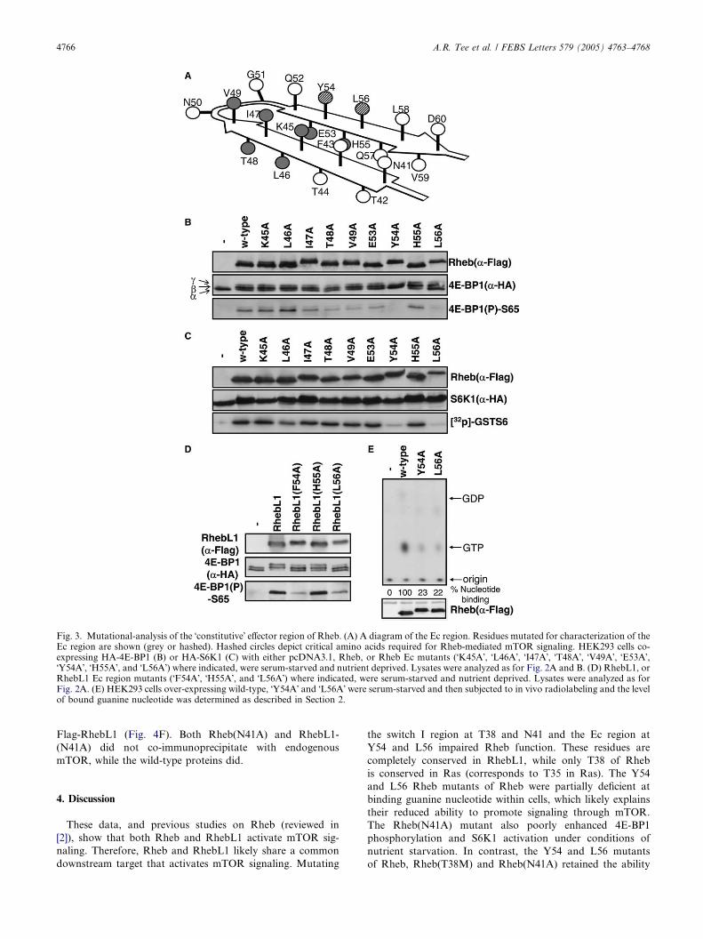
Fig. 3. Mutational-analysis of the �constitutive� effector region of Rheb. (A) A diagram of the Ec region. Residues mutated for characterization of theEc region are shown (grey or hashed). Hashed circles depict critical amino acids required for Rheb-mediated mTOR signaling. HEK293 cells co-expressing HA-4E-BP1 (B) or HA-S6K1 (C) with either pcDNA3.1, Rheb, or Rheb Ec mutants (�K45A�, �L46A�, �I47A�, �T48A�, �V49A�, �E53A�,�Y54A�, �H55A�, and �L56A�) where indicated, were serum-starved and nutrient deprived. Lysates were analyzed as for Fig. 2A and B. (D) RhebL1, orRhebL1 Ec region mutants (�F54A�, �H55A�, and �L56A�) where indicated, were serum-starved and nutrient deprived. Lysates were analyzed as forFig. 2A. (E) HEK293 cells over-expressing wild-type, �Y54A� and �L56A� were serum-starved and then subjected to in vivo radiolabeling and the levelof bound guanine nucleotide was determined as described in Section 2.
4766 A.R. Tee et al. / FEBS Letters 579 (2005) 4763–4768
Flag-RhebL1 (Fig. 4F). Both Rheb(N41A) and RhebL1-
(N41A) did not co-immunoprecipitate with endogenous
mTOR, while the wild-type proteins did.
4. Discussion
These data, and previous studies on Rheb (reviewed in
[2]), show that both Rheb and RhebL1 activate mTOR sig-
naling. Therefore, Rheb and RhebL1 likely share a common
downstream target that activates mTOR signaling. Mutating
the switch I region at T38 and N41 and the Ec region at
Y54 and L56 impaired Rheb function. These residues are
completely conserved in RhebL1, while only T38 of Rheb
is conserved in Ras (corresponds to T35 in Ras). The Y54
and L56 Rheb mutants of Rheb were partially deficient at
binding guanine nucleotide within cells, which likely explains
their reduced ability to promote signaling through mTOR.
The Rheb(N41A) mutant also poorly enhanced 4E-BP1
phosphorylation and S6K1 activation under conditions of
nutrient starvation. In contrast, the Y54 and L56 mutants
of Rheb, Rheb(T38M) and Rheb(N41A) retained the ability

Fig. 4. N41 within the switch I region is required for Rheb/mTOR signaling. HEK293 cells co-expressing HA-4E-BP1 (A) or HA-S6K1 (B) with twodifferent levels of expression of Rheb, or Rheb switch I mutants (�N41A�, and �T42A�) as indicated, were serum-starved and nutrient deprived. Thephosphorylation of exogenous 4E-BP1 was determined with anti-HA antibodies and Ser65 phospho-specific antibodies as indicated. The total levelsof Rheb and S6K1 are shown and S6K1 activity was measured. (C) HEK293 cells co-expressing HA-S6K1 with two different levels of wild-typeRhebL1 and switch I mutant (�N41A�) were serum-starved and nutrient deprived. S6K1 activity was measured and the total levels of RhebL1 proteinassessed. (D) HEK293 cells over-expressing wild-type, �T38M� and �N41A� were subjected to in vivo radiolabeling to examine [32P]GTP and [32P]GDPloading of Rheb as for Fig. 3E. (E) HEK293 cells co-expressing AU1-mTOR with Flag-tagged Rheb (wild-type, T38M or N41A) were serum-starvedprior to lysis. Flag-tagged Rheb was immunoprecipitated using anti-Flag antibodies, and the amount of associated AU1-mTOR was examined viawestern blot analysis. (F) Flag-tagged Rheb, Rheb(N41A), RhebL1, and RhebL1(N41A) proteins were expressed in HEK293 cells and serum-starvedbefore lysis. These Flag-tagged proteins were immunoprecipitated using anti-Flag antibodies and the level of co-purified endogenous mTOR(detected with mTAB1 antibody) was determined. As an mTOR protein control, mTOR was immunoprecipitated with mTAB1 (referred to as �a-mTOR�). The relative ratio of mTOR bound to Rheb or RhebL1 was quantitated using the Image/J software.
A.R. Tee et al. / FEBS Letters 579 (2005) 4763–4768 4767
to bind guanine nucleotides within cells. We reveal that
mTOR binds to Rheb and RhebL1 and this association re-
quires N41 within the putative switch I region. Therefore,
we identify mTOR as a common downstream target of both
Rheb and RhebL1.
During preparation of this manuscript, work carried out by
Long et al. [12,13] also showed that Rheb interacted with
mTOR. Our previous studies [11], similar to the observations
of Long et al. [12,13], showed that guanine deficient binding
mutants of Rheb interacted more potently to mTOR. Long
et al. also revealed that these guanine deficient binding mu-
tants of Rheb were inhibitory to the activity of mTOR [12].
These findings collectively show that mTOR does not function
as a typical small G-protein downstream effector, i.e., mTOR
is bound to Rheb regardless of its guanine nucleotide bound
status.
In conclusion, we reveal that RhebL1 promotes signal
transduction through mTOR and is a downstream target
of TSC1/2. We identify T38 and N41 within the putative
switch 1 region of both Rheb and RhebL1 as residues which
when mutated impairs their co-immunoprecipitation with
mTOR.
Acknowledgements: The British Heart Foundation supported this workthrough a BHF Intermediate Research Fellowship No. FS/04/002 (toAndrew R. Tee). This work was also supported in part by NationalInstitutes of Health grant GM51405 to J. Blenis.
References
[1] Reuther, G.W. and Der, C.J. (2000) The Ras branch of smallGTPases: Ras family members don�t fall far from the tree. Curr.Opin. Cell Biol. 12, 157–165.
[2] Tee, A.R. and Blenis, J. (2005) mTOR, translational control andhuman disease. Semin. Cell Dev. Biol. 16, 29–37.
[3] Tee, A.R., Manning, B.D., Roux, P.P., Cantley, L.C. and Blenis,J. (2003) Tuberous sclerosis complex gene products, Tuberin andHamartin, control mTOR signaling by acting as a GTPase-activating protein complex toward Rheb. Curr. Biol. 13, 1259–1268.
[4] Wittinghofer, A. (1998) Signal transduction via Ras. Biol. Chem.379, 933–937.
[5] Moodie, S.A., Paris, M., Villafranca, E., Kirshmeier, P., Willum-sen, B.M. and Wolfman, A. (1995) Different structural require-ments within the switch II region of the Ras protein forinteractions with specific downstream targets. Oncogene 11,447–454.
[6] Fujita-Yoshigaki, J., Shirouzu, M., Ito, Y., Hattori, S., Furuy-ama, S., Nishimura, S. and Yokoyama, S. (1995) A constitutive

4768 A.R. Tee et al. / FEBS Letters 579 (2005) 4763–4768
effector region on the C-terminal side of switch I of the Rasprotein. J. Biol. Chem. 270, 4661–4667.
[7] Tee, A.R., Fingar, D.C., Manning, B.D., Kwiatkowski, D.J.,Cantley, L.C. and Blenis, J. (2002) Tuberous sclerosis complex-1and -2 gene products function together to inhibit mammaliantarget of rapamycin (mTOR)-mediated downstream signaling.Proc. Natl. Acad. Sci. USA 99, 13571–13576.
[8] Schalm, S.S. and Blenis, J. (2002) Identification of a conservedmotif required for mTOR signaling. Curr. Biol. 12, 632–639.
[9] Fingar, D.C., Salama, S., Tsou, C., Harlow, E. and Blenis, J.(2002) Mammalian cell size is controlled by mTOR and itsdownstream targets S6K1 and 4EBP1/eIF4E. Genes Dev. 16,1472–1487.
[10] 2nd, Parise, L.V. and Cox, A.D. (2002) A distinct class ofdominant negative Ras mutants: cytosolic GTP-bound Ras
effector domain mutants that inhibit Ras signaling and transfor-mation and enhance cell adhesion. J. Biol. Chem. 277, 10813–10823.
[11] Smith, E.M., Finn, S.G., Tee, A.R., Browne, G.J. and Proud,C.G. (2005) The tuberous sclerosis protein TSC2 is not requiredfor the regulation of the mammalian target of rapamycin byamino acids and certain cellular stresses. J. Biol. Chem. 280,18717–18727.
[12] Long, X., Lin, Y., Ortiz-Vega, S., Yonezawa, K. and Avruch, J.(2005) Rheb binds and regulates the mTOR kinase. Curr. Biol. 15,702–713.
[13] Long, X., Ortiz-Vega, S., Lin, Y. and Avruch, J. (2005) Rhebbinding to mammalian target of rapamycin (mTOR) is regu-lated by amino acid sufficiency. J. Biol. Chem. 280, 23433–23436.
Related Documents