The regulation of protein synthesis by mTOR signaling: a potential target for cancer treatment?

Welcome message from author
This document is posted to help you gain knowledge. Please leave a comment to let me know what you think about it! Share it to your friends and learn new things together.
Transcript
The regulation of protein synthesis by mTOR signaling:
a potential target for cancer treatment?
Thesis_Weppler_v12_fc.pdf

Printed by: Datawyse | Universitaire Pers Maastricht Cover design: Jodil Willems © Sherry Weppler, Maastricht 2009 ISBN 978 90 5278 889 0
Thesis_Weppler_v12_fc.pdf
The regulation of protein synthesis by mTOR signaling:
a potential target for cancer treatment?
Dissertation
to obtain the degree of Doctor at Maastricht University,
on the authority of the Rector, Prof. dr. G.P.M.F. Mols
in accordance with the decision of the Board of Deans,
to be defended in public on
Thursday, December 3, 2009 at 12:00 p.m.
by
Sherry Anne Weppler
UNIVERSITAIREPERS MAASTRICHT
U P
M
Thesis_Weppler_v12_fc.pdf
Supervisors
Prof. dr. B.G. Wouters Prof. dr. P. Lambin
Co-supervisors
Dr. M.A.M. van Steensel Dr. G. Lammering
Assessment Committee
Prof. dr. F.C.S. Ramaekers (chairman) Dr. E.J. Bernhard (National Cancer Institute/NIH, USA) Prof. dr. J.F.C. Glatz Prof. dr. A.J. van der Kogel (Radboud University Nijmegen Medical Centre) Prof. dr. P.M. Steijlen
Thesis_Weppler_v12_fc.pdf
CONTENTS
Chapter 1 General introduction 9 Chapter 2 Hypoxia as a target for combined modality treatments 31 Chapter 3 Gene expression during acute and prolonged hypoxia 65 is regulated by distinct mechanisms of translational control Chapter 4 Expression of EGFR variant vIII promotes both 91 radiation resistance and hypoxia tolerance Chapter 5 Response of U87 glioma xenografts treated with 105 concurrent rapamycin and fractionated radiotherapy: possible role for thrombosis Chapter 6 Inhibition of 4E-BP1 phosphorylation and mRNA 125 translation requires simultaneous blockade of mTORC1 and PI3K/Akt signaling Chapter 7 Neuroendocrine carcinoma in Birt-Hogg-Dubé syndrome 149 Chapter 8 General discussion 159 Summary 170 Samenvatting 172 Acknowledgements 174 Curriculum vitae 177 List of publications 178
Thesis_Weppler_v12_fc.pdf
ABBREVIATIONS
4E-BP1 eIF4E binding protein 1 4E-T eIF4E transporter AICAR 5-aminoimidazole-4-carboxamide 1-β-D-ribonucleoside AMPK 5’ adenosine monophosphate-activated protein kinase ARCON accelerated radiotherapy combined with carbogen and nicotinamide ATF4 activating transcription factor 4 bFGF basic fibroblast growth factor BHD Birt-Hogg-Dubé BNIP3 BCL2/adenovirus E1B 19kDa interacting protein 3 BrdU bromodeoxyuridine CA-IX carbonic anhydrase 9 CHOP C/EBP homologous protein DMSO dimethyl sulfoxide DTT dithiothreitol ECM extracellular matrix eEF eukaryotic elongation factor EGF epidermal growth factor EGFR epidermal growth factor receptor EGFRvIII epidermal growth factor receptor variant 3 eIF eukaryotic initiation factor EPO erythropoietin ER endoplasmic reticulum FGF-BP fibroblast growth factor binding protein FKBP12 FK506 binding protein 12kDa FLCN folliculin FNIP folliculin interacting protein FRB FKBP12-rapamycin binding domain GADD34 growth arrest and DNA damage inducible gene 34 GLUT glucose transporter Hb hemoglobin HIF hypoxia inducible factor IGF insulin-like growth factor IGF-BP insulin-like growth factor binding protein IL interleukin IRE iron-responsive element IRES internal ribosome entry site IRP1 iron regulatory protein 1 IRS insulin receptor substrate mAbs monoclonal antibodies MAPK mitogen activated protein kinase MEFs mouse embryonic fibroblasts MMP matrix metalloproteinase mTOR mammalian target of rapamycin mTORC mammalian target of rapamycin complex NF-κB nuclear factor of kappa-light-chain-enhancer in activated B-cells
Thesis_Weppler_v12_fc.pdf
NOS nitric-oxide synthase ODC ornithine decarboxylase ORF open reading frame p70S6K ribosomal protein S6 kinase 70kDa P bodies processing bodies PDGF platelet derived growth factor PDK1 3-phosphoinositide-dependent kinase 1 PGK1 phosphoglycerate kinase 1 PI3K phosphatidylinositol-3-kinase PIP2 phosphatidylinositol 4,5 bisphosphate PIP3 phosphatidylinositol 3,4,5 triphosphate PKB protein kinase B PKCα protein kinase C alpha PK-M pyruvate kinase M PRAS40 proline-rich Akt substrate of 40kDa Protor protein observed with Rictor PTEN phosphatase and tensin homolog Raptor regulatory associated protein of mTOR REDD1 regulated in development and DNA damage responses Rheb ras homolog enriched in brain Rictor rapamycin insensitive companion of mTOR SIN1 stress-activated protein kinase-interacting protein TCD50 tumor control dose 50% TCP tumor control probability TIMP tissue inhibitor of metalloproteinases TNFα tumor necrosis factor alpha TKI tyrosine kinase inhibitor TOP terminal oligopyrimidine TORKinibs mTOR kinase domain inhibitors TOS TOR signaling motif TPZ tirapazamine TSC tuberous sclerosis complex uPA urokinase-type plasminogen activator uPAR urokinase-type plasminogen activator receptor UPR unfolded protein response UTR untranslated region VEGF vascular endothelial growth factor VEGFR vascular endothelial growth factor receptor
Thesis_Weppler_v12_fc.pdf

Thesis_Weppler_v12_fc.pdf

9
CHAPTER 1
General introduction
Thesis_Weppler_v12_fc.pdf
Chapter 1
10
Introduction
mTOR in normal physiology and disease
The mammalian target of rapamcyin (mTOR) is a central regulator of cell growth that has been highly conserved through evolution from yeast to mammals. mTOR responds to growth factors, nutrients and energy levels to promote protein synthesis and proliferation under replete conditions and to conserve energy and promote survival during periods of stress or starvation. The importance of TOR for development is clearly illustrated by the embry-onic lethality of TOR mutants in Drosophila melanogaster, Caenorhabditis elegans, and Mus musculus. TOR knockout embryos die shortly after implan-tation due to impaired trophoblast differentiation and failure of embryonic stem cells to proliferate (1, 2). Dysfunction of mTOR signaling has also been implicated in a number of human diseases including obesity, type II diabetes, muscle atrophy, cardiac hypertrophy, neurodegenerative disorders such as Huntington’s and Alzheimer’s disease, and cancer (3). Current research ef-forts are investigating the therapeutic potential of modulating mTOR signaling in the management of these disorders.
mTOR signaling complexes
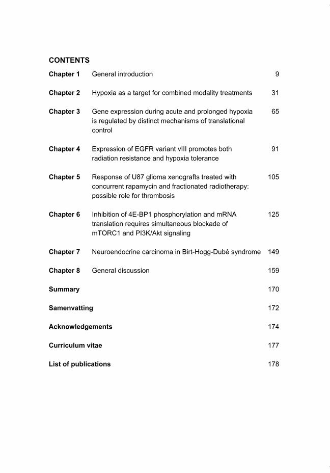
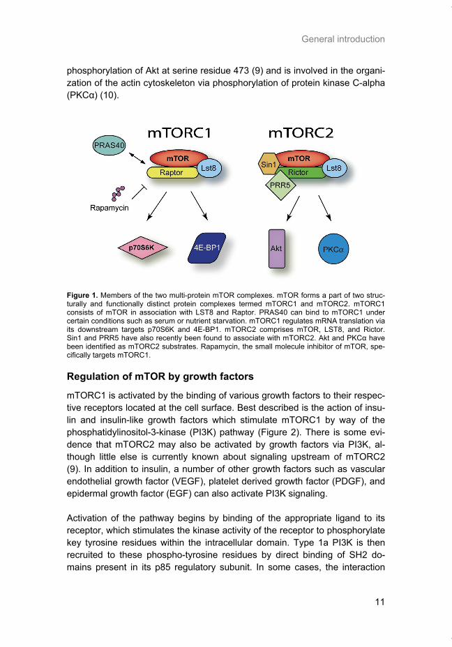
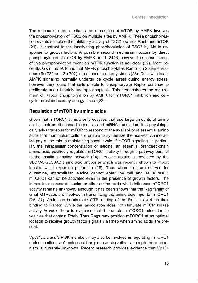
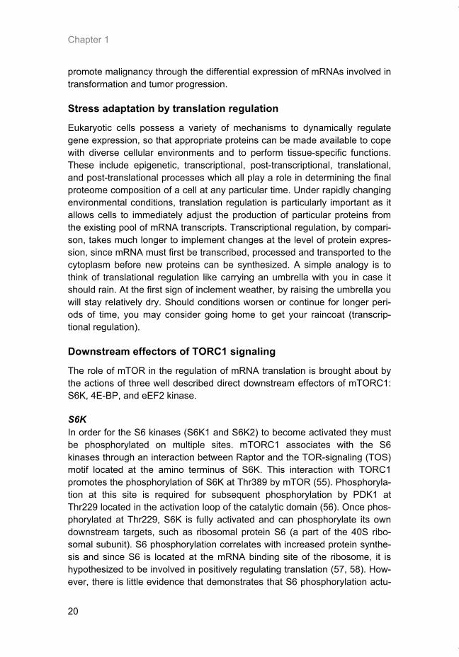
The mTOR kinase carries out its function within the context of two structurally distinct multi-protein complexes, mTORC1 and mTORC2 (Figure 1). mTORC1 consists of mTOR in association with LST8 and Raptor. LST8 and mTOR are the only common components present in both TOR complexes. LST8 binds constitutively to the catalytic domain of mTOR and stimulates its kinase activity (4). Raptor acts as a scaffolding protein to bring mTORC1 substrates such as p70S6K and 4E-BP1 within close proximity of the mTOR kinase domain. By phosphorylation of these substrates, mTORC1 plays a role in regulating the process of mRNA translation. A fourth partner of com-plex 1, PRAS40, binds mTORC1 under conditions of serum or nutrient depri-vation and acts as a negative regulator of mTORC1 (5). Phosphorylation of PRAS40 by Akt or mTOR itself appears to release its inhibitory effect on mTORC1 (6). mTORC2 contains mTOR, LST8, and Rictor in addition to the recently identi-fied proteins SIN1 and PRR5/Protor (7, 8). Much less is known about the up-stream regulation and function of this complex, although it is currently an area of active research. This complex has downstream substrates that are distinct from those of mTORC1, most likely as a consequence of the unique proteins present in this complex. Importantly, mTORC2 is responsible for
Thesis_Weppler_v12_fc.pdf
General introduction
11
phosphorylation of Akt at serine residue 473 (9) and is involved in the organi-zation of the actin cytoskeleton via phosphorylation of protein kinase C-alpha (PKCα) (10).
Figure 1. Members of the two multi-protein mTOR complexes. mTOR forms a part of two struc-turally and functionally distinct protein complexes termed mTORC1 and mTORC2. mTORC1 consists of mTOR in association with LST8 and Raptor. PRAS40 can bind to mTORC1 under certain conditions such as serum or nutrient starvation. mTORC1 regulates mRNA translation via its downstream targets p70S6K and 4E-BP1. mTORC2 comprises mTOR, LST8, and Rictor. Sin1 and PRR5 have also recently been found to associate with mTORC2. Akt and PKCα have been identified as mTORC2 substrates. Rapamycin, the small molecule inhibitor of mTOR, spe-cifically targets mTORC1.
Regulation of mTOR by growth factors
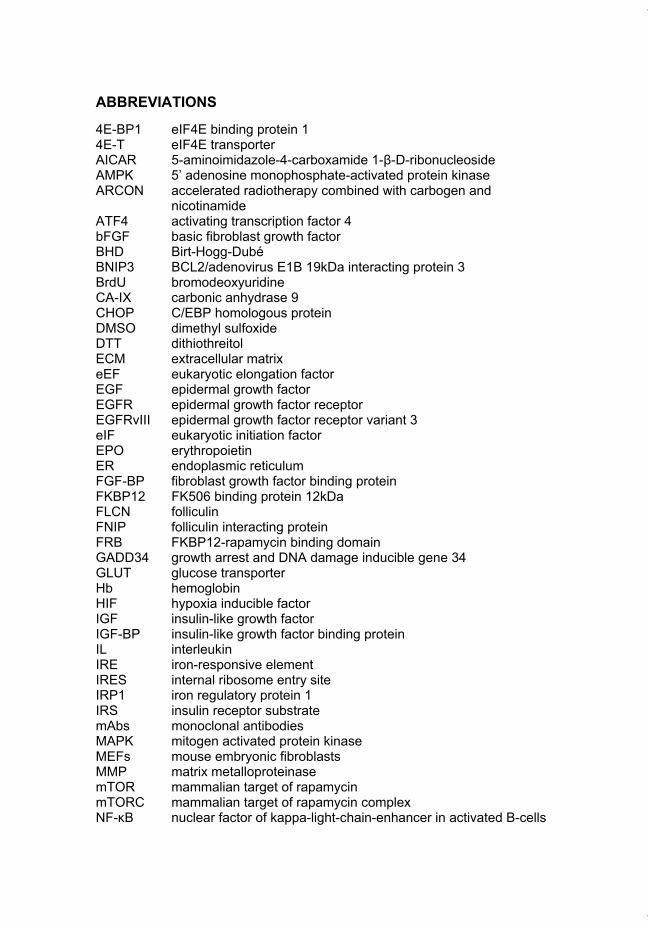
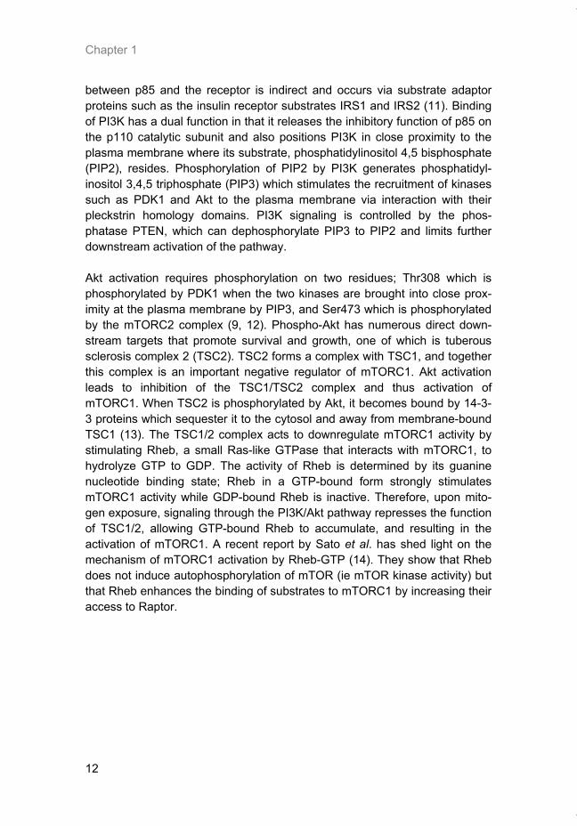
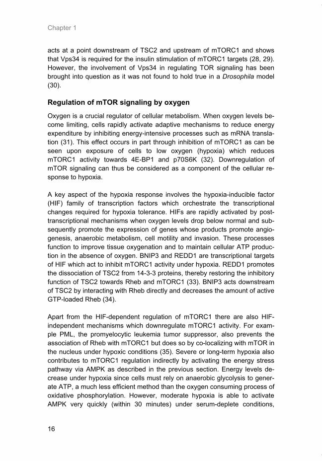
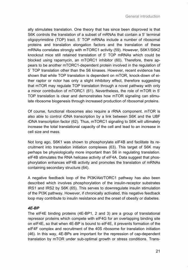
mTORC1 is activated by the binding of various growth factors to their respec-tive receptors located at the cell surface. Best described is the action of insu-lin and insulin-like growth factors which stimulate mTORC1 by way of the phosphatidylinositol-3-kinase (PI3K) pathway (Figure 2). There is some evi-dence that mTORC2 may also be activated by growth factors via PI3K, al-though little else is currently known about signaling upstream of mTORC2 (9). In addition to insulin, a number of other growth factors such as vascular endothelial growth factor (VEGF), platelet derived growth factor (PDGF), and epidermal growth factor (EGF) can also activate PI3K signaling. Activation of the pathway begins by binding of the appropriate ligand to its receptor, which stimulates the kinase activity of the receptor to phosphorylate key tyrosine residues within the intracellular domain. Type 1a PI3K is then recruited to these phospho-tyrosine residues by direct binding of SH2 do-mains present in its p85 regulatory subunit. In some cases, the interaction
Thesis_Weppler_v12_fc.pdf
Chapter 1
12
between p85 and the receptor is indirect and occurs via substrate adaptor proteins such as the insulin receptor substrates IRS1 and IRS2 (11). Binding of PI3K has a dual function in that it releases the inhibitory function of p85 on the p110 catalytic subunit and also positions PI3K in close proximity to the plasma membrane where its substrate, phosphatidylinositol 4,5 bisphosphate (PIP2), resides. Phosphorylation of PIP2 by PI3K generates phosphatidyl-inositol 3,4,5 triphosphate (PIP3) which stimulates the recruitment of kinases such as PDK1 and Akt to the plasma membrane via interaction with their pleckstrin homology domains. PI3K signaling is controlled by the phos-phatase PTEN, which can dephosphorylate PIP3 to PIP2 and limits further downstream activation of the pathway. Akt activation requires phosphorylation on two residues; Thr308 which is phosphorylated by PDK1 when the two kinases are brought into close prox-imity at the plasma membrane by PIP3, and Ser473 which is phosphorylated by the mTORC2 complex (9, 12). Phospho-Akt has numerous direct down-stream targets that promote survival and growth, one of which is tuberous sclerosis complex 2 (TSC2). TSC2 forms a complex with TSC1, and together this complex is an important negative regulator of mTORC1. Akt activation leads to inhibition of the TSC1/TSC2 complex and thus activation of mTORC1. When TSC2 is phosphorylated by Akt, it becomes bound by 14-3-3 proteins which sequester it to the cytosol and away from membrane-bound TSC1 (13). The TSC1/2 complex acts to downregulate mTORC1 activity by stimulating Rheb, a small Ras-like GTPase that interacts with mTORC1, to hydrolyze GTP to GDP. The activity of Rheb is determined by its guanine nucleotide binding state; Rheb in a GTP-bound form strongly stimulates mTORC1 activity while GDP-bound Rheb is inactive. Therefore, upon mito-gen exposure, signaling through the PI3K/Akt pathway represses the function of TSC1/2, allowing GTP-bound Rheb to accumulate, and resulting in the activation of mTORC1. A recent report by Sato et al. has shed light on the mechanism of mTORC1 activation by Rheb-GTP (14). They show that Rheb does not induce autophosphorylation of mTOR (ie mTOR kinase activity) but that Rheb enhances the binding of substrates to mTORC1 by increasing their access to Raptor.
Thesis_Weppler_v12_fc.pdf
General introduction
13
Figure 2. The mTOR signal transduction pathway. Activity of the mTOR-Raptor complex (mTORC1) is positively regulated by extracellular growth factors and by amino acids. Binding of growth factors to a cell surface receptor sets off a signaling cascade through the PI3K-PDK1-Akt pathway to the TSC1/2 complex. For complete activation of Akt, phosphorylation of Ser473 by mTOR-Rictor (mTORC2) is required. Currently little is known about the upstream regulation of mTORC2. TSC is a major negative regulator of mTORC1 which is inactivated by Akt which promotes sequestration of TSC2 by 14-3-3 proteins. Hypoxia and low energy levels, on the other hand, can stimulate TSC2 via REDD1 and AMPK respectively. This stimulates the TSC1/2 complex to activate GTP hydrolysis by Rheb, leading to accumulation of GDP-bound Rheb and inhibition of mTORC1. Hypoxia can also promote Rheb-GDP in a TSC-independent manner via BNIP3. Accumulation of GTP-bound Rheb positively regulates activity of mTORC1. Amino ac-ids, by activation of the Rag heterodimer, allow activation of mTORC1 by Rheb-GTP. Down-stream targets of mTORC1 include 4E-BP1, p70-S6K and eEF2K which all function in some aspect of mRNA translation.
Thesis_Weppler_v12_fc.pdf
Chapter 1
14
Deregulation of PI3K and mTOR in cancer
In cancer, mutagenic events often occur in genes whose encoded proteins function in the PI3K signaling pathway, thus resulting in constitutive activation of mTOR and uncontrolled proliferation in the absence of mitogenic signals. Activating mutations in PI3K subunits, mutation or amplification of various growth factor receptors and loss of PTEN expression are common events reported in human malignancies that play a role in the deregulation of PI3K signaling. One such mutation involves the epidermal growth factor receptor (EGFR) whereby genomic deletion of exons 2-7 results in expression of a truncated form of the receptor called EGFRvIII. This EGFR variant lacks a portion of the extracellular ligand-binding domain and exhibits constitutively elevated kinase activity in the absence of ligand. EGFRvIII expression is re-ported in 50% of gliomas, and is also common in breast, prostate and non-small cell lung cancer, but is not found in normal tissue (15). Clinical studies have demonstrated a correlation between EGFRvIII expression and poor prognosis for patients with glioblastoma. Signaling downstream of EGFRvIII is also different compared to wild-type EGFR, with a preferential activation of the PI3K pathway and relatively less stimulation of the MAPK and STAT3 signaling pathways (16). Current interest lies in combining EGFR targeted therapies with mTOR inhibitors, as this treatment strategy seems to be par-ticularly effective towards tumors which develop resistance against EGFR antagonists (17, 18).
Regulation of mTOR by cellular energy levels
Protein synthesis consumes a large proportion of the total cellular energy (estimated at 20-45% in mammalian cells), thus it is crucial that the rate of mRNA translation be linked to the ATP supply. Since mTOR is a key regula-tor of protein synthesis, it is not surprising that mTOR activity is coupled to cellular energy status. This connection is mediated by AMPK, a kinase that responds to changes in the ratio of AMP to ATP. It is important to note that the energy dependent regulation of mTOR signaling is dominant to other pathways such as insulin or amino acid stimulation, so that mTOR cannot be activated by other sources if energy levels are not permissive. AMPK be-comes activated in a multi-step process that is initiated by the binding of two AMP molecules to the γ-subunit. This induces a conformational change that allows the subsequent phosphorylation of the α-subunit on Thr172 by LKB1 (19). In contrast, under energy replete conditions when the ATP:AMP ratio is high, binding of ATP to AMPK inhibits its activation by preventing this phos-phorylation event. Experiments utilizing the AMP mimetic 5-aminoimidazole-4-carboxamide 1-β-D-ribonucleoside (AICAR) have shown that increased AMPK activity correlates with dephosphorylation of mTORC1 targets (20).
Thesis_Weppler_v12_fc.pdf
General introduction
15
The mechanism that mediates the repression of mTOR by AMPK involves the phosphorylation of TSC2 on multiple sites by AMPK. These phosphoryla-tion events stimulate the inhibitory activity of TSC2 towards Rheb and mTOR (21), in contrast to the inactivating phosphorylation of TSC2 by Akt in re-sponse to growth factors. A possible second mechanism occurs by direct phosphorylation of mTOR by AMPK on Thr2446, however the consequence of this phosphorylation event on mTOR function is not clear (22). More re-cently, Gwinn et al. found that AMPK phosphorylates Raptor on 2 serine resi-dues (Ser722 and Ser792) in response to energy stress (23). Cells with intact AMPK signaling normally undergo cell-cycle arrest during energy stress, however they found that cells unable to phosphorylate Raptor continue to proliferate and ultimately undergo apoptosis. This demonstrates the require-ment of Raptor phosphorylation by AMPK for mTORC1 inhibition and cell-cycle arrest induced by energy stress (23).
Regulation of mTOR by amino acids
Given that mTORC1 stimulates processes that use large amounts of amino acids, such as ribosome biogenesis and mRNA translation, it is physiologi-cally advantageous for mTOR to respond to the availability of essential amino acids that mammalian cells are unable to synthesize themselves. Amino ac-ids pay a key role in maintaining basal levels of mTOR signaling. In particu-lar, the intracellular concentration of leucine, an essential branched-chain amino acid, positively regulates mTORC1 activity through a pathway parallel to the insulin signaling network (24). Leucine uptake is mediated by the SLC7A5-SLC3A2 amino acid antiporter which was recently shown to import leucine while exporting glutamine (25). Thus when cells are starved for glutamine, extracellular leucine cannot enter the cell and as a result, mTORC1 cannot be activated even in the presence of growth factors. The intracellular sensor of leucine or other amino acids which influence mTORC1 activity remains unknown, although it has been shown that the Rag family of small GTPases are involved in transmitting the amino acid input to mTORC1 (26, 27). Amino acids stimulate GTP loading of the Rags as well as their binding to Raptor. While this association does not stimulate mTOR kinase activity in vitro, there is evidence that it promotes mTORC1 relocation to vesicles that contain Rheb. Thus Rags may position mTORC1 at an optimal location to receive growth factor signals via Rheb when amino acids are pre-sent. Vps34, a class 3 PI3K member, may also be involved in regulating mTORC1 under conditions of amino acid or glucose starvation, although the mecha-nism is currently unknown. Recent research provides evidence that Vps34
Thesis_Weppler_v12_fc.pdf
Chapter 1
16
acts at a point downstream of TSC2 and upstream of mTORC1 and shows that Vps34 is required for the insulin stimulation of mTORC1 targets (28, 29). However, the involvement of Vps34 in regulating TOR signaling has been brought into question as it was not found to hold true in a Drosophila model (30).
Regulation of mTOR signaling by oxygen
Oxygen is a crucial regulator of cellular metabolism. When oxygen levels be-come limiting, cells rapidly activate adaptive mechanisms to reduce energy expenditure by inhibiting energy-intensive processes such as mRNA transla-tion (31). This effect occurs in part through inhibition of mTORC1 as can be seen upon exposure of cells to low oxygen (hypoxia) which reduces mTORC1 activity towards 4E-BP1 and p70S6K (32). Downregulation of mTOR signaling can thus be considered as a component of the cellular re-sponse to hypoxia. A key aspect of the hypoxia response involves the hypoxia-inducible factor (HIF) family of transcription factors which orchestrate the transcriptional changes required for hypoxia tolerance. HIFs are rapidly activated by post-transcriptional mechanisms when oxygen levels drop below normal and sub-sequently promote the expression of genes whose products promote angio-genesis, anaerobic metabolism, cell motility and invasion. These processes function to improve tissue oxygenation and to maintain cellular ATP produc-tion in the absence of oxygen. BNIP3 and REDD1 are transcriptional targets of HIF which act to inhibit mTORC1 activity under hypoxia. REDD1 promotes the dissociation of TSC2 from 14-3-3 proteins, thereby restoring the inhibitory function of TSC2 towards Rheb and mTORC1 (33). BNIP3 acts downstream of TSC2 by interacting with Rheb directly and decreases the amount of active GTP-loaded Rheb (34). Apart from the HIF-dependent regulation of mTORC1 there are also HIF-independent mechanisms which downregulate mTORC1 activity. For exam-ple PML, the promyelocytic leukemia tumor suppressor, also prevents the association of Rheb with mTORC1 but does so by co-localizing with mTOR in the nucleus under hypoxic conditions (35). Severe or long-term hypoxia also contributes to mTORC1 regulation indirectly by activating the energy stress pathway via AMPK as described in the previous section. Energy levels de-crease under hypoxia since cells must rely on anaerobic glycolysis to gener-ate ATP, a much less efficient method than the oxygen consuming process of oxidative phosphorylation. However, moderate hypoxia is able to activate AMPK very quickly (within 30 minutes) under serum-deplete conditions,
Thesis_Weppler_v12_fc.pdf
General introduction
17
which correlates with inhibition of mTORC1 (36). This appears to be a con-sequence of rapidly decreasing ATP levels under hypoxia when growth fac-tors are not present. The fact that hypoxia influences mTOR through multiple mechanisms sug-gests that it may be particularly important for adaptation to this stress.
Possible link between folliculin and mTOR
An interesting new connection has been made between mTORC1 signaling and folliculin, a protein of unknown function. Germline mutations in the gene encoding folliculin cause a rare genetic disease called Birt-Hogg-Dubé (BHD) syndrome. The characteristic features of BHD include benign skin tumors originating from the hair follicles (fibrofolliculomas) on the face and upper torso, lung cysts leading to spontaneous pneumothorax, and an increased risk of developing renal carcinoma (37). Mutations in the BHD gene are inher-ited in an autosomal dominant fashion and have been characterized in a number of families. The majority of reported human mutations are either frameshift or nonsense mutations that are predicted to cause protein trunca-tion and result in haploinsufficiency (38). It is thought that folliculin acts as a typical tumor suppressor. Evidence to support this idea comes from renal tumors of BHD patients that have lost expression of the remaining wild-type allele, as well as loss-of-heterozygosity that has been reported at this locus in sporadic kidney tumors (39, 40). BHD syndrome displays phenotypic similari-ties with several familial hamartoma syndromes such as Peutz-Jeghers syn-drome, Cowden syndrome and Tuberous Sclerosis Complex. These diseases are characterized by benign tumors that develop in multiple tissues and ele-vated risk of malignant cancer. Hamartoma syndromes all share a common upregulation of mTORC1 signaling, so it is a logical assumption that folliculin is also somehow connected to mTOR. The link between folliculin and mTORC1 has recently been confirmed in a kidney-specific BHD knock-out mouse where strong activation of Akt/mTORC1 and MAPK pathways was observed (41). How exactly folliculin is regulating mTORC1 is still not clear, although there is some evidence to suggest that folliculin associates with AMPK via two recently identified interacting proteins, FNIP1 and 2 (42, 43). Folliculin may therefore be involved in energy and/or nutrient sensing through the AMPK and mTOR pathways.
Regulation of mRNA translation by mTOR
The result of stimulating mTORC1 activity is manifested in cellular changes such as increased cell growth (cell size and mass), increased proliferation,
Thesis_Weppler_v12_fc.pdf
Chapter 1
18
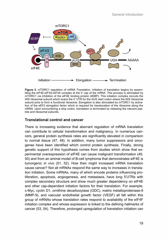
angiogenesis, and increased survival (44). These downstream effects are all thought to be mediated by mTORC1’s regulation of mRNA translation. The process of mRNA translation involves the sequential decoding of mRNA by the ribosome into protein. Ribosomes are composed of 2 subunits, termed the ‘small’ 40S subunit and the ‘large’ 60S subunit, which together are com-prised of 85-90 distinct proteins and four different ribosomal RNA molecules (45). The ribosome catalyzes the formation of peptide bonds between amino acids of the newly synthesized protein. Translation can be divided into three distinct stages: initiation, elongation, and termination. All stages require addi-tional translation factors which transiently associate with the ribosome and allow separate regulation of the various stages. For most mRNAs, initiation begins by recruiting the 40S ribosomal subunit to the 5’ cap structure of the mRNA (Figure 3). This occurs following formation of the eIF4F complex con-sisting of eIF4E (the cap-binding protein), eIF4G (a large scaffolding protein which is crucial for binding of the ribosome), and eIF4A (an RNA helicase) (46). As discussed later, mTOR plays an important role in regulating the availability of eIF4E to participate in this complex. Together with the methio-nyl-tRNA and certain initiation factors, the 40S subunit scans along the 5’ untranslated region (UTR) for the start codon. During elongation, the poly-peptide chain is assembled in a stepwise fashion according to the reading frame of the mRNA. Elongation requires two translation factors, eEF1 and eEF2. eEF2 is also regulated by mTOR and is required for translocation of the ribosome to the next codon in the mRNA (45). Termination occurs when the ribosome en-counters a stop codon, resulting in the release of the polypeptide chain and ribosomal subunits. It is common for multiple ribosomes to initiate translation of an mRNA molecule in succession, which allows the simultaneous produc-tion of several peptides from a single mRNA. This structure is termed a ‘polyribosome’ or ‘polysome’ and can be isolated to determine which mRNAs are actively synthesizing protein at any given time or condition.
Thesis_Weppler_v12_fc.pdf
General introduction
19
Figure 3. mTORC1 regulation of mRNA Translation. Initiation of translation begins by assem-bling the eIF4E-eIF4G-eIF4A complex at the 5’ cap of the mRNA. This process is stimulated by mTORC1 via inhibition of the eIF4E binding protein (4EBP). This initiation complex recruits the 40S ribosomal subunit which scans the 5’ UTR for the AUG start codon where the 60S ribosomal subunit joins to form a functional ribosome. Elongation is also stimulated by mTORC1 by activa-tion of the eEF2 elongation factor which is required for translocation of the ribosome along the mRNA. Upon encountering a stop codon, translation is terminated by releasing the nascent pep-tide and ribosomal subunits.
Translational control and cancer
There is increasing evidence that aberrant regulation of mRNA translation can contribute to cellular transformation and malignancy. In numerous can-cers, general protein synthesis rates are significantly elevated in comparison to normal tissue (47, 48). In addition, many tumor suppressors and onco-genes have been identified which control protein synthesis. Finally, strong genetic support of this hypothesis comes from studies which show that ex-perimental overexpression of eIF4E can cause malignant transformation (49, 50) and from an animal model of B-cell lymphoma that demonstrates eIF4E is tumorgenic in vivo (51, 52). How then might increased mRNA translation cause cancer? Not all mRNAs respond the same way to increases in transla-tion initiation. Some mRNAs, many of which encode proteins influencing pro-liferation, apoptosis, angiogenesis, and metastasis, have long 5’UTRs with complex secondary structure and show much greater dependency on eIF4E and other cap-dependent initiation factors for their translation. For example, c-Myc, cyclin D1, ornithine decarboxylase (ODC), matrix metalloproteinase-9 (MMP-9), and vascular endothelial growth factor (VEGF) all fall within this group of mRNAs whose translation rates respond to availability of the eIF4F initiation complex and whose expression is linked to the defining hallmarks of cancer (53, 54). Therefore, prolonged upregulation of translation initiation can
Thesis_Weppler_v12_fc.pdf
Chapter 1
20
promote malignancy through the differential expression of mRNAs involved in transformation and tumor progression.
Stress adaptation by translation regulation
Eukaryotic cells possess a variety of mechanisms to dynamically regulate gene expression, so that appropriate proteins can be made available to cope with diverse cellular environments and to perform tissue-specific functions. These include epigenetic, transcriptional, post-transcriptional, translational, and post-translational processes which all play a role in determining the final proteome composition of a cell at any particular time. Under rapidly changing environmental conditions, translation regulation is particularly important as it allows cells to immediately adjust the production of particular proteins from the existing pool of mRNA transcripts. Transcriptional regulation, by compari-son, takes much longer to implement changes at the level of protein expres-sion, since mRNA must first be transcribed, processed and transported to the cytoplasm before new proteins can be synthesized. A simple analogy is to think of translational regulation like carrying an umbrella with you in case it should rain. At the first sign of inclement weather, by raising the umbrella you will stay relatively dry. Should conditions worsen or continue for longer peri-ods of time, you may consider going home to get your raincoat (transcrip-tional regulation).
Downstream effectors of TORC1 signaling
The role of mTOR in the regulation of mRNA translation is brought about by the actions of three well described direct downstream effectors of mTORC1: S6K, 4E-BP, and eEF2 kinase.
S6K In order for the S6 kinases (S6K1 and S6K2) to become activated they must be phosphorylated on multiple sites. mTORC1 associates with the S6 kinases through an interaction between Raptor and the TOR-signaling (TOS) motif located at the amino terminus of S6K. This interaction with TORC1 promotes the phosphorylation of S6K at Thr389 by mTOR (55). Phosphoryla-tion at this site is required for subsequent phosphorylation by PDK1 at Thr229 located in the activation loop of the catalytic domain (56). Once phos-phorylated at Thr229, S6K is fully activated and can phosphorylate its own downstream targets, such as ribosomal protein S6 (a part of the 40S ribo-somal subunit). S6 phosphorylation correlates with increased protein synthe-sis and since S6 is located at the mRNA binding site of the ribosome, it is hypothesized to be involved in positively regulating translation (57, 58). How-ever, there is little evidence that demonstrates that S6 phosphorylation actu-
Thesis_Weppler_v12_fc.pdf
General introduction
21
ally stimulates translation. One theory that has since been disproved is that S6K controls the translation of a subset of mRNAs that contain a 5’ terminal oligopyrimidine (TOP) tract. 5’ TOP mRNAs include a number of ribosomal proteins and translation elongation factors and the translation of these mRNAs correlates strongly with mTORC1 activity (59). However, S6K1/S6K2 knockout mice still retained translation of 5’ TOP mRNAs which could be blocked using rapamycin, an mTORC1 inhibitor (60). Therefore, there ap-pears to be another mTORC1-dependent protein involved in the regulation of 5’ TOP translation other than the S6 kinases. However, recent evidence has shown that while TOP translation is dependent on mTOR, knock-down of ei-ther raptor or rictor has only a slight inhibitory effect, therefore suggesting that mTOR may regulate TOP translation through a novel pathway with only a minor contribution of mTORC1 (61). Nevertheless, the role of mTOR in 5’ TOP translation is clear and demonstrates how mTOR signaling can stimu-late ribosome biogenesis through increased production of ribosomal proteins. Of course, functional ribosomes also require a rRNA component. mTOR is also able to control rDNA transcription by a link between S6K and the UBF rDNA transcription factor (62). Thus, mTORC1 signaling to S6K will ultimately increase the total translational capacity of the cell and lead to an increase in cell size and mass. Not long ago, S6K1 was shown to phosphorylate eIF4B and facilitate its re-cruitment into translation initiation complexes (63). This target of S6K may perhaps be physiologically more important than S6 in regulating translation. eIF4B stimulates the RNA helicase activity of eIF4A. Data suggest that phos-phorylation enhances eIF4B activity and promotes the translation of mRNAs containing secondary structure (64). A negative feedback loop of the PI3K/Akt/TORC1 pathway has also been described which involves phosphorylation of the insulin-receptor substrates IRS1 and IRS2 by S6K (65). This serves to downregulate insulin stimulation of the PI3K pathway. However, if chronically activated, this negative feedback loop may contribute to insulin resistance and the onset of obesity or diabetes.
4E-BP The eIF4E binding proteins (4E-BP1, 2 and 3) are a group of translational repressor proteins which compete with eIF4G for an overlapping binding site on eIF4E, so that when 4E-BP is bound to eIF4E, it prevents formation of the eIF4F complex and recruitment of the 40S ribosome for translation initiation (46). In this way, 4E-BPs are important for the repression of cap-dependent translation by mTOR under sub-optimal growth or stress conditions. Trans-
Thesis_Weppler_v12_fc.pdf
Chapter 1
22
genic mice lacking 4E-BP1 and 4E-BP2 display elevated insulin resistance and sensitivity to diet-induced obesity, demonstrating a role for 4E-BPs as “metabolic brakes” (66). The interaction between 4E-BP and eIF4E is regulated by a complex series of phosphorylation events. Hypo-phosphorylated 4E-BPs bind eIF4E with high affinity, whereas hyper-phosphorylated 4E-BPs rapidly dissociate (67). The 4E-BPs also rely on a TOS motif for interaction with Raptor and mTORC1, just as the S6Ks. Evidence suggests that mTORC1 is responsible for phosphorylation of 4E-BP1 on Thr37 and Thr46 in response to nutrient signaling (68). Phosphorylation of 4E-BP1 occurs in a hierarchical manner such that phosphorylation of Thr37 and Thr46 is required to prime subse-quent phosphorylation of Thr70, followed by Ser65 (69). Thr70 and Ser65 are responsive to insulin and growth factors and respond to inhibition by rapamy-cin. However, in vitro kinase assays do not confirm mTOR as the kinase that directly phosphorylates these residues (68). Therefore it seems likely that an mTORC1-associated or mTORC1-controlled kinase is required for phos-phorylation of these sites.
eEF2 In addition to regulating translation initiation, mTORC1 also influences the elongation step of protein synthesis via eEF2. Association of eEF2 with the ribosome is controlled by phosphorylation of Thr56 in its GTP-binding domain (70). Phosphorylation of this residue is catalyzed by eEF2 kinase and inhibits the binding of eEF2 to the ribosome, thus impairing its activity. mTOR signal-ing negatively regulates eEF2 kinase activity by multiple inhibitory phosphory-lation events. One of these sites (Ser 366) has been shown to be a direct target of S6K1 (71). Phosphorylation of two additional sites (Ser78 and Ser359) is also dependent upon mTORC1, although the kinase(s) directly responsible has yet to be identified (72). Therefore, by blocking the inhibitory effect of eEF2 kinase upon eE2F, mTOR can promote translation elongation.
mTOR inhibitors in cancer therapy
The mTOR kinase was discovered as the result of studies conducted in yeast investigating a macrolide compound with antifungal properties called rapamy-cin, what would later become known as the first mTOR inhibitor (73). Rapamy-cin was first isolated from the bacterium Streptomyces hygroscopicus obtained from a soil sample collected on Easter Island (Rapa Nui) in the early 1970s (74). Due to the immunosuppressant properties of rapamycin, its development as an antifungal agent was not pursued. However, this same property made rapamycin attractive for the treatment of graft rejection after organ transplanta-
Thesis_Weppler_v12_fc.pdf
General introduction
23
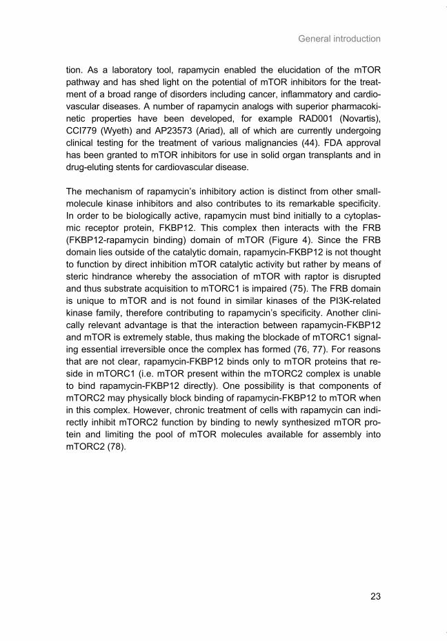
tion. As a laboratory tool, rapamycin enabled the elucidation of the mTOR pathway and has shed light on the potential of mTOR inhibitors for the treat-ment of a broad range of disorders including cancer, inflammatory and cardio-vascular diseases. A number of rapamycin analogs with superior pharmacoki-netic properties have been developed, for example RAD001 (Novartis), CCI779 (Wyeth) and AP23573 (Ariad), all of which are currently undergoing clinical testing for the treatment of various malignancies (44). FDA approval has been granted to mTOR inhibitors for use in solid organ transplants and in drug-eluting stents for cardiovascular disease. The mechanism of rapamycin’s inhibitory action is distinct from other small-molecule kinase inhibitors and also contributes to its remarkable specificity. In order to be biologically active, rapamycin must bind initially to a cytoplas-mic receptor protein, FKBP12. This complex then interacts with the FRB (FKBP12-rapamycin binding) domain of mTOR (Figure 4). Since the FRB domain lies outside of the catalytic domain, rapamycin-FKBP12 is not thought to function by direct inhibition mTOR catalytic activity but rather by means of steric hindrance whereby the association of mTOR with raptor is disrupted and thus substrate acquisition to mTORC1 is impaired (75). The FRB domain is unique to mTOR and is not found in similar kinases of the PI3K-related kinase family, therefore contributing to rapamycin’s specificity. Another clini-cally relevant advantage is that the interaction between rapamycin-FKBP12 and mTOR is extremely stable, thus making the blockade of mTORC1 signal-ing essential irreversible once the complex has formed (76, 77). For reasons that are not clear, rapamycin-FKBP12 binds only to mTOR proteins that re-side in mTORC1 (i.e. mTOR present within the mTORC2 complex is unable to bind rapamycin-FKBP12 directly). One possibility is that components of mTORC2 may physically block binding of rapamycin-FKBP12 to mTOR when in this complex. However, chronic treatment of cells with rapamycin can indi-rectly inhibit mTORC2 function by binding to newly synthesized mTOR pro-tein and limiting the pool of mTOR molecules available for assembly into mTORC2 (78).
Thesis_Weppler_v12_fc.pdf
Chapter 1
24
Figure 4. Inhibition of mTOR by rapamycin. The FRB domain of mTOR serves as the high-affinity binding site for the rapamycin-FKBP12 complex. Other important mTOR domains include the N-terminus HEAT domains which mediate protein-protein interactions and the kinase domain (KD) which harbors the catalytic site. The FAT and FATC domains are conserved regions found in protein which are members of the PI3K-like kinase (PIKK) family.
Aims of this thesis
In this thesis, I examine various aspects related to mTOR signaling in cancer with a focus on its role in the tumor microenvironment. The general aims of this thesis are to elucidate how mTOR signaling and the regulation of mRNA translation contribute to the response of tumor cells to microenvironmental stresses such as hypoxia or to cancer therapy, in order to exploit mTOR as a potential therapeutic target.
Specific objectives
Hypoxia is a common feature of solid tumors that contributes to mTOR regu-lation and modulates mRNA translation by both mTOR-dependent and inde-pendent mechanisms. Chapter 2 describes why hypoxia adversely affects patient prognosis and how we can exploit the unique properties of hypoxic tumors for cancer treatment. In chapter 3, the mechanisms regulating mRNA translation inhibition under hypoxia are characterized. By examining cells cul-tured under hypoxic conditions, the dynamic changes in global mRNA trans-lation, as well as several patterns of gene-specific changes in translation are quantified. In chapter 4, we investigated the involvement of a signaling molecule up-stream of mTOR (EGFRvIII) in tumor progression and response to therapy. A stable cell line model was generated and used to assess the impact of EGFRvIII on radiation sensitivity, in vivo tumor growth, and survival under hypoxia.
Thesis_Weppler_v12_fc.pdf
General introduction
25
Chapter 5 addresses the potential of mTOR as a therapeutic target in cancer using an animal model. In order to determine if mTOR inhibition could im-prove the local tumor control brought about by radiotherapy, rapamycin was given in combination with fractionated radiation to mice carrying glioblastoma xenografts. In chapter 6 we characterized the regulation of mRNA translation initiation mediated by the mTORC1 target 4E-BP1. In this study, a parallel mTORC1-independent pathway that signals to 4E-BP1 is described. A number of genetic syndromes that are characterized by benign hamartoma tumors, display overactive mTOR signaling. In chapter 7, we present an in-teresting case of Birt-Hogg-Dubé syndrome in which we investigated the in-volvement of mTORC1 in tumor tissue from the patient. In addition, prelimi-nary data suggesting the involvement of mTORC2 are presented in the dis-cussion.
Thesis_Weppler_v12_fc.pdf
Chapter 1
26
References 1. Gangloff, Y. G., Mueller, M., Dann, S. G., Svoboda, P., Sticker, M., Spetz, J. F., Um, S. H.,
Brown, E. J., Cereghini, S., Thomas, G., and Kozma, S. C. Disruption of the mouse mTOR gene leads to early postimplantation lethality and prohibits embryonic stem cell develop-ment. Mol Cell Biol, 24: 9508-9516, 2004.
2. Murakami, M., Ichisaka, T., Maeda, M., Oshiro, N., Hara, K., Edenhofer, F., Kiyama, H., Yonezawa, K., and Yamanaka, S. mTOR is essential for growth and proliferation in early mouse embryos and embryonic stem cells. Mol Cell Biol, 24: 6710-6718, 2004.
3. Tsang, C. K., Qi, H., Liu, L. F., and Zheng, X. F. Targeting mammalian target of rapamycin (mTOR) for health and diseases. Drug Discov Today, 12: 112-124, 2007.
4. Kim, D. H., Sarbassov, D. D., Ali, S. M., Latek, R. R., Guntur, K. V., Erdjument-Bromage, H., Tempst, P., and Sabatini, D. M. GbetaL, a positive regulator of the rapamycin-sensitive pathway required for the nutrient-sensitive interaction between raptor and mTOR. Mol Cell, 11: 895-904, 2003.
5. Vander Haar, E., Lee, S. I., Bandhakavi, S., Griffin, T. J., and Kim, D. H. Insulin signalling to mTOR mediated by the Akt/PKB substrate PRAS40. Nat Cell Biol, 9: 316-323, 2007.
6. Oshiro, N., Takahashi, R., Yoshino, K., Tanimura, K., Nakashima, A., Eguchi, S., Miyamoto, T., Hara, K., Takehana, K., Avruch, J., Kikkawa, U., and Yonezawa, K. The proline-rich Akt substrate of 40 kDa (PRAS40) is a physiological substrate of mammalian target of rapamy-cin complex 1. J Biol Chem, 282: 20329-20339, 2007.
7. Jacinto, E., Facchinetti, V., Liu, D., Soto, N., Wei, S., Jung, S. Y., Huang, Q., Qin, J., and Su, B. SIN1/MIP1 maintains rictor-mTOR complex integrity and regulates Akt phosphoryla-tion and substrate specificity. Cell, 127: 125-137, 2006.
8. Pearce, L. R., Huang, X., Boudeau, J., Pawlowski, R., Wullschleger, S., Deak, M., Ibrahim, A. F., Gourlay, R., Magnuson, M. A., and Alessi, D. R. Identification of Protor as a novel Rictor-binding component of mTOR complex-2. Biochem J, 405: 513-522, 2007.
9. Sarbassov, D. D., Guertin, D. A., Ali, S. M., and Sabatini, D. M. Phosphorylation and regu-lation of Akt/PKB by the rictor-mTOR complex. Science, 307: 1098-1101, 2005.
10. Sarbassov, D. D., Ali, S. M., Kim, D. H., Guertin, D. A., Latek, R. R., Erdjument-Bromage, H., Tempst, P., and Sabatini, D. M. Rictor, a novel binding partner of mTOR, defines a ra-pamycin-insensitive and raptor-independent pathway that regulates the cytoskeleton. Curr Biol, 14: 1296-1302, 2004.
11. Vivanco, I. and Sawyers, C. L. The phosphatidylinositol 3-Kinase AKT pathway in human cancer. Nat Rev Cancer, 2: 489-501, 2002.
12. Flynn, P., Wongdagger, M., Zavar, M., Dean, N. M., and Stokoe, D. Inhibition of PDK-1 activity causes a reduction in cell proliferation and survival. Curr Biol, 10: 1439-1442, 2000.
13. Cai, S. L., Tee, A. R., Short, J. D., Bergeron, J. M., Kim, J., Shen, J., Guo, R., Johnson, C. L., Kiguchi, K., and Walker, C. L. Activity of TSC2 is inhibited by AKT-mediated phosphory-lation and membrane partitioning. J Cell Biol, 173: 279-289, 2006.
14. Sato, T., Nakashima, A., Guo, L., and Tamanoi, F. Specific activation of mTORC1 by RHEB G-protein in vitro involves enhanced recruitment of its substrate protein. J Biol Chem, 2009.
15. Lammering, G. The Epidermal Growth Factor Receptor (EGFR)-Family Members as Tar-gets to Improve the Radiosensitivity of Human Malignant Solid Tumors
Ph.D. Thesis, pp. 212. Maastricht: Maastricht University, 2004. 16. Huang, P. H., Mukasa, A., Bonavia, R., Flynn, R. A., Brewer, Z. E., Cavenee, W. K., Fur-
nari, F. B., and White, F. M. Quantitative analysis of EGFRvIII cellular signaling networks reveals a combinatorial therapeutic strategy for glioblastoma. Proc Natl Acad Sci U S A, 104: 12867-12872, 2007.
17. Li, D., Shimamura, T., Ji, H., Chen, L., Haringsma, H. J., McNamara, K., Liang, M. C., Per-era, S. A., Zaghlul, S., Borgman, C. L., Kubo, S., Takahashi, M., Sun, Y., Chirieac, L. R., Padera, R. F., Lindeman, N. I., Janne, P. A., Thomas, R. K., Meyerson, M. L., Eck, M. J., Engelman, J. A., Shapiro, G. I., and Wong, K. K. Bronchial and peripheral murine lung car-cinomas induced by T790M-L858R mutant EGFR respond to HKI-272 and rapamycin com-bination therapy. Cancer Cell, 12: 81-93, 2007.
18. Bianco, R., Garofalo, S., Rosa, R., Damiano, V., Gelardi, T., Daniele, G., Marciano, R., Ciardiello, F., and Tortora, G. Inhibition of mTOR pathway by everolimus cooperates with EGFR inhibitors in human tumours sensitive and resistant to anti-EGFR drugs. Br J Cancer, 98: 923-930, 2008.
Thesis_Weppler_v12_fc.pdf
General introduction
27
19. Hawley, S. A., Boudeau, J., Reid, J. L., Mustard, K. J., Udd, L., Makela, T. P., Alessi, D. R., and Hardie, D. G. Complexes between the LKB1 tumor suppressor, STRAD alpha/beta and MO25 alpha/beta are upstream kinases in the AMP-activated protein kinase cascade. J Biol, 2: 28, 2003.
20. Bolster, D. R., Crozier, S. J., Kimball, S. R., and Jefferson, L. S. AMP-activated protein kinase suppresses protein synthesis in rat skeletal muscle through down-regulated mam-malian target of rapamycin (mTOR) signaling. J Biol Chem, 277: 23977-23980, 2002.
21. Inoki, K., Zhu, T., and Guan, K. L. TSC2 mediates cellular energy response to control cell growth and survival. Cell, 115: 577-590, 2003.
22. Cheng, S. W., Fryer, L. G., Carling, D., and Shepherd, P. R. Thr2446 is a novel mammalian target of rapamycin (mTOR) phosphorylation site regulated by nutrient status. J Biol Chem, 279: 15719-15722, 2004.
23. Gwinn, D. M., Shackelford, D. B., Egan, D. F., Mihaylova, M. M., Mery, A., Vasquez, D. S., Turk, B. E., and Shaw, R. J. AMPK phosphorylation of raptor mediates a metabolic check-point. Mol Cell, 30: 214-226, 2008.
24. Avruch, J., Long, X., Ortiz-Vega, S., Rapley, J., Papageorgiou, A., and Dai, N. Amino Acid Regulation of TOR Complex 1. Am J Physiol Endocrinol Metab, 2008.
25. Nicklin, P., Bergman, P., Zhang, B., Triantafellow, E., Wang, H., Nyfeler, B., Yang, H., Hild, M., Kung, C., Wilson, C., Myer, V. E., MacKeigan, J. P., Porter, J. A., Wang, Y. K., Cantley, L. C., Finan, P. M., and Murphy, L. O. Bidirectional transport of amino acids regulates mTOR and autophagy. Cell, 136: 521-534, 2009.
26. Kim, E., Goraksha-Hicks, P., Li, L., Neufeld, T. P., and Guan, K. L. Regulation of TORC1 by Rag GTPases in nutrient response. Nat Cell Biol, 10: 935-945, 2008.
27. Sancak, Y., Peterson, T. R., Shaul, Y. D., Lindquist, R. A., Thoreen, C. C., Bar-Peled, L., and Sabatini, D. M. The Rag GTPases bind raptor and mediate amino acid signaling to mTORC1. Science, 320: 1496-1501, 2008.
28. Byfield, M. P., Murray, J. T., and Backer, J. M. hVps34 is a nutrient-regulated lipid kinase required for activation of p70 S6 kinase. J Biol Chem, 280: 33076-33082, 2005.
29. Nobukuni, T., Joaquin, M., Roccio, M., Dann, S. G., Kim, S. Y., Gulati, P., Byfield, M. P., Backer, J. M., Natt, F., Bos, J. L., Zwartkruis, F. J., and Thomas, G. Amino acids mediate mTOR/raptor signaling through activation of class 3 phosphatidylinositol 3OH-kinase. Proc Natl Acad Sci U S A, 102: 14238-14243, 2005.
30. Juhasz, G., Hill, J. H., Yan, Y., Sass, M., Baehrecke, E. H., Backer, J. M., and Neufeld, T. P. The class III PI(3)K Vps34 promotes autophagy and endocytosis but not TOR signaling in Drosophila. J Cell Biol, 181: 655-666, 2008.
31. Wouters, B. G. and Koritzinsky, M. Hypoxia signalling through mTOR and the unfolded protein response in cancer. Nat Rev Cancer, 8: 851-864, 2008.
32. Arsham, A. M., Howell, J. J., and Simon, M. C. A novel hypoxia-inducible factor-independent hypoxic response regulating mammalian target of rapamycin and its targets. J Biol Chem, 278: 29655-29660, 2003.
33. DeYoung, M. P., Horak, P., Sofer, A., Sgroi, D., and Ellisen, L. W. Hypoxia regulates TSC1/2-mTOR signaling and tumor suppression through REDD1-mediated 14-3-3 shut-tling. Genes Dev, 22: 239-251, 2008.
34. Li, Y., Wang, Y., Kim, E., Beemiller, P., Wang, C. Y., Swanson, J., You, M., and Guan, K. L. Bnip3 mediates the hypoxia-induced inhibition on mammalian target of rapamycin by inter-acting with Rheb. J Biol Chem, 282: 35803-35813, 2007.
35. Bernardi, R., Guernah, I., Jin, D., Grisendi, S., Alimonti, A., Teruya-Feldstein, J., Cordon-Cardo, C., Simon, M. C., Rafii, S., and Pandolfi, P. P. PML inhibits HIF-1alpha translation and neoangiogenesis through repression of mTOR. Nature, 442: 779-785, 2006.
36. Liu, L., Cash, T. P., Jones, R. G., Keith, B., Thompson, C. B., and Simon, M. C. Hypoxia-induced energy stress regulates mRNA translation and cell growth. Mol Cell, 21: 521-531, 2006.
37. Nickerson, M. L., Warren, M. B., Toro, J. R., Matrosova, V., Glenn, G., Turner, M. L., Duray, P., Merino, M., Choyke, P., Pavlovich, C. P., Sharma, N., Walther, M., Munroe, D., Hill, R., Maher, E., Greenberg, C., Lerman, M. I., Linehan, W. M., Zbar, B., and Schmidt, L. S. Mu-tations in a novel gene lead to kidney tumors, lung wall defects, and benign tumors of the hair follicle in patients with the Birt-Hogg-Dube syndrome. Cancer Cell, 2: 157-164, 2002.
Thesis_Weppler_v12_fc.pdf
Chapter 1
28
38. Toro, J. R., Wei, M. H., Glenn, G. M., Weinreich, M., Toure, O., Vocke, C., Turner, M., Choyke, P., Merino, M. J., Pinto, P. A., Steinberg, S. M., Schmidt, L. S., and Linehan, W. M. BHD mutations, clinical and molecular genetic investigations of Birt-Hogg-Dube syndrome: a new series of 50 families and a review of published reports. J Med Genet, 45: 321-331, 2008.
39. Vocke, C. D., Yang, Y., Pavlovich, C. P., Schmidt, L. S., Nickerson, M. L., Torres-Cabala, C. A., Merino, M. J., Walther, M. M., Zbar, B., and Linehan, W. M. High frequency of so-matic frameshift BHD gene mutations in Birt-Hogg-Dube-associated renal tumors. J Natl Cancer Inst, 97: 931-935, 2005.
40. Khoo, S. K., Kahnoski, K., Sugimura, J., Petillo, D., Chen, J., Shockley, K., Ludlow, J., Knapp, R., Giraud, S., Richard, S., Nordenskjold, M., and Teh, B. T. Inactivation of BHD in sporadic renal tumors. Cancer Res, 63: 4583-4587, 2003.
41. Baba, M., Furihata, M., Hong, S. B., Tessarollo, L., Haines, D. C., Southon, E., Patel, V., Igarashi, P., Alvord, W. G., Leighty, R., Yao, M., Bernardo, M., Ileva, L., Choyke, P., War-ren, M. B., Zbar, B., Linehan, W. M., and Schmidt, L. S. Kidney-targeted Birt-Hogg-Dube gene inactivation in a mouse model: Erk1/2 and Akt-mTOR activation, cell hyperprolifera-tion, and polycystic kidneys. J Natl Cancer Inst, 100: 140-154, 2008.
42. Baba, M., Hong, S. B., Sharma, N., Warren, M. B., Nickerson, M. L., Iwamatsu, A., Esposito, D., Gillette, W. K., Hopkins, R. F., 3rd, Hartley, J. L., Furihata, M., Oishi, S., Zhen, W., Burke, T. R., Jr., Linehan, W. M., Schmidt, L. S., and Zbar, B. Folliculin encoded by the BHD gene interacts with a binding protein, FNIP1, and AMPK, and is involved in AMPK and mTOR signaling. Proc Natl Acad Sci U S A, 103: 15552-15557, 2006.
43. Hasumi, H., Baba, M., Hong, S. B., Hasumi, Y., Huang, Y., Yao, M., Valera, V. A., Linehan, W. M., and Schmidt, L. S. Identification and characterization of a novel folliculin-interacting protein FNIP2. Gene, 415: 60-67, 2008.
44. Bjornsti, M. A. and Houghton, P. J. The TOR pathway: a target for cancer therapy. Nat Rev Cancer, 4: 335-348, 2004.
45. Wang, X. and Proud, C. G. The mTOR pathway in the control of protein synthesis. Physiol-ogy (Bethesda), 21: 362-369, 2006.
46. Gingras, A. C., Raught, B., and Sonenberg, N. eIF4 initiation factors: effectors of mRNA recruitment to ribosomes and regulators of translation. Annu Rev Biochem, 68: 913-963, 1999.
47. Holland, E. C., Sonenberg, N., Pandolfi, P. P., and Thomas, G. Signaling control of mRNA translation in cancer pathogenesis. Oncogene, 23: 3138-3144, 2004.
48. Bader, A. G. and Vogt, P. K. An essential role for protein synthesis in oncogenic cellular transformation. Oncogene, 23: 3145-3150, 2004.
49. Smith, M. R., Jaramillo, M., Liu, Y. L., Dever, T. E., Merrick, W. C., Kung, H. F., and Sonenberg, N. Translation initiation factors induce DNA synthesis and transform NIH 3T3 cells. New Biol, 2: 648-654, 1990.
50. Lazaris-Karatzas, A., Smith, M. R., Frederickson, R. M., Jaramillo, M. L., Liu, Y. L., Kung, H. F., and Sonenberg, N. Ras mediates translation initiation factor 4E-induced malignant transformation. Genes Dev, 6: 1631-1642, 1992.
51. Ruggero, D., Montanaro, L., Ma, L., Xu, W., Londei, P., Cordon-Cardo, C., and Pandolfi, P. P. The translation factor eIF-4E promotes tumor formation and cooperates with c-Myc in lymphomagenesis. Nat Med, 10: 484-486, 2004.
52. Wendel, H. G., Silva, R. L., Malina, A., Mills, J. R., Zhu, H., Ueda, T., Watanabe-Fukunaga, R., Fukunaga, R., Teruya-Feldstein, J., Pelletier, J., and Lowe, S. W. Dissecting eIF4E ac-tion in tumorigenesis. Genes Dev, 21: 3232-3237, 2007.
53. Proud, C. G. Signalling to translation: how signal transduction pathways control the protein synthetic machinery. Biochem J, 403: 217-234, 2007.
54. Hanahan, D. and Weinberg, R. A. The hallmarks of cancer. Cell, 100: 57-70, 2000. 55. Schalm, S. S. and Blenis, J. Identification of a conserved motif required for mTOR signal-
ing. Curr Biol, 12: 632-639, 2002. 56. Biondi, R. M., Kieloch, A., Currie, R. A., Deak, M., and Alessi, D. R. The PIF-binding pocket
in PDK1 is essential for activation of S6K and SGK, but not PKB. Embo J, 20: 4380-4390, 2001.
57. Nielsen, P. J., Duncan, R., and McConkey, E. H. Phosphorylation of ribosomal protein S6. Relationship to protein synthesis in HeLa cells. Eur J Biochem, 120: 523-527, 1981.
58. Stewart, M. J. and Thomas, G. Mitogenesis and protein synthesis: a role for ribosomal pro-tein S6 phosphorylation? Bioessays, 16: 809-815, 1994.
Thesis_Weppler_v12_fc.pdf
General introduction
29
59. Jefferies, H. B., Fumagalli, S., Dennis, P. B., Reinhard, C., Pearson, R. B., and Thomas, G. Rapamycin suppresses 5'TOP mRNA translation through inhibition of p70s6k. Embo J, 16: 3693-3704, 1997.
60. Pende, M., Um, S. H., Mieulet, V., Sticker, M., Goss, V. L., Mestan, J., Mueller, M., Fuma-galli, S., Kozma, S. C., and Thomas, G. S6K1(-/-)/S6K2(-/-) mice exhibit perinatal lethality and rapamycin-sensitive 5'-terminal oligopyrimidine mRNA translation and reveal a mito-gen-activated protein kinase-dependent S6 kinase pathway. Mol Cell Biol, 24: 3112-3124, 2004.
61. Patursky-Polischuk, I., Stolovich-Rain, M., Hausner-Hanochi, M., Kasir, J., Cybulski, N., Avruch, J., Ruegg, M. A., Hall, M. N., and Meyuhas, O. The TSC-mTOR pathway mediates translational activation of TOP mRNAs by insulin largely in a raptor- or rictor-independent manner. Mol Cell Biol, 29: 640-649, 2009.
62. Hannan, K. M., Brandenburger, Y., Jenkins, A., Sharkey, K., Cavanaugh, A., Rothblum, L., Moss, T., Poortinga, G., McArthur, G. A., Pearson, R. B., and Hannan, R. D. mTOR-dependent regulation of ribosomal gene transcription requires S6K1 and is mediated by phosphorylation of the carboxy-terminal activation domain of the nucleolar transcription fac-tor UBF. Mol Cell Biol, 23: 8862-8877, 2003.
63. Holz, M. K., Ballif, B. A., Gygi, S. P., and Blenis, J. mTOR and S6K1 mediate assembly of the translation preinitiation complex through dynamic protein interchange and ordered phosphorylation events. Cell, 123: 569-580, 2005.
64. Raught, B., Peiretti, F., Gingras, A. C., Livingstone, M., Shahbazian, D., Mayeur, G. L., Polakiewicz, R. D., Sonenberg, N., and Hershey, J. W. Phosphorylation of eucaryotic trans-lation initiation factor 4B Ser422 is modulated by S6 kinases. Embo J, 23: 1761-1769, 2004.
65. Harrington, L. S., Findlay, G. M., Gray, A., Tolkacheva, T., Wigfield, S., Rebholz, H., Bar-nett, J., Leslie, N. R., Cheng, S., Shepherd, P. R., Gout, I., Downes, C. P., and Lamb, R. F. The TSC1-2 tumor suppressor controls insulin-PI3K signaling via regulation of IRS proteins. J Cell Biol, 166: 213-223, 2004.
66. Le Bacquer, O., Petroulakis, E., Paglialunga, S., Poulin, F., Richard, D., Cianflone, K., and Sonenberg, N. Elevated sensitivity to diet-induced obesity and insulin resistance in mice lacking 4E-BP1 and 4E-BP2. J Clin Invest, 117: 387-396, 2007.
67. Gingras, A. C., Gygi, S. P., Raught, B., Polakiewicz, R. D., Abraham, R. T., Hoekstra, M. F., Aebersold, R., and Sonenberg, N. Regulation of 4E-BP1 phosphorylation: a novel two-step mechanism. Genes Dev, 13: 1422-1437, 1999.
68. Wang, X., Beugnet, A., Murakami, M., Yamanaka, S., and Proud, C. G. Distinct signaling events downstream of mTOR cooperate to mediate the effects of amino acids and insulin on initiation factor 4E-binding proteins. Mol Cell Biol, 25: 2558-2572, 2005.
69. Gingras, A. C., Raught, B., Gygi, S. P., Niedzwiecka, A., Miron, M., Burley, S. K., Po-lakiewicz, R. D., Wyslouch-Cieszynska, A., Aebersold, R., and Sonenberg, N. Hierarchical phosphorylation of the translation inhibitor 4E-BP1. Genes Dev, 15: 2852-2864, 2001.
70. Browne, G. J. and Proud, C. G. Regulation of peptide-chain elongation in mammalian cells. Eur J Biochem, 269: 5360-5368, 2002.
71. Wang, X., Li, W., Williams, M., Terada, N., Alessi, D. R., and Proud, C. G. Regulation of elongation factor 2 kinase by p90(RSK1) and p70 S6 kinase. Embo J, 20: 4370-4379, 2001.
72. Browne, G. J. and Proud, C. G. A novel mTOR-regulated phosphorylation site in elongation factor 2 kinase modulates the activity of the kinase and its binding to calmodulin. Mol Cell Biol, 24: 2986-2997, 2004.
73. Heitman, J., Movva, N. R., and Hall, M. N. Targets for cell cycle arrest by the immunosup-pressant rapamycin in yeast. Science, 253: 905-909, 1991.
74. Vezina, C., Kudelski, A., and Sehgal, S. N. Rapamycin (AY-22,989), a new antifungal anti-biotic. I. Taxonomy of the producing streptomycete and isolation of the active principle. J Antibiot (Tokyo), 28: 721-726, 1975.
75. Oshiro, N., Yoshino, K., Hidayat, S., Tokunaga, C., Hara, K., Eguchi, S., Avruch, J., and Yonezawa, K. Dissociation of raptor from mTOR is a mechanism of rapamycin-induced in-hibition of mTOR function. Genes Cells, 9: 359-366, 2004.
76. Hosoi, H., Dilling, M. B., Shikata, T., Liu, L. N., Shu, L., Ashmun, R. A., Germain, G. S., Abraham, R. T., and Houghton, P. J. Rapamycin causes poorly reversible inhibition of mTOR and induces p53-independent apoptosis in human rhabdomyosarcoma cells. Cancer Res, 59: 886-894, 1999.
Thesis_Weppler_v12_fc.pdf

Chapter 1
30
77. Banaszynski, L. A., Liu, C. W., and Wandless, T. J. Characterization of the FKBP.rapamycin.FRB ternary complex. J Am Chem Soc, 127: 4715-4721, 2005.
78. Sarbassov, D. D., Ali, S. M., Sengupta, S., Sheen, J. H., Hsu, P. P., Bagley, A. F., Markhard, A. L., and Sabatini, D. M. Prolonged rapamycin treatment inhibits mTORC2 as-sembly and Akt/PKB. Mol Cell, 22: 159-168, 2006.
Thesis_Weppler_v12_fc.pdf
31
CHAPTER 2
Hypoxia as a target for combined modality treatments
European Journal of Cancer. 2002; 38(2):240-257. B.G. Wouters, S.A. Weppler, M. Koritzinsky, W. Landuyt, S. Nuyts, J. Theys, R.K. Chiu, P. Lambin
Thesis_Weppler_v12_fc.pdf
Chapter 2
32
Abstract
There is overwhelming evidence that solid human tumors grow within a unique microenvironment. This environment is characterized by an abnormal vasculature, which leads to an insufficient supply of oxygen and nutrients to the tumor cells. These characteristics of the environment limit the effective-ness of both radiotherapy and chemotherapy. Measurement of the oxygena-tion status of human tumors has unequivocally demonstrated the importance of this parameter on patient prognosis. Tumor hypoxia has been shown to be an independent prognostic indicator of poor outcome in prostate, head and neck and cervical cancers. Recent laboratory and clinical data have shown that hypoxia is also associated with a more malignant phenotype, affecting genomic stability, apoptosis, angiogenesis and metastasis. Several years ago, scientists realised that the unique properties within the tumor micro-environment could provide the basis for tumor-specific therapies. Efforts that are underway to develop therapies that exploit the tumor microenvironment can be categorised into three groups. The first includes agents that exploit the environmental changes that occur within the microenvironment such as hypoxia and reduced pH. This includes bioreductive drugs that are specifi-cally toxic to hypoxic cells, as well as hypoxia-specific gene delivery systems. The second category includes therapies designed to exploit the unique prop-erties of the tumor vasculature and include both angiogenesis inhibitors and vascular targeting agents. The final category includes agents that exploit the molecular and cellular responses to hypoxia. For example, many genes are induced by hypoxia and promoter elements from these genes can be used for the selective expression of therapeutic proteins in hypoxic tumor cells. An overview of the various properties ascribed to tumor hypoxia and the current efforts underway to exploit hypoxia for improving cancer treatment will be discussed.
Thesis_Weppler_v12_fc.pdf
Hypoxia as a therapeutic target
33
Introduction
Hypoxia is present in solid human tumors
During the past 10 years, it has become evident that solid human tumors very often contain regions that are deficient in oxygen. The presence of hypoxia has been demonstrated in cervical cancer [1,2], squamous cell carcinoma (SSC) of the head and neck [3,4], melanoma [5,6], breast [7,8] and more re-cently in prostate cancer [9]. The oxygen levels are typically very heteroge-neous both among patients and within individual tumors. Oxygenation status has primarily been measured using either polarographic oxygen electrodes (Eppendorf) or biochemical techniques that rely upon the antibody detection of nitroimidazole-based adducts within hypoxic tissue (pimonidazole, EF5, EF1). Electrode pO2 data have been used extensively in clinical studies and are often referred to as the ‘gold standard’ for determining tumor oxygenation status. However, these electrodes show no discrimination of cell type or vi-ability and thus will record readings from less significant (radiobiologically speaking) tissue. Since pimonidazole and EF5 are selectively reduced only in viable hypoxic cells, they have a theoretical advantage for determination of relevant hypoxia. This may also explain why Eppendorf pO2 values do not always correlate with the nitroimidazole-based hypoxia marker studies [10–12]. Reliable methods of identifying patients with hypoxic tumors will be in-creasingly important in the coming years as therapies targeting this aspect of the microenvironment approach use in the clinic.
Hypoxia is associated with poor prognosis
The presence of hypoxic cells in human tumors is considered as one of the multifactorial causes of tumor treatment resistance. Experimental and clinical evidence suggest that the hypoxic fraction in solid tumors reduces sensitivity to conventional treatment modalities, influences growth, and may increase malignant progression. Importantly, tumor hypoxia has been clinically dem-onstrated to predict an adverse treatment outcome in the radiotherapeutic management of cancer of the head and neck, uterine cervix and soft-tissue sarcomas [2,4,13–16]. In head and neck cancer in particular, there is strong evidence that hypoxia is associated with poor outcome of radiotherapy in terms of locoregional control, disease-free survival and overall survival [13]. This poor prognosis due to hypoxia is independent of known prognostic pa-rameters such as clinical stage. In some cases, the prognostic value of hy-poxia was shown to be independent of the treatment modality. Patients with hypoxic tumors in one series had a worse prognosis when treated with sur-
Thesis_Weppler_v12_fc.pdf
Chapter 2
34
gery alone [2]. This result implies that hypoxia may be associated with more advanced or aggressive tumors.
Mechanisms for worse prognosis
Treatment resistance
For many years, the importance of hypoxia in solid tumors was linked solely to the fact that hypoxic cells are intrinsically more resistant to treatment. For ionizing radiation, the dose required to produce the same amount of cell kill-ing is up to 3 times higher for hypoxic cells compared with well-oxygenated cells [17]. Chemotherapeutic drug resistance in hypoxic cells is also partially caused by reduced toxicity in the absence of molecular oxygen. Some agents, such as bleomycin, require free radicals in their mechanism of cell killing. Chemotherapeutic drug resistance can also be caused by the hypoxia-induced inhibition of cell cycle progression and proliferation, since a number of drugs specifically target highly proliferating cells. Proliferation decreases as a result of decreasing oxygen levels [18], and it has been shown that the drug toxicity falls off as a function of distance from blood vessels [19]. Fur-thermore, chemotherapeutic drug delivery to hypoxic areas is challenged since tumor hypoxia itself arises from insufficient and distorted vasculature. Thus the effective dose to hypoxic regions may be much less than to other parts of the tumor [19,20].
Increased malignancy
Recently, data have suggested that conditions within the tumor micro-environment, most notably hypoxia, can influence patient prognosis by means other than treatment resistance. These data have come from both the laboratory and the clinic.
Laboratory data There is a wealth of data from the laboratory that implicates hypoxia as a contributor to the malignant phenotype. Hypoxia has been implicated in pro-moting metastasis, angiogenesis, and selection of cells with a more malig-nant phenotype.
Metastasis Several experimental models have shown that tumor hypoxia is associated with an increased ability to form metastases. Young and coworkers demon-strated many years ago that murine tumor cells exposed to severe hypoxia increased their metastatic potential [21]. Similarly, in the murine KHT-C fi-brosarcoma model, hypoxic primary tumors exhibit a significant increase in
Thesis_Weppler_v12_fc.pdf
Hypoxia as a therapeutic target
35
pulmonary metastases [22]. Other in vitro experiments utilising the vasculosa area of early chick embryos to grow human glioblastoma cells demonstrated that microvessel density was significantly increased under hypoxia, and that migration of tumor cells outside of the main tumor mass occurred only under hypoxic conditions [23]. Hypoxia is able to promote tumor metastasis in two ways: (1) by inducing the expression of gene products involved in the metastatic cascade and (2) by providing selection pressure for a more aggressive phenotype (see next sec-tion). The initiation of metastasis is a multistep pathway that involves three major processes: degradation of the basement membrane and extracellular matrix (ECM), modulation of cell adhesion molecules, and cell migration. Hy-poxia plays a role in influencing several of these areas, thereby making it an attractive target to control tumor progression. The importance of matrix metalloproteinases (MMPs) in tumor invasion and metastasis is widely accepted. This family of enzymes is capable of degrad-ing constituents of the basement membrane and ECM, including fibrillar col-lagen, but may also contribute to metastasis through interactions with cell adhesion molecules and migration through the ECM [24]. Several studies have shown that MMP expression is associated with poor prognosis and de-creased overall survival [25–27]. Canning and co-workers have shown that MDA-MB-231, a highly metastatic breast carcinoma cell line, displays re-duced secretion of tissue inhibitor of metalloproteinase-1 (TIMP-1) and in-creased expression of MMP-9 under hypoxic conditions in vitro [28]. In addi-tion, the increased invasion of MDA-MB-231 cells through matrigel filters un-der hypoxia can be markedly reduced by addition of a MMP inhibitor. Simi-larly, in a rabbit model of myocardial infarction, cardiac myocytes show in-duced MMP-3 and MMP-9 expression, but downregulate TIMP-1 expression following infarction [29]. This pattern of MMP expression could be duplicated in vitro by culturing myocytes under hypoxic conditions, thus it seems that hypoxia is responsible for modulating MMP expression in several pathologi-cal conditions. Activation of MMPs under hypoxia may be mediated by increased expression of urokinase-type plasminogen activator receptor (uPAR). uPAR is a cell sur-face receptor responsible for the binding and activation of urokinase-type plasminogen activator (uPA). Activated uPA is able to convert plasminogen into plasmin, which can then act directly in ECM degradation, and initiate the MMP activation cascade [24]. Cell surface associated uPAR is upregulated under hypoxia in vitro, and also contributes to invasiveness [30]. Hypoxia mediates this increased expression by increasing both transcription and sta-
Thesis_Weppler_v12_fc.pdf
Chapter 2
36
bility of uPAR RNA [31]. There is also evidence that the association of uPAR with its ligand is directly involved in migration, independent of uPA-mediated proteolysis, which in combination with ECM degradation can markedly en-hance invasion [32]. Most research regarding the regulation of cell adhesion molecules by hypoxia has focused on endothelial cells with respect to angiogenesis, with relatively few studies having been conducted using tumor cells themselves. One such study revealed that cell surface integrins and other adhesion molecules, such as CD44 and N-CAM, were transiently downregulated upon exposure to hy-poxia, leading to an associated decrease in adhesion to ECM components that returned to normal levels after reoxygenation [33]. If similar changes should occur in vivo, this could have a significant effect on the migration of malignant cells from a hypoxic environment to a new site of tumor growth. In addition to its pro-inflammatory properties, interleukin-8 (IL-8) has been associated with the tumorigenicity, angiogenesis, and metastasis of numer-ous tumors including melanoma, prostate, bladder, pancreas and ovarian cancer. In vitro exposure of several different cell types to hypoxia leads to elevated levels of both IL-8 mRNA and protein [34,35]. The hypoxic regula-tion of IL-8 mRNA involves increases in both the stability and transcription of the message and is dependent upon the cooperation of the AP-1 and NF-B transcription factors. In vivo analysis by immunohistochemistry and in situ hybridisation of tumor sections has localised IL-8 expression adjacent to ne-crotic zones, lending even further evidence to the argument that IL-8 expres-sion is regulated by hypoxia within the tumor micro-environment [34,36]. IL-8 expression is often correlated with an aggressive phenotype and has the abil-ity to cause nonmetastatic cell lines transfected with IL-8 cDNA to become highly tumorigenic and invasive [37,38]. IL-8 transfected cells show upregula-tion of MMP-2 and MMP-9 mRNA, collagenase activity, and increased inva-siveness through Matrigel-coated filters.
Selection Hypoxia-mediated selection of tumor cells with a diminished apoptotic poten-tial under hypoxic conditions has been suggested as an important biological mechanism for tumor progression [39]. Graeber and colleagues used embry-onic fibroblasts derived from wt and p53-deficient mice to investigate the role of p53 in hypoxia-induced apoptosis and showed that oncogenic transforma-tion predisposed cells to hypoxia-induced killing through an apoptotic path-way modulated by p53. They also demonstrated that apoptotic regions were more prevalent in p53+/+ tumors than in p53–/– tumors and that apoptotic ar-eas colocalised with hypoxic regions, distal to adjacent blood vessels. Based
Thesis_Weppler_v12_fc.pdf
Hypoxia as a therapeutic target
37
on the observation that in a mixture of transformed p53–/– and p53+/+ cells in a 1 to 1000 ratio, p53–/– cells had overtaken p53+/+ cells after multiple rounds of hypoxia and aerobic recovery, they concluded that hypoxia could also select for apoptosis-resistant cells. Drawn primarily from these experimental results, a mathematical model has recently been developed that describes the effects of alternating periods of hypoxia and normoxia on tumors that contain wild-type and mutant p53 cells [40]. Based on independent experimental results, the model can predict the time it takes for a subpopulation of mutant p53 tu-mor cells to become the dominant population within defined tumor regions, both in vitro and in vivo, and provides a qualitative insight into the behaviour of mixed populations of wild-type and mutant cells growing under normoxic and hypoxic conditions. By studying the role of the human papilloma virus (HPV) E6 and E7 genes in sensitising human cervical epithelial cells to hy-poxia, Kim and colleagues [41] consolidated the results of Graeber and col-leagues and extended the relevance of these observations made in geneti-cally manipulated rodent cells to human neoplasia. Furthermore, studies us-ing three-dimensional cultures of human multicell spheroids have also shown that tumor cells bearing mutant p53 are able to sustain longer periods of cel-lular proliferation in hypoxic conditions than those with the wild-type gene [42]. The selective pressure resulting from hypoxia is not limited to the selection of cells with reduced apoptotic potential. It has also been shown to provide a possible selection force for cells that have altered oncogenic pathways that result in a switch to a more angiogenic phenotype [43]. By promoting the clonal expansion of cells with reduced apoptosis and in-creased angiogenesis, hypoxia can contribute to the development and malig-nancy of tumors. Recent clinical results showing that hypoxic cervical can-cers with a low apoptotic index are highly aggressive strongly support this basic experimental concept [44].
Angiogenesis Tumor progression requires the formation of new blood vessels—the process of angiogenesis—in order to provide nutrients and remove catabolites from the expanding tumor mass. Angiogenesis is also essential for the efficient dissemination of primary tumor cells during metastasis. The early steps of angiogenesis and tumor metastasis are nearly identical, as both processes involve degradation of the ECM and directed migration of either vascular or neoplastic cells. In addition, angiogenesis requires proliferation of the migrat-ing endothelial cells. Therefore, it is not surprising to find that many of the molecules that facilitate tumor cell invasion during metastasis are also in-
Thesis_Weppler_v12_fc.pdf
Chapter 2
38
volved in angiogenesis (i.e. MMPs, the uPA system and cell adhesion mole-cules), and may also be regulated by hypoxia in this function. Initiation of angiogenesis begins when cells within the tumor micro-environment respond to hypoxia by the production of the vascular endothelial growth factor (VEGF) [45]. In vitro studies by Rofstad’s group have shown that D-12 melanoma cells expressing low VEGF levels under aerobic condi-tions significantly increase VEGF secretion under hypoxia, and demonstrate increased angiogenesis and metastatic efficiency in mice [46]. In addition to VEGF, hypoxia is also responsible for inducing the expression of the VEGF receptors (VEGFR1 and VEGFR2) through HIF-1 mediated transcription [47]. Thus, it would seem that hypoxia efficiently promotes an angiogenic signal by regulating both the VEGF ligand and its receptors. Basic fibroblast growth factor (bFGF), like VEGF, is a potent angiogenic fac-tor, but its expression in endothelium does not appear to be directly regulated by hypoxia. bFGF binds with high affinity to heparan sulphate proteoglycans in the ECM where it remains sequestered in an inactive form until released by the FGF binding protein (FGF-BP). Upon mobilisation by FGF-BP, bFGF can exert its biological effects by signaling through one of its four receptor tyro-sine kinases [48]. Hypoxia may play an indirect role in upregulating bFGF activity by inducing FGF-BP through the p38 signal transduction pathway [49, 50]. Hypoxia can also regulate the amount of extracellular bFGF available to stimulate endothelial cells by inducing its secretion, along with that of platelet-derived growth factor, from macrophages that infiltrate the tumor micro-environment [51]. Integrins αvβ3 and αvβ5 are expressed on the angiogenic endothelium where they mediate adhesion with ECM components such as vitronectin. Human umbilical endothelial cells (HUVECs) exposed to 1% oxygen show increased expression of αv and β3 subunits, while β5 expression remained constant compared with aerobic controls [52]. A concomitant increase in the attach-ment to fibrinogen, an αvβ3-mediated process, was also observed under hy-poxia. There is evidence that this integrin regulates matrix degradation through the binding of proteolytically active MMP-2, which facilitates collagen degradation in vitro [53]. Cell–matrix interactions can augment VEGF signal transduction through complexes of αvβ3 and VEGFR-2, whereby binding of vitronectin to its receptor results in increased VEGFR-2 kinase activity [54].
Clinical data Several clinical studies support the association between hypoxia and malig-nancy. Data in primary uterine cervical carcinoma [1,2,15,55], soft-tissue sar-
Thesis_Weppler_v12_fc.pdf
Hypoxia as a therapeutic target
39
coma [4,56] and SSC of the head and neck [3,13,14,57–61] showed that tu-mor hypoxia was prognostic for poorer outcome, irrespective of the treatment modality. Different end-points were evaluated, locoregional control, disease-free survival, disease-specific survival or overall survival. In the study of Brizel and colleagues [13], 63 patients with head and neck cancer receiving primary radiotherapy underwent pre-treatment polarographic tumor oxygen measurement of the primary tumor or a metastatic neck lymph node. The median pO2 for the primary lesions was 4.8 mm Hg, and it was 4.3 mm Hg for the cervical nodes. Hypoxia adversely affected 2-year local control (30 versus 73%, P=0.01), disease-free survival (26 versus 73%, P= 0.005), and survival (35 versus 83%, P=0.02). In general, tumor hypoxia does not depend on clinical tumor size, clinical stage, histological type, grade, extent of necrosis, or patient haemoglobin levels, and is therefore an independent predictor of outcome. Based on these results, it has been proposed that tumor hypoxia may directly influence ma-lignancy and that the poor prognosis of hypoxic tumors is not simply a result of resistance to therapy [2,14]. Indeed, tumor hypoxia has been shown to promote lymph-vascular space involvement and parametrial infiltration in SCC of the uterine cervix [2]. Moreover, positive correlations between the lactate concentration of the primary tumor and the incidence of lymph node metastases have been demonstrated in cervical carcinoma [62] and in carci-noma of the head and neck [63]. High lactate level is indicative of extensive anaerobic metabolism and, hence, poor oxygenation in the tumor tissue [64]. There is substantial evidence that hypoxia is associated with clinical metas-tases and several mechanisms have been suggested. Nordsmark and col-leagues demonstrated an inverse relationship between the tumor cell poten-tial doubling time (Tpot) and the median tumor pO2 in human soft tissue sar-comas [56]. The authors suggested that a high proliferation rate was confined to more hypoxic tumors. In human cervical carcinoma, a low apoptotic index was associated with highly aggressive tumors [44]. Although experimental studies suggest that apoptotic cell kill is compromised in hypoxic tumors due to TP53 mutations [39], no association between mutant TP53 and hypoxia could be found in human soft-tissue sarcomas [16] or in cervical cancers [65]. In cervical cancers, a high incidence of metastases in squamous cell carci-noma of the uterine cervix is associated with poor oxygenation of the primary tumor and not with vascular density [66]. The exact mechanisms by which tumor hypoxia leads to distant metastases are still to be elucidated. Some suggestions for improving treatment strate-gies come from the study of Rofstad and colleagues in SCC of the uterine
Thesis_Weppler_v12_fc.pdf
Chapter 2
40
cervix treated with radiotherapy. The authors argue that treatment failure was primarily a result of hypoxia-induced radiation resistance rather than hypoxia-induced lymph-node metastasis, suggesting that novel treatment strategies aiming at improving tumor oxygenation or enhancing the radiation sensitivity of hypoxic tumor cells may prove beneficial to improve radiation therapy of advanced cervical carcinoma [67].
Gene expression
The multiple roles assigned to hypoxia, including the induction of angiogene-sis, apoptosis and metastasis, likely result in large part from changes in gene expression that accompany hypoxia. A significant number and wide variety of hypoxia-induced genes have been described. Changes in the expression of many of these genes serve to counteract hypoxia and increase oxygenation, while others affect the cellular adaptation to decreased oxygen levels or me-diate death signal pathways. Upregulation of growth factors and hormones such as vascular endothelial growth factor (VEGF) [68], platelet-derived endothelial cell growth fac-tor/thymidine phosphorylase (PDECGF/TP) [69] and erythropoietin (EPO) [70] results in endothelial cell proliferation and increased red blood cell pro-duction and serves to restore oxygen availability. Expression of the VEGF receptor Flt-1 is also induced in endothelial cells under hypoxic conditions [71]. Induction of the messenger molecule nitric oxide synthase (NOS) under hypoxia has been postulated as a mechanism to stimulate vasodilation result-ing in increased blood flow [72]. As an adaptation to oxygen deprivation, cells need to shift their adenosine triphosphate (ATP) production from oxidative phosphorylation to anaerobic glycolysis. Thus, the activity of glycolytic enzymes such as phosphoglycerate kinase-1 (PGK-1) [73] and pyruvate kinase M (PK-M) [74] is increased during hypoxia, and the expression of glucose importer proteins (GLUTs) [75,76] are also induced. Several genes involved in regulating cell survival, metabolism and prolifera-tion have been reported to be induced by hypoxia, including c-jun [77], insu-lin-like growth factor-2 (IGF-2), IGF-binding protein 1 and 3 (IGFBP-1 and IGFBP-3), transforming growth factor β (TGF-β) [78], placental growth factor (P1GF) [79], urokinase receptor [80], tyrosine hydroxylase (TH) [81], p27Kip1 [82] and p21Waf1 [83].
Thesis_Weppler_v12_fc.pdf
Hypoxia as a therapeutic target
41
The regulation of gene expression under hypoxia has been shown to occur through many different mechanisms, including transcription, mRNA stability, translation and post-translational modifications. VEGF expression in particu-lar is controlled at several levels by hypoxia, including increased transcription initiated by the transcription factor HIF-1 [84], enhancement of message sta-bility by association with an RNA-binding protein HuR [85], and by increased production of a required chaperone protein ORP150 [86]. The 5'UTR of VEGF mRNA has also been shown to contain a functional internal ribosomal entry site (IRES), which facilitates cap-independent translation. This may serve as an advantage under hypoxic conditions where translation is low and competition for cap-dependent translation factors is high [87–89]. Cells exposed to hypoxia upregulate the expression of several transcription factors, including hypoxia-inducible factor (HIF-1) [90], p53 [91], AP-1 [92], C/EBPβ [93], early growth response 1 (Egr-1) [94] and nuclear factor κB (NFκB) [95]. Perhaps the most important within this group is HIF-1, which induces the expression of more than 30 known genes (for a review see Ref.[96]), including EPO [90,97], VEGF [98], NOS2 [99], Flt-1 [100], GLUT-1 and GLUT-3, PK-M [101] and IGF-2. The transcription of the HIF-1-responsive genes is stimulated through the binding of HIF-1 and other tran-scriptional activators to a hypoxia responsive element (HRE) in the gene promoter [102–105]. The HIF-1 transcription factor itself, is regulated by a post-translational mechanism. HIF-1 is a heterodimer consisting of the two subunits, HIF-1α and HIF-1β (identical to the aryl hydrocarbon receptor nuclear translocator (ARNT)) which are both ubiquitously expressed [71,106]. HIF-1β protein is stable, while HIF-1α is targeted for ubiquitination by the von Hippel-Lindau tumor suppressor protein (VHL) and rapidly degraded by the proteasome un-der well-oxygenated conditions [107–109]. VHL recognises a hydroxylated prolyl residue (P564) in the HIF-1α protein, which remains unhydroxylated under hypoxic conditions [110,111]. Thus, HIF-1α is stabilised during hypoxia and can dimerise with its partner HIF-1β to induce the transcription of HRE-responsive genes.
How do we combat hypoxia?
The realisation that hypoxia is a common characteristic of human tumors that adversely effects patient prognosis suggests that targeting hypoxia will be an effective means of improving treatment. Scientists and clinicians alike are using two fundamentally different approaches to tackle the problems of hy-poxia. The first approach is to improve or restore normal tumor oxygenation,
Thesis_Weppler_v12_fc.pdf
Chapter 2
42
and the second approach is to exploit the unique property of tumor hypoxia for targeting treatment to the tumor. The success of these two approaches will ultimately depend upon the relative importance of hypoxia in treatment resistance and malignancy.
Improve oxygenation
Attempts to increase the oxygen supply to the hypoxic yet potentially viable tumor cells has been a major goal of experimental and clinical research for over 40 years. Various strategies have been considered including hyperbaric or increased oxygen breathing, the administration of hypoxic cell sensitisers, and, more recently, erythropoietin to improve the haemoglobin level and to avoid repeated transfusions. Although most of the early attempts to over-come hypoxia have led to mixed results, in head and neck cancer a large meta-analysis of these trials has shown that oxygen modification results in a significant improvement in local control and disease-specific survival [112,113].
Erythropoietin (EPO) EPO is a glycoprotein hormone produced by the kidney in response to tissue hypoxia that stimulates red blood cell production in the bone marrow. Cur-rently, there is active interest in using recombinant human EPO in patients with low haemoglobin (Hb) levels in order to improve tumor oxygenation. The hypothesis is that some hypoxic tumors may result from low Hb levels in anaemic patients. Hb concentration has been shown to be an important prognostic factor for the outcome of various cancer types treated by radio-therapy. Most of the clinical studies published have shown better tumor con-trol in patients with higher Hb levels than in patients with Hb in the lower part of—or below—the normal range. There seems to be a good documentation for the effect of Hb on radiation response in carcinoma of the uterine cervix [114–117], in head and neck cancer [118–122], in bronchogenic carcinoma [123–125], in bladder carcinoma [126–129] and prostate carcinoma [130]. Overall, patients with low haemoglobin levels have lower local control and survival. The only prospective study on the effect of transfusion on tumor control is a small study in carcinoma of the cervix [131]. Patients who were transfused to maintain their Hb level above 135 g/l showed significantly im-proved local control rates. Recombinant human EPO (r-HuEPO) has been evaluated in normal subjects, as well as in subjects with various anaemic conditions. In oncology, EPO is known to increase the Hb level in cancer patients without interfering with their course of radiation therapy. In a study by Lavey and colleagues [132], the 40
Thesis_Weppler_v12_fc.pdf
Hypoxia as a therapeutic target
43
participating patients had a Hb value <135 g/l and a malignant tumor located above the diaphragm without evidence of distant metastasis for which they were scheduled to undergo a 5–8 week course of daily radiation therapy. Half the patients also received 150–300 mg/kg of EPO subcutaneously (s.c.) three times per week starting 0–10 days prior to the first dose of radiation. The EPO and control groups did not differ significantly in patient age, gender, tu-mor type, initial Hb, erythropoietin or iron bioavailability. The Hb level in-creased more than 6% during radiation therapy in all 20 of the EPO patients, but in only 2/20 of the control patients (P<0.001). The Hb rose from a mean ± standard deviation (S.D.) of 119±13 g/l to >140 g/l during radiation therapy in 80% of the EPO group compared with 5% of the control group (P<0.001). The mean change in Hb concentration during radiation (an average rise of 5% per week) in the EPO group was significantly higher than in the control group (P<0.001). Abels and colleagues also showed that approximately 50–60% of anaemic cancer patients receiving chemotherapy responded with a Hb rise of at least 20 g/l to EPO therapy given three times weekly at a dose of 150 I.U./kg over a period of 12 weeks [133]. In a subsequent open-label dose titration study, doses up to 300 IU/kg, were sometimes required, demonstrating the relative resistance to the effect of EPO in these patients. In another study, 60 anae-mic patients treated with neoadjuvant radio-chemotherapy and EPO experi-enced more pathological responses compared with that of a historical control group (67% versus 27%) [134]. At the moment, several phase III trials are running to test the hypothesis that an increase of Hb with EPO during radio- or chemo-therapy has the ability to improve outcome.
ARCON The ARCON protocol (accelerated radiotherapy combined with carbogen and nicotinamide) is currently being evaluated in the clinic. Carbogen (95% O2+5% CO2) is used to reduce diffusion limited or chronic hypoxia, and nicotinamide is added to reduce acute hypoxia resulting from temporary vas-culature shutdown [135–140]. The use of these agents simultaneously has indeed been shown to increase the radiation damaging effect in a variety of rodent tumor models [141–145]. Increased oxygenation of tumors treated with carbogen and nicotinamide has been demonstrated in patients [140]. Promising results have been obtained in several non-randomised clinical studies using this combination in conjunction with accelerated irradiation. The Nijmegen radiotherapy group reported a sig-nificant beneficial effect for the treatment of stage T3–T4 SCC laryngeal tu-mors compared with historical conventional radiation therapy data, both in
Thesis_Weppler_v12_fc.pdf
Chapter 2
44
terms of locoregional control and survival [146,147]. Phase II clinical results obtained for bladder carcinoma also showed a significantly increased local control and overall survival from the triple combination treatment, when com-pared with previous experiences using standard radiotherapy [148]. However, these positive findings were not confirmed by a phase I/II study of the European Organization for Research and Treatment of Cancer (EORTC) that involved head and neck SCC tumors of various localizations [149]. EORTC studies involving non-small cell lung cancer [150], and glioblastoma were also negative [151]. A randomised phase III clinical trial will be started shortly to ultimately determine the success of this protocol.
Radiosensitisers Many years have been dedicated to the search and development of com-pounds that could substitute for oxygen at the time of radiotherapy. This ap-proach was based on the concept that these compounds could mimic the effects of oxygen at the time of radiation delivery, thereby increasing DNA damage and restoring radiosensivity. However, most of the compounds de-veloped could not be administered to patients at effective concentrations with acceptable toxicity. None the less, hypoxic sensitisers continue to be devel-oped and used in some instances. Nimorazole, a 5-nitroimidazole derivative, has been widely used as an antimicrobial agent against Trichomonas vaginalis and other protozoa including Entamoeba histolytica and Giardia intestinalis with little reported toxicity. Similarly, significant or chronic toxicity has been absent from the phase I and II studies involving the use of nimora-zole [152,153]. In a large double-blind randomised phase III trial in Denmark, nimorazole was reported to significantly improve the effect of radiotherapy of supraglottic and pharyngeal tumors, while the toxicity of the drug was mild [154]. This result was highly significant, and nimorazole has now been incor-porated into the standard treatment of most head and neck cancer patients in Denmark.
Exploit the microenvironment
The second approach in combating hypoxia is fundamentally different from attempts to restore or replace oxygen. In this scenario, the unique property of tumor hypoxia is used as an advantage for targeting cancer treatment. There are three primary means by which this targeting is currently being attempted. The first is to target the lack of oxygen per se, for example by using bioreduc-tive drugs that are only toxic in the absence of oxygen. The second is to ex-ploit the unique features of the tumor vasculature that are both responsible
Thesis_Weppler_v12_fc.pdf
Hypoxia as a therapeutic target
45
for and a consequence of tumor hypoxia. Finally, one can target the known molecular and cellular biological responses to hypoxia.
Exploit hypoxia per se
Bioreductive drugs Bioreductive drugs are compounds that are reduced by biological enzymes to their toxic, active metabolites. They are designed such that this metabolism occurs only or preferentially in the absence of oxygen. The use of these drugs in combination with traditional therapies has the potential to greatly improve treatment outcome by increasing cytotoxicity to the hypoxic fraction. Tirapazamine (TPZ) is the leading compound in this class of agents and has shown promising results in a number of clinical trials when used in combina-tion with cisplatin and/or radiotherapy [155–157]. A wide number of cell lines are sensitive to TPZ, regardless of their p53 status, and require 50–150 times higher dose for the same toxicity under aerobic conditions [158]. The mecha-nism of this preferential toxicity is mediated by an enzymatically catalysed one-electron reduction of TPZ, which yields a highly reactive radical capable of causing cell death by producing various types of DNA damage [159]. In the presence of oxygen, the TPZ radical is rapidly oxidized back to the non-toxic parental compound, thus minimizing toxicity to well-oxygenated tissues. Pre-clinical in vitro testing has shown TPZ to have a synergistic effect on cell kill when given prior to cisplatin [160]. This synergism reflects the findings in animal studies [158,161] and clinical trials [162,163] showing that this com-bined chemotherapy potentiates the antitumor efficacy of cisplatin without increasing systemic toxicity. The mechanism of this synergism has yet to be elucidated, but has been postulated to involve the inhibition of cisplatin-induced DNA cross-link repair [160,164]. Another promising bioreductive drug nearing clinical trial is AQ4N, a prodrug that is activated by reduction in hypoxic cells producing a stable product (AQ4) that intercalates within DNA and blocks topoisomerase II action. A key advantage to this drug is that the active AQ4 is stable, thus allowing diffusion to aerobic regions where it can act to produce a ‘bystander’ effect, or be ef-fective in areas of transient/acute hypoxia [165]. In murine tumor models, AQ4N is not effective as a single agent, but shows substantial antitumor ac-tivity when combined with methods to increase the hypoxic fraction (physical clamping or hydralazine), radiation, or anticancer drugs [166,167].
Gene therapy Poor prognosis for many cancer patients prescribed conventional drug or ra-diation treatments has increased interest in clinical protocols based on gene
Thesis_Weppler_v12_fc.pdf
Chapter 2
46
therapy. The aim is to transfer genetic material to the tumor cell or its micro-environment in quantities sufficient to obtain a therapeutic level of expres-sion. However, strategies devised to date have limited efficiency, most nota-bly due to deficiencies in the delivery systems employed. A recent approach to this problem employs the concept of targeting anaerobic bacteria to the hypoxic/necrotic areas of solid tumors. An association between bacteria and tumors dates back more than 100 years ago when William Coley found that certain patients who contracted bacterial infections recovered remarkably well from certain cancers. Currently, Clostridium spp. [168,169] and attenu-ated Salmonella typhimurium auxotrophs [170,171] are being investigated at several research centres as systems to deliver anti-tumor compounds spe-cifically to the tumor site. The latter strain grows under aerobic and anaerobic conditions, with selectivity for tumors reported as a consequence of its auxotrophic nature. The specificity of clostridia for tumors resides in its obli-gate requirement for anaerobic conditions, giving Clostridium an advantage over Salmonella. Intravenously (i.v.) injected spores of a non-pathogenic clostridial species have been shown to localise to, and germinate in, the hy-poxic/necrotic regions of solid tumors. Although growth alone in the tumor is not sufficient for therapeutic efficacy, the possibility now exists to engineer Clostridium spp. to produce a variety of therapeutic proteins with anticancer properties. Clostridia can thus be used as highly selective in-situ cell factories able to produce and secrete antitumor therapeutics specifically at the tumor site. Moreover, it has been shown that the immune response does not hinder repeated administration of clostridial spores, that colonization can be im-proved using vascular targeting treatment using Combretastatin A4-phosphate (CA-4P) (see next section) and that gene expression can be stopped at any time using suitable antibiotics [172]. We [173] and others [174,175] demonstrated that it is possible to express therapeutic proteins, not only in vitro, but also in vivo after administration of the recombinant clostridia to tumor-bearing animals [176]. Moreover, the specificity of this gene delivery system can be further increased, by placing the therapeutic gene under the regulation of a radio-induced promoter, leading to spatial and temporal con-trol of gene expression [177]. Taken together, these experiments demon-strate that the principle of using the Clostridium vector system, or other an-aerobic bacteria such as Bifidobacterium [178], is feasible and holds consid-erable promise for tumor-specific therapy.
Exploit tumor vasculature Abundant evidence has demonstrated that solid tumors require an expansion of the blood supply to provide their oxygen and nutritional requirements. Yet in tumors, this process of angiogenesis results in disproportional and inade-quate vascular architecture, with vessels that are structurally and functionally
Thesis_Weppler_v12_fc.pdf
Hypoxia as a therapeutic target
47
different from those in normal tissues [179–181]. Consequently, this abnor-mal intra-tumoral vessel network, which elicits a high rate of endothelial cell proliferation [182], offers an ideal target for novel therapeutic strategies, such as anti-angiogenesis and vascular targeting.
Anti-angiogenesis Angiogenesis is a complex biological process that offers potential therapeutic targets at many points [183]. The target population most often consists of actively dividing and migrating vascular endothelium from established normal host and tumor vessels. Many of the current strategies for therapeutic anti-angiogenesis involve the blockade of angiogenic growth factors and the sup-pression of endothelial cell recruitment through small molecule receptor blockers, specific antibodies or the use of endogenous inhibitors. The five classes of angiogenesis antagonists in current clinical trials include mole-cules that block matrix breakdown, inhibit endothelial cells directly, block acti-vators of angiogenesis, inhibit endothelial specific integrin/survival signalling and distinct mechanisms of action. Due to the large number of currently in-vestigational anti-angiogenic approaches, we limit our discussion to a select number of drugs currently subject to clinical investigation. The initial step in the angiogenic process is the degradation of the basement membrane surrounding the endothelial cells [184]. MMPs play a critical role in the degradative process [185]. Thus, inhibitors of MMPs are an obvious choice for anti-angiogenic strategies. Synthetic molecules such as marimas-tat, prinomastat, and BAY 12-9566 have been investigated as such agents. Unfortunately, phase III clinical trials using these inhibitors alone or in combi-nation with chemotherapy have demonstrated no clinical efficacy [186]. The apparent explanation for this observation is that MMPs may be more impor-tant in the early stages of cancer and may not be required once the metasta-ses have been established. Another method to target the enzymatic break-down of the basement membrane and surrounding tissue is to disrupt the uPA system [187]. The urokinase inhibitor penicillamine is currently being tested in a phase II clinical trial for glioblastoma. Molecules that inhibit endothelial cell migration and proliferation include the endogenous molecules angiostatin and endostatin [188], as well as the po-tent teratogen thalidomide. Angiostatin, a fragment of the precursor plasmi-nogen was the first isolated tumor-derived angiogenesis inhibitor [189]. Treatment of experimental animals with angiostatin causes regression of the primary tumor, prevents angiogenesis and metastatic growth [189,190]. En-dostatin is a C-terminal fragment of collagen type XVIII [191]. Interestingly, the activity of endostatin and angiostatin are synergistic when combined sug-
Thesis_Weppler_v12_fc.pdf
Chapter 2
48
gesting different molecular targets [192]. Both of these molecules are cur-rently the subject of phase I clinical trials. Thalidomide has also been shown to have anti-angiogenic properties and in vitro data suggest that it also inhib-its endothelial cell and tumor cell proliferation [193,194]. Recent reports from phase II clinical trials have shown encouraging results [195,196]. VEGF, its receptor and its signalling pathway are attractive targets for anti-angiogenic strategies. A series of compounds that target this pathway includ-ing small molecule inhibitors of the VEGF-R, such as SU5416 [197], SU6668 [198], a ribozyme that degrades VEGF mRNA (angiozyme) [199] and anti-bodies directed against VEGF [200,201] or VEGF-R (PTK-787/ ZK22584) [202] have been developed and are under clinical investigation. Interactions between tumor cells and the ECM are vitally important for inva-sion and migration. In particular, αvβ3 and αvβ5 integrins, serve as major receptors for ECM-mediated cell adhesion and migration [203]. These in-tegrin molecules have been demonstrated to be upregulated during repair, retinal neovascular-isation and tumor neo-angiogenesis [204–206]. This ad-hesion event is mediated by an arg-gly-asp (RGD) peptide motif and small peptides containing such a motif have been demonstrated to inhibit integrin function [207]. Angiogenesis is inhibited both by antibodies directed against these integrins and by peptide antagonists that block integrin–extracellular matrix interactions. A humanised monoclonal antibody directed against αvβ3, designated Vitaxin [208,209] and a small molecule blocker of αvβ3, EMD121974 are currently the subject of clinical investigation. A number of anti-angiogenic strategies work through mechanisms distinct from those described above. CAI is an inhibitor of calcium influx [210] cur-rently in phase I studies in combination with paclitaxel against solid tumors. Interleukin-12 (IL-12) is a multifunctional cytokine determined to be anti-angiogenic [211–213] by inducing interferon gamma and interferon-γ-inducible 10 kDa protein (IP10) [214]. Furthermore, the group B streptococ-cus toxin, CM101 that selectively targets proliferating blood vessels has completed phase I trials with encouraging results [215].
Vascular targeting The concept of ‘vascular targeting’ was championed many years ago [181,216] and has recently become a very active area of research. This con-cept refers to the use of agents that exploit vasculature features that are unique within the tumor. Several advantages of targeting the vasculature have been presented including: (i) potential efficacy against any solid tumor since the main target is the endothelial cell lining, (ii) lack of treatment-
Thesis_Weppler_v12_fc.pdf
Hypoxia as a therapeutic target
49
induced resistance, since endothelial cells are genetically stable, (iii) acces-sibility of the drug and target, and (iv) indirect killing of many thousands of tumor cells from vessel damage and subsequent nutrient deprivation. This approach would also result in killing of those cells that are at intermediate levels of hypoxia, resistant to classical therapies [217]. Five different ap-proaches to vascular targeting have been attempted in clinical settings (see Table 1). The specificity of hyperthermia and photodynamic therapy for vasculature is somewhat limited as is the accessibility of these modalities for a variety of tumor sites. Flavone acetic acid (FAA) has been shown to be active in a vari-ety of murine tumors [218–220]. This activity was accompanied by the induc-tion of tumor necrosis factor α (TNFα), blood flow changes and the induction of haemorrhagic necrosis. However, changes in blood flow were not ob-served in patients and therefore this agent was ineffective in clinical trials [221,222]. Its structural analogue, the 5,6-dimethylxanthenone 4-acetic acid (DMXAA) compound, appears to induce TNFα more strongly in tumors than in normal tissues and to exert specific anti-tumor activity independently in humans [223,224]. DMXAA is presently being tested in a phase I trial both in the United Kingdom and New Zealand. Various tubulin-interfering drugs have also been reported to provide anti-tumor activity through vasculature shutdown and the induction of haemor-rhagic necrosis. This was demonstrated for the tubulin-binding drugs vincris-tine and vinblastine (both well known chemotherapeutics), colchicine, as well as the structurally similar compound homoharringtonine [218,220,225]. How-ever, these effects were only observed at doses near the maximum systemi-cally tolerable concentrations. More recently, the combretastatin family of tubulin-binding compounds with more selective anti-tumor activity has been introduced [226]. CA-4P has been selected from this family for preclinical and clinical evaluation [219,227–232]. A single CA-4P dose of 1/3 to 1/10 of the maximum tolerable dose (rat or mouse experiments, respectively) results in rapid blood vessel damage, and subsequently tumor necrosis. The efficacy is somewhat tumor-dependent—being more effective in the mouse KHT sarcoma [230] and the WAG/Rij rat rhabdomyosarcoma [232] models than the mouse C3H mammary carcinoma [219]. Typically CA-4P results in central tumor necrosis, leaving a viable rim of cells on the edge of the tumor. CA-4P also appears to be much more ef-fective in large tumors (>7 cm3) compared with small (<1 cm3) tumors [232]. The mechanism of action of CA-4P seems to result from a cell shape change that occurs in newly formed endothelial cells, resulting in blood vessel occlu-
Thesis_Weppler_v12_fc.pdf
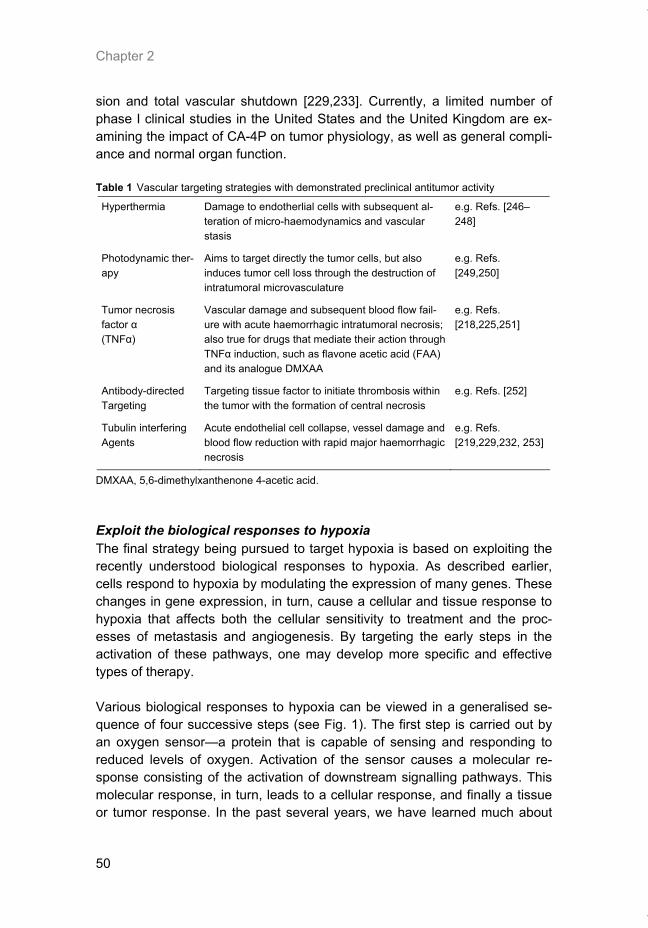
Chapter 2
50
sion and total vascular shutdown [229,233]. Currently, a limited number of phase I clinical studies in the United States and the United Kingdom are ex-amining the impact of CA-4P on tumor physiology, as well as general compli-ance and normal organ function. Table 1 Vascular targeting strategies with demonstrated preclinical antitumor activity
Hyperthermia Damage to endotherlial cells with subsequent al-teration of micro-haemodynamics and vascular stasis
e.g. Refs. [246–248]
Photodynamic ther-apy
Aims to target directly the tumor cells, but also induces tumor cell loss through the destruction of intratumoral microvasculature
e.g. Refs. [249,250]
Tumor necrosis factor α (TNFα)
Vascular damage and subsequent blood flow fail-ure with acute haemorrhagic intratumoral necrosis; also true for drugs that mediate their action through TNFα induction, such as flavone acetic acid (FAA) and its analogue DMXAA
e.g. Refs. [218,225,251]
Antibody-directed Targeting
Targeting tissue factor to initiate thrombosis within the tumor with the formation of central necrosis
e.g. Refs. [252]
Tubulin interfering Agents
Acute endothelial cell collapse, vessel damage and blood flow reduction with rapid major haemorrhagic necrosis
e.g. Refs. [219,229,232, 253]
DMXAA, 5,6-dimethylxanthenone 4-acetic acid.
Exploit the biological responses to hypoxia The final strategy being pursued to target hypoxia is based on exploiting the recently understood biological responses to hypoxia. As described earlier, cells respond to hypoxia by modulating the expression of many genes. These changes in gene expression, in turn, cause a cellular and tissue response to hypoxia that affects both the cellular sensitivity to treatment and the proc-esses of metastasis and angiogenesis. By targeting the early steps in the activation of these pathways, one may develop more specific and effective types of therapy. Various biological responses to hypoxia can be viewed in a generalised se-quence of four successive steps (see Fig. 1). The first step is carried out by an oxygen sensor—a protein that is capable of sensing and responding to reduced levels of oxygen. Activation of the sensor causes a molecular re-sponse consisting of the activation of downstream signalling pathways. This molecular response, in turn, leads to a cellular response, and finally a tissue or tumor response. In the past several years, we have learned much about
Thesis_Weppler_v12_fc.pdf

Hypoxia as a therapeutic target
51
one of the main hypoxic biological response pathways in mammalian cells—that involving the HIF-1 transcription factor. This pathway serves as a good example of this general response sequence and for how this knowledge can be translated into new cancer therapies. Two recent reports suggest that the oxygen sensor in the HIF-1 pathway is a prolyl hydroxlyase [110,111]. This enzyme, designated HIF-PH, requires oxy-gen for its activity (hydroxylation of proline residues). In this example, the mo-lecular response to hypoxia is initiated as a result of reduced hydroxylation of a proline residue in the HIF-1α subunit (P564). Reduced hydroxylation pre-vents the recognition of HIF-1α by the VHL ubiquitin ligase, thereby prevent-ing ubiquitination. As a result, HIF-1 is stabilised and can transactivate its many targets, such as EPO, VEGF and GLUT-1. These changes in gene ex-pression lead to a cellular response that may consist of increased glycolysis in the tumor cells or activation of endothelial cell proliferation and migration by binding of VEGF to its receptor. Finally, this leads to a tumor or tissue re-sponse that consists of increased angiogenesis, and to increased survival of tumor cells resulting from a switch to anaerobic metabolism [234]. The important part of this illustration is that a detailed biological understand-ing of this pathway offers a plethora of options for targeting cancer treatment to the tumor. For example, an attractive molecular treatment would be one based on augmenting the activity of the oxygen sensor itself. Since the multi-ple cellular and tissue effects stem from this one initial protein, it provides a very specific and potent treatment target. There are already many examples of research directed against the second level of this pathway. Several com-pounds designed to alter the activity of HIF-1 [235,236], VHL [237], or the ubiquitin system itself [238–240] are being explored in cancer treatment. At the level of the cellular response, antibodies and inhibitors of both VEGF and its receptor Flk-1 have been developed (as discussed under the anti-angiogenesis strategies). Recent reports suggest that inhibiting the ability of tumor cells to shift to glycolysis would also be advantageous [234,241]. Fi-nally, targeting treatment to the cellular or tissue response of this pathway would consist of the more generalised anti-angiogenesis and hypoxia-targeted therapeutics (both discussed earlier). It is clear that as one moves downwards in this pathway from the oxygen sensor to the cellular and tissue responses, the targets become less specific in nature. Elements of this pathway can be exploited as well as inhibited. For example, the DNA recognition sequence for the HIF-1 transcription factor is well de-scribed. This HRE can be inserted within gene therapy constructs, to limit the expression of therapeutic proteins to hypoxic areas of tumors [242–245].
Thesis_Weppler_v12_fc.pdf
Chapter 2
52
Dachs and colleagues [243] established the potential for tumor hypoxia to be exploited for targeted gene expression by showing that the HRE from the mouse PGK-1 gene could be used to drive expression of heterologous genes within the mass of a solid tumor. The HIF-1 pathway is relatively well understood and serves as a good exam-ple of how knowledge of the biological responses to hypoxia can translate into new therapies. However, there are numerous other molecular and cellu-lar responses to hypoxia that are independent of HIF-1, perhaps each with unique oxygen sensors. Continued research into the basic molecular and cellular responses of hypoxia will undoubtedly contribute further to the devel-opment of novel hypoxia-based cancer therapies.
Figure 1. Biological responses to hypoxia can be viewed in terms of four successive steps. The HIF-1 pathway serves as an example of such a response. The first step is to sense that oxygen is limiting and in the HIF-1 pathway this is carried out by an oxygen-dependent prolyl hydroxy-lase. The second step is the initiation of a molecular response through the activation of down-stream signalling pathways. In this example, this results in the activation of several classes of genes as a result of stabilisation of the HIF-1α subunit. A cellular response occurs due to these changes in gene expression, in this case resulting in a switch to anaerobic metabolism and se-cretion of angiogenic factors. Finally, a tumor/tissue response occurs. In the HIF-1 pathway, this may be the induction of angiogenesis in the tumor micro-environment together with increased survival and proliferation of the tumor cells. Each of these steps in the biological response to hypoxia is an opportunity for targeting therapy as indicated below each box.
Thesis_Weppler_v12_fc.pdf
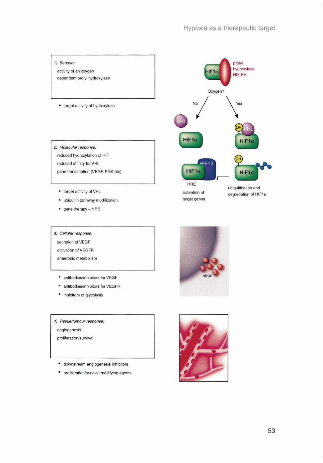
Hypoxia as a therapeutic target
53
Thesis_Weppler_v12_fc.pdf
Chapter 2
54
References 1. Knocke TH, Weitmann HD, Feldmann HJ, Selzer E, Potter R. Intratumoral pO2-
measurements as predictive assay in the treatment of carcinoma of the uterine cervix. Ra-diother Oncol 1999, 53, 99–104.
2. Hockel M, Schlenger K, Aral B, Mitze M, Schaffer U, Vaupel P. Association between tumor hypoxia and malignant progression in advanced cancer of the uterine cervix. Cancer Res 1996, 56, 4509–4515.
3. Nordsmark M, Overgaard M, Overgaard J. Pretreatment oxygenation predicts radiation response in advanced squamous cell carcinoma of the head and neck. Radiother Oncol 1996, 41, 31–39.
4. Brizel DM, Sibley GS, Prosnitz LR, Scher RL, Dewhirst MW. Tumor hypoxia adversely af-fects the prognosis of carcinoma of the head and neck. Int J Radiat Oncol Biol Phys 1997, 38, 285-289.
5. Lartigau E, Randrianarivelo H, Avril MF, et al. Intratumoral oxygen tension in metastatic melanoma. Melanoma Res 1997, 7, 400–406.
6. Rofstad EK, Maseide K. Radiobiological and immunohistochemical assessment of hypoxia in human melanoma xenografts: acute and chronic hypoxia in individual tumours. Int J Ra-diat Biol 1999, 75, 1377–1393.
7. Vaupel P, Schlenger K, Knoop C, Hockel M. Oxygenation of human tumors: evaluation of tissue oxygen distribution in breast cancers by computerized O2 tension measurements. Cancer Res 1991, 51, 3316–3322.
8. Runkel S, Wischnik A, Teubner J, Kaven E, Gaa J, Melchert F. Oxygenation of mammary tumors as evaluated by ultrasound-guided computerized-pO2-histography. Adv Exp Med Biol 1994, 345, 451–458.
9. Movsas B, Chapman JD, Horwitz EM, et al. Hypoxic regions exist in human prostate carci-noma. Urology 1999, 53, 11–18.
10. Nordsmark M, Loncaster J, Chou SC, et al. Invasive oxygen measurements and pimonida-zole labeling in human cervix carcinoma. Int J Radiat Oncol Biol Phys 2001, 49, 581–586.
11. Kavanagh MC, Tsang V, Chow S, et al. A comparison in individual murine tumors of tech-niques for measuring oxygen levels. Int J Radiat Oncol Biol Phys 1999, 44, 1137–1146.
12. Lee J, Siemann DW, Koch CJ, Lord EM. Direct relationship between radiobiological hy-poxia in tumors and monoclonal antibody detection of EF5 cellular adducts. Int J Cancer 1996, 67, 372–378.
13. Brizel DM, Dodge RK, Clough RW, Dewhirst MW. Oxygenation of head and neck cancer: changes during radiotherapy and impact on treatment outcome. Radiother Oncol 1999, 53, 113–117.
14. Brizel DM, Scully SP, Harrelson JM, et al. Tumor oxygenation predicts for the likelihood of distant metastases in human soft tissue sarcoma. Cancer Res 1996, 56, 941–943.
15. Fyles AW, Milosevic M, Wong R, et al. Oxygenation predicts radiation response and sur-vival in patients with cervix cancer. Radiother Oncol 1998, 48, 149–156.
16. Nordsmark M, Alsner J, Keller J, et al. Hypoxia in human soft tissue sarcomas: adverse impact on survival and no association with p53 mutations. Br J Cancer 2001, 84, 1070–1075.
17. Gray LH, Conger AD, Ebert M, Hornsey S, Scott OC. Concentration of oxygen dissolved in tissues at the time of irradiation as a factor in radiotherapy. Br J Radiol 1953, 26, 638–648.
18. Bedford JS, Mitchell JB. The effect of hypoxia on the growth and radiation response of mammalian cells in culture. Br J Radiol 1974, 47, 687–696.
19. Durand RE. The influence of microenvironmental factors during cancer therapy. In Vivo 1994, 8, 691–702.
20. Baish JW, Gazit Y, Berk DA, Nozue M, Baxter LT, Jain RK. Role of tumor vascular architec-ture in nutrient and drug delivery: an invasion percolation-based network model. Microvasc Res 1996, 51, 327–346.
21. Young SD, Marshall RS, Hill RP. Hypoxia induces DNA overreplication and enhances me-tastatic potential of murine tumor cells. Proc Natl Acad Sci USA 1988, 85, 9533–9537.
22. De Jaeger K, Kavanagh MC, Hill RP. Relationship of hypoxia to metastatic ability in rodent tumours. Br J Cancer 2001, 84, 1280–1285.
23. Plasswilm L, Tannapfel A, Cordes N, et al. Hypoxia-induced tumour cell migration in an in vivo chicken model. Pathobiology 2000, 68, 99–105.
Thesis_Weppler_v12_fc.pdf
Hypoxia as a therapeutic target
55
24. Curran S, Murray GI. Matrix metalloproteinases: molecular aspects of their roles in tumour invasion and metastasis. Eur J Cancer 2000, 36, 1621–1630.
25. Murray GI, Duncan ME, O’Neil P, McKay JA, Melvin WT, Fothergill JE. Matrix metallopro-teinase-1 is associated with poor prognosis in oesophageal cancer. J Pathol 1998, 185, 256–261.
26. Murray GI, Duncan ME, O’Neil P, Melvin WT, Fothergill JE. Matrix metalloproteinase-1 is associated with poor prognosis in colorectal cancer. Nat Med 1996, 2, 461–462.
27. Sier CF, Kubben FJ, Ganesh S, et al. Tissue levels of matrix metalloproteinases MMP-2 and MMP-9 are related to the overall survival of patients with gastric carcinoma. Br J Can-cer 1996, 74, 413–417.
28. Canning MT, Postovit LM, Clarke SH, Graham CH. Oxygen mediated regulation of gelati-nase and tissue inhibitor of metalloproteinases-1 expression by invasive cells. Exp Cell Res 2001, 367, 88–94.
29. Romanic AM, Burns-Kurtis CL, Gout B, Berrebi-Bertrand I, Ohlstein EH. Matrix metallopro-teinase expression in cardiac myocytes following myocardial infarction in the rabbit. Life Sci 2001, 68, 799–814.
30. Graham CH, Forsdike J, Fitzgerald CJ, Macdonald-Goodfellow S. Hypoxia-mediated stimula-tion of carcinoma cell invasiveness via upregulation of urokinase receptor expression. Int J Cancer 1999, 80, 617–623.
31. Maity A, Solomon D. Both increased stability and transcription contribute to the induction of the urokinase plasminogen activator receptor (uPAR) message by hypoxia. Exp Cell Res 2000, 255, 250–257.
32. MacDonald TJ, DeClerck YA, Laug WE. Urokinase induces receptor mediated brain tumor cell migration and invasion. J Neurooncol 1998, 40, 215–226.
33. Hasan NM, Adams GE, Joiner MC, Marshall JF, Hart IR. Hypoxia facilitates tumour cell detachment by reducing expression of surface adhesion molecules and adhesion to ex-tracellular matrices without loss of cell viability. Br J Cancer 1998, 77, 1799–1805.
34. Xu L, Xie K, Mukaida N, Matsushima K, Fidler IJ. Hypoxiainduced elevation in interleukin-8 expression by human ovarian carcinoma cells. Cancer Res 1999, 59, 5822–5829.
35. Shi Q, Le X, Abbruzzese JL, et al. Cooperation between transcription factor AP-1 and NF-kappaB in the induction of interleukin-8 in human pancreatic adenocarcinoma cells by hy-poxia. J Interferon Cytokine Res 1999, 19, 1363–1371.
36. Kunz M, Hartmann A, Flory E, et al. Anoxia-induced up-regulation of interleukin-8 in human malignant melanoma. A potential mechanism for high tumor aggressiveness. Am J Pathol 1999, 155, 753–763.
37. Inoue K, Slaton JW, Kim SJ, et al. Interleukin 8 expression regulates tumorigenicity and metastasis in human bladder cancer. Cancer Res 2000, 60, 2290–2299.
38. Luca M, Huang S, Gershenwald JE. Expression of interleukin-8 by human melanoma cells up-regulates MMP-2 activity and increases tumor growth and metastasis. Am J Pathol 1997, 151, 1105–1113.
39. Graeber TG, Osmanian C, Jacks T, et al. Hypoxia-mediated selection of cells with dimin-ished apoptotic potential in solid tumours. Nature 1996, 379, 88–91.
40. Gammack D, Byrne HM, Lewis CE. Estimating the selective advantage of mutant p53 tu-mour cells to repeated rounds of hypoxia. Bull Math Biol 2001, 63, 135–166.
41. Kim CY, Tsai MH, Osmanian C, et al. Selection of human cervical epithelial cells that pos-sess reduced apoptotic potential to low-oxygen conditions. Cancer Res 1997, 57, 4200–4204.
42. Royds JA, Dower SK, Qwarnstrom EE, Lewis CE. Response of tumour cells to hypoxia: role of p53 and NFkB. Mol Pathol 1998, 51, 55–61.
43. Zundel W, Schindler C, Haas-Kogan D, et al. Loss of PTEN facilitates HIF-1-mediated gene expression. Genes Dev 2000, 14, 391–396.
44. Hockel M, Schlenger K, Hockel S, Vaupel P. Hypoxic cervical cancers with low apoptotic index are highly aggressive. Cancer Res 1999, 59, 4525–4528.
45. Semenza GL. Regulation of hypoxia-induced angiogenesis: a chaperone escorts VEGF to the dance. J Clin Invest 2001, 108, 39–40.
46. Rofstad EK, Danielsen T. Hypoxia-induced metastasis of human melanoma cells: involve-ment of vascular endothelial growth factor-mediated angiogenesis. Br J Cancer 1999, 80, 1697–1707.
47. Veikkola T, Karkkainen M, Claesson-Welsh L, Alitalo K. Regulation of angiogenesis via vascular endothelial growth factor receptors. Cancer Res 2000, 60, 203–212.
Thesis_Weppler_v12_fc.pdf
Chapter 2
56
48. Aigner A, Butscheid M, Kunkel P, et al. An FGF-binding protein (FGF-BP) exerts its biologi-cal function by parallel paracrine stimulation of tumor cell and endothelial cell proliferation through FGF-2 release. Int J Cancer 2001, 92, 510–517.
49. Harris VK, Coticchia CM, Kagan BL, Ahmad S, Wellstein A, Riegel AT. Induction of the angiogenic modulator fibroblast growth factor-binding protein by epidermal growth factor is mediated through both MEK/ERK and p38 signal transduction pathways. J Biol Chem 2000, 275, 10802–10811.
50. Conrad PW, Rust RT, Han J, Millhorn DE, Beitner-Johnson D. Selective activation of p38alpha and p38gamma by hypoxia. Role in regulation of cyclin D1 by hypoxia in PC12 cells. J Biol Chem 1999, 274, 23570–23576.
51. Kuwabara K, Ogawa S, Matsumoto M, et al. Hypoxia-mediated induction of acidic/basic fibroblast growth factor and plateletderived growth factor in mononuclear phagocytes stimu-lates growth of hypoxic endothelial cells. Proc Natl Acad Sci USA 1995, 92, 4606–4610.
52. Walton HL, Corjay MH, Mohamed SN, Mousa SA, Santomenna LD, Reilly TM. Hypoxia induces differential expression of the integrin receptors alpha(vbeta3) and alpha(vbeta5) in cultured human endothelial cells. J Cell Biochem 2000, 78, 674–680.
53. Brooks PC, Stromblad S, Sanders LC, et al. Localization of matrix metalloproteinase MMP-2 to the surface of invasive cells by interaction with integrin alpha v beta 3. Cell 1996, 85, 683–693.
54. Soldi R, Mitola S, Strasly M, Defilippi P, Tarone G, Bussolino F. Role of alphavbeta3 in-tegrin in the activation of vascular endothelial growth factor receptor-2. Embo J 1999, 18, 882–892.
55. Sundfor K, Lyng H, Trope CG, Rofstad EK. Treatment outcome in advanced squamous cell carcinoma of the uterine cervix: relationships to pretreatment tumor oxygenation and vascu-larization. Radiother Oncol 2000, 54, 101–107.
56. Nordsmark M, Hoyer M, Keller J, Nielsen OS, Jensen OM, Overgaard J. The relationship between tumor oxygenation and cell proliferation in human soft tissue sarcomas. Int J Ra-diat Oncol Biol Phys 1996, 35, 701–708.
57. Adam MF, Gabalski EC, Bloch DA, et al. Tissue oxygen distribution in head and neck can-cer patients. Head Neck 1999, 21, 146–153.
58. Becker A, Hansgen G, Bloching M, Weigel C, Lautenschlager C, Dunst J. Oxygenation of squamous cell carcinoma of the head and neck: comparison of primary tumors, neck node metastases, and normal tissue. Int J Radiat Oncol Biol Phys 1998, 42, 35–41.
59. Nordsmark M, Overgaard J. A confirmatory prognostic study on oxygenation status and loco-regional control in advanced head and neck squamous cell carcinoma treated by ra-diation therapy. Radiother Oncol 2000, 57, 39–43.
60. Stadler P, Becker A, Feldmann HJ, et al. Influence of the hypoxic subvolume on the sur-vival of patients with head and neck cancer. Int J Radiat Oncol Biol Phys 1999, 44, 749–754.
61. Rudat V, Vanselow B, Wollensack P, et al. Repeatability and prognostic impact of the pre-treatment pO(2) histography in patients with advanced head and neck cancer. Radiother Oncol 2000, 57, 31–37.
62. Schwickert G, Walenta S, Sundfor K, Rofstad EK, Mueller-Klieser W. Correlation of high lactate levels in human cervical cancer with incidence of metastasis. Cancer Res 1995, 55, 4757–4759.
63. Walenta S, Salameh A, Lyng H, et al. Correlation of high lactate levels in head and neck tumors with incidence of metastasis. Am J Pathol 1997, 150, 409–415.
64. Vaupel P, Kallinowski F, Okunieff P. Blood flow, oxygen and nutrient supply, and metabolic microenvironment of human tumors: a review. Cancer Res 1989, 49, 6449–6465.
65. Birner P, Schindl M, Obermair A, Plank C, Breitenecker G, Oberhuber G. Overexpression of hypoxia-inducible factor 1alpha is a marker for an unfavorable prognosis in early-stage invasive cervical cancer. Cancer Res 2000, 60, 4693–4696.
66. Sundfor K, Lyng H, Rofstad EK. Tumour hypoxia and vascular density as predictors of me-tastasis in squamous cell carcinoma of the uterine cervix. Br J Cancer 1998, 78, 822–827.
67. Rofstad EK, Sundfor K, Lyng H, Trope CG. Hypoxia-induced treatment failure in advanced squamous cell carcinoma of the uterine cervix is primarily due to hypoxia-induced radiation resistance rather than hypoxia-induced metastasis. Br J Cancer 2000, 83, 354–359.
68. Shweiki D, Itin A, Soffer D, Keshet E. Vascular endothelial growth factor induced by hy-poxia may mediate hypoxia-initiated angiogenesis. Nature 1992, 359, 843–845.
Thesis_Weppler_v12_fc.pdf
Hypoxia as a therapeutic target
57
69. Kourembanas S, Hannan RL, Faller DV. Oxygen tension regulates the expression of the platelet-derived growth factor-B chain gene in human endothelial cells. J Clin Invest 1990, 86, 670–674.
70. Goldberg MA, Glass GA, Cunningham JM, Bunn HF. The regulated expression of erythro-poietin by two human hepatoma cell lines. Proc Natl Acad Sci USA 1987, 84, 7972–7976.
71. Wang GL, Jiang BH, Rue EA, Semenza GL. Hypoxia-inducible factor 1 is a basic-helix-loop-helix-PAS heterodimer regulated by cellular O2 tension. Proc Natl Acad Sci USA 1995, 92, 5510–5514.
72. McQuillan LP, Leung GK, Marsden PA, Kostyk SK, Kourembanas S. Hypoxia inhibits ex-pression of eNOS via transcriptional and posttranscriptional mechanisms. Am J Physiol 1994, 267, H1921–H1927.
73. Firth JD, Ebert BL, Pugh CW, Ratcliffe PJ. Oxygen-regulated control elements in the phosphoglycerate kinase 1 and lactate dehydrogenase A genes: similarities with the erythropoietin 30 enhancer. Proc Natl Acad Sci USA 1994, 91, 6496–6500.
74. Semenza GL, Roth PH, Fang HM, Wang GL. Transcriptional regulation of genes encoding glycolytic enzymes by hypoxiainducible factor 1. J Biol Chem 1994, 269, 23757–23763.
75. Sivitz WI, Lund DD, Yorek B, Grover-McKay M, Schmid PG. Pretranslational regulation of two cardiac glucose transporters in rats exposed to hypobaric hypoxia. Am J Physiol 1992, 263, E562–E569.
76. Ebert BL, Gleadle JM, O’Rourke JF, Bartlett SM, Poulton J, Ratcliffe PJ. Isoenzyme-specific regulation of genes involved in energy metabolism by hypoxia: similarities with the regulation of erythropoietin. Biochem J 1996, 313, 809–814.
77. Ausserer WA, Bourrat-Floeck B, Green CJ, Laderoute KR, Sutherland RM. Regulation of c-jun expression during hypoxic and low-glucose stress. Mol Cell Biol 1994, 14, 5032–5042.
78. Santilli SM, Fiegel VD, Aldridge DE, Knighton DR. Rabbit aortic endothelial cell hypoxia induces secretion of transforming growth factor beta and augments macrophage adhesion in vitro. Ann Vasc Surg 1991, 5, 429–438.
79. Gleadle JM, Ebert BL, Firth JD, Ratcliffe PJ. Regulation of angiogenic growth factor ex-pression by hypoxia, transition metals, and chelating agents. Am J Physiol 1995, 268, C1362–C1368.
80. Graham CH, Fitzpatrick TE, McCrae KR. Hypoxia stimulates urokinase receptor expression through a heme protein- dependent pathway. Blood 1998, 91, 3300–3307.
81. Czyzyk-Krzeska MF, Furnari BA, Lawson EE, Millhorn DE. Hypoxia increases rate of tran-scription and stability of tyrosine hydroxylase mRNA in pheochromocytoma (PC12) cells. J Biol Chem 1994, 269, 760–764.
82. Krtolica A, Krucher NA, Ludlow JW. Hypoxia-induced pRB hypophosphorylation results from downregulation of CDK and upregulation of PP1 activities. Oncogene 1998, 17, 2295–2304.
83. Carmeliet P, Dor Y, Herbert JM, et al. Role of HIF-1alpha in hypoxia-mediated apoptosis, cell proliferation and tumour angiogenesis. Nature 1998, 394, 485–490.
84. Forsythe JA, Jiang BH, Iyer NV, et al. Activation of vascular endothelial growth factor gene transcription by hypoxia-inducible factor 1. Mol Cell Biol 1996, 16, 4604–4613.
85. Levy NS, Chung S, Furneaux H, Levy AP. Hypoxic stabilization of vascular endothelial growth factor mRNA by the RNAbinding protein HuR. J Biol Chem 1998, 273, 6417–6423.
86. Ozawa K, Kondo T, Hori O, et al. Expression of the oxygenregulated protein ORP150 ac-celerates wound healing by modulating intracellular VEGF transport. J Clin Invest 2001, 108, 41–50.
87. Stein I, Itin A, Einat P, Skaliter R, Grossman Z, Keshet E. Translation of vascular endothe-lial growth factor mRNA by internal ribosome entry: implications for translation under hy-poxia. Mol Cell Biol 1998, 18, 3112–3119.
88. Akiri G, Nahari D, Finkelstein Y, Le SY, Elroy-Stein O, Levi BZ. Regulation of vascular en-dothelial growth factor (VEGF) expression is mediated by internal initiation of translation and alternative initiation of transcription. Oncogene 1998, 17, 227–236.
89. Miller DL, Dibbens JA, Damert A, Risau W, Vadas MA, Goodall GJ. The vascular endothe-lial growth factor mRNA contains an internal ribosome entry site. FEBS Lett 1998, 434, 417–420.
90. Semenza GL, Wang GL. A nuclear factor induced by hypoxia via de novo protein synthesis binds to the human erythropoietin gene enhancer at a site required for transcriptional acti-vation. Mol Cell Biol 1992, 12, 5447–5454.
Thesis_Weppler_v12_fc.pdf
Chapter 2
58
91. Graeber TG, Peterson JF, Tsai M, Monica K, Fornace Jr AJ, Giaccia AJ. Hypoxia induces accumulation of p53 protein, but activation of a G1-phase checkpoint by low-oxygen condi-tions is independent of p53 status. Mol Cell Biol 1994, 14, 6264–6277.
92. Yao KS, Xanthoudakis S, Curran T, O’Dwyer PJ. Activation of AP-1 and of a nuclear redox factor, Ref-1, in the response of HT29 colon cancer cells to hypoxia. Mol Cell Biol 1994, 14, 5997–6003.
93. Yan SF, Zou YS, Mendelsohn M, et al. Nuclear factor interleukin 6 motifs mediate tissue-specific gene transcription in hypoxia. J Biol Chem 1997, 272, 4287–4294.
94. Yan SF, Lu J, Zou YS, Soh-Won J, et al. Hypoxia-associated induction of early growth re-sponse-1 gene expression. J Biol Chem 1999, 274, 15030–15040.
95. Koong AC, Chen EY, Giaccia AJ. Hypoxia causes the activation of nuclear factor kappa B through the phosphorylation of I kappa B alpha on tyrosine residues. Cancer Res 1994, 54, 1425–1430.
96. Semenza GL. Hypoxia-inducible factor 1: control of oxygen homeostasis in health and dis-ease. Pediatr Res 2001, 49, 614–617.
97. Madan A, Curtin PT. A 24-base-pair sequence 30 to the human erythropoietin gene con-tains a hypoxia-responsive transcriptional enhancer. Proc Natl Acad Sci USA 1993, 90, 3928–3932.
98. Minchenko A, Salceda S, Bauer T, Caro J. Hypoxia regulatory elements of the human vas-cular endothelial growth factor gene. Cell Mol Biol Res 1994, 40, 35–39.
99. Melillo G, Musso T, Sica A, Taylor LS, Cox GW, Varesio L. A hypoxia-responsive element mediates a novel pathway of activation of the inducible nitric oxide synthase promoter. J Exp Med 1995, 182, 1683–1693.
100. Gerber HP, Condorelli F, Park J, Ferrara N. Differential transcriptional regulation of the two vascular endothelial growth factor receptor genes. Flt-1, but not Flk-1/KDR, is up-regulated by hypoxia. J Biol Chem 1997, 272, 23659–23667.
101. Iyer NV, Kotch LE, Agani F, et al. Cellular and developmental control of O2 homeostasis by hypoxia-inducible factor 1 alpha. Genes Dev 1998, 12, 149–162.
102. Beck I, Weinmann R, Caro J. Characterization of hypoxiaresponsive enhancer in the hu-man erythropoietin gene shows presence of hypoxia-inducible 120-Kd nuclear DNA-binding protein in erythropoietin-producing and nonproducing cells. Blood 1993, 82, 704–711.
103. Wang GL, Semenza GL. Characterization of hypoxia-inducible factor 1 and regulation of DNA binding activity by hypoxia. J Biol Chem 1993, 268, 21513–21518.
104. Ebert BL, Bunn HF. Regulation of transcription by hypoxia requires a multiprotein complex that includes hypoxia-inducible factor 1, an adjacent transcription factor, and p300/CREB binding protein. Mol Cell Biol 1998, 18, 4089–4096.
105. Salceda S, Caro J. Hypoxia-inducible factor 1alpha (HIF-1alpha) protein is rapidly degraded by the ubiquitin-proteasome system under normoxic conditions. Its stabilization by hypoxia depends on redox-induced changes. J Biol Chem 1997, 272, 22642–22647.
106. Wiener CM, Booth G, Semenza GL. In vivo expression of mRNAs encoding hypoxia-inducible factor 1. Biochem Biophys Res Commun 1996, 225, 485–488.
107. Maxwell PH, Wiesener MS, Chang GW, et al. The tumour suppressor protein VHLtargets hypoxia-inducible factors for oxygen-dependent proteolysis. Nature 1999, 399, 271–275.
108. Huang LE, Gu J, Schau M, Bunn HF. Regulation of hypoxiainducible factor 1alpha is medi-ated by an O2-dependent degradation domain via the ubiquitin-proteasome pathway. Proc Natl Acad Sci USA 1998, 95, 7987–7992.
109. Kallio PJ, Wilson WJ, O’Brien S, Makino Y, Poellinger L. Regulation of the hypoxia-inducible transcription factor 1alpha by the ubiquitin-proteasome pathway. J Biol Chem 1999, 274, 6519–6525.
110. Jaakkola P, Mole DR, Tian YM, et al. Targeting of HIF-alpha to the von Hippel-Lindau ubiq-uitylation complex by O2-regulated prolyl hydroxylation. Science 2001, 292, 468–472.
111. Ivan M, Kondo K, Yang H, et al. HIFalpha targeted for VHLmediated destruction by proline hydroxylation: implications for O2 sensing. Science 2001, 292, 464–468.
112. Overgaard J, Horsman MR. Modification of hypoxia-induced radioresistance in tumors by the use of oxygen and sensitizers. Semin Radiat Oncol 1996, 6, 10–21.
113. Saunders M, Dische S. Clinical results of hypoxic cell radiosensitisation from hyperbaric oxygen to accelerated radiotherapy, carbogen and nicotinamide. Br J Cancer Suppl 1996, 27, S271–S278.
114. Overgaard J. Sensitization of hypoxic tumour cells—clinical experience. Int J Radiat Biol 1989, 56, 801–811.
Thesis_Weppler_v12_fc.pdf
Hypoxia as a therapeutic target
59
115. Pedersen D, Sogaard H, Overgaard J, Bentzen SM. Prognostic value of pretreatment fac-tors in patients with locally advanced carcinoma of the uterine cervix treated by radiother-apy alone. Acta Oncol 1995, 34, 787–795.
116. Girinski T, Pejovic-Lenfant MH, Bourhis J, et al. Prognostic value of hemoglobin concentra-tions and blood transfusions in advanced carcinoma of the cervix treated by radiation ther-apy: results of a retrospective study of 386 patients. Int J Radiat Oncol Biol Phys 1989, 16, 37–42.
117. Werner-Wasik M, Schmid CH, Bornstein L, Ball HG, Smith DM, Madoc-Jones H. Prognostic factors for local and distant recurrence in stage I and II cervical carcinoma. Int J Radiat On-col Biol Phys 1995, 32, 1309–1317.
118. Fein DA, Lee WR, Hanlon AL, et al. Pretreatment hemoglobin level influences local control and survival of T1-T2 squamous cell carcinomas of the glottic larynx. J Clin Oncol 1995, 13, 2077–2083.
119. Lee WR, Berkey B, Marcial V, et al. Anemia is associated with decreased survival and in-creased locoregional failure in patients with locally advanced head and neck carcinoma: a secondary analysis of RTOG 85-27. Int J Radiat Oncol Biol Phys 1998, 42, 1069–1075.
120. van Acht MJ, Hermans J, Boks DE, Leer JW. The prognostic value of hemoglobin and a decrease in hemoglobin during radiotherapy in laryngeal carcinoma. Radiother Oncol 1992, 23, 229–235.
121. Warde P, O’Sullivan B, Bristow RG, et al. T1/T2 glottic cancer managed by external beam radiotherapy: the influence of pretreatment hemoglobin on local control. Int J Radiat Oncol Biol Phys 1998, 41, 347–353.
122. Dubray B, Mosseri V, Brunin F, et al. Anemia is associated with lower local-regional control and survival after radiation therapy for head and neck cancer: a prospective study. Radiol-ogy 1996, 201, 553–558.
123. Dische S, Warburton MF, Saunders MI. Radiation myelitis and survival in the radiotherapy of lung cancer. Int J Radiat Oncol Biol Phys 1988, 15, 75–81.
124. Macchiarini P, Silvano G, Janni A, Mussi A, Chella A, Angeletti CA. Results of treatment and lessons learned from pathologically staged T4 non-small cell lung cancer. J Surg Oncol 1991, 47, 209–214.
125. Sasai K, Ono K, Hiraoka M, et al. The effect of arterial oxygen content on the results of radiation therapy for epidermoid bronchogenic carcinoma. Int J Radiat Oncol Biol Phys 1989, 16, 1477–1481.
126. Cole CJ, Pollack A, Zagars GK, Dinney CP, Swanson DA, von Eschenbach AC. Local con-trol of muscle-invasive bladder cancer: preoperative radiotherapy and cystectomy versus cystectomy alone. Int J Radiat Oncol Biol Phys 1995, 32, 331–340.
127. Hannisdal E, Fossa SD, Host H. Blood tests and prognosis in bladder carcinomas treated with definitive radiotherapy. Radiother Oncol 1993, 27, 117–122.
128. Wijkstrom H, Nilsson B, Tribukait B. DNA analysis in predicting survival of irradiated pa-tients with transitional cell carcinoma of bladder. Br J Urol 1992, 69, 49–55.
129. Greven KM, Solin LJ, Hanks GE. Prognostic factors in patients with bladder carcinoma treated with definitive irradiation. Cancer 1990, 65, 908–912.
130. Dunphy EP, Petersen IA, Cox RS, Bagshaw MA. The influence of initial hemoglobin and blood pressure levels on results of radiation therapy for carcinoma of the prostate. Int J Ra-diat Oncol Biol Phys 1989, 16, 1173–1178.
131. Bush RS. The significance of anemia in clinical radiation therapy. Int J Radiat Oncol Biol Phys 1986, 12, 2047–2050.
132. Lavey RS. Clinical trial experience using erythropoietin during radiation therapy. Strahlen-ther Onkol 1998, 174(Suppl. 4), 24–30.
133. Abels RI. Use of recombinant human erythropoietin in the treatment of anemia in patients who have cancer. Semin Oncol 1992, 19(Suppl. 8), 29–35.
134. Glaser CM, Millesi W, Kornek GV, et al. Impact of hemoglobin level and use of recombinant erythropoietin on efficacy of preoperative chemoradiation therapy for squamous cell carci-noma of the oral cavity and oropharynx. Int J Radiat Oncol Biol Phys 2001, 50, 705–715.
135. Rojas A, Joiner MC, Denekamp J. Extrapolations from laboratory and preclinical studies for the use of carbogen and nicotinamide in radiotherapy. Radiother Oncol 1992, 24, 123–124.
136. Chaplin DJ, Horsman MR, Trotter MJ. Effect of nicotinamide on the microregional hetero-geneity of oxygen delivery within a murine tumor. J Natl Cancer Inst 1990, 82, 672–676.
137. Rojas A. Radiosensitization with normobaric oxygen and carbogen. Radiother Oncol 1991, 20(Suppl. 1), 65–70.
Thesis_Weppler_v12_fc.pdf
Chapter 2
60
138. Martin L, Lartigau E, Weeger P, et al. Changes in the oxygenation of head and neck tumors during carbogen breathing. Radiother Oncol 1993, 27, 123–130.
139. Horsman MR, Nordsmark M, Khalil AA, et al. Reducing acute and chronic hypoxia in tu-mours by combining nicotinamide with carbogen breathing. Acta Oncol 1994, 33, 371–376.
140. Laurence VM, Ward R, Dennis IF, Bleehen NM. Carbogen breathing with nicotinamide improves the oxygen status of tumours in patients. Br J Cancer 1995, 72, 198–205.
141. Siemann DW, Horsman MR, Chaplin DJ. The radiation response of KHT sarcomas follow-ing nicotinamide treatment and carbogen breathing. Radiother Oncol 1994, 31, 117–122.
142. Fenton BM. The effects of carbogen and nicotinamide on intravascular oxyhaemoglobin saturations in SCCVII and KHT murine tumours. Br J Cancer 1995, 71, 945–949.
143. Rojas A, Hirst VK, Calvert AS, Johns H. Carbogen and nicotinamide as radiosensitizers in a murine mammary carcinoma using conventional and accelerated radiotherapy. Int J Radiat Oncol Biol Phys 1996, 34, 357–365.
144. Denekamp J, Fowler JF. ARCON—current status: summary of a workshop on preclinical and clinical studies. Acta Oncol 1997, 36, 517–525.
145. Fenton BM, Lord EM, Paoni SF. Enhancement of tumor perfusion and oxygenation by car-bogen and nicotinamide during single- and multifraction irradiation. Radiat Res 2000, 153, 75–83.
146. Kaanders JH, Pop LA, Marres HA, et al. Accelerated radiotherapy with carbogen and nicotinamide (ARCON) for laryngeal cancer. Radiother Oncol 1998, 48, 115–122.
147. Bussink J, Kaanders JH, Van der Kogel AJ. Clinical outcome and tumour microenvironmen-tal effects of accelerated radiotherapy with carbogen and nicotinamide. Acta Oncol 1999, 38, 875–882.
148. Hoskin PJ, Saunders MI, Dische S. Hypoxic radiosensitizers in radical radiotherapy for patients with bladder carcinoma: hyperbaric oxygen, misonidazole, and accelerated radio-therapy, carbogen, and nicotinamide. Cancer 1999, 86, 1322–1328.
149. Bernier J, Denekamp J, Rojas A, et al. ARCON: accelerated radiotherapy with carbogen and nicotinamide in head and neck squamous cell carcinomas. The experience of the Co-operative group of radiotherapy of the European Organization for Research and Treatment of Cancer (EORTC). Radiother Oncol 2000, 55, 111–119.
150. Bernier J, Denekamp J, Rojas A, et al. ARCON: accelerated radiotherapy with carbogen and nicotinamide in non small cell lung cancer: a phase I/II study by the EORTC. Radiother Oncol 1999, 52, 149–156.
151. Miralbell R, Mornex F, Greiner R, et al. Accelerated radiotherapy, carbogen, and nicotina-mide in glioblastoma multiforme: report of European Organization for Research and Treat-ment of Cancer trial 22933. J Clin Oncol 1999, 17, 3143–3149.
152. Overgaard J, Overgaard M, Timothy AR. Studies of the pharmacokinetic properties of ni-morazole. Br J Cancer 1983, 48, 27–34.
153. Timothy AR, Overgaard J, Overgaard M. A phase I clinical study of Nimorazole as a hy-poxic radiosensitizer. Int J Radiat Oncol Biol Phys 1984, 10, 1765–1768.
154. Overgaard J, Hansen HS, Overgaard M, et al. A randomized double-blind phase III study of nimorazole as a hypoxic radiosensitizer of primary radiotherapy in supraglottic larynx and pharynx carcinoma. Results of the Danish Head and Neck Cancer Study (DAHANCA) Pro-tocol 5-85. Radiother Oncol 1998, 46, 135–146.
155. von Pawel J, von Roemeling R, Gatzemeier U, et al. Tirapazamine plus cisplatin versus cisplatin in advanced non-smallcell lung cancer: a report of the international CATAPULT I study group. Cisplatin and tirapazamine in subjects with advanced previously untreated non-small-cell lung tumors. J Clin Oncol 2000, 18, 1351–1359.
156. Rischin D, Peters L, Hicks R, et al. Phase I trial of concurrent tirapazamine, cisplatin, and radiotherapy in patients with advanced head and neck cancer. J Clin Oncol 2001, 19, 535–542.
157. Craighead PS, Pearcey R, Stuart G. A phase I/II evaluation of tirapazamine administered intravenously concurrent with cisplatin and radiotherapy in women with locally advanced cervical cancer. Int J Radiat Oncol Biol Phys 2000, 48, 791–795.
158. Wouters BG, Wang LH, Brown JM. Tirapazamine: a new drug producing tumor specific enhancement of platinum-based chemotherapy in non-small-cell lung cancer. Ann Oncol 1999, 10(Suppl. 5), S29–S33.
159. Brown JM. SR 4233 (tirapazamine): a new anticancer drug exploiting hypoxia in solid tu-mours. Br J Cancer 1993, 67, 1163–1170.
Thesis_Weppler_v12_fc.pdf
Hypoxia as a therapeutic target
61
160. Kovacs MS, Hocking DJ, Evans JW, Siim BG, Wouters BG, Brown JM. Cisplatin anti-tumour potentiation by tirapazamine results from a hypoxia-dependent cellular sensitization to cisplatin. Br J Cancer 1999, 80, 1245–1251.
161. Siemann DW, Hinchman CA. Potentiation of cisplatin activity by the bioreductive agent tirapazamine. Radiother Oncol 1998, 47, 215–520.
162. Treat J, Johnson E, Langer C, et al. Tirapazamine with cisplatin in patients with advanced non-small-cell lung cancer: a phase II study. J Clin Oncol 1998, 16, 3524–3527.
163. Miller VA, Ng KK, Grant SC, et al. Phase II study of the combination of the novel bioreduc-tive agent, tirapazamine, with cisplatin in patients with advanced non-small-cell lung cancer. Ann Oncol 1997, 8, 1269–1271.
164. Goldberg Z, Evans J, Birrell G, Brown JM. An investigation of the molecular basis for the synergistic interaction of tirapazamine and cisplatin. Int J Radiat Oncol Biol Phys 2001, 49, 175–182.
165. Patterson LH, McKeown SR. AQ4N: a new approach to hypoxia-activated cancer chemo-therapy. Br J Cancer 2000, 83, 1589–1993.
166. Patterson LH, McKeown SR, Ruparelia K, et al. Enhancement of chemotherapy and radio-therapy of murine tumours by AQ4N, a bioreductively activated anti-tumour agent. Br J Cancer 2000, 82, 1984–1990.
167. McKeown SR, Hejmadi MV, McIntyre IA, McAleer JJ, Patterson LH. AQ4N: an alkylamino-anthraquinone N-oxide showing bioreductive potential and positive interaction with radiation in vivo. Br J Cancer 1995, 72, 76–81.
168. Minton NP, Mauchline ML, Lemmon MJ, et al. Chemotherapeutic tumour targeting using clostridial spores. FEMS Microbiol Rev 1995, 17, 357–364.
169. Lambin P, Theys J, Landuyt W, et al. Colonisation of Clostridium in the body is restricted to hypoxic and necrotic areas of tumours. Anaerobe 1998, 4, 183–188.
170. Low KB, Ittensohn M, Le T, et al. Lipid A mutant Salmonella with suppressed virulence and TNFalpha induction retain tumor-targeting in vivo. Nat Biotechnol 1999, 17, 37–41.
171. Pawelek JM, Low KB, Bermudes D. Tumor-targeted Salmonella as a novel anticancer vec-tor. Cancer Res 1997, 57, 4537–4544.
172. Theys J, Landuyt W, Nuyts S, et al. Improvement of Clostridium tumour targeting vectors evaluated in rat rhabdomyosarcomas. FEMS Immunol Med Microbiol 2001, 30, 37–41.
173. Theys J, Nuyts S, Landuyt W, et al. Stable Escherichia coli-Clostridium acetobutylicum shuttle vector for secretion of murine tumor necrosis factor alpha. Appl Environ Microbiol 1999, 65, 4295–4300.
174. Fox ME, Lemmon MJ, Mauchline ML, et al. Anaerobic bacteria as a delivery system for cancer gene therapy: in vitro activation of 5-fluorocytosine by genetically engineered clos-tridia. Gene Ther 1996, 3, 173–178.
175. Lemmon MJ, van Zijl P, Fox ME, et al. Anaerobic bacteria as a gene delivery system that is controlled by the tumor microenvironment. Gene Ther 1997, 4, 791–796.
176. Theys J, Landuyt W, Nuyts S, et al. Specific targeting of cytosine deaminase to solid tu-mors by engineered Clostridium acetobutylicum. Cancer Gene Ther 2001, 8, 294–297.
177. Nuyts S, Van Mellaert L, Theys J, Landuyt W, Lambin P, Anne J. The use of radiation-induced bacterial promoters in anaerobic conditions: a means to control gene expression in clostridiummediated therapy for cancer. Radiat Res 2001, 155, 716–723.
178. Yazawa K, Fujimori M, Amano J, Kano Y, Taniguchi S. Bifidobacterium longum as a deliv-ery system for cancer gene therapy: selective localization and growth in hypoxic tumors. Cancer Gene Ther 2000, 7, 269–274.
179. Konerding MA, Malkusch W, Klapthor B, et al. Evidence for characteristic vascular patterns in solid tumours: quantitative studies using corrosion casts. Br J Cancer 1999, 80, 724–732.
180. Konerding MA, Miodonski AJ, Lametschwandtner A. Microvascular corrosion casting in the study of tumor vascularity: a review. Scanning Microsc 1995, 9, 1233–1243.
181. Denekamp J. Vascular attack as a therapeutic strategy for cancer. Cancer Metastasis Rev 1990, 9, 267–282.
182. Denekamp J, Hobson B. Endothelial-cell proliferation in experimental tumours. Br J Cancer 1982, 46, 711–720.
183. Folkman J. Tumor angiogenesis: therapeutic implications. N Engl J Med 1971, 285, 1182–1186.
184. Hanahan D, Folkman J. Patterns and emerging mechanisms of the angiogenic switch dur-ing tumorigenesis. Cell 1996, 86, 353–364.
Thesis_Weppler_v12_fc.pdf
Chapter 2
62
185. Hiraoka N, Allen E, Apel IJ, Gyetko MR, Weiss SJ. Matrix metalloproteinases regulate ne-ovascularization by acting as pericellular fibrinolysins. Cell 1998, 95, 365–377.
186. Zucker S, Cao J, Chen WT. Critical appraisal of the use of matrix metalloproteinase inhibi-tors in cancer treatment. Oncogene 2000, 19, 6642–6650.
187. Towle MJ, Lee A, Maduakor EC, Schwartz CE, Bridges AJ, Littlefield BA. Inhibition of urokinase by 4-substituted benzo[b]thiophene-2- carboxamidines: an important new class of selective synthetic urokinase inhibitor. Cancer Res 1993, 53, 2553–2559.
188. Marshall E. The power of the front page of The New York Times. Science 1998, 280, 996–997.
189. O’Reilly MS, Holmgren L, Shing Y, et al. Angiostatin: a novel angiogenesis inhibitor that mediates the suppression of metastases by a Lewis lung carcinoma. Cell 1994, 79, 315–328.
190. Holmgren L, O’Reilly MS, Folkman J. Dormancy of micrometastases: balanced proliferation and apoptosis in the presence of angiogenesis suppression. Nat Med 1995, 1, 149–153.
191. O’Reilly MS, Boehm T, Shing Y, et al. Endostatin: an endogenous inhibitor of angiogenesis and tumor growth. Cell 1997, 88, 277–285.
192. Yokoyama Y, Dhanabal M, Griffioen AW, Sukhatme VP, Ramakrishnan S. Synergy be-tween angiostatin and endostatin: inhibition of ovarian cancer growth. Cancer Res 2000, 60, 2190–2196.
193. D’Amato RJ, Loughnan MS, Flynn E, Folkman J. Thalidomide is an inhibitor of angiogene-sis. Proc Natl Acad Sci USA 1994, 91, 4082–4085.
194. Minchinton AI, Fryer KH, Wendt KR, Clow KA, Hayes MM. The effect of thalidomide on experimental tumors and metastases. Anticancer Drugs 1996, 7, 339–343.
195. Figg WD, Dahut W, Duray P, et al. A randomized phase II trial of thalidomide, an angio-genesis inhibitor, in patients with androgen-independent prostate cancer. Clin Cancer Res 2001, 7, 1888–1893.
196. Fine HA, Figg WD, Jaeckle K, et al. Phase II trial of the antiangiogenic agent thalidomide in patients with recurrent highgrade gliomas. J Clin Oncol 2000, 18, 708–715.
197. Fong TA, Shawver LK, Sun L, et al. SU5416 is a potent and selective inhibitor of the vascu-lar endothelial growth factor receptor (Flk-1/KDR) that inhibits tyrosine kinase catalysis, tu-mor vascularization, and growth of multiple tumor types. Cancer Res 1999, 59, 99–106.
198. Lair AD, Vajkoczy P, Shawver LK, et al. SU6668 is a potent antiangiogenic and antitumor agent that induces regression of established tumors. Cancer Res 2000, 60, 4152–4160.
199. Usman N, Blatt LM. Nuclease-resistant synthetic ribozymes: developing a new class of therapeutics. J Clin Invest 2000, 106, 1197–1202.
200. Asano M, Yukita A, Matsumoto T, Hanatani M, Suzuki H. An anti-human VEGF monoclonal antibody, MV833, that exhibits potent anti-tumor activity in vivo. Hybridoma 1998, 17, 185–190.
201. Rubenstein JL, Kim J, Ozawa T, et al. Anti-VEGF antibody treatment of glioblastoma pro-longs survival but results in increased vascular cooption. Neoplasia 2000, 2, 306–314.
202. Wood JM, Bold G, Buchdunger E, et al. PTK787/ZK 222584, a novel and potent inhibitor of vascular endothelial growth factor receptor tyrosine kinases, impairs vascular endothelial growth factor-induced responses and tumor growth after oral administration. Cancer Res 2000, 60, 2178–2189.
203. Friedlander M, Brooks PC, Shaffer RW, Kincaid CM, Varner JA, Cheresh DA. Definition of two angiogenic pathways by distinct alpha v integrins. Science 1995, 270, 1500–1502.
204. Brooks PC, Clark RA, Cheresh DA. Requirement of vascular integrin alpha v beta 3 for angiogenesis. Science 1994, 264, 569–571.
205. Brooks PC,Montgomery AM, RosenfeldM, et al. Integrin alpha v beta 3 antagonists promote tumor regression by inducing apoptosis of angiogenic blood vessels. Cell 1994, 97, 1157–1164.
206. Brooks PC, Stromblad S, Klemke R, Visscher D, Sarkar FH, Cheresh DA. Antiintegrin alpha v beta 3 blocks human breast cancer growth and angiogenesis in human skin. J Clin Invest 1995, 96, 1815–1822.
207. Pasqualini R, Koivunen E, Ruoslahti E. Alpha v integrins as receptors for tumor targeting by circulating ligands. Nat Biotechnol 1997, 15, 542–546.
208. Wu H, Beuerlein G, Nie Y, et al. Stepwise in vitro affinity maturation of Vitaxin, an alphav beta3- specific humanized mAb. Proc Natl Acad Sci USA 1998, 95, 6037–6042.
Thesis_Weppler_v12_fc.pdf
Hypoxia as a therapeutic target
63
209. Gutheil JC, Campbell TN, Pierce PR, et al. Targeted antiangiogenic therapy for cancer using Vitaxin: a humanized monoclonal antibody to the integrin alphavbeta3. Clin Cancer Res 2000, 6, 3056–3061.
210. Kohn EC, Felder CC, Jacobs W, et al. Structure-function analysis of signal and growth inhibition by carboxyamido-triazole, CAI. Cancer Res 1994, 54, 935–942.
211. Cavallo F, Di Carlo E, Butera M, et al. Immune events associated with the cure of estab-lished tumors and spontaneous metastases by local and systemic interleukin 12. Cancer Res 1999, 59, 414–421.
212. Voest EE, Kenyon BM, O’Reilly MS, Truitt G, D’Amato RJ, Folkman J. Inhibition of angio-genesis in vivo by interleukin 12. J Natl Cancer Inst 1995, 87, 581–586.
213. Coughlin CM, Salhany KE, Wysocka M, et al. Interleukin-12 and interleukin-18 synergisti-cally induce murine tumor regression which involves inhibition of angiogenesis. J Clin In-vest 1998, 101, 1441–1452.
214. Haicheur N, Escudier B, Dorval T, et al. Cytokines and soluble cytokine receptor induction after IL-12 administration in cancer patients. Clin Exp Immunol 2000, 119, 28–37.
215. DeVore RF, Hellerqvist CG, Wakefield GB, et al. Phase I study of the antineovasculariza-tion drug CM101. Clin Cancer Res 1997, 3, 365–372.
216. Denekamp J. Vascular endothelium as the vulnerable element in tumours. Acta Radiol Oncol 1984, 23, 217–225.
217. Wouters BG, Brown JM. Cells at intermediate oxygen levels can be more important than the ‘‘hypoxic fraction’’ in determining tumor response to fractionated radiotherapy. Radiat Res 1997, 147, 541–550.
218. Hill SA, Lonergan SJ, Denekamp J, Chaplin DJ. Vinca alkaloids: anti-vascular effects in a murine tumour. Eur J Cancer 1993, 9, 1320–1324.
219. Horsman MR, Ehrnrooth E, Ladekarl M, Overgaard J. The effect of combretastatin A-4 disodium phosphate in a C3H mouse mammary carcinoma and a variety of murine sponta-neous tumors. Int J Radiat Oncol Biol Phys 1998, 42, 895–898.
220. Zwi LJ, Baguley BC, Gavin JB, Wilson WR. The use of vascularised spheroids to investi-gate the action of flavone acetic acid on tumour blood vessels. Br J Cancer 1990, 62, 231–237.
221. Mahadevan V, Malik ST, Meager A, Fiers W, Lewis GP, Hart IR. Role of tumor necrosis factor in flavone acetic acid-induced tumor vasculature shutdown. Cancer Res 1990, 50, 5537–5542.
222. Futami H, Eader LA, Komschlies KL, et al. Flavone acetic acid directly induces expression of cytokine genes in mouse splenic leukocytes but not in human peripheral blood leuko-cytes. Cancer Res 1991, 51, 6596–6602.
223. Joseph WR, Cao Z, Mountjoy KG, Marshall ES, Baguley BC, Ching LM. Stimulation of tu-mors to synthesize tumor necrosis factor-alpha in situ using 5,6-dimethylxanthenone-4-acetic acid: a novel approach to cancer therapy. Cancer Res 1999, 59, 633–638.
224. Ching LM, Joseph WR, Crosier KE, Baguley BC. Induction of tumor necrosis factor-alpha messenger RNA in human and murine cells by the flavone acetic acid analogue 5,6-dimethylxanthenone-4-acetic acid (NSC 640488). Cancer Res 1994, 54, 870–872.
225. Baguley BC, Holdaway KM, Thomsen LL, Zhuang L, Zwi LJ. Inhibition of growth of colon 38 adenocarcinoma by vinblastine and colchicine: evidence for a vascular mechanism. Eur J Cancer 1991, 27, 482–487.
226. Pettit GR, Singh SB, Boyd MR, et al. Antineoplastic agents. 291. Isolation and synthesis of combretastatins A-4, A-5, and A-6(1a). J Med Chem 1995, 38, 1666–1672.
227. Pettit GR, Singh SB, Hamel E, Lin CM, Alberts DS, Garcia-Kendall D. Isolation and struc-ture of the strong cell growth and tubulin inhibitor combretastatin A-4. Experientia 1989, 45, 209–211.
228. Pettit GR, Temple Jr C, Narayanan VL, et al. Antineoplastic agents 322. synthesis of com-bretastatin A-4 prodrugs. Anticancer Drug Des 1995, 10, 299–309.
229. Dark GG, Hill SA, Prise VE, Tozer GM, Pettit GR, Chaplin DJ. Combretastatin A-4, an agent that displays potent and selective toxicity toward tumor vasculature. Cancer Res 1997, 57, 1829–1834.
230. Li L, Rojiani A, Siemann DW. Targeting the tumor vasculature with combretastatin A-4 disodium phosphate: effects on radiation therapy. Int J Radiat Oncol Biol Phys 1998, 42, 899–903.
Thesis_Weppler_v12_fc.pdf
Chapter 2
64
231. Tozer GM, Prise VE, Wilson J, et al. Combretastatin A-4 phosphate as a tumor vascular-targeting agent: early effects in tumors and normal tissues. Cancer Res 1999, 59, 1626–1634.
232. Landuyt W, Verdoes O, Darius DO, et al. Vascular targeting of solid tumours: a major ‘in-verse’ volume-response relationship following combretastatin A-4 phosphate treatment of rat rhabdomyosarcomas. Eur J Cancer 2000, 36, 1833–1843.
233. Galbraith SM, Chaplin DJ, Lee F, et al. Effects of combretastatin A4 phosphate on endothe-lial cell morphology in vitro and relationship to tumour vascular targeting activity in vivo. Anticancer Res 2001, 21, 93–102.
234. Seagroves TN, Ryan HE, Lu H, et al. Transcription factor hif-1 is a necessary mediator of the pasteur effect in mammalian cells. Mol Cell Biol 2001, 21, 3436–3444.
235. Sun X, Kanwar JR, Leung E, Lehnert K, Wang D, Krissansen GW. Gene transfer of an-tisense hypoxia inducible factor-1 alpha enhances the therapeutic efficacy of cancer immu-notherapy. Gene Ther 2001, 8, 638–645.
236. Kung AL, Wang S, Klco JM, Kaelin WG, Livingston DM. Suppression of tumor growth through disruption of hypoxia-inducible transcription. Nat Med 2000, 6, 1335–1340.
237. Kim MS, Kwon HJ, Lee YM, et al. Histone deacetylases induce angiogenesis by negative regulation of tumor suppressor genes. Nat Med 2001, 7, 437–443.
238. Cusack Jr JC, Liu R, Houston M, et al. Enhanced chemosensitivity to CPT-11 with protea-some inhibitor PS-341: implications for systemic nuclear factor-kappaB inhibition. Cancer Res 2001, 61, 3535–3540.
239. Russo SM, Tepper JE, Baldwin Jr AS, et al. Enhancement of radiosensitivity by protea-some inhibition: implications for a role of NF-kappaB. Int J Radiat Oncol Biol Phys 2001, 50, 183–193.
240. Adams J, Palombella VJ, Elliott PJ. Proteasome inhibition: a new strategy in cancer treat-ment. Invest New Drugs 2000, 18, 109–121.
241. Ryan HE, Poloni M, McNulty W, et al. Hypoxia-inducible factor-1alpha is a positive factor in solid tumor growth. Cancer Res 2000, 60, 4010–4015.
242. Dachs GU, Tozer GM. Hypoxia modulated gene expression: angiogenesis, metastasis and therapeutic exploitation. Eur J Cancer 2000, 36, 1649–1660.
243. Dachs GU, Patterson AV, Firth JD, et al. Targeting gene expression to hypoxic tumor cells. Nat Med 1997, 3, 515–520.
244. Shibata T, Akiyama N, Noda M, Sasai K, Hiraoka M. Enhancement of gene expression under hypoxic conditions using fragments of the human vascular endothelial growth factor and the erythropoietin genes. Int J Radiat Oncol Biol Phys 1998, 42, 913–916.
245. Shibata T, Giaccia AJ, Brown JM. Development of a hypoxia-responsive vector for tumor-specific gene therapy. Gene Ther 2000, 7, 493–498.
246. Song CW. Effect of local hyperthermia on blood flow and microenvironment: a review. Can-cer Res 1984, 44(Suppl.), 4721s–4730s.
247. Reinhold HS, Endrich B. Tumour microcirculation as a target for hyperthermia. Int J Hyper-thermia 1986, 2, 111–137.
248. Dewhirst MW, Prosnitz L, Thrall D, et al. Hyperthermic treatment of malignant diseases: current status and a view toward the future. Semin Oncol 1997, 24, 616–625.
249. Dougherty TJ, Gomer CJ, Henderson BW, et al. Photodynamic therapy. J Natl Cancer Inst 1998, 90, 889–905.
250. Pass HI. Photodynamic therapy in oncology: mechanisms and clinical use. J Natl Cancer Inst 1993, 85, 443–456.
251. Watanabe N, Niitsu Y, Umeno H, et al. Toxic effect of tumor necrosis factor on tumor vas-culature in mice. Cancer Res 1988, 48, 2179–2183.
252. Huang X, Molema G, King S, Watkins L, Edgington TS, Thorpe PE. Tumor infarction in mice by antibody-directed targeting of tissue factor to tumor vasculature. Science 1997, 275, 547–550.
253. Chaplin DJ, Pettit GR, Parkins CS, Hill SA. Antivascular approaches to solid tumour ther-apy: evaluation of tubulin binding agents. Br J Cancer 1996, 27(Suppl.), S86–S88.
Thesis_Weppler_v12_fc.pdf
65
CHAPTER 3
Gene expression during acute and prolonged hypoxia is regulated by distinct mechanisms of translational control
EMBO J. 2006 Mar 8;25(5):1114-25. Marianne Koritzinsky, Michaël G Magagnin, Twan van den Beucken, Renaud Seigneuric, Kim Savelkouls, Josée Dostie, Stéphane Pyron-net, Randal J Kaufman, Sherry A Weppler, Jan Willem Voncken, Philippe Lambin, Constantinos Koumenis, Nahum Sonenberg and Bradly G Wouters
Thesis_Weppler_v12_fc.pdf
Chapter 3
66
Abstract
Hypoxia has recently been shown to activate the endoplasmic reticulum kinase PERK, leading to phosphorylation of eIF2α and inhibition of mRNA translation initiation. Using a quantitative assay, we show that this inhibition exhibits a biphasic response mediated through two distinct pathways. The first occurs rapidly, reaching a maximum at 1–2 h and is due to phosphoryla-tion of eIF2α. Continued hypoxic exposure activates a second, eIF2α inde-pendent pathway that maintains repression of translation. This phase is char-acterized by disruption of eIF4F and sequestration of eIF4E by its inhibitor 4E-BP1 and transporter 4E-T. Quantitative RT-PCR analysis of polysomal RNA indicates that the translation efficiency of individual genes varies widely during hypoxia. Furthermore, the translation efficiency of individual genes is dynamic, changing dramatically during hypoxic exposure due to the initial phosphorylation and subsequent dephosphorylation of eIF2α. Together, our data indicate that acute and prolonged hypoxia regulates mRNA translation through distinct mechanisms, each with important contributions to hypoxic gene expression.
Thesis_Weppler_v12_fc.pdf
Translational control of gene expression during hypoxia
67
Introduction
The presence of hypoxic and anoxic areas in human tumors is well docu-mented, and is prognostic for poor outcome (reviewed in Harris, 2002; Wout-ers et al, 2002). The clinical importance of tumor hypoxia results from its abil-ity to protect cells against both radiation and chemotherapy and from the fact that it can provide a selection pressure for apoptotically resistant cells (Grae-ber et al, 1996). Furthermore, the cellular response to hypoxia causes impor-tant changes in gene expression that affect cell behavior and influence pa-tient prognosis. There has been particular focus on changes mediated through the family of hypoxia-inducible transcription factors (HIFs). HIF-1 and HIF-2 promote transcription of more than 60 putative downstream genes (for a review see Semenza, 2003) that affect hypoxia tolerance, energy homeo-stasis, angiogenesis and tumor growth. Although the transcriptional response to hypoxia is clearly very important (Ryan et al, 1998; Tang et al, 2004; Leek et al, 2005), tumor cells also experience short, transient exposures to hypoxia and/or anoxia that occur over time frames too fast for an effective transcrip-tional response. Transient changes in oxygenation occur owing to the ab-normal vasculature found in most tumors, characterized by immature, leaky and improperly formed vessels. Perfusion of these vessels can change dy-namically in time, leading to rapid but transient episodes of severe hypoxia in the tumor cells dependent upon them (Bennewith and Durand, 2004; Cardenas-Navia et al, 2004). Consequently, post-transcriptional responses are presumably important for adaptation to cycling oxygenation in tumors. Control of mRNA translation during hypoxia is emerging as an important cel-lular response to hypoxia (Koumenis et al, 2002; Koritzinsky et al, 2005; Wouters et al, 2005). As protein synthesis is energy costly, inhibition of mRNA translation may represent an active response to prevent loss of en-ergy homeostasis during hypoxia. Indeed, it has been shown that overall mRNA translation is severely but reversibly inhibited during hypoxia (Koumenis et al, 2002; Erler et al, 2004; Bi et al, 2005) with kinetics that pre-cede ATP depletion (Lefebvre et al, 1993). Furthermore, regulation of mRNA translation can have a significant and rapid impact on individual gene expres-sion. This is because the sensitivity of individual genes to changes in overall translation varies widely and in a manner that reflects the molecular mecha-nisms responsible for controlling translation (Johannes et al, 1999; Harding et al, 2000). Regulation of gene expression through control of mRNA translation is important during various pathologies including cancer (Holland et al, 2004). The mechanisms responsible for inhibiting translation during hypoxia are not yet fully understood.
Thesis_Weppler_v12_fc.pdf
Chapter 3
68
We have previously investigated the involvement of the endoplasmic reticu-lum (ER) kinase PERK in the hypoxia-induced downregulation of protein syn-thesis (Koumenis et al, 2002). PERK is activated as part of the evolutionarily conserved unfolded protein response (UPR) (reviewed in Schroder and Kaufman, 2005). It phosphorylates eIF2α, a subunit of eIF2, which in its GTP-bound form recruits the aminoacylated tRNA to the 40S ribosomal subunit. The exchange of GDP for GTP is mediated by the guanine nucleotide ex-change factor eIF2B. Ser51-phosphorylated eIF2α inhibits eIF2B, resulting in inhibition of translation initiation. eIF2α phosphorylation results in a set of mo-lecular events collectively termed the integrated stress response. These in-clude the inhibition of global mRNA translation in conjunction with induced expression of the transcription factor ATF4 and its downstream target genes (Harding et al, 2003). We showed that hypoxia rapidly activated PERK, which led to reversible phosphorylation of eIF2α (Koumenis et al, 2002). Hypoxia-induced inhibition of protein synthesis was severely attenuated in cells with-out functional PERK. After prolonged periods of hypoxia, PERK-deficient cells did show partial inhibition, suggesting that protein synthesis is regulated through additional mechanisms. Another candidate mechanism for inhibiting translation during hypoxia is dis-ruption of the cap-binding protein complex eIF4F, which consists of eIF4E, eIF4A and eIF4G (for recent reviews see Gebauer and Hentze, 2004; Holcik and Sonenberg, 2005). eIF4E participates in a protein bridge between the mRNA and the ribosome by its simultaneous interaction with the mRNA 5' cap structure and the large scaffolding protein eIF4G, which in turn interacts with eIF3 that is bound to the 40S ribosomal subunit. eIF4E is regulated through a set of binding proteins (4E-BPs) that bind reversibly to eIF4E in their hypophosphorylated form, and this obstructs the interaction between eIF4E and eIF4G. The 4E-BP1 protein becomes hyperphosphorylated in re-sponse to a number of stimuli, such as insulin, hormones, growth factors, mitogens and cytokines, as a result of activation of the PI3-kinase/Akt/mTOR pathway (Hay and Sonenberg, 2004). It remains unclear to what degree the lack of eIF4F assembly contributes to inhibition of translation during tumor hypoxia. Several studies have investi-gated the combined consequences of ischemia/reperfusion on eIF4F-related proteins in rat brains (reviewed in DeGracia et al, 2002). Proteolysis of eIF4G was reported during ischemia and reperfusion in vivo (Neumar et al, 1998; Martin de la Vega et al, 2001), but not in neuronal cells cultured in vitro (NGF differentiated PC12 cells) (Martin et al, 2000). The reports addressing the expression and phosphorylation status of eIF4E during ischemia are conflict-ing, but 4E-BP1 dephosphorylation has been demonstrated both in vivo and
Thesis_Weppler_v12_fc.pdf
Translational control of gene expression during hypoxia
69
in vitro (Martin et al, 2000; Martin de la Vega et al, 2001). The acuteness and complexity of ischemia/reperfusion stress and the high sensitivity of neurons to deprivation and reconstitution of both oxygen and nutrients are distinct properties of this model system and thus difficult to extrapolate to tumor hy-poxia. In rat hepatocytes, 4E-BP1 becomes dephosphorylated and associ-ates with eIF4E rapidly (15-60 min) upon mild hypoxia, but this could not ex-plain the observed down-regulation of protein synthesis (Tinton and Buc-Calderon, 1999). More recently, it was reported that hypoxia could influence 4E-BP1 phosphorylation by affecting the activity of mTOR (Arsham et al, 2003). Serum-starved and hypoxic human embryonic kidney cells failed to activate mTOR, phosphorylate 4E-BP1 and dissociate 4E-BP1 from eIF4E in response to insulin treatment. Nonetheless, it remains unknown whether hy-poxia alone is sufficient to disrupt the eIF4F complex and to what extent this influences overall translation during hypoxia. Here we show that hypoxia in-duces a biphasic inhibition of mRNA translation characterized by transient phosphorylation of eIF2α and subsequent dissociation of eIF4F. These two mechanisms operate independently of each other and both have important consequences for gene expression during hypoxia
Materials and methods
Cell culture
Exponentially growing cervical carcinoma HeLa cells (American Type Culture Collection CCL-2), lung adenocarcinoma A549 cells, normal human fibro-blasts (AG1522) or MEFs that were WT or had a homozygous knock-in muta-tion for eIF2α (S51A) (Scheuner et al, 2001) were grown on glass dishes or chamber slides in DMEM media supplemented with 10% fetal calf serum. The MEF media also contained MEM nonessential amino acids and 55 μM 2-mercaptoethanol (all Sigma-Aldrich). For preparation of extracts and viability assessments, see Supplementary data.
Hypoxic conditions
Cells were transferred to a hypoxic culture chamber (MACS VA500 mi-croaerophilic workstation, Don Whitley Scientific). The composition of the at-mosphere in the chamber consisted of 5% H2, 5% CO2, 0.0% O2 and residual N2.
Thesis_Weppler_v12_fc.pdf
Chapter 3
70
m7GTP resin precipitation
A 1 mg portion of HeLa extract was incubated with 25 µl of m7GTP sepha-rose resin (Amersham Biosciences) for 3 h at 4°C. The resin was washed, boiled in Laemmli buffer and the polypeptides were resolved by SDS-PAGE.
Western blotting
Cell extracts were boiled in Laemmli buffer and polypeptides were resolved by SDS-PAGE and transferred onto 0.2 μm nitrocellulose membranes (Am-ersham Corp.). For primary antibodies, see Supplementary data. Detection of peroxidase-coupled secondary antibodies was performed with Enhanced Chemiluminescence (Amersham Corp.).
Immunofluorescence
Cells were fixed with 4% paraformaldehyde and permeabilized in 4% para-formaldehyde and 0.1% Triton X-100. For antibodies, see Supplementary data. Cells were mounted in the ProLongTM Antifade Kit (Molecular Probes) and analyzed with a Zeiss inverted LSM 410 laser scan confocal microscope.
Polysomal fractionation and analysis
Polysomal fractionation and analysis were performed as described previously (Koritzinsky et al, 2005); see Supplementary data.
RNA isolation and reverse transcription
RNA isolation and reverse transcription were performed as described previ-ously (Koritzinsky et al, 2005); see Supplementary data.
Quantitative PCR analysis
Real-time PCR was performed in either ABI 7700 or ABI 7500 (Applied Bio-systems). For primers and probes, see Supplementary data. Unfractionated samples were normalized by 18S rRNA signal. Samples from polysome frac-tions were normalized by 18S rRNA measured by PCR divided by 18S rRNA measured by spectrometry during fractionation, corrected for loading. This facilitated correction for any differences in RNA isolation or reverse transcrip-tase efficiency between samples. The abundance of every gene was calcu-lated relative to a master reference using standard curves.
Supplementary data
Supplementary data are available online at www.embojournal.org.
Thesis_Weppler_v12_fc.pdf
Translational control of gene expression during hypoxia
71
Results
Kinetics of translation inhibition
To determine the effects of hypoxia on mRNA translation initiation in HeLa cells, we examined the association of ribosomes with mRNA at various time points. In this assay, the number of ribosomes found within the ‘polysomal’ fraction of mRNA (mRNA containing two or more ribosomes) is a reflection of de novo protein synthesis. This technique is advantageous to other methods such as 35S incorporation, which requires prior amino-acid starvation, a pro-cedure that can itself influence translation initiation (Kimball and Jefferson, 2000). Figure 1A shows that at all time points examined, hypoxia causes a large decrease in polysomal mRNA and a corresponding increase in free ri-bosomes and ribosomal subunits. The reduction in translation is not influ-enced by cell death, as cell viability remains above 90% following 16 h of hy-poxia (data not shown). Furthermore, the inhibition of translation is com-pletely reversible upon reoxygenation (data not shown). To assess quantitatively overall mRNA translation from the polysome profiles, we calculated the percentage of rRNA participating in polysomes and defined this as the overall translation efficiency. This value is reduced from 62 to 24% after 1 h of hypoxia, and then recovers somewhat stabilizing at ~30% (Figure 1B). The drop in translation reproducibly exhibited this biphasic response with maximum inhibition after 1-2 h, followed by a small recovery. The magnitude of inhibition is comparable to that observed following complete disruption of the cellular redox environment with 1 mM dithiothreitol (DTT) (17%) (data not shown). Analysis of the polysome profiles in Figure 1A shows that hypoxia also causes a change in the distribution of the polysomal mRNA, with proportionally less signal in the higher molecular weight fractions. This indicates that the average number of ribosomes per mRNA transcript is also decreased during hypoxia, reflecting a reduction in translation initiation efficiency even for those transcripts that remain translated. From the polysome profiles, we calculated the average number of ribosomes per translated transcript (i.e. mRNAs containing two or more ribosomes) at different time points during hypoxia (Figure 1C). The kinet-ics of this parameter follow in large part that of the overall translation.
Thesis_Weppler_v12_fc.pdf
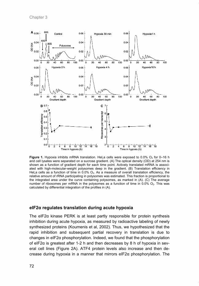
Chapter 3
72
Figure 1. Hypoxia inhibits mRNA translation. HeLa cells were exposed to 0.0% O2 for 0–16 h and cell lysates were separated on a sucrose gradient. (A) The optical density (OD) at 254 nm is shown as a function of gradient depth for each time point. Actively translated mRNA is associ-ated with high-molecular-weight polysomes deep in the gradient. (B) Translation efficiency in HeLa cells as a function of time in 0.0% O2. As a measure of overall translation efficiency, the relative amount of rRNA participating in polysomes was estimated. This fraction is proportional to the integrated area under the curve containing polysomes, as marked in (A). (C) The average number of ribosomes per mRNA in the polysomes as a function of time in 0.0% O2. This was calculated by differential integration of the profiles in (A).
eIF2α regulates translation during acute hypoxia
The eIF2α kinase PERK is at least partly responsible for protein synthesis inhibition during acute hypoxia, as measured by radioactive labeling of newly synthesized proteins (Koumenis et al, 2002). Thus, we hypothesized that the rapid inhibition and subsequent partial recovery in translation is due to changes in eIF2α phosphorylation. Indeed, we found that the phosphorylation of eIF2α is greatest after 1-2 h and then decreases by 8 h of hypoxia in sev-eral cell lines (Figure 2A). ATF4 protein levels also increase and then de-crease during hypoxia in a manner that mirrors eIF2α phosphorylation. The
Thesis_Weppler_v12_fc.pdf
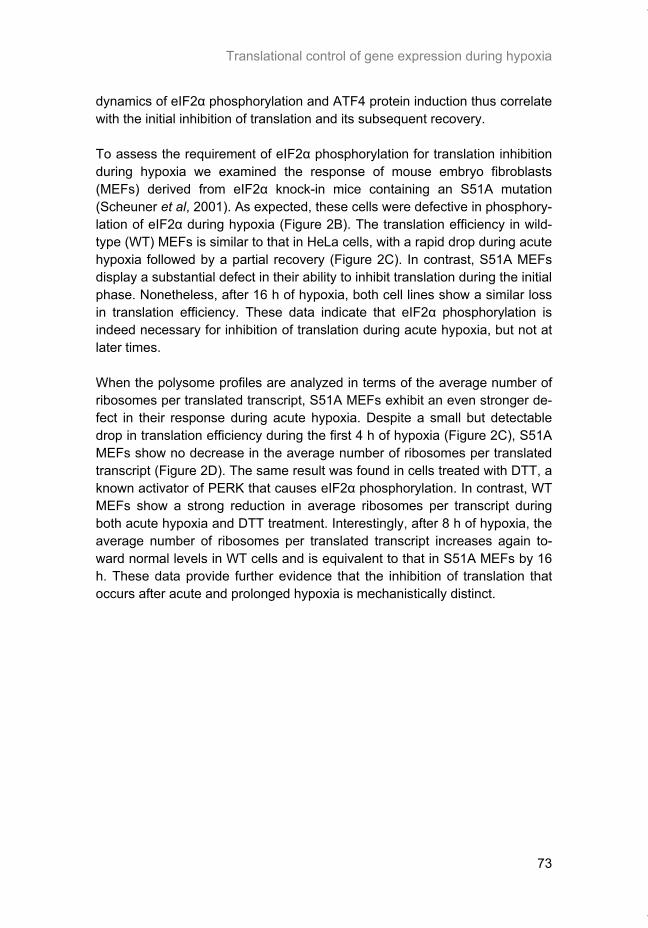
Translational control of gene expression during hypoxia
73
dynamics of eIF2α phosphorylation and ATF4 protein induction thus correlate with the initial inhibition of translation and its subsequent recovery. To assess the requirement of eIF2α phosphorylation for translation inhibition during hypoxia we examined the response of mouse embryo fibroblasts (MEFs) derived from eIF2α knock-in mice containing an S51A mutation (Scheuner et al, 2001). As expected, these cells were defective in phosphory-lation of eIF2α during hypoxia (Figure 2B). The translation efficiency in wild-type (WT) MEFs is similar to that in HeLa cells, with a rapid drop during acute hypoxia followed by a partial recovery (Figure 2C). In contrast, S51A MEFs display a substantial defect in their ability to inhibit translation during the initial phase. Nonetheless, after 16 h of hypoxia, both cell lines show a similar loss in translation efficiency. These data indicate that eIF2α phosphorylation is indeed necessary for inhibition of translation during acute hypoxia, but not at later times. When the polysome profiles are analyzed in terms of the average number of ribosomes per translated transcript, S51A MEFs exhibit an even stronger de-fect in their response during acute hypoxia. Despite a small but detectable drop in translation efficiency during the first 4 h of hypoxia (Figure 2C), S51A MEFs show no decrease in the average number of ribosomes per translated transcript (Figure 2D). The same result was found in cells treated with DTT, a known activator of PERK that causes eIF2α phosphorylation. In contrast, WT MEFs show a strong reduction in average ribosomes per transcript during both acute hypoxia and DTT treatment. Interestingly, after 8 h of hypoxia, the average number of ribosomes per translated transcript increases again to-ward normal levels in WT cells and is equivalent to that in S51A MEFs by 16 h. These data provide further evidence that the inhibition of translation that occurs after acute and prolonged hypoxia is mechanistically distinct.
Thesis_Weppler_v12_fc.pdf
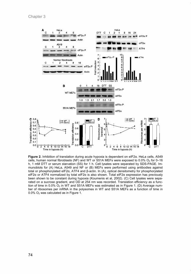
Chapter 3
74
Figure 2. Inhibition of translation during acute hypoxia is dependent on eIF2α. HeLa cells, A549 cells, human normal fibroblasts (NF) and WT or S51A MEFs were exposed to 0.0% O2 for 0–16 h, 1 mM DTT or serum starvation (SS) for 1 h. Cell lysates were separated by SDS-PAGE. Im-munoblots for (A) HeLa, A549 and NF or (B) MEFs were performed using antibodies against total or phosphorylated eIF2α, ATF4 and β-actin. In (A), optical densitometry for phosphorylated eIF2α or ATF4 normalized by total eIF2α is also shown. Total eIF2α expression has previously been shown to be constant during hypoxia (Koumenis et al, 2002). (C) Cell lysates were sepa-rated on a sucrose gradient, and OD at 254 nm was recorded. Translation efficiency as a func-tion of time in 0.0% O2 in WT and S51A MEFs was estimated as in Figure 1. (D) Average num-ber of ribosomes per mRNA in the polysomes in WT and S51A MEFs as a function of time in 0.0% O2 was calculated as in Figure 1.
Thesis_Weppler_v12_fc.pdf
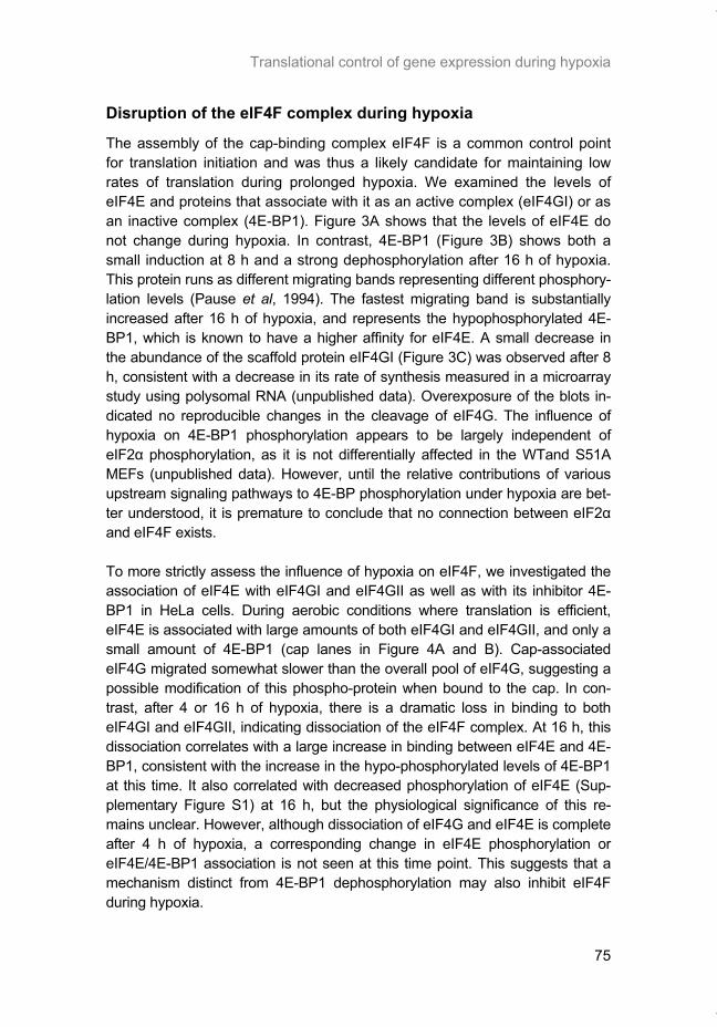
Translational control of gene expression during hypoxia
75
Disruption of the eIF4F complex during hypoxia
The assembly of the cap-binding complex eIF4F is a common control point for translation initiation and was thus a likely candidate for maintaining low rates of translation during prolonged hypoxia. We examined the levels of eIF4E and proteins that associate with it as an active complex (eIF4GI) or as an inactive complex (4E-BP1). Figure 3A shows that the levels of eIF4E do not change during hypoxia. In contrast, 4E-BP1 (Figure 3B) shows both a small induction at 8 h and a strong dephosphorylation after 16 h of hypoxia. This protein runs as different migrating bands representing different phosphory-lation levels (Pause et al, 1994). The fastest migrating band is substantially increased after 16 h of hypoxia, and represents the hypophosphorylated 4E-BP1, which is known to have a higher affinity for eIF4E. A small decrease in the abundance of the scaffold protein eIF4GI (Figure 3C) was observed after 8 h, consistent with a decrease in its rate of synthesis measured in a microarray study using polysomal RNA (unpublished data). Overexposure of the blots in-dicated no reproducible changes in the cleavage of eIF4G. The influence of hypoxia on 4E-BP1 phosphorylation appears to be largely independent of eIF2α phosphorylation, as it is not differentially affected in the WTand S51A MEFs (unpublished data). However, until the relative contributions of various upstream signaling pathways to 4E-BP phosphorylation under hypoxia are bet-ter understood, it is premature to conclude that no connection between eIF2α and eIF4F exists. To more strictly assess the influence of hypoxia on eIF4F, we investigated the association of eIF4E with eIF4GI and eIF4GII as well as with its inhibitor 4E-BP1 in HeLa cells. During aerobic conditions where translation is efficient, eIF4E is associated with large amounts of both eIF4GI and eIF4GII, and only a small amount of 4E-BP1 (cap lanes in Figure 4A and B). Cap-associated eIF4G migrated somewhat slower than the overall pool of eIF4G, suggesting a possible modification of this phospho-protein when bound to the cap. In con-trast, after 4 or 16 h of hypoxia, there is a dramatic loss in binding to both eIF4GI and eIF4GII, indicating dissociation of the eIF4F complex. At 16 h, this dissociation correlates with a large increase in binding between eIF4E and 4E-BP1, consistent with the increase in the hypo-phosphorylated levels of 4E-BP1 at this time. It also correlated with decreased phosphorylation of eIF4E (Sup-plementary Figure S1) at 16 h, but the physiological significance of this re-mains unclear. However, although dissociation of eIF4G and eIF4E is complete after 4 h of hypoxia, a corresponding change in eIF4E phosphorylation or eIF4E/4E-BP1 association is not seen at this time point. This suggests that a mechanism distinct from 4E-BP1 dephosphorylation may also inhibit eIF4F during hypoxia.
Thesis_Weppler_v12_fc.pdf
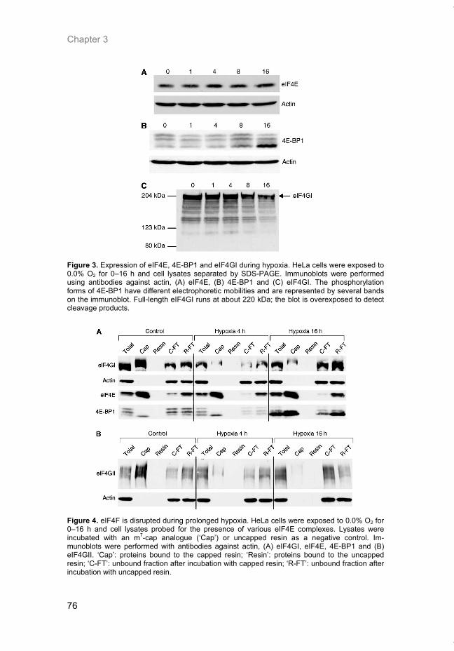
Chapter 3
76
Figure 3. Expression of eIF4E, 4E-BP1 and eIF4GI during hypoxia. HeLa cells were exposed to 0.0% O2 for 0–16 h and cell lysates separated by SDS-PAGE. Immunoblots were performed using antibodies against actin, (A) eIF4E, (B) 4E-BP1 and (C) eIF4GI. The phosphorylation forms of 4E-BP1 have different electrophoretic mobilities and are represented by several bandson the immunoblot. Full-length eIF4GI runs at about 220 kDa; the blot is overexposed to detectcleavage products.
Figure 4. eIF4F is disrupted during prolonged hypoxia. HeLa cells were exposed to 0.0% O2 for 0–16 h and cell lysates probed for the presence of various eIF4E complexes. Lysates were incubated with an m7-cap analogue (‘Cap’) or uncapped resin as a negative control. Im-munoblots were performed with antibodies against actin, (A) eIF4GI, eIF4E, 4E-BP1 and (B) eIF4GII. ‘Cap’: proteins bound to the capped resin; ‘Resin’: proteins bound to the uncapped resin; ‘C-FT’: unbound fraction after incubation with capped resin; ‘R-FT’: unbound fraction after incubation with uncapped resin.
Thesis_Weppler_v12_fc.pdf
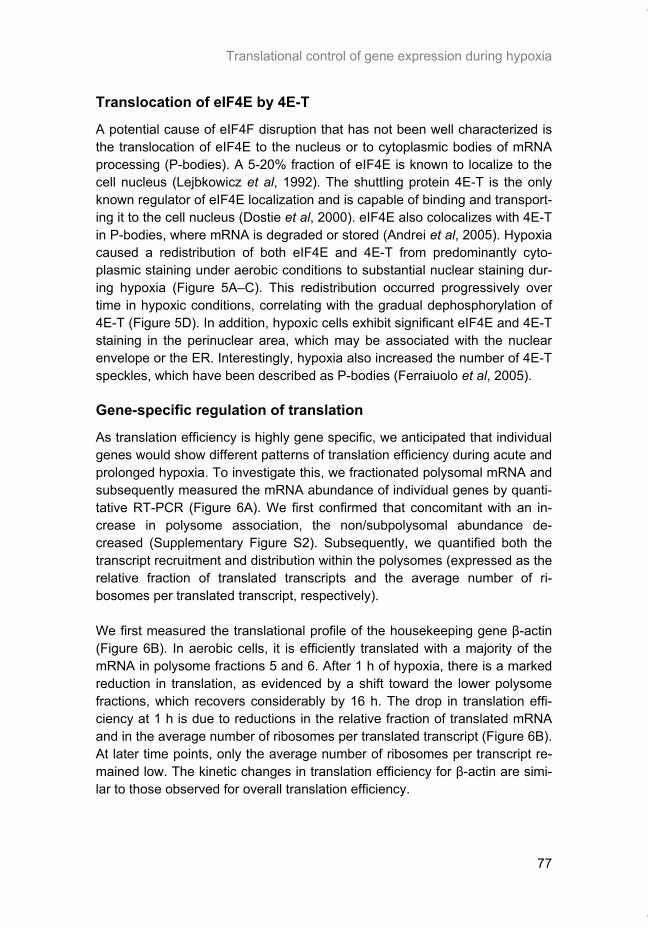
Translational control of gene expression during hypoxia
77
Translocation of eIF4E by 4E-T
A potential cause of eIF4F disruption that has not been well characterized is the translocation of eIF4E to the nucleus or to cytoplasmic bodies of mRNA processing (P-bodies). A 5-20% fraction of eIF4E is known to localize to the cell nucleus (Lejbkowicz et al, 1992). The shuttling protein 4E-T is the only known regulator of eIF4E localization and is capable of binding and transport-ing it to the cell nucleus (Dostie et al, 2000). eIF4E also colocalizes with 4E-T in P-bodies, where mRNA is degraded or stored (Andrei et al, 2005). Hypoxia caused a redistribution of both eIF4E and 4E-T from predominantly cyto-plasmic staining under aerobic conditions to substantial nuclear staining dur-ing hypoxia (Figure 5A–C). This redistribution occurred progressively over time in hypoxic conditions, correlating with the gradual dephosphorylation of 4E-T (Figure 5D). In addition, hypoxic cells exhibit significant eIF4E and 4E-T staining in the perinuclear area, which may be associated with the nuclear envelope or the ER. Interestingly, hypoxia also increased the number of 4E-T speckles, which have been described as P-bodies (Ferraiuolo et al, 2005).
Gene-specific regulation of translation
As translation efficiency is highly gene specific, we anticipated that individual genes would show different patterns of translation efficiency during acute and prolonged hypoxia. To investigate this, we fractionated polysomal mRNA and subsequently measured the mRNA abundance of individual genes by quanti-tative RT-PCR (Figure 6A). We first confirmed that concomitant with an in-crease in polysome association, the non/subpolysomal abundance de-creased (Supplementary Figure S2). Subsequently, we quantified both the transcript recruitment and distribution within the polysomes (expressed as the relative fraction of translated transcripts and the average number of ri-bosomes per translated transcript, respectively). We first measured the translational profile of the housekeeping gene β-actin (Figure 6B). In aerobic cells, it is efficiently translated with a majority of the mRNA in polysome fractions 5 and 6. After 1 h of hypoxia, there is a marked reduction in translation, as evidenced by a shift toward the lower polysome fractions, which recovers considerably by 16 h. The drop in translation effi-ciency at 1 h is due to reductions in the relative fraction of translated mRNA and in the average number of ribosomes per translated transcript (Figure 6B). At later time points, only the average number of ribosomes per transcript re-mained low. The kinetic changes in translation efficiency for β-actin are simi-lar to those observed for overall translation efficiency.
Thesis_Weppler_v12_fc.pdf
Chapter 3
78
Figure 5. 4E-T and eIF4E relocal-ize during hypoxia. HeLa cells were treated with 0.0% O2 for 0-16 h. Cells were stained with DAPI and (A) a polyclonal antibody against eIF4E, (B) a polyclonal antibody against 4E-T or (C) a monoclonal antibody against eIF4E and a polyclonal antibody against 4E-T. Cells were visual-ized by confocal microscopy and individual pictures merged to de-termine colocalization. (D) Cell lysates were separated by SDS-PAGE and immunoblots per-formed using antibodies against 4E-T. The ratio of the individual bands was quantified with optical densitometry. A cross-reacting band is indicated (*).
Thesis_Weppler_v12_fc.pdf
Translational control of gene expression during hypoxia
79
Figure 6. Gene-specific regulation of translation during hypoxia. HeLa cells were exposed to 0.0% O2 for 0-16 h and cell lysates were separated on sucrose gradients. (A) Fractions were collected as indicated, RNA was isolated and reverse transcribed. Thereafter, the total mRNA abundance of (B) β-actin, (C) CAIX, (D) ATF4, (E) CHOP and (F) GADD34 was determined using real-time quantitative PCR. The left panel shows total mRNA levels from unfractionated samples, normalized by 18S rRNA signal. The following three panels use black, gray and white bars to represent the gene abundance in polysome fractions following 0, 1 or 16 h hypoxia, respectively. The last two graphs show components of translation efficiency. This includes the relative fraction of transcripts in polysomes (i.e. corrected for total mRNA abundance) and the average number of ribosomes per mRNA. Graphs show the average from two independent experiments, and the histograms show the results from one representative experiment.
Thesis_Weppler_v12_fc.pdf
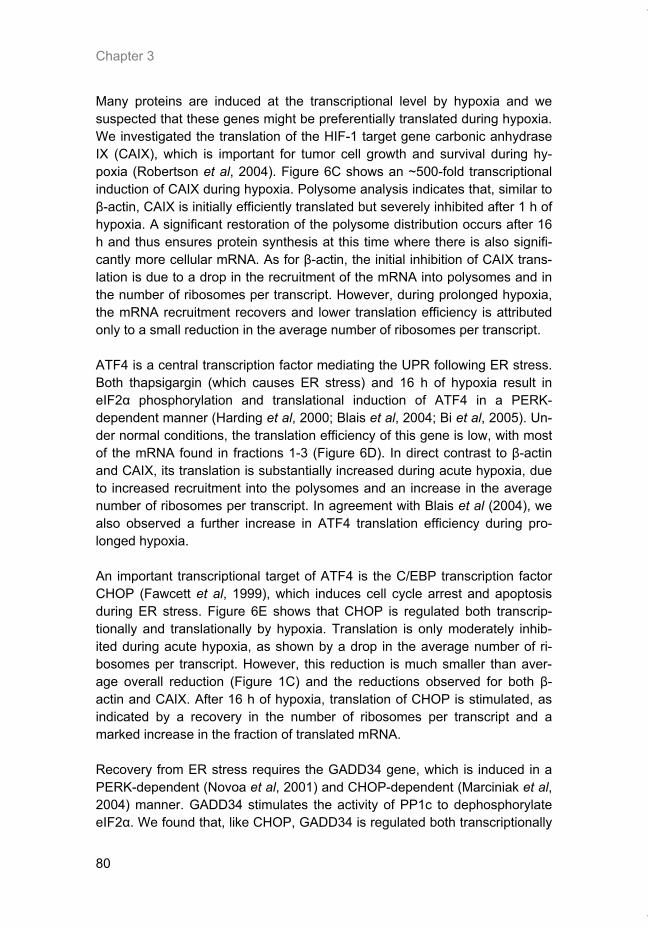
Chapter 3
80
Many proteins are induced at the transcriptional level by hypoxia and we suspected that these genes might be preferentially translated during hypoxia. We investigated the translation of the HIF-1 target gene carbonic anhydrase IX (CAIX), which is important for tumor cell growth and survival during hy-poxia (Robertson et al, 2004). Figure 6C shows an ~500-fold transcriptional induction of CAIX during hypoxia. Polysome analysis indicates that, similar to β-actin, CAIX is initially efficiently translated but severely inhibited after 1 h of hypoxia. A significant restoration of the polysome distribution occurs after 16 h and thus ensures protein synthesis at this time where there is also signifi-cantly more cellular mRNA. As for β-actin, the initial inhibition of CAIX trans-lation is due to a drop in the recruitment of the mRNA into polysomes and in the number of ribosomes per transcript. However, during prolonged hypoxia, the mRNA recruitment recovers and lower translation efficiency is attributed only to a small reduction in the average number of ribosomes per transcript. ATF4 is a central transcription factor mediating the UPR following ER stress. Both thapsigargin (which causes ER stress) and 16 h of hypoxia result in eIF2α phosphorylation and translational induction of ATF4 in a PERK-dependent manner (Harding et al, 2000; Blais et al, 2004; Bi et al, 2005). Un-der normal conditions, the translation efficiency of this gene is low, with most of the mRNA found in fractions 1-3 (Figure 6D). In direct contrast to β-actin and CAIX, its translation is substantially increased during acute hypoxia, due to increased recruitment into the polysomes and an increase in the average number of ribosomes per transcript. In agreement with Blais et al (2004), we also observed a further increase in ATF4 translation efficiency during pro-longed hypoxia. An important transcriptional target of ATF4 is the C/EBP transcription factor CHOP (Fawcett et al, 1999), which induces cell cycle arrest and apoptosis during ER stress. Figure 6E shows that CHOP is regulated both transcrip-tionally and translationally by hypoxia. Translation is only moderately inhib-ited during acute hypoxia, as shown by a drop in the average number of ri-bosomes per transcript. However, this reduction is much smaller than aver-age overall reduction (Figure 1C) and the reductions observed for both β-actin and CAIX. After 16 h of hypoxia, translation of CHOP is stimulated, as indicated by a recovery in the number of ribosomes per transcript and a marked increase in the fraction of translated mRNA. Recovery from ER stress requires the GADD34 gene, which is induced in a PERK-dependent (Novoa et al, 2001) and CHOP-dependent (Marciniak et al, 2004) manner. GADD34 stimulates the activity of PP1c to dephosphorylate eIF2α. We found that, like CHOP, GADD34 is regulated both transcriptionally
Thesis_Weppler_v12_fc.pdf
Translational control of gene expression during hypoxia
81
and translationally during hypoxia. Interestingly, its translation efficiency is highest after 4 h of hypoxia, which coincides with the start of recovery from eIF2α phosphorylation and overall translation inhibition (Figure 6F). In con-trast, GADD34 mRNA is unable to completely bypass the translation inhibi-tion after 16 h.
Gene-specific regulation of translation—dependence upon eIF2α
The gene-specific changes in translation noted above likely reflect the under-lying eIF2α-and eIF4F-dependent mechanisms of translation control during hypoxia. We thus analyzed gene-specific translation in WT and S51A MEFs to establish the dependence of individual genes on eIF2α regulation (Figure 7). In contrast to WT MEFs, S51A MEFs show no loss in translation efficiency of β-actin or CAIX during acute hypoxia (Figure 7A and B). The loss in trans-lation efficiency for these genes in the WT cells is similar to that observed in HeLa cells and is due primarily to a reduction in the average number of ri-bosomes per transcript. For both these genes, S51A MEFs show virtually no reduction in this parameter during the acute phase of hypoxia. However, in contrast to acute hypoxia, the translation efficiency during prolonged hypoxia is similar for these two genes in both cell lines. For ATF4 (Supplementary Figure S2), CHOP (data not shown) and GADD34 (Figure 7C), acute hypoxia causes a stimulation of translation in WT MEFs that is similar to HeLa cells. However, the translational induction is entirely absent in S51A MEFs. The increase in translation efficiency for GADD34 in WT MEFs during acute hypoxia results mainly from an increase in the aver-age number of ribosomes per transcript (Figure 7C). Cells that are defective in eIF2α phosphorylation show impaired regulation of this parameter. Thus, for all genes examined, the observed changes in translation efficiency during acute hypoxia are dependent on eIF2α phosphorylation.
Thesis_Weppler_v12_fc.pdf
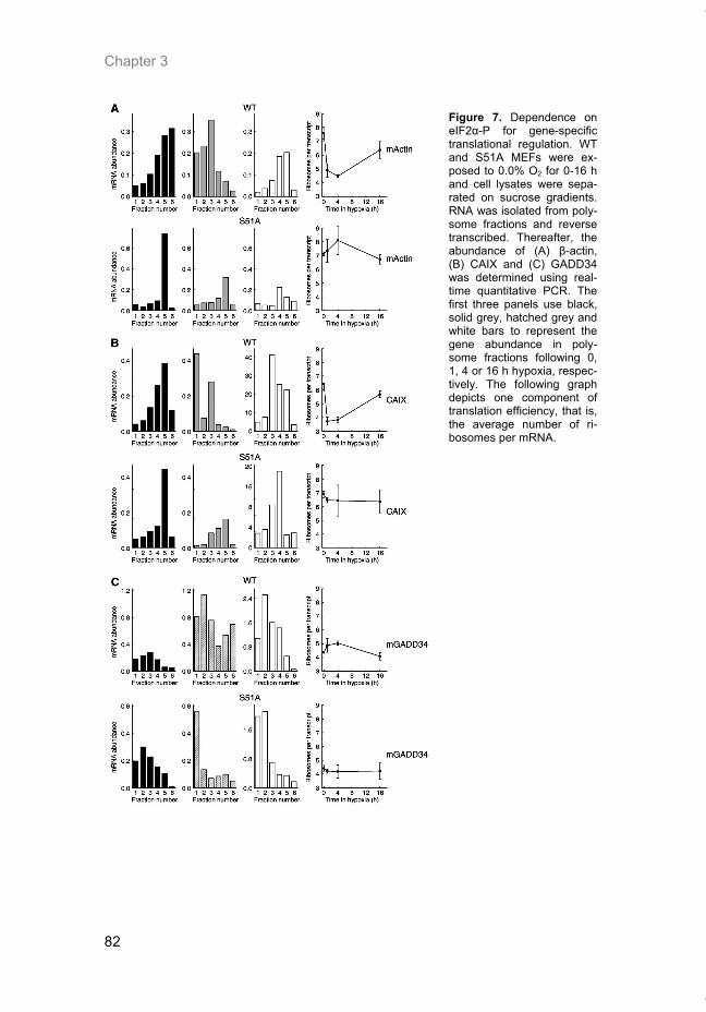
Chapter 3
82
Figure 7. Dependence on eIF2α-P for gene-specific translational regulation. WT and S51A MEFs were ex-posed to 0.0% O2 for 0-16 h and cell lysates were sepa-rated on sucrose gradients. RNA was isolated from poly-some fractions and reverse transcribed. Thereafter, the abundance of (A) β-actin, (B) CAIX and (C) GADD34 was determined using real-time quantitative PCR. The first three panels use black, solid grey, hatched grey and white bars to represent the gene abundance in poly-some fractions following 0, 1, 4 or 16 h hypoxia, respec-tively. The following graph depicts one component of translation efficiency, that is, the average number of ri-bosomes per mRNA.
Thesis_Weppler_v12_fc.pdf
Translational control of gene expression during hypoxia
83
Discussion
Rapid and persistent downregulation of protein synthesis is thought to be a means of energy preservation and to protect against the lethal effects of hy-poxia (Koumenis et al, 2002; Wouters et al, 2005). Here we show that the inhibition of global mRNA translation during hypoxia exhibits a biphasic re-sponse (Figure 8). The initial rapid inhibition (i.e. 15 min-4 h) is primarily de-pendent on eIF2α phosphorylation, whereas inhibition during prolonged hy-poxia is independent of eIF2α. Phosphorylation of eIF2α under conditions of anoxia is extremely rapid, occurring almost as quickly as we can establish hypoxia in our system (15-30 min). We have previously shown that eIF2α is also phosphorylated under more moderate hypoxic conditions, although to a smaller degree and after longer times (Koumenis et al, 2002). We speculate that this rapid anoxic response may be especially important during the acute exposures to hypoxia/anoxia that frequently occur in tumors due to the tran-sient opening and closing of blood vessels. This rapid response may explain the importance of eIF2α and ATF4 in the tolerance of cells to oxidative stress, which also occurs during hypoxia/reoxygenation cycles (Harding et al, 2003). This hypothesis is supported by a recent study by Bi et al (2005), who showed that activation of the PERK-eIF2α pathway during hypoxia contrib-utes to overall tumor growth. Human tumor cells expressing a dominant-negative PERK allele as well as MEFs lacking PERK or expressing the S51A eIF2α produce smaller tumors with increased cell death in hypoxic areas than their WT counterparts (Bi et al, 2005). Thus, although activation of eIF2α phosphorylation in response to hypoxia is transient, this response appears critical for long-term cell survival within hypoxic regions of tumors. Inhibition of translation during prolonged anoxia is associated with disruption of the mRNA cap-binding complex eIF4F and sequestration of eIF4E by both dephosphorylated 4E-BP1 and 4E-T. To our knowledge, this report is the first to show a physiological stress-induced regulation of the localization of eIF4E and its transporter 4E-T. Accumulation of eIF4E in the cell nucleus or P-bodies renders it unavailable for cytoplasmic translation. Relocalization of eIF4E may have additional roles apart from reducing cap-dependent protein translation, including effects on mRNA processing, transport and degrada-tion. Moderate hypoxia (1%) has also been shown to affect the eIF4F com-plex through inhibition of mTOR (Arsham et al, 2003). However, the kinetics and relative contribution of the eIF4F pathway on inhibition of global and gene-specific translation during more moderate hypoxia remain to be deter-mined (see below).
Thesis_Weppler_v12_fc.pdf
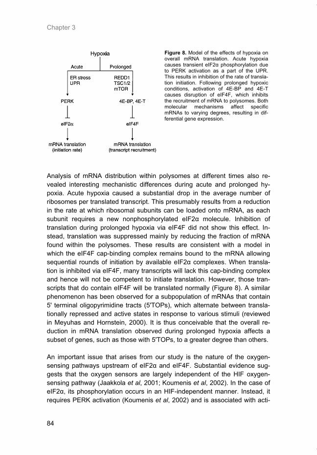
Chapter 3
84
Figure 8. Model of the effects of hypoxia on overall mRNA translation. Acute hypoxia causes transient eIF2α phosphorylation due to PERK activation as a part of the UPR. This results in inhibition of the rate of transla-tion initiation. Following prolonged hypoxic conditions, activation of 4E-BP and 4E-T causes disruption of eIF4F, which inhibits the recruitment of mRNA to polysomes. Both molecular mechanisms affect specific mRNAs to varying degrees, resulting in dif-ferential gene expression.
Analysis of mRNA distribution within polysomes at different times also re-vealed interesting mechanistic differences during acute and prolonged hy-poxia. Acute hypoxia caused a substantial drop in the average number of ribosomes per translated transcript. This presumably results from a reduction in the rate at which ribosomal subunits can be loaded onto mRNA, as each subunit requires a new nonphosphorylated eIF2α molecule. Inhibition of translation during prolonged hypoxia via eIF4F did not show this effect. In-stead, translation was suppressed mainly by reducing the fraction of mRNA found within the polysomes. These results are consistent with a model in which the eIF4F cap-binding complex remains bound to the mRNA allowing sequential rounds of initiation by available eIF2α complexes. When transla-tion is inhibited via eIF4F, many transcripts will lack this cap-binding complex and hence will not be competent to initiate translation. However, those tran-scripts that do contain eIF4F will be translated normally (Figure 8). A similar phenomenon has been observed for a subpopulation of mRNAs that contain 5' terminal oligopyrimidine tracts (5'TOPs), which alternate between transla-tionally repressed and active states in response to various stimuli (reviewed in Meyuhas and Hornstein, 2000). It is thus conceivable that the overall re-duction in mRNA translation observed during prolonged hypoxia affects a subset of genes, such as those with 5'TOPs, to a greater degree than others. An important issue that arises from our study is the nature of the oxygen-sensing pathways upstream of eIF2α and eIF4F. Substantial evidence sug-gests that the oxygen sensors are largely independent of the HIF oxygen-sensing pathway (Jaakkola et al, 2001; Koumenis et al, 2002). In the case of eIF2α, its phosphorylation occurs in an HIF-independent manner. Instead, it requires PERK activation (Koumenis et al, 2002) and is associated with acti-
Thesis_Weppler_v12_fc.pdf
Translational control of gene expression during hypoxia
85
vation of the UPR in response to ER stress (Romero-Ramirez et al, 2004; Bi et al, 2005). The upstream signaling that leads to eIF4F disruption is less clear, with perhaps both HIF-dependent and -independent components. Hy-poxia has been shown to prevent insulin stimulation of mTOR and phos-phorylation of its substrate 4E-BP1 during conditions of moderate hypoxia and serum starvation (Arsham et al, 2003). Similarly, Brugarolas et al (2004) showed that induction of REDD1 during hypoxia resulted in activation of the mTOR inhibitory complex TSC1/TSC2. As we also observe a decrease in the phosphorylation of 4E-BP1 after prolonged hypoxia, the eIF4F-dependent changes in translation reported here may also be due in part to inhibition of mTOR via REDD1 and TSC1/2. However, it is unlikely that this accounts en-tirely for eIF4F disruption and translation inhibition during hypoxia. REDD1 is a HIF-dependent gene and both mTOR inhibition and translation inhibition during hypoxia occur in HIF1α-knockout cells (Koumenis et al, 2002; Arsham et al, 2003). Furthermore, our data indicate that eIF4F disruption occurs be-fore substantial binding of eIF4E to 4E-BP1. Here we have identified redistri-bution of eIF4E into the cell nucleus via 4E-T as an additional mechanism for eIF4F disruption during hypoxia. Further work will be needed to establish to what degree inhibition of translation is due to suppression of mTOR/4E-BP1 phosphorylation and 4E-T activation, as well as to the requirements of HIF in both of these pathways. The fact that both eIF2α and eIF4F independently affect translation during hypoxia has important implications for the regulation of gene expression. mRNAs preferentially translated during acute hypoxia must be less depend-ent on eIF2α availability, whereas mRNAs that are actively translated during prolonged hypoxia must be less dependent on eIF4F. The translation of ATF4, which contains two upstream open reading frames (uORFs) in its 5'UTR, is perhaps the best example of a mammalian gene that displays this type of preferential translation (Harding et al, 2000). When eIF2α availability is high, translation begins at the 5' most uORF and re-initiation occurs effi-ciently at the subsequent uORF, preventing translation from the correct start codon of ATF4. When eIF2α is phosphorylated, there is a higher probability of skipping the second uORF and re-initiating at the bona fide start codon (Lu et al, 2004). Here, we found that in addition to ATF4, the downstream genes CHOP and GADD34 are also translationally induced during acute hypoxia. The S51A MEFs, which are unable to phosphorylate eIF2α, are defective in this translational regulation. This result is consistent with a report showing that ER stress-induced expression of GADD34 protein can be prevented by keeping eIF2α dephosphorylated (Novoa et al, 2003). The bypass of transla-tion inhibition may thus facilitate the ability of GADD34 to dephosphorylate eIF2α and promote recovery from ER stress.
Thesis_Weppler_v12_fc.pdf
Chapter 3
86
Our results predict that during prolonged hypoxia, gene transcripts with lower dependency on eIF4F should be preferentially translated. The translation of ATF4 and CHOP was in fact stimulated in HeLa cells after 16 h of hypoxia when the eIF4F complex was disrupted. Preferential translation under condi-tions of limiting cap-binding complex activity can occur through a higher than average affinity for eIF4F (Lawson et al, 1988). Another group of mRNAs that can be translated independently of the eIF4F complex are those that contain an internal ribosomal entry site (IRES) in their 5'UTR (Carter et al, 2000; Hol-cik and Sonenberg, 2005). The presence of an IRES enables translation ini-tiation under conditions where eIF4F-dependent translation is inhibited. Im-portantly, both mouse HIF-1α and VEGF have been shown to contain func-tional IRESs within their 5'UTR (Stein et al, 1998; Lang et al, 2002), although their biological importance is not yet firmly established. This provides a mechanism to ensure their translation during prolonged hypoxia where eIF4F is disrupted. The mechanism responsible for the selective translation of ATF4 and CHOP during prolonged hypoxia remains to be identified. In conclusion, we have shown that mRNA translation is inhibited through mul-tiple independent pathways with differing activation kinetics during hypoxia. These distinct modes of translational control influence the translation of indi-vidual genes to varying degrees and consequently can influence hypoxia-regulated protein expression in complex ways. An important finding is that inhibition of translation via eIF2α is transient, leading to dynamic changes in the translation efficiency of genes over the first 8 h of hypoxia. Our selected analysis of gene translation during hypoxia suggests that many genes may be differentially regulated by hypoxia. A complete survey of the genome for differentially translated genes during various exposures to hypoxia and their dependence of eIF2α is possible and will undoubtedly identify novel and im-portant hypoxia-regulated proteins (Koritzinsky et al, 2005).
Acknowledgements
This work was financially supported by Netherlands Organization for Scien-tific Research (NWO), the Dutch Cancer Society (KWF Kankerbestrijding) and the Euroxy grant from the 6th framework of the EU to BGW, from the Norwegian Research Council to MK and from grant CA94214 from the Na-tional Institutes of Health (NIH) to CK.
Thesis_Weppler_v12_fc.pdf
Translational control of gene expression during hypoxia
87
References Andrei MA, Ingelfinger D, Heintzmann R, Achsel T, Rivera-Pomar R, Luhrmann R (2005) A role
for eIF4E and eIF4E-transporter in targeting mRNPs to mammalian processing bodies. RNA 11: 717–727
Arsham AM, Howell JJ, Simon MC (2003) A novel hypoxia-inducible factor-independent hypoxic response regulating mammalian target of rapamycin and its targets. J Biol Chem 278: 29655–29660
Bennewith KL, Durand RE (2004) Quantifying transient hypoxia in human tumor xenografts by flow cytometry. Cancer Res 64: 6183–6189
Bi M, Naczki C, Koritzinsky M, Fels D, Blais J, Hu N, Harding H, Novoa I, Varia M, Raleigh J, Scheuner D, Kaufman RJ, Bell J, Ron D, Wouters BG, Koumenis C (2005) ER stress-regulated translation increases tolerance to extreme hypoxia and promotes tumor growth. EMBO J 24: 3470–3481
Blais JD, Filipenko V, Bi M, Harding HP, Ron D, Koumenis C, Wouters BG, Bell JC (2004) Acti-vating transcription factor 4 is translationally regulated by hypoxic stress. Mol Cell Biol 24: 7469–7482
Brugarolas J, Lei K, Hurley RL, Manning BD, Reiling JH, Hafen E, Witters LA, Ellisen LW, Kaelin Jr WG (2004) Regulation of mTOR function in response to hypoxia by REDD1 and the TSC1/TSC2 tumor suppressor complex. Genes Dev 18: 2893–2904
Cardenas-Navia LI, Yu D, Braun RD, Brizel DM, Secomb TW, Dewhirst MW (2004) Tumor-dependent kinetics of partial pressure of oxygen fluctuations during air and oxygen breath-ing.
Cancer Res 64: 6010–6017 Carter MS, Kuhn KM, Sarnow P (2000) Cellular internal ribosome entry site elements and the
use of cDNA microarrays in their investigation. In Translational Control of Gene Expression, Sonenberg N, Hershey JW, Mathews MB (eds) pp 615–636. Cold Spring Harbor, NY: Cold Spring Harbor Laboratory Press
DeGracia DJ, Kumar R, Owen CR, Krause GS, White BC (2002) Molecular pathways of protein synthesis inhibition during brain reperfusion: implications for neuronal survival or death. J Cereb Blood Flow Metab 22: 127–141
Dostie J, Ferraiuolo M, Pause A, Adam SA, Sonenberg N (2000) A novel shuttling protein, 4E-T, mediates the nuclear import of the mRNA 50 cap-binding protein, eIF4E. EMBO J 19: 3142–3156
Erler JT, Cawthorne CJ, Williams KJ, Koritzinsky M, Wouters BG, Wilson C, Miller C, Demona-cos C, Stratford IJ, Dive C (2004) Hypoxia-mediated down-regulation of Bid and Bax in tu-mors occurs via hypoxia-inducible factor 1-dependent and –independent mechanisms and contributes to drug resistance. Mol Cell Biol 24: 2875–2889
Fawcett TW, Martindale JL, Guyton KZ, Hai T, Holbrook NJ (1999) Complexes containing acti-vating transcription factor (ATF)/cAMP-responsive-element-binding protein (CREB) interact with the CCAAT/enhancer-binding protein (C/EBP)-ATF composite site to regulate Gadd153 expression during the stress response. Biochem J 339 (Part 1): 135–141
Ferraiuolo MA, Basak S, Dostie J, Murray EL, Schoenberg DR, Sonenberg N (2005) A role for the eIF4E-binding protein 4E-T in P-body formation and mRNA decay. J Cell Biol 170: 913–924
Gebauer F, Hentze MW (2004) Molecular mechanisms of translational control. Nat Rev Mol Cell Biol 5: 827–835
Graeber TG, Osmanian C, Jacks T, Housman DE, Koch CJ, Lowe SW, Giaccia AJ (1996) Hy-poxia-mediated selection of cells with diminished apoptotic potential in solid tumours. Na-ture 379: 88–91
Harding HP, Novoa I, Zhang Y, Zeng H, Wek R, Schapira M, Ron D (2000) Regulated translation initiation controls stress-induced gene expression in mammalian cells. Mol Cell 6: 1099–1108
Harding HP, Zhang Y, Zeng H, Novoa I, Lu PD, Calfon M, Sadri N, Yun C, Popko B, Paules R, Stojdl DF, Bell JC, Hettmann T, Leiden JM, Ron D (2003) An integrated stress response regulates amino acid metabolism and resistance to oxidative stress. Mol Cell 11: 619–633
Harris AL (2002) Hypoxia—a key regulatory factor in tumour growth. Nat Rev Cancer 2: 38–47 Hay N, Sonenberg N (2004) Upstream and downstream of mTOR. Genes Dev 18: 1926–1945 Holcik M, Sonenberg N (2005) Translational control in stress and apoptosis. Nat Rev Mol Cell
Biol 6: 318–327
Thesis_Weppler_v12_fc.pdf
Chapter 3
88
Holland EC, Sonenberg N, Pandolfi PP, Thomas G (2004) Signaling control of mRNA translation in cancer pathogenesis. Oncogene23: 3138–3144
Jaakkola P, Mole DR, Tian YM, Wilson MI, Gielbert J, Gaskell SJ, Kriegsheim A, Hebestreit HF, Mukherji M, Schofield CJ, Maxwell PH, Pugh CW, Ratcliffe PJ (2001) Targeting of HIF-alpha to the von Hippel–Lindau ubiquitylation complex by O2-regulated prolyl hydroxylation. Science 292: 468–472
Johannes G, Carter MS, Eisen MB, Brown PO, Sarnow P (1999) Identification of eukaryotic mRNAs that are translated at reduced cap binding complex eIF4F concentrations using a cDNA microarray. Proc Natl Acad Sci USA 96: 13118–13123
Kimball SR, Jefferson LS (2000) Regulation of translation initiation in mammalian cells by amino acids. In Translational Control of Gene Expression, Sonenberg N, Hershey JW, Mathews MB (eds) pp 561–579. Cold Spring Harbor, NY: Cold Spring Harbor Laboratory Press
Koritzinsky M, Seigneuric R, Magagnin MG, Beucken T, Lambin P, Wouters BG (2005) The hy-poxic proteome is influenced by gene-specific changes in mRNA translation. Radiother On-col 76: 177–186
Koumenis C, Naczki C, Koritzinsky M, Rastani S, Diehl A, Sonenberg N, Koromilas A, Wouters BG (2002) Regulation of protein synthesis by hypoxia via activation of the endoplasmic re-ticulum kinase PERK and phosphorylation of the translation initiation factor eIF2alpha. Mol Cell Biol 22: 7405–7416
Lang KJ, Kappel A, Goodall GJ (2002) Hypoxia-inducible factor-1alpha mRNA contains an inter-nal ribosome entry site that allows efficient translation during normoxia and hypoxia. Mol Biol Cell 13: 1792–1801
Lawson TG, Cladaras MH, Ray BK, Lee KA, Abramson RD, Merrick WC, Thach RE (1988) Dis-criminatory interaction of purified eukaryotic initiation factors 4F plus 4A with the 50 ends of reovirus messenger RNAs. J Biol Chem 263: 7266–7276
Leek RD, Stratford I, Harris AL (2005) The role of hypoxia-inducible factor-1 in three-dimensional tumor growth, apoptosis, and regulation by the insulin-signaling pathway. Cancer Res 65: 4147–4152
Lefebvre VH, Van Steenbrugge M, Beckers V, Roberfroid M, Buc-Calderon P (1993) Adenine nucleotides and inhibition of protein synthesis in isolated hepatocytes incubated under dif-ferent pO2 levels. Arch Biochem Biophys 304: 322–331
Lejbkowicz F, Goyer C, Darveau A, Neron S, Lemieux R, Sonenberg N (1992) A fraction of the mRNA 50 cap-binding protein, eukaryotic initiation factor 4E, localizes to the nucleus. Proc Natl Acad Sci USA 89: 9612–9616
Lu PD, Harding HP, Ron D (2004) Translation reinitiation at alternative open reading frames regulates gene expression in an integrated stress response. J Cell Biol 167: 27–33
Marciniak SJ, Yun CY, Oyadomari S, Novoa I, Zhang Y, Jungreis R, Nagata K, Harding HP, Ron D (2004) CHOP induces death by promoting protein synthesis and oxidation in the stressed endoplasmic reticulum. Genes Dev 18: 3066–3077
Martin de la Vega C, Burda J, Nemethova M, Quevedo C, Alcazar A, Martin ME, Danielisova V, Fando JL, Salinas M (2001) Possible mechanisms involved in the down-regulation of trans-lation during transient global ischaemia in the rat brain. Biochem J 357: 819–826
Martin ME, Munoz FM, Salinas M, Fando JL (2000) Ischaemia induces changes in the associa-tion of the binding protein 4E-BP1 and eukaryotic initiation factor (eIF) 4G to eIF4E in dif-ferentiated PC12 cells. Biochem J 351 (Part 2): 327–334
Meyuhas O, Hornstein E (2000) Translational control of TOP mRNAs. In Translational Control of Gene Expression, Sonenberg N, Hershey JW, Mathews MB (eds) pp 671–694. Cold Spring Harbor, NY: Cold Spring Harbor Laboratory Press
Neumar RW, DeGracia DJ, Konkoly LL, Khoury JI, White BC, Krause GS (1998) Calpain medi-ates eukaryotic initiation factor 4G degradation during global brain ischemia. J Cereb Blood Flow Metab 18: 876–881
Novoa I, Zeng H, Harding HP, Ron D (2001) Feedback inhibition of the unfolded protein re-sponse by GADD34-mediated dephosphorylation of eIF2alpha. J Cell Biol 153: 1011–1022
Novoa I, Zhang Y, Zeng H, Jungreis R, Harding HP, Ron D (2003) Stress-induced gene expres-sion requires programmed recovery from translational repression. EMBO J 22: 1180–1187
Pause A, Belsham GJ, Gingras AC, Donze O, Lin TA, Lawrence Jr JC, Sonenberg N (1994) Insulin-dependent stimulation of protein synthesis by phosphorylation of a regulator of 50-cap function. Nature 371: 762–767
Robertson N, Potter C, Harris AL (2004) Role of carbonic anhydrase IX in human tumor cell growth, survival, and invasion. Cancer Res 64: 6160–6165
Thesis_Weppler_v12_fc.pdf
Translational control of gene expression during hypoxia
89
Romero-Ramirez L, Cao H, Nelson D, Hammond E, Lee AH, Yoshida H, Mori K, Glimcher LH, Denko NC, Giaccia AJ, Le QT, Koong AC (2004) XBP1 is essential for survival under hy-poxic conditions and is required for tumor growth. Cancer Res 64: 5943–5947
Ryan HE, Lo J, Johnson RS (1998) HIF-1 alpha is required for solid tumor formation and embry-onic vascularization. EMBO J 17: 3005–3015
Scheuner D, Song B, McEwen E, Liu C, Laybutt R, Gillespie P, Saunders T, Bonner-Weir S, Kaufman RJ (2001) Translational control is required for the unfolded protein response and in vivo glucose homeostasis. Mol Cell 7: 1165–1176
Schroder M, Kaufman RJ (2005) The mammalian unfolded protein response. Annu Rev Biochem 74: 739–789
Semenza GL (2003) Targeting HIF-1 for cancer therapy. Nat Rev Cancer 3: 721–732 Stein I, Itin A, Einat P, Skaliter R, Grossman Z, Keshet E (1998) Translation of vascular endothe-
lial growth factor mRNA by internal ribosome entry: implications for translation under hy-poxia. Mol Cell Biol 18: 3112–3119
Tang N, Wang L, Esko J, Giordano FJ, Huang Y, Gerber HP, Ferrara N, Johnson RS (2004) Loss of HIF-1alpha in endothelial cells disrupts a hypoxia-driven VEGF autocrine loop nec-essary for tumorigenesis. Cancer Cell 6: 485–495
Tinton SA, Buc-Calderon PM (1999) Hypoxia increases the association of 4E-binding protein 1 with the initiation factor 4E in isolated rat hepatocytes. FEBS Lett 446: 55–59
Wouters BG, van den Beucken T, Magagnin MG, Koritzinsky M, Fels D, Koumenis C (2005) Control of the hypoxic response through regulation of mRNA translation. Semin Cell Dev Biol 16: 487–501
Wouters BG, Weppler SA, Koritzinsky M, Landuyt W, Nuyts S, Theys J, Chiu RK, Lambin P (2002) Hypoxia as a target for combined modality treatments. Eur J Cancer 38: 240–257
Thesis_Weppler_v12_fc.pdf

Thesis_Weppler_v12_fc.pdf
91
CHAPTER 4
Expression of EGFR variant vIII promotes both radiation resistance and hypoxia tolerance
Radiotherapy and Oncology. 2007; 83(3):333-9. Sherry A. Weppler*, Younan Li*, Ludwig Dubois, Natasja Lieuwes, Barry Jutten, Philippe Lambin, Bradly G. Wouters, Guido Lammering. * These authors contributed equally to the manuscript
Thesis_Weppler_v12_fc.pdf
Chapter 4
92
Abstract
Background and purpose: EGFRvIII has been described to function as an oncoprotein with constitutive activation promoting neoplastic transformation and tumorigenicity. The present study was undertaken to test whether EGFRvIII also contributes to hypoxia tolerance. Material and methods: The human glioma cell line U373 was genetically modified to stably express EGFRvIII. Western blotting and immunohisto-chemistry verified the expression of EGFRvIII. Tumor xenografts were pro-duced by injecting U373 control and EGFRvIII positive cells subcutaneously into the lateral flank of recipient mice. Colony formation assays were per-formed after ionizing radiation at 4 Gy and after exposure to anoxia for 1-4 days. Results: EGFRvIII accelerated tumor growth leading to a 3.5-fold increase in tumor size compared to control tumors at 40 days after cell injection. EGFRvIII promoted clonogenic survival by almost 2-fold and 4-fold after 4 Gy and 4 days of anoxia, respectively. EGFRvIII was also associated with a sub-stantially bigger colony size after anoxic treatment. Conclusions: EGFRvIII expression stimulates the growth of tumor xenografts and strongly promotes survival after irradiation and under hypoxic stress.
Thesis_Weppler_v12_fc.pdf
EGFRvIII promotes hypoxia tolerance
93
Introduction
The epidermal growth factor receptor (EGFR) is a well characterized proto-oncogene that is expressed in multiple cancers where it has been shown to promote tumor progression and therapy resistance [1, 3, 12, 19]. EGFR tar-geted strategies are actively under investigation and EGFR-specific tyrosine kinase inhibitors (TKI) and monoclonal antibodies (mAb) have shown great promise [4, 6-8, 17]. This is illustrated by the high number of currently running clinical trials investigating anti-EGFR strategies in cancer treatment. How-ever, some of the first clinical reports failed to corroborate the promising anti-tumor effects seen in preclinical studies, implicating persistent growth path-ways despite blockade of wild-type EGFR [5, 21]. The presence of naturally occurring mutations of EGFR may account for the limited clinical response to EGFR-targeted therapies [10, 13, 18]. A commonly described variant harbors an in-frame deletion of exons 2-7 resulting in a truncated version of the re-ceptor which lacks a portion of the extracellular ligand binding domain. This variant, called EGFRvIII, has not been detected in normal tissue, but is found in many malignancies, such as glioblastoma, non-small lung cell carcinoma, breast cancer, prostate cancer and just recently also in head and neck can-cer [15,16, 20]. Ligand-independent activation of EGFRvIII may explain the relative inability of blocking mAbs to downregulate the receptor [2, 26]. The present study was undertaken to test the hypothesis that EGFRvIII express-ing tumor cells not only contribute to therapy resistance but also to hypoxia tolerance. This would have important implications for our understanding of the tumor microenvironment and for the optimization of EGFR-targeted strategies in cancer therapy.
Materials and methods
Generation of a stable EGFRvIII expressing cell line
U373 human glioma cells were obtained from the American Type Culture Collection (Manassas, VA, USA) and cultured in MEMα medium (Invitrogen, Breda, NL) supplemented with 10% fetal calf serum (FCS). The phβAc.EGFRvIII plasmid, a generous gift from D.Bigner (Duke University, NC, USA), was transfected into U373 cells using PolyFect (Qiagen, Venlo, NL) according to the manufacturer’s directions. Forty-eight hours after trans-fection, cells were trypsinized and seeded at low density for selection in 300 µg/ml Geneticin (Invitrogen, Breda, NL). The plates were incubated for two weeks to allow formation of resistant colonies. Several colonies were chosen for expansion and labeled U373-vIII clones A-G.
Thesis_Weppler_v12_fc.pdf
Chapter 4
94
Immunoblotting
The cell pellet obtained from a 6 cm plate was lysed in 50 µl RIPA buffer (150 mM NaCl, 1% NP-40, 0.5% sodium deoxycholate, 0.1% SDS, 50 mM Tris-HCl, pH 7.5, 1x protease inhibitor cocktail) and incubated on ice for 30 min. Cell debris was removed by centrifugation at 10,000g for 15 min. The protein concentration in the supernatant was determined using the DC Assay (Bio-Rad, Veenendaal, NL). Forty micrograms of each sample was resolved on an 8% SDS-PAGE gel and blotted onto a Hybond ECL nitrocellulose membrane (GE Healthcare, Deigem, BE) by electrotransfer. The membrane was blocked with 5% milk-TBST (20 mM Tris–HCl, pH 7.6, 140 mM NaCl, 0.1% Tween 20) and incubated overnight in a 1:1000 dilution of EGFR (15F8) rabbit mono-clonal antibody (Cell Signaling Technology, Danvers, MA, USA). The mem-brane was washed with TBST and incubated for 1 h with a goat anti-rabbit secondary antibody (Cell Signaling Technology) at 1:3000 dilution. Immobi-lized proteins were detected using SuperSignal West Pico chemiluminescent substrate (Perbio, Etten-Leur, NL) and by exposing the blot to X-ray film.
Immunohistochemistry
Cells grown in chamber slides in vitro were fixed with 10% formalin and stained for EGFR using a 1:200 dilution of EGFR (sc-03) rabbit polyclonal antibody (Santa Cruz Biotechnology, Heidelberg, DE). The primary antibody was detected by an EnVision peroxidase-linked secondary antibody (DAKO, Heverlee, BE) used in combination with DAB+ chromogen and substrate (DAKO, Heverlee, BE). Slides were counterstained with hematoxylin and mounted in DPX medium (Brunschwig chemie, Amsterdam, NL). Seven micrometer thick tumor sections were stained using the same primary antibody as above at 1:100 dilution. A biotinylated goat anti-rabbit secondary (DAKO) was used at a 1:200 dilution followed by Vectastain ABC solution (Brunschwig chemie) and detection with DAB.
Radiation of cells
Cells were seeded in 6 cm dishes in order to reach ~80% confluence at the time of irradiation. Growth medium containing 0.5% FCS was added to cells 16 h before irradiation. During irradiation, dishes were placed in a Plexiglas jig filled with water at 37 °C. Cells were irradiated using an MCN 225 indus-trial X-ray tube (Philips, Eindhoven, NL) operated at 225 kV and 10 mA to deliver a dose of 4 Gy at a rate of 0.85 Gy/min. Immediately following irradia-tion, cells were returned to the incubator for 24 h at which time they were plated in MEMα + 10% FCS for the clonogenic survival assay.
Thesis_Weppler_v12_fc.pdf
EGFRvIII promotes hypoxia tolerance
95
Clonogenic survival assay
Cells were counted using a Coulter Z Series particle counter (Beckman Coul-ter, Mijdrecht, NL) and seeded in triplicate 6 cm dishes. For hypoxia experi-ments, cells were seeded for clonogenic survival prior to hypoxic exposure at 0% oxygen for 1-4 days. After the hypoxic treatment, the plates were re-moved from the hypoxic chamber (a MACS VA500 microaerophilic work-station supplied by Don Whitley Scientific, Shipley, UK) and incubated under standard culture conditions until colonies formed (~14 days in total). Colonies were fixed and stained with 2% bromophenol blue in 70% ethanol. Plating efficiency was determined by counting colonies consisting of ≥50 cells and correcting for the number of cells seeded.
Tumor xenograft growth
Animal experiments were performed using adult NMRI (nu/nu) female mice (28-32 g) from the animal facility of the Catholic University of Leuven in Bel-gium. The animal facilities and experiments were in accordance with local institutional guidelines for animal welfare and were approved by the Animal Ethics Committee of the university. Three million U373 and U373-vIII(+) cells were resuspended in 100 µl growth medium and injected subcutaneously into the lateral flank of recipient mice. Tumors were measured with callipers in three orthogonal diameters and used to calculate tumor volume based on the formula A x B x C x π/6. Animals were followed until the ethically allowed tu-mor burden was reached, at which time tumors were excised, fixed in 1-4% formaldehyde, and embedded in paraffin. Mean tumor volumes were calcu-lated for each group.
In vitro growth under aerobic and hypoxic conditions
For growth under aerobic conditions, 105 cells were seeded in triplicate on 6 cm dishes. The plates were incubated under normal culture conditions in a 5% CO2 incubator for 1-6 days. To monitor growth under hypoxia, 5 x 10⁵ cells were seeded in 10 cm dishes. The following day (day 0), dishes were placed in the hypoxic chamber for 1-4 days. Plates were harvested by wash-ing two times with phosphate-buffered saline followed by trypsinization. Total cell numbers were enumerated using a Coulter Z Series particle counter (Beckman Coulter, Mijdrecht, NL). Cell numbers were normalized to the amount of cells present on day 0. Doubling times were calculated from the slope of the best-fit line during the exponential phase of growth.
Thesis_Weppler_v12_fc.pdf
Chapter 4
96
Statistics
Statistical analysis was carried out using the program SPSS 12.0.1 for Win-dows (SPSS Inc., 2003, Chicago, IL, USA). A non-parametric Mann-Whitney U test was used to assess differences in xenograft tumor growth. A Student’s t-test was used to assess differences in radiation survival and a one-way ANOVA was used to determine differences in growth under hypoxia. P values <0.05 were considered to be significant.
Results
Generation of a stable EGFRvIII model
U373 glioma cells were transfected with an EGFRvIII expression plasmid in which EGFRvIII transcription is driven from a human β-actin promoter. Sev-eral clones with stable integration of the plasmid were selected and screened for EGFRvIII protein expression. Clones A, D, and F have high expression of EGFRvIII as seen by the presence of two bands corresponding to different glycosylated forms of the mutant receptor (Fig. 1a). In contrast, clones B, C, E, and G express only wild-type EGFR similar to the parental U373 cells de-spite displaying Geneticin resistance. We selected clone D as an EGFR-vIII positive cell line and clone B as an EGFR-vIII negative control for use in fur-ther experiments. We will refer to clones B and D as U373-vIII(-) and U373-vIII(+), respectively. Immunohistochemical staining was performed using an antibody that recognizes both wild-type and variant forms of EGFR. The pa-rental U373 and U373-vIII(-) cell lines show low EGFR expression which is localized mainly to the plasma membrane (Fig. 1b). In contrast, U373-vIII(+) show higher expression in both the cytoplasm and at the plasma membrane with some cells also demonstrating strong nuclear staining.
Intrinsic radiation resistance imparted by EGFRvIII
Previous studies have demonstrated a role for EGFRvIII in radiation resis-tance [10, 11]. We confirmed that our cell line model is also protected from radiation induced killing when EGFR-vIII is expressed (Fig. 2). In a clono-genic survival assay, U373-vIII(+) showed an almost 2-fold increase in sur-vival after irradiation with 4 Gy (P = 0.005), suggesting that EGFR-vIII is func-tional in our model and able to activate downstream survival pathways.
Thesis_Weppler_v12_fc.pdf
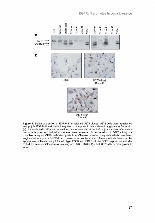
EGFRvIII promotes hypoxia tolerance
97
Figure 1. Stable expression of EGFRvIII in selected U373 clones. U373 cells were transfected with phβAc.EGFRvIII and stable integration of the plasmid was selected by growth in Geneticin. (a) Untransfected U373 cells, as well as transfected cells, either before (transient) or after selec-tion (stable pool and individual clones), were screened for expression of EGFRvIII by im-munoblot analysis. CHO+ indicates lysate from Chinese hamster ovary cells which have been engineered to express EGFRvIII and serve as a positive control. Arrows indicate bands at the appropriate molecular weight for wild type EGFR and EGFRvIII. (b) EGFR expression was de-tected by immunohistochemical staining of U373, U373-vIII(-) and U373-vIII(+) cells grown in vitro.
Thesis_Weppler_v12_fc.pdf
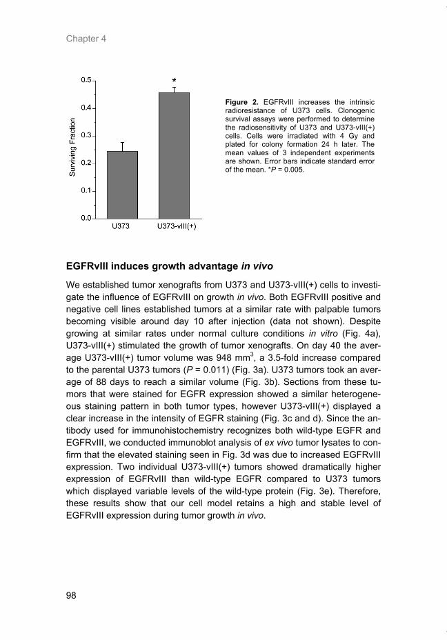
Chapter 4
98
Figure 2. EGFRvIII increases the intrinsic radioresistance of U373 cells. Clonogenic survival assays were performed to determine the radiosensitivity of U373 and U373-vIII(+) cells. Cells were irradiated with 4 Gy and plated for colony formation 24 h later. The mean values of 3 independent experiments are shown. Error bars indicate standard error of the mean. *P = 0.005.
EGFRvIII induces growth advantage in vivo
We established tumor xenografts from U373 and U373-vIII(+) cells to investi-gate the influence of EGFRvIII on growth in vivo. Both EGFRvIII positive and negative cell lines established tumors at a similar rate with palpable tumors becoming visible around day 10 after injection (data not shown). Despite growing at similar rates under normal culture conditions in vitro (Fig. 4a), U373-vIII(+) stimulated the growth of tumor xenografts. On day 40 the aver-age U373-vIII(+) tumor volume was 948 mm3, a 3.5-fold increase compared to the parental U373 tumors (P = 0.011) (Fig. 3a). U373 tumors took an aver-age of 88 days to reach a similar volume (Fig. 3b). Sections from these tu-mors that were stained for EGFR expression showed a similar heterogene-ous staining pattern in both tumor types, however U373-vIII(+) displayed a clear increase in the intensity of EGFR staining (Fig. 3c and d). Since the an-tibody used for immunohistochemistry recognizes both wild-type EGFR and EGFRvIII, we conducted immunoblot analysis of ex vivo tumor lysates to con-firm that the elevated staining seen in Fig. 3d was due to increased EGFRvIII expression. Two individual U373-vIII(+) tumors showed dramatically higher expression of EGFRvIII than wild-type EGFR compared to U373 tumors which displayed variable levels of the wild-type protein (Fig. 3e). Therefore, these results show that our cell model retains a high and stable level of EGFRvIII expression during tumor growth in vivo.
Thesis_Weppler_v12_fc.pdf
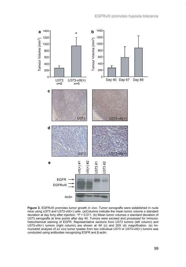
EGFRvIII promotes hypoxia tolerance
99
Figure 3. EGFRvIII promotes tumor growth in vivo. Tumor xenografts were established in nude mice using U373 and U373-vIII(+) cells. (a)Columns indicate the mean tumor volume ± standard deviation at day forty after injection. *P = 0.011. (b) Mean tumor volumes ± standard deviation of U373 xenografts at time points after day 40. Tumors were excised and processed for immuno-histochemical staining of EGFR. Representative sections from U373 tumors (left column) and U373-vIII(+) tumors (right column) are shown at 4X (c) and 20X (d) magnification. (e) Im-munoblot analysis of ex vivo tumor lysates from two individual U373 or U373-vIII(+) tumors was conducted using antibodies recognizing EGFR and β-actin.
Thesis_Weppler_v12_fc.pdf
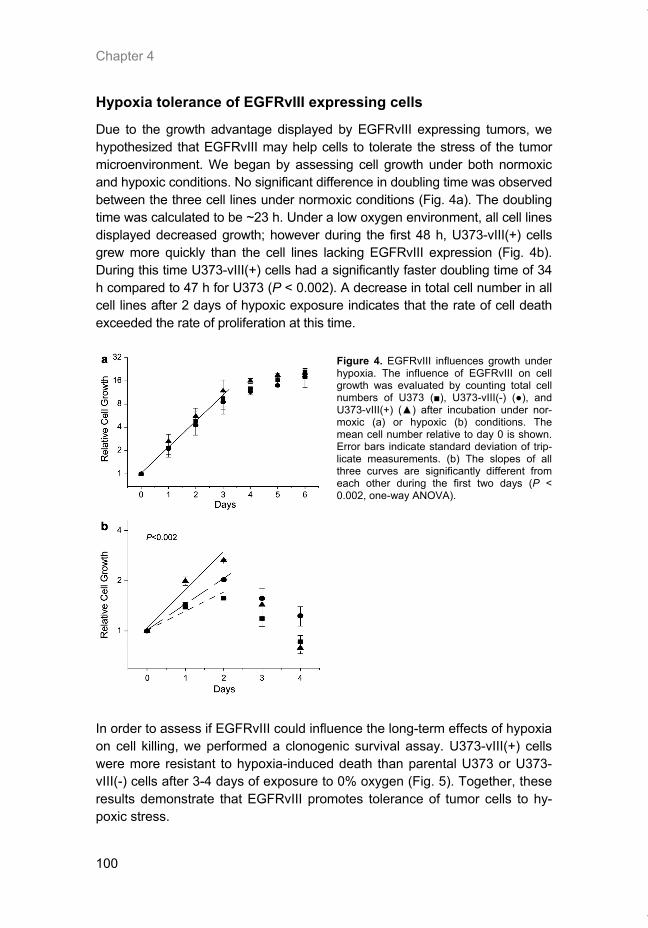
Chapter 4
100
Hypoxia tolerance of EGFRvIII expressing cells
Due to the growth advantage displayed by EGFRvIII expressing tumors, we hypothesized that EGFRvIII may help cells to tolerate the stress of the tumor microenvironment. We began by assessing cell growth under both normoxic and hypoxic conditions. No significant difference in doubling time was observed between the three cell lines under normoxic conditions (Fig. 4a). The doubling time was calculated to be ~23 h. Under a low oxygen environment, all cell lines displayed decreased growth; however during the first 48 h, U373-vIII(+) cells grew more quickly than the cell lines lacking EGFRvIII expression (Fig. 4b). During this time U373-vIII(+) cells had a significantly faster doubling time of 34 h compared to 47 h for U373 (P < 0.002). A decrease in total cell number in all cell lines after 2 days of hypoxic exposure indicates that the rate of cell death exceeded the rate of proliferation at this time.
Figure 4. EGFRvIII influences growth under hypoxia. The influence of EGFRvIII on cell growth was evaluated by counting total cell numbers of U373 (■), U373-vIII(-) (●), and U373-vIII(+) (▲) after incubation under nor-moxic (a) or hypoxic (b) conditions. The mean cell number relative to day 0 is shown. Error bars indicate standard deviation of trip-licate measurements. (b) The slopes of all three curves are significantly different from each other during the first two days (P < 0.002, one-way ANOVA).
In order to assess if EGFRvIII could influence the long-term effects of hypoxia on cell killing, we performed a clonogenic survival assay. U373-vIII(+) cells were more resistant to hypoxia-induced death than parental U373 or U373-vIII(-) cells after 3-4 days of exposure to 0% oxygen (Fig. 5). Together, these results demonstrate that EGFRvIII promotes tolerance of tumor cells to hy-poxic stress.
Thesis_Weppler_v12_fc.pdf
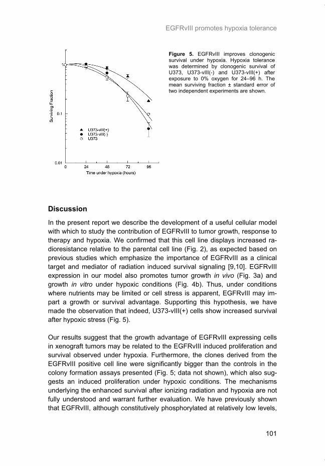
EGFRvIII promotes hypoxia tolerance
101
Figure 5. EGFRvIII improves clonogenic survival under hypoxia. Hypoxia tolerance was determined by clonogenic survival of U373, U373-vIII(-) and U373-vIII(+) after exposure to 0% oxygen for 24–96 h. The mean surviving fraction ± standard error of two independent experiments are shown.
Discussion
In the present report we describe the development of a useful cellular model with which to study the contribution of EGFRvIII to tumor growth, response to therapy and hypoxia. We confirmed that this cell line displays increased ra-dioresistance relative to the parental cell line (Fig. 2), as expected based on previous studies which emphasize the importance of EGFRvIII as a clinical target and mediator of radiation induced survival signaling [9,10]. EGFRvIII expression in our model also promotes tumor growth in vivo (Fig. 3a) and growth in vitro under hypoxic conditions (Fig. 4b). Thus, under conditions where nutrients may be limited or cell stress is apparent, EGFRvIII may im-part a growth or survival advantage. Supporting this hypothesis, we have made the observation that indeed, U373-vIII(+) cells show increased survival after hypoxic stress (Fig. 5). Our results suggest that the growth advantage of EGFRvIII expressing cells in xenograft tumors may be related to the EGFRvIII induced proliferation and survival observed under hypoxia. Furthermore, the clones derived from the EGFRvIII positive cell line were significantly bigger than the controls in the colony formation assays presented (Fig. 5; data not shown), which also sug-gests an induced proliferation under hypoxic conditions. The mechanisms underlying the enhanced survival after ionizing radiation and hypoxia are not fully understood and warrant further evaluation. We have previously shown that EGFRvIII, although constitutively phosphorylated at relatively low levels,
Thesis_Weppler_v12_fc.pdf
Chapter 4
102
responds to ionizing radiation with an immediate activation after clinically relevant doses of ionizing radiation [9]. This radiation induced activation pref-erentially stimulates PI3K-Akt signaling, which is known to be involved in cel-lular proliferation and apoptosis [9,14, 24, 25]. Whether this can explain the difference in clonogenic survival of almost 2- and 4-fold after 4 Gy and 4 days of anoxia, respectively (Fig. 2–5), remains unclear. Despite its role in prolif-eration and antiapoptosis, EGFRvIII may alter the capacity for DNA repair via activation of the Akt-mammalian target of rapamycin (mTOR) pathway and the subsequent upregulation of translation of DNA repair proteins [19,22,23]. Our results might also indicate a possible selection of EGFRvIII expressing tumor cells under hypoxic conditions in growing tumors, which would lead to an accumulation of EGFRvIII positive cancer cells in hypoxic areas of the tumor. This might, however, impair on the effectiveness of targeted anti-EGFR drugs due to the biodistribution properties of the drugs in solid tumors. In summary, we have implicated EGFRvIII as a contributor to cell survival under conditions of radiotherapy as well as hypoxic stress. This finding im-proves our understanding of the contribution of hypoxia on the negative influ-ence of radiosensitivity in tumor cells. It also highlights the need for monitor-ing the expression of EGFRvIII in human tumors in order to individualize and optimize molecular targeted therapies. The mechanisms underlying these vIII-dependent phenotypical changes warrant further investigation.
Acknowledgements
We acknowledge financial support from the Association for International Cancer Research (AICR Grant 05-277 awarded to G.L., P.L. and B.W.), the Dutch Cancer Society (KWF Grant 2006-3519 awarded to G.L., P.L. and B.W.) and the EU 6th framework programme (EUROXY Grant LSCH-CT-2003-502932 awarded to P.L. and B.W.).
Thesis_Weppler_v12_fc.pdf
EGFRvIII promotes hypoxia tolerance
103
References [1] Ang KK, Berkey BA, Tu X, et al. Impact of epidermal growth factor receptor expression on
survival and pattern of relapse in patients with advanced head and neck carcinoma. Cancer Res 2002;62:7350–6.
[2] Batra SK, Castelino-Prabhu S, Wikstrand CJ, et al. Epidermal growth factor ligand-independent, unregulated, cell-transforming potential of a naturally occurring human mutant EGFRvIII gene. Cell Growth Differ 1995;6:1251–9.
[3] Baumann M, Krause M. Targeting the epidermal growth factor receptor in radiotherapy: radiobiological mechanisms, preclinical and clinical results. Radiother Oncol 2004;72:257–66.
[4] Bonner JA, Harari PM, Giralt J, et al. Radiotherapy plus cetuximab for squamous-cell carci-noma of the head and neck. N Engl J Med 2006;354:567–78.
[5] Cohen EE, Rosen F, Stadler WM, et al. Phase II trial of ZD1839 in recurrent or metastatic squamous cell carcinoma of the head and neck. J Clin Oncol 2003;21:1980–7.
[6] Heimberger AB, Learn CA, Archer GE, et al. Brain tumors in mice are susceptible to block-ade of epidermal growth factor receptor (EGFR) with the oral, specific, EGFR-tyrosine kinase inhibitor ZD1839 (iressa). Clin Cancer Res 2002;8:3496–502.
[7] Krause M, Ostermann G, Petersen C, et al. Decreased repopulation as well as increased reoxygenation contribute to the improvement in local control after targeting of the EGFR by C225 during fractionated irradiation. Radiother Oncol 2005;76:162–7.
[8] Krause M, Schutze C, Petersen C, et al. Different classes of EGFR inhibitors may have different potential to improve local tumour control after fractionated irradiation: a study on C225 in FaDu hSCC. Radiother Oncol 2005;74:109–15.
[9] Lammering G, Hewit TH, Valerie K, et al. EGFRvIII-mediated radioresistance through a strong cytoprotective response. Oncogene 2003;22:5545–53.
[10] Lammering G, Hewit TH, Holmes M, et al. Inhibition of the type III epidermal growth factor receptor variant mutant receptor by dominant-negative EGFR-CD533 enhances malignant glioma cell radiosensitivity. Clin Cancer Res 2004;10:6732–43.
[11] Lammering G, Valerie K, Lin PS, Hewit TH, Schmidt-Ullrich RK. Radiation-induced activa-tion of a common variant of EGFR confers enhanced radioresistance. Radiother Oncol 2004;72:267–73.
[12] Lammering G. Molecular predictor and promising target: will EGFR now become a star in radiotherapy? Radiother Oncol 2005;74:89–91.
[13] Lynch TJ, Bell DW, Sordella R, et al. Activating mutations in the epidermal growth factor receptor underlying responsiveness of non-small-cell lung cancer to gefitinib. N Engl J Med 2004;350:2129–39.
[14] Mellinghoff IK, Wang MY, Vivanco I, et al. Molecular determinants of the response of glioblastomas to EGFR kinase inhibitors. N Engl J Med 2005;353:2012–24.
[15] Moscatello DK, Holgado-Madruga M, Godwin AK, et al. Frequent expression of a mutant epidermal growth factor receptor in multiple human tumors. Cancer Res 1995;55:5536–9.
[16] Okamoto I, Kenyon LC, Emlet DR, et al. Expression of constitutively activated EGFRvIII in non-small cell lung cancer. Cancer Sci 2003;94:50–6.
[17] Ono M, Kuwano M. Molecular mechanisms of epidermal growth factor receptor (EGFR) activation and response to gefitinib and other EGFR-targeting drugs. Clin Cancer Res 2006;12:7242–51.
[18] Paez JG, Janne PA, Lee JC, et al. EGFR mutations in lung cancer: correlation with clinical response to gefitinib therapy. Science 2004;304:1497–500.
[19] Schmidt-Ullrich RK, Contessa JN, Lammering G, Amorino G, Lin PS. ERBB receptor tyro-sine kinases and cellular radiation responses. Oncogene 2003;22:5855–65.
[20] Sok JC, Coppelli FM, Thomas SM, et al. Mutant epidermal growth factor receptor (EGFRvIII) contributes to head and neck cancer growth and resistance to EGFR targeting. Clin Cancer Res 2006;12:5064–73.
[21] Soulieres D, Senzer NN, Vokes EE, Hidalgo M, Agarwala SS, Siu LL. Multicenter phase II study of erlotinib, an oral epidermal growth factor receptor tyrosine kinase inhibitor, in pa-tients with recurrent or metastatic squamous cell cancer of the head and neck. J Clin Oncol 2004;22:77–85.
[22] Sunavala-Dossabhoy G, Fowler M, De Benedetti A. Translation of the radioresistance kinase TLK1B is induced by gammairradiation through activation of mTOR and phosphory-lation of 4E-BP1. BMC Mol Biol 2004;5:1.
Thesis_Weppler_v12_fc.pdf

Chapter 4
104
[23] Sunavala-Dossabhoy G, Balakrishnan SK, Sen S, Nuthalapaty S, De Benedetti A. The radioresistance kinase TLK1B protects the cells by promoting repair of double strand breaks. BMC Mol Biol 2005;6:19.
[24] Toulany M, Dittmann K, Baumann M, Rodemann HP. Radiosensitization of Ras-mutated human tumor cells in vitro by the specific EGF receptor antagonist BIBX1382BS. Radiother Oncol 2005;74:117–29.
[25] Toulany M, Dittmann K, Kruger M, Baumann M, Rodemann HP. Radioresistance of K-Ras mutated human tumor cells is mediated through EGFR-dependent activation of PI3K-AKT pathway. Radiother Oncol 2005;76:143–50.
[26] Wikstrand CJ, McLendon RE, Friedman AH, Bigner DD. Cell surface localization and den-sity of the tumor-associated variant of the epidermal growth factor receptor, EGFRvIII. Can-cer Res 1997;57:4130–40.
Thesis_Weppler_v12_fc.pdf
105
CHAPTER 5
Response of U87 glioma xenografts treated with concurrent rapamycin and fractionated radiotherapy: Possible role for thrombosis
Radiotherapy and Oncology. 2007; 82(1):96-104. Sherry A. Weppler, Mechthild Krause, Agnieszka Zyromska, Philippe Lambin, Michael Baumann, and Bradly G. Wouters.
Thesis_Weppler_v12_fc.pdf
Chapter 5
106
Abstract
Background and purpose: Rapamycin, a highly specific mTOR inhibitor, has shown anti-proliferative and anti-angiogenic properties, as well as an en-hancement in tumor growth delay when used in combination with radiation in mouse xenograft models. Our goal was to determine if rapamycin can also have a positive effect on the local tumor control achieved by radiotherapy. Materials and methods: Nude mice bearing U87 glioblastoma xenografts were treated with concomitant rapamycin and radiotherapy over a 5 day frac-tionation schedule. Animals received graded total doses ranging from 24 to 100 Gy. Experimental endpoints were tumor growth delay and local tumor control. In addition, histological evaluation of tumor sections was performed to examine changes occurring within the tumor microenvironment as a result of treatment. Analysis of proliferation, mTOR signalling, hypoxia, and vessel thrombosis was conducted. Results: As a single agent, rapamycin reduced the in vitro growth of U87 cells by 70% and caused a 4 day growth delay of tumor xenografts. In combination with radiation, no further increase in tumor growth delay was observed when compared to radiation alone. The tumor control dose 50% (TCD50) was 46.8 Gy (95% CI 41; 53 Gy) in tumors treated with radiation alone and was slightly but not significantly lower at 42.8 Gy (95% CI 36; 49 Gy) after simultaneous treatment with rapamycin. Histological evaluation revealed evidence of ele-vated hypoxia following rapamycin treatment that may be due to vessel thrombosis. Conclusions: The influence of rapamycin on thrombosis and tumor hypoxia may be a confounding factor limiting its effectiveness in combination with ra-diotherapy.
Thesis_Weppler_v12_fc.pdf
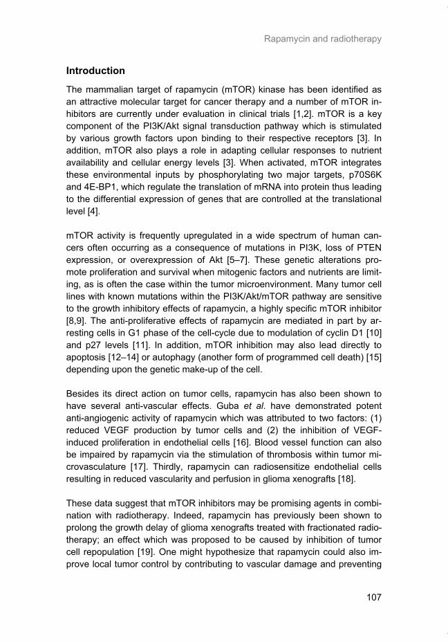
Rapamycin and radiotherapy
107
Introduction
The mammalian target of rapamycin (mTOR) kinase has been identified as an attractive molecular target for cancer therapy and a number of mTOR in-hibitors are currently under evaluation in clinical trials [1,2]. mTOR is a key component of the PI3K/Akt signal transduction pathway which is stimulated by various growth factors upon binding to their respective receptors [3]. In addition, mTOR also plays a role in adapting cellular responses to nutrient availability and cellular energy levels [3]. When activated, mTOR integrates these environmental inputs by phosphorylating two major targets, p70S6K and 4E-BP1, which regulate the translation of mRNA into protein thus leading to the differential expression of genes that are controlled at the translational level [4]. mTOR activity is frequently upregulated in a wide spectrum of human can-cers often occurring as a consequence of mutations in PI3K, loss of PTEN expression, or overexpression of Akt [5–7]. These genetic alterations pro-mote proliferation and survival when mitogenic factors and nutrients are limit-ing, as is often the case within the tumor microenvironment. Many tumor cell lines with known mutations within the PI3K/Akt/mTOR pathway are sensitive to the growth inhibitory effects of rapamycin, a highly specific mTOR inhibitor [8,9]. The anti-proliferative effects of rapamycin are mediated in part by ar-resting cells in G1 phase of the cell-cycle due to modulation of cyclin D1 [10] and p27 levels [11]. In addition, mTOR inhibition may also lead directly to apoptosis [12–14] or autophagy (another form of programmed cell death) [15] depending upon the genetic make-up of the cell. Besides its direct action on tumor cells, rapamycin has also been shown to have several anti-vascular effects. Guba et al. have demonstrated potent anti-angiogenic activity of rapamycin which was attributed to two factors: (1) reduced VEGF production by tumor cells and (2) the inhibition of VEGF-induced proliferation in endothelial cells [16]. Blood vessel function can also be impaired by rapamycin via the stimulation of thrombosis within tumor mi-crovasculature [17]. Thirdly, rapamycin can radiosensitize endothelial cells resulting in reduced vascularity and perfusion in glioma xenografts [18]. These data suggest that mTOR inhibitors may be promising agents in combi-nation with radiotherapy. Indeed, rapamycin has previously been shown to prolong the growth delay of glioma xenografts treated with fractionated radio-therapy; an effect which was proposed to be caused by inhibition of tumor cell repopulation [19]. One might hypothesize that rapamycin could also im-prove local tumor control by contributing to vascular damage and preventing
Thesis_Weppler_v12_fc.pdf
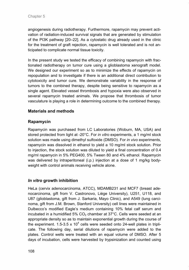
Chapter 5
108
angiogenesis during radiotherapy. Furthermore, rapamycin may prevent acti-vation of radiation-induced survival signals that are generated by stimulation of the PI3K pathway [20–22]. As a cytostatic drug already used in the clinic for the treatment of graft rejection, rapamycin is well tolerated and is not an-ticipated to complicate normal tissue toxicity. In the present study we tested the efficacy of combining rapamycin with frac-tionated radiotherapy on tumor cure using a glioblastoma xenograft model. We designed our experiment so as to minimize the effects of rapamycin on repopulation and to investigate if there is an additional direct contribution to cytotoxicity and tumor cure. We demonstrate variability in the response of tumors to the combined therapy, despite being sensitive to rapamycin as a single agent. Elevated vessel thrombosis and hypoxia were also observed in several rapamycin treated animals. We propose that thrombosis of tumor vasculature is playing a role in determining outcome to the combined therapy.
Materials and methods
Rapamycin
Rapamycin was purchased from LC Laboratories (Woburn, MA, USA) and stored protected from light at -20°C. For in vitro experiments, a 1 mg/ml stock solution was made using dimethyl sulfoxide (DMSO). For in vivo experiments, rapamycin was dissolved in ethanol to yield a 10 mg/ml stock solution. Prior to injection, the stock solution was diluted to yield a final concentration of 0.4 mg/ml rapamycin in 5% PEG400, 5% Tween 80 and 4% ethanol. Rapamycin was delivered by intraperitoneal (i.p.) injection at a dose of 1 mg/kg body-weight with control animals receiving vehicle alone.
In vitro growth inhibition
HeLa (cervix adenocarcinoma, ATCC), MDAMB231 and MCF7 (breast ade-nocarcinoma, gift from V. Castronovo, Liège University), U251, U118, and U87 (glioblastoma, gift from J. Sarkaria, Mayo Clinic), and A549 (lung carci-noma, gift from J.M. Brown, Stanford University) cell lines were maintained in Dulbecco’s modified Eagle’s medium containing 10% fetal calf serum and incubated in a humidified 5% CO2 chamber at 37°C. Cells were seeded at an appropriate density so as to maintain exponential growth during the course of the experiment. 1.5-3.5 x 10⁴ cells were seeded onto 24-well plates in tripli-cate. The following day, serial dilutions of rapamycin were added to the plates. Control wells were treated with an equal volume of DMSO. After 5 days of incubation, cells were harvested by trypsinization and counted using
Thesis_Weppler_v12_fc.pdf
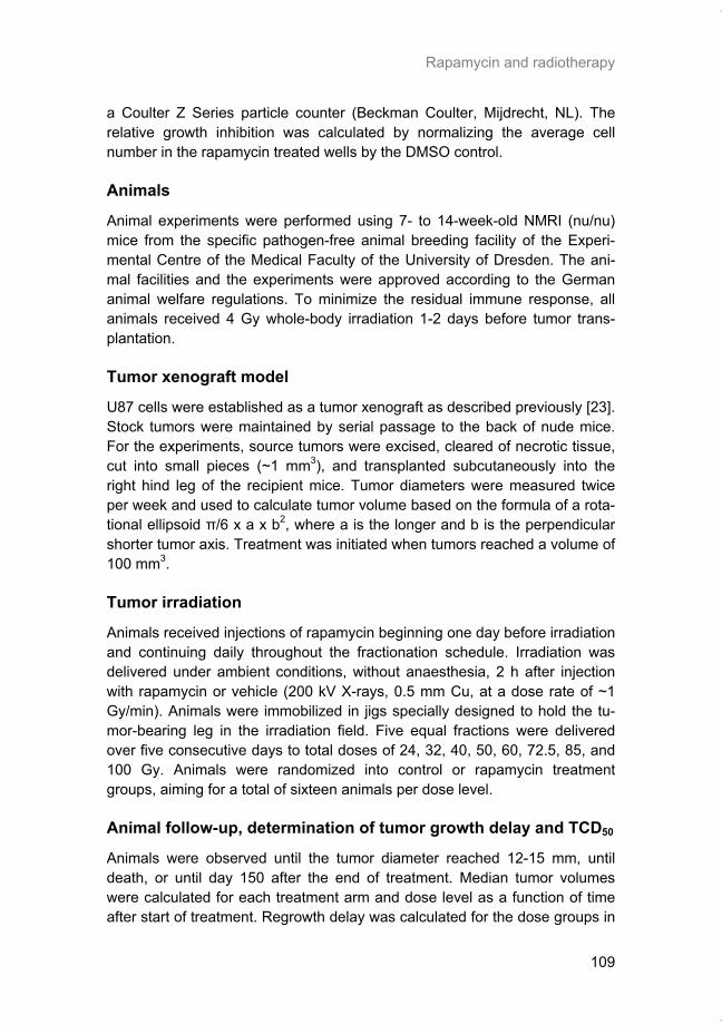
Rapamycin and radiotherapy
109
a Coulter Z Series particle counter (Beckman Coulter, Mijdrecht, NL). The relative growth inhibition was calculated by normalizing the average cell number in the rapamycin treated wells by the DMSO control.
Animals
Animal experiments were performed using 7- to 14-week-old NMRI (nu/nu) mice from the specific pathogen-free animal breeding facility of the Experi-mental Centre of the Medical Faculty of the University of Dresden. The ani-mal facilities and the experiments were approved according to the German animal welfare regulations. To minimize the residual immune response, all animals received 4 Gy whole-body irradiation 1-2 days before tumor trans-plantation.
Tumor xenograft model
U87 cells were established as a tumor xenograft as described previously [23]. Stock tumors were maintained by serial passage to the back of nude mice. For the experiments, source tumors were excised, cleared of necrotic tissue, cut into small pieces (~1 mm3), and transplanted subcutaneously into the right hind leg of the recipient mice. Tumor diameters were measured twice per week and used to calculate tumor volume based on the formula of a rota-tional ellipsoid π/6 x a x b2, where a is the longer and b is the perpendicular shorter tumor axis. Treatment was initiated when tumors reached a volume of 100 mm3.
Tumor irradiation
Animals received injections of rapamycin beginning one day before irradiation and continuing daily throughout the fractionation schedule. Irradiation was delivered under ambient conditions, without anaesthesia, 2 h after injection with rapamycin or vehicle (200 kV X-rays, 0.5 mm Cu, at a dose rate of ~1 Gy/min). Animals were immobilized in jigs specially designed to hold the tu-mor-bearing leg in the irradiation field. Five equal fractions were delivered over five consecutive days to total doses of 24, 32, 40, 50, 60, 72.5, 85, and 100 Gy. Animals were randomized into control or rapamycin treatment groups, aiming for a total of sixteen animals per dose level.
Animal follow-up, determination of tumor growth delay and TCD50
Animals were observed until the tumor diameter reached 12-15 mm, until death, or until day 150 after the end of treatment. Median tumor volumes were calculated for each treatment arm and dose level as a function of time after start of treatment. Regrowth delay was calculated for the dose groups in
Thesis_Weppler_v12_fc.pdf
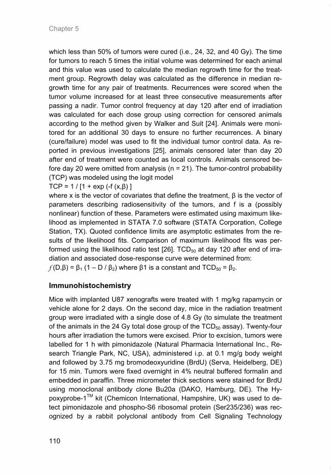
Chapter 5
110
which less than 50% of tumors were cured (i.e., 24, 32, and 40 Gy). The time for tumors to reach 5 times the initial volume was determined for each animal and this value was used to calculate the median regrowth time for the treat-ment group. Regrowth delay was calculated as the difference in median re-growth time for any pair of treatments. Recurrences were scored when the tumor volume increased for at least three consecutive measurements after passing a nadir. Tumor control frequency at day 120 after end of irradiation was calculated for each dose group using correction for censored animals according to the method given by Walker and Suit [24]. Animals were moni-tored for an additional 30 days to ensure no further recurrences. A binary (cure/failure) model was used to fit the individual tumor control data. As re-ported in previous investigations [25], animals censored later than day 20 after end of treatment were counted as local controls. Animals censored be-fore day 20 were omitted from analysis (n = 21). The tumor-control probability (TCP) was modeled using the logit model TCP = 1 / [1 + exp (-f (x,β) ] where x is the vector of covariates that define the treatment, β is the vector of parameters describing radiosensitivity of the tumors, and f is a (possibly nonlinear) function of these. Parameters were estimated using maximum like-lihood as implemented in STATA 7.0 software (STATA Corporation, College Station, TX). Quoted confidence limits are asymptotic estimates from the re-sults of the likelihood fits. Comparison of maximum likelihood fits was per-formed using the likelihood ratio test [26]. TCD50 at day 120 after end of irra-diation and associated dose-response curve were determined from: f (D,β) = β1 (1 – D / β2) where β1 is a constant and TCD50 = β2.
Immunohistochemistry
Mice with implanted U87 xenografts were treated with 1 mg/kg rapamycin or vehicle alone for 2 days. On the second day, mice in the radiation treatment group were irradiated with a single dose of 4.8 Gy (to simulate the treatment of the animals in the 24 Gy total dose group of the TCD50 assay). Twenty-four hours after irradiation the tumors were excised. Prior to excision, tumors were labelled for 1 h with pimonidazole (Natural Pharmacia International Inc., Re-search Triangle Park, NC, USA), administered i.p. at 0.1 mg/g body weight and followed by 3.75 mg bromodeoxyuridine (BrdU) (Serva, Heidelberg, DE) for 15 min. Tumors were fixed overnight in 4% neutral buffered formalin and embedded in paraffin. Three micrometer thick sections were stained for BrdU using monoclonal antibody clone Bu20a (DAKO, Hamburg, DE). The Hy-poxyprobe-1TM kit (Chemicon International, Hampshire, UK) was used to de-tect pimonidazole and phospho-S6 ribosomal protein (Ser235/236) was rec-ognized by a rabbit polyclonal antibody from Cell Signaling Technology
Thesis_Weppler_v12_fc.pdf
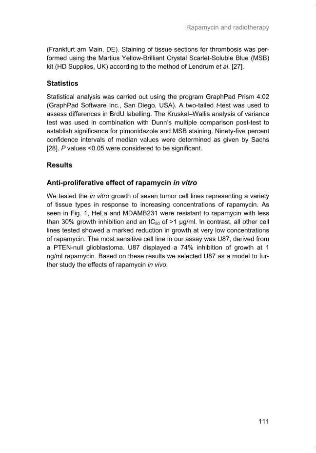
Rapamycin and radiotherapy
111
(Frankfurt am Main, DE). Staining of tissue sections for thrombosis was per-formed using the Martius Yellow-Brilliant Crystal Scarlet-Soluble Blue (MSB) kit (HD Supplies, UK) according to the method of Lendrum et al. [27].
Statistics
Statistical analysis was carried out using the program GraphPad Prism 4.02 (GraphPad Software Inc., San Diego, USA). A two-tailed t-test was used to assess differences in BrdU labelling. The Kruskal–Wallis analysis of variance test was used in combination with Dunn’s multiple comparison post-test to establish significance for pimonidazole and MSB staining. Ninety-five percent confidence intervals of median values were determined as given by Sachs [28]. P values <0.05 were considered to be significant.
Results
Anti-proliferative effect of rapamycin in vitro
We tested the in vitro growth of seven tumor cell lines representing a variety of tissue types in response to increasing concentrations of rapamycin. As seen in Fig. 1, HeLa and MDAMB231 were resistant to rapamycin with less than 30% growth inhibition and an IC50 of >1 µg/ml. In contrast, all other cell lines tested showed a marked reduction in growth at very low concentrations of rapamycin. The most sensitive cell line in our assay was U87, derived from a PTEN-null glioblastoma. U87 displayed a 74% inhibition of growth at 1 ng/ml rapamycin. Based on these results we selected U87 as a model to fur-ther study the effects of rapamycin in vivo.
Thesis_Weppler_v12_fc.pdf
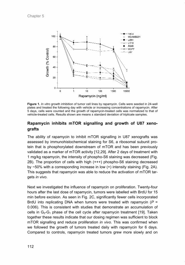
Chapter 5
112
Figure 1. In vitro growth inhibition of tumor cell lines by rapamycin. Cells were seeded in 24-well plates and treated the following day with vehicle or increasing concentrations of rapamycin. After 5 days, cells were counted and the growth of rapamycin-treated cells was normalized to that of vehicle-treated cells. Results shown are means ± standard deviation of triplicate samples.
Rapamycin inhibits mTOR signalling and growth of U87 xeno-grafts
The ability of rapamycin to inhibit mTOR signalling in U87 xenografts was assessed by immunohistochemical staining for S6, a ribosomal subunit pro-tein that is phosphorylated downstream of mTOR and has been previously validated as a marker of mTOR activity [12,29]. After 2 days of treatment with 1 mg/kg rapamycin, the intensity of phospho-S6 staining was decreased (Fig. 2B). The proportion of cells with high (+++) phospho-S6 staining decreased by ~50% with a corresponding increase in low (+) intensity staining (Fig. 2A). This suggests that rapamycin was able to reduce the activation of mTOR tar-gets in vivo. Next we investigated the influence of rapamycin on proliferation. Twenty-four hours after the last dose of rapamycin, tumors were labelled with BrdU for 15 min before excision. As seen in Fig. 2C, significantly fewer cells incorporated BrdU into replicating DNA when tumors were treated with rapamycin (P = 0.006). This is consistent with studies that demonstrate an accumulation of cells in G0-G1 phase of the cell cycle after rapamycin treatment [19]. Taken together these results indicate that our dosing regimen was sufficient to block mTOR signalling and reduce proliferation in vivo. This was confirmed when we followed the growth of tumors treated daily with rapamycin for 6 days. Compared to controls, rapamycin treated tumors grew more slowly and on
Thesis_Weppler_v12_fc.pdf
Rapamycin and radiotherapy
113
average required 4 additional days in order to reach the same relative tumor volume (Fig. 2D).
Figure 2. Rapamycin inhibits mTOR signalling and proliferation in vivo. Nude mice bearing sub-cutaneous U87 xenografts were treated with 1 mg/kg rapamycin for 2 days. Control animals received vehicle alone. On day 3, animals were injected with BrdU and euthanized. Tumors were excised and processed for immunohistochemical staining. (A) Phosphorylation of ribosomal pro-tein S6 was used as a marker of mTOR activity. Staining intensity was scored on a scale from zero to +++ in sections from 5 mice per group. (B) Phospho-S6 staining in representative control and rapamycin treated tumors. Arrows indicate the various levels of staining intensity. (C) BrdU uptake of cells in S-phase. Columns represent the mean labelling index ± standard deviation from 5 mice per group. *P = 0.006. (D) Tumor growth delay of animals treated with vehicle (con-trol) or rapamycin for 6 days. Data points represent the median number of days to reach a fixed tumor volume and error bars represent 95% confidence intervals.
Thesis_Weppler_v12_fc.pdf
Chapter 5
114
Tumor growth delay with combination therapy
We next determined if rapamycin could delay tumor regrowth if used in com-bination with fractionated radiotherapy. Rapamycin was given for 6 days along with equal fractions of radiation on days 2-6. In the lowest dose group of 24 Gy, we observed a 26 day growth delay for tumors to reach 5 times the starting volume, however there was no additional effect of rapamycin (Fig. 3A). Tumors treated with the combination therapy began to regress sooner but tumor regrowth began at approximately the same time as the tumors re-ceiving radiation alone. The higher dose groups showed a similar pattern with no enhancement of growth delay (data not shown). Interestingly, we ob-served a large variation in the response of tumors within the rapa + RT treatment arm which accounts for the large error bars in Fig. 3A. To illustrate this variability, the growth of individual tumors is plotted in Fig. 3B. In the up-per graph, the majority of tumors treated with 24 Gy alone responded in a similar fashion with regrowth beginning around day 25. In contrast, the group receiving concurrent rapamycin had 4 out of 8 tumors that behaved similar to the radiation only controls, one that had a delayed regrowth, and 3 tumors that were cured. The data suggest that some tumors receiving the combined treatment may have responded better although the group as a whole did not show an improvement in growth delay.
Local tumor control
Animals were treated with fractionated radiotherapy as described above for the growth delay experiment. A total of eight different doses ranging from 24 to 100 Gy were given in five equal fractions and the animals were observed long-term for local tumor recurrence. Fig. 4 depicts the rate of tumor cure (data points) and the predicted probability of tumor control (fitted curves). The TCD50 for the control group was calculated to be 46.8 Gy (95% CI 41; 53). Concurrent rapamycin treatment shifted the dose-response curve towards increased radiosensitivity with a reduction of the TCD50 to 42.8 Gy (95% CI 36; 49), although this value did not reach significance when compared to ra-diation alone.
Thesis_Weppler_v12_fc.pdf
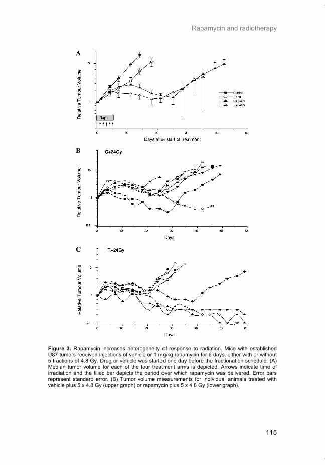
Rapamycin and radiotherapy
115
Figure 3. Rapamycin increases heterogeneity of response to radiation. Mice with established U87 tumors received injections of vehicle or 1 mg/kg rapamycin for 6 days, either with or without 5 fractions of 4.8 Gy. Drug or vehicle was started one day before the fractionation schedule. (A) Median tumor volume for each of the four treatment arms is depicted. Arrows indicate time of irradiation and the filled bar depicts the period over which rapamycin was delivered. Error bars represent standard error. (B) Tumor volume measurements for individual animals treated with vehicle plus 5 x 4.8 Gy (upper graph) or rapamycin plus 5 x 4.8 Gy (lower graph).
Thesis_Weppler_v12_fc.pdf
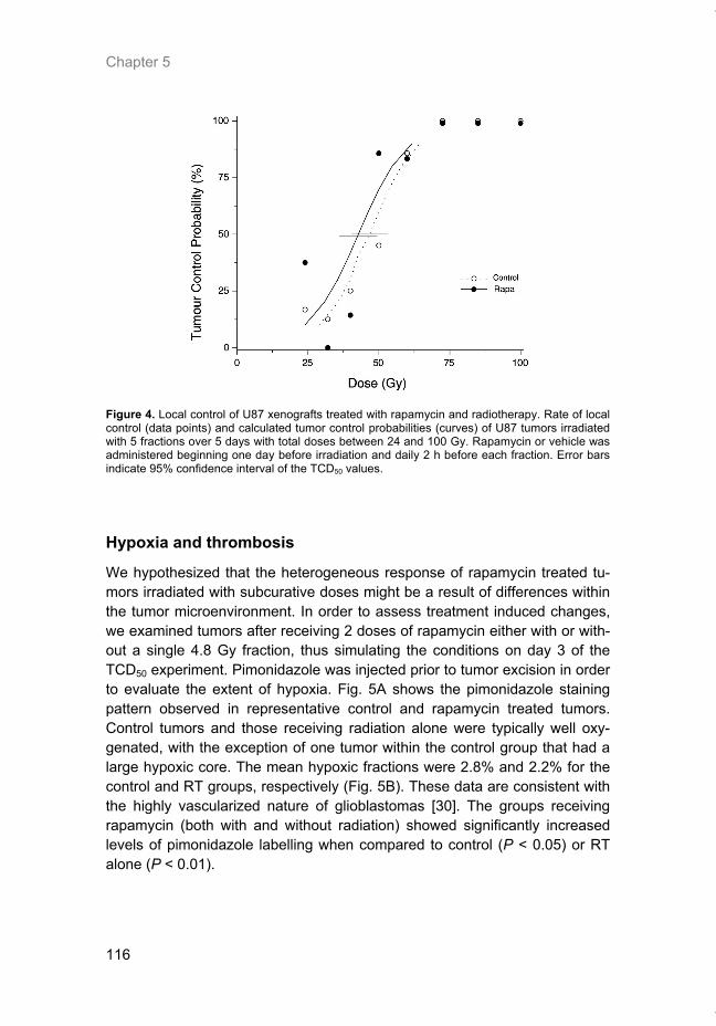
Chapter 5
116
Figure 4. Local control of U87 xenografts treated with rapamycin and radiotherapy. Rate of local control (data points) and calculated tumor control probabilities (curves) of U87 tumors irradiated with 5 fractions over 5 days with total doses between 24 and 100 Gy. Rapamycin or vehicle was administered beginning one day before irradiation and daily 2 h before each fraction. Error bars indicate 95% confidence interval of the TCD50 values.
Hypoxia and thrombosis
We hypothesized that the heterogeneous response of rapamycin treated tu-mors irradiated with subcurative doses might be a result of differences within the tumor microenvironment. In order to assess treatment induced changes, we examined tumors after receiving 2 doses of rapamycin either with or with-out a single 4.8 Gy fraction, thus simulating the conditions on day 3 of the TCD50 experiment. Pimonidazole was injected prior to tumor excision in order to evaluate the extent of hypoxia. Fig. 5A shows the pimonidazole staining pattern observed in representative control and rapamycin treated tumors. Control tumors and those receiving radiation alone were typically well oxy-genated, with the exception of one tumor within the control group that had a large hypoxic core. The mean hypoxic fractions were 2.8% and 2.2% for the control and RT groups, respectively (Fig. 5B). These data are consistent with the highly vascularized nature of glioblastomas [30]. The groups receiving rapamycin (both with and without radiation) showed significantly increased levels of pimonidazole labelling when compared to control (P < 0.05) or RT alone (P < 0.01).
Thesis_Weppler_v12_fc.pdf
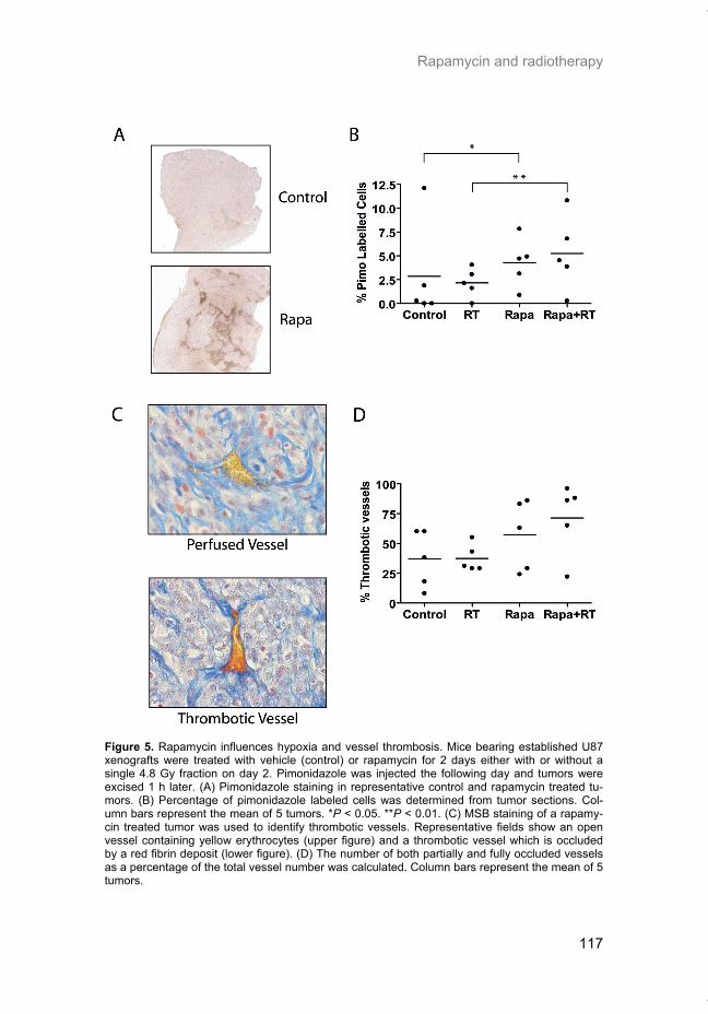
Rapamycin and radiotherapy
117
Figure 5. Rapamycin influences hypoxia and vessel thrombosis. Mice bearing established U87 xenografts were treated with vehicle (control) or rapamycin for 2 days either with or without a single 4.8 Gy fraction on day 2. Pimonidazole was injected the following day and tumors were excised 1 h later. (A) Pimonidazole staining in representative control and rapamycin treated tu-mors. (B) Percentage of pimonidazole labeled cells was determined from tumor sections. Col-umn bars represent the mean of 5 tumors. *P < 0.05. **P < 0.01. (C) MSB staining of a rapamy-cin treated tumor was used to identify thrombotic vessels. Representative fields show an open vessel containing yellow erythrocytes (upper figure) and a thrombotic vessel which is occluded by a red fibrin deposit (lower figure). (D) The number of both partially and fully occluded vessels as a percentage of the total vessel number was calculated. Column bars represent the mean of 5 tumors.
Thesis_Weppler_v12_fc.pdf
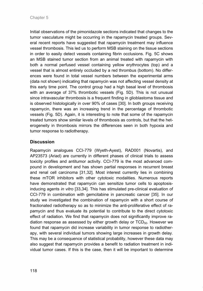
Chapter 5
118
Initial observations of the pimonidazole sections indicated that changes to the tumor vasculature might be occurring in the rapamycin treated groups. Sev-eral recent reports have suggested that rapamycin treatment may influence vessel thrombosis. This led us to perform MSB staining on the tissue sections in order to easily detect vessels containing fibrin occlusions. Fig. 5C shows an MSB stained tumor section from an animal treated with rapamycin with both a normal perfused vessel containing yellow erythrocytes (top) and a vessel that is almost entirely occluded by a red thrombus (bottom). No differ-ences were found in total vessel numbers between the experimental arms (data not shown) indicating that rapamycin was not affecting vessel density at this early time point. The control group had a high basal level of thrombosis with an average of 37% thrombotic vessels (Fig. 5D). This is not unusual since intravascular thrombosis is a frequent finding in glioblastoma tissue and is observed histologically in over 90% of cases [30]. In both groups receiving rapamycin, there was an increasing trend in the percentage of thrombotic vessels (Fig. 5D). Again, it is interesting to note that some of the rapamycin treated tumors show similar levels of thrombosis as controls, but that the het-erogeneity in thrombosis mirrors the differences seen in both hypoxia and tumor response to radiotherapy.
Discussion
Rapamycin analogues CCI-779 (Wyeth-Ayest), RAD001 (Novartis), and AP23573 (Ariad) are currently in different phases of clinical trials to assess toxicity profiles and antitumor activity. CCI-779 is the most advanced com-pound in development and has shown partial responses in recurrent breast and renal cell carcinoma [31,32]. Most interest currently lies in combining these mTOR inhibitors with other cytotoxic modalities. Numerous reports have demonstrated that rapamycin can sensitize tumor cells to apoptosis-inducing agents in vitro [33,34]. This has stimulated pre-clinical evaluation of CCI-779 in combination with gemcitabine in pancreatic cancer [35]. In our study we investigated the combination of rapamycin with a short course of fractionated radiotherapy so as to minimize the anti-proliferative effect of ra-pamycin and thus evaluate its potential to contribute to the direct cytotoxic effect of radiation. We find that rapamycin does not significantly improve ra-diation response as assessed by either growth delay or TCD50. However we found that rapamycin did increase variability in tumor response to radiother-apy, with several individual tumors showing large increases in growth delay. This may be a consequence of statistical probability, however these data may also suggest that rapamycin provides a benefit to radiation treatment in indi-vidual tumor cases. If this is the case, then it will be important to determine
Thesis_Weppler_v12_fc.pdf
Rapamycin and radiotherapy
119
the biological factors that mediate this differential response in order to poten-tially identify patients that may benefit from combination treatment. Differences in the pharmacokinetics of rapamycin have been observed in clinical trials [36] and would help to explain a lack of enhancement of radio-therapy if delivery of rapamycin to the tumor was impaired. Although we can-not rule out this possibility entirely, it seems unlikely to play a major role since animals receiving rapamycin alone responded in a homogeneous manner with respect to proliferation and growth delay (Fig. 2C and D). Our studies point toward changes in the microenvironment as a potential con-tributing factor in the response to the combination of rapamycin and radiation. We found that rapamycin treated animals showed increased amounts of thrombosis and also an increase in the hypoxic fraction measured by pimoni-dazole binding. However, similar to the results observed in individual tumor response, these microenvironmental changes after rapamycin treatment were also variable amongst the individual tumors examined. It is tempting to speculate that the increase in both vessel thrombosis and resulting hypoxia in a subset of rapamycin treated tumors may explain the variation in their re-sponse to radiotherapy. The complete or partial restriction to blood flow by thrombotic vessels would produce local areas of acute hypoxia causing an increase in radioresistance. If this is the case, then administration of rapamy-cin prior to radiotherapy, as was done in our study, may decrease radiosensi-tivity due to transient increases in acute hypoxia and thus offset any potential increase in toxicity of the combination treatment. This hypothesis is sup-ported by the finding that the combination treatment showed a substantial gain in the group of animals which received the lowest dose per fraction (24 Gy group). It is well known that the influence of hypoxia on overall response in fractionated therapy increases with increasing dose per fraction. It would be interesting to test this hypothesis by administering rapamycin in an adju-vant setting and/or further reducing the dose per fraction so as to minimize the consequences of (acute) hypoxia. It should be pointed out however, that at this time we cannot rule out the possibility that rapamycin-induced throm-bosis may be beneficial if the additional vascular damage translates into in-creased cytotoxicity to tumor cells, as has been documented using vascular-targeting agents in combination with radiotherapy [37,38]. Our analysis of vessel thrombosis was conducted after the initial investigation of tumor hypoxia. In hindsight, the hypoxic fraction may have been under-estimated in our experiment if you take into account the possible inhibitory effect of thrombosis on pimonidazole delivery to the tumor. Intrinsic markers of hypoxia or oxygen probe measurements would be useful to determine if
Thesis_Weppler_v12_fc.pdf
Chapter 5
120
this is in fact occurring in our system. In addition, sequential measurements of hypoxia over time would shed light on the relative effect of rapamycin on hypoxia in individual tumors. The ability of rapamycin to contribute to response through its effects on the vasculature may also be influenced by tumor type. Glioblastomas are charac-terized by frequent thrombosis and areas of necrosis and in addition, these patients are at high risk for developing deep-vein thrombosis and pulmonary embolism suggesting a systemic dysfunction of coagulation [30]. Therefore, a fraction of these tumors may already have high levels of thrombosis making it difficult to observe additional thrombosis induced by rapamycin. Previous studies documenting rapamycin-induced thrombosis were conducted using pancreatic and colon adenocarcinoma models and did not find evidence of thrombosis in control tumors [17,39]. It would thus be interesting to evaluate rapamycin in combination with radiation in other tumor types showing less intrinsic vasculature occlusion. Subsequent studies should also consider in-cluding tumor lines which are less sensitive than U87 to rapamycin as a sin-gle agent since the magnitude of this response may mask interactions with radiation. Any variability we observed in tumor response may simply reflect the sto-chastic nature of thrombus formation and its consequences. Impaired perfu-sion in a single vessel due to thrombus formation can potentially affect thou-sands or millions of individual tumor cells and thus have a large impact on tumor response. The fact that the hypoxic fraction can vary tremendously amongst individual untreated tumors derived from the same cell line and grown in the same host supports a role for the stochastic nature of blood vessel formation and/or function in determining hypoxia and radiosensitivity of individual tumors [40]. Finally, there is also some recent evidence suggesting that pre-treatment levels of hypoxia could contribute to the ability of rapamycin to affect tumor response. As described previously, the activity of mTOR is regulated by nu-merous upstream pathways that monitor growth factors, nutrients, and en-ergy metabolism. Recently hypoxia was also implicated in the regulation of mTOR signalling through two different pathways. Hypoxia causes activation of the TSC1/2 complex, which functions to inhibit mTOR. This can occur both via induction of the HIF-dependent gene REDD1, and/or through activation of AMPK [41,42]. This leads us to speculate that rapamycin may be less effec-tive in hypoxic regions of tumors since mTOR may already be at least par-tially inactivated by TSC. Thus the amount of hypoxia present at the start of treatment may play a part in determining sensitivity to rapamycin in vivo.
Thesis_Weppler_v12_fc.pdf
Rapamycin and radiotherapy
121
Whether this is relevant in our model is subject to debate since Kaper et al. argue that hypoxia’s ability to inhibit mTOR is diminished in tumors where mTOR activity has been highly upregulated by mutation of the PI3K pathway [43]. It is clear that additional preclinical work is needed to establish the degree and consequences of vascular thrombosis and hypoxia on both tumors and normal tissues treated with rapamycin and irradiation. It would be interesting to monitor perfusion during the course of therapy via in vivo imaging tech-niques such as DCE-MRI and to compare this with treatment outcome. Also, alternative sequencing and timing of rapamycin and radiation delivery should be investigated in order to determine if there is an optimal therapeutic win-dow. Our results suggest that it will be important to consider the effects of rapamycin on tumor oxygenation and thrombosis and to use caution in the design of future clinical trials combining mTOR inhibitors with radiotherapy.
Acknowledgements
This work was supported in part by a grant from the Nederlands Vereniging voor Oncologie awarded to S.A.W. and by Grant Ba 1433-4 by the Deutsche Forschungsgemeinschaft to M.B. The authors thank Ms. D. Pfitzmann, Ms. K. Schumann, and Ms. E. Lootze for their excellent technical assistance with the animal experiments conducted in Dresden. We also thank Mrs. S. Balschukat (Dresden), and Mrs. Hanna Andrusewicz and Dr. Jan Sir (Department of Pa-thology in Bydgoszcz) for their help with the histological assessments.
Thesis_Weppler_v12_fc.pdf
Chapter 5
122
References [1] Dancey JE. Inhibitors of the mammalian target of rapamycin. Expert Opin Investig Drugs
2005;14:313–28. [2] Rubio-Viqueira B, Hidalgo M. Targeting mTOR for cancer treatment. Curr Opin Investig
Drugs 2006;7:501–12. [3] Avruch J, Lin Y, Long X, Murthy S, Ortiz-Vega S. Recent advances in the regulation of the
TOR pathway by insulin and nutrients. Curr Opin Clin Nutr Metab Care 2005;8:67–72. [4] Petroulakis E, Mamane Y, Le Bacquer O, Shahbazian D, Sonenberg N. mTOR signaling:
implications for cancer and anticancer therapy. Br J Cancer 2006;94:195–9. [5] Vivanco I, Sawyers CL. The phosphatidylinositol 3-Kinase AKT pathway in human cancer.
Nat Rev Cancer 2002;2:489–501. [6] Choe G, Horvath S, Cloughesy TF, et al. Analysis of the phosphatidylinositol 30-kinase
signaling pathway in glioblastoma patients in vivo. Cancer Res 2003;63:2742–6. [7] Kang S, Bader AG, Zhao L, Vogt PK. Mutated PI 3-kinases: cancer targets on a silver plat-
ter. Cell Cycle 2005;4:578–81. [8] Guertin DA, Sabatini DM. An expanding role for mTOR in cancer. Trends Mol Med
2005;11:353–61. [9] Yu K, Toral-Barza L, Discafani C, et al. mTOR, a novel target in breast cancer: the effect of
CCI-779, an mTOR inhibitor, in preclinical models of breast cancer. Endocr Relat Cancer 2001;8:249–58.
[10] Hashemolhosseini S, Nagamine Y, Morley SJ, Desrivieres S, Mercep L, Ferrari S. Rapa-mycin inhibition of the G1 to S transition is mediated by effects on cyclin D1 mRNA and pro-tein stability. J Biol Chem 1998;273:14424–9.
[11] Kawamata S, Sakaida H, Hori T, Maeda M, Uchiyama T. The upregulation of p27Kip1 by rapamycin results in G1 arrest in exponentially growing T-cell lines. Blood 1998;91:561–9.
[12] Teachey DT, Obzut DA, Cooperman J, Fang J, Carroll M, Choi JK, Houghton PJ, Brown VI, Grupp SA. The mTOR inhibitor CCI-779 induces apoptosis and inhibits growth in preclinical models of primary adult human ALL. Blood 2006;107:1149–55.
[13] Huang S, Bjornsti MA, Houghton PJ. Rapamycins: mechanism of action and cellular resis-tance. Cancer Biol Ther 2003;2:222–32.
[14] Hosoi H, Dilling MB, Shikata T, et al. Rapamycin causes poorly reversible inhibition of mTOR and induces p53-independent apoptosis in human rhabdomyosarcoma cells. Cancer Res 1999;59:886–94.
[15] Takeuchi H, Kondo Y, Fujiwara K, et al. Synergistic augmentation of rapamycin-induced autophagy in malignant glioma cells by phosphatidylinositol 3-kinase/protein kinase B in-hibitors. Cancer Res 2005;65:3336–46.
[16] Guba M, von Breitenbuch P, Steinbauer M, et al. Rapamycin inhibits primary and metas-tatic tumor growth by antiangiogenesis: involvement of vascular endothelial growth factor. Nat Med 2002;8:128–35.
[17] Guba M, Yezhelyev M, Eichhorn ME, et al. Rapamycin induces tumor-specific thrombosis via tissue factor in the presence of VEGF. Blood 2005;105:4463–9.
[18] Shinohara ET, Cao C, Niermann K, et al. Enhanced radiation damage of tumor vasculature by mTOR inhibitors. Oncogene 2005;24:5414–22.
[19] Eshleman JS, Carlson BL, Mladek AC, Kastner BD, Shide KL, Sarkaria JN. Inhibition of the mammalian target of rapamycin sensitizes U87 xenografts to fractionated radiation therapy. Cancer Res 2002;62:7291–7.
[20] Albert JM, Kim KW, Cao C, Lu B. Targeting the Akt/mammalian target of rapamycin path-way for radiosensitization of breast cancer. Mol Cancer Ther 2006;5:1183–9.
[21] Toulany M, Dittmann K, Baumann M, Rodemann HP. Radiosensitization of Ras-mutated human tumor cells in vitro by the specific EGF receptor antagonist BIBX1382BS. Radiother Oncol 2005;74:117–29.
[22] Gottschalk AR, Doan A, Nakamura JL, Haas-Kogan DA, Stokoe D. Inhibition of phosphati-dylinositol-3-kinase causes cell death through a protein kinase B (PKB)-dependent mecha-nism and growth arrest through a PKB-independent mechanism. Int J Radiat Oncol Biol Phys 2005;61:1183–8.
[23] Taghian A, DuBois W, Budach W, Baumann M, Freeman J, Suit H. In vivo radiation sensi-tivity of glioblastoma multiforme. Int J Radiat Oncol Biol Phys 1995;32:99–104.
[24] Walker AM, Suit HD. Assessment of local tumor control using censored tumor response data. Int J Radiat Oncol Biol Phys 1983;9:383–6.
Thesis_Weppler_v12_fc.pdf
Rapamycin and radiotherapy
123
[25] Krause M, Schutze C, Petersen C, et al. Different classes of EGFR inhibitors may have different potential to improve local tumour control after fractionated irradiation: a study on C225 in FaDu hSCC. Radiother Oncol 2005;74:109–15.
[26] Edwards AWF. Likelihood. Expanded edition. Baltimore: Johns Hopkins University Press; 1992.
[27] Lendrum AC, Fraser DS, Slidders W, Henderson R. Studies on the character and staining of fibrin. J Clin Pathol 1962;15:401–13.
[28] Sachs L. Angewandte statistik. Berlin: Springer; 1992. [29] Kenerson HL, Aicher LD, True LD, Yeung RS. Activated mammalian target of rapamycin
pathway in the pathogenesis of tuberous sclerosis complex renal tumors. Cancer Res 2002;62:5645–50.
[30] Brat DJ, Van Meir EG. Vaso-occlusive and prothrombotic mechanisms associated with tumor hypoxia, necrosis, and accelerated growth in glioblastoma. Lab Invest 2004;84:397–405.
[31] Chan S, Scheulen ME, Johnston S, et al. Phase II study of temsirolimus (CCI-779), a novel inhibitor of mTOR, in heavily pretreated patients with locally advanced or metastatic breast cancer. J Clin Oncol 2005;23:5314–22.
[32] Atkins MB, Hidalgo M, Stadler WM, et al. Randomized phase II study of multiple dose lev-els of CCI-779, a novel mammalian target of rapamycin kinase inhibitor, in patients with ad-vanced refractory renal cell carcinoma. J Clin Oncol 2004;22:909–18.
[33] Beuvink I, Boulay A, Fumagalli S, et al. The mTOR inhibitor RAD001 sensitizes tumor cells to DNA-damaged induced apoptosis through inhibition of p21 translation. Cell 2005;120:747–59.
[34] Mondesire WH, Jian W, Zhang H, et al. Targeting mammalian target of rapamycin synergis-tically enhances chemotherapyinduced cytotoxicity in breast cancer cells. Clin Cancer Res 2004;10:7031–42.
[35] Ito D, Fujimoto K, Mori T, et al. In vivo antitumor effect of the mTOR inhibitor CCI-779 and gemcitabine in xenograft models of human pancreatic cancer. Int J Cancer 2006;118:2337–43.
[36] Gallant-Haidner HL, Trepanier DJ, Freitag DG, Yatscoff RW. Pharmacokinetics and me-tabolism of sirolimus. Ther Drug Monit 2000;22:31–5.
[37] Hoang T, Huang S, Armstrong E, Eickhoff JC, Harari PM. Augmentation of radiation re-sponse with the vascular targeting agent ZD6126. Int J Radiat Oncol Biol Phys 2006;64:1458–65.
[38] Murata R, Siemann DW, Overgaard J, Horsman MR. Interaction between combretastatin A-4 disodium phosphate and radiation in murine tumors. Radiother Oncol 2001;60:155–61.
[39] Bruns CJ, Koehl GE, Guba M, et al. Rapamycin-induced endothelial cell death and tumor vessel thrombosis potentiate cytotoxic therapy against pancreatic cancer. Clin Cancer Res 2004;10:2109–19.
[40] Evans SM, Jenkins WT, Joiner B, Lord EM, Koch CJ. 2- Nitroimidazole (EF5) binding pre-dicts radiation resistance in individual 9L s.c. tumors. Cancer Res 1996;56:405–11.
[41] Liu L, Cash TP, Jones RG, Keith B, Thompson CB, Simon MC. Hypoxia-induced energy stress regulates mRNA translation and cell growth. Mol Cell 2006;21:521–31.
[42] Brugarolas J, Lei K, Hurley RL, et al. Regulation of mTOR function in response to hypoxia by REDD1 and the TSC1/TSC2 tumor suppressor complex. Genes Dev 2004;18:2893–904.
[43] Kaper F, Dornhoefer N, Giaccia AJ. Mutations in the PI3K/PTEN/TSC2 pathway contribute to mammalian target of rapamycin activity and increased translation under hypoxic condi-tions. Cancer Res 2006;66:1561–9.
Thesis_Weppler_v12_fc.pdf

Thesis_Weppler_v12_fc.pdf
125
CHAPTER 6
Inhibition of 4E-BP1 phosphorylation and mRNA translation requires simultaneous blockade of mTORC1 and PI3K/Akt signaling
Manuscript submitted SA Weppler, M Koritzinsky, JW Voncken, P Lambin, and BG Wout-ers.
Thesis_Weppler_v12_fc.pdf
Chapter 6
126
Abstract
The mammalian target of rapamycin (mTOR) kinase is a central regulator of cell metabolism and growth whose activity is often altered in cancer. An im-portant consequence of mTOR activation is stimulation of cap-dependent translation, which is mediated by phosphorylation and inactivation of the eu-karyotic initiation factor 4E-binding protein 1 (4E-BP1). Here, we demonstrate that short exposures to rapamycin, a specific inhibitor of mTOR complex 1 (mTORC1), has only a modest inhibitory effect on 4E-BP1 phosphorylation and mRNA translation despite completely blocking phosphorylation of an-other mTORC1 substrate p70S6K. We describe a synergistic effect of com-bined mTORC1 and phosphatidylinositol 3-kinase (PI3K) inhibition that is specific for dephosphorylation of 4E-BP1 at both Thr70 and Ser65, and which leads to a rapid suppression of translation in a 4E-BP1 dependent manner. Similar results are observed using rapamycin in combination with an inhibitor of protein kinase B (PKB, also termed Akt), suggesting the existence of a PI3K/Akt-dependent signaling pathway sufficient to maintain 4E-BP1 phos-phorylation in the presence of mTORC1 inhibition. In addition, the differential sensitivity of mTORC1 targets to inhibition of PI3K/Akt alone implies that mTORC1 complexes may exhibit substrate specificity dependent upon acti-vation of specific upstream signaling pathways to mTOR. Together these re-sults suggest that targeting deregulated translation in cancer may be im-proved by strategies that block both mTORC1 dependent and independent signals regulating 4E-BP1.
Thesis_Weppler_v12_fc.pdf
Regulation of 4E-BP1 phosphorylation
127
Introduction
The mammalian target of rapamycin (mTOR) kinase exists in at least two distinct complexes with independent functions and regulation (1). mTOR complex 1 (mTORC1) consists of the mTOR kinase, LST8 (also known as GβL) and raptor, a scaffolding protein that serves to bring mTORC1 sub-strates in close proximity to mTOR’s catalytic domain (2). The eukaryotic ini-tiation factor 4E (eIF4E) binding protein (4E-BP1) and p70S6K, both regula-tors of mRNA translation, are the best characterized targets of mTORC1. mTOR complex 2 (TORC2) also contains LST8, but instead of raptor associ-ates with rictor (3). TORC2 has been shown to phosphorylate Akt at Ser473 and protein kinase Cα, but to date no evidence of 4E-BP1 or p70S6K phos-phorylation has been demonstrated (4, 5). mTOR regulates several processes relevant to cancer including mRNA trans-lation, ribosome biogenesis, autophagy and metabolism (1). A direct link be-tween mTOR and cancer was established following discovery that the tuber-ous sclerosis complex (TSC) functions as a negative regulator of mTORC1. Mutations in tsc1 or tsc2 genes thus activate mTOR and are responsible for the development of harmatomas in TSC patients. The TSC1/2 complex regu-lates mTORC1 in response to many upstream signals including amino acid and oxygen availability, energy status, insulin and other growth factors (6). These signals influence mTOR via TSC1/2 through signaling pathways that are also frequently altered in cancer, including the PI3K/Akt pathway. In re-sponse to insulin and other growth factors, PI3K/Akt signaling activates mTORC1 by phosphorylation and inhibition of TSC2 (7). PI3K activation is antagonized by the tumor suppressor PTEN, and its importance in cancer has been directly linked to its ability to control mTORC1 activity (8). Regulation of translation is a critical function of mTOR that when deregulated contributes to cancer development. This is evidenced by the fact that both mTORC1 targets, 4E-BP1 and p70S6K, are involved in regulation of transla-tion. 4E-BP1 functions as a negative regulator of translation by competing with eIF4G for binding to the rate limiting translation initiation factor eIF4E (9). Growth promoting signals that stimulate mTOR lead to phosphorylation of 4E-BP1, which prevents it from binding to eIF4E and therefore stimulates translation. The importance of this mTOR effector has been demonstrated in a mouse model for cancer in which overexpression of eIF4E can bypass the requirement for mTOR activation (8). In addition to its general role in protein synthesis, deregulation of translational control by eIF4E and 4E-BP1 is thought to differentially alter the expression of cancer related genes in a manner that promotes malignant conversion (10). eIF4E is often found to be
Thesis_Weppler_v12_fc.pdf
Chapter 6
128
overexpressed in cancer, and its expression is sufficient to transform cells (11). 4E-BP1 also has a demonstrated role in regulating gene expression and functions as a potential biomarker of malignant progression and adverse prognosis in breast, ovary, and prostate tumors (12, 13). The binding of 4E-BP1 to eIF4E is regulated by phosphorylation at multiple sites. Non-phosphorylated or hypo-phosphorylated 4E-BP1 binds strongly to eIF4E and prevents cap-dependent translation, whereas hyperphosphory-lated 4E-BP1 dissociates from eIF4E, allowing translation to initiate (14). The phosphorylation of 4E-BP1 is complex involving at least six phospho-residues. Four of these sites have been linked to mTOR signaling and are phosphorylated in a hierarchical manner, so that phosphorylation at Thr37/Thr46 is required for subsequent phosphorylation of Thr70, which in turn is required for Ser65 phosphorylation (14-16). Although these sites all show dependency on mTOR, their sensitivity to changes in phosphorylation by different signals that regulate mTOR are distinct. For example, phosphory-lation of Thr37/Thr46 is affected significantly by amino acid availability, but not by insulin which preferentially stimulates Ser65 phosphorylation (6). The ability of 4E-BP1 to bind eIF4E and thus inhibit translation correlates best with the phosphorylation status of Ser65, although this is somewhat contro-versial (6, 9, 17). It remains unclear as to which, if any, of the 4E-BP1 phos-pho-sites are directly phosphorylated by mTOR and it is likely that other kinases are also involved (9, 18). The importance of mTOR in regulation of mRNA translation, cell growth and metabolism coupled with its deregulation in cancer has made it an attractive therapeutic target. Rapamycin is a macrolide antibiotic that inhibits mTOR with high selectivity (19). Rapamycin interacts with the immunophilin FKBP12, which then binds to the FRB-domain of mTOR and in doing so, weakens its interaction with raptor. As a consequence, rapamycin prevents phosphorylation of the raptor dependent mTORC1 substrates, p70S6K and 4E-BP1 (20, 21), but not rictor dependent TORC2 substrates like Akt (22) although longer treatments may indirectly effect mTORC2 activity by interfer-ing with assembly of mTOR into new TORC2 complexes (23). The ability of rapamycin to inhibit proliferation or other cancer associated phenotypes is highly variable between tumors and dependent on their genetic background (24, 25). For example, loss of PTEN or activation of Akt leads to constitutive mTOR activation and can significantly sensitize cells to the ef-fects of mTOR inhibitors (26, 27). Combinations of rapamycin with other small-molecule inhibitors or cytotoxic agents have also shown promising re-sults, and found to provide better inhibition of targets downstream of
Thesis_Weppler_v12_fc.pdf
Regulation of 4E-BP1 phosphorylation
129
mTORC1 (28-30). However, it is unclear whether this beneficial effect results from a more complete inhibition of mTORC1, or via mTORC1 independent regulation of its targets. Our objective was to examine the ability of mTORC1 and/or PI3K inhibition to regulate phosphorylation and function of 4E-BP1. Surprisingly, our data indicate that 4E-BP1 Ser65 and Ser70 phosphorylation and translation initiation is largely resistant to inhibition of either mTORC1 or PI3K. However, a strong synergistic effect restricted to the mTORC1 target 4E-BP1 is observed when these two targets are inhibited in combination. These data implicate PI3K/Akt in the regulation of 4E-BP1 and translation in an mTORC1 independent pathway and suggest a more effective way of tar-geting deregulated translation in cancer.
Materials and methods
Cell lines
HT29 and HCT116 colorectal carcinomas, A549 lung carcinoma, and HeLa cervix carcinoma were from the American Type Culture Collection, Tig3 hu-man fibroblasts were a gift from J.Fukami (31), and U87 glioblastoma cells were a gift from J. Sarkaria (Mayo Clinic, MN, USA). All the above cell lines were maintained in Dulbecco’s modified Eagle’s medium (DMEM) (Sigma-Aldrich, Zwijndrecht, NL) supplemented with 10% fetal calf serum (FCS) (HyClone, Logan UT, USA). DU145 prostate carcinoma cells were main-tained in McCoy’s 5A medium (Invitrogen, Breda, NL) with 10% FCS. U373 glioma cells were cultured as described (32). U87 cells containing an ecdy-sone inducible-PTEN construct were generated and validated previously (33).
Reagents and antibodies
Rapamycin, LY294002, wortmannin, and Akti-1/2 were purchased from Cal-biochem (Darmstadt, DE). Ponasterone A was from Sigma-Aldrich (Zwijndrecht, NL). The following antibodies were purchased from Cell Signal-ing Technology (Danvers, MA): Phospho-p70S6K (Thr389), Phospho-4E-BP1 (Ser65), Phospho-4E-BP1 (Thr70), Phospho-4E-BP1 (Thr37/46), Phospho-Akt (Ser473), Akt, PTEN, Raptor, and mTOR. The β-actin monoclonal anti-body (Clone C4) was purchased from MP Biomedicals (Amsterdam, NL). The eIF4E monoclonal antibody (Clone 87) was supplied by BD Biosciences (Breda, NL), and the eIF4GI antibody was purchased from Abcam (Cam-bridgeshire, UK). Secondary antibodies were purchased from Sigma-Aldrich (goat-anti-mouse) and Cell Signaling Technology (goat-anti-rabbit).
Thesis_Weppler_v12_fc.pdf
Chapter 6
130
Western blot analysis
Cell extracts were prepared, resolved by SDS-PAGE and transferred to a nitrocellulose membrane as described previously (32). The membrane was blocked with 5% milk-TBST (20mM Tris-HCl, pH 7.6, 140mM NaCl, 0.1% Tween 20) and incubated overnight with the appropriate primary antibody. The membrane was washed with TBST and incubated for 1 h with a horse-radish peroxidase-linked secondary antibody. Chemiluminescence detection was carried out using SuperSignal West Pico from Perbio (Etten-Leur, NL).
m7GTP-agarose affinity chromatography
eIF4E and associated proteins were isolated as described (34) but with the following amendments. 300 μg of cytoplasmic extract was incubated with 50 μl of m7GTP-Sepharose 4B resin (GE Healthcare Life Sciences) overnight at 4°C. The resin was washed, boiled in Laemmli sample buffer and the isolated proteins were resolved by SDS-PAGE and detected by immunoblotting.
Polysome analysis
Polysome-associated mRNA was isolated by sucrose gradient centrifugation as described previously (35). The absorbance at 254 nm was recorded con-tinuously as the gradient was fractionated.
RNA interference
4E-BP1 expression was modulated by stably transfecting U87 cells with pRetroSuper (36) containing a short hairpin RNA with the targeting sequence gtttgagatggacatttaa. The empty pRetroSuper vector was used as a control. Effective reduction of 4E-BP1 protein levels using this vector has been vali-dated previously (37). Pre-designed siRNA against raptor, as well as a non-targeting negative control siRNA, were purchased from Applied Biosystems (Nieuwerkerk a/d Ijssel, NL). Raptor siRNA had the sequence ggattat-gaggtcgtataatt and was used at a concentration of 20 nM. A SignalSilence siRNA kit for mTOR was purchased from Cell Signaling Technology. mTOR siRNA was transfected at a concentration of 50 nM. For all siRNA transfec-tions, cells were transfected in 6-well plates using 12 μl Oligofectamine (Invi-trogen) in a total volume of 2 ml growth medium according to the manufac-turer’s directions. Cell lysates were analyzed 72 h after transfection.
Clonogenic survival assay
24 hr after treatment with rapamycin and LY294002, cells were trypsinized and counted using a Coulter Z Series particle counter (Beckman Coulter, Mij-
Thesis_Weppler_v12_fc.pdf
Regulation of 4E-BP1 phosphorylation
131
drecht, NL). Cells were seeded in triplicate 6 cm dishes at two different densi-ties in complete growth medium. The dishes were incubated under standard culture conditions for a period of 12 days for colony formation. Colonies were fixed and stained with 2% bromophenol blue in 70% ethanol. Plating effi-ciency was determined by counting colonies consisting of ≥ 50 cells and cor-recting for the number of cells seeded.
Statistics
A two-tailed Student’s t-test was used to assess differences in translation inhibition and clonogenic survival. P values less than 0.05 were considered to be significant.
Results
4E-BP1 phosphorylation is independently maintained by mTORC1 and PI3K
Given the demonstrated influence of mTOR and PI3K signaling on 4E-BP1 phosphorylation and mRNA translation, we evaluated the consequences of inhibiting either or both of these pathways using the mTORC1 inhibitor rapa-mycin and the PI3K inhibitor LY294002. Two glioma cell lines were treated with these inhibitors, either alone or in combination, for a period of 2 h at which time we examined phosphorylation of 4E-BP1 and p70S6K (Figure 1a). In both U87 and U373 cell lines, treatment with rapamycin alone resulted in a complete loss of p70S6K phosphorylation. Consistent with its reported role in signaling to mTOR via TSC1/2, inhibition of PI3K with LY294002 was also sufficient to block phosphorylation of p70S6K. Despite this clear inhibition of mTORC1 activity with either inhibitor, phosphorylation of 4E-BP1 at Ser65 was only marginally reduced following either of these treatments alone. How-ever, a strong synergistic effect resulting in near complete loss of 4E-BP1 Ser65 phosphorylation was observed when the two inhibitors were used in combination (Figure 1a, lanes 4 and 8). No decrease in 4E-BP1 Ser65 phosphorylation similar to that observed with the combination treatment was found by increasing the concentration of ra-pamycin alone from 10nM to 100nM or the concentration of LY294002 alone from 5μM to 10μM (Supplementary Figure S1). This suggests that the differ-ential outcome of mTORC1 and PI3K inhibition on 4E-BP1 and p70S6K does not result from dissimilar sensitivities to any residual kinase activity present after treatment with the inhibitors (at least not at the concentrations used in these experiments). In contrast, a clear dose-dependent decrease in 4E-BP1
Thesis_Weppler_v12_fc.pdf
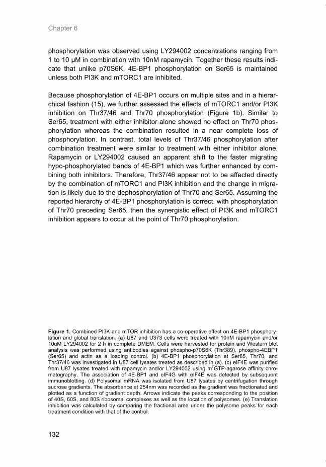
Chapter 6
132
phosphorylation was observed using LY294002 concentrations ranging from 1 to 10 μM in combination with 10nM rapamycin. Together these results indi-cate that unlike p70S6K, 4E-BP1 phosphorylation on Ser65 is maintained unless both PI3K and mTORC1 are inhibited. Because phosphorylation of 4E-BP1 occurs on multiple sites and in a hierar-chical fashion (15), we further assessed the effects of mTORC1 and/or PI3K inhibition on Thr37/46 and Thr70 phosphorylation (Figure 1b). Similar to Ser65, treatment with either inhibitor alone showed no effect on Thr70 phos-phorylation whereas the combination resulted in a near complete loss of phosphorylation. In contrast, total levels of Thr37/46 phosphorylation after combination treatment were similar to treatment with either inhibitor alone. Rapamycin or LY294002 caused an apparent shift to the faster migrating hypo-phosphorylated bands of 4E-BP1 which was further enhanced by com-bining both inhibitors. Therefore, Thr37/46 appear not to be affected directly by the combination of mTORC1 and PI3K inhibition and the change in migra-tion is likely due to the dephosphorylation of Thr70 and Ser65. Assuming the reported hierarchy of 4E-BP1 phosphorylation is correct, with phosphorylation of Thr70 preceding Ser65, then the synergistic effect of PI3K and mTORC1 inhibition appears to occur at the point of Thr70 phosphorylation.
Figure 1. Combined PI3K and mTOR inhibition has a co-operative effect on 4E-BP1 phosphory-lation and global translation. (a) U87 and U373 cells were treated with 10nM rapamycin and/or 10uM LY294002 for 2 h in complete DMEM. Cells were harvested for protein and Western blot analysis was performed using antibodies against phospho-p70S6K (Thr389), phospho-4EBP1 (Ser65) and actin as a loading control. (b) 4E-BP1 phosphorylation at Ser65, Thr70, and Thr37/46 was investigated in U87 cell lysates treated as described in (a). (c) eIF4E was purified from U87 lysates treated with rapamycin and/or LY294002 using m7GTP-agarose affinity chro-matography. The association of 4E-BP1 and eIF4G with eIF4E was detected by subsequent immunoblotting. (d) Polysomal mRNA was isolated from U87 lysates by centrifugation through sucrose gradients. The absorbance at 254nm was recorded as the gradient was fractionated and plotted as a function of gradient depth. Arrows indicate the peaks corresponding to the position of 40S, 60S, and 80S ribosomal complexes as well as the location of polysomes. (e) Translation inhibition was calculated by comparing the fractional area under the polysome peaks for each treatment condition with that of the control.
Thesis_Weppler_v12_fc.pdf
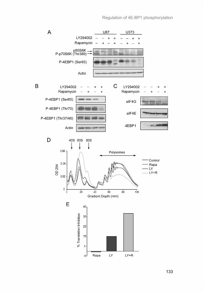
Regulation of 4E-BP1 phosphorylation
133
Thesis_Weppler_v12_fc.pdf
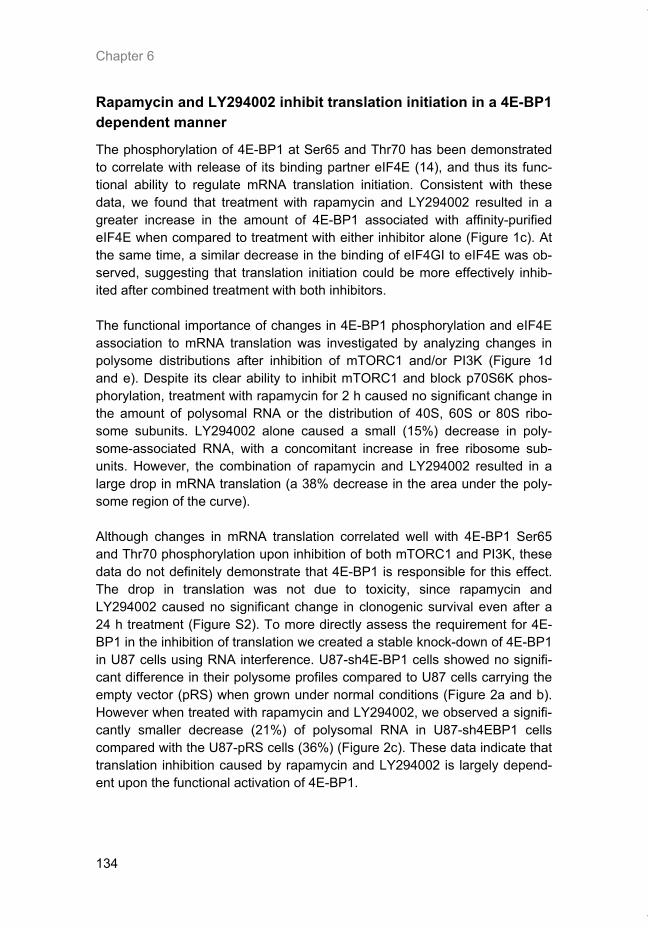
Chapter 6
134
Rapamycin and LY294002 inhibit translation initiation in a 4E-BP1 dependent manner
The phosphorylation of 4E-BP1 at Ser65 and Thr70 has been demonstrated to correlate with release of its binding partner eIF4E (14), and thus its func-tional ability to regulate mRNA translation initiation. Consistent with these data, we found that treatment with rapamycin and LY294002 resulted in a greater increase in the amount of 4E-BP1 associated with affinity-purified eIF4E when compared to treatment with either inhibitor alone (Figure 1c). At the same time, a similar decrease in the binding of eIF4GI to eIF4E was ob-served, suggesting that translation initiation could be more effectively inhib-ited after combined treatment with both inhibitors. The functional importance of changes in 4E-BP1 phosphorylation and eIF4E association to mRNA translation was investigated by analyzing changes in polysome distributions after inhibition of mTORC1 and/or PI3K (Figure 1d and e). Despite its clear ability to inhibit mTORC1 and block p70S6K phos-phorylation, treatment with rapamycin for 2 h caused no significant change in the amount of polysomal RNA or the distribution of 40S, 60S or 80S ribo-some subunits. LY294002 alone caused a small (15%) decrease in poly-some-associated RNA, with a concomitant increase in free ribosome sub-units. However, the combination of rapamycin and LY294002 resulted in a large drop in mRNA translation (a 38% decrease in the area under the poly-some region of the curve). Although changes in mRNA translation correlated well with 4E-BP1 Ser65 and Thr70 phosphorylation upon inhibition of both mTORC1 and PI3K, these data do not definitely demonstrate that 4E-BP1 is responsible for this effect. The drop in translation was not due to toxicity, since rapamycin and LY294002 caused no significant change in clonogenic survival even after a 24 h treatment (Figure S2). To more directly assess the requirement for 4E-BP1 in the inhibition of translation we created a stable knock-down of 4E-BP1 in U87 cells using RNA interference. U87-sh4E-BP1 cells showed no signifi-cant difference in their polysome profiles compared to U87 cells carrying the empty vector (pRS) when grown under normal conditions (Figure 2a and b). However when treated with rapamycin and LY294002, we observed a signifi-cantly smaller decrease (21%) of polysomal RNA in U87-sh4EBP1 cells compared with the U87-pRS cells (36%) (Figure 2c). These data indicate that translation inhibition caused by rapamycin and LY294002 is largely depend-ent upon the functional activation of 4E-BP1.
Thesis_Weppler_v12_fc.pdf
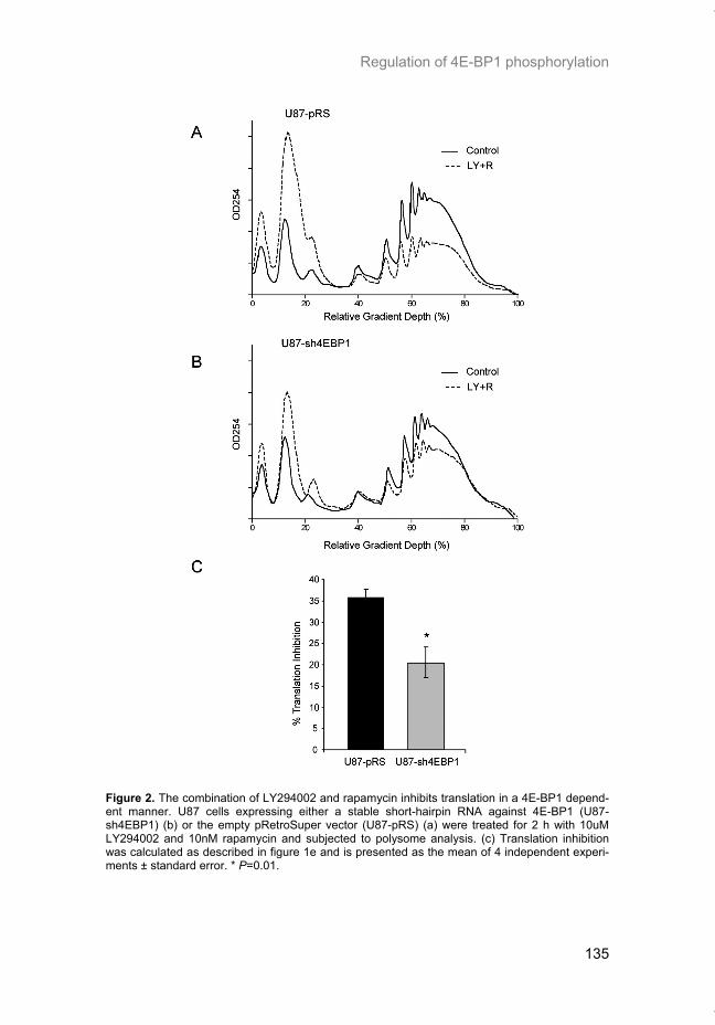
Regulation of 4E-BP1 phosphorylation
135
Figure 2. The combination of LY294002 and rapamycin inhibits translation in a 4E-BP1 depend-ent manner. U87 cells expressing either a stable short-hairpin RNA against 4E-BP1 (U87-sh4EBP1) (b) or the empty pRetroSuper vector (U87-pRS) (a) were treated for 2 h with 10uM LY294002 and 10nM rapamycin and subjected to polysome analysis. (c) Translation inhibition was calculated as described in figure 1e and is presented as the mean of 4 independent experi-ments ± standard error. * P=0.01.
Thesis_Weppler_v12_fc.pdf
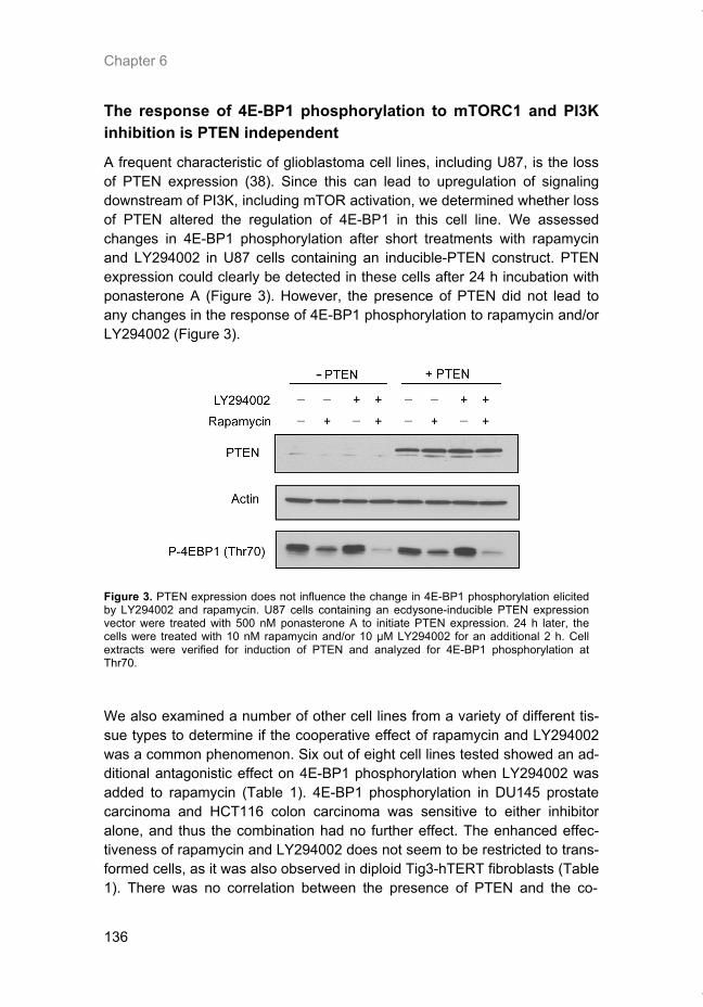
Chapter 6
136
The response of 4E-BP1 phosphorylation to mTORC1 and PI3K inhibition is PTEN independent
A frequent characteristic of glioblastoma cell lines, including U87, is the loss of PTEN expression (38). Since this can lead to upregulation of signaling downstream of PI3K, including mTOR activation, we determined whether loss of PTEN altered the regulation of 4E-BP1 in this cell line. We assessed changes in 4E-BP1 phosphorylation after short treatments with rapamycin and LY294002 in U87 cells containing an inducible-PTEN construct. PTEN expression could clearly be detected in these cells after 24 h incubation with ponasterone A (Figure 3). However, the presence of PTEN did not lead to any changes in the response of 4E-BP1 phosphorylation to rapamycin and/or LY294002 (Figure 3).
Figure 3. PTEN expression does not influence the change in 4E-BP1 phosphorylation elicited by LY294002 and rapamycin. U87 cells containing an ecdysone-inducible PTEN expression vector were treated with 500 nM ponasterone A to initiate PTEN expression. 24 h later, the cells were treated with 10 nM rapamycin and/or 10 μM LY294002 for an additional 2 h. Cell extracts were verified for induction of PTEN and analyzed for 4E-BP1 phosphorylation at Thr70. We also examined a number of other cell lines from a variety of different tis-sue types to determine if the cooperative effect of rapamycin and LY294002 was a common phenomenon. Six out of eight cell lines tested showed an ad-ditional antagonistic effect on 4E-BP1 phosphorylation when LY294002 was added to rapamycin (Table 1). 4E-BP1 phosphorylation in DU145 prostate carcinoma and HCT116 colon carcinoma was sensitive to either inhibitor alone, and thus the combination had no further effect. The enhanced effec-tiveness of rapamycin and LY294002 does not seem to be restricted to trans-formed cells, as it was also observed in diploid Tig3-hTERT fibroblasts (Table 1). There was no correlation between the presence of PTEN and the co-
Thesis_Weppler_v12_fc.pdf
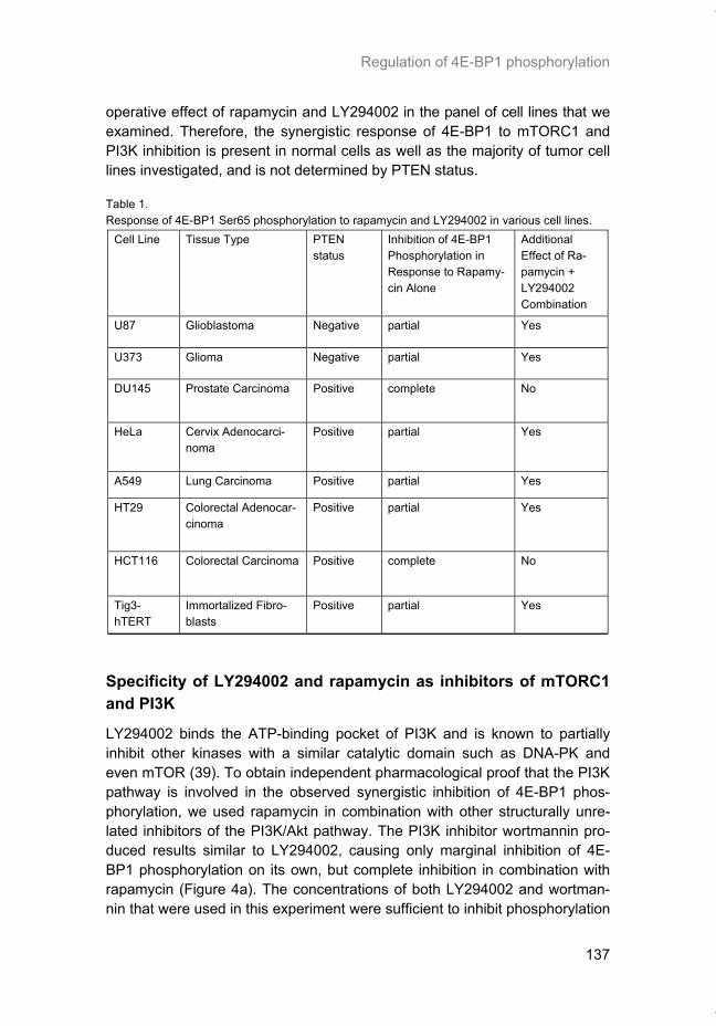
Regulation of 4E-BP1 phosphorylation
137
in the panel of cell lines that we examined. Therefore, the synergistic response of 4E-BP1 to mTORC1 and PI3K inhibition is present in normal cells as well as the majority of tumor cell lines investigated, and is not determined by PTEN status. Table 1. Response of 4E-BP1 Ser65 phosphorylation to rapamycin and LY294002 in various cell lines.
Cell Line Tissue Type PTEN status
Inhibition of 4E-BP1 Phosphorylation in Response to Rapamy-cin Alone
Additional Effect of Ra-pamycin + LY294002 Combination
U87 Glioblastoma Negative partial Yes
U373 Glioma Negative partial Yes
DU145 Prostate Carcinoma Positive complete No
HeLa Cervix Adenocarci-noma
Positive partial Yes
A549 Lung Carcinoma Positive partial Yes
HT29 Colorectal Adenocar-cinoma
Positive partial Yes
HCT116 Colorectal Carcinoma Positive complete No
Tig3-hTERT
Immortalized Fibro-blasts
Positive partial Yes
Specificity of LY294002 and rapamycin as inhibitors of mTORC1 and PI3K
LY294002 binds the ATP-binding pocket of PI3K and is known to partially inhibit other kinases with a similar catalytic domain such as DNA-PK and even mTOR (39). To obtain independent pharmacological proof that the PI3K pathway is involved in the observed synergistic inhibition of 4E-BP1 phos-phorylation, we used rapamycin in combination with other structurally unre-lated inhibitors of the PI3K/Akt pathway. The PI3K inhibitor wortmannin pro-duced results similar to LY294002, causing only marginal inhibition of 4E-BP1 phosphorylation on its own, but complete inhibition in combination with rapamycin (Figure 4a). The concentrations of both LY294002 and wortman-nin that were used in this experiment were sufficient to inhibit phosphorylation
Thesis_Weppler_v12_fc.pdf
operative effect of rapamycin and LY294002
Chapter 6
138
of Akt. Rapamycin on the other hand, did not have any appreciable effect on Akt phosphorylation at Ser473. More importantly, inhibition of Akt in U87 cells with the Akt1/2 inhibitor (Akti-1/2) also resulted in only a small inhibition of 4E-BP1 phosphorylation on its own, but a near complete attenuation of Ser65 phosphorylation when given in combination with rapamycin (Figure 4b). Taken together, these results reinforce the existence of a PI3K/Akt depend-ent pathway that signals to 4E-BP1 in an mTORC1 independent manner. Rapamycin is considered to be one of the most specific small molecule inhibi-tors known due to its formation of a stable complex with both FKBP12 and mTOR. The biological consequences of rapamycin are largely assumed to result from inhibition of mTORC1, since it us unable to block mTOR activity of the TORC2 complex. However, it remains possible that rapamycin affects mTOR activities that are separate from that associated with mTORC1. In or-der to assess the contribution of combined mTORC1 and PI3K inhibition on 4E-BP1 regulation independently of rapamycin, we used a genetic approach to inhibit the mTORC1 specific protein raptor. Figure 4c shows that we were able to attain near complete loss of detectable raptor protein using siRNA. Inhibition of raptor reduced the basal levels of 4E-BP1 phosphorylation at Ser65 to levels similar to those caused by treatment of the parental cells with rapamycin or LY294002 alone (Figure 4c). Importantly, treatment of the rap-tor knockdown cells with LY294002 resulted in complete loss of 4E-BP1 Ser65 phosphorylation. These data support mTORC1 as the target of rapa-mycin in the synergistic effect observed on 4E-BP1 phosphorylation in com-bination with PI3K inhibition. Knockdown of raptor also led to a small de-crease in mTOR protein, possibly due to destabilization of the mTORC1 complex. However, this is unlikely to have influenced 4E-BP1 phosphoryla-tion since knockdown of mTOR to ~50% of control levels had no significant effect on the basal or inhibitor treated levels of 4EBP1 Ser65 phosphorylation (Figure S3). Consequently mTOR protein levels do not seem to be rate limit-ing for mTORC1 activity. As expected, rictor siRNA also did not promote 4E-BP1 dephosphorylation either basally or after treatment with rapamycin (data not shown).
Thesis_Weppler_v12_fc.pdf
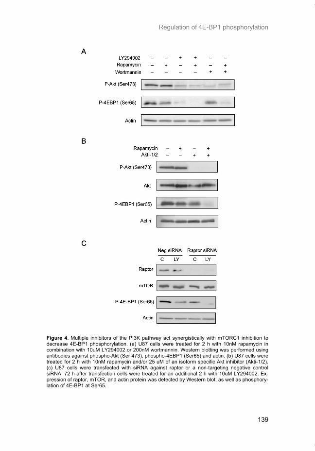
Regulation of 4E-BP1 phosphorylation
139
Figure 4. Multiple inhibitors of the PI3K pathway act synergistically with mTORC1 inhibition to decrease 4E-BP1 phosphorylation. (a) U87 cells were treated for 2 h with 10nM rapamycin in combination with 10uM LY294002 or 200nM wortmannin. Western blotting was performed using antibodies against phospho-Akt (Ser 473), phospho-4EBP1 (Ser65) and actin. (b) U87 cells were treated for 2 h with 10nM rapamycin and/or 25 uM of an isoform specific Akt inhibitor (Akti-1/2). (c) U87 cells were transfected with siRNA against raptor or a non-targeting negative control siRNA. 72 h after transfection cells were treated for an additional 2 h with 10uM LY294002. Ex-pression of raptor, mTOR, and actin protein was detected by Western blot, as well as phosphory-lation of 4E-BP1 at Ser65.
Thesis_Weppler_v12_fc.pdf
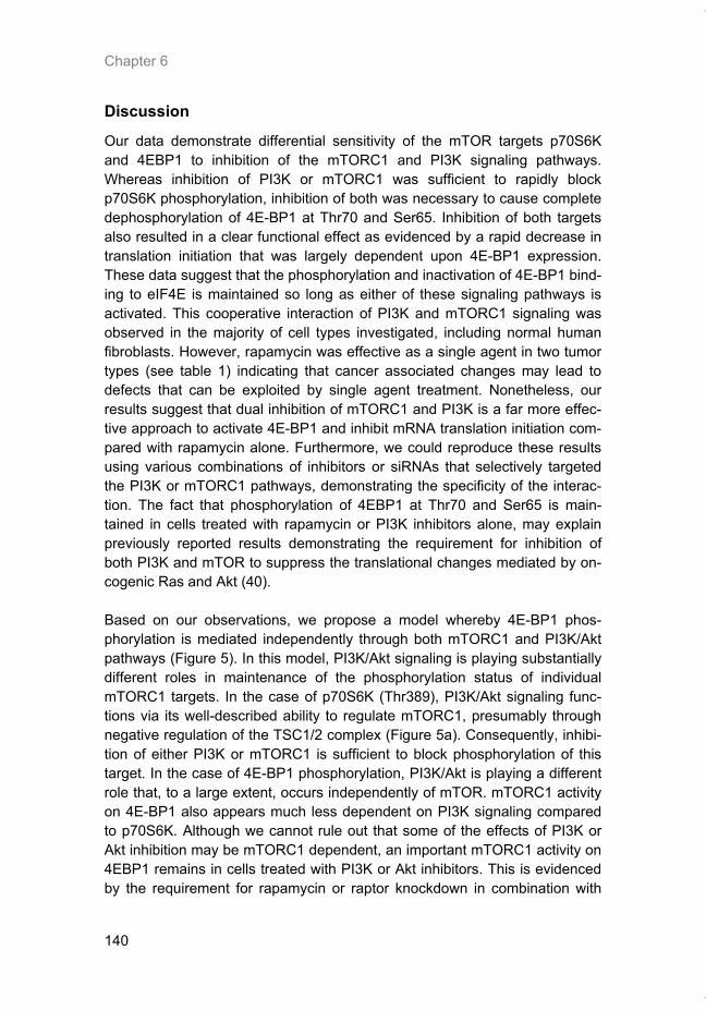
Chapter 6
140
Discussion
Our data demonstrate differential sensitivity of the mTOR targets p70S6K and 4EBP1 to inhibition of the mTORC1 and PI3K signaling pathways. Whereas inhibition of PI3K or mTORC1 was sufficient to rapidly block p70S6K phosphorylation, inhibition of both was necessary to cause complete dephosphorylation of 4E-BP1 at Thr70 and Ser65. Inhibition of both targets also resulted in a clear functional effect as evidenced by a rapid decrease in translation initiation that was largely dependent upon 4E-BP1 expression. These data suggest that the phosphorylation and inactivation of 4E-BP1 bind-ing to eIF4E is maintained so long as either of these signaling pathways is activated. This cooperative interaction of PI3K and mTORC1 signaling was observed in the majority of cell types investigated, including normal human fibroblasts. However, rapamycin was effective as a single agent in two tumor types (see table 1) indicating that cancer associated changes may lead to defects that can be exploited by single agent treatment. Nonetheless, our results suggest that dual inhibition of mTORC1 and PI3K is a far more effec-tive approach to activate 4E-BP1 and inhibit mRNA translation initiation com-pared with rapamycin alone. Furthermore, we could reproduce these results using various combinations of inhibitors or siRNAs that selectively targeted the PI3K or mTORC1 pathways, demonstrating the specificity of the interac-tion. The fact that phosphorylation of 4EBP1 at Thr70 and Ser65 is main-tained in cells treated with rapamycin or PI3K inhibitors alone, may explain previously reported results demonstrating the requirement for inhibition of both PI3K and mTOR to suppress the translational changes mediated by on-cogenic Ras and Akt (40). Based on our observations, we propose a model whereby 4E-BP1 phos-phorylation is mediated independently through both mTORC1 and PI3K/Akt pathways (Figure 5). In this model, PI3K/Akt signaling is playing substantially different roles in maintenance of the phosphorylation status of individual mTORC1 targets. In the case of p70S6K (Thr389), PI3K/Akt signaling func-tions via its well-described ability to regulate mTORC1, presumably through negative regulation of the TSC1/2 complex (Figure 5a). Consequently, inhibi-tion of either PI3K or mTORC1 is sufficient to block phosphorylation of this target. In the case of 4E-BP1 phosphorylation, PI3K/Akt is playing a different role that, to a large extent, occurs independently of mTOR. mTORC1 activity on 4E-BP1 also appears much less dependent on PI3K signaling compared to p70S6K. Although we cannot rule out that some of the effects of PI3K or Akt inhibition may be mTORC1 dependent, an important mTORC1 activity on 4EBP1 remains in cells treated with PI3K or Akt inhibitors. This is evidenced by the requirement for rapamycin or raptor knockdown in combination with
Thesis_Weppler_v12_fc.pdf
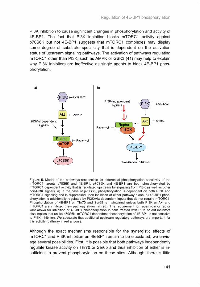
Regulation of 4E-BP1 phosphorylation
141
PI3K inhibition to cause significant changes in phosphorylation and activity of 4E-BP1. The fact that PI3K inhibition blocks mTORC1 activity against p70S6K but not 4E-BP1 suggests that mTORC1 complexes may display some degree of substrate specificity that is dependent on the activation status of upstream signaling pathways. The activation of pathways regulating mTORC1 other than PI3K, such as AMPK or GSK3 (41) may help to explain why PI3K inhibitors are ineffective as single agents to block 4E-BP1 phos-phorylation.
Figure 5. Model of the pathways responsible for differential phosphorylation sensitivity of the mTORC1 targets p70S6K and 4E-BP1. p70S6K and 4E-BP1 are both phosphorylated by mTORC1 dependent activity that is regulated upstream by signaling from PI3K as well as other non-PI3K signals. a) In the case of p70S6K, phosphorylation is dependent on both PI3K and mTORC1 signaling and is suppressed upon inhibition of either pathway alone. b) 4E-BP1 phos-phorylation is additionally regulated by PI3K/Akt dependent inputs that do not require mTORC1. Phosphorylation of 4E-BP1 on Thr70 and Ser65 is maintained unless both PI3K or Akt and mTORC1 are inhibited (new pathway shown in red). The requirement for rapamycin or raptor knockdown for inhibition of 4E-BP1 phosphorylation in cells treated with PI3K or Akt inhibitors also implies that unlike p70S6K, mTORC1 dependent phosphorylation of 4E-BP1 is not sensitive to PI3K inhibition. We speculate that additional upstream regulatory pathways are important for this activity (pathway in red arrows). Although the exact mechanisms responsible for the synergistic effects of mTORC1 and PI3K inhibition on 4E-BP1 remain to be elucidated, we envis-age several possibilities. First, it is possible that both pathways independently regulate kinase activity on Thr70 or Ser65 and thus inhibition of either is in-sufficient to prevent phosphorylation on these sites. Although, there is little
Thesis_Weppler_v12_fc.pdf
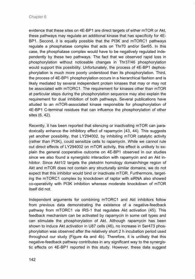
Chapter 6
142
evidence that these sites on 4E-BP1 are direct targets of either mTOR or Akt, these pathways may regulate an additional kinase that has specificity for 4E-BP1. Second, it is equally possible that the PI3K and mTORC1 pathways regulate a phosphatase complex that acts on Thr70 and/or Ser65. In this case, the phosphatase complex would have to be negatively regulated inde-pendently by these two pathways. The fact that we observed rapid loss in phosphorylation without noticeable changes in Thr37/46 phosphorylation would support this possibility. Unfortunately, the process of 4E-BP1 dephos-phorylation is much more poorly understood than its phosphorylation. Third, the process of 4E-BP1 phosphorylation occurs in a hierarchical fashion and is likely mediated by several independent protein kinases that may or may not be associated with mTORC1. The requirement for kinases other than mTOR at particular steps during the phosphorylation sequence may also explain the requirement for dual inhibition of both pathways. Several publications have alluded to an mTOR-associated kinase responsible for phosphorylation of 4E-BP1 C-terminal residues that can influence the phosphorylation of other sites (6, 42). Recently, it has been reported that silencing or inactivating mTOR can para-doxically enhance the inhibitory effect of rapamycin (43, 44). This suggests yet another possibility, that LY294002, by inhibiting mTOR catalytic activity (rather than PI3K), could sensitize cells to rapamycin. While we cannot rule out direct effects of LY294002 on mTOR activity, this effect is unlikely to ex-plain the general cooperative outcome on 4E-BP1 observed in our studies since we also found a synergistic interaction with rapamycin and an Akt in-hibitor. Since Akt1/2 targets the plekstrin homology domain/hinge region of Akt and mTOR does not contain any structurally similar domains, we do not expect that this inhibitor would bind or inactivate mTOR. Furthermore, target-ing the mTORC1 complex by knockdown of raptor with siRNA also showed co-operativity with PI3K inhibition whereas moderate knockdown of mTOR itself did not. Independent arguments for combining mTORC1 and Akt inhibitors follow from previous data demonstrating the existence of a negative-feedback pathway from mTORC1 via IRS-1 that regulates Akt activation (45). This feedback mechanism can be activated by rapamycin in some cell types and can stimulate the phosphorylation of Akt. Although rapamycin has been shown to induce Akt activation in U87 cells (46), no increase in Ser473 phos-phorylation was observed after the relatively short 2 h incubation period used throughout our study (Figure 4a and 4b). Therefore, it is unlikely that this negative-feedback pathway contributes in any significant way to the synergis-tic effects on 4E-BP1 reported in this study. However, these data suggest
Thesis_Weppler_v12_fc.pdf
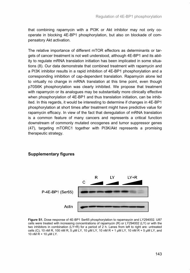
Regulation of 4E-BP1 phosphorylation
143
that combining rapamycin with a PI3K or Akt inhibitor may not only co-operate in blocking 4E-BP1 phosphorylation, but also on blockade of com-pensatory Akt activation. The relative importance of different mTOR effectors as determinants or tar-gets of cancer treatment is not well understood, although 4E-BP1 and its abil-ity to regulate mRNA translation initiation has been implicated in some situa-tions (8). Our data demonstrate that combined treatment with rapamycin and a PI3K inhibitor results in a rapid inhibition of 4E-BP1 phosphorylation and a corresponding inhibition of cap-dependent translation. Rapamycin alone led to virtually no change in mRNA translation at this time point, even though p70S6K phosphorylation was clearly inhibited. We propose that treatment with rapamycin or its analogues may be substantially more clinically effective when phosphorylation of 4E-BP1 and thus translation initiation, can be inhib-ited. In this regards, it would be interesting to determine if changes in 4E-BP1 phosphorylation at short times after treatment might have predictive value for rapamycin efficacy. In view of the fact that deregulation of mRNA translation is a common feature of many cancers and represents a critical function downstream of commonly mutated oncogenes and tumor suppressor genes (47), targeting mTORC1 together with PI3K/Akt represents a promising therapeutic strategy.
Supplementary figures
Figure S1. Dose response of 4E-BP1 Ser65 phosphorylation to rapamaycin and LY294002. U87 cells were treated with increasing concentrations of rapamycin (R) or LY294002 (LY) or with the two inhibitors in combination (LY+R) for a period of 2 h. Lanes from left to right are: untreated cells (C), 10 nM R, 100 nM R, 5 μM LY, 10 μM LY, 10 nM R + 1 μM LY, 10 nM R + 5 μM LY, and 10 nM R + 10 μM LY.
Thesis_Weppler_v12_fc.pdf
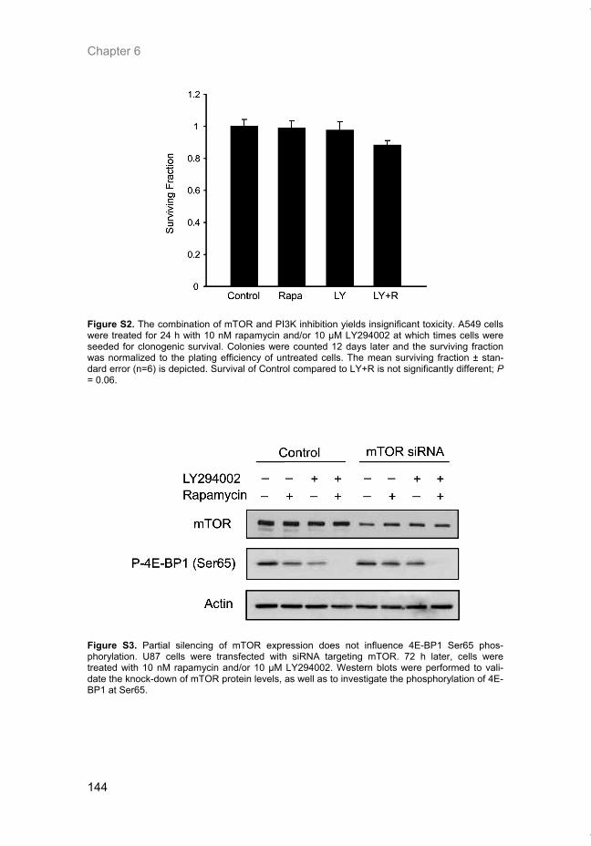
Chapter 6
144
Figure S2. The combination of mTOR and PI3K inhibition yields insignificant toxicity. A549 cells were treated for 24 h with 10 nM rapamycin and/or 10 μM LY294002 at which times cells were seeded for clonogenic survival. Colonies were counted 12 days later and the surviving fraction was normalized to the plating efficiency of untreated cells. The mean surviving fraction ± stan-dard error (n=6) is depicted. Survival of Control compared to LY+R is not significantly different; P = 0.06.
Figure S3. Partial silencing of mTOR expression does not influence 4E-BP1 Ser65 phos-phorylation. U87 cells were transfected with siRNA targeting mTOR. 72 h later, cells were treated with 10 nM rapamycin and/or 10 μM LY294002. Western blots were performed to vali-date the knock-down of mTOR protein levels, as well as to investigate the phosphorylation of 4E-BP1 at Ser65.
Thesis_Weppler_v12_fc.pdf
Regulation of 4E-BP1 phosphorylation
145
References 1. Guertin, D. A. and Sabatini, D. M. Defining the role of mTOR in cancer. Cancer Cell, 12: 9-
22, 2007. 2. Hara, K., Maruki, Y., Long, X., Yoshino, K., Oshiro, N., Hidayat, S., Tokunaga, C., Avruch,
J., and Yonezawa, K. Raptor, a binding partner of target of rapamycin (TOR), mediates TOR action. Cell, 110: 177-189, 2002.
3. Sabatini, D. M. mTOR and cancer: insights into a complex relationship. Nat Rev Cancer, 6: 729-734, 2006.
4. Jacinto, E., Loewith, R., Schmidt, A., Lin, S., Ruegg, M. A., Hall, A., and Hall, M. N. Mam-malian TOR complex 2 controls the actin cytoskeleton and is rapamycin insensitive. Nat Cell Biol, 6: 1122-1128, 2004.
5. Sarbassov, D. D., Guertin, D. A., Ali, S. M., and Sabatini, D. M. Phosphorylation and regu-lation of Akt/PKB by the rictor-mTOR complex. Science, 307: 1098-1101, 2005.
6. Wang, X., Beugnet, A., Murakami, M., Yamanaka, S., and Proud, C. G. Distinct signaling events downstream of mTOR cooperate to mediate the effects of amino acids and insulin on initiation factor 4E-binding proteins. Mol Cell Biol, 25: 2558-2572, 2005.
7. Cai, S. L., Tee, A. R., Short, J. D., Bergeron, J. M., Kim, J., Shen, J., Guo, R., Johnson, C. L., Kiguchi, K., and Walker, C. L. Activity of TSC2 is inhibited by AKT-mediated phosphory-lation and membrane partitioning. J Cell Biol, 173: 279-289, 2006.
8. Wendel, H. G., De Stanchina, E., Fridman, J. S., Malina, A., Ray, S., Kogan, S., Cordon-Cardo, C., Pelletier, J., and Lowe, S. W. Survival signalling by Akt and eIF4E in oncogene-sis and cancer therapy. Nature, 428: 332-337, 2004.
9. Hay, N. and Sonenberg, N. Upstream and downstream of mTOR. Genes Dev, 18: 1926-1945, 2004.
10. Larsson, O., Li, S., Issaenko, O. A., Avdulov, S., Peterson, M., Smith, K., Bitterman, P. B., and Polunovsky, V. A. Eukaryotic translation initiation factor 4E induced progression of pri-mary human mammary epithelial cells along the cancer pathway is associated with targeted translational deregulation of oncogenic drivers and inhibitors. Cancer Res, 67: 6814-6824, 2007.
11. Richter, J. D. and Sonenberg, N. Regulation of cap-dependent translation by eIF4E inhibi-tory proteins. Nature, 433: 477-480, 2005.
12. Armengol, G., Rojo, F., Castellvi, J., Iglesias, C., Cuatrecasas, M., Pons, B., Baselga, J., and Ramon, Y. C. S. 4E-Binding Protein 1: A Key Molecular "Funnel Factor" in Human Cancer with Clinical Implications. Cancer Res, 67: 7551-7555, 2007.
13. Zhou, X., Tan, M., Stone Hawthorne, V., Klos, K. S., Lan, K. H., Yang, Y., Yang, W., Smith, T. L., Shi, D., and Yu, D. Activation of the Akt/mammalian target of rapamycin/4E-BP1 pathway by ErbB2 overexpression predicts tumor progression in breast cancers. Clin Can-cer Res, 10: 6779-6788, 2004.
14. Gingras, A. C., Gygi, S. P., Raught, B., Polakiewicz, R. D., Abraham, R. T., Hoekstra, M. F., Aebersold, R., and Sonenberg, N. Regulation of 4E-BP1 phosphorylation: a novel two-step mechanism. Genes Dev, 13: 1422-1437, 1999.
15. Gingras, A. C., Raught, B., Gygi, S. P., Niedzwiecka, A., Miron, M., Burley, S. K., Po-lakiewicz, R. D., Wyslouch-Cieszynska, A., Aebersold, R., and Sonenberg, N. Hierarchical phosphorylation of the translation inhibitor 4E-BP1. Genes Dev, 15: 2852-2864, 2001.
16. Mothe-Satney, I., Brunn, G. J., McMahon, L. P., Capaldo, C. T., Abraham, R. T., and Law-rence, J. C., Jr. Mammalian target of rapamycin-dependent phosphorylation of PHAS-I in four (S/T)P sites detected by phospho-specific antibodies. J Biol Chem, 275: 33836-33843, 2000.
17. Wang, X., Li, W., Parra, J. L., Beugnet, A., and Proud, C. G. The C terminus of initiation factor 4E-binding protein 1 contains multiple regulatory features that influence its function and phosphorylation. Mol Cell Biol, 23: 1546-1557, 2003.
18. Averous, J. and Proud, C. G. When translation meets transformation: the mTOR story. Oncogene, 25: 6423-6435, 2006.
19. Huang, S., Bjornsti, M. A., and Houghton, P. J. Rapamycins: mechanism of action and cellular resistance. Cancer Biol Ther, 2: 222-232, 2003.
20. Kim, D. H., Sarbassov, D. D., Ali, S. M., King, J. E., Latek, R. R., Erdjument-Bromage, H., Tempst, P., and Sabatini, D. M. mTOR interacts with raptor to form a nutrient-sensitive complex that signals to the cell growth machinery. Cell, 110: 163-175, 2002.
Thesis_Weppler_v12_fc.pdf
Chapter 6
146
21. Oshiro, N., Yoshino, K., Hidayat, S., Tokunaga, C., Hara, K., Eguchi, S., Avruch, J., and Yonezawa, K. Dissociation of raptor from mTOR is a mechanism of rapamycin-induced in-hibition of mTOR function. Genes Cells, 9: 359-366, 2004.
22. Sarbassov, D. D., Ali, S. M., Kim, D. H., Guertin, D. A., Latek, R. R., Erdjument-Bromage, H., Tempst, P., and Sabatini, D. M. Rictor, a novel binding partner of mTOR, defines a ra-pamycin-insensitive and raptor-independent pathway that regulates the cytoskeleton. Curr Biol, 14: 1296-1302, 2004.
23. Sarbassov, D. D., Ali, S. M., Sengupta, S., Sheen, J. H., Hsu, P. P., Bagley, A. F., Markhard, A. L., and Sabatini, D. M. Prolonged rapamycin treatment inhibits mTORC2 as-sembly and Akt/PKB. Mol Cell, 22: 159-168, 2006.
24. Noh, W. C., Mondesire, W. H., Peng, J., Jian, W., Zhang, H., Dong, J., Mills, G. B., Hung, M. C., and Meric-Bernstam, F. Determinants of rapamycin sensitivity in breast cancer cells. Clin Cancer Res, 10: 1013-1023, 2004.
25. Yu, K., Toral-Barza, L., Discafani, C., Zhang, W. G., Skotnicki, J., Frost, P., and Gibbons, J. J. mTOR, a novel target in breast cancer: the effect of CCI-779, an mTOR inhibitor, in pre-clinical models of breast cancer. Endocr Relat Cancer, 8: 249-258, 2001.
26. Aoki, M., Blazek, E., and Vogt, P. K. A role of the kinase mTOR in cellular transformation induced by the oncoproteins P3k and Akt. Proc Natl Acad Sci U S A, 98: 136-141, 2001.
27. Neshat, M. S., Mellinghoff, I. K., Tran, C., Stiles, B., Thomas, G., Petersen, R., Frost, P., Gibbons, J. J., Wu, H., and Sawyers, C. L. Enhanced sensitivity of PTEN-deficient tumors to inhibition of FRAP/mTOR. Proc Natl Acad Sci U S A, 98: 10314-10319, 2001.
28. Buck, E., Eyzaguirre, A., Brown, E., Petti, F., McCormack, S., Haley, J. D., Iwata, K. K., Gibson, N. W., and Griffin, G. Rapamycin synergizes with the epidermal growth factor re-ceptor inhibitor erlotinib in non-small-cell lung, pancreatic, colon, and breast tumors. Mol Cancer Ther, 5: 2676-2684, 2006.
29. Mabuchi, S., Altomare, D. A., Cheung, M., Zhang, L., Poulikakos, P. I., Hensley, H. H., Schilder, R. J., Ozols, R. F., and Testa, J. R. RAD001 inhibits human ovarian cancer cell proliferation, enhances cisplatin-induced apoptosis, and prolongs survival in an ovarian cancer model. Clin Cancer Res, 13: 4261-4270, 2007.
30. Takeuchi, H., Kondo, Y., Fujiwara, K., Kanzawa, T., Aoki, H., Mills, G. B., and Kondo, S. Synergistic augmentation of rapamycin-induced autophagy in malignant glioma cells by phosphatidylinositol 3-kinase/protein kinase B inhibitors. Cancer Res, 65: 3336-3346, 2005.
31. Fukami, J., Anno, K., Ueda, K., Takahashi, T., and Ide, T. Enhanced expression of cyclin D1 in senescent human fibroblasts. Mech Ageing Dev, 81: 139-157, 1995.
32. Weppler, S. A., Li, Y., Dubois, L., Lieuwes, N., Jutten, B., Lambin, P., Wouters, B. G., and Lammering, G. Expression of EGFR variant vIII promotes both radiation resistance and hy-poxia tolerance. Radiother Oncol, 83: 333-339, 2007.
33. Stolarov, J., Chang, K., Reiner, A., Rodgers, L., Hannon, G. J., Wigler, M. H., and Mittal, V. Design of a retroviral-mediated ecdysone-inducible system and its application to the ex-pression profiling of the PTEN tumor suppressor. Proc Natl Acad Sci U S A, 98: 13043-13048, 2001.
34. Constantinou, C. and Clemens, M. J. Regulation of translation factors eIF4GI and 4E-BP1 during recovery of protein synthesis from inhibition by p53. Cell Death Differ, 14: 576-585, 2007.
35. Koritzinsky, M., Magagnin, M. G., van den Beucken, T., Seigneuric, R., Savelkouls, K., Dostie, J., Pyronnet, S., Kaufman, R. J., Weppler, S. A., Voncken, J. W., Lambin, P., Koumenis, C., Sonenberg, N., and Wouters, B. G. Gene expression during acute and pro-longed hypoxia is regulated by distinct mechanisms of translational control. Embo J, 25: 1114-1125, 2006.
36. Brummelkamp, T. R., Bernards, R., and Agami, R. Stable suppression of tumorigenicity by virus-mediated RNA interference. Cancer Cell, 2: 243-247, 2002.
37. Koritzinsky, M., Rouschop, K. M., van den Beucken, T., Magagnin, M. G., Savelkouls, K., Lambin, P., and Wouters, B. G. Phosphorylation of eIF2alpha is required for mRNA transla-tion inhibition and survival during moderate hypoxia. Radiother Oncol, 83: 353-361, 2007.
38. Ohgaki, H. and Kleihues, P. Genetic pathways to primary and secondary glioblastoma. Am J Pathol, 170: 1445-1453, 2007.
39. Brunn, G. J., Williams, J., Sabers, C., Wiederrecht, G., Lawrence, J. C., Jr., and Abraham, R. T. Direct inhibition of the signaling functions of the mammalian target of rapamycin by the phosphoinositide 3-kinase inhibitors, wortmannin and LY294002. Embo J, 15: 5256-5267, 1996.
Thesis_Weppler_v12_fc.pdf
Regulation of 4E-BP1 phosphorylation
147
40. Rajasekhar, V. K., Viale, A., Socci, N. D., Wiedmann, M., Hu, X., and Holland, E. C. Onco-genic Ras and Akt signaling contribute to glioblastoma formation by differential recruitment of existing mRNAs to polysomes. Mol Cell, 12: 889-901, 2003.
41. Inoki, K., Ouyang, H., Zhu, T., Lindvall, C., Wang, Y., Zhang, X., Yang, Q., Bennett, C., Harada, Y., Stankunas, K., Wang, C. Y., He, X., MacDougald, O. A., You, M., Williams, B. O., and Guan, K. L. TSC2 integrates Wnt and energy signals via a coordinated phosphory-lation by AMPK and GSK3 to regulate cell growth. Cell, 126: 955-968, 2006.
42. Heesom, K. J. and Denton, R. M. Dissociation of the eukaryotic initiation factor-4E/4E-BP1 complex involves phosphorylation of 4E-BP1 by an mTOR-associated kinase. FEBS Lett, 457: 489-493, 1999.
43. Edinger, A. L., Linardic, C. M., Chiang, G. G., Thompson, C. B., and Abraham, R. T. Differ-ential effects of rapamycin on mammalian target of rapamycin signaling functions in mam-malian cells. Cancer Res, 63: 8451-8460, 2003.
44. Iwamaru, A., Kondo, Y., Iwado, E., Aoki, H., Fujiwara, K., Yokoyama, T., Mills, G. B., and Kondo, S. Silencing mammalian target of rapamycin signaling by small interfering RNA en-hances rapamycin-induced autophagy in malignant glioma cells. Oncogene, 26: 1840-1851, 2007.
45. Harrington, L. S., Findlay, G. M., Gray, A., Tolkacheva, T., Wigfield, S., Rebholz, H., Bar-nett, J., Leslie, N. R., Cheng, S., Shepherd, P. R., Gout, I., Downes, C. P., and Lamb, R. F. The TSC1-2 tumor suppressor controls insulin-PI3K signaling via regulation of IRS proteins. J Cell Biol, 166: 213-223, 2004.
46. Fan, Q. W., Knight, Z. A., Goldenberg, D. D., Yu, W., Mostov, K. E., Stokoe, D., Shokat, K. M., and Weiss, W. A. A dual PI3 kinase/mTOR inhibitor reveals emergent efficacy in glioma. Cancer Cell, 9: 341-349, 2006.
47. Bilanges, B. and Stokoe, D. Mechanisms of translational deregulation in human tumors and therapeutic intervention strategies. Oncogene, 26: 5973-5990, 2007.
Thesis_Weppler_v12_fc.pdf

Thesis_Weppler_v12_fc.pdf
149
CHAPTER 7
Neuroendocrine carcinoma in Birt-Hogg-Dubé syndrome
Manuscript submitted S.A. Weppler, T. Claessens, M. van Geel, D. Creytons, M. Vreeburg, B.G. Wouters, M.A.M. van Steensel.
Thesis_Weppler_v12_fc.pdf
Chapter 7
150
Abstract
Birt-Hogg-Dubé syndrome (BHD) is a dominantly inherited disorder charac-terized by an increased risk of developing kidney cancer, pneumothorax as a consequence of lung cysts and benign hair follicle tumors called fibrofollicu-lomas. It is caused by mutations in the BHD gene, coding for folliculin (FLCN), a protein possibly involved in mTOR signaling. BHD syndrome is generally considered to be a relatively benign condition for which annual fol-low-up suffices. However, the possibility that the pre-existing gene defect might act to modify the behavior of other cancers that arise in patients with BHD syndrome has so far not been considered. We describe a patient with BHD who succumbed to a malignant neuro-endocrine tumor of prostatic or bladder origin within 6 months of its discovery. He also had a papillary renal cell carcinoma. We propose that the behavior of our patient's cancer might have been modulated by absence of the BHD gene. We demonstrate loss of FLCN in the tumor and show that it does not cause mTOR upregulation, contrary to previous findings in a mouse model. Our observation could possibly lead to a different approach towards BHD patient surveillance and treatment in the future.
Thesis_Weppler_v12_fc.pdf
mTOR and Birt-Hogg-Dubé syndrome
151
Introduction
Birt-Hogg-Dubé syndrome (BHD, MIM #135150) is an autosomal-dominantly inherited cancer syndrome characterized by fibrofolliculomas, lung cysts leading to pneumothorax, and mainly chromophobic/oncocytic renal cell car-cinoma (1). The disease is caused by heterozygous mutations in the BHD gene encoding folliculin (FLCN). Almost all human mutations reported so far lead to putative protein truncation (2). A conditional kidney-specific BHD knockout mouse displays activation of mTOR signaling (3). Thus, loss of FLCN may result in inappropriate mTOR activity. Considering that mTOR signaling is increasingly implicated in tumor progression (4) the spectrum of malignancies associated thus far with BHD syndrome seems limited. Studies of multiple extended pedigrees have so far not yielded any firm evidence to the contrary. We present a patient with BHD syndrome who developed a neu-roendocrine carcinoma of prostate or bladder origin that behaved in a highly malignant fashion, causing our patient’s demise within 6 months after his ini-tial diagnosis. He also had fibrofolliculomas, lung cysts and a papillary renal cell carcinoma. We find loss of FLCN in the tumor and suggest that this may have contributed to tumorigenesis, although we find no evidence for mTOR deregulation in our patient’s tissues.
Materials and methods
Immunohistochemical stainings were performed on frozen sections according to a standard protocol. Briefly, tissue samples were flash frozen in liquid ni-trogen and transferred to -80ºC within one hour after sampling. 5 µm sections were cut with a microtome at -20ºC. For staining, slides were fixed in acetone for 10 minutes at -20ºC and washed twice in TBS, followed by an incubation with H2O2 (3%) in methanol for 10 minutes. After another washing step, slides were blocked (TBS/Triton X-100 0.3%/goat serum 5%) and primary antibody was added in a 1:50 dilution to be incubated overnight at 4ºC. Next, secon-dary antibody (goat anti-rabbit, 1:200) was added and incubated at room temperature for 30 minutes. The slides were then washed (3x5 minutes) in washing buffer. ABC reagent was then added and incubated at room tem-perature for 30 minutes, followed by three wash steps with washing buffer. DAB was then added in 10 ml washing buffer with 0.1% H2O2 followed by rinsing in demineralized water during development of the stain. H&E stained slides were used for orientation and histological examination. Photographs were taken using a Leica DM-4000 microscope with the Leica Application Suite software.
Thesis_Weppler_v12_fc.pdf
Chapter 7
152
Case report
The patient, a 50 year-old man of Dutch origin, was originally diagnosed with Birt-Hogg-Dubé syndrome in 2005 when he visited the outpatient clinic of the department of dermatology. Several family members were also found to be affected. Our findings in the family are described elsewhere (5). Using direct sequencing, we found a novel heterozygous insertion mutation 1408_1418delGGGAGCCCTGT in the BHD gene in all affected family mem-bers, including the present patient. Following local guidelines in place at the time, we performed abdominal CT imaging and found a homogeneous hypo-dense mass in the upper pole of the left kidney that the radiologist judged to be benign, possibly an oncocytoma. A wait-and-see policy was thus adopted, as the patient seemed otherwise healthy. A year later, he visited the outpatient clinic of the department of urology be-cause of visible hematuria since six months, particularly after exercise. There were no other complaints, with the exception of incidental hematospermia. Physical examination at the time showed no abnormalities. Rectal examination in particular was unremarkable. Routine lab examination showed the following normal results: ESR 11, Hb 8.3 mmol/l, Creatinine 74 µmol/l, alkaline phos-phatase 83 U/l, gammaGT 23 U/l and PSA 1.0. Routine urinalysis was also normal. An abdominal CT-scan was performed and showed the previously found lesion in the left kidney, in addition to a hypodense mass in the left pros-tate lobe (figure 1a). An MRI likewise demonstrated the presence of a mass in the left prostate with extension underneath the bladder and into the mesorectal fat (figure 1b). At the time, it was interpreted as inflammatory. In an attempt to further characterize the process, a transrectal prostate biopsy was taken, which revealed only inflammatory changes. A malignancy was considered unlikely and the patient was discharged. About five months later, he developed acute urinary retention for which he was treated with alpha-sympathicolytic agents and a trans-urethral catheter. A trans-urethral prostate resection was attempted a month after the initial presentation. During surgery, a mass was seen on the posterior bladder wall, blocking the ureteric ostia. Tissue samples were taken and a suprapubic catheter was placed. A post-operative MRI of the abdomen showed a large pelvic mass and extensive lymphadenopathy (figure 2). The tumor invaded pelvic wall muscles, prostate, seminal vesicles and bladder. A second large tumor was seen that impinged upon the rectum. In addition, ex-tensive lymphadenopathy was now present, in particular on the left para-iliacal side. A thoracic CT-scan did not show any evidence for intrathoracic metasta-ses. Histopathological examination of the tissue samples obtained during sur-gery was consistent with a neuroendocrine carcinoma of unknown origin. Two weeks after surgery, the patient developed deep venous thrombosis of the left
Thesis_Weppler_v12_fc.pdf
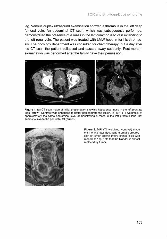
mTOR and Birt-Hogg-Dubé syndrome
153
leg. Venous duplex ultrasound examination showed a thrombus in the left deep femoral vein. An abdominal CT scan, which was subsequently performed, demonstrated the presence of a mass in the left common iliac vein extending to the left renal vein. The patient was treated with LMW heparin for his thrombo-sis. The oncology department was consulted for chemotherapy, but a day after his CT scan the patient collapsed and passed away suddenly. Post-mortem examination was performed after the family gave their permission.
Figure 1. (a) CT scan made at initial presentation showing hypodense mass in the left prostate lobe (arrow). Contrast was enhanced to better demonstrate the lesion. (b) MRI (T1-weighted) at approximately the same anatomical level demonstrating a mass in the left prostate lobe that seems to invade the perirectal fat (arrow).
Figure 2. MRI (T1 weighted, contrast) made 5.5 months later illustrating dramatic progres-sion of tumor growth (more cranial slice with respect to 1b). Note that the bladder is almost replaced by tumor.
Thesis_Weppler_v12_fc.pdf
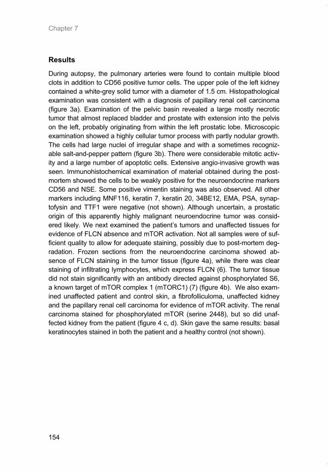
Chapter 7
154
Results
During autopsy, the pulmonary arteries were found to contain multiple blood clots in addition to CD56 positive tumor cells. The upper pole of the left kidney contained a white-grey solid tumor with a diameter of 1.5 cm. Histopathological examination was consistent with a diagnosis of papillary renal cell carcinoma (figure 3a). Examination of the pelvic basin revealed a large mostly necrotic tumor that almost replaced bladder and prostate with extension into the pelvis on the left, probably originating from within the left prostatic lobe. Microscopic examination showed a highly cellular tumor process with partly nodular growth. The cells had large nuclei of irregular shape and with a sometimes recogniz-able salt-and-pepper pattern (figure 3b). There were considerable mitotic activ-ity and a large number of apoptotic cells. Extensive angio-invasive growth was seen. Immunohistochemical examination of material obtained during the post-mortem showed the cells to be weakly positive for the neuroendocrine markers CD56 and NSE. Some positive vimentin staining was also observed. All other markers including MNF116, keratin 7, keratin 20, 34BE12, EMA, PSA, synap-tofysin and TTF1 were negative (not shown). Although uncertain, a prostatic origin of this apparently highly malignant neuroendocrine tumor was consid-ered likely. We next examined the patient’s tumors and unaffected tissues for evidence of FLCN absence and mTOR activation. Not all samples were of suf-ficient quality to allow for adequate staining, possibly due to post-mortem deg-radation. Frozen sections from the neuroendocrine carcinoma showed ab-sence of FLCN staining in the tumor tissue (figure 4a), while there was clear staining of infiltrating lymphocytes, which express FLCN (6). The tumor tissue did not stain significantly with an antibody directed against phosphorylated S6, a known target of mTOR complex 1 (mTORC1) (7) (figure 4b). We also exam-ined unaffected patient and control skin, a fibrofolliculoma, unaffected kidney and the papillary renal cell carcinoma for evidence of mTOR activity. The renal carcinoma stained for phosphorylated mTOR (serine 2448), but so did unaf-fected kidney from the patient (figure 4 c, d). Skin gave the same results: basal keratinocytes stained in both the patient and a healthy control (not shown).
Thesis_Weppler_v12_fc.pdf
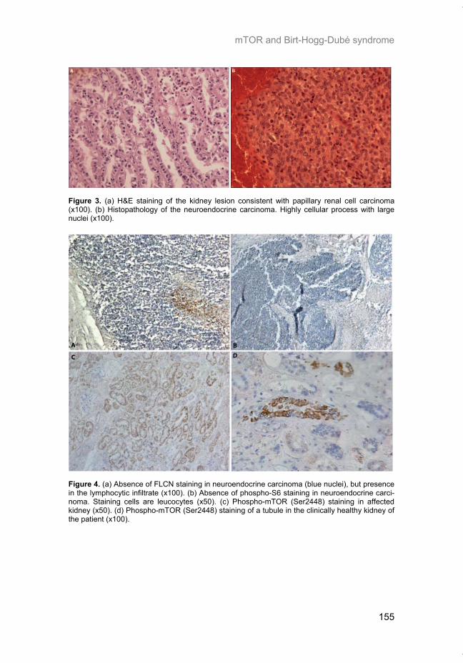
mTOR and Birt-Hogg-Dubé syndrome
155
Figure 3. (a) H&E staining of the kidney lesion consistent with papillary renal cell carcinoma (x100). (b) Histopathology of the neuroendocrine carcinoma. Highly cellular process with large nuclei (x100).
Figure 4. (a) Absence of FLCN staining in neuroendocrine carcinoma (blue nuclei), but presence in the lymphocytic infiltrate (x100). (b) Absence of phospho-S6 staining in neuroendocrine carci-noma. Staining cells are leucocytes (x50). (c) Phospho-mTOR (Ser2448) staining in affected kidney (x50). (d) Phospho-mTOR (Ser2448) staining of a tubule in the clinically healthy kidney of the patient (x100).
Thesis_Weppler_v12_fc.pdf
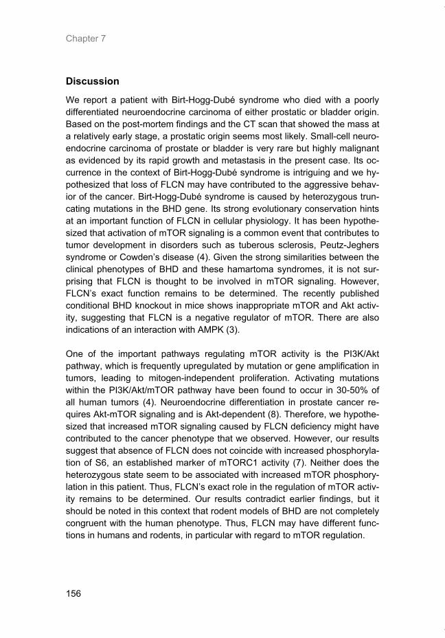
Chapter 7
156
Discussion
We report a patient with Birt-Hogg-Dubé syndrome who died with a poorly differentiated neuroendocrine carcinoma of either prostatic or bladder origin. Based on the post-mortem findings and the CT scan that showed the mass at a relatively early stage, a prostatic origin seems most likely. Small-cell neuro-endocrine carcinoma of prostate or bladder is very rare but highly malignant as evidenced by its rapid growth and metastasis in the present case. Its oc-currence in the context of Birt-Hogg-Dubé syndrome is intriguing and we hy-pothesized that loss of FLCN may have contributed to the aggressive behav-ior of the cancer. Birt-Hogg-Dubé syndrome is caused by heterozygous trun-cating mutations in the BHD gene. Its strong evolutionary conservation hints at an important function of FLCN in cellular physiology. It has been hypothe-sized that activation of mTOR signaling is a common event that contributes to tumor development in disorders such as tuberous sclerosis, Peutz-Jeghers syndrome or Cowden’s disease (4). Given the strong similarities between the clinical phenotypes of BHD and these hamartoma syndromes, it is not sur-prising that FLCN is thought to be involved in mTOR signaling. However, FLCN’s exact function remains to be determined. The recently published conditional BHD knockout in mice shows inappropriate mTOR and Akt activ-ity, suggesting that FLCN is a negative regulator of mTOR. There are also indications of an interaction with AMPK (3). One of the important pathways regulating mTOR activity is the PI3K/Akt pathway, which is frequently upregulated by mutation or gene amplification in tumors, leading to mitogen-independent proliferation. Activating mutations within the PI3K/Akt/mTOR pathway have been found to occur in 30-50% of all human tumors (4). Neuroendocrine differentiation in prostate cancer re-quires Akt-mTOR signaling and is Akt-dependent (8). Therefore, we hypothe-sized that increased mTOR signaling caused by FLCN deficiency might have contributed to the cancer phenotype that we observed. However, our results suggest that absence of FLCN does not coincide with increased phosphoryla-tion of S6, an established marker of mTORC1 activity (7). Neither does the heterozygous state seem to be associated with increased mTOR phosphory-lation in this patient. Thus, FLCN’s exact role in the regulation of mTOR activ-ity remains to be determined. Our results contradict earlier findings, but it should be noted in this context that rodent models of BHD are not completely congruent with the human phenotype. Thus, FLCN may have different func-tions in humans and rodents, in particular with regard to mTOR regulation.
Thesis_Weppler_v12_fc.pdf
mTOR and Birt-Hogg-Dubé syndrome
157
In conclusion, we suggest that our patient’s FLCN mutation may have con-tributed to the pathogenesis of his small-cell neuroendocrine carcinoma. We further propose that follow-up of BHD patients should perhaps be more ag-gressive than currently recommended because, conceivably, loss of FLCN may predispose to more tumor types than renal cell carcinoma alone, or al-ternatively may render other tumors more malignant. The apparent absence of mTOR deregulation in our patient’s tumors might suggest that therapies targeting mTORC1 may not be useful for BHD-associated malignancies.
Acknowledgements
MvS is supported by grants from the university Hospital Maastricht, the GROW research institute for oncology and developmental biology and the Netherlands Organization for Scientific Research ZONMW (907-00-202). SW is supported by a grant from the university Hospital Maastricht. This work is supported by the Myrovlytis Trust. SW, TC and MvS are members of the European Birt-Hogg-Dubé Consortium.
References 1. Schulz, T. and Hartschuh, W. Characteristics of the Birt-Hogg-Dube/Hornstein-Knickenberg
syndrome. Am J Dermatopathol, 22: 293-294, 2000. 2. Schmidt, L. S., Nickerson, M. L., Warren, M. B., Glenn, G. M., Toro, J. R., Merino, M. J.,
Turner, M. L., Choyke, P. L., Sharma, N., Peterson, J., Morrison, P., Maher, E. R., Walther, M. M., Zbar, B., and Linehan, W. M. Germline BHD-mutation spectrum and phenotype analysis of a large cohort of families with Birt-Hogg-Dube syndrome. Am J Hum Genet, 76: 1023-1033, 2005.
3. Hasumi, H., Baba, M., Hong, S. B., Hasumi, Y., Huang, Y., Yao, M., Valera, V. A., Linehan, W. M., and Schmidt, L. S. Identification and characterization of a novel folliculin-interacting protein FNIP2. Gene, 415: 60-67, 2008.
4. Shaw, R. and Cantley, L. Ras, PI(3)K and mTOR signalling controls tumour cell growth. Nature, 441: 424-430, 2006.
5. van Steensel, M. A., Verstraeten, V. L., Frank, J., Kelleners-Smeets, N. W., Poblete-Gutierrez, P., Marcus-Soekarman, D., Bladergroen, R. S., Steijlen, P. M., and van Geel, M. Novel mutations in the BHD gene and absence of loss of heterozygosity in fibrofolliculomas of Birt-Hogg-Dube patients. J Invest Dermatol, 127: 588-593, 2007.
6. Takagi, Y., Kobayashi, T., Shiono, M., Wang, L., Piao, X., Sun, G., Zhang, D., Abe, M., Hagiwara, Y., Takahashi, K., and Hino, O. Interaction of folliculin (Birt-Hogg-Dube gene product) with a novel Fnip1-like (FnipL/Fnip2) protein. Oncogene, 27: 5339-5347, 2008.
7. Ruvinsky, I. and Meyuhas, O. Ribosomal protein S6 phosphorylation: from protein synthe-sis to cell size. Trends Biochem Sci, 31: 342-348, 2006.
8. Wu, C. and Huang, J. Phosphatidylinositol 3-kinase-AKT-mammalian target of rapamycin pathway is essential for neuroendocrine differentiation of prostate cancer. J Biol Chem, 282: 3571-3583, 2007.
Thesis_Weppler_v12_fc.pdf

Thesis_Weppler_v12_fc.pdf

159
CHAPTER 8
General discussion
Thesis_Weppler_v12_fc.pdf
Chapter 8
160
Discussion
Tumorigenesis in humans is often a slow and multistep process character-ized by changes in cell physiology that overcome the inherent defense mechanisms that limit tissue growth. Such changes have been defined as the ‘hallmarks of cancer’ and are thought to be essential steps in the develop-ment and progression of all solid tumors (1). A number of these hallmarks, such as self-sufficiency in growth signals, evading apoptosis, and sustained angiogenesis can be achieved through changes in pathways that influence signaling to mTOR. As a benign proliferative lesion grows, areas of hypoxia develop when tissue demand for oxygen and nutrients exceeds the vascular supply. The cellular response and eventual adaptation to hypoxia allows tu-mor cells to remain viable during long periods with little or fluctuating oxygen, and on a tissue-level activates the “angiogenic switch”. Thus hypoxia toler-ance is also an acquired characteristic of solid tumors that may be necessary for tumor progression and could be considered an additional hallmark of can-cer.
Regulation of mRNA translation is a biological response to hy-poxia
Chapter 2 outlines the clinical importance of tumor hypoxia as a prognostic indicator of poor outcome and discusses several ways of targeting hypoxia in order to improve cancer therapy. Hypoxia not only impedes the effectiveness of radiation or chemotherapy but also promotes a more malignant phenotype by stimulating angiogenesis, metastasis, and by selecting for cells with re-duced apoptotic potential. One way to combat hypoxia in tumors is to inter-fere with the biological response that is initiated when reduced oxygen levels are encountered. In chapter 3 we investigated one of the main biological responses to hypoxia, the control of mRNA translation. We have shown that the inhibition of transla-tion under hypoxia occurs via two distinct mechanisms, the first involving eIF2α which occurs rapidly during the first 4 hours of hypoxia and the second involving eIF4E which maintains repression of translation during prolonged hypoxic exposure. Different patterns of gene-specific translation efficiency were characterized during acute and prolonged hypoxia. For example, actin and CAIX are efficiently translated under control conditions but show a rapid decrease in the average number of ribosomes per transcript during acute hypoxia that partially recovers during prolonged hypoxia. The rapid decrease in translation efficiency of these genes was dependent on eIF2α. In contrast, ATF4, a transcription factor activated upon ER stress, displays increased
Thesis_Weppler_v12_fc.pdf
General discussion
161
translation under hypoxia therefore illustrating that specific genes are able to overcome the general repression in global translation under hypoxia in order to increase their expression levels. Microarray studies utilizing polysome-associated RNA have revealed a number of genes that are preferentially translated under hypoxia (2, 3). The exact mechanisms responsible for in-creased translation under hypoxia are still being investigated but seem to depend upon gene-specific sequences or secondary structure of the 5’ and 3’ UTRs. ATF4 harbors upstream open reading frames (ORFs) in its 5’UTR which prevent ribosome initiation at the proper start codon under normal conditions; however under ER stress the phosphorylation of eIF2α impairs ribosome initiation increasing the probability that ribosomes will read through the upstream ORFs and initiate translation at the correct ATF4 start site (4). It is probable that the same mechanism regulates ATF4 expression under hy-poxia. Other genes may rely upon internal ribosome entry sites (IRES) to ini-tiate cap-independent translation under conditions where the eIF4F initiation complex is limiting. A majority of advanced breast cancers overexpress both 4E-BP1 and eIF4G which facilitates a hypoxia-mediated switch between cap-dependent to cap-independent mRNA translation (5). This increase in 4E-BP1 expression enables the inhibition of cap-dependent translation under higher oxygen levels while high eIF4G expression allows the selective trans-lation of IRES-harbouring mRNAs under hypoxia. However the existence of IRES mediated translation of endogenous genes remains controversial and there is yet no irrefutable evidence that it contributes significantly to the se-lective translation of particular genes under hypoxia (6, 7). A recent report by Zimmer et al. has described a mechanism that increases the translation of HIF2α under hypoxia that involves binding of iron regulatory protein 1 (IRP1) to an iron-responsive element (IRE) in the HIF2α 5’UTR (8). The IRP1/IRE association represses translation under normoxic conditions; however hy-poxia impairs IRP1 binding therefore allowing efficient translation of HIF2α under low oxygen.
EGFRvIII promotes resistance to hypoxia
In chapter 4 we investigated a constitutively active tyrosine kinase receptor that signals to mTORC1 via the PI3K-Akt pathway, EGFRvIII. We generated a glioma cell line that stably expresses EGFRvIII which we used to determine the effect of EGFRvIII expression on radiation sensitivity, tumor growth and hypoxia tolerance. Despite having no effect on the growth rate of cells grown under optimal culture conditions, EGFRvIII stimulated the growth of xenograft tumors in mice. This result suggests that EGFRvIII is able to promote prolif-eration and/or survival under conditions specific to the tumor microenviron-ment. Indeed, when cultured under hypoxia, EGFRvIII expressing cells had
Thesis_Weppler_v12_fc.pdf
Chapter 8
162
an increased growth rate and were less susceptible to hypoxia-induced cell death. Although the mechanism involved has yet to be investigated, it is in-teresting to speculate that mTOR might play a role. EGFR activation of the PI3K-mTOR pathway has been shown to upregulate HIF-1α expression lead-ing to increased survivin expression and subsequent resistance to apoptosis (9). Based on our results, we reason that EGFR over-expressing or EGFRvIII positive cells would accumulate in hypoxic regions due to the selective ad-vantage that increased proliferation and survival under hypoxia imparts to these cells. Indeed, a correlation between EGFR expression and regions of tumor hypoxia has been reported (10). Treatment of tumors with EGFR in-hibitors also leads to a decrease in hypoxic fraction (11, 12). Increased cell viability within hypoxic regions of tumors would also promote resistance to radiation therapy since hypoxic cells require a 3 fold higher radiation dose in order to achieve an equivalent amount of toxicity as oxygenated tissue (13). Thus, in addition to the effect of EGFRvIII that we found on intrinsic radiosen-sitivity (as seen by a nearly two-fold increase in survival after irradiation with 4 Gy in vitro), increased hypoxia in EGFRvIII expressing tumors can signifi-cantly impair tumor response to radiation therapy. The relationship between EGFR and hypoxia also works in the reverse direc-tion with hypoxia stimulating translation of EGFR mRNA (14). Since EGFRvIII and wild-type EGFR share the same UTR sequences, it is likely that the same regulation would apply to our model. Targeting of mTOR in this situa-tion could be advantageous if it would block the hypoxia-induced expression of EGFR. A number of recent studies have examined the combination of mTOR and EGFR inhibitors and have shown promising results (15-17).
Targeting mTOR in combination with radiotherapy
In chapter 5 we focused our interest on mTOR as a therapeutic target by in-vestigating the combination of rapamycin with radiation therapy in a mouse tumor xenograft model. We demonstrated that rapamycin was effective both in vitro and in vivo to reduce the proliferation of tumor cells. When given in combination with fractionated radiotherapy, there was however no additional effect of rapamycin on tumor cure or on tumor growth delay. This finding is in contrast to two other reports that mTOR inhibitors can enhance the growth delay achieved by fractionated radiation (18, 19). However, several key dif-ferences exist in the design of our study which may explain the conflicting results. In our experiment, we delivered the rapamycin treatment over a short period of 6 days to look specifically at the interaction with radiation. This lim-ited the anti-proliferative effect that rapamycin would have on tumor cell re-population between radiation fractions or on the growth rate of tumor cells
Thesis_Weppler_v12_fc.pdf
General discussion
163
surviving radiotherapy. We also used higher radiation doses per fraction than the other studies as our primary goal was to measure tumor cure rate. In the other studies, rapamycin or RAD001 was given over a prolonged period (> 18 days). Given the fast doubling time of U87 xenografts (~4 days), overall treatment time will play a significant role in the outcome of such a growth de-lay assay. It still remains to be determined if rapamycin would be beneficial with respect to local tumor control and survival in a typical clinical setting where radiation is delivered over a 4-6 week period in 2 Gy fractions. We found a high degree of heterogeneity in the response of individual ani-mals receiving the combination of rapamycin and radiation which suggested that a subset of tumors may have benefited although the group as a whole did not. Upon ex vivo examination of tumors we found that rapamycin treat-ment increased the presence of hypoxia in tumor sections. This may be due to an increasing trend in the amount of thrombotic vessels within the tumor tissue which could limit perfusion. Alternatively, this may be a result of meta-bolic effects of mTOR inhibition. Ronellenfitsch et al. show that rapamycin can protect glioma cells against hypoxia-induced cell death by maintaining ATP levels and thus preserving cell viability (20). Sustaining energy homeo-stasis by limiting translation may allow tumor cells to better tolerate hypoxic stress. This is supported by evidence that the inhibition of translation via 4E-BP1 is important in order for tumor cells to survive chronic hypoxia (21). We speculate that these microenvironmental effects of rapamycin may have limited any additional increase in cytotoxicity that may have resulted from the combination with radiotherapy. Different scheduling of rapamycin and radia-tion may be useful to alleviate any complications of rapamycin-induced hy-poxia (ie sequential versus concurrent treatment) as has been demonstrated with anti-angiogenic agents (22).
Differential effects of mTOR inhibition on downstream targets
In chapter 6, we conducted an in-depth examination of the effects of rapamy-cin on the downstream targets of mTORC1. We found that p70S6K and 4E-BP1 responded differently to rapamycin treatment as seen by a more effec-tive inhibition of p70S6K phosphorylation than of 4E-BP1. Curiously, by com-bining rapamycin with LY294002, a PI3K inhibitor, 4E-BP1 phosphorylation could be blocked more effectively than by either inhibitor alone and there was also a greater repression on the biological consequences of mTOR regulation as revealed by changes in global translation. This was unexpected given what is currently known about the mTOR signaling pathway and the specifici-ties of these two kinase inhibitors. Previous studies have described both ra-
Thesis_Weppler_v12_fc.pdf
Chapter 8
164
pamycin-sensitive and insensitive functions of mTORC1 towards 4E-BP1 and have shown that expression of a kinase-inactive mTOR mutant can enhance the inhibitory effect of rapamycin towards 4E-BP1 phosphorylation (23). These results indicate that rapamycin is not able to block all aspects of mTORC1 function in some cell lines. Dose-dependent effects of mTOR inhi-bition have also been described, with CCI-779 displaying an FKBP12-independent inhibition of mTOR kinase activity at high micromolar concentra-tions (24). This suggests that high-dose rapamycin may be more effective to inhibit signaling from both mTOR complexes and could overcome the resis-tance of some cell lines to low-dose rapamycin treatment. We also saw an enhanced inhibition of 4E-BP1 phosphorylation when using rapamycin in combination with an Akt inhibitor, suggesting that there is a PI3K/Akt-dependent stimulation of 4E-BP1 phosphorylation that is independ-ent of mTORC1. This parallel pathway does not influence the phosphoryla-tion of p70S6K but can maintain phosphorylation of 4E-BP1 and global trans-lation when mTORC1 is inactive. This parallel pathway may be similar to one described by Pore et al. which concerns a PI3K/Akt- dependent regulation of HIF-1α translation that does not require mTOR (25). In conclusion, our results suggest that a more effective inhibition of all mTORC1 downstream targets will lead to a better inhibition of translation and perhaps a more robust anticancer activity.
Folliculin dependent regulation of mTOR
Upregulation of mTOR signaling has been described as a common feature in a number of genetic disorders that are associated with the development of both benign and malignant tumors referred to as hamartoma syndromes (26). The genes which are mutated in these syndromes are tumor suppressors such as PTEN, TSC1/2, or LKB1 that all act as negative regulators of the various signaling pathways that activate mTOR. The clinical manifestation of Birt-Hogg-Dubé syndrome is similar to the phenotypes of the hamartoma family of diseases, and mouse models of BHD have established a connection between loss of folliculin expression and upregulation of mTOR and MAPK signaling (27, 28). Thus, these findings point to a potential function of fol-liculin in suppressing tumor formation by downregulating mTOR signaling by an unknown mechanism. In the case report presented in chapter 7, we describe a BHD patient who died as a result of an aggressive neuroendocrine carcinoma of the prostate. This was an unusual case since BHD is not commonly associated with this
Thesis_Weppler_v12_fc.pdf
General discussion
165
type of tumor. We hypothesized that increased mTORC1 activity as a conse-quence of lost folliculin expression in the tumor may have contributed to the aggressiveness of this patient’s cancer. However, despite observing loss of folliculin expression in the prostate tumor we found no evidence of S6 phos-phorylation, a target downstream of mTORC1. Interestingly, this finding is consistent with recently published results from Hartman et al. who also re-ported downregulation of S6 phosphorylation in human cell lines expressing BHD siRNA (29). Therefore it appears that loss of folliculin does not correlate with increased mTORC1 activity in humans, unlike what is observed in vari-ous mouse models of the disease. This discrepancy may be due to species specific differences or may be related to the time during development during which folliculin expression is lost (i.e. during embryonic development in the conditional knock-out mice or after tissue specific differentiation has occurred as in the human situation). We have continued to explore the mechanistic basis behind these surprising observations and have recently obtained preliminary data suggesting that mTORC2 may be more relevant for BHD than mTORC1 (figure 1). We have expressed either wild-type folliculin or a series of patient-derived folliculin mu-tants in HEK293 cells. Expression of the wild-type folliculin protein leads to reduced phosphorylation of Akt at Ser473 (the site which is phosphorylated by mTORC2 (30)). In contrast, expression of mutant folliculin constructs which result in the expression of truncated proteins (c.1733insC or Y463X) resulted in increased phosphorylation of Akt at the same site. The truncated mutants also appear to elevate Akt activity as seen by enhanced FoxO3a phosphorylation, a downstream target of Akt. In comparison to these truncat-ing mutations, the folliculin K508R missense mutant did not have the same stimulatory effect on Akt phosphorylation. These new data suggest that the C-terminus of folliculin functions as a negative regulator of mTORC2 and that these truncating mutations cause activation of mTORC2 and Akt.
Thesis_Weppler_v12_fc.pdf
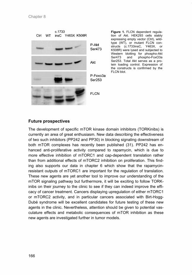
Chapter 8
166
Figure 1. FLCN dependent regula-tion of Akt. HEK293 cells stably expressing empty vector (Ctrl), wild-type (WT), or mutant FLCN con-structs (c.1733insC, Y463X, or K508R) were lysed and subjected to Western blotting for phospho-Akt Ser473 and phospho-FoxO3a Ser253. Total Akt serves as a pro-tein loading control. Expression of the constructs is confirmed by the FLCN blot.
Future prospectives
The development of specific mTOR kinase domain inhibitors (TORKinibs) is currently an area of great enthusiasm. New data describing the effectiveness of two such inhibitors (PP242 and PP30) in blocking signaling downstream of both mTOR complexes has recently been published (31). PP242 has en-hanced anti-proliferative activity compared to rapamycin, which is due to more effective inhibition of mTORC1 and cap-dependent translation rather than from additional effects of mTORC2 inhibition on proliferation. This find-ing also supports our data in chapter 6 which show that the rapamycin-resistant outputs of mTORC1 are important for the regulation of translation. These new agents are yet another tool to improve our understanding of the mTOR signaling pathway but furthermore, it will be exciting to follow TORK-inibs on their journey to the clinic to see if they can indeed improve the effi-cacy of cancer treatment. Cancers displaying upregulation of either mTORC1 or mTORC2 activity, and in particular cancers associated with Birt-Hogg-Dubé syndrome will be excellent candidates for future testing of these new agents in the clinic. Nevertheless, attention should be given to potential vas-culature effects and metabolic consequences of mTOR inhibition as these new agents are investigated further in tumor models.
Thesis_Weppler_v12_fc.pdf
General discussion
167
References 1. Hanahan, D. and Weinberg, R. A. The hallmarks of cancer. Cell, 100: 57-70, 2000. 2. Blais, J. D., Filipenko, V., Bi, M., Harding, H. P., Ron, D., Koumenis, C., Wouters, B. G.,
and Bell, J. C. Activating transcription factor 4 is translationally regulated by hypoxic stress. Mol Cell Biol, 24: 7469-7482, 2004.
3. Koritzinsky, M., Seigneuric, R., Magagnin, M. G., van den Beucken, T., Lambin, P., and Wouters, B. G. The hypoxic proteome is influenced by gene-specific changes in mRNA translation. Radiother Oncol, 76: 177-186, 2005.
4. Harding, H. P., Novoa, I., Zhang, Y., Zeng, H., Wek, R., Schapira, M., and Ron, D. Regu-lated translation initiation controls stress-induced gene expression in mammalian cells. Mol Cell, 6: 1099-1108, 2000.
5. Braunstein, S., Karpisheva, K., Pola, C., Goldberg, J., Hochman, T., Yee, H., Cangiarella, J., Arju, R., Formenti, S. C., and Schneider, R. J. A hypoxia-controlled cap-dependent to cap-independent translation switch in breast cancer. Mol Cell, 28: 501-512, 2007.
6. Young, R. M., Wang, S. J., Gordan, J. D., Ji, X., Liebhaber, S. A., and Simon, M. C. Hy-poxia-mediated selective mRNA translation by an internal ribosome entry site-independent mechanism. J Biol Chem, 283: 16309-16319, 2008.
7. Kozak, M. A second look at cellular mRNA sequences said to function as internal ribosome entry sites. Nucleic Acids Res, 33: 6593-6602, 2005.
8. Zimmer, M., Ebert, B. L., Neil, C., Brenner, K., Papaioannou, I., Melas, A., Tolliday, N., Lamb, J., Pantopoulos, K., Golub, T., and Iliopoulos, O. Small-molecule inhibitors of HIF-2a translation link its 5'UTR iron-responsive element to oxygen sensing. Mol Cell, 32: 838-848, 2008.
9. Peng, X. H., Karna, P., Cao, Z., Jiang, B. H., Zhou, M., and Yang, L. Cross-talk between epidermal growth factor receptor and hypoxia-inducible factor-1alpha signal pathways in-creases resistance to apoptosis by up-regulating survivin gene expression. J Biol Chem, 281: 25903-25914, 2006.
10. Swinson, D. E. and O'Byrne, K. J. Interactions between hypoxia and epidermal growth factor receptor in non-small-cell lung cancer. Clin Lung Cancer, 7: 250-256, 2006.
11. Krause, M., Ostermann, G., Petersen, C., Yaromina, A., Hessel, F., Harstrick, A., van der Kogel, A. J., Thames, H. D., and Baumann, M. Decreased repopulation as well as in-creased reoxygenation contribute to the improvement in local control after targeting of the EGFR by C225 during fractionated irradiation. Radiother Oncol, 76: 162-167, 2005.
12. Solomon, B., Binns, D., Roselt, P., Weibe, L. I., McArthur, G. A., Cullinane, C., and Hicks, R. J. Modulation of intratumoral hypoxia by the epidermal growth factor receptor inhibitor gefitinib detected using small animal PET imaging. Mol Cancer Ther, 4: 1417-1422, 2005.
13. Basic Clinical Radiobiology, second edition, p. 254. London: Arnold, 1997. 14. Franovic, A., Gunaratnam, L., Smith, K., Robert, I., Patten, D., and Lee, S. Translational up-
regulation of the EGFR by tumor hypoxia provides a nonmutational explanation for its over-expression in human cancer. Proc Natl Acad Sci U S A, 104: 13092-13097, 2007.
15. Jimeno, A., Kulesza, P., Wheelhouse, J., Chan, A., Zhang, X., Kincaid, E., Chen, R., Clark, D. P., Forastiere, A., and Hidalgo, M. Dual EGFR and mTOR targeting in squamous cell carcinoma models, and development of early markers of efficacy. Br J Cancer, 96: 952-959, 2007.
16. Buck, E., Eyzaguirre, A., Brown, E., Petti, F., McCormack, S., Haley, J. D., Iwata, K. K., Gibson, N. W., and Griffin, G. Rapamycin synergizes with the epidermal growth factor re-ceptor inhibitor erlotinib in non-small-cell lung, pancreatic, colon, and breast tumors. Mol Cancer Ther, 5: 2676-2684, 2006.
17. Wang, M. Y., Lu, K. V., Zhu, S., Dia, E. Q., Vivanco, I., Shackleford, G. M., Cavenee, W. K., Mellinghoff, I. K., Cloughesy, T. F., Sawyers, C. L., and Mischel, P. S. Mammalian target of rapamycin inhibition promotes response to epidermal growth factor receptor kinase inhibi-tors in PTEN-deficient and PTEN-intact glioblastoma cells. Cancer Res, 66: 7864-7869, 2006.
18. Eshleman, J. S., Carlson, B. L., Mladek, A. C., Kastner, B. D., Shide, K. L., and Sarkaria, J. N. Inhibition of the mammalian target of rapamycin sensitizes U87 xenografts to fraction-ated radiation therapy. Cancer Res, 62: 7291-7297, 2002.
Thesis_Weppler_v12_fc.pdf
Chapter 8
168
19. Manegold, P. C., Paringer, C., Kulka, U., Krimmel, K., Eichhorn, M. E., Wilkowski, R., Jauch, K. W., Guba, M., and Bruns, C. J. Antiangiogenic therapy with mammalian target of rapamycin inhibitor RAD001 (Everolimus) increases radiosensitivity in solid cancer. Clin Cancer Res, 14: 892-900, 2008.
20. Ronellenfitsch, M. W., Brucker, D. P., Burger, M. C., Wolking, S., Tritschler, F., Rieger, J., Wick, W., Weller, M., and Steinbach, J. P. Antagonism of the mammalian target of rapamy-cin selectively mediates metabolic effects of epidermal growth factor receptor inhibition and protects human malignant glioma cells from hypoxia-induced cell death. Brain, 132: 1509-1522, 2009.
21. Dubois, L., Magagnin, M. G., Cleven, A. H., Weppler, S. A., Grenacher, B., Landuyt, W., Lieuwes, N., Lambin, P., Gorr, T. A., Koritzinsky, M., and Wouters, B. G. Inhibition of 4E-BP1 sensitizes U87 glioblastoma xenograft tumors to irradiation by decreasing hypoxia tol-erance. Int J Radiat Oncol Biol Phys, 73: 1219-1227, 2009.
22. Williams, K. J., Telfer, B. A., Brave, S., Kendrew, J., Whittaker, L., Stratford, I. J., and Wedge, S. R. ZD6474, a potent inhibitor of vascular endothelial growth factor signaling, combined with radiotherapy: schedule-dependent enhancement of antitumor activity. Clin Cancer Res, 10: 8587-8593, 2004.
23. Edinger, A. L., Linardic, C. M., Chiang, G. G., Thompson, C. B., and Abraham, R. T. Differ-ential effects of rapamycin on mammalian target of rapamycin signaling functions in mam-malian cells. Cancer Res, 63: 8451-8460, 2003.
24. Shor, B., Zhang, W. G., Toral-Barza, L., Lucas, J., Abraham, R. T., Gibbons, J. J., and Yu, K. A new pharmacologic action of CCI-779 involves FKBP12-independent inhibition of mTOR kinase activity and profound repression of global protein synthesis. Cancer Res, 68: 2934-2943, 2008.
25. Pore, N., Jiang, Z., Shu, H. K., Bernhard, E., Kao, G. D., and Maity, A. Akt1 activation can augment hypoxia-inducible factor-1alpha expression by increasing protein translation through a mammalian target of rapamycin-independent pathway. Mol Cancer Res, 4: 471-479, 2006.
26. Inoki, K., Corradetti, M. N., and Guan, K. L. Dysregulation of the TSC-mTOR pathway in human disease. Nat Genet, 37: 19-24, 2005.
27. Baba, M., Furihata, M., Hong, S. B., Tessarollo, L., Haines, D. C., Southon, E., Patel, V., Igarashi, P., Alvord, W. G., Leighty, R., Yao, M., Bernardo, M., Ileva, L., Choyke, P., War-ren, M. B., Zbar, B., Linehan, W. M., and Schmidt, L. S. Kidney-targeted Birt-Hogg-Dube gene inactivation in a mouse model: Erk1/2 and Akt-mTOR activation, cell hyperprolifera-tion, and polycystic kidneys. J Natl Cancer Inst, 100: 140-154, 2008.
28. Chen, J., Futami, K., Petillo, D., Peng, J., Wang, P., Knol, J., Li, Y., Khoo, S. K., Huang, D., Qian, C. N., Zhao, P., Dykyma, K., Zhang, R., Cao, B., Yang, X. J., Furge, K., Williams, B. O., and Teh, B. T. Deficiency of FLCN in mouse kidney led to development of polycystic kidneys and renal neoplasia. PLoS ONE, 3: e3581, 2008.
29. Hartman, T. R., Nicolas, E., Klein-Szanto, A., Al-Saleem, T., Cash, T. P., Simon, M. C., and Henske, E. P. The role of the Birt-Hogg-Dube protein in mTOR activation and renal tumori-genesis. Oncogene, 28: 1594-1604, 2009.
30. Sarbassov, D. D., Guertin, D. A., Ali, S. M., and Sabatini, D. M. Phosphorylation and regu-lation of Akt/PKB by the rictor-mTOR complex. Science, 307: 1098-1101, 2005.
31. Feldman, M. E., Apsel, B., Uotila, A., Loewith, R., Knight, Z. A., Ruggero, D., and Shokat, K. M. Active-site inhibitors of mTOR target rapamycin-resistant outputs of mTORC1 and mTORC2. PLoS Biol, 7: e38, 2009.
Thesis_Weppler_v12_fc.pdf

169
Summary
Samenvatting
Acknowledgements
Curriculum vitae
List of publications
Thesis_Weppler_v12_fc.pdf
170
SUMMARY
The mTOR kinase, as a central integration point for sensors of growth fac-tors, nutrients and energy sources, plays a key role in tumor biology. In the cell, mTOR is a part of two different multi-protein complexes, mTORC1 and mTORC2. The main function of mTORC1 is to regulate the production of new proteins within the cell. In this dissertation I have investigated several key aspects of mTOR signaling in cancer with a focus on its role in the tumor mi-croenvironment. During the process of tumor development, the normally tight regulation of mTOR and protein synthesis is often lost, leading to increased proliferation and survival, and alteration of the microenvironment (this thesis). This is illus-trated in chapter 4 where we show that EGFRvIII, a constitutively active kinase receptor that signals through the PI3K/Akt/mTOR pathway, stimulates tumor growth and promotes survival after irradiation or under low-oxygen conditions (hypoxia). Hypoxia is a common feature of solid tumors and an important determinant of poor treatment outcome, thus strategies that can reduce or eliminate hypoxic tumor cells are highly desirable (reviewed in chapter 2). The ability of cells to repress mTOR activity and protein synthesis is an important part of the cellular response to hypoxia. In chapter 3 we show that hypoxia inhibits protein synthesis in two separate phases by using two distinct mechanisms, one of which requires 4E-BP1, an mTOR substrate. The ability of 4E-BP1 to inhibit protein synthesis is determined by its level of phosphorylation. This prompted us to conduct an indepth study of 4E-BP1 regulation in chapter 6. We provide evidence to suggest that in addition to mTORC1, there is a PI3K/Akt-dependent but mTORC1-independent signal to 4E-BP1 that must be blocked in order to effectively inhibit protein synthesis via this mechanism. Thus, targeting protein synthesis in cancer might be im-proved by strategies that block 4E-BP1 more effectively. Due to the attractiveness of mTOR as a therapeutic target in cancer, a num-ber of mTOR inhibitors are currently being tested, many of which are analogs based upon the structure of the original mTOR inhibitor, rapamycin. Previous studies have shown that rapamycin can inhibit tumor regrowth when used in combination with radiation. However, in chapter 5, we saw no additional ef-fect of rapamycin to limit local tumor control by radiation, although we did ob-serve heterogeneity in the response of individual tumors that may have been influenced by increased areas of hypoxia and intravascular thrombosis after rapamycin treatment.
Thesis_Weppler_v12_fc.pdf
Summary
171
Our knowledge of the mTOR signaling pathway continues to evolve as we learn more about the intricacies of its regulatory proteins and feedback path-ways. Human genetics can be a valuable tool in helping us to dissect the in-teractions between signaling proteins within a pathway. In chapter 8, we de-scribe a patient with Birt-Hogg-Dubé syndrome (a familial cancer disease) in which the mutant folliculin protein is suspected to promote tumor formation by upregulation of mTOR activity through an unknown mechanism. We show that tumors associated with this syndrome do not express common markers of increased mTORC1 activity, but rather that activation of mTORC2 may be more relevant to this disease. In conclusion, mTOR and protein synthesis are attractive targets for cancer therapy. However, the future success of these strategies will depend upon both basic and translational research to understand how best to inhibit these processes for maximum gain of anti-tumor effect without undesirable activa-tion of feedback loops or normal tissue side-effects.
Thesis_Weppler_v12_fc.pdf
172
SAMENVATTING
mTOR fungeert als centraal integratiepunt voor sensoren van groeifactoren, voedingsstoffen en energiebronnen. Op die manier heeft mTOR een erg be-langrijke rol in tumorbiologie. In de cel kan mTOR deel uitmaken van twee verschillende multi-eiwit complexen, mTORC1 en mTORC2. De hoofdfunctie van mTORC1 is het reguleren van de productie van nieuwe eiwitten binnen de cel. In dit proefschrift heb ik een aantal sleutelaspecten van mTOR-signalering in kanker onderzocht. De focus lag daarbij op de rol die mTOR speelt in de tumor micro-omgeving.
Gedurende het ontwikkelingsproces van een tumor gaat de strikte regulering van mTOR en eiwitsynthese vaak verloren. Dit leidt tot toegenomen prolifera-tie en overleving van tumorcellen, en tot een verandering van de tumor mi-cro-omgeving (dit proefschrift). Dit wordt beschreven in hoofdstuk 4 waar we laten zien dat EGFRvIII, een constitutief geactiveerde kinase receptor die signaleert via de PI3K/Akt/mTOR-route, tumorgroei stimuleert. Expressie van EGFRvIII verbetert ook het overleven van tumoren na bestraling of bij bloot-stelling aan verlaagde zuurstofconcentraties (hypoxie). Hypoxie komt veel-vuldig voor in vaste tumoren en is een belangrijke determinerende factor voor een slechte behandelingsuitkomst. Het is dan ook zeer wenselijk dat strate-gieën ontwikkeld worden die het aantal hypoxische cellen in tumoren kunnen verminderen of elimineren (zoals beschreven in hoofdstuk 2). Het vermogen van cellen om mTOR-activiteit en eiwitsynthese te onderdrukken vormt een belangrijk onderdeel van de cellulaire respons tegenover hypoxie. In hoofd-stuk 3 tonen we aan dat hypoxie de eiwitsynthese remt in twee verschillende fases, door gebruik te maken van twee verschillende mechanismen. Eén van deze mechanismen vereist het mTOR-substraat 4E-BP1. De mogelijkheid van 4E-BP1 om eiwitsynthese te remmen hangt af van de mate waarin het is gefosforyleerd. Dit zette ons aan om de regulatie van 4E-BP1 in detail te onderzoeken (hoofdstuk 6). Onze data suggereren dat naast het mTORC1 signaal wellicht een PI3K/Akt-afhankelijk maar mTORC1-onafhankelijk signaal naar 4E-BP1 gaat, dat geblokkeerd moet worden om via dit mechanisme de eiwitsynthese te kunnen remmen. Zodoende zou het aangrijpen op de eiwitsynthese bij kanker mogelijk verbeterd kunnen worden met behulp van strategieën die 4E-BP1 effectiever blokkeren. Omwille van het feit dat mTOR aantrekkelijk is als een therapeutisch doelwit bij kanker, worden momenteel een aantal mTOR-inhibitoren getest. Vele van deze remmers zijn analogen die gebaseerd zijn op de structuur van de origi-nele mTOR-remmer, rapamycine. Eerdere studies hebben laten zien dat ra-
Thesis_Weppler_v12_fc.pdf
Samenvatting
173
pamycine, in combinatie met bestraling, de hergroei van tumoren kan rem-men. Echter, in hoofdstuk 5 hebben we geen additioneel effect van rapamy-cine op lokale tumorcontrole door bestraling gevonden. Wat we wel duidelijk observeerden, was een heterogeniteit in respons van individuele tumoren. Dit kan mogelijk verklaard worden door een toename van hypoxie en intravascu-laire thrombose na behandeling met rapamycine. Onze kennis van de mTOR-signaleringsroute blijft zich verder ontwikkelen doordat we steeds meer te weten komen over de gecompliceerdheid van de verschillende mTOR-regulatorische eiwitten en de feedback mechanismen. Humane genetische informatie kan een waardevol instrument zijn om de in-teracties tussen verschillende signaleringseiwitten binnen een transductie-weg in een cel te ontleden. In hoofdstuk 8 beschrijven we een patiënt met het Birt-Hogg-Dubé syndroom (een familiale kankeraandoening) waarbij vermoed wordt dat het gemuteerd folliculin-eiwit tumorvorming promoot door verhoging van de mTOR-activiteit via een nog onbekend mechanisme. Wij tonen aan dat tumoren die met dit syndroom geassocieerd zijn, geen merkers van ver-hoogde mTORC1-activiteit tot expressie brengen, maar dat eerder activering van mTORC2 relevanter is voor deze ziekte. Tot besluit kunnen we stellen dat mTOR en eiwitsynthese aantrekkelijke doe-len zijn voor kankertherapie. Het toekomstige succes van deze strategieën zal afhankelijk zijn van zowel basaal en translationeel onderzoek, dat ons in staat moet stellen om te begrijpen hoe we deze processen het best inhibe-ren, zodat we een maximaal anti-tumor effect kunnen verkrijgen zonder on-gewenste activering van feedbackmechanismen of neveneffecten op gezond weefsel.
Thesis_Weppler_v12_fc.pdf
174
ACKNOWLEDGEMENTS
According to a well-known African proverb, “it takes a village to raise a child”. In the same way, it takes many people to bring a Ph.D. thesis to fruition. Therefore, I would like to offer my thanks and gratitude to the following peo-ple.
I would like to begin by thanking my supervisors Brad Wouters and Philippe Lambin for giving me the opportunity to come to Maastricht and be involved in the start-up of the MAASTRO lab. I also appreciate the many opportunities that I had to travel to conferences in such exotic locations as Puerto Rico, Australia, the USA, as well as within Europe. Brad, I’ve learned a tremendous amount from you and am sure that it will serve me well in the future.
I would also like to thank Guido Lammering and Willem Voncken for serving as members of my Ph.D. portfolio committee. Your comments and criticisms were always very helpful, and I now know to plan at least 1 hour of discus-sion per committee member present! Also thanks to Guido and Maurice van Steensel for serving as co-promoters. Over the years, I have had many colleagues at the MAASTRO lab (unfortu-nately too many to name everyone individually), so a big thanks to all past and present lab members. A special thanks to all the technicians who really do a great job of keeping the lab running in spite of the efforts of all those pesky Ph.D. students. Jan, Kim, and Roland—you were my trusted coffee companions over the years. Thanks to you guys I got the ‘Belgian perspective’ on everything from politics to football to beer. Roland, my fellow Canuck, it seems like a lifetime ago that we made the decision to pack-up everything in Ottawa and move to Maastricht. You have been the one constant element through this whole jour-ney from beginning to end. Thanks for everything man…I’m sure I still owe you a Duvel. More recently I began working at the department of Dermatology, so I would like to thank everyone who has made me feel welcome there, especially Tijs Claessens with whom I worked most closely. Good luck with your future Ph.D. research Tijs, and with climbing mountains both on your bike and in the lab. During my studies I had the wonderful experience to spend several months in Dresden, Germany at the lab of Prof. dr. Michael Baumann to conduct the
Thesis_Weppler_v12_fc.pdf
Acknowledgemenst
175
rapamycin and radiotherapy study. I would like to thank Mechthild Krause who helped to design and carry out the experiments and Dorothy Pfitzmann for teaching me the technical aspects of working with mice. Also thanks to Agnieska Zyromska for her contribution to the follow-up study. Hopefully we will have the opportunity to meet in person one day in the future. Thanks again to Willem Voncken (this time for his contagious and unquench-able enthusiasm for good science) and to his research group for all of the interesting discussions and for making journal club, pizza meetings, etc. more enjoyable and thought provoking. Especially to Hanneke, Frank, and Peggy who commiserated with me throughout the Ph.D. process. Thanks also to Ramon Langen and the others from the Pulmonology depart-ment who participate in our weekly lab meetings. Your comments and feed-back are always appreciated. During my time in Maastricht, I established a strong “French connection” through the friendships I made with Florence, Celine & Vincent. As foreigners in a strange land we flocked together, even if only to avoid Dutch ‘food’ or for shopping trips across the border. Thanks for the memories and I hope we will continue to stay in touch. The ProTones band provided me with a welcome musical interlude away from the lab. Thanks to everyone for letting me ‘meeblazen’. Maybe next year we’ll go on tour? Also thanks to Kim & Jos for worrying about me, so that I would be guilted into writing more quickly. I like to think that you guys contributed to my educa-tion through our frequent trips to John Mullins for quiz night. Een heel groot “merci” aan de familie Willems, mijn nieuwe schoonfamilie. Ik heb van jullie alle limburgse dingen geleerd te waarderen. Nu alleen nog maar praten in het plat…..maar ik kan wel zeggen dat ik toch een beetje in-geburgerd ben door jullie. Mom and Dad, thanks for all the years of patience and support. Even though most of this book is nothing more than Greek to you, I know that you can see all the hard work, the trials and tribulations that are behind the words on every page. And to the rest of my family, Dianne & Dave, Dale & Emily, Jessie & Sarah, I hope that you can be proud and enjoy bragging that there is now a doctor in the family.
Thesis_Weppler_v12_fc.pdf

Acknowledgemenst
176
And last, but most importantly, my heartfelt thanks to Jodil, for always starting dinner and taking care of the home-front when I was delayed at the lab. I love that we can discuss experiments over breakfast (usually in ‘du-nglish’, our own special language), and that you sympathize whole-heartedly when I have Western blot issues. Infinite thanks for the emotional support you’ve given me during the tough times and for the welcome distractions you provide from work. I look forward to many more years of scientific discovery by your side.
Thesis_Weppler_v12_fc.pdf
177
CURRICULUM VITAE
Sherry Weppler was born on August 15, 1975 in Hanover, Canada. She was awarded her Ontario Secondary School Diploma from John Diefenbaker Secondary School in 1994. At this time she enrolled in an Honours Bachelor of Science program specializing in Microbiology and Immunology at the Uni-versity of Western Ontario in London, Canada. Her graduation project was under the supervision of Dr. Katherine Dobinson and involved the molecular characterization of a trypsin-like protease from the plant pathogenic fungus Verticillium dahliae. In 1998 she was awarded her bachelors degree with dis-tinction (cum laude), and moved to the University of Ottawa where she began a Master of Science in the department of Microbiology and Immunology. Un-der the supervision of Dr. Chaim Birnboim, she conducted research describ-ing spontaneous and nitrous oxide-induced mutations in a murine tumor model. After completion of her M.Sc. in 2001, she moved to the Netherlands in order to pursue her doctorate at Maastricht University under the guidance of Prof. Brad Wouters and Prof. Philippe Lambin in the newly established Maastricht Radiation Oncology (MAASTRO) laboratory. She is currently working at Maastricht University as a post-doctoral fellow under the supervi-sion of Dr. Maurice van Steensel on a collaborative project between MAAS-TRO and the department of Dermatology that is investigating the involvement of mTOR and HIF in Birt-Hogg-Dubé syndrome.
Thesis_Weppler_v12_fc.pdf
178
LIST OF PUBLICATIONS
Wouters, B.G., Weppler, S.A., Koritzinsky, M., Landuyt, W., Nuyts, S., Theys, J., Chiu, R.K., Lambin, P. Hypoxia as a target for combined modality treatments. Eur J Cancer 2002, 38 (2): 240-257.
Koritzinsky, M., Magagnin, M.G., van den Beucken, T., Seigneuric, R., Sav-elkouls, K., Dostie, J., Pyronnet, S., Kaufman, R.J., Weppler, S.A., Voncken, J.W., Lambin, P., Koumenis, C., Sonenberg, N., Wouters, B.G. Gene expres-sion during acute and prolonged hypoxia is regulated by distinct mechanisms of translational control. EMBO J 2006, 25 (5): 1114-25. Weppler, S.A., Krause, M., Zyromska, A., Lambin, P., Baumann, M., Wout-ers, B.G. Response of U87 glioma xenografts treated with concurrent rapa-mycin and fractionated radiotherapy: possible role for thrombosis. Radiother Oncol 2007, 82 (1): 96-104. Aerts, H.J.W.L., Dubois, L., Hackeng, T.M., Straathof, R., Chiu, R.K., Lieu-wes, N.G., Jutten, B., Weppler, S.A., Lammering, G., Wouters, B.G., Lambin, P. Development and evaluation of a Cetuximab-based imaging probe to tar-get EGFR and EGFRvIII. Radiother Oncol 2007, 83 (3): 326-332. Weppler, S.A.*, Li, Y.*, Dubois, L., Lieuwes, N., Jutten, B., Lambin, P., Wouters, B.G., Lammering, G. Expression of EGFR variant vIII promotes both radiation resistance and hypoxia tolerance. Radiother Oncol 2007, 83 (3): 333-339. (* These authors contributed equally to this work). Dubois, L.*, Magagnin, M.G.*, Cleven, A.H., Weppler, S.A., Grenacher, B., Landuyt, W., Lieuwes, N., Lambin, P., Gorr, T.A., Koritzinsky, M., Wouters., B.G. Inhibition of 4E-BP1 sensitizes U87 glioblastoma xenograft tumors to irradiation by decreasing hypoxia tolerance. Int J Radiat Oncol Biol Phys 2009, 73 (4): 1219-27. Weppler, S.A., Koritzinsky, M., Voncken, J.W., Lambin, P., Wouters, B.G. Inhibition of 4E-BP1 phosphorylation and mRNA translation requires simulta-neous blockade of mTORC1 and PI3K/Akt signaling. (manuscript submitted). Weppler, S.A., Claessens, T., van Geel, M., Creytons, D., Vreeburg, M., Wouters, B.G., van Steensel, M.A.M. Neuroendocrine carcinoma in Birt-Hogg-Dubé syndrome. (manuscript submitted).
Thesis_Weppler_v12_fc.pdf
Related Documents