Analysis of congenital hypomyelinating Egr2 Lo/Lo nerves identifies Sox2 as an inhibitor of Schwann cell differentiation and myelination Nam Le, Rakesh Nagarajan, James Y. T. Wang, Toshiyuki Araki, Robert E. Schmidt, and Jeffrey Milbrandt* Department of Pathology and Immunology, Washington University School of Medicine, 660 South Euclid Avenue, Box 8118, St. Louis, MO 63110 Edited by Eric M. Shooter, Stanford University School of Medicine, Stanford, CA, and approved January 3, 2005 (received for review October 21, 2004) Egr2 is a transcription factor required for peripheral nerve myeli- nation in rodents, and mutations in Egr2 are associated with congenital hypomyelinating neuropathy (CHN) in humans. To fur- ther study its role in myelination, we generated mice harboring a hypomorphic Egr2 allele (Egr2 Lo ) that survive for up to 3 weeks postnatally, a period of active myelination in rodents. These Egr2 Lo/Lo mice provided the opportunity to study the molecular effects of Egr2 deficiency on Schwann cell biology, an analysis that was not possible previously, because of the perinatal lethality of Egr2-null mice. Egr2 Lo/Lo mice phenocopy CHN, as evidenced by the severe hypomyelination and increased numbers of proliferating Schwann cells of the peripheral nerves. Comparison of sciatic nerve gene expression profiles during development and after crush injury with those of Egr2 Lo/Lo Schwann cells revealed that they are developmentally arrested, with down-regulation of myelination- related genes and up-regulation of genes associated with imma- ture and promyelinating Schwann cells. One of the abnormally elevated genes in Egr2 Lo/Lo Schwann cells, Sox2, encodes a tran- scription factor that is crucial for maintenance of neural stem cell pluripotency. Wild-type Schwann cells infected with Sox2 adeno- virus or lentivirus inhibited expression of myelination-associated genes (e.g., myelin protein zero; Mpz), and failed to myelinate axons in vitro, but had an enhanced proliferative response to -neuregulin. The characterization of a mouse model of CHN has provided insight into Schwann cell differentiation and allowed the identification of Sox2 as a negative regulator of myelination. D eficits in Schwann cell function are associated with a number of human diseases, from debilitating conditions like Charcot– Marie–Tooth disease (CMT) and diabetic neuropathy, to fre- quently lethal diseases, such as congenital hypomyelinating neu- ropathy (CHN) (1). Developmentally, Schwann cells progress from an immature state, expressing markers such as L1, Ncam, and Cyclin D1, to the nonmyelinating or the promyelinating state. Whereas nonmyelinating Schwann cells continue to express L1 and Ncam and are associated with multiple axons, promyelinating Schwann cells establish a one-to-one relationship with axons and are marked by expression of the POU domain transcription factor suppressed cAMP-inducible POU (SCIP) (2). Shortly after birth, promyelinating Schwann cells exit from the cell cycle and differ- entiate into myelinating Schwann cells, a process that involves the expression of genes such as Egr2, Mpz, Pmp22, Prx, and Gjb1 and the formation of myelin. A similar process occurs after axonal injury, in which Schwann cells must revert to a proliferative immature state and later redifferentiate and myelinate regenerating axons (3). Studies of Egr2-null mice led to the discovery that this transcrip- tion factor is a prime regulator of Schwann cell myelination. Although most Egr2 null mice die at birth, a few Egr2-null mice survive for up to 2 weeks. Nerves from these rare survivors are hypomyelinated and are populated with Schwann cells that fail to exit the cell cycle (4). Enforced expression of Egr2 in cultured Schwann cells activates expression of numerous genes associated with myelination, including crucial myelin structural proteins and enzymes involved in lipid synthesis (5), further supporting the importance of Egr2 in regulating myelination. More importantly, mutations in Egr2 are found in patients with CHN, CMT, or Dejerine–Sottas syndrome (6–9). Most Egr2 mutations in CHN patients occur in the zinc-finger DNA-binding domain. These DNA-binding mutants act in a dominant-negative fashion to inhibit wild-type Egr2 activity (5). Additionally, a reces- sive form of CHN is caused by a mutation in the R1 domain, a site of interaction for the Nab proteins that modulate Egr2 activity (6). While these data indicate that alterations in Egr2 activity result in Schwann cell dysfunction and aberrant myelination, the molecular alterations that occur in these cells are unknown. To understand the molecular consequences of Egr2-deficiency in the context of CHN, we have studied mice homozygous for an Egr2 hypomorphic allele (Egr2 Lo/Lo ) that consistently survive into the third postnatal week. These mice display characteristics of CHN, including severe hypomyelination and an absence of Schwann cell differentiation. Global gene expression analysis of CHN Schwann cells from these Egr2 Lo/Lo mice identified numerous proteins that are likely to play important roles in Schwann cell development and may directly contribute to Schwann cell dysfunction in CHN. Further examination of one of these candidates, Sox2, indicates a role for this transcription factor in maintaining the undifferentiated Schwann cell state. Methods Microarray Analysis. We compared microarray data from three different paradigms in this study. Two of the data sets, nerve crush injury (10) and development (11), were previously generated. The third data set was generated from two independent samples of sciatic nerve total RNA (10 g) for each genotype, Egr2 Lo/Lo and wild type. Each sample contained nerves from 10 animals. RNA extraction, probe generation, and chip hybridization to MU74A V2 microarrays were described (10), and chips were scaled to 1,500 (MAS 5.0, Affymetrix). For each paradigm, pairwise comparisons were performed (a total of nine analyses). The baseline points, wild type (for Egr2 Lo/Lo ), uninjured (for nerve crush), or embryonic day (E)17 (for development), were compared with mutant (Egr2 Lo/Lo ), or to each monitored time point in the nerve crush or development paradigms, respectively. For each comparison, genes called absent in all chips were excluded. These filtered data sets were subjected to significance analysis of microarrays by using the pairwise com- parison option with the following parameters: significantly altered 2-fold, and a false-discovery rate of 10% (12). The list of differentially regulated genes in injury or development was ob- tained by taking the union of significantly altered genes in each This paper was submitted directly (Track II) to the PNAS office. Abbreviations: CHN, congenital hypomyelinating neuropathy; CMT, Charcot–Marie–Tooth disease; En, embryonic day n;Pn, postnatal day n; EGFP, enhanced GFP; Mpz, myelin protein zero; SCIP, suppressed cAMP-inducible POU. *To whom correspondence should be addressed. E-mail: [email protected]. © 2005 by The National Academy of Sciences of the USA 2596 –2601 PNAS February 15, 2005 vol. 102 no. 7 www.pnas.orgcgidoi10.1073pnas.0407836102

Welcome message from author
This document is posted to help you gain knowledge. Please leave a comment to let me know what you think about it! Share it to your friends and learn new things together.
Transcript
Analysis of congenital hypomyelinating Egr2Lo/Lo
nerves identifies Sox2 as an inhibitor of Schwanncell differentiation and myelinationNam Le, Rakesh Nagarajan, James Y. T. Wang, Toshiyuki Araki, Robert E. Schmidt, and Jeffrey Milbrandt*
Department of Pathology and Immunology, Washington University School of Medicine, 660 South Euclid Avenue, Box 8118, St. Louis, MO 63110
Edited by Eric M. Shooter, Stanford University School of Medicine, Stanford, CA, and approved January 3, 2005 (received for review October 21, 2004)
Egr2 is a transcription factor required for peripheral nerve myeli-nation in rodents, and mutations in Egr2 are associated withcongenital hypomyelinating neuropathy (CHN) in humans. To fur-ther study its role in myelination, we generated mice harboring ahypomorphic Egr2 allele (Egr2Lo) that survive for up to 3 weekspostnatally, a period of active myelination in rodents. TheseEgr2Lo/Lo mice provided the opportunity to study the moleculareffects of Egr2 deficiency on Schwann cell biology, an analysis thatwas not possible previously, because of the perinatal lethality ofEgr2-null mice. Egr2Lo/Lo mice phenocopy CHN, as evidenced by thesevere hypomyelination and increased numbers of proliferatingSchwann cells of the peripheral nerves. Comparison of sciatic nervegene expression profiles during development and after crushinjury with those of Egr2Lo/Lo Schwann cells revealed that they aredevelopmentally arrested, with down-regulation of myelination-related genes and up-regulation of genes associated with imma-ture and promyelinating Schwann cells. One of the abnormallyelevated genes in Egr2Lo/Lo Schwann cells, Sox2, encodes a tran-scription factor that is crucial for maintenance of neural stem cellpluripotency. Wild-type Schwann cells infected with Sox2 adeno-virus or lentivirus inhibited expression of myelination-associatedgenes (e.g., myelin protein zero; Mpz), and failed to myelinateaxons in vitro, but had an enhanced proliferative response to�-neuregulin. The characterization of a mouse model of CHN hasprovided insight into Schwann cell differentiation and allowed theidentification of Sox2 as a negative regulator of myelination.
Deficits in Schwann cell function are associated with a numberof human diseases, from debilitating conditions like Charcot–
Marie–Tooth disease (CMT) and diabetic neuropathy, to fre-quently lethal diseases, such as congenital hypomyelinating neu-ropathy (CHN) (1). Developmentally, Schwann cells progress froman immature state, expressing markers such as L1, Ncam, andCyclin D1, to the nonmyelinating or the promyelinating state.Whereas nonmyelinating Schwann cells continue to express L1 andNcam and are associated with multiple axons, promyelinatingSchwann cells establish a one-to-one relationship with axons andare marked by expression of the POU domain transcription factorsuppressed cAMP-inducible POU (SCIP) (2). Shortly after birth,promyelinating Schwann cells exit from the cell cycle and differ-entiate into myelinating Schwann cells, a process that involves theexpression of genes such as Egr2, Mpz, Pmp22, Prx, and Gjb1 andthe formation of myelin. A similar process occurs after axonalinjury, in which Schwann cells must revert to a proliferativeimmature state and later redifferentiate and myelinate regeneratingaxons (3).
Studies of Egr2-null mice led to the discovery that this transcrip-tion factor is a prime regulator of Schwann cell myelination.Although most Egr2 null mice die at birth, a few Egr2-null micesurvive for up to 2 weeks. Nerves from these rare survivors arehypomyelinated and are populated with Schwann cells that fail toexit the cell cycle (4). Enforced expression of Egr2 in culturedSchwann cells activates expression of numerous genes associatedwith myelination, including crucial myelin structural proteins and
enzymes involved in lipid synthesis (5), further supporting theimportance of Egr2 in regulating myelination.
More importantly, mutations in Egr2 are found in patients withCHN, CMT, or Dejerine–Sottas syndrome (6–9). Most Egr2mutations in CHN patients occur in the zinc-finger DNA-bindingdomain. These DNA-binding mutants act in a dominant-negativefashion to inhibit wild-type Egr2 activity (5). Additionally, a reces-sive form of CHN is caused by a mutation in the R1 domain, a siteof interaction for the Nab proteins that modulate Egr2 activity (6).While these data indicate that alterations in Egr2 activity result inSchwann cell dysfunction and aberrant myelination, the molecularalterations that occur in these cells are unknown.
To understand the molecular consequences of Egr2-deficiency inthe context of CHN, we have studied mice homozygous for an Egr2hypomorphic allele (Egr2Lo/Lo) that consistently survive into thethird postnatal week. These mice display characteristics of CHN,including severe hypomyelination and an absence of Schwann celldifferentiation. Global gene expression analysis of CHN Schwanncells from these Egr2Lo/Lo mice identified numerous proteins thatare likely to play important roles in Schwann cell development andmay directly contribute to Schwann cell dysfunction in CHN.Further examination of one of these candidates, Sox2, indicates arole for this transcription factor in maintaining the undifferentiatedSchwann cell state.
MethodsMicroarray Analysis. We compared microarray data from threedifferent paradigms in this study. Two of the data sets, nerve crushinjury (10) and development (11), were previously generated. Thethird data set was generated from two independent samples ofsciatic nerve total RNA (10 �g) for each genotype, Egr2Lo/Lo andwild type. Each sample contained nerves from 10 animals. RNAextraction, probe generation, and chip hybridization to MU74A V2microarrays were described (10), and chips were scaled to 1,500(MAS 5.0, Affymetrix).
For each paradigm, pairwise comparisons were performed (atotal of nine analyses). The baseline points, wild type (forEgr2Lo/Lo), uninjured (for nerve crush), or embryonic day (E)17(for development), were compared with mutant (Egr2Lo/Lo), or toeach monitored time point in the nerve crush or developmentparadigms, respectively. For each comparison, genes called absentin all chips were excluded. These filtered data sets were subjectedto significance analysis of microarrays by using the pairwise com-parison option with the following parameters: significantly altered�2-fold, and a false-discovery rate of 10% (12). The list ofdifferentially regulated genes in injury or development was ob-tained by taking the union of significantly altered genes in each
This paper was submitted directly (Track II) to the PNAS office.
Abbreviations: CHN, congenital hypomyelinating neuropathy; CMT, Charcot–Marie–Toothdisease; En, embryonic day n; Pn, postnatal day n; EGFP, enhanced GFP; Mpz, myelin proteinzero; SCIP, suppressed cAMP-inducible POU.
*To whom correspondence should be addressed. E-mail: [email protected].
© 2005 by The National Academy of Sciences of the USA
2596–2601 � PNAS � February 15, 2005 � vol. 102 � no. 7 www.pnas.org�cgi�doi�10.1073�pnas.0407836102
pairwise comparison within each paradigm. A summary of theseanalyses is in Table 1, which is published as supporting informationon the PNAS web site. The array data are available upon request.
Supporting Methods. Generation of the Egr2Lo targeting construct,sciatic nerve histology and ultrastructure, immunohistochemistry,western analysis, quantitative RT-PCR, sciatic nerve crush injury,primary Schwann cell cultures, proliferation assays, adenoviralinfection, lentiviral infection, in vitro myelination assay, and K-means clustering of microarray profiles, are all detailed in Support-ing Methods, which is published as supporting information on thePNAS web site.
ResultsGeneration of Egr2Lo/Lo Mice. Because most Egr2-null mice die atbirth, a model of CHN with a longer lifespan would enhance theidentification of important Schwann cell deficits in this disease. Inattempting to introduce specific point mutants into the Egr2 locus,we found that insertion of a PGK-Neo cassette into the first intronof Egr2 generated a hypomorphic Egr2 allele (Egr2Lo). The de-creased expression from this mutant allele is presumably due to theintroduction of the PGK promoter, as has been observed at otherloci (13). Mice heterozygous (Egr2�/Lo) for the hypomorphic allelewere overtly normal, and homozygous mice (Egr2Lo/Lo) wereproduced at expected Mendelian ratios. Southern analysis con-firmed homologous recombination at the Egr2 locus (Fig. 6, whichis published as supporting information on the PNAS web site).
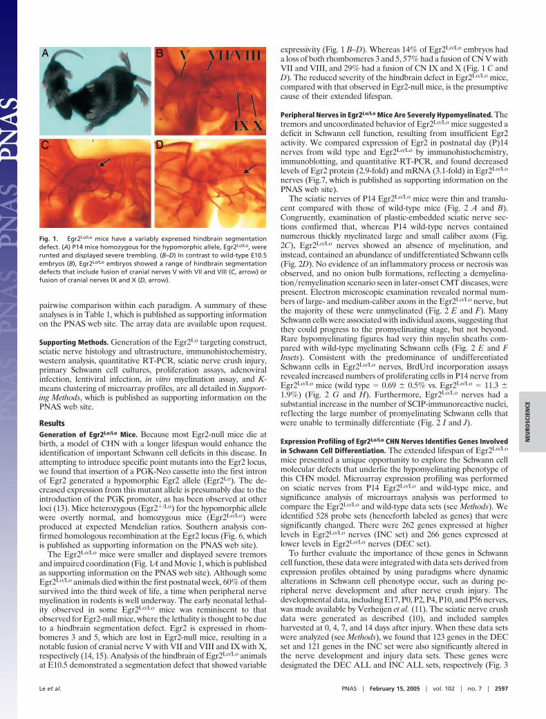
The Egr2Lo/Lo mice were smaller and displayed severe tremorsand impaired coordination (Fig. 1A and Movie 1, which is publishedas supporting information on the PNAS web site). Although someEgr2Lo/Lo animals died within the first postnatal week, 60% of themsurvived into the third week of life, a time when peripheral nervemyelination in rodents is well underway. The early neonatal lethal-ity observed in some Egr2Lo/Lo mice was reminiscent to thatobserved for Egr2-null mice, where the lethality is thought to be dueto a hindbrain segmentation defect. Egr2 is expressed in rhom-bomeres 3 and 5, which are lost in Egr2-null mice, resulting in anotable fusion of cranial nerve V with VII and VIII and IX with X,respectively (14, 15). Analysis of the hindbrain of Egr2Lo/Lo animalsat E10.5 demonstrated a segmentation defect that showed variable
expressivity (Fig. 1 B–D). Whereas 14% of Egr2Lo/Lo embryos hada loss of both rhombomeres 3 and 5, 57% had a fusion of CN V withVII and VIII, and 29% had a fusion of CN IX and X (Fig. 1 C andD). The reduced severity of the hindbrain defect in Egr2Lo/Lo mice,compared with that observed in Egr2-null mice, is the presumptivecause of their extended lifespan.
Peripheral Nerves in Egr2Lo/Lo Mice Are Severely Hypomyelinated. Thetremors and uncoordinated behavior of Egr2Lo/Lo mice suggested adeficit in Schwann cell function, resulting from insufficient Egr2activity. We compared expression of Egr2 in postnatal day (P)14nerves from wild type and Egr2Lo/Lo by immunohistochemistry,immunoblotting, and quantitative RT-PCR, and found decreasedlevels of Egr2 protein (2.9-fold) and mRNA (3.1-fold) in Egr2Lo/Lo
nerves (Fig.7, which is published as supporting information on thePNAS web site).
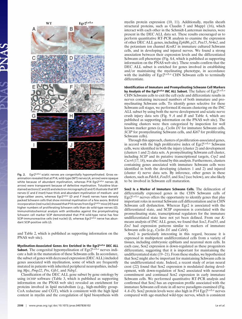
The sciatic nerves of P14 Egr2Lo/Lo mice were thin and translu-cent compared with those of wild-type mice (Fig. 2 A and B).Congruently, examination of plastic-embedded sciatic nerve sec-tions confirmed that, whereas P14 wild-type nerves containednumerous thickly myelinated large and small caliber axons (Fig.2C), Egr2Lo/Lo nerves showed an absence of myelination, andinstead, contained an abundance of undifferentiated Schwann cells(Fig. 2D). No evidence of an inflammatory process or necrosis wasobserved, and no onion bulb formations, reflecting a demyelina-tion�remyelination scenario seen in later-onset CMT diseases, werepresent. Electron microscopic examination revealed normal num-bers of large- and medium-caliber axons in the Egr2Lo/Lo nerve, butthe majority of these were unmyelinated (Fig. 2 E and F). ManySchwann cells were associated with individual axons, suggesting thatthey could progress to the promyelinating stage, but not beyond.Rare hypomyelinating figures had very thin myelin sheaths com-pared with wild-type myelinating Schwann cells (Fig. 2 E and FInsets). Consistent with the predominance of undifferentiatedSchwann cells in Egr2Lo/Lo nerves, BrdUrd incorporation assaysrevealed increased numbers of proliferating cells in P14 nerve fromEgr2Lo/Lo mice (wild type � 0.69 � 0.5% vs. Egr2Lo/Lo � 11.3 �1.9%) (Fig. 2 G and H). Furthermore, Egr2Lo/Lo nerves had asubstantial increase in the number of SCIP-immunoreactive nuclei,reflecting the large number of promyelinating Schwann cells thatwere unable to terminally differentiate (Fig. 2 I and J).
Expression Profiling of Egr2Lo/Lo CHN Nerves Identifies Genes Involvedin Schwann Cell Differentiation. The extended lifespan of Egr2Lo/Lo
mice presented a unique opportunity to explore the Schwann cellmolecular defects that underlie the hypomyelinating phenotype ofthis CHN model. Microarray expression profiling was performedon sciatic nerves from P14 Egr2Lo/Lo and wild-type mice, andsignificance analysis of microarrays analysis was performed tocompare the Egr2Lo/Lo and wild-type data sets (see Methods). Weidentified 528 probe sets (henceforth labeled as genes) that weresignificantly changed. There were 262 genes expressed at higherlevels in Egr2Lo/Lo nerves (INC set) and 266 genes expressed atlower levels in Egr2Lo/Lo nerves (DEC set).
To further evaluate the importance of these genes in Schwanncell function, these data were integrated with data sets derived fromexpression profiles obtained by using paradigms where dynamicalterations in Schwann cell phenotype occur, such as during pe-ripheral nerve development and after nerve crush injury. Thedevelopmental data, including E17, P0, P2, P4, P10, and P56 nerves,was made available by Verheijen et al. (11). The sciatic nerve crushdata were generated as described (10), and included samplesharvested at 0, 4, 7, and 14 days after injury. When these data setswere analyzed (see Methods), we found that 123 genes in the DECset and 121 genes in the INC set were also significantly altered inthe nerve development and injury data sets. These genes weredesignated the DEC ALL and INC ALL sets, respectively (Fig. 3
Fig. 1. Egr2Lo/Lo mice have a variably expressed hindbrain segmentationdefect. (A) P14 mice homozygous for the hypomorphic allele, Egr2Lo/Lo, wererunted and displayed severe trembling. (B–D) In contrast to wild-type E10.5embryos (B), Egr2Lo/Lo embryos showed a range of hindbrain segmentationdefects that include fusion of cranial nerves V with VII and VIII (C, arrow) orfusion of cranial nerves IX and X (D, arrow).
Le et al. PNAS � February 15, 2005 � vol. 102 � no. 7 � 2597
NEU
ROSC
IEN
CE
and Table 2, which is published as supporting information on thePNAS web site).
Myelination-Associated Genes Are Enriched in the Egr2Lo/Lo DEC ALLSubset. The congenital hypomyelination of Egr2Lo/Lo nerves indi-cate a halt in the maturation of these Schwann cells. In accordance,the subset of genes with decreased expression (DEC ALL) includedgenes associated with myelination, some of which are frequentlymutated in patients with inherited peripheral neuropathies, includ-ing Mpz, Pmp22, Prx, Gjb1, and Ndrg1.
Classification of the DEC ALL gene subset by gene ontology byusing DCHIP software (Table 3, which is published as supportinginformation on the PNAS web site) revealed an enrichment forproteins involved in lipid metabolism (e.g., high-mobility group-CoA reductase and CGT), which is consistent with the high lipidcontent in myelin and the coregulation of lipid biosynthesis with
myelin protein expression (10, 11). Additionally, myelin sheathstructural proteins, such as Claudin 5 and Mupp1 (16), whichinteract with each other in the Schmidt-Lanterman incisures, werepresent in the DEC ALL data set. These results encouraged us toperform quantitative RT-PCR analysis to examine the expressionof other DEC ALL genes, including EphB6, p21, Pea15, Nr4a2, andthe potassium ion channel KcnK1 in immature cultured Schwanncells, and in developing and injured nerves. We found a strongassociation between their expression levels and the differentiatedSchwann cell phenotype (Fig. 8A, which is published as supportinginformation on the PNAS web site). These results confirm that theDEC ALL subset is enriched for genes involved in establishingand�or maintaining the myelinating phenotype, in accordancewith the inability of Egr2Lo/Lo CHN Schwann cells to terminallydifferentiate.
Identification of Immature and Promyelinating Schwann Cell Markersby Analysis of the Egr2Lo/Lo INC ALL Subset. The failure of Egr2Lo/Lo
CHN Schwann cells to exit the cell cycle and differentiate results innerves containing increased numbers of both immature and pro-myelinating Schwann cells. To identify genes selective for theseSchwann cell stages, we performed K-means clustering on the INCALL subset by using both the nerve development and sciatic nervecrush injury data sets (Fig. 9 A and B and Table 4, which arepublished as supporting information on the PNAS web site). Theresulting clusters were then categorized by inspection for wellknown marker genes (e.g., Cyclin D1 for immature Schwann cells,SCIP for promyelinating Schwann cells, and Ki67 for proliferatingSchwann cells).
Through this approach, clusters of proliferation-associated genes,in accord with the high proliferative index of Egr2Lo/Lo Schwanncells, were identified in both the injury (cluster 2) and development(clusters 1 and 2) data sets. A promyelinating Schwann cell cluster,including SCIP and its putative transcriptional targets, Crp2 andCxcr4 (17, 18), was also found by this analysis. Furthermore, clusterscontaining genes associated with immature Schwann cells wereidentified in both the developing (clusters 1 and 2) and injured(cluster 4) nerve data sets. By inference, other genes in theseclusters, such as Pde8A, FoxD3, and Sox2 (see below), are also likelyto be involved in Schwann cell maturation.
Sox2 Is a Marker of Immature Schwann Cells. The delineation ofdifferentially expressed genes in the CHN Schwann cells ofEgr2Lo/Lo nerves offers the opportunity to identify genes that playimportant roles in normal Schwann cell differentiation and in CHNSchwann cell dysfunction. Whereas Egr2 is associated with thedifferentiated state, and SCIP expression is correlated with thepromyelinating state, transcriptional regulators for the immatureundifferentiated state have not yet been defined. From our K-means analysis of INC ALL genes, we found that Sox2, FoxD3, andEts1 had expression patterns similar to markers of immatureSchwann cells (e.g., Cyclin D1 and Cd44).
Sox2 is particularly interesting in this regard, because it isexpressed in multipotent undifferentiated cells from a variety oftissues, including embryonic epiblasts and neuronal stem cells. Ineach case, Sox2 expression is down-regulated as these progenitorsdifferentiate, suggesting that it is important for maintaining theundifferentiated state (19–21). From these studies, we hypothesizedthat Sox2 might also be important for maintaining Schwann cells inthe undifferentiated state. Indeed, a recent study of avian neuralcrest (22) found that Sox2 expression is modulated during devel-opment, with down-regulation of Sox2 associated with neuronalcommitment and continued Sox2 expression in early immatureSchwann cells. We performed quantitative RT-PCR analysis andconfirmed that Sox2 has an expression profile associated with theimmature Schwann cell state in all nerve paradigms examined (Fig.4 A–D). Sox2 protein levels were also high in P14 Egr2Lo/Lo nervescompared with age-matched wild-type nerves, which is consistent
Fig. 2. Egr2Lo/Lo sciatic nerves are congenitally hypomyelinated. Gross ex-amination revealed that at P14, wild-type (WT) nerves (A, arrow) were opaquewhite because of abundant myelination, whereas P14 Egr2Lo/Lo nerves (B,arrow) were transparent because of defective myelination. Toluidine blue-stained sections (C and D) and electron micrographs (E and F) illustrate that WTnerves (C and E Inset) have thick and abundant myelination of medium- andlarge-caliber axons, whereas Egr2Lo/Lo (D and F Inset) nerves have denselypacked Schwann cells that show minimal myelination of a few axons. BrdUrdincorporation (red nuclei) showed that P14 nerves from Egr2Lo/Lo mice (H) havehigher numbers of proliferating Schwann cells than do wild-type nerves (G).Immunohistochemical analysis with antibodies against the promyelinatingSchwann cell marker SCIP demonstrated that P14 wild-type nerve has fewSCIP-immunoreactive cells (red nuclei) (I), whereas Egr2Lo/Lo nerve has abun-dant SCIP-positive cells (J).
2598 � www.pnas.org�cgi�doi�10.1073�pnas.0407836102 Le et al.
with the undifferentiated state of the Schwann cells in these CHNnerves (Fig. 4E). Immunohistochemical studies using antibodies toSox2 and L1, a marker of undifferentiated Schwann cells, furtherdemonstrated that the increased Sox2 expression in Schwann cellsof Egr2Lo/Lo CHN sciatic nerve correlated with increased L1expression (Fig. 4 F and G). Likewise, after crush injury and axonalWallerian degeneration, as seen by loss of neurofilament staining,Sox2 was also significantly up-regulated in dedifferentiatedSchwann cells (Fig. 4 H and I). The increased Sox2 expression afterinjury again paralleled the induction of L1 (Fig. 4 J and K), thusleading us to conclude that Sox2 is an important marker forimmature Schwann cells.
Sox2 Is a Critical Regulator of Schwann Cell Differentiation. Schwanncell progression from the undifferentiated state to the myelinatingstate requires the down-regulation of immature Schwann cellmarkers with concomitant up-regulation of genes associated withdifferentiated Schwann cells. To determine whether Sox2 caninfluence the expression of genes associated with myelination, weinfected Schwann cells cultured in the presence of forskolin (6 �M),a treatment that induces expression of myelination-associated genes(23), with adenovirus-expressing Sox2. Using quantitative RT-PCR, we found that Sox2 suppressed the expression of severalmyelination-related genes, including Egr2, Mpz, Gjb1, Prx, andNdrg1 (Fig. 5A). In contrast, genes expressed in undifferentiatedSchwann cells, such as L1, Cyclin D1, and Ncam, were not affectedby Sox2 overexpression. Additionally, we used Mpz immunostain-ing to confirm that forskolin-treated Schwann cells infected withadenovirus-expressing GFP have elevated Mpz levels, whereasthose infected with Sox2-expressing adenovirus have low levels ofMpz (Fig. 5 B and C).
Enforced Sox2 expression inhibited the induction of severalcrucial Schwann cell differentiation genes. We therefore assessedwhether Schwann cells constitutively expressing Sox2 could myelin-ate axons. For these experiments, we produced lentiviruses express-ing either Sox2 or enhanced GFP (EGFP) and performed myeli-nation assays in vitro with lentivirus-infected Schwann cells.Schwann cells infected with the EGFP lentivirus effectively my-elinated DRG axons, as observed by the presence of many myelinbasic protein-immunoreactive myelin profiles (Fig. 5 D–F). How-ever, we rarely detected myelin profiles in cultures containing
Schwann cells infected with the Sox2 lentivirus, indicating that theywere essentially incapable of myelinating DRG axons. Thus, con-sistent with its ability to suppress expression of crucial myelinationgenes such as Egr2 and Mpz, continuous expression of Sox2 inSchwann cells maintained them in the undifferentiated state andprevented them from myelinating axons.
Although Schwann cells infected with the Sox2 lentivirus wereunable to myelinate axons, they rapidly populated the cocultures.To determine whether Sox2 affected Schwann cell growth, theproliferative capacity of these Schwann cells was assessed byBrdUrd incorporation (Fig. 5 G–I). When cultured in serum-freedefined media, Schwann cells infected with either EGFP or Sox2lentivirus proliferated at a rate indistinguishable from uninfectedSchwann cells (BrdUrd index: uninfected, 7.7 � 2.1%; GFP-infected, 8.6 � 3.0%; Sox2-infected, 7.1 � 1.5%). However, inresponse to �-neuregulin, a potent mitogenic factor for Schwanncells, Sox2-expressing Schwann cells proliferated twice as fast as diduninfected or EGFP-expressing Schwann cells (BrdUrd index:uninfected, 20.8 � 4.5%; EGFP-infected, 22.3 � 4.6%; Sox2-infected, 40.0 � 6.7%). Thus, enforced expression of Sox2 enhancedthe proliferative response of Schwann cells to �-neuregulin stimu-lation, supporting its role in the maintenance of the immatureSchwann cell state.
DiscussionInherited peripheral neuropathies are common genetic dis-eases that occur in �1 of 2,500 people (24). Mutations in anumber of genes, including Egr2, have been associated withthese conditions; however, little is known of the molecularalterations underlying these neuropathies. Using a hypomor-phic allele of Egr2 to circumvent the neonatal lethality oftraditional Egr2-null mice, we generated a longer-lived mousemodel of CHN, the most severe form of myelinopathy. His-tologically, Egr2Lo/Lo sciatic nerves were very similar to thoseof CHN patients, with a marked deficiency in myelination ofaxons from birth and an absence of axonopathy and onion bulbformations, hallmarks that distinguish CHN from other pe-ripheral neuropathies. The molecular alterations in dysfunc-tional CHN Schwann cells from the Egr2Lo/Lo mice largelyref lected their developmental arrest and the role of Egr2 inpromoting Schwann cell differentiation (25).
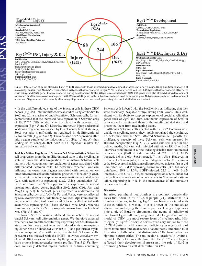
Fig. 3. Intersection of genes altered in Egr2Lo/Lo CHN nerve with those altered during development or after sciatic nerve injury. Using significance analysis ofmicroarrays analysis (see Methods), we identified 528 genes that were altered in Egr2Lo/Lo CHN sciatic nerves (red set), 1,353 genes that were altered after nervecrush injury, and 2,637 genes that were altered during development. Of the 528 genes associated with CHN, 438 genes were also altered during development(green set) or after nerve crush injury (yellow set). Whereas 244 genes in this subset were altered in all three paradigms, 106 genes were altered in developmentalone, and 88 genes were altered only after injury. Representative functional gene categories are included for each subset.
Le et al. PNAS � February 15, 2005 � vol. 102 � no. 7 � 2599
NEU
ROSC
IEN
CE
Curiously, however, a number of genes were altered in Egr2Lo/Lo
nerves and after nerve injury, but were not differentially regulatedduring development (Table 5, which is published as supporting
information on the PNAS web site). This expression pattern waspresent, even in the absence of the inflammatory processes thatnormally occur after nerve injury, perhaps reflecting a generalstress-related response in Egr2Lo/Lo Schwann cells that results froma failure to properly enter the differentiation pathway. Significance
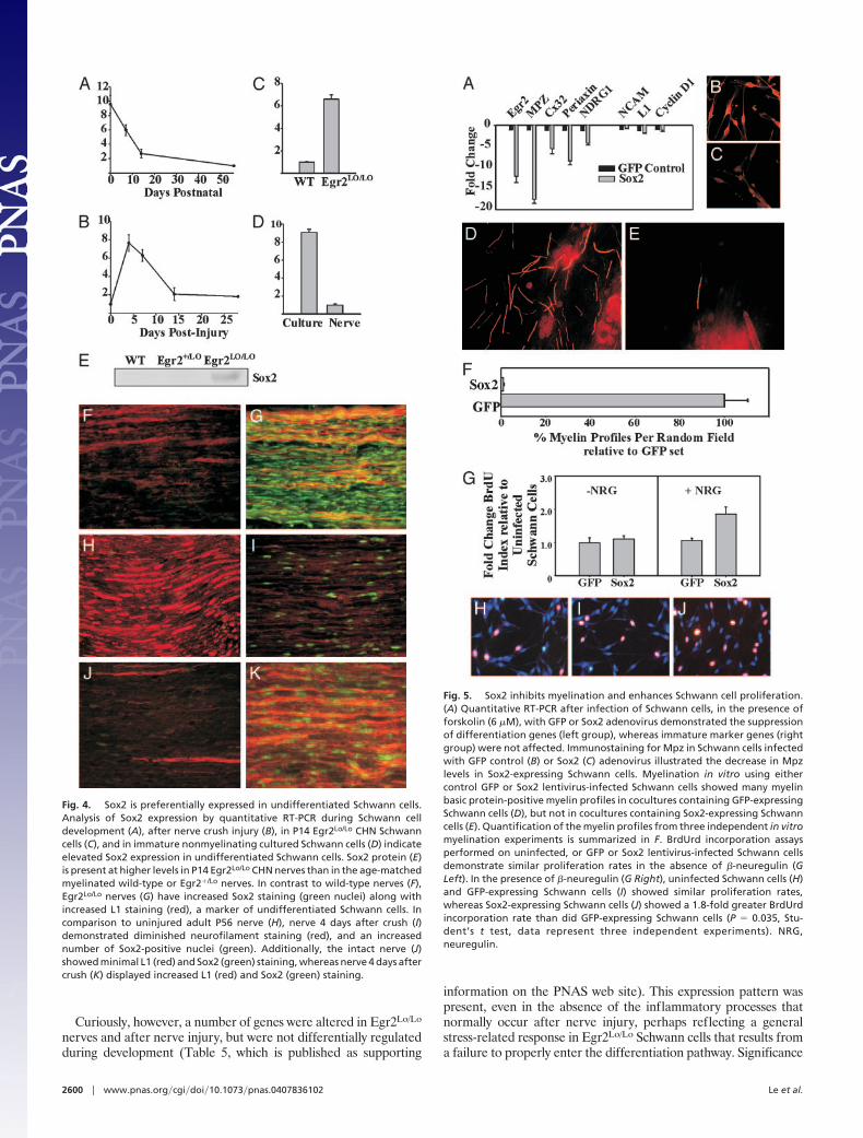
Fig. 4. Sox2 is preferentially expressed in undifferentiated Schwann cells.Analysis of Sox2 expression by quantitative RT-PCR during Schwann celldevelopment (A), after nerve crush injury (B), in P14 Egr2Lo/Lo CHN Schwanncells (C), and in immature nonmyelinating cultured Schwann cells (D) indicateelevated Sox2 expression in undifferentiated Schwann cells. Sox2 protein (E)is present at higher levels in P14 Egr2Lo/Lo CHN nerves than in the age-matchedmyelinated wild-type or Egr2�/Lo nerves. In contrast to wild-type nerves (F),Egr2Lo/Lo nerves (G) have increased Sox2 staining (green nuclei) along withincreased L1 staining (red), a marker of undifferentiated Schwann cells. Incomparison to uninjured adult P56 nerve (H), nerve 4 days after crush (I)demonstrated diminished neurofilament staining (red), and an increasednumber of Sox2-positive nuclei (green). Additionally, the intact nerve (J)showed minimal L1 (red) and Sox2 (green) staining, whereas nerve 4 days aftercrush (K) displayed increased L1 (red) and Sox2 (green) staining.
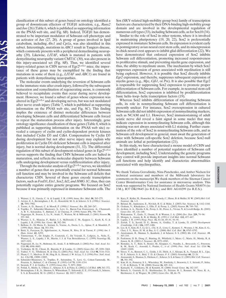
Fig. 5. Sox2 inhibits myelination and enhances Schwann cell proliferation.(A) Quantitative RT-PCR after infection of Schwann cells, in the presence offorskolin (6 �M), with GFP or Sox2 adenovirus demonstrated the suppressionof differentiation genes (left group), whereas immature marker genes (rightgroup) were not affected. Immunostaining for Mpz in Schwann cells infectedwith GFP control (B) or Sox2 (C) adenovirus illustrated the decrease in Mpzlevels in Sox2-expressing Schwann cells. Myelination in vitro using eithercontrol GFP or Sox2 lentivirus-infected Schwann cells showed many myelinbasic protein-positive myelin profiles in cocultures containing GFP-expressingSchwann cells (D), but not in cocultures containing Sox2-expressing Schwanncells (E). Quantification of the myelin profiles from three independent in vitromyelination experiments is summarized in F. BrdUrd incorporation assaysperformed on uninfected, or GFP or Sox2 lentivirus-infected Schwann cellsdemonstrate similar proliferation rates in the absence of �-neuregulin (GLeft). In the presence of �-neuregulin (G Right), uninfected Schwann cells (H)and GFP-expressing Schwann cells (I) showed similar proliferation rates,whereas Sox2-expressing Schwann cells (J) showed a 1.8-fold greater BrdUrdincorporation rate than did GFP-expressing Schwann cells (P � 0.035, Stu-dent’s t test, data represent three independent experiments). NRG,neuregulin.
2600 � www.pnas.org�cgi�doi�10.1073�pnas.0407836102 Le et al.
classification of this subset of genes based on ontology identified agroup of downstream effectors of TGF�1 activation, e.g., Runx2and Jun (26) (Table 6, which is published as supporting informationon the PNAS web site, and Fig. 8B). Indeed, TGF�1 has demon-strated to be important modulator of Schwann cell phenotype andan activator of Jun (27, 28). A group of genes involved in lipidcatabolism, including Abc1 and Cpt1a, was also identified in thissubset. Interestingly, mutations in ABC1 result in Tangiers disease,which commonly presents with a peripheral demyelinating neurop-athy (29). Likewise, LITAF, which is mutated in patients withdemyelinating neuropathy variant CMT1C (30), was also present inthis injury-associated set (Fig. 8B). Thus, we identified severalinjury-related genes in CHN nerves of Egr2Lo/Lo mice; the impor-tance of these genes may be exemplified by the finding thatmutations in some of them (e.g., LITAF and ABC1) are found inpatients with demyelinating neuropathies.
The molecular events underlying the reversion of Schwann cellsto the immature state after crush injury, followed by the subsequentmaturation and remyelination of regenerating axons, is commonlybelieved to recapitulate events that occur during nerve develop-ment. However, we found a subset of genes whose expression wasaltered in Egr2Lo/Lo and developing nerves, but was not modulatedafter nerve crush injury (Table 7, which is published as supportinginformation on the PNAS web site, and Fig. 8C). This findingimplies the existence of distinct molecular differences betweendeveloping Schwann cells and differentiated Schwann cells forcedto repeat the maturation process after injury. Interestingly, geneontology significance classification of these genes (Table 8, which ispublished as supporting information on the PNAS web site) re-vealed a category of cyclin and cyclin-dependent protein kinasesthat included Cyclin D3 and Cdk4. Compensation by Cyclin D3during development but not after nerve injury may explain whyproliferation in Cyclin D1-deficient Schwann cells is impaired afterinjury, but is normal during development (31, 32). The differentialregulation of this subset of development-related genes in Egr2Lo/Lo
nerves supports the finding that CHN Schwann cells are halted inmaturation, and reflects the molecular disparity between Schwanncells undergoing development versus redifferentiation after injury.
Through the molecular analysis of Egr2Lo/Lo nerves, we identifieda number of genes that are potentially crucial for normal Schwanncell function and may be involved in the Schwann cell deficits thatcharacterize CHN. Several of these genes encode transcriptionfactors, such as FoxD3, Ets1, Sox2, Id2, and HMG 1C; thus, they canpotentially regulate entire genetic programs. We focused on Sox2because it was primarily expressed in immature Schwann cells. The
Sox (SRY-related high-mobility group box) family of transcriptionfactors are characterized by their DNA-binding high-mobility groupdomain and are involved in the developmental regulation ofnumerous cell types (33), including Schwann cells, as for Sox10 (34).
Similar to the role of Sox2 in other systems, where it is involvedin maintaining pluripotency (19, 20, 22), Sox2 is preferentiallyexpressed in immature Schwann cells. Its expression is evident earlyin postmigratory avian neural crest stem cells, and its misexpressionin chick neural crest appears to inhibit glial differentiation (22). Wehave demonstrated that enforced expression of Sox2 inhibitsSchwann cell differentiation, promoting increased responsivenessto proliferative stimuli, and preventing myelin gene expression, andthus, the ability to myelinate axons. The mechanism by which Sox2suppresses expression of genes associated with myelination is stillbeing explored. However, it is possible that Sox2 directly inhibitsEgr2 expression, and thereby, suppresses subsequent expression ofmyelin genes (e.g., Mpz, Gjb1, or Prx). It is also possible that Egr2is responsible for suppressing Sox2 expression to promote properdifferentiation of Schwann cells. For example, in neuronal stem celldifferentiation, Sox2 expression is inhibited by prodifferentiationbasic helix–loop–helix transcription factors (19).
Whereas Sox2 inhibits differentiation of myelinating Schwanncells, its role in nonmyelinating Schwann cell differentiation ispresently unclear. For instance, Sox2 overexpression in culturedSchwann cells did not inhibit expression of nonmyelinating markers,such as NCAM and L1. However, Sox2 immunostaining of adultsciatic nerve did reveal a faint signal in some nuclei that mayindicate expression in nonmyelinating Schwann cells, although thisstaining was not always associated with L1 staining. Further exam-ination of the role of Sox2 in nonmyelinating Schwann cells, and inSchwann cell development in general, must await the generation ofmice with Schwann cell-specific Sox2 deletion, because Sox2-nullanimals are lethal at periimplantation (21).
In this study, we have characterized a mouse model of CHN andhave identified a number of potential regulators of Schwann celldevelopment, including Sox2. Understanding the genetic programsthey control will provide important insights into normal Schwanncell functions and help identify and characterize abnormalitiesassociated with myelinopathies.
We thank Tatiana Gorodinsky, Nina Panchenko, and Amber Nielson fortechnical assistance and members of the Milbrandt laboratory forcomments on the manuscript.We thank members of the Siteman CancerCenter Bioinformatics Core for assistance in microarray analysis. Thiswork was supported by National Institutes of Health Grants NS4074 (toJ.M.), R37 DK19645 (to R.E.S.), and R01 AG10299 (to R.E.S.).
1. Warner, L. E., Garcia, C. A. & Lupski, J. R. (1999) Annu. Rev. Med. 50, 263–275.2. Arroyo, E. J., Bermingham, J. R., Jr., Rosenfeld, M. G. & Scherer, S. S. (1998) J. Neurosci.
18, 7891–7902.3. Toews, A. D., Barrett, C. & Morell, P. (1998) J. Neurosci. Res. 53, 260–267.4. Topilko, P., Schneider-Maunoury, S., Levi, G., Baron-Van Evercooren, A., Chennoufi,
A. B. Y., Seitanidou, T., Babinet, C. & Charnay, P. (1994) Nature 371, 796–799.5. Nagarajan, R., Svaren, J., Le, N., Araki, T., Watson, M. & Milbrandt, J. (2001) Neuron 30,
355–368.6. Warner, L. E., Mancias, P., Butler, I. J., McDonald, C. M., Keppen, L., Koob, K. G. &
Lupski, J. R. (1998) Nat. Genet. 18, 382–384.7. Bellone, E., Di Maria, E., Soriani, S., Varese, A., Doria, L. L., Ajmar, F. & Mandich, P.
(1999) Hum. Mutat. 14, 353–354.8. Botti, S., Pareyson, D., Sghirlanzoni, A., Nemni, R., Riva, D. & Taroni, F. (1998) Am. J.
Hum. Genet. 63, A352.9. Timmerman, V., De Jonghe, P., Ceuterick, C., De Vriendt, E., Lofgren, A., Nelis, E.,
Warner, L. E., Lupski, J. R., Martin, J. J. & Van Broeckhoven, C. (1999) Neurology 52,1827–1832.
10. Nagarajan, R., Le, N., Mahoney, H., Araki, T. & Milbrandt, J. (2002) Proc. Natl. Acad. Sci.USA 99, 8998–9003.
11. Verheijen, M. H., Chrast, R., Burrola, P. & Lemke, G. (2003) Genes Dev. 17, 2450–2464.12. Tusher, V. G., Tibshirani, R. & Chu, G. (2001) Proc. Natl. Acad. Sci. USA 98, 5116–5121.13. Pham, C. T., MacIvor, D. M., Hug, B. A., Heusel, J. W. & Ley, T. J. (1996) Proc. Natl. Acad.
Sci. USA 93, 13090–13095.14. Schneider-Maunoury, S., Topilko, P., Seitanidou, T., Levi, G., Cohen-Tannoudji, M.,
Pournin, S., Babinet, C. & Charnay, P. (1993) Cell 75, 1199–1214.15. Swiatek, P. J. & Gridley, T. (1993) Genes Dev. 7, 2071–2084.16. Poliak, S., Matlis, S., Ullmer, C., Scherer, S. S. & Peles, E. (2002) J. Cell Biol. 159, 361–372.17. Bermingham, J. R., Jr., Shumas, S., Whisenhunt, T., Sirkowski, E. E., O’Connell, S., Scherer,
S. S. & Rosenfeld, M. G. (2002) J. Neurosci. 22, 10217–10231.
18. Kury, P., Koller, H., Hamacher, M., Cornely, C., Hasse, B. & Muller, H. W. (2003) Mol. Cell.Neurosci. 24, 1–9.
19. Bylund, M., Andersson, E., Novitch, B. G. & Muhr, J. (2003) Nat. Neurosci. 6, 1162–1168.20. Graham, V., Khudyakov, J., Ellis, P. & Pevny, L. (2003) Neuron 39, 749–765.21. Avilion, A. A., Nicolis, S. K., Pevny, L. H., Perez, L., Vivian, N. & Lovell-Badge, R. (2003)
Genes Dev. 17, 126–140.22. Wakamatsu, Y., Endo, Y., Osumi, N. & Weston, J. A. (2004) Dev. Dyn. 229, 74–86.23. Morgan, L., Jessen, K. R. & Mirsky, R. (1991) J. Cell Biol. 112, 457–467.24. Lupski, J. R. (1997) Hosp. Pract. 32, 83–84, 89–91, 94–95.25. Zorick, T. S., Syroid, D. E., Brown, A., Gridley, T. & Lemke, G. (1999) Development
(Cambridge, U.K.) 126, 1397–1406.26. Lee, K. S., Kim, H. J., Li, Q. L., Chi, X. Z., Ueta, C., Komori, T., Wozney, J. M., Kim, E. G.,
Choi, J. Y., Ryoo, H. M. & Bae, S. C. (2000) Mol. Cell. Biol. 20, 8783–8792.27. Awatramani, R., Shumas, S., Kamholz, J. & Scherer, S. S. (2002) Mo. Cell. Neurosci. 19,
307–319.28. Parkinson, D. B., Dong, Z., Bunting, H., Whitfield, J., Meier, C., Marie, H., Mirsky, R. &
Jessen, K. R. (2001) J. Neurosci. 21, 8572–8585.29. Remaley, A. T., Rust, S., Rosier, M., Knapper, C., Naudin, L., Broccardo, C., Peterson,
K. M., Koch, C., Arnould, I., Prades, C., et al. (1999) Proc. Natl. Acad. Sci. USA 96,12685–12690.
30. Street, V. A., Bennett, C. L., Goldy, J. D., Shirk, A. J., Kleopa, K. A., Tempel, B. L., Lipe,H. P., Scherer, S. S., Bird, T. D. & Chance, P. F. (2003) Neurology 60, 22–26.
31. Atanasoski, S., Shumas, S., Dickson, C., Scherer, S. S. & Suter, U. (2001) Mol. Cell. Neurosci.18, 581–592.
32. Kim, H. A., Pomeroy, S. L., Whoriskey, W., Pawlitzky, I., Benowitz, L. I., Sicinski, P., Stiles,C. D. & Roberts, T. M. (2000) Neuron 26, 405–416.
33. Kamachi, Y., Uchikawa, M. & Kondoh, H. (2000) Trends Genet. 16, 182–187.34. Britsch, S., Goerich, D. E., Riethmacher, D., Peirano, R. I., Rossner, M., Nave, K. A.,
Birchmeier, C. & Wegner, M. (2001) Genes Dev. 15, 66–78.
Le et al. PNAS � February 15, 2005 � vol. 102 � no. 7 � 2601
NEU
ROSC
IEN
CE
Related Documents