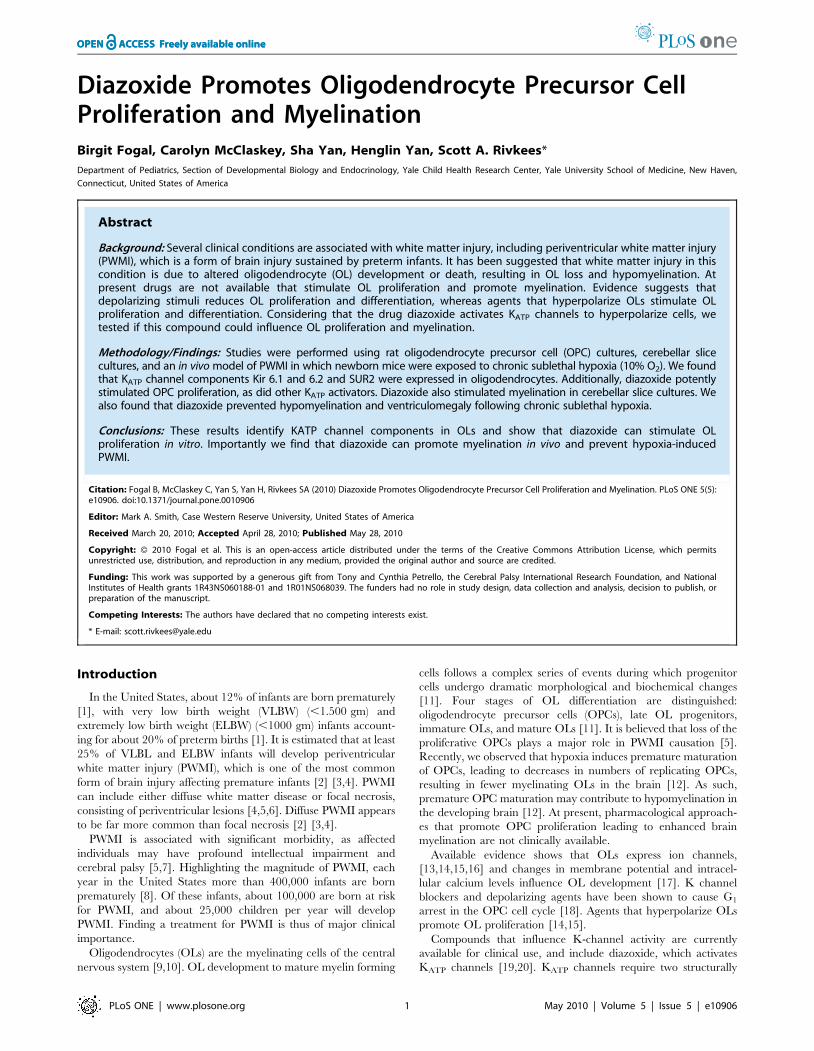
Diazoxide Promotes Oligodendrocyte Precursor Cell Proliferation and Myelination Birgit Fogal, Carolyn McClaskey, Sha Yan, Henglin Yan, Scott A. Rivkees* Department of Pediatrics, Section of Developmental Biology and Endocrinology, Yale Child Health Research Center, Yale University School of Medicine, New Haven, Connecticut, United States of America Abstract Background: Several clinical conditions are associated with white matter injury, including periventricular white matter injury (PWMI), which is a form of brain injury sustained by preterm infants. It has been suggested that white matter injury in this condition is due to altered oligodendrocyte (OL) development or death, resulting in OL loss and hypomyelination. At present drugs are not available that stimulate OL proliferation and promote myelination. Evidence suggests that depolarizing stimuli reduces OL proliferation and differentiation, whereas agents that hyperpolarize OLs stimulate OL proliferation and differentiation. Considering that the drug diazoxide activates K ATP channels to hyperpolarize cells, we tested if this compound could influence OL proliferation and myelination. Methodology/Findings: Studies were performed using rat oligodendrocyte precursor cell (OPC) cultures, cerebellar slice cultures, and an in vivo model of PWMI in which newborn mice were exposed to chronic sublethal hypoxia (10% O 2 ). We found that K ATP channel components Kir 6.1 and 6.2 and SUR2 were expressed in oligodendrocytes. Additionally, diazoxide potently stimulated OPC proliferation, as did other K ATP activators. Diazoxide also stimulated myelination in cerebellar slice cultures. We also found that diazoxide prevented hypomyelination and ventriculomegaly following chronic sublethal hypoxia. Conclusions: These results identify KATP channel components in OLs and show that diazoxide can stimulate OL proliferation in vitro. Importantly we find that diazoxide can promote myelination in vivo and prevent hypoxia-induced PWMI. Citation: Fogal B, McClaskey C, Yan S, Yan H, Rivkees SA (2010) Diazoxide Promotes Oligodendrocyte Precursor Cell Proliferation and Myelination. PLoS ONE 5(5): e10906. doi:10.1371/journal.pone.0010906 Editor: Mark A. Smith, Case Western Reserve University, United States of America Received March 20, 2010; Accepted April 28, 2010; Published May 28, 2010 Copyright: ß 2010 Fogal et al. This is an open-access article distributed under the terms of the Creative Commons Attribution License, which permits unrestricted use, distribution, and reproduction in any medium, provided the original author and source are credited. Funding: This work was supported by a generous gift from Tony and Cynthia Petrello, the Cerebral Palsy International Research Foundation, and National Institutes of Health grants 1R43NS060188-01 and 1R01NS068039. The funders had no role in study design, data collection and analysis, decision to publish, or preparation of the manuscript. Competing Interests: The authors have declared that no competing interests exist. * E-mail: [email protected] Introduction In the United States, about 12% of infants are born prematurely [1], with very low birth weight (VLBW) (,1.500 gm) and extremely low birth weight (ELBW) (,1000 gm) infants account- ing for about 20% of preterm births [1]. It is estimated that at least 25% of VLBL and ELBW infants will develop periventricular white matter injury (PWMI), which is one of the most common form of brain injury affecting premature infants [2] [3,4]. PWMI can include either diffuse white matter disease or focal necrosis, consisting of periventricular lesions [4,5,6]. Diffuse PWMI appears to be far more common than focal necrosis [2] [3,4]. PWMI is associated with significant morbidity, as affected individuals may have profound intellectual impairment and cerebral palsy [5,7]. Highlighting the magnitude of PWMI, each year in the United States more than 400,000 infants are born prematurely [8]. Of these infants, about 100,000 are born at risk for PWMI, and about 25,000 children per year will develop PWMI. Finding a treatment for PWMI is thus of major clinical importance. Oligodendrocytes (OLs) are the myelinating cells of the central nervous system [9,10]. OL development to mature myelin forming cells follows a complex series of events during which progenitor cells undergo dramatic morphological and biochemical changes [11]. Four stages of OL differentiation are distinguished: oligodendrocyte precursor cells (OPCs), late OL progenitors, immature OLs, and mature OLs [11]. It is believed that loss of the proliferative OPCs plays a major role in PWMI causation [5]. Recently, we observed that hypoxia induces premature maturation of OPCs, leading to decreases in numbers of replicating OPCs, resulting in fewer myelinating OLs in the brain [12]. As such, premature OPC maturation may contribute to hypomyelination in the developing brain [12]. At present, pharmacological approach- es that promote OPC proliferation leading to enhanced brain myelination are not clinically available. Available evidence shows that OLs express ion channels, [13,14,15,16] and changes in membrane potential and intracel- lular calcium levels influence OL development [17]. K channel blockers and depolarizing agents have been shown to cause G 1 arrest in the OPC cell cycle [18]. Agents that hyperpolarize OLs promote OL proliferation [14,15]. Compounds that influence K-channel activity are currently available for clinical use, and include diazoxide, which activates K ATP channels [19,20]. K ATP channels require two structurally PLoS ONE | www.plosone.org 1 May 2010 | Volume 5 | Issue 5 | e10906

Welcome message from author
This document is posted to help you gain knowledge. Please leave a comment to let me know what you think about it! Share it to your friends and learn new things together.
Transcript
Diazoxide Promotes Oligodendrocyte Precursor CellProliferation and MyelinationBirgit Fogal, Carolyn McClaskey, Sha Yan, Henglin Yan, Scott A. Rivkees*
Department of Pediatrics, Section of Developmental Biology and Endocrinology, Yale Child Health Research Center, Yale University School of Medicine, New Haven,
Connecticut, United States of America
Abstract
Background: Several clinical conditions are associated with white matter injury, including periventricular white matter injury(PWMI), which is a form of brain injury sustained by preterm infants. It has been suggested that white matter injury in thiscondition is due to altered oligodendrocyte (OL) development or death, resulting in OL loss and hypomyelination. Atpresent drugs are not available that stimulate OL proliferation and promote myelination. Evidence suggests thatdepolarizing stimuli reduces OL proliferation and differentiation, whereas agents that hyperpolarize OLs stimulate OLproliferation and differentiation. Considering that the drug diazoxide activates KATP channels to hyperpolarize cells, wetested if this compound could influence OL proliferation and myelination.
Methodology/Findings: Studies were performed using rat oligodendrocyte precursor cell (OPC) cultures, cerebellar slicecultures, and an in vivo model of PWMI in which newborn mice were exposed to chronic sublethal hypoxia (10% O2). We foundthat KATP channel components Kir 6.1 and 6.2 and SUR2 were expressed in oligodendrocytes. Additionally, diazoxide potentlystimulated OPC proliferation, as did other KATP activators. Diazoxide also stimulated myelination in cerebellar slice cultures. Wealso found that diazoxide prevented hypomyelination and ventriculomegaly following chronic sublethal hypoxia.
Conclusions: These results identify KATP channel components in OLs and show that diazoxide can stimulate OLproliferation in vitro. Importantly we find that diazoxide can promote myelination in vivo and prevent hypoxia-inducedPWMI.
Citation: Fogal B, McClaskey C, Yan S, Yan H, Rivkees SA (2010) Diazoxide Promotes Oligodendrocyte Precursor Cell Proliferation and Myelination. PLoS ONE 5(5):e10906. doi:10.1371/journal.pone.0010906
Editor: Mark A. Smith, Case Western Reserve University, United States of America
Received March 20, 2010; Accepted April 28, 2010; Published May 28, 2010
Copyright: � 2010 Fogal et al. This is an open-access article distributed under the terms of the Creative Commons Attribution License, which permitsunrestricted use, distribution, and reproduction in any medium, provided the original author and source are credited.
Funding: This work was supported by a generous gift from Tony and Cynthia Petrello, the Cerebral Palsy International Research Foundation, and NationalInstitutes of Health grants 1R43NS060188-01 and 1R01NS068039. The funders had no role in study design, data collection and analysis, decision to publish, orpreparation of the manuscript.
Competing Interests: The authors have declared that no competing interests exist.
* E-mail: [email protected]
Introduction
In the United States, about 12% of infants are born prematurely
[1], with very low birth weight (VLBW) (,1.500 gm) and
extremely low birth weight (ELBW) (,1000 gm) infants account-
ing for about 20% of preterm births [1]. It is estimated that at least
25% of VLBL and ELBW infants will develop periventricular
white matter injury (PWMI), which is one of the most common
form of brain injury affecting premature infants [2] [3,4]. PWMI
can include either diffuse white matter disease or focal necrosis,
consisting of periventricular lesions [4,5,6]. Diffuse PWMI appears
to be far more common than focal necrosis [2] [3,4].
PWMI is associated with significant morbidity, as affected
individuals may have profound intellectual impairment and
cerebral palsy [5,7]. Highlighting the magnitude of PWMI, each
year in the United States more than 400,000 infants are born
prematurely [8]. Of these infants, about 100,000 are born at risk
for PWMI, and about 25,000 children per year will develop
PWMI. Finding a treatment for PWMI is thus of major clinical
importance.
Oligodendrocytes (OLs) are the myelinating cells of the central
nervous system [9,10]. OL development to mature myelin forming
cells follows a complex series of events during which progenitor
cells undergo dramatic morphological and biochemical changes
[11]. Four stages of OL differentiation are distinguished:
oligodendrocyte precursor cells (OPCs), late OL progenitors,
immature OLs, and mature OLs [11]. It is believed that loss of the
proliferative OPCs plays a major role in PWMI causation [5].
Recently, we observed that hypoxia induces premature maturation
of OPCs, leading to decreases in numbers of replicating OPCs,
resulting in fewer myelinating OLs in the brain [12]. As such,
premature OPC maturation may contribute to hypomyelination in
the developing brain [12]. At present, pharmacological approach-
es that promote OPC proliferation leading to enhanced brain
myelination are not clinically available.
Available evidence shows that OLs express ion channels,
[13,14,15,16] and changes in membrane potential and intracel-
lular calcium levels influence OL development [17]. K channel
blockers and depolarizing agents have been shown to cause G1
arrest in the OPC cell cycle [18]. Agents that hyperpolarize OLs
promote OL proliferation [14,15].
Compounds that influence K-channel activity are currently
available for clinical use, and include diazoxide, which activates
KATP channels [19,20]. KATP channels require two structurally
PLoS ONE | www.plosone.org 1 May 2010 | Volume 5 | Issue 5 | e10906
diverse subunits [19,20]. One subunit is a member of the pore-
forming inward rectifier Kir6.x family of potassium channels,
while the other subunit is a sulfonylurea receptor (SUR) [19,20],
that belongs to the ATP-binding cassette superfamily.
We are unaware of studies that have examined the expression of
Kir6 or SUR proteins in OLs. It is also not known if KATP channel
antagonists or agonists effect OL development or function. We
postulated that alteration in KATP channel activity could have
potential utility as a therapeutic agent in white mater injury. To
test this hypothesis we studied cultured OLs and validated models
of PWMI [21].
Materials and Methods
AnimalsThis study was conducted in accordance with USDA guidelines
for the use of experimental animals and was approved by the
Institutional Animal Care and Use Committee (IACUC) of Yale
University School of Medicine. CD1 and C57BL/6 mice and
Sprague Dawley rats were obtained from Charles River
Laboratories (Wilmington, MA). The Yale IACUC approval
number for this study was #2008-11198. The Yale University
Public Health Services approval number is A3230-01.
Cell culturePurified OPC cultures were prepared as described [22,23]. In
brief, primary rat mixed glial cell cultures were isolated from
whole brains of postnatal day (P) 1 rats, dissociated into single cells,
and cultured into poly-D-lysine (PDL, Sigma-Aldrich, St. Louis,
MO) coated T75 tissue culture flasks. Plating medium consisted of
Dulbecco’s modified Eagle’s medium (DMEM, Invitrogen,
Carlsbad, CA) supplemented with 10% fetal bovine serum (FBS;
InVitrogen, Carlsbad, CA), 2 mM L-glutamine, 100 mM strepto-
mycin, and 10 mg/ml penicillin. Tissue cultures were maintained
at 37uC in a humidified 5% CO2 incubator, and medium was
exchanged every 3 days. Once confluent (after 7–9 days),
microglia were separated by mechanical shaking of flasks on a
rotary shaker for 60 min at 200 rpm and removed. After addition
of fresh medium, the remaining cells were allowed to recover
overnight before repeating the mechanical shaking for an
additional 16 h at 200 rpm to isolate OPCs. To ensure purity of
OPC cultures, the isolated cells were transferred to a tissue culture
dish, from which the loosely attached OPCs were detached by
gentle shaking after 60 min, leaving behind attached microglia
and astrocytes. OPCs were plated onto PDL coated 96 well plates
using an automated dispenser and allowed to adhere to the plates
over the next 1–2 days. In agreement with others [24], this
procedure yields 98% A2B5-positive (OPC marker), and 2%
MBP-positive (mature OL marker) cells. GFAP-positive (astrocyte
marker) or Ox2A-positive (microglia marker) cells cannot be
detected in cultures prepared in this manner.
Drug exposureCells were exposed to KATP channel stimulators for 72–96 h.
The compounds included commercially available KATP channel
activators ZM26600 (2.5 uM), Pinacidil (10 uM), Y26763
(200 nM), Levcromakalim (2.5 uM), P1075 (100 nM) and diaz-
oxide. Cultures were washed two times with DMEM to remove
any residual serum and then the respective solutions of KATP
channel activators were added daily in an incubation buffer of
DMEM with 0.5% FBS +10 mg/ml biotin and N2 supplement.
Cells were then placed at 37uC in a humidified atmosphere
containing 5% CO2.
Quantification of cell numberTo evaluate cell proliferation in response to KATP channel
stimulators, we utilized the CyQUANTH NF Cell Proliferation
assay (Invitrogen, Carlsbad, CA) according to the manufacturer’s
instructions. This assay measures cellular DNA content as a direct
index of cell proliferation. Since cellular DNA content is highly
proportional to cell number, this is a very accurate way to assess
cell proliferation specifically [25]. At the end of drug exposure,
medium was removed and a stock solution of the green-fluorescent
CyQUANT GR dye (prepared according to manufacturer’s
instructions) was added. Upon binding to DNA, the GR dye
shows a measurable enhancement in fluorescent intensity. Cells
were returned to the incubator (37uC) for 2 h, which we
determined to result in maximal and stable changes in fluores-
cence. Fluorescence was measured using an Envision Multilabel
reader (Perkin-Elmer; Excitation: 480 nm, Emission: 530 nm).
Calcium imagingCalcium imaging was performed by using an Olympus Fluoview
confocal laser scanning microscope (IX70, Melville, NY), an
UPlanFl 206objective (N.A. 0.5), and Fluoview image processing
software (v2.1). To assess intracellular calcium levels the calcium
ionophore Fluo-3 was used.
Cells were first evaluated before the addition of any drug. After
addition of drug, calcium levels were recorded. Quantification and
statistical analysis of quantified data was performed by using
Fluoview software, Microsoft Excel 2000 (Microsoft, Inc., Red-
mond, WA), and GraphPad Prism (v3.0, GraphPad Software, Inc.,
San Diego, CA), as described [26].
Cerebellar Slice CulturesCerebellar organotypic cultures of mice (postnatal 0 day) were
prepared as described [27,28]. 300-mm thick slices were trans-
ferred onto membranes of 30 mm Millipore culture inserts with a
0.4 mm pore size, and maintained in six-well tissue culture plates
containing 1 ml of medium at 35uC in room air with 5% CO2.
The nutrient medium consisted of 50% basal medium with Earle’s
salts, 25% horse serum, 25% Hank’s balance solution, 1 mM L-
glutamine, and 5 mg/ml glucose. After 24 h the medium was
removed and culture plates were washed twice with phosphate-
buffered saline before addition of tolbutamide, diazoxide, or
vehicle in N1 medium. After five days cultures were immuno-
stained for MBP as described below. Total numbers of MBP-
positive cells and myelinated fibers per mm2 area were determined
for each slice by manual counting by an individual who was
blinded to treatment conditions. Each treatment condition
contained six slices from three different animals.
Polymerase chain reactioncDNA was made from OPCs and mature OLs. DNA was
amplified from cells and used in PCR reactions with SUR or
KIR6 subtype-specific primers, as detailed [29].
Western blot analysisCells were washed in ice-cold phosphate buffered saline (PBS)
and lysed using hot lysis buffer containing 42 mM Tris-HCl
(pH 6.8), 1.3% sodium dodecylsulfate (SDS), 6.5% glycerol, and
0.1 mM sodium orthovanadate. Protein concentrations were
determined using the bicinchonic acid method (BCA kit, Pierce
Technologies, Rockford, IL). Before loading, samples were mixed
with 10 mM dithiothreitol and 0.1% bromophenol blue, and
boiled for 5 min. Proteins were separated on an SDS-polyacryl-
amide gel, transferred to a polyvinylidene fluoride (PVDF)
Diazoxide and Myelination
PLoS ONE | www.plosone.org 2 May 2010 | Volume 5 | Issue 5 | e10906
membrane, and blocked with 5% non-fat dry milk in 20 mM Tris-
HCl, pH 7.6, 150 mM sodium chloride and 0.1% tween-20 (TBS-
T) for 1 hr. Anti Kir6.1 and 6.2 and SUR1 and 2a antisera were
obtained from Santa Cruz Biotechnology (Carlsbad, CA).
Secondary antibodies were diluted in blocking buffer and
incubated for 1 h at RT. Horseradish peroxidase-conjugated goat
anti-rabbit antibody (1:5000 dilution) was used to detect labeled
product. After washing, proteins were detected using an enhanced
chemiluminescence kit (Pierce Biotech).
In vivo HypoxiaC57BL/6 mice were exposed to low or normal oxygen
conditions from P3-P12 as described [21,30]. In brief, litters of
pups (P3) were placed with the dam in a Plexiglas chamber, in
which oxygen levels were maintained at 9.561.0%., O2 levels
were continuously monitored using a Cameron Instrument
(Ontario, Canada) dual channel oxygen monitor attached to O2
electrodes placed at each end of the chamber. Control animals
were kept in room air (,22%) outside the Plexiglas chamber.
Because C57BL/6 dams do not normally care for pups in hypoxic
conditions, pups (hypoxia and control) were cross-fostered with
CD1 dams, which were placed in the same cage before birth of
pups. Animals were removed from the chamber daily for less than
15 min to allow for diazoxide (10 mg/kg i.p.) or vehicle
administration, as well as observation of weight gain. Mice were
euthanized at P12, brains were harvested and shock frozen in ice
cold (220uC) 2-methylbutane and stored at 280uC until
assessment of ventricular size or MBP immunocytochemistry (see
below). At least eight animals were studied in each treatment
group. There were no difference in mortality observed between
the vehicle and diazoxide treated pups. Mortality rates were less
than 12% for each treatment group (p.0.05).
Quantification of ventricular sizeVentricle size was determined as reported [30,31]. Animals
were weighed, anesthetized, and decapitated. Brains were shock
frozen in ice cold (220uC) 2-methylbutane and stored at 280uC.
Coronal sections spanning the brain were cut in a cryostat at a
thickness of 16 mm in a Zeiss cryostat. Sections were mounted onto
glass slides and stained with Phoenix Blue (Thermo Scientific,
Waltham, MA). Serial sections through the midstriatum were
photographed to include the lateral ventricle region. Ventricular
sizes were quantified using Sigma Scan Pro Image Analysis
Version 5.0.0 (SPSS Inc., Chicago, IL). The ventricular area,
outlined by the Phoenix Blue staining, was measured and
numerically integrated across the thickness of the slice. Images
were obtained using a Leica florescence microscope.
ImmunostainingTissue sections were fixed with 4% paraformaldehyde in PBS
for 30 min at room temperature and incubated with 10% normal
goat serum plus 0.3% Triton-X-100 in 0.1 M PBS (pH 7.4)
overnight to block nonspecific binding. The sections were
incubated with monoclonal MBP antibody (SMI-99, Covance,
Princeton, NJ) at a dilution of 1:1000 in PBS with 10% normal
goat serum plus 0.1% Triton-X-100 at 4uC overnight, followed by
incubation of Alexa Fluor 495 anti-mouse IgG (Molecular Probes)
at a dilution of 1:400. The intensity of labeling was assessed using
Image J Version 1.42q (National Institutes of Health, Bethesda
MD) at the mid-level of the corpus callosum from three slides per
animal.
For A2B5 and O1, immunocytochemistry was performed as
described [32]. Cultures were washed twice with L-15 medium.
Cells were then incubated with murine monoclonal antibodies, or
A2B5, O1 hybridoma culture supernatants for 20 min at 37uC.
Cells were incubated with Alexa Fluor 594- or Alexa Fluor 488-
conjugated antimouse secondary antibody (1:200) for 20 min at
37uC.
For Kir and SUR immunostaining, cells were fixed with 4%
paraformaldehyde and treated with 0.2% Triton X-100 and 10%
goat serum. Next, cells were incubated with monoclonal anti-
Kir6.1 and 6.2 and SUR1 and 2a (Santa Cruz Biotechnology;
Carlsbad, CA) (1:100-500) or mouse IgG (control) plus 10%
normal goat serum overnight at 4uC. After washing three times
with PBS, cells were incubated with goat anti-mouse IgG
conjugated to Alexa Fluor 594, or goat anti-rabbit IgG conjugated
to Alexa Fluor 488 for 90 min at room temperature. Cells were
examined by fluorescence microscopy.
Statistical AnalysisAll experiments were repeated at least three times and statistical
analyses were performed using Graph Pad Prism software.
Comparisons among multiple groups were made by ANOVA,
with Bonferonni post-test comparisons. Paired comparisons were
made by the Students-T test. In all experiments significance was
assessed at p,0.05.
Results
Expression of SUR and KIR6 in OLsWe postulated that OLs contain KATP channels that could be
pharmacological targets. To begin to address this issue, we
examined the expression KATP channel components SUR and
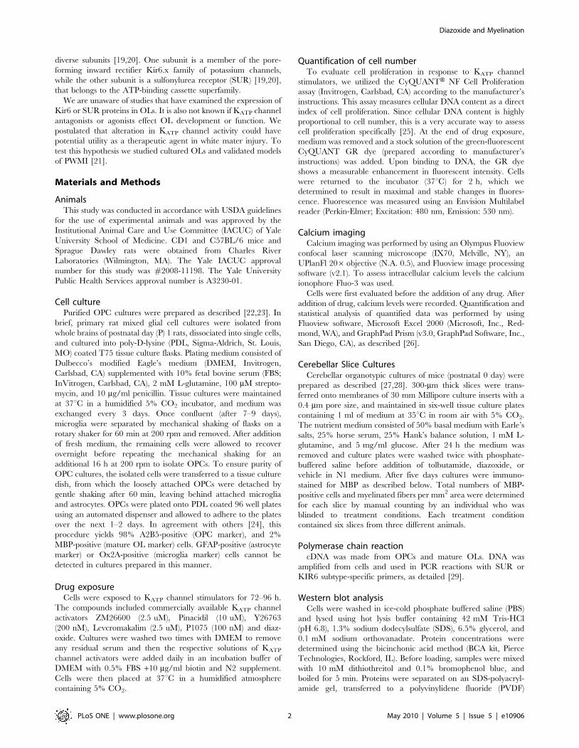
KIR6.1 and 6.2 in OLs. First, we assessed KIR6.1 and 6.2 and
SUR gene expression in OLs by PCR. cDNA was made from
OPCs and mature OLs. For OPC studies, 98% of cells were
A2B5-postive; for mature OL studies, 95% of cells were MBP-
positive.
DNA was amplified from cells and used in PCR reactions with
SUR or KIR6 subtype-specific primers, as reported [29]. KIR6.1
and KIR6.2, as well as SUR1 and SUR2 mRNA expression was
observed in both, OPCs (Fig. 1) and mature OLs (data not shown).
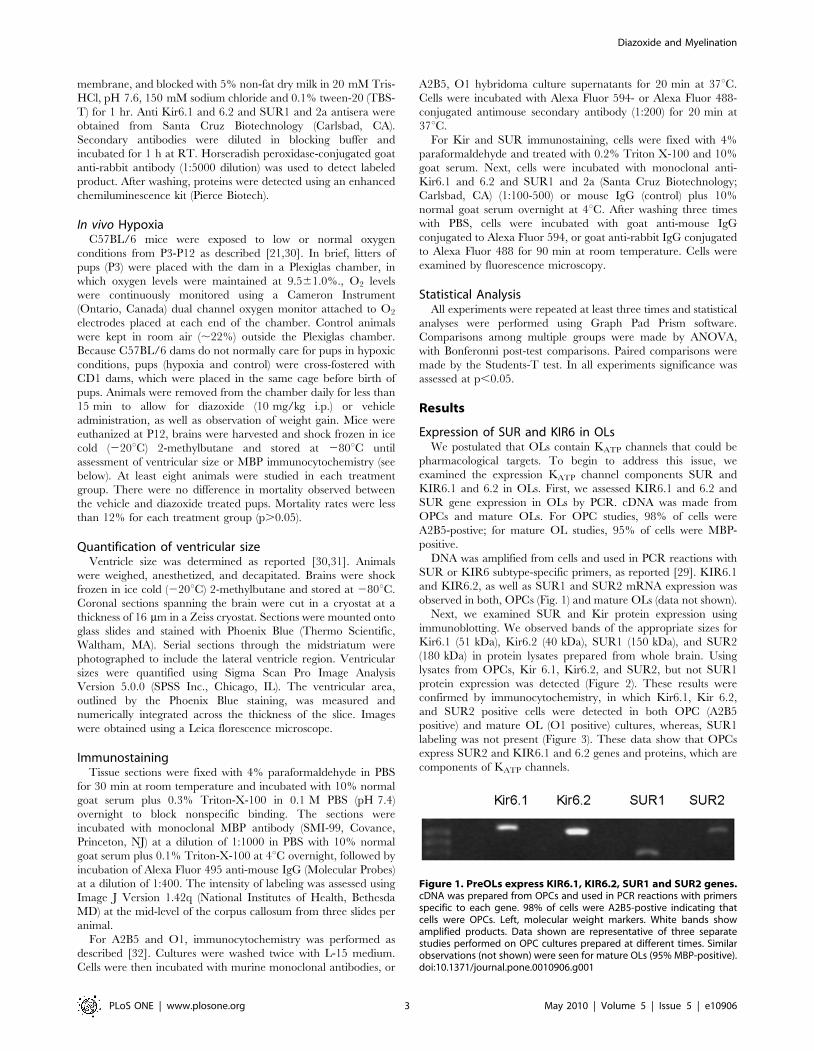
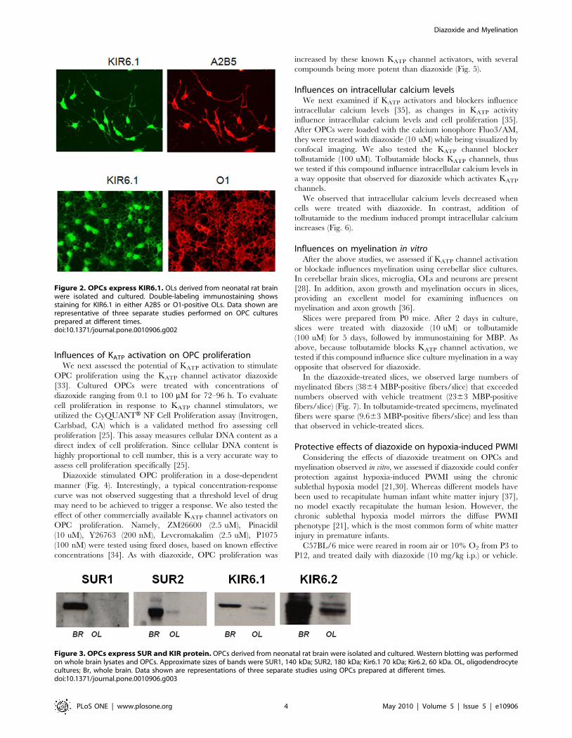
Next, we examined SUR and Kir protein expression using
immunoblotting. We observed bands of the appropriate sizes for
Kir6.1 (51 kDa), Kir6.2 (40 kDa), SUR1 (150 kDa), and SUR2
(180 kDa) in protein lysates prepared from whole brain. Using
lysates from OPCs, Kir 6.1, Kir6.2, and SUR2, but not SUR1
protein expression was detected (Figure 2). These results were
confirmed by immunocytochemistry, in which Kir6.1, Kir 6.2,
and SUR2 positive cells were detected in both OPC (A2B5
positive) and mature OL (O1 positive) cultures, whereas, SUR1
labeling was not present (Figure 3). These data show that OPCs
express SUR2 and KIR6.1 and 6.2 genes and proteins, which are
components of KATP channels.
Figure 1. PreOLs express KIR6.1, KIR6.2, SUR1 and SUR2 genes.cDNA was prepared from OPCs and used in PCR reactions with primersspecific to each gene. 98% of cells were A2B5-postive indicating thatcells were OPCs. Left, molecular weight markers. White bands showamplified products. Data shown are representative of three separatestudies performed on OPC cultures prepared at different times. Similarobservations (not shown) were seen for mature OLs (95% MBP-positive).doi:10.1371/journal.pone.0010906.g001
Diazoxide and Myelination
PLoS ONE | www.plosone.org 3 May 2010 | Volume 5 | Issue 5 | e10906
Influences of KATP activation on OPC proliferationWe next assessed the potential of KATP activation to stimulate
OPC proliferation using the KATP channel activator diazoxide
[33]. Cultured OPCs were treated with concentrations of
diazoxide ranging from 0.1 to 100 mM for 72–96 h. To evaluate
cell proliferation in response to KATP channel stimulators, we
utilized the CyQUANTH NF Cell Proliferation assay (Invitrogen,
Carlsbad, CA) which is a validated method fro assessing cell
proliferation [25]. This assay measures cellular DNA content as a
direct index of cell proliferation. Since cellular DNA content is
highly proportional to cell number, this is a very accurate way to
assess cell proliferation specifically [25].
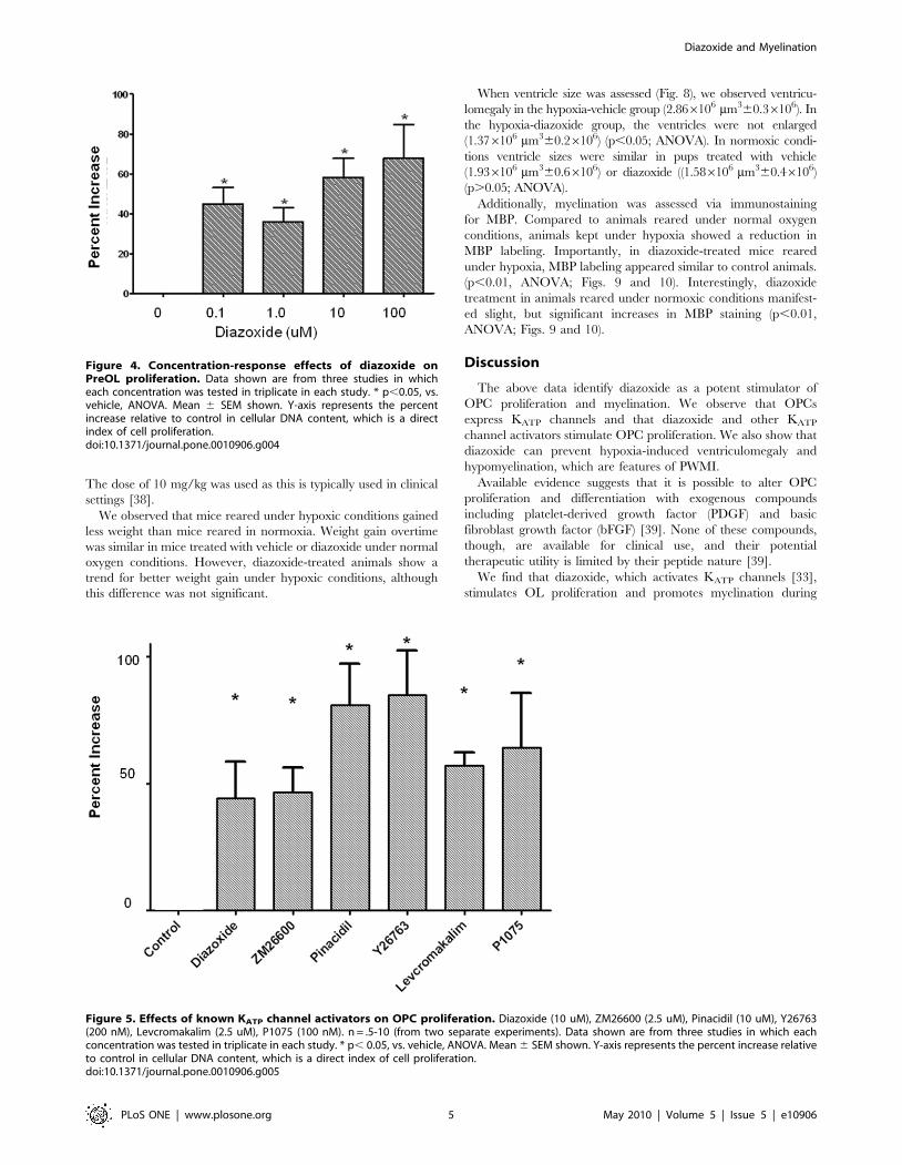
Diazoxide stimulated OPC proliferation in a dose-dependent
manner (Fig. 4). Interestingly, a typical concentration-response
curve was not observed suggesting that a threshold level of drug
may need to be achieved to trigger a response. We also tested the
effect of other commercially available KATP channel activators on
OPC proliferation. Namely, ZM26600 (2.5 uM), Pinacidil
(10 uM), Y26763 (200 nM), Levcromakalim (2.5 uM), P1075
(100 nM) were tested using fixed doses, based on known effective
concentrations [34]. As with diazoxide, OPC proliferation was
increased by these known KATP channel activators, with several
compounds being more potent than diazoxide (Fig. 5).
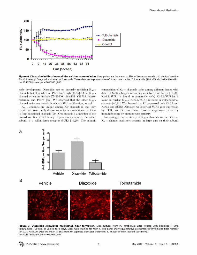
Influences on intracellular calcium levelsWe next examined if KATP activators and blockers influence
intracellular calcium levels [35], as changes in KATP activity
influence intracellular calcium levels and cell proliferation [35].
After OPCs were loaded with the calcium ionophore Fluo3/AM,
they were treated with diazoxide (10 uM) while being visualized by
confocal imaging. We also tested the KATP channel blocker
tolbutamide (100 uM). Tolbutamide blocks KATP channels, thus
we tested if this compound influence intracellular calcium levels in
a way opposite that observed for diazoxide which activates KATP
channels.
We observed that intracellular calcium levels decreased when
cells were treated with diazoxide. In contrast, addition of
tolbutamide to the medium induced prompt intracellular calcium
increases (Fig. 6).
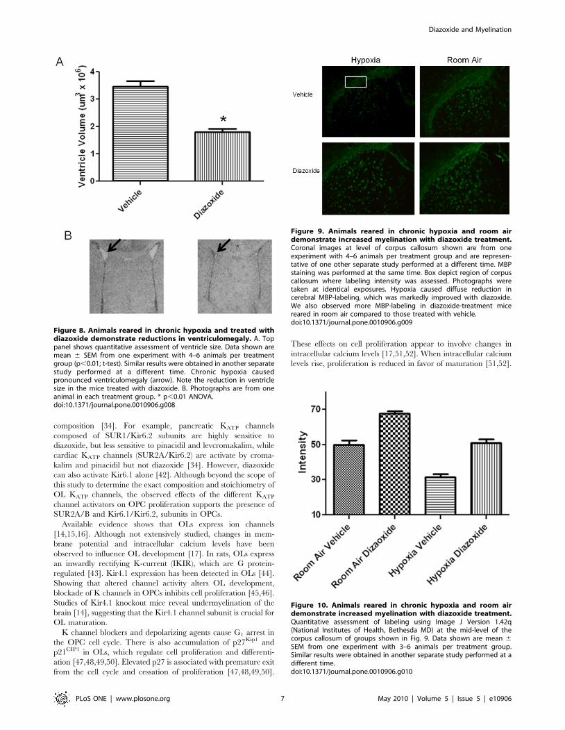
Influences on myelination in vitroAfter the above studies, we assessed if KATP channel activation
or blockade influences myelination using cerebellar slice cultures.
In cerebellar brain slices, microglia, OLs and neurons are present
[28]. In addition, axon growth and myelination occurs in slices,
providing an excellent model for examining influences on
myelination and axon growth [36].
Slices were prepared from P0 mice. After 2 days in culture,
slices were treated with diazoxide (10 uM) or tolbutamide
(100 uM) for 5 days, followed by immunostaining for MBP. As
above, because tolbutamide blocks KATP channel activation, we
tested if this compound influence slice culture myelination in a way
opposite that observed for diazoxide.
In the diazoxide-treated slices, we observed large numbers of
myelinated fibers (3864 MBP-positive fibers/slice) that exceeded
numbers observed with vehicle treatment (2363 MBP-positive
fibers/slice) (Fig. 7). In tolbutamide-treated specimens, myelinated
fibers were sparse (9.663 MBP-positive fibers/slice) and less than
that observed in vehicle-treated slices.
Protective effects of diazoxide on hypoxia-induced PWMIConsidering the effects of diazoxide treatment on OPCs and
myelination observed in vitro, we assessed if diazoxide could confer
protection against hypoxia-induced PWMI using the chronic
sublethal hypoxia model [21,30]. Whereas different models have
been used to recapitulate human infant white matter injury [37],
no model exactly recapitulate the human lesion. However, the
chronic sublethal hypoxia model mirrors the diffuse PWMI
phenotype [21], which is the most common form of white matter
injury in premature infants.
C57BL/6 mice were reared in room air or 10% O2 from P3 to
P12, and treated daily with diazoxide (10 mg/kg i.p.) or vehicle.
Figure 2. OPCs express KIR6.1. OLs derived from neonatal rat brainwere isolated and cultured. Double-labeling immunostaining showsstaining for KIR6.1 in either A2B5 or O1-positive OLs. Data shown arerepresentative of three separate studies performed on OPC culturesprepared at different times.doi:10.1371/journal.pone.0010906.g002
Figure 3. OPCs express SUR and KIR protein. OPCs derived from neonatal rat brain were isolated and cultured. Western blotting was performedon whole brain lysates and OPCs. Approximate sizes of bands were SUR1, 140 kDa; SUR2, 180 kDa; Kir6.1 70 kDa; Kir6.2, 60 kDa. OL, oligodendrocytecultures; Br, whole brain. Data shown are representations of three separate studies using OPCs prepared at different times.doi:10.1371/journal.pone.0010906.g003
Diazoxide and Myelination
PLoS ONE | www.plosone.org 4 May 2010 | Volume 5 | Issue 5 | e10906
The dose of 10 mg/kg was used as this is typically used in clinical
settings [38].
We observed that mice reared under hypoxic conditions gained
less weight than mice reared in normoxia. Weight gain overtime
was similar in mice treated with vehicle or diazoxide under normal
oxygen conditions. However, diazoxide-treated animals show a
trend for better weight gain under hypoxic conditions, although
this difference was not significant.
When ventricle size was assessed (Fig. 8), we observed ventricu-
lomegaly in the hypoxia-vehicle group (2.866106 mm360.36106). In
the hypoxia-diazoxide group, the ventricles were not enlarged
(1.376106 mm360.26106) (p,0.05; ANOVA). In normoxic condi-
tions ventricle sizes were similar in pups treated with vehicle
(1.936106 mm360.66106) or diazoxide ((1.586106 mm360.46106)
(p.0.05; ANOVA).
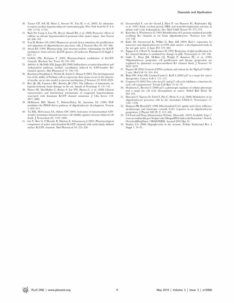
Additionally, myelination was assessed via immunostaining
for MBP. Compared to animals reared under normal oxygen
conditions, animals kept under hypoxia showed a reduction in
MBP labeling. Importantly, in diazoxide-treated mice reared
under hypoxia, MBP labeling appeared similar to control animals.
(p,0.01, ANOVA; Figs. 9 and 10). Interestingly, diazoxide
treatment in animals reared under normoxic conditions manifest-
ed slight, but significant increases in MBP staining (p,0.01,
ANOVA; Figs. 9 and 10).
Discussion
The above data identify diazoxide as a potent stimulator of
OPC proliferation and myelination. We observe that OPCs
express KATP channels and that diazoxide and other KATP
channel activators stimulate OPC proliferation. We also show that
diazoxide can prevent hypoxia-induced ventriculomegaly and
hypomyelination, which are features of PWMI.
Available evidence suggests that it is possible to alter OPC
proliferation and differentiation with exogenous compounds
including platelet-derived growth factor (PDGF) and basic
fibroblast growth factor (bFGF) [39]. None of these compounds,
though, are available for clinical use, and their potential
therapeutic utility is limited by their peptide nature [39].
We find that diazoxide, which activates KATP channels [33],
stimulates OL proliferation and promotes myelination during
Figure 5. Effects of known KATP channel activators on OPC proliferation. Diazoxide (10 uM), ZM26600 (2.5 uM), Pinacidil (10 uM), Y26763(200 nM), Levcromakalim (2.5 uM), P1075 (100 nM). n = .5-10 (from two separate experiments). Data shown are from three studies in which eachconcentration was tested in triplicate in each study. * p, 0.05, vs. vehicle, ANOVA. Mean 6 SEM shown. Y-axis represents the percent increase relativeto control in cellular DNA content, which is a direct index of cell proliferation.doi:10.1371/journal.pone.0010906.g005
Figure 4. Concentration-response effects of diazoxide onPreOL proliferation. Data shown are from three studies in whicheach concentration was tested in triplicate in each study. * p,0.05, vs.vehicle, ANOVA. Mean 6 SEM shown. Y-axis represents the percentincrease relative to control in cellular DNA content, which is a directindex of cell proliferation.doi:10.1371/journal.pone.0010906.g004
Diazoxide and Myelination
PLoS ONE | www.plosone.org 5 May 2010 | Volume 5 | Issue 5 | e10906
early development. Diazoxide acts on inwardly rectifying KATP
channels that close when ATP levels are high [33,35]. Other KATP
channel activators include ZM26600, pinacidil, Y26763, levcro-
makalim, and P1075 [33]. We observed that the other KATP
channel activators tested stimulated OPC proliferation, as well.
KATP channels are unique among Kir channels in that they
require two structurally diverse subunits in a stoichiometry of 4:4
to form functional channels [20]. One subunit is a member of the
inward rectifier Kir6.0 family of potassium channels; the other
subunit is a sulfonylurea receptor (SUR) [19,20]. The subunit
composition of KATP channels varies among different tissues, with
different SUR subtypes interacting with Kir6.1 or Kir6.2 [19,20].
Kir6.2/SUR1 is found in pancreatic cells; Kir6.2/SUR2A is
found in cardiac KATP. Kir6.1/SUR1 is found in mitochondrial
channels [40,41]. We observed that OL expressed both Kir6.1 and
Kir6.2 and SUR2. Although we observed SUR1 gene expression
by PCR, we did not detect protein expression either by
immunoblotting or immunocytochemistry.
Interestingly, the sensitivity of KATP channels to the different
KATP channel activators depends in large part on their subunit
Figure 7. Diazoxide stimulates myelinated fiber formation. Slice cultures from P0 cerebellum were treated with diazoxide (1 uM),tolbutamide (100 uM), or vehicle for 5 days. Slices were stained for MBP. A. Top panel shows quantitative assessment of myelinated fiber number(p,0.01; ANOVA). Data are mean 6 SEM from six separate slices per treatment. B. Images of MBP labeled specimens.doi:10.1371/journal.pone.0010906.g007
Figure 6. Diazoxide inhibits intracellular calcium accumulation. Data points are the mean 6 SEM of 30 separate cells. 100 depicts baselineFluo-3 intensity. Drugs administered at 0 seconds. These data are representative of 3 separate studies. Tolbutamide (100 uM), diazoxide (10 uM).doi:10.1371/journal.pone.0010906.g006
Diazoxide and Myelination
PLoS ONE | www.plosone.org 6 May 2010 | Volume 5 | Issue 5 | e10906
composition [34]. For example, pancreatic KATP channels
composed of SUR1/Kir6.2 subunits are highly sensitive to
diazoxide, but less sensitive to pinacidil and levcromakalim, while
cardiac KATP channels (SUR2A/Kir6.2) are activate by croma-
kalim and pinacidil but not diazoxide [34]. However, diazoxide
can also activate Kir6.1 alone [42]. Although beyond the scope of
this study to determine the exact composition and stoichiometry of
OL KATP channels, the observed effects of the different KATP
channel activators on OPC proliferation supports the presence of
SUR2A/B and Kir6.1/Kir6.2, subunits in OPCs.
Available evidence shows that OLs express ion channels
[14,15,16]. Although not extensively studied, changes in mem-
brane potential and intracellular calcium levels have been
observed to influence OL development [17]. In rats, OLs express
an inwardly rectifying K-current (IKIR), which are G protein-
regulated [43]. Kir4.1 expression has been detected in OLs [44].
Showing that altered channel activity alters OL development,
blockade of K channels in OPCs inhibits cell proliferation [45,46].
Studies of Kir4.1 knockout mice reveal undermyelination of the
brain [14], suggesting that the Kir4.1 channel subunit is crucial for
OL maturation.
K channel blockers and depolarizing agents cause G1 arrest in
the OPC cell cycle. There is also accumulation of p27Kip1 and
p21CIP1 in OLs, which regulate cell proliferation and differenti-
ation [47,48,49,50]. Elevated p27 is associated with premature exit
from the cell cycle and cessation of proliferation [47,48,49,50].
These effects on cell proliferation appear to involve changes in
intracellular calcium levels [17,51,52]. When intracellular calcium
levels rise, proliferation is reduced in favor of maturation [51,52].
Figure 8. Animals reared in chronic hypoxia and treated withdiazoxide demonstrate reductions in ventriculomegaly. A. Toppanel shows quantitative assessment of ventricle size. Data shown aremean 6 SEM from one experiment with 4–6 animals per treatmentgroup (p,0.01; t-test). Similar results were obtained in another separatestudy performed at a different time. Chronic hypoxia causedpronounced ventriculomegaly (arrow). Note the reduction in ventriclesize in the mice treated with diazoxide. B. Photographs are from oneanimal in each treatment group. * p,0.01 ANOVA.doi:10.1371/journal.pone.0010906.g008
Figure 9. Animals reared in chronic hypoxia and room airdemonstrate increased myelination with diazoxide treatment.Coronal images at level of corpus callosum shown are from oneexperiment with 4–6 animals per treatment group and are represen-tative of one other separate study performed at a different time. MBPstaining was performed at the same time. Box depict region of corpuscallosum where labeling intensity was assessed. Photographs weretaken at identical exposures. Hypoxia caused diffuse reduction incerebral MBP-labeling, which was markedly improved with diazoxide.We also observed more MBP-labeling in diazoxide-treatment micereared in room air compared to those treated with vehicle.doi:10.1371/journal.pone.0010906.g009
Figure 10. Animals reared in chronic hypoxia and room airdemonstrate increased myelination with diazoxide treatment.Quantitative assessment of labeling using Image J Version 1.42q(National Institutes of Health, Bethesda MD) at the mid-level of thecorpus callosum of groups shown in Fig. 9. Data shown are mean 6SEM from one experiment with 3–6 animals per treatment group.Similar results were obtained in another separate study performed at adifferent time.doi:10.1371/journal.pone.0010906.g010
Diazoxide and Myelination
PLoS ONE | www.plosone.org 7 May 2010 | Volume 5 | Issue 5 | e10906
When intracellular calcium levels fall, proliferation is increased
and cells do not mature [51,52]. Consistent with this notion, we
find that diazoxide results in decreases in intracellular calcium
levels, whereas tolbutamide triggers increases in intracellular
calcium levels. It is important to highlight that we only examined
acute changes in intracellular calcium levels in response to
treatment with diazoxide and tolbutamide. Future studies are
indicated to discern the duration of such responses and whether
chronic exposure to if KATP channel agonists and antagonist
influences changes in cell membrane potential and intracellular
calcium levels.
When we examined myelination in slice culture models, we
observed that diazoxide stimulated myelination and tolbutamide
inhibited myelination. Interestingly, in newborn mice treated with
diazoxide in room air, we observed that diazoxide stimulated
myelination, above that observed in control animals. Further
studies are thus indicated to assess if KATP channel agonists could
result in hypermyelination or abnormal dysmyelination.
It is also important to note that the cerebellar slice culture
model and the in vivo studies of developing pups provided
complementary models for assessing effects of diazoxide on
myelination. In both models, we observed that there was more
myelination in the diazoxide-treated groups than those treated
with vehicle. The presence of increased myelination in the two
different model systems supports the notion that diazoxide can
indeed promote myelination in different brain regions.
We recognize that no in vivo model completely recapitulates
human white matter injury. Thus in future studies it will be
interesting to examine effects in other models of white matter
injury, such as those caused by hypoxic-ischemic or inflammatory
insults. Whereas it is likely that the favorable effects of diazoxide
on myelination in the models used represents are direct effects on
OLs, additional studies are indicated to determine if this is a direct
effect of diazoxide or an indirect effect mediated by other brain
cell types. Studies of ultrastructural analysis will also be revealing
in assess axon myelination.
Previously, we reported that caffeine confers protective effects
against hypoxia-induced white matter injury in development [31].
In comparison with those observations, in the identical model, we
find diazoxide to be more effective in promoting myelination. We
also recently performed high-throughput screening of chemical
libraries using the GenPlus Custom Collection (MicroSource
Discovery Systems, Inc; Gaylordsville, CT) and a library from
Maybridge (Cornwall, UK). Of 14,700 compounds screened, none
were as potent as diazoxide in stimulating OPC proliferation
under the same experimental conditions (SAR and BF, unpub-
lished observations).
Currently, diazoxide is FDA approved for the treatment of
hyperinsulinism in infants [53]. Diazoxide has been used in infants
for extended periods with a favorable safety profile [54].
Considering that no FDA approved drugs are currently available
for the treatment of PWMI or other hypo- or demyelinating
conditions, further studies are indicated to assess the utility of
diazoxide as a potential therapeutic. It is also possible that other
KATP channel activators that specifically target OL KATP channel
components may prove to be even more effective.
Acknowledgments
Ryan Poulsen and Sarah Rhenzi are thanked for technical assistance.
Author Contributions
Conceived and designed the experiments: BF HY SAR. Performed the
experiments: BF CM SY HY SAR. Analyzed the data: BF CM SY HY
SAR. Wrote the paper: BF SAR.
Rererences
1. Muglia LJ, Katz M (2010) The Enigma of Spontaneous Preterm Birth.N Engl J Med 362: 529–535.
2. Bodensteiner JB, Johnsen SD (2006) Magnetic resonance imaging (MRI)
findings in children surviving extremely premature delivery and extremely lowbirthweight with cerebral palsy. J Child Neurol 21: 743–747.
3. Wilson-Costello D, Friedman H, Minich N, Fanaroff AA, Hack M (2005)
Improved survival rates with increased neurodevelopmental disability for
extremely low birth weight infants in the 1990s. Pediatrics 115: 997–1003.
4. Volpe JJ (2001) Neurobiology of periventricular leukomalacia in the premature
infant. Pediatr Res 50: 553–562.
5. Back SA (2006) Perinatal white matter injury: the changing spectrum of
pathology and emerging insights into pathogenetic mechanisms. Ment Retard
Dev Disabil Res Rev 12: 129–140.
6. Kinney HC, Back SA (1998) Human oligodendroglial development: relationshipto periventricular leukomalacia. Semin Pediatr Neurol 5: 180–189.
7. Volpe JJ (2009) Brain injury in premature infants: a complex amalgam of
destructive and developmental disturbances. Lancet Neurol 8: 110–124.
8. Rushing S, Ment LR (2004) Preterm birth: a cost benefit analysis. Semin
Perinatol 28: 444–450.
9. Kinney HC (2005) Human myelination and perinatal white matter disorders.J Neurol Sci 228: 190–192.
10. McLaurin JA, Yong VW (1995) Oligodendrocytes and myelin. Neurol Clin 13:
23–49.
11. Craig A, Luo NL, Beardsley DJ, Wingate-Pearse N, Walker DW, et al. (2003)
Quantitative analysis of rodent oligodendrocyte lineage progression and its
correlation with human. Exp Neurol 181.
12. Akundi RS, Rivkees SA (2009) Hypoxia alters cell cycle regulatory protein
expression and induces premature maturation of oligodendrocyte precursor cells.
PLoS ONE 4: e4739.
13. Gipson K, Bordey A (2002) Analysis of the K+ current profile of mature ratoligodendrocytes in situ. J Membr Biol 189: 201–212.
14. Neusch C, Rozengurt N, Jacobs RE, Lester HA, Kofuji P (2001) Kir4.1
potassium channel subunit is crucial for oligodendrocyte development and invivo myelination. J Neurosci 21: 5429–5438.
15. Williamson AV, Compston DA, Randall AD (1997) Analysis of the ion channel
complement of the rat oligodendrocyte progenitor in a commonly studied invitro preparation. Eur J Neurosci 9: 706–720.
16. Barres BA, Koroshetz WJ, Swartz KJ, Chun LL, Corey DP (1990) Ion channel
expression by white matter glia: the O-2A glial progenitor cell. Neuron 4:
507–524.
17. Knutson P, Ghiani CA, Zhou JM, Gallo V, McBain CJ (1997) K+ channel
expression and cell proliferation are regulated by intracellular sodium and
membrane depolarization in oligodendrocyte progenitor cells. J Neurosci 17:
2669–2682.
18. Bolton S, Butt AM (2006) Cyclic AMP-mediated regulation of the resting
membrane potential in myelin-forming oligodendrocytes in the isolated intact rat
optic nerve. Exp Neurol 202: 36–43.
19. Babenko AP, Gonzalez G, Bryan J (2000) Pharmaco-topology of sulfonylurea
receptors. Separate domains of the regulatory subunits of K(ATP) channel
isoforms are required for selective interaction with K(+) channel openers. J Biol
Chem 275: 717–720.
20. Babenko AP, Aguilar-Bryan L, Bryan J (1998) A view of sur/KIR6.X, KATP
channels. Annu Rev Physiol 60: 667–687.
21. Ment LR, Schwartz M, Makuch RW, Stewart WB (1998) Association of chronic
sublethal hypoxia with ventriculomegaly in the developing rat brain. Brain
ResearchDevelopmentalBrain Research 111: 197–203.
22. McCarthy KD, de Vellis J (1980) Preparation of separate astroglial and
oligodendroglial cell cultures from rat cerebral tissue. J Cell Biol 85: 890–902.
23. Othman T, Yan H, Rivkees SA (2003) Oligodendrocytes express functional A1
adenosine receptors that stimulate cellular migration. Glia 44: 166–172.
24. Dugas JC, Tai YC, Speed TP, Ngai J, Barres BA (2006) Functional genomic
analysis of oligodendrocyte differentiation. J Neurosci 26: 10967–10983.
25. Jones LJ, Gray M, Yue ST, Haugland RP, Singer VL (2001) Sensitive
determination of cell number using the CyQUANT cell proliferation assay.
J Immunol Methods 254: 85–98.
26. Porter GA, Jr., Makuck RF, Rivkees SA (2002) Reduction in intracellular
calcium levels inhibits myoblast differentiation. J Biol Chem 277: 28942–28947.
27. Yan H, Rivkees SA (2006) Hypoglycemia influences oligodendrocyte develop-
ment and myelin formation. Neuroreport 17: 55–59.
28. Marin-Teva JL, Dusart I, Colin C, Gervais A, van Rooijen N, et al. (2004)
Microglia promote the death of developing Purkinje cells. Neuron 41: 535–547.
29. Ye P, Li L, Richards RG, DiAugustine RP, D’Ercole AJ (2002) Myelination is
altered in insulin-like growth factor-I null mutant mice. J Neurosci 22:
6041–6051.
Diazoxide and Myelination
PLoS ONE | www.plosone.org 8 May 2010 | Volume 5 | Issue 5 | e10906
30. Turner CP, Seli M, Ment L, Stewart W, Yan H, et al. (2003) A1 adenosine
receptors mediate hypoxia-induced ventriculomegaly. Proc Natl Acad Sci U S A
100: 11718–11722.
31. Back SA, Craig A, Luo NL, Ren J, Akundi RS, et al. (2006) Protective effects of
caffeine on chronic hypoxia-induced perinatal white matter injury. Ann Neurol
60: 696–705.
32. Yan H, Rivkees SA (2002) Hepatocyte growth factor stimulates the proliferation
and migration of oligodendrocyte precursor cells. J Neurosci Res 69: 597–606.
33. Atwal KS (1994) Pharmacology and structure-activity relationships for KATP
modulators: tissue-selective KATP openers. J Cardiovasc Pharmacol 24 Suppl 4:
S12–17.
34. Gribble FM, Reimann F (2002) Pharmacological modulation of K(ATP)
channels. Biochem Soc Trans 30: 333–339.
35. Adebiyi A, McNally EM, Jaggar JH (2008) Sulfonylurea receptor-dependent and
-independent pathways mediate vasodilation induced by ATP-sensitive K+channel openers. Mol Pharmacol 74: 736–743.
36. Bouslama-Oueghlani L, Wehrle R, Sotelo C, Dusart I (2003) The developmental
loss of the ability of Purkinje cells to regenerate their axons occurs in the absence
of myelin: an in vitro model to prevent myelination. J Neurosci 23: 8318–8329.
37. Rice JE, III, Vannucci RC, Brierley JB (1981) The influence of immaturity on
hypoxic-ischemic brain damage in the rat. Annals of Neurology 9: 131–141.
38. Pinney SE, MacMullen C, Becker S, Lin YW, Hanna C, et al. (2008) Clinical
characteristics and biochemical mechanisms of congenital hyperinsulinism
associated with dominant KATP channel mutations. J Clin Invest 118:
2877–2886.
39. McKinnon RD, Matsui T, Dubois-Dalcq M, Aaronson SA (1990) FGF
modulates the PDGF-driven pathway of oligodendrocyte development. Neuron
5: 603–614.
40. Tai KK, McCrossan ZA, Abbott GW (2003) Activation of mitochondrial ATP-
sensitive potassium channels increases cell viability against rotenone-induced cell
death. J Neurochem 84: 1193–1200.
41. Liu Y, Ren G, O’Rourke B, Marban E, Seharaseyon J (2001) Pharmacological
comparison of native mitochondrial K(ATP) channels with molecularly defined
surface K(ATP) channels. Mol Pharmacol 59: 225–230.
42. Groenendaal F, van der Grond J, Eken P, van Haastert IC, Rademaker KJ,
et al. (1997) Early cerebral proton MRS and neurodevelopmental outcome ininfants with cystic leukomalacia. Dev Med Child Neurol 39: 373–379.
43. Karschin A, Wischmeyer E (1995) Identification of G protein-regulated inwardly
rectifying K+ channels in rat brain oligodendrocytes. Neurosci Lett 183:135–138.
44. Kalsi AS, Greenwood K, Wilkin G, Butt AM (2004) Kir4.1 expression byastrocytes and oligodendrocytes in CNS white matter: a developmental study in
the rat optic nerve. J Anat 204: 475–485.
45. Pappas CA, Ullrich N, Sontheimer H (1994) Reduction of glial proliferation byK+ channel blockers is mediated by changes in pHi. Neuroreport 6: 193–196.
46. Gallo V, Zhou JM, McBain CJ, Wright P, Knutson PL, et al. (1996)Oligodendrocyte progenitor cell proliferation and lineage progression are
regulated by glutamate receptor-mediated K+ channel block. J Neurosci 16:2659–2670.
47. Pagano M (2004) Control of DNA synthesis and mitosis by the Skp2-p27-Cdk1/
2 axis. Mol Cell 14: 414–416.48. Blain SW, Scher HI, Cordon-Cardo C, Koff A (2003) p27 as a target for cancer
therapeutics. Cancer Cell 3: 111–115.49. Coqueret O (2003) New roles for p21 and p27 cell-cycle inhibitors: a function for
each cell compartment? Trends Cell Biol 13: 65–70.
50. Desdouets C, Brechot C (2000) p27: a pleiotropic regulator of cellular phenotypeand a target for cell cycle dysregulation in cancer. Pathol Biol (Paris) 48:
203–210.51. Maysami S, Nguyen D, Zobel F, Pitz C, Heine S, et al. (2006) Modulation of rat
oligodendrocyte precursor cells by the chemokine CXCL12. Neuroreport 17:1187–1190.
52. Simpson PB, Russell JT (1998) Mitochondrial Ca2+ uptake and release influence
metabotropic and ionotropic cytosolic Ca2+ responses in rat oligodendrocyteprogenitors. J Physiol 508 (Pt 2): 413–426.
53. US Food and Drug Adminstration Website. Diazoxide. (2010) Available: http://www.accessdata.fda.gov/Scripts/cder/DrugsatFDA/index.cfm?fuseaction = Search.
Overview&DrugName=DIAZOXIDE. Accessed 2010 May 12.
54. Stanley CA (2006) Hypoglycemia in the neonate. Pediatr Endocrinol Rev 4Suppl 1: 76–81.
Diazoxide and Myelination
PLoS ONE | www.plosone.org 9 May 2010 | Volume 5 | Issue 5 | e10906
Related Documents