AN INVESTIGATION INTO THE ROLE OF RANKL AND SCLEROSTIN IN DENTOALVEOLAR ANKYLOSIS A thesis submitted in partial fulfilment of the requirements for the degree of Doctor of Clinical Dentistry (Orthodontics) Dr Shelley Coburn, BDS (University of Otago) Orthodontic Unit School of Dentistry Faculty of Health Science The University of Adelaide South Australia AUSTRALIA June 2015

Welcome message from author
This document is posted to help you gain knowledge. Please leave a comment to let me know what you think about it! Share it to your friends and learn new things together.
Transcript

AN INVESTIGATION INTO THE ROLE OF
RANKL AND SCLEROSTIN IN DENTOALVEOLAR ANKYLOSIS
A thesis submitted in partial fulfilment of the requirements for the degree of
Doctor of Clinical Dentistry (Orthodontics)
Dr Shelley Coburn, BDS (University of Otago)
Orthodontic Unit
School of Dentistry
Faculty of Health Science
The University of Adelaide
South Australia
AUSTRALIA
June 2015

2
1 CONTENTS
1.1 Table of Contents
1 CONTENTS .............................................................................................................. 2
1.1 Table of Contents ............................................................................................... 2
1.2 List of Figures .................................................................................................... 5
1.3 List of Tables ...................................................................................................... 6
1.4 List of abbreviations ........................................................................................... 8
2 ACKNOWLEDGEMENTS ....................................................................................... 10
3 THESIS DECLARATION ........................................................................................ 11
4 ABSTRACT ............................................................................................................ 12
5 LITERATURE REVIEW .......................................................................................... 15
5.1 The Periodontal Complex ................................................................................. 15
5.1.1 The Periodontal Ligament ......................................................................... 15
5.1.2 Cementum ................................................................................................. 18
5.1.3 Bone .......................................................................................................... 21
5.2 The Bone Remodelling Cycle ........................................................................... 29
5.2.1 Activation of the remodelling cycle ............................................................ 31
5.2.2 Bone resorption ......................................................................................... 31
5.2.3 The reversal phase .................................................................................... 38
5.2.4 Bone formation .......................................................................................... 39
5.2.5 Termination of the remodelling cycle ......................................................... 40
5.3 The OPG/RANK/RANKL Triad ......................................................................... 41
5.3.1 Discovery of the molecular triad OPG/RANK/RANKL ................................ 41
5.3.2 RANKL expression by osteocytes ............................................................. 49
5.4 Sclerostin ......................................................................................................... 54
5.4.1 Role of sclerostin ....................................................................................... 54
5.4.2 Sclerostin deficiency in humans ................................................................ 55
5.4.3 Expression of SOST/Sclerostin ................................................................. 56
5.4.4 Possible mechanisms of action ................................................................. 59
3
5.4.5 Regulation of SOST/Sclerostin expression ................................................ 65
5.4.6 Clinical applications of sclerostin ............................................................... 67
5.4.7 Dentistry and sclerostin ............................................................................. 67
5.5 Dentoalveolar ankylosis ................................................................................... 69
5.5.1 Prevalence ................................................................................................ 70
5.5.2 Aetiology .................................................................................................... 72
5.5.3 Diagnosis ................................................................................................... 73
5.5.4 Histology .................................................................................................... 75
5.5.5 Sequelae and treatment of ankylosed teeth .............................................. 76
5.5.6 Experimentally induced ankylosis .............................................................. 78
5.6 Features of the rodent dentoalveolar complex ................................................. 80
5.7 References ....................................................................................................... 82
6 STATEMENT OF PURPOSE ................................................................................. 96
6.1 Aims ................................................................................................................. 96
6.2 Null Hypothesis ................................................................................................ 97
6.2.1 Article 1: .................................................................................................... 97
6.2.2 Article 2: .................................................................................................... 97
7 ARTICLE 1 ............................................................................................................. 98
Expression of RANKL in a rat model of dentoalveolar ankylosis following hypothermal
insult 98
7.1 Abstract ............................................................................................................ 99
7.2 Introduction .................................................................................................... 100
7.3 Methods and Materials ................................................................................... 105
7.4 Results ........................................................................................................... 109
7.5 Discussion ...................................................................................................... 123
7.6 Conclusions ................................................................................................... 127
7.7 References ..................................................................................................... 128
8 ARTICLE 2 ........................................................................................................... 131
Expression of sclerostin in a rat model of dentoalveolar ankylosis following
hypothermal insult .................................................................................................... 131
8.1 Abstract .......................................................................................................... 132
4
8.2 Introduction .................................................................................................... 133
8.3 Methods and materials ................................................................................... 137
8.4 Results ........................................................................................................... 141
8.4.1 Summary of results .................................................................................. 157
8.5 Discussion ...................................................................................................... 157
8.6 Conclusions ................................................................................................... 161
8.7 References ..................................................................................................... 162
9 CONCLUDING REMARKS ................................................................................... 166
9.1 Conclusions ................................................................................................... 166
9.2 Limitations of the study .................................................................................. 167
9.3 Suggestions for future work: .......................................................................... 168
10 APPENDICES ................................................................................................... 169
10.1 Optimisation of immunohistochemical detection for RANKL and sclerostin ... 169
10.2 Materials Utilised ............................................................................................ 169
10.3 Tartrateresistant acid phosphatase (TRAP) staining protocol ....................... 174
10.4 Immunohistological staining protocol ............................................................. 175
10.4.1 RANKL ................................................................................................. 175
10.4.2 Sclerostin ............................................................................................. 177

5
1.2 List of Figures
Literature Review:
Figure 1. Histological stains for cementum ................................................................... 18
Figure 2. Cellular stratified cementum ........................................................................... 20
Figure 3. Expression of markers during osteoblasttoosteocyte ontogeny ................... 26
Figure 4. Visualisation of early embedding osteocytes in 12day murine calvaria ........ 27
Figure 5. Schematic illustration of the local communication mechanisms involved in the
remodelling of bone. ...................................................................................................... 30
Figure 6. Proposed roles of osteocytes under different mechanical conditions ............. 36
Figure 7. Mechanisms of action for OPG, RANKL and RANK ...................................... 44
Figure 8. Essential signalling pathways activated by RANKL interactions with RANK .. 49
Figure 9. MicroCT reconstruction of femurs of 12 week mice ...................................... 51
Figure 10. Chronological portraits of a patient with sclerosteosis from the age of 3 ...... 55
Figure 11. Canaliculi and/or lacunae of osteocytes in human bone positive for sclerostin.
...................................................................................................................................... 56
Figure 12. Sclerostin expression by cementocytes in a human third permanent molar. 56
Figure 13. Schematic diagram of the proposed regulation of a remodelling cortical
osteon by osteocytic sclerostin expression ................................................................... 58
Figure 14. The Wnt/βcatenin signalling pathway ......................................................... 62
Figure 15. The potential role of osteocytederived sclerostin in regulating the expression
of osteocyte RANKL. ..................................................................................................... 64
Figure 16. Schematic model for the regulation of the control of bone formation by
sclerostin. ...................................................................................................................... 66
Figure 17. Infraocclusion of a primary molar tooth below the level of the occlusal plane
...................................................................................................................................... 69
Figure 18. Tooth 11 demonstrates infraocclusion and ankylosis subsequent to avulsion
and replantation ............................................................................................................ 69

6
Article 1:
Figure 1. Essential signaling pathways activated by RANKL interactions with RANK . 101
Figure 2. Sampling regions in the rat molar tooth ........................................................ 108
Figure 3. Positive and negative controls for RANKL ................................................... 109
Figure 4. H and E sections of experimental tooth (external control, day 0) ................. 111
Figure 5. H and E sections of experimental tooth (day 4, day 7) ................................. 112
Figure 6. H and E sections of experimental tooth (day 14, day 28) ............................. 113
Figure 7. TRAPpositive cells (external control, day 0) ............................................... 114
Figure 8. TRAPpositive cells (day 7, day 14) ............................................................. 115
Figure 9. TRAPpositive cells (day 28) ........................................................................ 116
Figure 10. Expression of RANKL ................................................................................ 117
Figure 11. RANKL expression in the pulp of a rat in the experimental teeth ............... 119
Figure 12. RANKL expression in the PDL of a rat in the experimental teeth ............... 121
Figure 13. Epithelial rests of Malassez ........................................................................ 121
Figure 14. Osteocytes stain negatively for RANKL ..................................................... 122
Figure 15. Alveolar bone at day 14 (maximal ankylosis) ............................................. 123
Article 2:
Figure 1. Sampling regions in the rat molar tooth ........................................................ 140
Figure 2. H and E sections of experimental tooth (external control, day 0) ................. 143
Figure 3. H and E sections of experimental tooth (day 4, day 7) ................................. 144
Figure 4. H and E sections of experimental tooth (day 14, day 28) ............................. 145
Figure 5. Negative control ........................................................................................... 146
Figure 6. Lack of positive sclerostin expression in pulp and PDL................................ 147
Figure 7. Positive sclerostin expression (cementocytes, osteocytes) .......................... 148
Figure 8. Osteocytes showing positive staining for sclerostin ..................................... 149
Figure 9. Empty lacunae and positive sclerostin staining ............................................ 149
Figure 10. Empty lacunae and lack of osteocytes near the furcation area .................. 151
Figure 11. Expression of sclerostin in osteocytes in alveolar bone in the experimental
teeth ............................................................................................................................ 152
7
1.3 List of Tables
Literature Review:
Table 1. Acronyms and synonyms for proteins from the OPG/RANKL/RANK pathway. 42
Table 2. Expression patterns of OPG, RANKL, RANK .................................................. 48
Article 1:
Table 1: Expression patterns of RANKL and RANK .................................................... 100
Table 2: Percentage of bone in the sampled area of the experimental tooth .............. 111
Article 2:
Table 1. Intraclass Correlation Coefficients ................................................................. 141
Table 2. Percentage of bone in the sampled area of PDL of the experimental tooth .. 143
Table 3. Empty lacunae counts: Interaction of group and day of sacrifice .................. 150
Table 4. Empty lacunae counts: Interaction of group and zone .................................. 150
Table 5. Least squares means for outcome: sclerostin positive cells vs time, zone and
side ............................................................................................................................. 153
Table 6. Global P values for outcome: sclerostin positive cells vs time, zone and side
.................................................................................................................................... 153
Table 7. Differences of Least Squares Means for outcome: positive cells vs zone ..... 154
Table 8. Least Squares Means for outcome: sclerostin positive cells vs time and
zone*side .................................................................................................................... 154
Table 9. Global P values for outcome: positive cells versus time and zone*side ........ 155
Table 10. Differences of Least Squares Means for outcome: positive cells vs time and
zone*side .................................................................................................................... 155
Table 11. Global P values for outcome: sclerostin positive cells vs zone and time*side
.................................................................................................................................... 156
Table 12. Positive cell counts: interaction of zone and day of sacrifice, adjusting for
group ........................................................................................................................... 156

8
1.4 List of abbreviations
Ab Antibody
ABC Avidinbiotin complex
AEC 3Amino9ethylcarbazole
Ag Antigen
ALP Alkaline phosphatase
ATP Adenosine5’triphosphate
BMP Bone morphogenetic protein
BMU Basic metabolic unit
CSF Colony stimulating factor
ECM Extracellular matrix
EDTA Ethylenediaminetetraacetic acid
ERM Epithelial rests of Malassez
Ig Immunoglobulin
IGF Insulinlike growth factor
IL Interleukin
M Molar (molarity)
MCSF Macrophage colonystimulating factor
MMP Matrix metalloproteinases
mRNA Messenger ribonucleic acid
OPG Osteoprotegerin
OY Osteocyte
PBS Phosphate buffered solution
PDL Periodontal ligament

9
PTH Parathyroid hormone
PTHrP Parathyroid hormonerelated protein
RANK Receptor activator of nuclear factor kappa
RANKL Receptor activator of nuclear factor kappa ligand
RNA Ribonucleic acid
SCL Sclerostin
SOST The gene encoding for sclerostin
TGF Transforming growth factor
TNF Tumour necrosis factor
TRAP Tartrateresistant acid phosphatase
Abbreviations of length
mm Millimetre
m Micrometre
Abbreviations of volume
ml Millilitre
µl Microlitre
Abbreviations of weight
g Gram
kg Kilogram
mg Milligram
ng Nanogram

10
2 ACKNOWLEDGEMENTS
I wish to express my appreciation and gratitude to the following people for their
invaluable assistance in the completion of this thesis.
Professor W.J. Sampson, Emeritus Professor and Visiting Research Fellow, The
University of Adelaide, for his advice and guidance. His patience and enthusiasm for
teaching and research is invaluable.
Professor C.W. Dreyer, P.R. Begg Chair of Orthodontics, The University of Adelaide, for
his comprehensive knowledge of the subject matter and pragmatic advice.
Dr Kencana Dharmapatni, School of Medical Sciences, The University of Adelaide, for
her expertise and assistance especially with the laboratory work. Without her dedicated
input this project would never have been completed.
Ms Suzanne Edwards, Discipline of Public Health, The University of Adelaide, for her
expert statistical advice.
Mr Jim Manavis, Laboratory Manager, Hanson Institute Centre for Neurological
Diseases, for his assistance in using the Nanozoomer Digital Microscope.
And finally, to my parents, Marion and Derek. Thank you for raising me to believe I can
accomplish whatever I set my mind to.

11
3 THESIS DECLARATION
I certify that this work contains no material which has been accepted for the award of
any other degree or diploma in my name, in any university or other tertiary institution
and, to the best of my knowledge and belief, contains no material previously published
or written by another person, except where due reference has been made in the text. In
addition, I certify that no part of this work will, in the future, be used in a submission in
my name, for any other degree or diploma in any university or other tertiary institution
without the prior approval of the University of Adelaide and where applicable, any
partner institution responsible for the jointaward of this degree.
I give consent to this copy of my thesis when deposited in the University Library, being
made available for loan and photocopying, subject to the provisions of the Copyright Act
1968.
The author acknowledges that copyright of published works contained within this thesis
resides with the copyright holder(s) of those works.
I also give permission for the digital version of my thesis to be made available on the
web, via the University’s digital research repository, the Library Search and also through
web search engines, unless permission has been granted by the University to restrict
access for a period of time.
__________________
Shelley Coburn
30th June 2015
12
4 ABSTRACT
Background: Dentoalveolar ankylosis may occur spontaneously or as a sequela to
dental trauma and results in an area of bone fusing to the tooth (Kurol 1981). Infra
occlusion following dentoalveolar ankylosis results in a number of significant orthodontic
sequelae. These include overeruption of the opposing tooth, tipping of adjacent teeth
towards the ankylosed tooth, a loss of arch length, space loss and a shift of the dental
midline (Messer and Cline 1980; Andlaw 1974; Ponduri et al. 2009). Vertical alveolar
growth may also be hindered (Kjaer et al. 2008). The understanding of the biological
processes behind the formation and repair of the ankylotic lesion is incomplete.
Following dental trauma, the periodontal ligament (PDL) may be the source of the cells
that repopulate a tooth root defect that determine whether ankylosis occurs (Erausquin
and Devoto 1970; Lin et al. 2000; Melcher 1970; Line et al. 1974). When the PDL space
is repopulated by cells from a source outside the true PDL tissues (such as the alveolar
bone) healing may occur by way of dentoalveolar ankylosis.
Research in the field of bone biology has recently focused on the role of the osteocyte.
This cell, with its unique location embedded in bone, may have an essential role in bone
metabolism. Osteocytes produce sclerostin, a protein that inhibits bone formation. There
is also evidence that the osteocyte may be a major source of receptor activator of NFκβ
(RANKL) which is essential for osteoclastogenesis. This project aims to investigate the
expression of RANKL and sclerostin in a rat model of dentoalveolar ankylosis induced
by a hypothermal insult.
The null hypothesis is that an applied cold insult and subsequent ankylosis does not
affect the expression of RANKL and sclerostin within the dentoalveolar complex.
13
Methods: Dentoalveolar ankylosis was induced in fifteen, eight week old, male,
SpragueDawley rats (5 groups of 3 rats each) by application of dry ice to the upper
right first molar tooth. An additional 3 rats served as untreated controls and the
experimental rats were sacrificed at days 0, 4, 7, 14 and 28. Immunohistochemical
detection of RANKL and sclerostin was performed and the number of RANKL and
sclerostin positive and negative cells as well as the number of empty lacunae
representing dead osteocytes were calculated and compared between groups.
Results: The cold insult resulted in dentoalveolar ankylosis, with the periodontal
ligament (PDL) almost completely replaced by bone in the furcation region of the root 14
days after injury with regeneration of the PDL evident after 28 days. Resorption of the
ankylotic bone and cementum was evident in the furcation region. There was also a
statistically significant increase in the number of empty lacunae due to osteocyte death
that coincides with the incidence of maximal ankylosis.
RANKL was detected in bone marrow stromal cells, osteoblasts and bone lining cells,
osteoclasts, endotheliallike cells lining vessels, epithelial cells, odontoblasts and
periodontal fibroblasts. However, clear staining in osteocytes was not evident. Epithelial
rests of Malassez showed strong expression of RANKL.
When ankylosis was present, there was a statistically significant difference in sclerostin
expression between the areas of bone closest to, and farthest away, from the furcation
area. There was a nonstatistically significant trend towards reduced sclerostin
expression at days 7 and 14 followed by a slight increase in expression at day 28. The
slight increase in sclerostin expression at day 28 may indicate the establishment of a
healing response.
In considering these results, it should be noted that this experiment uses a model of
ankylosis in which the ankylotic lesion develops following a thermal insult. The factors
that initiate ankylosis in a clinical situation are incompletely understood and may differ
from this model.

14
Conclusions: Whilst RANKL was not detected in osteocytes in this model of ankylosis
there was strong expression of RANKL by ERM in the PDL and a significant change in
sclerostin expression near the area of ankylosis. This may contribute evidence that
RANKL, sclerostin and the osteocyte might have a role in influencing the regeneration
of the PDL following dentoalveolar ankylosis.
The null hypothesis that an applied cold insult and subsequent ankylosis does not affect
the expression of RANKL and sclerostin within the dentoalveolar complex is rejected.

15
5 LITERATURE REVIEW
5.1 The Periodontal Complex
The periodontium can be defined as those tissues supporting and investing the tooth. It
is comprised of root cementum, the periodontal ligament (PDL), the bone lining the
tooth socket (the alveolar bone) and the part of the gingival tissue facing the tooth
(dentogingival junction).
5.1.1 The Periodontal Ligament
The periodontal ligament is of key importance in dentistry and orthodontics. It is
reportedly involved in tooth eruption; it provides support for the tooth and resists the
forces of mastication; its proprioception permits positioning of the jaws and the control
of the muscle forces during mastication and animal experiments suggest it provides a
cell reservoir for tissue homeostasis, repair and regeneration. The ability of the PDL to
remodel facilitates orthodontic tooth movement (Berkovitz 1990; Nanci and Bosshardt
2006).
Whilst reviewing research on the PDL, it must be noted that the majority of
investigations into the periodontal ligament have been performed using animal material
(Berkovitz 1990). The periodontal ligament consists of a fibrous stroma in a matrix of
ground substance which contains cells, blood vessels and nerves. The fibrous stoma
mainly consists of collagen and the majority of the cells are fibroblasts (Berkovitz 1990).
The PDL measures 0.15mm to 0.38mm in width with the thinnest area at the middle
third of the root and the thickness of which decreases with age (Nanci and Bosshardt
2006).
The periodontal ligament is approximately 70% water, which may provide support to the
ligament in its ability to withstand loading. In areas of injury and inflammation, an

16
increase in tissue fluids in the ground substance may be observed (Nanci and
Bosshardt 2006).
Type I collagen is the major fibrous component of the PDL, which is also the major
protein found in skin, bones, tendons and dentine (Berkovitz 1990). Approximately 20%
of PDL collagen is type III (Butler et al. 1975) and this is spread throughout the ligament
(Wang et al. 1980). It is thought that type III collagen may be related to the overall
turnover and determination of fibre diameter and provision of mobility (Berkovitz 1990).
Type XII collagen (a nonfibril linking form) has also been shown to be present but its
function is yet to be determined (Berkovitz 1990).
The PDL collagen fibres are arranged in principal bundles of different orientations
according to function in different parts of the ligament. Branching networks course
around neurovascular bundles. Near cementum the PDL fibres are 310µm wide while
near the alveolar wall the fibres are less numerous but are 1020µm wide. Most fibres
measure 14µm in diameter. The fibres have a complex 3D arrangement which
ensures that, irrespective of the direction of force applied, some bundles are always
placed in tension (Berkovitz 1990).
Collagen has a rapid turnover with a halflife of several days (Sodek 1977; Orlowski
1978; Rippin 1976). PDL collagen turnover is approximately 5 times faster than the
turnover of collagen in skin (Imberman et al. 1986). This high turnover may be in
relation to the need for rapid adaptation of the tissue and has also been suggested to be
related to masticatory forces (Rippin 1976).
Approximately 50% of the PDL (excluding blood vessels) is taken up by cells (Berkovitz
1990) with the principal cell of the PDL being the fibroblast. It is a large cell with an
extensive cytoplasm and a large number of organelles associated with protein synthesis
and secretion. It has a welldeveloped cytoskeleton and frequent adherens and gap
junctions. Fibroblasts are aligned along the general direction of the PDL fibre bundles

17
and may extend cytoplasmic processes in close fibre approximation (Nanci and
Bosshardt 2006).
The fibroblast shape is difficult to determine in routine histological sections because the
cell’s cytoplasm takes up little stain and consequently is deduced mainly from the
nucleus which stains more intensely (Berkovitz 1990). Scanning electron microscopy
has led to the suggestion that the fibroblasts are pleomorphic and four general cell
types exist: Irregular oblongshaped cells that are oriented along principal PDL fibres
characterised by roughened surfaces and the absence of cell processes; stellate
shaped, fibroblastlike cells with multiple cellular processes observed in lacunar spaces
among the principal PDL fibres; nodular, spheroid shaped cells often seen in
perivascular areas; and cells of an elongated, stellate shape usually oriented along
principal PDL fibres and with numerous pseudopodiaIike cellular processes (Roberts
and Chamberlain 1978). However, care must be taken before accepting this
classification as it is possible that specimen preparation could result in the introduction
of artefacts. Cells may have the same basic shape but a lack of preferential orientation
could give an appearance of pleomorphism (Berkovitz 1990). Although periodontal
fibroblasts may appear polarised when viewed in longitudinal section, their outline in
other planes suggest that they are flat, irregular discs, lacking any polarity in either
morphology or distribution of intracytoplasmic contents (Shore and Berkovitz 1979).
The epithelial cells in the PDL are remnants of Hertwig’s Root Sheath (HERS) and are
known as the epithelial cell rests of Malassez. The cells lie close to cementum as
clusters which form an epithelial network and seem to be more evident or abundant in
furcation areas (Nanci and Bosshardt 2006). It has been postulated that the HERS give
rise to cementoblasts and may also be the source of a special subpopulation of
fibroblasts that contributes to the pool of periodontal ligament fibroblasts (Bosshardt
2005). Clinical studies in animals have demonstrated that the presence of epithelial cell
rests of Malassez may prevent root resorption (Wallace and Vergona 1990; Fujiyama et
al. 2004) and induce acellular cementum formation (Fujiyama et al. 2004). It has been
suggested that the HERS may be the ultimate regulator of the PDL, responsible for

18
maintaining its width, homeostasis and providing a shield against resorption and
ankylosis (Luan et al. 2006).
The PDL also contains undifferentiated mesenchymal cells or progenitor cells (Nanci
and Bosshardt 2006). New cells are produced whilst the total cell population of the
ligament remains steady. This indicates that the selective deletion of cells by apoptosis
must be occurring to balance the production of new cells. These cells may be used not
only for periodontal ligament repair but may also provide cells for cementum and bone
formation (Nanci and Bosshardt 2006).
5.1.2 Cementum
Cementum is a thin, avascular, mineralised connective tissue which coats the root
surfaces and acts as an interface between the periodontal fibres and the tooth dentine
(Cho and Garant 2000; Nanci and Bosshardt 2006). Cementum is traditionally classified
as cellular or acellular based on the presence or absence of cementocytes within the
cementum. It is further classified as intrinsic or extrinsic fibre cementum based on
whether the collagen fibres in the matrix are formed by cementoblasts or fibroblasts
respectively (Figure 1) (Cho and Garant 2000; Nanci and Bosshardt 2006).
Figure 1. Histological stains for cementum
Mouse mandibular molar stained with Alcian blue and nuclear fast red (AB-NFR) to
visualise acellular extrinsic fibre cementum (AEFC) and cellular intrinsic fibre cementum
(CIFC). Magnifications are indicated above the panels [adapted from (Foster 2012)]
Alveolar
Alveolar Alveolar

19
Acellular afibrillar cementum is located over cervical enamel at the cementoenamel
junction and its functional significance is, as yet, unknown (Cho and Garant 2000).
Acellular extrinsic fibre cementum (AEFC) is found on the cervical half to twothirds of
the root and is considered to be acellular as the cells that form it remain on the surface
(Nanci and Bosshardt 2006). A large number of principal PDL fibres insert into this type
of cementum (where they are called Sharpey’s fibres) demonstrating that this tissue
plays a major role in attaching the tooth to the PDL (Cho and Garant 2000; Nanci and
Bosshardt 2006). AEFC is thin (50200µm) (Yamamoto et al. 2010) and forms slowly
(Nanci and Bosshardt 2006).
Cellular intrinsic fibre cementum (CIFC) is found along the apical third of the root and in
furcation areas (Nanci and Bosshardt 2006) and is much thicker than acellular extrinsic
cementum (Yamamoto et al. 2010). Characteristic features of this type of cementum
include the presence of cementocytes trapped within the matrix they produce and
collagen produced by cementoblasts with fibres arranged mostly parallel to the root
(Cho and Garant 2000). This type of cementum forms rapidly and is less well
mineralised than AEFC (Nanci and Bosshardt 2006). CIFC may also be produced as a
repair tissue that fills resorptive defects and root fractures and consequently may be
found further coronally in this situation (Nanci and Bosshardt 2006).
Cellular mixed stratified cementum is derived from consecutively deposited, alternating
layers of AEFC and CIF (Figure 2) (Nanci and Bosshardt 2006). Individual layers of
CIFC are 10100µm thick (Yamamoto et al. 2010). This stratified cementum forms in
response to functional movement of the teeth within their alveolar sockets (Yamamoto
et al. 2010). Reshaping of the root surface is required to compensate for tooth
movement and cellular intrinsic fibre cementum with few or no extrinsic fibres is
deposited (Yamamoto et al. 2010). Whilst present in human teeth, cellular mixed
stratified cementum is not found in rodent teeth (Nanci and Bosshardt 2006).

20
Cementum composition resembles bone with approximately 50% mineralised and 50%
unmineralised matrix (Nanci and Bosshardt 2006). Type I collagen is the primary
component of the unmineralised matrix accounting for up to 90% with other collagen
fibres also present in small amounts. Noncollagenous proteins found in cementum are
similar to those found in bone and include bone sialoprotein, dentin matrix protein 1
(DMP1), dentin sialoprotein, fibronectin, osteocalcin, osteonectin, osteopontin,
tenascin, proteoglycans, proteolipids, and several growth factors (Bosshardt 2005).
Figure 2. Cellular stratified cementum
H and E staining of human cementum illustrating the stratified layers [adapted from
(Foster 2012)]
Developmental differences between bone and cementum and between AEFC and CIFC
are unclear. Historically, it has been suggested that cementoblasts are positional
osteoblasts and that cementum is a type of bone; although cementum is avascular, non
innervated and does not remodel or undergo physiological turnover (Foster 2012). A
newer model (Bosshardt 2005) suggests that that cementoblasts producing both AEFC
and CIFC are unique phenotypes that differ from osteoblasts. This theory proposes that
cells descending from HERS may give direct rise to cells that form new cementum and
periodontal ligament tissues, or play an indirect role by producing the necessary
signalling molecules for cell recruitment and differentiation.
21
5.1.3 Bone
The human skeleton has many functions beyond acting as the structural support system
for the body. Bone is a dynamic connective tissue and must constantly respond to
external forces such as loading of the skeleton and to internal and external signals such
as cytokines, growth factors and hormones (Jähn and Bonewald 2012). In addition,
bone cells constantly communicate with each other and the cells of the immune and
haematopoietic systems through factors such as osteoprotegrin (OPG), receptor
activator of NFκβ (RANKL), ephrins and sclerostin (Jähn and Bonewald 2012).
5.1.3.1 Histological structure of bone
The adult human skeleton is composed of 80% cortical bone and 20% trabecular bone
(Clarke 2008). Cortical bone is dense and solid and surrounds the marrow space
whereas the trabecular bone is composed of a honeycomblike network of trabecular
plates and rods interspersed in the bone marrow compartment. Both types are
comprised of osteons. Cortical osteons are called Haversian systems which are
cylindrical in shape and form a branching network within the cortical bone. Trabecular
bone also comprises osteons that are semilunar in shape (Clarke 2008). Cortical and
trabecular bone are normally formed in a lamellar pattern in which collagen fibrils are
laid down in alternating orientations which provides the bone with significant strength.
Woven bone is where the lamellar pattern is absent and collagen fibrils are laid down in
a disorganised manner. Woven bone is weaker than lamellar bone and is normally
produced during formation of primary bone. It may also be seen in high turnover states
such as Paget’s disease (Clarke 2008).
The outer surface of cortical bone is surrounded by the periosteum which is comprised
of fibrous connective tissue. The periosteum contains blood vessels, nerve fibres,
osteoblasts and osteoclasts. The endosteum is a membranous structure that covers the
inner surface of the cortical bone, trabecular bone and blood vessel canals (Volkman’s
canals). It also contains blood vessels, osteoblasts and osteoclasts (Clarke 2008).

22
5.1.3.2 Bone biochemistry
Bone consists of 50 to 70% mineral, 20 to 40% organic matrix, 5 to 10% water and less
than 3% lipids. The mineral content is mostly hydroxyapatite with small amounts of
carbonate, magnesium and acid phosphate. Bone mineral provides mechanical rigidity
and load bearing strength and organic matrix provides elasticity and flexibility (Clarke
2008).
Matrix maturation is associated with expression of alkaline phosphatase and non
collagenous proteins including osteocalcin, osteopontin and bone sialoprotein that are
present in bone. These calcium and phosphate binding proteins may help regulate
ordered deposition of mineral by regulating the size of hydroxyapatite crystals formed
(Clarke 2008). Confirmed mineralisation promoters include dentin matrix protein 1 and
bone sialoprotein. Vitamin D has an indirect role in stimulating mineralisation of
unmineralised bone matrix and is responsible for maintaining serum calcium and
phosphorus in adequate concentrations to allow passive mineralisation of unmineralised
bone matrix (Clarke 2008).
5.1.3.3 The Alveolar Process
The alveolar process consists of the jaw bone that houses the sockets for the teeth. It
includes the buccal and oral cortical plates of compact bone, and central spongiosa and
bone lining the alveolus. The cortical plate and the bone lining the alveolar sockets meet
at the alveolar crest (Nanci and Bosshardt 2006; Cho and Garant 2000).
The alveolar wall has a double fibre orientation and is known as bundle bone and
provides attachment for the PDL fibre bundles. The bundle bone consists of Sharpey’s
fibres (which become mineralised once they insert into bone) and bone laid down by
osteoblasts between the Sharpey’s fibres which is irregularly arranged and less dense
(Saffar et al. 1997). Bundle bone covers the entire socket so that the supporting bone
never contacts the PDL. Alveolar bone must respond to the functional demand placed
on it by the forces of mastication so the bone of the socket wall is constantly remodelled

23
and its structural organisation varies along the wall (Saffar et al. 1997; Cho and Garant
2000; Nanci and Bosshardt 2006). In humans this layer of bundle bone is 100 200µm
thick (Saffar et al. 1997).
The cortical plates consist of surface layers (lamellae) of bone supported by Haversian
systems and the central part of the alveolar process is occupied by trabecular (or
spongy) bone. Intertrabecular spaces can be filled by yellow marrow, rich in adipose
cells, and there may also be some red or hematopoietic marrow. In the region of the
anterior teeth the cortical plate and alveolar bone are fused together with the absence of
trabecular bone (Nanci and Bosshardt 2006).
5.1.3.4 Cellular Components
Bone is a living tissue which is created and maintained by three major cell types –
osteoclasts, osteoblasts and osteocytes.
5.1.3.4.1 Osteoclasts
Osteoclasts resorb the mineralised extracellular matrix (ECM) of bone and arise from
haemopoietic precursors that also give rise to macrophages. The precursor cells are
recruited to the bone surface where they fuse to form multinucleated cells (Takahashi et
al. 2008). Osteoclasts are rare in bone and only 23 cells per µm of bone are found
(Jähn and Bonewald 2012). Active osteoclasts reside in specialised cavities on the bone
surface known as Howship’s lacunae. They have a ruffled border consisting of finger
like cytoplasmic projections that is turned to the bone surface (Roodman 1996). The
osteoclast seals the cavity with its ruffled border and secretes proteins and proteolytic
enzymes to break down the organic bone ECM (Hill 1998). Within the ruffled border the
pH is reduced to 23 which enhances the mineral degradation (Roodman 1996). The
sole function of these cells is to resorb bone. A mature osteoclast can be described
histologically as a multinucleated, tartrate resistant, acid phosphatase (TRAP) positive
cell with the presence of a resorption lacunae or ‘pit’ on a mineralised surface (Jähn and

24
Bonewald 2012). Osteoclast precursors require supporting factors for osteoclast
formation such as macrophage colonystimulating factor (MCSF) (Kodama et al. 1991).
Although TRAP is expressed in a variety of other tissues including the gut, kidney, and
lung, bone appears to express the highest levels of TRAP among normal tissues. As
such, TRAP can serve as a marker enzyme for osteoclasts in bone (Roodman 1996).
5.1.3.4.2 Osteoblasts
Osteoblasts are the cells that form bone by secreting a type I collagenrich ECM that
eventually undergoes mineralisation. At least 45 maturation stages of the osteoblast
cell lineage are commonly accepted: Preosteoblasts (immature osteoblasts), mature
osteoblasts, osteoid osteocytes, early osteocytes and mature osteocytes (which are the
terminally differentiated osteoblast embedded in the bone matrix) (Aubin 2008). Lining
cells on the bone surface are also considered terminally differentiated osteoblasts but
their lineage is not yet completely validated (Jähn and Bonewald 2012).
Osteoblasts arise from multipotent progenitor cells of mesenchyme origin,
mesenchymal stem cells. These progenitor cells have the ability to also differentiate into
chondrocytes or adipocytes under the appropriate conditions (Friedenstein et al. 1987).
The osteoprogenitors are found in the periosteum, endosteum and the marrow stroma
(Aubin 2008). The mature osteoblast has a cuboidal or polygonal shape with abundant
endoplasmic reticulum and enlarged golgi apparatus (Jähn and Bonewald 2012).
Bone lining cells are of interest due to their controversial origin. They are thought to be
resting osteoblasts, preosteoblasts or postosteoblasts. They have been acknowledged
as active cells that participate in bone resorption and formation and may be involved in
haemostatic processes as a result of their communication with the osteocyte network
and are an element of the strain sensing network (Jähn and Bonewald 2012). They
could also be a source for active osteoblasts and reservoir for preosteoblasts if
subjected to the right stimulus. For example, rats treated with parathyroid hormone

25
(PTH) led to the reactivation of bone lining cells to boneforming osteoblasts (Dobnig
and Turner 1995).
5.1.3.4.3 Osteocytes
Osteocytes have a very unique location in bone. They are trapped within spaces in
bone called lacunae and their dendritic processes extend through small tunnels or
canaliculi that connect the entire bone volume. Osteocytes compose 90% to 95% of all
bone cells in adult bone and are the longestlived bone cell, able to survive decades
within their mineralised environment (Bonewald 2011).
Osteocytes are the terminally differentiated cell of the osteoblast lineage (Bonewald
2011). Osteoblasts synthesise osteoid, an unmineralised bone matrix composed of
collagen and other organic components (Knothe Tate et al. 2004). The transformation
from osteoblast to entrapped osteocyte takes about 3 days during which time the cell
produces three times its own cell volume of extracellular matrix (Palumbo 1986). The
differentiating osteoblasts undergo a significant morphological change as they become
embedded in the ECM. The most significant of these changes include the development
of long slender cell processes, the loss of many cytoplasmic organelles and the
transformation into a stellateshaped cell (Aubin 2008). In the transition from osteoblast
to osteocyte a number of cellular markers are expressed (Figure 3). Matrixproducing
osteoblasts express Cbfa1 and Osterix which are necessary for osteoblast
differentiation. This is followed by alkaline phosphatase and collagen, necessary for the
production of osteoid. Osteocalcin is produced by the late osteoblast and the osteocyte
and differentiation to a mature osteocyte may be indicated by expression of sclerostin
(Bonewald 2011).

26
Figure 3. Expression of markers during osteoblast-to-osteocyte ontogeny
[Adapted from (Bonewald 2011)].
Osteocyte processes make up a complex network that connects mature osteocytes to
each other and to the osteoblasts lining the bone surface at the mineralising face
(Figure 4) (Kamioka et al. 2001; KleinNulend and Bonewald 2008). Osteocytes also
appear to be able to retract and extend their dendritic processes (Kamioka et al. 2001).
A limited number of processes have been shown to reach the vascular facing surface of
the osteoblast layer (Kamioka et al. 2001) and dynamic imaging of osteocytes has
demonstrated dendritic processes extending into marrow spaces (Veno et al. 2006). On
average 4.8 osteocyte processes contact one osteoblast (Kamioka et al. 2001).
Osteoblasts closest to osteoidosteocytes contain numerous processes and osteoblasts
farther away contain only a few processes. It is rare to see an osteoblast connect with
more than one osteocyte (Kamioka et al. 2001). Osteocytes communicate via gap
junctions at the end of the processes (Doty 1981).

27
Figure 4. Visualisation of early embedding osteocytes in 12-day murine calvaria
Immuno-fluorescent staining (a) and an acid-etched resin embedded sample visualised
by transmission electron microscopy (b) illustrates the complexity of the osteocyte
network and their interface with the bone surface. [Adapted from (Bonewald 2011)]
Mechanical stimulation may also induce cell signaling (Jähn and Bonewald 2012). The
most commonly accepted theory as to how this occurs is loadinduced fluid flow (Knothe
Tate 2003) where interstitial fluid is squeezed thorough the porous ECM and the
lacunarcanalicular system in response to bone deformation by physiological loading.
The shear stresses then act on the outer structure of the osteocyte (Jähn and Bonewald
2012). Molecules involved in mechanotransduction in bone include nitric oxide,
adenosine triphosphate (ATP), prostaglandin (PGE2) and calcium (Jähn and Bonewald
2012).

28
Osteocytes are multifunctional and have a role in mineral homeostasis, regulating both
calcium and phosphate availability (KleinNulend and Bonewald 2008; Bonewald 2011).
The osteocyte has also been identified as the orchestrator of bone formation and
resorption with the discovery of sclerostin, which inhibits osteoblastic bone formation
(Winkler et al. 2003). Osteocytes can also support osteoclast formation via the
production of RANKL and OPG (Kramer et al. 2010).
One osteocyte cell line has been generated for experimental study of osteocyte
function, the MLOY4 cell. This cell line has dendritic processes and is very similar to
primary osteocytes (Kato et al. 1997; Bonewald 1999). The role of the osteocyte in bone
homeostasis and remodelling will be examined in more detail in a later section.

29
5.2 The Bone Remodelling Cycle
Bone is a dynamic tissue that is constantly formed and resorbed in response to changes
in mechanical loading, serum calcium levels and a variety of paracrine and endocrine
factors (Clarke 2008). The ability of alveolar bone to remodel rapidly allows the
physiological drift of teeth which accompanies jaw development and also allows teeth to
be repositioned in response to functional and applied forces (Sodek and McKee 2000).
The constant remodelling of bone ensures correct mineral homeostasis and
maintenance of the bone’s structural integrity and strength. This remodelling is achieved
by a coordinated action of osteoclasts and osteoblasts and also from osteocytes
embedded in bone matrix and osteoblastderived lining cells that cover the surface of
the bone. The coordinated action of these cells together is termed the “Basic
Multicellular Unit” (BMU). The cellular activity within the BMU is matched or ‘coupled’ so
that the amount of bone destroyed by osteoclasts matches that formed by osteoblasts
(Sims and Gooi 2008). The sequential, cyclical manner in which bone remodelling
occurs is often referred to as the ActivationResorptionFormation (ARF) cycle
(Henriksen et al. 2009). The stages of the bone remodelling cycle will be considered in
further detail and the process is summarised in Figure 5.

30
Figure 5. Schematic illustration of the local communication mechanisms involved in the
remodelling of bone.
(A) Activation. Microcracks lead to osteocyte death and production of pro-osteoclastic
signals or removal of inhibitory signals directly in the osteocytes or indirectly involving
activation of RANKL and M-CSF production by the bone lining cells. Anti-osteoclastic
cytokines OPG and TGF-β (red) regulate the number of osteoclasts. (B) Osteoclasts
resorb bone whilst producing signals that lead to initiation of; (C) the reversal phase,
which involves cleaning of the resorption pit by bone lining cells followed by bone
formation. Direct cell-to-cell contact through the Ephrin–Eph signaling may be important
in this phase. (D) Bone lining cells differentiate into bone forming osteoblasts or are
replaced by these through a process also controlled by osteoclast derived molecules
(IGF-I, TGF-β). (E) When bone formation is complete secretion of sclerostin by the newly
generated osteocytes may terminate the cycle leading to a resting state, where bone
lining cells again cover the bone surface. PTH appears to be involved in the regulation of
most steps in the cycle, i.e. it stimulates production of RANKL, EphrinB2–EphB2
signaling, works though IGF-I (red arrows) and finally reduces sclerostin expression
(black repressor) by osteocytes. [from (Henriksen et al. 2009)]

31
5.2.1 Activation of the remodelling cycle
Evidence shows that osteocytes may be the central regulatory cell of the remodelling
process. They play an important role in their response to mechanical stresses which
create microcracks in bone. The osteocytes respond to mechanical stress by
undergoing apoptosis which has been shown experimentally in osteocytes near the
microcrack (Verborgt et al. 2000; Verborgt et al. 2002). It is this loss or damage of
osteocytes that appears to be essential for osteoclastogenesis and increased resorption
(Noble et al. 2003; Gu et al. 2005; Kurata et al. 2006; Henriksen et al. 2009).
5.2.2 Bone resorption
5.2.2.1 Osteoclast formation
For bone remodelling to begin, osteoclasts must be formed by the attraction of
haemopoietic myelomonocytic precursors to the resorption site. Fusion of the
mononuclear precursors occurs and the resultant multinucleated cell attaches to the
bone surface to conduct its resorptive function. The osteoblast lineage plays a vital role
in the control of this process (Henriksen et al. 2009).
Osteoclast formation may be initiated by a multitude of factors released from nearby
cells (preosteoblasts, osteoblasts, lining cells, osteocytes) (Sims and Gooi 2008).
Cytokines that stimulate osteoclast formation may also be released from immune cells
within the bone marrow, although these must work through the osteoblast lineage cells
(Sims and Gooi 2008). Most signalling mechanisms converge to a common mechanism
to promote osteoclast formation which requires receptor activation of NFκβ ligand
(RANKL) as the essential mediator (Dougall et al. 1999). RANKL interacts with its
receptor (RANK) which is expressed on the mononuclear haemopoietic osteoclast
precursors to initiate osteoclast formation. Experiments have shown that mice lacking
RANKL or RANK have few osteoclasts and severe osteopetrosis, indicating the critical
importance of this interaction in osteoclast formation (Dougall et al. 1999).
32
The osteoblast is also responsible for the production of the nonsignalling decoy
receptor osteoprotegerin (OPG) which is a soluble member of the tumour necrosis
factor (TNF) receptor superfamily. It effectively inhibits osteoclast formation by binding
to RANKL thereby preventing RANKL binding to RANK (Simonet et al. 1997).
Osteoblasts, can therefore, regulate the formation of osteoclasts through the modulation
of RANKL and OPG expression.
Another osteoblastsecreted factor required for osteoclast formation is macrophage
colony stimulating factor1 (MCSF). A lack of MCSF causes osteopetrosis in mice
(Kodama et al. 1991; Hattersley et al. 1991). MCSF is reported to act directly on
osteoclast precursors but by itself cannot support osteoclast differentiation (Kodama et
al. 1991). The main role of MCSF in osteoclastogenesis is to stimulate cell survival and
proliferation and to induce RANK expression (Arai et al. 1999).
5.2.2.2 Osteocyte control of osteoclastogenesis
It has been demonstrated that osteocytes secrete RANKL and MCSF which would
enable them to support osteoclastogenesis (Zhao et al. 2002). Recent research
demonstrates that the role of the osteocyte is far from insignificant and has a central
role in bone metabolism that warrants further investigation.
A number of studies have examined the role of osteocyte signalling following apoptosis.
Verborgt and colleagues (2002), studied the expression of Bax, a proapoptotic gene
product, and Bcl2, an antiapoptotic gene product in a rat model of fatigued bone. The
results demonstrated that Bax and Bcl2 in osteocytes were expressed differently as a
function of distance from microdamage sites. The peak of the proapoptotic gene
product was at the location of the microcrack whereas the antiapoptotic gene product
was at its greatest some distance from the damage. The study concluded that
osteocytes near sites of microinjury that do not undergo apoptosis are prevented from
doing so by active protective mechanisms. The zone of apoptotic osteocytes near the

33
injury were surrounded by surviving osteocytes actively expressing the antiapoptotic
gene product, Bcl2. The expression of these gene products may provide important
signals in the guidance of resorption that occurs in association with osteocyte apoptosis
following fatigue (Verborgt et al. 2002).
Apoptotic osteocytes could regulate osteoclastogenesis either directly (directly
impacting osteoclast precursors) or indirectly (acting upon neighbouring healthy
osteocytes that in turn impact osteoclast precursors). AlDujaili and colleagues (2011)
used an in vitro conditioned medium based model and the MLOY4 cell line to delineate
between the direct and indirect effects of osteocytes subject to serum starvation. The
results indicated that apoptotic osteocytes regulated osteoclasts directly through soluble
RANKL secretion and indirectly by MCSF and VEGF secretion by neighbouring healthy
osteocytes (AlDujaili et al. 2011). It appears that osteocytes have a role in the direct
and indirect stimulation of osteoclast precursor recruitment and differentiation.
This division of labour between osteocyte populations was also reinforced by Kennedy
et al. (2012). The experiment used cyclic loading of rat ulnae to induce bone fatigue.
Expression of genes associated with osteoclastogenesis (RANKL, OPG, VEGF) and
apoptosis (caspase3) were assessed by qPCR. RANKL expression was low or absent
in adult cortical bone osteocytes but was induced by injury. The results of the
experiment demonstrated a spatial and temporal relationship between injury, osteocyte
apoptosis and proosteoclastogenic signalling in response to fatigueinduced
microdamage. Osteocyte apoptosis triggers a bone remodelling response following
microdamage but it is the neighbouring nonapoptotic osteocytes that are the main
source of the proosteoclastogenic signals exhibiting upregulated expression of RANKL
and VEGF and decreased expression of OPG (Kennedy et al. 2012).
The size of the injury could also be critical in affecting the initiation of bone remodelling.
An increase in microcrack size resulted in a proportionately greater release of RANKL
and a decrease in OPG initially following the injury (Mulcahy et al. 2011). The high

34
levels of RANKL were indicative of the instigation of bone resorption (Mulcahy et al.
2011).
Osteocytes play an important role in maintaining the mineralising function of mature
osteoblasts (Tatsumi et al. 2007). To investigate this Tatsumi and colleagues (2007)
used targeted expression of diphtheria toxin (DT) receptor in a transgenic mouse model
and selectively ablated osteocytes (Tatsumi et al. 2007). This resulted in the death of
7080% of the osteocytes which was enough to cause a serious malfunction of the
osteocyte network although osteoblasts remained intact. After eight days the transgenic
mice demonstrated regions of empty lacunae, TRAPpositive osteoclasts and ALP
positive osteoblasts present in the cavities excavated by the osteoclasts. There were no
change in the number of osteoblasts but the mineralising function of these cells was
significantly reduced following the osteocyte ablation. The presence of unmineralised
matrix, an increase in the osteoid surface of trabecular bone and microfractures in the
cortical bone were observed (Tatsumi et al. 2007).
Osteocytes are also critical for the maintenance of structural integrity and mechanical
competence of cortical bone. Forty days after DT receptor administration the bone
strength was compromised with the continued presence of empty lacunae, thin cortical
bone, increased intracortical porosity and microstructural changes in the trabeculae
(Tatsumi et al. 2007). The changes following osteocyte ablation appeared to be
reversible. Ninety days after DT receptor administration normal osteocytes resided in
most lacunae and trabecular bone volume and bone strength returned to control levels
(Tatsumi et al. 2007).
Tatsumi et al (2007) also used RTPCR to conduct a gene analysis. It was found that
mRNA for RANKL increased in transgenic mice two days after injection with DT. This
suggested that osteocytes may control osteoclast differentiation indirectly through
modulation of RANKL expression in osteoblasts. Osteocytes contact osteoblasts and
lining cells on the bone surface via gap junctions and this cell to cell contact may

35
normally retrain RANKL gene activation from aberrant activation. When the osteocyte
network is disrupted this may result in aberrantly elevated RANKL expression and
osteoclastogenesis (Tatsumi et al. 2007). The gene analysis also revealed that the
expression of sclerostin (a negative regulator of osteoclastic bone formation) mRNA
decreased markedly following osteocyte ablation, which would cause stimulation of
bone formation (Tatsumi et al. 2007). It is possible that in addition to negative regulators
such as sclerostin, that one or more positive regulators are produced by osteocytes to
maintain the mineralising function of osteoblasts. DMP1 and FGF23 are osteocyte
derived factors that regulate mineralisation and phosphate homeostasis and their
expression was also reduced in this model. The authors concluded that further studies
are required to identify osteocyte products that regulate osteoblastic as well as
osteoclastic activities (Tatsumi et al. 2007).
By using tail suspension or hind limb elevation, unloading can be simulated in mice
(Bikle and Halloran 1999). Tatsumi et al. (2007) used the model of osteocyte ablation to
study the role of osteocytes in mechanotransduction. Results showed that control mice
lost a substantial portion of trabecular volume during a one week period of tail
suspension; however, the osteocyte deficient mice were resistant to the unloading
induced bone loss. This suggested that the presence of osteocytes is essential for
mechanotransduction in bone (Tatsumi et al. 2007). The osteocyte ablated mice
showed no increase in RANKL and OPG mRNA during tail suspension in contrast to
controls. This suggested that the increase in RANKL expression in osteoblasts during
unloading did not occur in the absence of osteocytes and is illustrated in Figure 6.
Sclerostin mRNA increased during tail suspension but did not in the osteocyteablated
mice (Tatsumi et al. 2007).

36
Figure 6. Proposed roles of osteocytes under different mechanical conditions
Under normal loading conditions (mechanical input +), osteocytes function to keep
osteoclastic (Oc) bone resorption in check and to maintain mineralisation by osteoblasts
(Ob). Thus, when osteocytes are ablated, atypically elevated bone resorption with
impaired mineralisation takes place. In response to unloading (mechanical input -),
osteocytes execute the stimulation of bone resorption and suppression of bone
formation, resulting in marked bone loss and micro-structural deterioration in a short
period. When osteocytes are ablated specifically during tail suspension, those changes
do not take place, and bone is resistant to disuse-induced atrophy. During reloading
experiments following unloading (mechanical input -/+), it is suggested that osteocytes
are dispensable for this recovery phase and that osteoclasts (Oc) and osteoblasts (Ob)
respond to the reloading stimulus, bypassing osteocytes, with reversal of elevated bone
resorption and release from suppressed bone formation, respectively.
[Adapted from (Tatsumi et al. 2007)]
37
5.2.2.3 Attraction of precursors and attachment to the bone surface
Osteoblasts also govern osteoclast differentiation by controlling the attraction of
osteoclast precursors towards each other (to result in fusion) and towards the bone
surface (attachment) via the release of chemoattractants. These may be deposited in
bone matrix itself during formation, may be derived from active osteoblasts, or be
released from apoptotic osteocytes (Sims and Gooi 2008). When the bone is
demineralised in the first stage of bone resorption, bone matrixderived factors such as
α2HS glycoprotein and osteocalcin are released. There has been experimental evidence
to suggest that these factors, along with collagen I fragments attract monocytic
osteoclast precursors to the bone site (Malone et al. 1982). Osteocalcin and collagen I
are also secreted by the osteoblast during osteoid formation and are involved in
regulating mineralisation (Sims and Gooi 2008). However, when osteocalcin knockout
mice are examined there is no osteoclast defect present and bone resorption occurs
normally (Ducy et al. 1996).
5.2.2.4 Preparation of the bone surface
Bonelining cells are essential to allow migration of osteoclast precursors, their fusion
and attachment to the bony surface (Everts et al. 2002). These cells were initially
thought to belong to the monocyte/macrophage lineage; however Everts and colleagues
demonstrated that they belong to the osteoblast lineage (Everts et al. 2002) and are
usually described as terminally differentiated osteoblasts.
Cytokines and prostanoids act to stimulate the bone lining cells to digest the thin layer
of osteoid (nonmineralised matrix) via the release of collagenase. As a result the
mineralised matrix is now exposed for osteoclastic resorption (Chambers et al. 1985).
Hauge and colleagues described a specialised structure; the bone remodelling
compartment (BRC) which forms after site selection osteoid degradation, when the
lining cells separate from the underlying osteocytes (Hauge et al. 2001) possibly by a
disruption of the gap junctions between their processes (Parfitt 2001). The resulting

38
BRC is a sinus that is lined on one side (marrow side) by flattened cells of osteoblastic
origin and on the other (osseous side) by the remodelling surface, forming a canopy
over the remodelling area. It is suggested that due to the vascularity of the BRC that
osteoclasts may arrive through the circulation (Parfitt 2001), although there is still some
uncertainty in this area. In particular, the arrival of the osteoblasts is mysterious due to
their stromal origin.
5.2.3 The reversal phase
Once osteoclasts have resorbed the bone and have detached from the bone surface,
remnants of demineralised nondigested bone collagen are left behind (Everts et al.
2002). Bone lining cells are essential to remove these remnants before new bone is
formed. Lining cells move into resorption pits, possibly as a result of the action of the
cysteine proteinases and matrix metalloproteinases (MMPs) and complete the process
of bone resorption. This occurs by a degradation of the remaining demineralised
collagen matrix and a modification of the bone surface within the resorption pits and the
formation of a ‘reversal’ or ‘cement’ line represented by a thin layer of fibrillar collagen
on the cleaned surfaces (Everts et al. 2002). In vitro studies have shown that
osteoblasts or their precursors preferentially form bone in resorption pits (Gray et al.
1996; Jones et al. 1994) and that demineralisation promotes the proliferation of
osteoblasts (Schwartz et al. 2000).

39
5.2.4 Bone formation
5.2.4.1 Osteoclastic control
There is evidence which suggests that during the reversal phase osteoclasts could
initiate osteoblast differentiation and subsequent bone formation through the contact
dependent action of osteoclastmediated expression of Ephrin B2 (a membranebound
ligand) on EphB4, its receptor in the osteoblast. Signalling in the other direction
(originating from the osteoblast) leads to a suppression of osteoclast differentiation
(Zhao et al. 2006).
When bone matrix is resorbed by osteoclasts, factors that were embedded in bone
matrix during formation are released. The osteoclast can activate selected factors which
may then stimulate the osteoblast and bone formation (Sims and Gooi 2008). These
factors include insulinlike growth factor (IGF) I and II, acidic and basic fibroblast growth
factor (FGF), transforming growth factor β (TGFβ) 1 and 2, bone morphogenic proteins
(BMPs) 2, 3, 4, 6 and 7 and plateletderived growth factor (PDGF) (Sims and Gooi
2008). The amount of bone resorbed determines the concentration of the released
factors; however the concentration of these factors in the matrix was previously
determined by the osteoblasts in their last remodelling cycle.
The important role of the osteoclast in osteoblast function can be demonstrated
experimentally in mice. Colonystimulating factor1 receptor (CSF1R) signalling is
essential for osteoclastogenesis and Csf1r-/- osteopetrotic mice show a significant
osteoclast deficiency. The osteoblasts in these mice were shown to also be negative for
CSF1R. The Csf1r-/- mice showed bone defects developing later than the development
of osteoclasts in normal embryos and the transplanted Csf1r-/- femoral anlagen
developed normally in the presence of wild type osteoclasts. It is suggested that this is
caused by a deficiency of the osteoclastmediated regulation of osteoblasts (Dai et al.
2004).

40
5.2.4.2 Osteoblastic Control
Osteoblast differentiation and deposition of osteoid is also controlled by cells of the
osteoblast lineage. Osteoblasts communicate via gap junctions and also membrane
bound and secreted factors. There are many factors but those of importance include
ephrins, parathyroid hormonerelated protein (PTHrP) and sclerostin (encoded by the
SOST gene). The interaction of ephrinB2 with the EphB4 receptor has been shown to
stimulate osteoblast differentiation via signalling from the osteoclast (Zhao et al. 2006).
Recent research has demonstrated that parathyroid hormone (PTH) or PTHrP may
regulate ephrinB2 to act in a paracrine or autocrine manner on EphB4 or EphB2 in the
osteoblast. It has been demonstrated that the production of mRNA for ephrinB2 is
increased by PTH and PTHrP in a dosedependent manner. This will then favour
osteoblast differentiation and bone formation (Allan et al. 2008). Circulating PTH is
known to act as a systemic regulator of bone remodelling mainly in response to
hypocalcaemia. PTHrP is a paracrine factor and is produced by preosteoblasts and
bone marrow cells. Both act on preosteoblasts and osteoblasts through the PTH1R to
promote osteoblast differentiation and inhibit apoptosis. PTHrP is also a stimulus of
RANKL production (Sims and Gooi 2008).
5.2.5 Termination of the remodelling cycle
As the remodelling cycle ends osteoblasts differentiate into bone lining cells which
remain on the bone surface or into osteocytes becoming embedded in the bone matrix.
Bone matrix mineralisation continues which is dependent on osteocyte signaling (Sims
and Gooi 2008). When the matrix is sufficiently mineralised it is presumed to be stable
until a signal from within the matrix or from the cells on or near the bone surface trigger
the cycle to begin again (Sims and Gooi 2008). A key development in recent years is
the discovery of sclerostin which is encoded by the SOST gene and inhibits bone
formation (Winkler et al. 2003). The role of this important protein is covered in detail in a
later section.

41
5.3 The OPG/RANK/RANKL Triad
In 1965, Epker and Frost demonstrated that interactions between osteoblasts and
osteoclasts are essential for bone remodelling. This equilibrium is tightly regulated by
physical parameters and polypeptides and any disturbance in this equilibrium can lead
to the development of skeletal abnormalities. It has been demonstrated experimentally
that cellcell contact is required between osteoblasts and osteoclasts in order for
osteoblasts to induce osteoclastogenesis. The requirement of an osteoblastic stromal
cell factor or factors that mediated osteoclastogenesis was hypothesised. In 1997 and
1998, four groups, working independently identified the novel set of cytokines within the
TNF family that are required for bone remodelling. Whilst there are many synonyms in
use (Table 1) these three important factors are osteoprotegerin (OPG), receptor
activator of NFκβ (RANK) and RANKL (RANK ligand).
5.3.1 Discovery of the molecular triad OPG/RANK/RANKL
Researchers in the Amgen group discovered the naturally occurring inhibitor of RANKL
when work was being conducted on transgenic mice in an attempt to find molecules that
could interfere with TNF signalling. It was observed that mice overexpressing a
particular cDNA, developed osteopetrosis due to a lack of osteoclasts (Simonet et al.
1997). The bone protecting molecule was named osteoprotegerin (OPG). Independent
from this work and at the same time, Yasuda and colleagues at the Snow Brand Milk
Products Co. in Japan discovered a molecule when screening osteoclast inhibitory and
stimulatory factors (Yasuda et al. 1998). This molecule inhibited osteoclastogenesis in
bone marrow stromal cells by interfering in the interaction of stromal cells with
osteoclasts which suppressed osteoclast survival (Akatsu et al. 1998). They named this
factor osteoclastogenesis inhibitory factor (OCIF) and subsequent analysis (Akatsu et
al. 1998; Lacey et al. 1998; Yasuda et al. 1998; Mizuno et al. 1998) has shown OPG
and OCIF to be identical. OPG was shown to suppress osteoclast activity, survival and
adhesion to bone surfaces (Kearns et al. 2008).

42
Table 1. Acronyms and synonyms for proteins from the OPG/RANKL/RANK pathway.
Items in italics are the American Society for Bone and Mineral Research (ASBMR)
preferred terms [adapted from (Kearns et al. 2008)]
Soon after the discovery of OPG, both groups, using OPG as a probe, identified its
ligand, called OPG ligand (OPGL) and osteoclast differentiation factor (ODF)
respectively (Yasuda et al. 1998; Lacey et al. 1998). This protein was identical to two
previously described molecules: TNFrelated activation induced cytokine (TRANCE)
(Wong et al. 1997a; Wong et al. 1997b) and receptor activator of NFκβ ligand (RANKL)
(Anderson et al. 1997).
Acronym Synonym
Ligand
RANKL OPGL ODF TRANCE SOFA TNFSF-11
RANK ligand Osteprotegerin ligand Osteoclast differentiation factor TNF-related activation-induced cytokine Stromal osteoclast-forming activity TNF superfamily 11
Receptor
RANK ODAR TNFRSF-11A
Receptor activator of NF-κβ Osteoclast differentiation and activation receptor TNF superfamily receptor 11A
Decoy Receptor
OPG OCIF TR-1 FDCR-1 TNFRSF-11B
Osteoprotegerin Osteoclastogenesis inhibitory factor TNF receptor-like molecule 1 Follicular dendritic receptor 1 TNF superfamily receptor 11B

43
The receptor identified to mediate RANKL activity has been termed receptor activator of
nuclear factor κβ (RANK). RANK was discovered through sequencing cDNAs from a
human bonemarrowderived myeloid dendriticcell cDNA library (Dougall et al. 1999).
The essential role for RANK in bone resorption was identified by the high bone mass
phenotype of RANKknockout mice that were almost devoid of osteoclasts (Dougall et
al. 1999). RANK and RANKLknockout mice were virtual phenocopies of each other,
indicating that they had few, if any roles beyond their mutual interactions (Kearns et al.
2008).
Research has led to the consensus that, in this molecular triad, OPG functions as a
soluble decoy receptor by binding to RANKL, thereby preventing RANKL from binding
and activating RANK (Figure 7). OPG inhibition of RANKL leads to the rapid arrest of
osteoclast formation, attachment to bone, activation and survival.
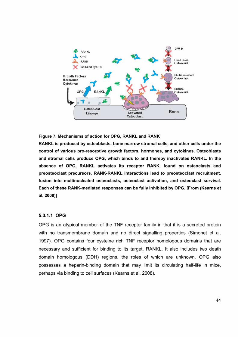
44
Figure 7. Mechanisms of action for OPG, RANKL and RANK
RANKL is produced by osteoblasts, bone marrow stromal cells, and other cells under the
control of various pro-resorptive growth factors, hormones, and cytokines. Osteoblasts
and stromal cells produce OPG, which binds to and thereby inactivates RANKL. In the
absence of OPG, RANKL activates its receptor RANK, found on osteoclasts and
preosteoclast precursors. RANK-RANKL interactions lead to preosteoclast recruitment,
fusion into multinucleated osteoclasts, osteoclast activation, and osteoclast survival.
Each of these RANK-mediated responses can be fully inhibited by OPG. [From (Kearns et
al. 2008)]
5.3.1.1 OPG
OPG is an atypical member of the TNF receptor family in that it is a secreted protein
with no transmembrane domain and no direct signalling properties (Simonet et al.
1997). OPG contains four cysteine rich TNF receptor homologous domains that are
necessary and sufficient for binding to its target, RANKL. It also includes two death
domain homologous (DDH) regions, the roles of which are unknown. OPG also
possesses a heparinbinding domain that may limit its circulating halflife in mice,
perhaps via binding to cell surfaces (Kearns et al. 2008).

45
OPG has been shown to be produced by numerous tissues and associated cells in
addition to osteoblasts. The production of OPG by cultured osteoblasts was shown to
increase with cell differentiation, suggesting that mature osteoblasts are not well suited
to support osteoclastogenesis (Gori et al. 2000). Other cells that produce OPG include
endothelial cells (CollinOsdoby et al. 2001), vascular smooth muscle cells (Olesen et
al. 2005) and fibroblasts in the synovial tissue (Haynes 2003).
Its production is stimulated by treatment with bone morphogenic protein, IL1, TNFα,
TNFβ, vitamin D and oestrogen (Brändström et al. 1998; Hofbauer et al. 1998;
Hofbauer et al. 1999; Vidal et al. 1998). Its secretion is suppressed by prostaglandin E2,
glucocorticoids, and PTH (Brändström et al. 1998; Lee and Lorenzo 1999; Vidal et al.
1998).
The importance of OPG in the regulation of bone remodelling was shown by Simonet
and colleagues (Simonet et al. 1997) and their investigations in mice overexpressing
OPG. The mice had a severe osteopetrosis that presented early after birth and
progressively worsened. Interestingly and in contrast to other osteopetrotic animals, the
affected mice had normally shaped and sized bones and tooth eruption was normal.
Histologically, the animals showed a marked reduction in trabecular osteoclasts but no
deficiency of osteoclast precursors. This suggested that OPG’s role is in the latter
stages of osteoclast differentiation. The key role of OPG was also demonstrated by the
systemic administration of OPG which produces an increase in bone density and blocks
the loss of bone induced by ovariectomy (Simonet et al. 1997).
The physiological role of OPG in bone remodelling was investigated by targeted
deletion of the endogenous OPG gene in mice (Bucay et al. 1998; Mizuno et al. 1998).
These studies show that endogenous OPG is not required for embryonic bone formation
but is essential for maintenance of postnatal trabecular and cortical bone mass
throughout the skeleton. By one month of age, there is a noticeable decrease in bone

46
mineral density of OPG/ mice and changes become more severe with increasing age.
Some abnormalities noted include multiple fractures, kyphosis of the spine, and
profound osteoporosis in the humerus, femur and tibia. Cortical bone is composed of
mostly woven bone with an increased density of osteocytes which are features
consistent with increased bone turnover. As previously shown, the numbers of
osteoclast precursors are not affected and this suggests that the osteoporosis in OPG/
mice is most likely due to poorly regulated osteoclast recruitment and activation in vivo.
Calcification was also noted in the large arteries including the aorta. Endogenous OPG
expression is localised within the smooth muscle layer of the aortic and renal arteries,
suggesting a role for OPG in maintaining normal structure in larger arteries (Bucay et al.
1998).
5.3.1.2 RANKL
RANKL is a type II transmembrane protein of 317 amino acids of which 3 isoforms exist
(Ikeda et al. 2001). It is typically membranebound and found on osteoblasts and
activated Tcells with RANKL1 and RANKL2 encoding for its transmembrane forms.
RANKL3 is a soluble form as it lacks the transmembrane domain. The secreted protein
is derived from the membrane form by either proteolytic cleavage or alternative splicing
(Ikeda et al. 2001).
Most of the factors that are known to stimulate osteoclastic formation and activity induce
RANKL expression via osteoblastic stromal cells although RANKL is expressed in a
number of other tissues (Table 2). It is also expressed by synovial cells and activated T
cells in joints of patients with inflammatory arthritis and may contribute in part, to the
joint destruction in patients with rheumatoid arthritis (Boyce and Xing 2008).
RANKL is involved in numerous aspects of osteoclast differentiation and function. It has
been implicated in the fusion of osteoclast precursors into multinucleated cells (Lacey et
al. 1998), their differentiation into mature osteoclasts (Lacey et al. 1998), their

47
attachment to bone surfaces (O’Brien et al. 2000), their activation to resorb bone
(Burgess et al. 1999; Lacey et al. 1998) and osteoclast survival by avoiding apoptosis
(Lacey et al. 2000).
RANKL plays a dominant role in the regulation of bone resorption and no factor or
combination of factors has been shown to restore bone resorption when RANKL is not
present. Udagawa and colleagues (1999) demonstrated that although MCSF and
RANKL both support the survival of osteoclasts in culture, only RANKL induced pit
forming activity of osteoclasts. The data shows that the survival of osteoclasts is
necessary but not sufficient for inducing bone resorbing activity. Addition of osteotropic
factors such as 1α25(OH)2D3 and PTH to MCSF and RANKL bone marrow cultures
did not stimulate pitforming activity but when osteoblasts were added, it was found that
RANKL mRNA was upregulated by these osteotropic factors (Udagawa et al. 1999).
Interestingly, in MCSF deficient mice the osteoclast population has been shown to
recover over time (Begg et al. 1993); a phenomenon that has not been shown to occur
with a RANKL deficiency.
5.3.1.3 RANK
Receptor activator of nuclear factor κβ (RANK) is the receptor that mediates all known
activity for RANKL. RANK is a type I transmembrane protein of 616 amino acids and is
a member of the TNF receptor family (Anderson et al. 1997). It is expressed primarily on
cells of the macrophage/monocytic lineage, including preosteoclastic cells, T and B
cells, dendritic cells, and fibroblasts (Anderson et al. 1997; Hsu et al. 1999). It is also
found in numerous tissues including bone, bone marrow, spleen; skeletal muscle, liver,
heart, lung, brain, mammary tissue and skin (see Table 2).
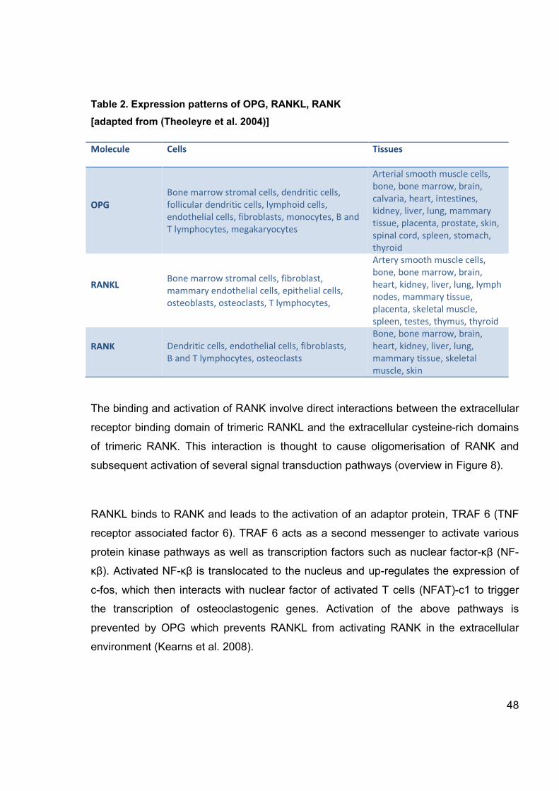
48
Table 2. Expression patterns of OPG, RANKL, RANK
[adapted from (Theoleyre et al. 2004)]
The binding and activation of RANK involve direct interactions between the extracellular
receptor binding domain of trimeric RANKL and the extracellular cysteinerich domains
of trimeric RANK. This interaction is thought to cause oligomerisation of RANK and
subsequent activation of several signal transduction pathways (overview in Figure 8).
RANKL binds to RANK and leads to the activation of an adaptor protein, TRAF 6 (TNF
receptor associated factor 6). TRAF 6 acts as a second messenger to activate various
protein kinase pathways as well as transcription factors such as nuclear factorκβ (NF
κβ). Activated NFκβ is translocated to the nucleus and upregulates the expression of
cfos, which then interacts with nuclear factor of activated T cells (NFAT)c1 to trigger
the transcription of osteoclastogenic genes. Activation of the above pathways is
prevented by OPG which prevents RANKL from activating RANK in the extracellular
environment (Kearns et al. 2008).
Molecule Cells Tissues
OPG
Bone marrow stromal cells, dendritic cells, follicular dendritic cells, lymphoid cells, endothelial cells, fibroblasts, monocytes, B and T lymphocytes, megakaryocytes
Arterial smooth muscle cells, bone, bone marrow, brain, calvaria, heart, intestines, kidney, liver, lung, mammary tissue, placenta, prostate, skin, spinal cord, spleen, stomach, thyroid
RANKL Bone marrow stromal cells, fibroblast, mammary endothelial cells, epithelial cells, osteoblasts, osteoclasts, T lymphocytes,
Artery smooth muscle cells, bone, bone marrow, brain, heart, kidney, liver, lung, lymph nodes, mammary tissue, placenta, skeletal muscle, spleen, testes, thymus, thyroid
RANK Dendritic cells, endothelial cells, fibroblasts, B and T lymphocytes, osteoclasts
Bone, bone marrow, brain, heart, kidney, liver, lung, mammary tissue, skeletal muscle, skin

49
Figure 8. Essential signalling pathways activated by RANKL interactions with RANK
[from (Kearns et al. 2008)]
5.3.2 RANKL expression by osteocytes
The discussion of RANKL has centred on its expression by osteoblasts. However, two
independent studies have emerged that challenge this concept by providing evidence
that osteocytes are a major source of RANKL and are essential for bone remodelling
(Nakashima et al. 2011; Xiong and O’Brien 2012).
The research group from the Tokyo Medical and Dental University (Nakashima et al.
2011) set out to determine the most physiologically relevant osteoclastogenesis
supporting cells amongst the mesenchymal lineage cells in bone. They purified
osteoblast and osteocyte populations utilising known osteocytespecific genes and
demonstrated that the RANKL levels were significantly higher in the osteocytes when
compared with osteoblasts and other mesenchymal cells. Osteoclast precursor cells
were cocultured with osteoblasts or osteocytes in the presence of 1,25dihydroxy
vitamin D3 (1,25(OH)2D3) and prostaglandin E2 (PGE2) which revealed that the
osteocytes have a stronger capacity to support osteoclastogenesis than osteoblasts.
Osteocytes derived from long bones also showed higher levels of RANKL and a more
potent ability to support osteoclastogenesis.

50
Nakashima and colleagues also created mice that were T cell specific Tnsfs-11
(encodes for RANKL) deficient. The mice did not show any discernible osteopetrotic
phenotype which indicates that RANKL expressed on T cells does not significantly
contribute to regulation of osteoclastogenesis. The Tnsf-11 gene was specifically
deleted in osteocytes. This diminished the osteoclastogenesissupporting ability of
osteocytes but not osteoblasts. These mice initially showed no gross abnormalities, no
defects in tooth eruption or any growth retardation. However, bone volume was
increased and at 12 weeks of age, the marrow cavity was abnormally filled with
trabecular bone (Figure 9). These mice showed a severe osteopetrotic phenotype as a
result of a reduction in the number of osteoclasts. There was no abnormality in
osteocyte density, morphology or marker gene expression (including SOST) between
wildtype and osteocytespecific Tnsf-11-deficient mice, which suggested the crucial
role of osteocytederived RANKL in the regulation of osteoclastogenesis in vivo.
Because the mice showed no abnormal phenotype at birth but developed a postnatal
phenotype and bone mass increased with age, the authors concluded that the RANKL
expressed by osteocytes contributes more to bone remodelling after birth than to the
skeletal development in the embryo. This suggests the other cells producing RANKL
may have a larger role in the regulation of osteoclastic bone resorption in the embryonic
stage and in tooth eruption and the growth of long bones (Nakashima et al. 2011).

51
Figure 9. Micro-CT reconstruction of femurs of 12 week mice
Longitudinal and axial views. [Adapted from (Nakashima et al. 2011)]
Xiong and colleagues (2011) generated mice in which RANKL was conditionally deleted
in various geneticallydefined cell populations corresponding to different stages of
osteocyte and chondrocyte differentiation. RANKL produced by osteocytes was also
found to be responsible for the majority of the osteoclast differentiation that drives the
remodelling of cancellous bone, as their mice with deletion of the Tnfs-11 (encoding for
RANKL) gene had increasing bone mineral density with increasing age. Hypertrophic
chondrocytes were proposed as the source of RANKL required for the resorption of the
primary spongiosa.
In addition, a model of unloadinginduced bone loss was used in which mice were tail
suspended in an attempt to demonstrate a role for osteocyte RANKL in pathological
bone remodelling. Control mice showed an increase in RANKL mRNA and osteoclast
numbers and an increase in trabecular spacing and decreased cortical thickness. Mice
lacking RANKL in osteocytes were protected from the loss of bone and changes in the
bone architecture. This was considered to indicate that osteocytes are important in the
pathogenesis of diseases caused by abnormal bone remodelling (Xiong et al. 2011).

52
Expression of RANKL in osteoblast progenitors was stimulated by hormones known to
increase bone resorption in vitro. However, the results show that RANKL expression in
these cells does not participate in physiological bone remodelling. It was suggested
(Xiong et al. 2011) that it may contribute to osteoclast differentiation under different
circumstances such as dietary calcium deficiency or lactation. It must also be kept in
mind that it is possible that RANKL expression in these cultures could be an in vitro
phenomenon and not reflect a true physiological role.
Deleting RANKL from osteocytes leads to an increase in bone mass. In this model,
deletion of RANKL led to the femoral cancellous bone volume doubling and osteoclast
surface decreasing by 75%. It was concluded that RANKL produced by osteocytes is
responsible for most of the osteoclastogenesis in cancellous bone (Xiong et al. 2011).
5.3.2.1 How RANKL from osteocytes reaches osteoblasts
If RANKL is produced by osteocytes which are the key cells in bone remodelling, it is
important to examine how the RANKL produced by these cells which are embedded
within mineralised matrix can reach the osteoblasts. Osteocytes have up to 50 cellular
processes that extend thorough the bone matrix that connect osteocytes to each other
and to the bone surface via gap junctions (Bonewald 2011). These connections and
processes make up the lacunacanalicular network (Bonewald 2011). Imaging studies
(Kamioka et al. 2001) have shown that osteocytes widely spread their processes into
the osteoblast layer, and that some of these processes elongate to the vascularfacing
surface of the osteoblast layer. It is plausible that membranebound RANKL produced
by osteocytes could be carried on these processes. Soluble RANKL has a small enough
molecular weight that it can pass through the osteocyte lacunaecanalicular network
(Xiong et al. 2011). Molecules travelling from the osteocyte to the osteoblast and
osteoclast via these means may be involved in the regulation of RANKL in these cells
(Tatsumi et al. 2007).

53
Osteocytes can be difficult to study in vitro as the dendritic processes are lost when
osteocytes are isolated from bone matrix and placed into conventional 2D cultures
(Honma et al. 2013). Honma et al (2013) developed a coculture system of osteoclast
precursors and osteocytes embedded in collagen gel that allowed the physiological
properties of osteocytes to be maintained for about a week. Results showed that
osteocytic RANKL was provided as a membranebound form to osteoclast precursors
through osteocyte dendritic processes. The contribution of soluble RANKL to the
osteoclastogenesis supported by osteocytes was found to be minor (Honma et al.
2013).
Tatsumi and colleagues (2007) have postulated that it is the cellcell contact between
osteocytes and osteoblasts that normally retrains RANKL gene expression from
aberrant activation and that osteocyte ablation or disruption of the network releases this
negative regulation. This would result in the aberrantly elevated RANKL expression and
osteoclastogenesis that was found in their investigation.

54
5.4 Sclerostin
Osteocytes have been acknowledged to sense loading stimuli and to regulate
remodelling and bone turnover processes, but little is known about how cells embedded
in a mineralised matrix carry out this regulatory role. Research suggested that mature
osteocytes send an inhibitory signal to osteoblasts at the boneforming surface via
canalicular processes and it was also proposed that the same signal maintained bone
lining cells in a quiescent state against their natural tendency to reactivate the
remodelling process (Martin 2000). This could explain the exponential decline in bone
formation rates as new osteocytes were formed. If this signal was sensitive to
mechanical loading the correct balance could be obtained between bone formation and
its inhibition. This previously unknown signal has been identified as sclerostin, the
secreted protein product of the SOST gene, located on chromosome 17q1221.
Sclerostin is a negative regulator of osteoblasts and inhibits bone formation and
appears to be exclusively osteocyte derived (van Bezooijen et al. 2004). Information
about sclerostin has mainly been obtained by studying rare human bone disorders and
in vitro experimentation.
5.4.1 Role of sclerostin
Sclerostin has been proposed to have a number of roles in bone formation and
homeostasis. It may have a role in bone modelling whereby it may keep bone lining
cells in a quiescent state, preventing activation of osteoblasts and formation of bone
without previous resorption. Its role in bone remodelling may be to serve as a negative
feedback signal to osteoblasts to prevent overfilling of the basic multicellular unit (van
Bezooijen et al. 2004; Poole et al. 2005).
The effects of sclerostin have been shown experimentally by using SOST knockout
mice which show increased radiodensity, bone mineral density, cortical and trabecular
volume, bone formation and bone strength (Poole et al. 2005).

55
5.4.2 Sclerostin deficiency in humans
A lack of sclerostin in humans results in the rare conditions of sclerosteosis and van
Buchem disease, first described in the 1950’s (van Buchem et al. 1955; Truswell 1958).
The skeletal manifestations of both diseases are the result of endosteal hyperostosis
and are characterised by progressive generalized osteosclerosis. The manifestations
are most pronounced in the mandible, skull and include characteristic enlargement of
the jaw and facial bones leading to facial distortion, increased intracranial pressure with
entrapment of cranial nerves which is often associated with facial palsy, hearing loss,
and/or loss of smell. Those patients with sclerosteosis have a more severe phenotype
compared with patients with van Buchem disease and usually present with syndactyly
(Figure 10). Bone biopsies of those affected show increased bone formation including a
predominance of cuboidal active osteoblasts (Moester et al. 2010).
Figure 10. Chronological portraits of a patient with sclerosteosis from the age of 3
[From (Moester et al. 2010)]

56
5.4.3 Expression of SOST/Sclerostin
SOST mRNA is expressed in many tissues, especially during embryogenesis. In
humans these tissues include the heart, aorta, liver and kidney. Expression of the
sclerostin protein has only been reported in cells embedded within a mineralised matrix
e.g. osteocytes (Figure 11), cementocytes (Figure 12) (van Bezooijen et al. 2004;
Winkler et al. 2003) and mineralised hypertrophic chondrocytes in the human growth
plate (van Bezooijen et al. 2009). Osteoclasts, as well as osteoblasts and lining cells
(whether located in the Haversian canals or at the periosteum) have been shown to be
consistently negative for sclerostin (Poole et al. 2005).
Figure 11. Canaliculi and/or lacunae of osteocytes in human bone positive for sclerostin.
[from (Winkler et al. 2003)]
Figure 12. Sclerostin expression by cementocytes in a human third permanent molar.
(B) Detail of (A) showing sclerostin-positive canaliculi of cementocytes [from (van
Bezooijen et al. 2009)]

57
Sclerostin expression has been shown to be cell differentiationdependent with
detection only present in latedifferentiated human and mouse cells, using in vitro
experiments (van Bezooijen et al. 2004). Researchers have closely examined the stage
at which normal osteocytes express sclerostin in human bone. It was concluded that
sclerostin secretion by osteocytes is a delayed event in which the cells only secrete
sclerostin after the onset of mineralisation to inhibit cortical bone formation and osteon
infilling by cells of the osteoblast lineage (Figure 13) (Poole et al. 2005). The more
sclerostinnegative osteocytes were found in an individual osteon, the more likely it was
that the osteon was in the process of bone formation. Most new osteocytes were
negative for sclerostin staining for at least 16 days. There was a trend towards a higher
percentage of sclerostinpositive cells in the cortex than the cancellous bone and it was
also demonstrated that sclerostin negative cells were located closer to surfaces
(Haversian canal or endo/periosteal surface) than sclerostin positive cells (Poole et al.
2005). Late stage osteoblasts/early osteocytes are the target for sclerostin action
(Wijenayaka et al. 2011) which was demonstrated experimentally by examining normal
human bone derived osteoblastic cells (NHBC) treated throughout differentiation with
recombinant sclerostin. Observation revealed that gene expression was primarily
affected at the time of mineralisation.
Positive sclerostin staining is evident in osteocyte canaliculi and this may suggest that
this could be a method whereby sclerostin is transported to neighbouring cells, intra
cellularly via the gap junctions present at the tips of the canaliculi. Sclerostin has also
been demonstrated to be present in lacunar walls giving evidence that sclerostin is a
secreted protein and its transport may occur extracellularly from lacunae to lacunae
(van Bezooijen et al. 2004).

58
Figure 13. Schematic diagram of the proposed regulation of a remodelling cortical osteon
by osteocytic sclerostin expression
[from (Poole et al. 2005)]

59
5.4.4 Possible mechanisms of action
A number of experiments have demonstrated the role of sclerostin on bone formation.
5.4.4.1 Inhibition of bone formation
Clinically, patients with sclerosteosis show a high bone mass due to increased
formation with premature termination codons in the SOST gene which suggests an
inhibitory role of sclerostin on bone formation. Experimentally, this role has been
demonstrated by adding sclerostin to osteogenic cultures which has been shown to
result in inhibited proliferation and differentiation of mouse and human osteoblasts
(Poole et al. 2005; Sutherland et al. 2004; van Bezooijen et al. 2004; Winkler et al.
2003) and overexpression of sclerostin in vivo induces osteopenia in mice in which
bone formation decreased but resorption was unaffected (Winkler et al. 2003). van
Bezooijen and colleagues (2004) suggested that sclerostin produced by osteocytes is
transported to the bone surface where it inhibits the later stages of bone formation.
5.4.4.1.1 Osteoblast apoptosis
The balance of proliferation, cell differentiation and apoptosis affects the pool of
osteoblasts available to maintain bone homeostasis. Many factors affect osteoblast
survival and osteoblasts undergo programmed cell death due to increased sensitivity to
apoptosis–inducing agents, decreased concentrations of vital growth factors or
enhanced response to apoptosisinducing factors (Jilka et al. 1998), for example TNFα
and high levels of glucocorticoids have been reported to increase the apoptosis of
osteoblasts. Using cell cultures, sclerostin has also been shown to induce osteoblast
apoptosis (Sutherland et al. 2004). The pathway by which sclerostin causes apoptosis
has been likened to that reported for growth factor deprivation. This pathway may serve
as the basis for the method by which sclerostin is able to modulate the survival of
osteoblasts. By promoting osteoblast apoptosis a decrease in osteoblast numbers will
result and this will ultimately lead to decreased bone formation.

60
5.4.4.2 Member of the DAN family of glycoproteins
Sclerostin has an amino acid sequence which indicates a cystine knot structure and has
been assigned as part of the DAN (differential screeningselected gene aberrant in
neuroblastoma) family of glycoproteins (AvsianKretchmer and Hsueh 2004). These
proteins are largely bone morphogenic protein (BMP) antagonists. BMPs are secreted
cytokines and although sclerostin has a relatively low binding affinity for BMPs, it has
been shown to antagonise BMPstimulated bone formation (van Bezooijen et al. 2004).
The mechanism by which sclerostin antagonised BMPstimulated bone formation differs
from that of classical BMP antagonists; however, it has since been suggested that
sclerostin, like other DAN family members such as WISE (which shares 38% amino acid
identity to sclerostin), acts to antagonise canonical Wnt signalling (van Bezooijen et al.
2004; Li et al. 2005; Semënov et al. 2005).
5.4.4.3 Antagonist to canonical Wnt signalling
5.4.4.3.1 Wnt/β catenin signalling pathway
The canonical Wnt pathway is a complex biochemical sequence that when activated,
results in increased osteoblastic cell differentiation and inhibition of bone resorption by
blocking the receptor activator RANKL/RANK (Jackson et al. 2005). There are over a
dozen forms of Wnt proteins. Wnt proteins are secreted glycoproteins that play key
roles in development and homeostasis through their roles in cell differentiation,
proliferation and apoptosis (Johnson 2008).
The key components of the pathway in bone include (Johnson 2008):
1) Cell surface coreceptors, LRP5/6 and Frizzled
2) Intracellular proteins
3) TCF nuclear transcription factors that bind βcatenin and regulate gene
expression.

61
The signalling pathway is controlled by two cell surface coreceptors, Frizzled and low
density lipoprotein receptorrelated proteins (LRP) 5 or 6 that bind the Wnt protein
ligand. In the absence of Wnt, there are a number of intracellular proteins including
Dishevelled (Dsh), and the ‘degradation complex’ of Axin, adenomatous polyposis coli
(APC), glycogen synthase kinase (GSK3β) and betacatenin. The degradation complex
in addition to other proteins coordinate the phosphorylation of βcatenin. The molecule
ubequitin ligase then binds to βcatenin which is then ubiquitinated and degraded by the
proteosome. This maintains a low intracellular level of βcatenin. Transcription factor
TCF and other proteins such as ’Groucho’ bind to DNA and prevent DNA transcription.
In activation, Wnt binds to the transmembrane receptor Frizzled and its coreceptor
LRP5/6. This binding causes activation of the protein Dishevelled which results in
phosphorylation of GSK3β inhibiting its activity and dissociation of the proteins of the
degradation complex. βcatenin is no longer modified and destroyed and so it
accumulates in the cytosol. As the amount of βcatenin rises, it migrates into the
nucleus where it binds to TCF transcription factor to activate a RNA polymerase and
gene transcription. Osteoblastic activity is stimulated (Figure 14). An increase in
LRP5/6mediated canonical Wnt signalling leads to an increase in bone mass (Li et al.
2005).

62
Figure 14. The Wnt/β-catenin signalling pathway
[from (http://www.intechopen.com/source/html/42019/media/image2.png)]
5.4.4.3.2 Sclerostin binds to low-density lipoprotein receptor-related protein
Sclerostin has been shown to inhibit the Wntsignalling pathway. It does this by binding
to LRP5 (lowdensity lipoprotein receptorrelated protein) and its coreceptor LRP6 to
inhibit canonical Wntsignalling (Li et al. 2005; Semënov et al. 2005). Despite sclerostin
binding to LRP5/6, it does not appear that Wnt proteins and sclerostin compete for
binding of this coreceptor (Li 2005). When sclerostin binds, LPR5 cannot restrain axin
and the Wnt pathway cannot be activated. The degradation complex remains intact and
as βcatenin does not accumulate DNA transcription does not occur and osteoblast
function is inhibited. Further research has also shown that LRP4 plays a role in skeletal
development and bone metabolism as it has been demonstrated to bind sclerostin in
addition to other BMP antagonists, Wise and Dickkopf1 (DKK1) (Choi et al. 2009;
Leupin et al. 2009).

63
5.4.4.3.3 A catabolic action through promotion of osteoclast formation
Wijenayaka and colleagues (2011) have explored the possibility of a catabolic role for
sclerostin in addition to the antianabolic roles already established. To investigate this
experimentally, exogenous recombinant sclerostin (rhSCL) was added to cultures of
human primary osteoblastic cells differentiated to an osteocytelike stage and in MLO
Y4 cells (the mouse osteocytelike cell line). It was found that a rhSCL dose
dependently upregulated the expression of RANKL mRNA and downregulated the
expression of OPG, which caused a primarily RANKL driven increase in the RANKL:
OPG mRNA ratio in both experimental cell types. To study the effects of rhSCL on
resulting osteoclastic activity MLOY4 cells primed with rhSCL were cocultured with
mouse splenocytes or human peripheral blood mononuclear cells (PBMC). Osteoclastic
resorption increased approximately 7fold compared to untreated cocultures with a
moderate increase in number and size of TRAPpositive multinucleated osteoclastlike
cells. The rhSCL did not induce apoptosis of MLOY4 cells which demonstrated that the
increase in osteoclastic activity was not due to dying osteocytes. When recombinant
OPG was added, the effects of rhSCL on osteoclast formation and activity were
completely supressed, demonstrating that RANKL signalling was essential for the
sclerostin effect. The authors concluded that sclerostin may have a catabolic action,
promoting osteoclastogenesis by its effect on osteocytes in a RANKLdependent
manner (Wijenayaka et al. 2011) (Figure 15).

64
Figure 15. The potential role of osteocyte-derived sclerostin in regulating the expression
of osteocyte RANKL.
A catabolic stimulus (e.g. mechanical unloading) may cause sclerostin expression in
osteocytes (OY) to respond in an autocrine manner (curved dotted arrow), increasing the
local expression of RANKL relative to OPG; or in a paracrine manner (straight dotted
arrow) by inducing such expression in sclerostin-negative OY (including immature OY
immediately below the osteoblastic lining cells and in the lining cells themselves). An
effect of sclerostin and OY-derived RANKL on both endosteal and intra-cortical
remodelling may occur either at the stage of osteoclast precursor
recruitment/chemotaxis, proliferation/differentiation and/or on the activity of mature
osteoclasts. Osteocytes may also ‘talk to’ cells in the bone marrow via a limited number
of cell processes extending into the marrow space. This may result in effects on bone
marrow stromal cell expression of RANKL and OPG and subsequent regulation of
osteoclastogenesis by these cells. A role for apoptosing OY is not ruled out here as the
initial stimulus for sclerostin expression - perhaps secondarily to microdamage and local
bone unloading; or that the resorbing osteoclast is exposed to higher RANKL levels
during engulfment of the expressing OY.
[adapted from (Wijenayaka et al. 2011)]

65
5.4.5 Regulation of SOST/Sclerostin expression
Osteocytes have long been implicated in mechanosensing due to their location and
morphology (Knothe Tate et al. 2004). The Wnt pathway may play an important role in
the anabolic bone response to deformation and loading and increased Wnt signalling
has been found after loading osteoblastic cells in vitro (Hens et al. 2005). Mechanical
loading of ulnar limbs in mice has demonstrated a significant reduction in sclerostin
levels which led the authors to hypothesise that the osteocytes detected the mechanical
loading and this caused a reduction in secretion of sclerostin. This in turn reduced the
inhibition of LRP5 signalling in nearby osteoblasts which led to enhanced osteogenesis
(Robling et al. 2008). In the same study, the hind limbs were subject to unloading which
yielded a significant increase in limb sclerostin expression (Robling et al. 2008). This
was coroberated by Lin and colleagues (Lin et al. 2009) who demonstrated that, in
response to unloading (tail suspension) in mice, an increased amount of sclerostin was
secreted by osteocytes which inhibited Wnt/βcatenin signalling, thereby inducing
apoptosis and suppressing osteoblast activity. This led to decreased bone formation
and as a result, bone mass decreased. SOST / mice were also shown to be resistant to
the usual unloadinginduced bone loss.
It appears that the Wnt signalling pathway is held in the “off” position in bone due to the
high levels of sclerostin produced by osteocytes. Therefore, activation of the pathway
requires either a bypass of the inhibition of LRP5/6 by sclerostin or a decrease in levels
of sclerostin to permit Wnt ligandmediated activation of the pathway. It has been
proposed that PTH stimulation of bone formation involves a reduction in SOST gene
expression (Johnson 2008).
Other systemic factors that have been shown to affect SOST/sclerostin expression
include 1,25 dihydroxy vitamin D3 alone or in combination with retinoic acid, which
increases SOST expression in human osteoblastic cells in vitro. Glucocorticoids affect
sclerostin differently depending on the experimental conditions. In vitro, SOST
expression on osteoblastic cells was suppressed by dexamethasone whilst in vivo,

66
SOST expression was increased in mice treated with prednisolone (Figure 16) (Moester
et al. 2010).
Figure 16. Schematic model for the regulation of the control of bone formation by
sclerostin.
Sclerostin may exert its inhibitory effect on bone formation by preventing the activation
of lining cells as well as the inactivation of active osteoblasts. Glucocorticoids stimulate
sclerostin expression and, thereby, inhibit bone formation, whereas intermittent PTH and
loading inhibit sclerostin expression in osteocytes and, thereby, stimulate bone
formation. [from (Moester et al. 2010)]
The research to date indicates that sclerostin is an antagonist for Wnt signalling and the
loss of SOST/sclerostin function likely leads to the hyperactivation of Wnt signalling that
underlies bone growth characteristic of sclerosteosis patients. More research is required
to determine the precise mechanisms by which sclerostin binding to LPR5/6 interferes
with canonical Wnt signalling, to further identify factors that regulate SOST/sclerostin
expression, and to determine any potential additional functions of sclerostin.

67
5.4.6 Clinical applications of sclerostin
Sclerostin has been the subject of intensive investigation in the medical field as
research grows showing that the osteocyte plays a key role in bone homeostasis.
Sclerostin is of particular interest because of its potential therapy for the stimulation of
bone formation as required in the treatment of osteoporosis.
Antibodies to sclerostin have been developed that inhibit its biological activity. These
antibodies have already been shown to increase the bone formation rate, density and
strength in ovariectomised rats (Li et al. 2009). Clinical trials have also begun involving
48 healthy postmenopausal women who were given an injection of monoclonal
antibody against sclerostin. Results showed increased bone formation markers and
bone mineral density and the therapy was well tolerated (Padhi et al. 2011).
5.4.7 Dentistry and sclerostin
Research has been conducted on the role of sclerostin on the alveolar bone and the
development of dental structures. When sclerostin was knocked out of mice, increased
dimensions of mandibular basal bone but no differences in the tooth and the pulp
chamber were shown. There was an increase in the width of the cementum and a
moderate decrease in the width of the periodontal ligament (Kuchler et al. 2014).
Sclerostin appears to affect the bone and cementum rather than producing anomalies in
tooth structure such as dentine (Kuchler et al. 2014).
Sclerostin is not normally found in the periodontal ligament (PDL). When PDL cultures
underwent mineralising treatment researchers were able to demonstrate increased
levels of SOST mRNA using real time PCR, immunohistochemistry and western blots
(Jäger et al. 2010).

68
There have been two clinically relevant investigations into possible applications of
sclerostin utilising animal models. Pharmacologic inhibition of sclerostin using
sclerostin‐neutralising monoclonal antibody (Scl‐Ab) is known to increase bone
formation, bone mass and bone strength in models of osteopenia and fracture repair
(Taut et al. 2013). Taut and colleagues designed an investigation to assess the
therapeutic potential of Scl‐Ab to stimulate alveolar bone regeneration after the
establishment of experimental periodontitis in rats. Experimental periodontitis reduced
bone volume fraction (BVF) by 30% and tissue mineral density (TMD) by 32%. After 6
weeks of treatment significant increases in alveolar bone volume and density were
observed with no statistically significant difference between the animals with
periodontitis and intact healthy animals.
The effect of orthodontic tooth movement has been studied using a NiTi closed coil
spring applied between the incisors and molars of mice (Nishiyama et al. 2015). After 4
days the mice were examined using multimodal confocal fluorescence imaging analysis.
In the tensile areas of bone there was downregulation of sclerostin and in areas of
compression sclerostin was significantly higher than control sites. A study at the
University of Adelaide (Sanmuganathan 2011), in which separators were placed
between rat molars to provide the orthodontic force has found similar results. It seems
that when significant local strains are sensed by the osteocyte population on the side of
tension the sclerostin levels are decreased and the cells are released from their
suppressed Wnt signalling state and new bone is formed. Conversely, on the side of
compression, sclerostin levels are increased with continued Wnt signalling suppression,
leading to a prevention of local bone formation.

69
5.5 Dentoalveolar ankylosis
Dentoalveolar ankylosis is defined as the fusing of cementum with alveolar bone (Kurol,
1981). Once ankylosis has occurred, further tooth eruption and bone deposition
becomes impossible. Ankylosis is commonly found in two clinical situations: those of
infraocclusion which mainly affects deciduous or primary teeth (Figure 17) and in the
permanent dentition in which it is associated with impaction or external root replacement
resorption following trauma and is frequently observed after reimplantation of avulsed
permanent teeth (Figure 18).
Figure 17. Infra-occlusion of a primary molar tooth below the level of the occlusal plane
[from (Kjaer et al. 2008)]
Figure 18. Tooth 11 demonstrates infra-occlusion and ankylosis subsequent to avulsion
and replantation [from (Campbell et al. 2005)]

70
5.5.1 Prevalence
Ankylosed teeth often present at a level below the occlusal plane, a condition referred to
as submergence or infraocclusion, which results from the inability of the ankylosed
tooth to follow the normal vertical development of the neighbouring teeth that are
continuing to develop and erupt. The condition is as much as ten times more common in
the deciduous dentition compared with the permanent dentition (Kurol 1981; Messer
and Cline 1980; Biederman 1956), is twice as common in the mandible as the maxilla
and does not appear to a have a dominance in either the right or left side (Biederman
1962; Messer and Cline 1980) or between males and females (Kurol 1981). The most
common tooth reportedly affected by ankylosis is the deciduous second molar
(Biederman 1962; Biederman and Centre 1956), although some studies have found a
higher prevalence of ankylosis in mandibular deciduous first molars (Messer and Cline
1980; Steigman et al. 1974). However, as this tooth shows only slight infraocclusion
and usually exfoliates on schedule, the diagnosis of ankylosis may be missed,
explaining the higher prevalence of ankylosis reported in second molars (Messer and
Cline 1980). Any tooth, deciduous or permanent, may become ankylosed anywhere
along its eruptive path, either before or after emergence (Biederman and Centre 1956).
Infraocclusion affecting primary molars can be found in children as young as 3 years of
age with review articles citing prevalence ranging from 1.338.5% (Andlaw 1974; Kurol
1981). This most likely reflects the use of different inclusion criteria (e.g. no contact with
opposing tooth, presence of permanent successors, degree of immobility), age of
children investigated and ethnic factors (Andlaw 1974; Kurol 1981). Kurol (1981)
reported a difference in prevalence between different age groups with the maximum
prevalence reported as 14.3% in 89 year old children and the minimum of 1.9% in 12
year old children. This may reflect the normal exfoliation of teeth, including those that
were in infraocclusion. The prevalence amongst siblings is significantly higher at 18.1%
suggesting a familial tendency in the infraocclusion of primary molars and that a
multifactorial, polygenic inheritance pattern is most likely with possible contribution from
environmental factors (Kurol 1981).

71
In individuals with bilateral infraocclusion of deciduous molars in which a successor
was absent on one side only, the side with no successor showed more infraocclusion
and no infraoccluded molars with aplasia of the successor were found to exfoliate
spontaneously (Kurol and Thilander 1984).
Traumatic dental injuries commonly involve anterior teeth with effects on the dental hard
tissues as well as the gingiva, periodontal tissue, pulp and alveolar bone. Healing of
dental injuries depends on the stage of root development, the extent of damage
particularly to the periodontal tissues, and the effect of bacterial contamination from the
mouth. A significant complication following a traumatic injury is external root resorption
which may be classified into surface resorption, inflammatory resorption, replacement
resorption (ankylosis) (Andreasen 1975; Hecova et al. 2010) and in some cases
transient replacement resorption (Andreasen 1975).
External root resorption is most common following avulsion rather than luxation injuries
and more common following intrusive luxation compared with extrusive or lateral
luxations. This is most likely due to higher damage to the PDL with intrusive luxation
and avulsion injuries. In a study of 889 injured permanent teeth, replacement root
resorption was present in 42.9% of avulsed and replanted teeth (Hecova et al. 2010). Of
the replanted teeth, 12.2% required eventual extraction due to replacement resorption
and marked infraocclusion and an additional 26.5% were extracted due to inflammatory
root resorption (Hecova et al. 2010). Root resorption has been observed predominantly
on the apical part of the root (58%), while 29% was located coronally and 19% was
located on the central part of the root (CronaLarsson et al. 1991).

72
5.5.2 Aetiology
Ankylosis is preceded by a defective or discontinuous periodontal ligament (PDL) due to
incomplete development of the periodontal membrane, local lysis or local ossification
(Biederman and Centre 1956). Part of the bone surface is therefore obliterated and
bone growth accompanying tooth eruption is halted (Biederman and Centre 1956). Early
theories in an attempt to describe the alteration in the PDL included a genetic or
congenital gap in the PDL, excessive masticatory pressure or trauma causing local
injury to PDL which leads to ossification occurring as a healing process or a disturbance
to local metabolism (Biederman and Centre 1956; Biederman 1962). Biederman
postulated that masticatory trauma was unlikely to be the cause as there was not a
greater incidence in ankylosis in adults who have higher masticatory forces. Neither was
there a predominance of the incidence in left or right sides and Biederman suggested
that if masticatory trauma was the cause then one side should predominate. He found
that the same jaw was commonly affected and that this supported the concept of a
disturbed local metabolism, whilst not being absolute proof.
Other causes of ankylosis that have been proposed include localised infection
(Adamson 1952), chemical or thermal irritation (Line et al. 1974; Messer and Cline
1980; Kurol and Magnusson 1984), root canal treatment (Erausquin and Devoto 1970),
tooth reimplantation (Löe and Waerhaug 1961), failure of normal eruptive process to
keep pace with vertical alveolar growth (Dixon 1963) and an aberration of the normal
process of resorption and deposition of bone and cementum during tooth exfoliation
(Messer and Cline 1980).
In addition to an increased prevalence of infraocclusion in siblings (Kurol 1981)
suggesting a genetic origin, studies have shown that infraocclusion is frequently
observed in the presence of other dental anomalies such as tooth agenesis, microform
teeth, delayed tooth development, palatallydisplaced canines and mandibular second
premolar distal angulation (Bjerklin et al. 1992; Baccetti 1998; Baccetti 2000; Garib et al.
2009; Shalish et al. 2010). These dental features arising in combination have been

73
named dental anomaly patterns (DAP) by Peck (2009). Their occurrence suggests that
not only is infraocclusion under genetic control, but that there is a common genetic
origin. This is clinically relevant as an infraoccluded tooth may be considered a marker
for future dental problems such as tooth agenesis and palatallydisplaced canines.
Ankylosis may occur as a repair process following injury. A healthy periodontal ligament
must have the capacity to resist induction to osteogenesis through cell signalling
systems such as the release of cytokines and growth factors that accurately ‘measure’
and maintain the PDL width, ensuring separation of the root and alveolar bone (Melcher
1970; McCulloch 1995). Traumatic injury disrupts this homeostasis. A number of studies
have demonstrated that it seems to be the source of the cells that repopulate the PDL
space following trauma or injury that determine whether or not ankylosis occurs
(Erausquin and Devoto 1970; Lin et al. 2000; Melcher 1970; Line et al. 1974). Following
trauma to the PDL the root side progenitor cells lose their ability to differentiate into
fibroblasts. When the space is repopulated by cells from a source outside the true PDL
tissues (alveolar bone, bone marrow or soft connective tissue) osteoclasis and
osteogenesis occur, resulting in healing via a dentoalveolar ankylosis. Having vital PDL
cells present after trauma to the tissue appears crucial for repair and regeneration to a
functional PDL. Experiments with the reimplantation of teeth after varied periods of
extraoral dry time demonstrated that the presence of a normal PDL over ankylotic
repair may be related to the presence of epithelial rests of Malassez (Löe and
Waerhaug 1961).
5.5.3 Diagnosis
Methods of detecting ankylosis in a clinical setting are limited to infraocclusion of the
tooth, sound on percussion and mobility, supported by radiographic examination of the
PDL, but the lack of sensitivity of these methods can lead to a false negative diagnosis.
Raghoebar and colleagues (1989) calculated the number of true positives and false
negatives when comparing clinical (percussion sounds and radiographic appearance of

74
the PDL) to the histological presence of ankylosis and found a sensitivity of 23% and a
specificity of 100%.
The sound produced on tooth percussion is a commonly employed diagnostic test. It
has been demonstrated in an animal model that percussion sounds changed from dull
to high pitched when ankylosis affected more than 20% of the root surface and these
teeth had no mobility present (Andersson et al. 1984). However, if the amount of
ankylosis is small it may not be detected by percussion. In the same animal model when
ankylosis was present but consisted of less than 10% of the root surface, percussion
sounds were normal (Andersson et al. 1984).
Despite the limitations of percussion it is still more sensitive than radiographic
examination of the change in width of the PDL. Radiographs may be subject to errors
from overlapping structures and bone marrow space which may result in incorrect
identification of the PDL (Andersson et al. 1984). The location of the lesion is also
important as while ankylosis can be detected proximally with radiographs, it cannot be
detected when it occurs on the lingual and labial surfaces of the root (Andersson et al.
1984). After trauma, clinical tests such as mobility testing and percussion could detect
ankylosis 3 weeks earlier than radiographic examination (Andreasen 1975) and a
subsequent investigation found lack of mobility, a metallic percussion tone and no pain
to percussion were highly reliable compared to radiographic obliteration of the PDL
space which appeared to be only partly reliable (Andreasen et al. 2006)
Infraocclusion of the tooth below the occlusal plane has been suggested as the most
important and reliable clinical sign of ankylosis (Raghoebar et al. 1989) and this may be
accompanied by tipping of adjacent teeth and overeruption of opposing teeth.

75
5.5.4 Histology
Dentoalveolar ankylosis presents histologically as a bony union between tooth and
bone. Studies on extracted teeth have provided information on the specific histological
presentation. Kurol and Magnusson detected the presence of ankylosis at the inner
surface of the roots in the apical third but not at the apex in younger children and more
coronally in older children (Kurol and Magnusson 1984). Raghoebar and colleagues
found ankylosis in the bifurcation and interradicular area 81% of the time with a few
teeth showing ankylosis on the outer root surface (Raghoebar et al. 1989).
Kurol and Magnusson (1984) described the ankylotic area as repair of a previously
resorbed root area by a hard tissue resembling bone directly contacting dentine. Cell
inclusions were present in the hard tissue and resting lines were evident especially in
the older children. The authors suggested that as ankylosis did not appear to be static it
may be part of an ongoing remodelling process following root resorption and be
intermittent in nature. The altered relationship between the tooth and surrounding
tissues may be a developmental rather than a functional disturbance.
Andersson et al. (1984) described two types of ankylosis using a monkey experimental
model. In most teeth the ankylosis was preceded by resorption of the cementum and
dentine with no cementum present at the ankylotic site. In some teeth direct apposition
of bone onto the cemental surface occurred without previous resorption of the
cementum – this type was slightly more common in apical areas of the root. Ankylotic
areas consisted of either thin bony trabeculae or wide bony areas with most teeth
showing the presence of both.
Pulp changes were also observed with Raghoebar and colleagues finding occasional
pulp calcifications in the root canal and Kurol and Magnusson noting that pulps of
ankylosed teeth showed more degenerative changes such as fibrosis and calcification
(Kurol and Magnusson 1984; Raghoebar et al. 1989).

76
5.5.5 Sequelae and treatment of ankylosed teeth
The presence of an ankylosed deciduous molar tooth does not appear to affect coronal
morphology or cause hypoplasia/hypomineralisation of the succedaneous bicuspid as
the crown of this tooth is usually fully formed by the time of the clinical diagnosis of
ankylosis (Messer and Cline 1980). In addition, ankylosis does not appear to influence
the rate of development of the successive tooth (Steigman et al. 1974). The successive
bicuspids showed rotations of the crown associated with a lack of mesiodistal space in
the arch and a higher incidence of periodontal pathology (Messer and Cline 1980).
When the deciduous molar ankyloses very early, there have been reports of ectopic
positioning of the permanent successor (Kjaer et al. 2008). In the initial stages of
development the permanent tooth germ is located laterally to the deciduous molar.
When the eruption of the deciduous molar is arrested the permanent tooth germ may
fail to migrate to an interradicular position below the nonerupting deciduous molar
(Kjaer et al. 2008). Instead, it has been shown to erupt independently to obtain a
position occlusal to the ankylosed molar (Kjaer et al. 2008).
The majority of ankylosed first primary molars tended to occur bilaterally and future
ankylosis of other molars, particularly the deciduous mandibular second molars, was
likely. The ankylosed primary first molar tends to show only mild to moderate infra
occlusion and exfoliates on schedule. It is assumed that during the process of
exfoliation, the ankylosing tissue is resorbed, thus allowing exfoliation (Messer and
Cline 1980; Steigman et al. 1974). If the infraocclusion is mild and the permanent
successor is present the tooth may simply be observed for any progression in infra
occlusion and radiographic evidence of interference with the successive permanent
tooth (Andlaw 1974). There has been shown to be no risk of subsequent bone loss of
the first permanent molar when the deciduous second molar became ankylosed and
was allowed to exfoliate naturally (Kurol and Olson, 1991).
However, the ankylosed primary mandibular second molar tends to have a later onset
and becomes progressively more severely infraoccluded than first molars. The infra

77
occlusion may result in overeruption of the opposing tooth and tipping of adjacent teeth
which may lead to a loss of arch length, space loss, a shift of the dental midline and
difficulty with future extraction of the infraoccluded tooth (Messer and Cline 1980;
Andlaw 1974; Ponduri et al. 2009). Treatment options for moderate infraocclusion
include prosthodontic buildup of the affected tooth to reestablish the occlusion (Andlaw
1974; Ekim and HatibovicKofman 2001). Adjacent teeth that have tipped towards the
infraoccluded teeth require orthodontic uprighting which will facilitate easier extraction
or alignment (Ekim and HatibovicKofman 2001). If the infraocclusion is progressive it
may also result in hindrance of the vertical alveolar growth which would lead to a poor
prognosis for the succedaneous tooth in the case of primary molar teeth (Kjaer et al.
2008). In these situations extraction may be indicated with space maintenance if
required (Messer and Cline 1980). When considering extraction of ankylosed
permanent teeth the timing must take into account the patient’s age and expected future
growth in order to maximise the quality of alveolar bone for future prosthetic or implant
placement (Steiner 1997). Luxation of the tooth to release the ankylosis and allow
continued eruption of the tooth has also been proposed (Biederman 1962).
Where the ankylosed primary mandibular second molar has no successor early
orthodontic and prosthodontic consultation is recommended and prosthetic
replacement, implants and orthodontic space closure can be considered based on the
patient’s age, occlusal status and the development and condition of the infraoccluded
tooth (Kurol and Thilander 1984; Ekim and HatibovicKofman 2001). It has been
observed that these molars without successors show very slow root resorption,
particularly after the age of 13 years (Kurol and Thilander 1984). If the deciduous molar
is considered useful it may be left in the arch and may remain for many years although
progressive resorption may lead to eventual tooth loss. Where extraction is considered,
early extraction maximises spontaneous mesial drift whereas late extraction may
include the added risk of loss of additional alveolar bone. Both may require extensive
orthodontic treatment (Kurol and Thilander 1984).

78
In the case of ankylosed maxillary primary molars there tends to be severe and
progressive infraocclusion and early extraction is almost always indicated to avoid
tipping of adjacent teeth and subsequent loss of arch length (Messer and Cline 1980).
One case report has shown continued eruption and exfoliation of a severely infra
occluded maxillary deciduous second molar after using orthodontic appliances to create
space around the tooth (Ponduri et al. 2009). However, this must be considered unusual
and normally, ankylosed teeth cannot be moved using orthodontic force (Mitchell and
West 1975). An ankylosed tooth is a difficult circumstance to manage in an orthodontic
treatment plan. If surgical luxation is not successful the only remaining way to bring the
tooth into correct alignment is to move the tooth with the alveolar bone using a
segmental osteotomy and distraction of this segment. This is usually considered for
ankylosed incisors in which orthodontic space closure is more difficult and
disadvantageous than in the case of a primary molar. Various methods of this technique
show success but these are isolated case reports (Medeiros and Bezerra 1997;
Isaacson et al. 2001; Kofod et al. 2005; Alcan 2006). When all of these methods prove
unsuccessful, extraction of the ankylosed tooth may be required.
5.5.6 Experimentally induced ankylosis
Ankylosis may be induced in an animal model in a laboratory setting. Studies using
monkeys and dogs are limited in their possibilities because of costs, duration and
ethical considerations. Therefore, researchers have looked to develop a method
applicable to smaller research animals such as rats (Hellsing et al. 1993).
Removal and replantation of molars has been employed to generate dentoalveolar
ankylosis in rats (Hellsing et al. 1993). Rat molars were extracted and the PDL was
devitalised by submerging the tooth in 10% Dakin’s solution or leaving to air dry before
the teeth were replanted. Storage of the molars in Dakin’s solution for 5 minutes before
replantation resulted in the highest incidence of molar immobility (70%). Immobility as a

79
sign of ankylosis was established after 2 weeks and lasted until the 6th week when the
teeth became mobile again. Histologically the ankylosed teeth showed replacement of
the PDL with bone in addition to resorption of either the supracrestal or peripheral part
of the root. Pulpal changes included replacement of pulpal tissues by a collagenous
connective tissue or the presence of a bonelike tissue. Some teeth showed extensive
root resorption, mobility, inflammation of neighbouring soft tissue and necrotic pulps
with bacterial invasion during the experimental period. Using this model only 17 of 37 rat
molars became immobile illustrating the difficulty in reliably producing ankylosis with this
method (Hellsing et al. 1993).
Dentoalveolar ankylosis may occur subsequent to intentional surgical injury as shown
by Andreasen and Skougaard (1972). However, this type of ankylosis appears to be
transient in nature as the surrounding periodontal membrane removed the ankylotic
area by a resorption process and a new periodontal membrane was reestablished.
Root canal treatment in rats using formaldehydecontaining cements has been shown to
frequently cause partial and sometimes complete dentoalveolar ankylosis following
necrosis of the periodontal tissues (Erausquin and Devoto 1970). A 50% formalin
solution resulted in the appearance of thin bony trabeculae after 7 days and total
ankylosis after 30 days (Erausquin and Devoto 1970)
A cold thermal insult applied to the occlusal surface of a rat molar has been
demonstrated to induce aseptic resorption (Dreyer et al. 2000). The cold insult resulted
in cell death with shrinkage and lysis of odontoblasts and other cells in the pulp as well
as degeneration of the periodontal ligament and subsequent resorption of the root
surface. Repair of the resorption occurred and was proportional to the level of thermal
insult with shorter single exposures to cold having more rapid repair when compared to
longer and multiple exposures. Ankylosis was seen in some but not all animals in this
study (Dreyer et al. 2000).

80
5.6 Features of the rodent dentoalveolar complex
With any animal model, consideration must be given to the applicability to humans.
Rodent models are able to be used for dental research as tooth shape and position
between species tends to be highly conserved and significant variation is rare
(Cobourne and Sharpe 2010). This is because interactions between the odontogenic
epithelium and neural crest derived ectomesenchyme are mediated by a small number
of signalling molecule families under close genetic control (Cobourne and Sharpe
2010). The rat is a good model as the rat molar has similar anatomical, genetic,
radiographic and histological aspects to that of a human (Klausen 1991)
There are differences between the human and rodent dentition. In humans, the primary
incisor, canine and molar teeth undergo successional replacement, whilst an
accessional permanent molar dentition is added as the posterior jaw dimension
increases (Cobourne and Sharpe 2010). In the rodent dentition, tooth replacement does
not occur and the animals develop only a single primary dentition. The rodent dentition
is also highly reduced in the number of teeth with each quadrant having only a single
incisor tooth and three molars separated by a large edentulous region (Cobourne and
Sharpe 2010).
Human teeth exhibit physiological mesial drift (Moorrees et al. 1969) whilst rat molars
drift distally throughout life (Kraw and Enlow 1967) with the distal alveolar walls
characterised by resorption (Milne et al. 2009). In rodents the interradicular bone is of
the woven or cancellous type composed of osseous trabeculae enclosing a network of
vascular channels, some of which are continuous with the PDL and there is no distinct
lamina dura (Milne et al. 2009). Due to the small size of the jaws, secondary osteons
are absent and marrow spaces are usually limited to the bone at the level of the apical
third of the roots (Milne et al. 2009).

81
The alveolar bone in the rat shows a very high rate of activity under normal conditions
compared with other bones in the rat skeleton and other species. There are 6
osteoclasts per mm of bone surface, compared to 0.1 in adult human trabecular bone.
As such, the total duration of each remodelling cycle is about 6 days, as compared to 60
to 120 days in adult human trabecular bone (Vignery and Baron 1980). In addition,
osteocytes in the bones of large animals, unlike in rodents, are organised into osteons
(Atkins and Findlay 2012). It is possible that this additional degree of hierarchy and
positioning results in increased complexity to the role of osteocytes, not observed in a
rodent model (Atkins and Findlay 2012).
There are similarities between human and rat cementum. Primary cementum
predominantly consists of radial collagen fibres in both species. Secondary human and
rat cementum can be represented as a woven fabric with wide radial and narrower
circumferential collagen fibres (Ho et al. 2009). Both species have cementum attached
to root dentin via collagen fibre bridges. Cementum and the cementodentinal junction
from a 9 to 12monthold rat is more mineralised, with a higher packing density, and a
noticeably decreased collagen fibre hydration when compared with a physiologically
equivalent 40 to 55 year old human (Ho et al. 2009).

82
5.7 References
Adamson, K., 1952. The problem of impacted teeth in orthodontics. Australian Dental Journal, 56, pp.74–84.
Akatsu, T. et al., 1998. Osteoclastogenesis inhibitory factor suppresses osteoclast survival by interfering in the interaction of stromal cells with osteoclast. Biochemical and biophysical research communications, 250(2), pp.229–34.
Alcan, T., 2006. A Miniature Toothborne Distractor for the Alignment of Ankylosed Teeth. The Angle Orthodontist, 76(1), pp.77–83.
AlDujaili, S.A. et al., 2011. Apoptotic osteocytes regulate osteoclast precursor recruitment and differentiation in vitro. Journal of cellular biochemistry, 112(9), pp.2412–23.
Allan, E.H. et al., 2008. EphrinB2 Regulation by PTH and PTHrP Revealed by Molecular Profiling in Differentiating Osteoblasts. Journal of bone and mineral research, 23(8), pp.1170–1181.
Anderson, D.M. et al., 1997. A homologue of the TNF receptor and its ligand enhance Tcell growth and dendriticcell function. Nature, 390(6656), pp.175–9.
Andersson, L. et al., 1984. Tooth ankylosis. International Journal of Oral Surgery, 13, pp.423–431.
Andlaw, R., 1974. Submerged Deciduous Molars. A Review, with Special Reference to the Rationale of Treatment. Journal of the International Association of Dentistry for Children, 5, pp.59–66.
Andreasen, J.O., 1975. Periodontal healing after replantation of traumatically avulsed human teeth. Assessment by mobility testing and radiography. Acta Odontologica Scandinavica, 33, pp.325–335.
Andreasen, J.O. et al., 2006. Traumatic intrusion of permanent teeth. Part 1. An epidemiological study of 216 intruded permanent teeth. Dental traumatology, 22(2), pp.83–9.
Andreasen, J.O. & Skougaard, M.R., 1972. Reversibility of surgically induced dental ankylosis in rats. International Journal of Oral Surgery, 1, pp.98–102.
Arai, F. et al., 1999. Commitment and differentiation of osteoclast precursor cells by the sequential expression of cFms and receptor activator of nuclear factor kappaβ (RANK) receptors. The Journal of experimental medicine, 190(12), pp.1741–54.

83
Atkins, G.J. & Findlay, D.M., 2012. Osteocyte regulation of bone mineral: a little give and take. Osteoporosis international, 23(8), pp.2067–79.
Aubin, J.E., 2008. Mesenchymal Stem Cells and Osteoblast Differentiation. In J. Bilezikian, L. Raisz, & T.J. Martin, eds. Principles of Bone Biology. Academic Press, pp. 85–107.
AvsianKretchmer, O. & Hsueh, A.J.W., 2004. Comparative genomic analysis of the eightmembered ring cystine knotcontaining bone morphogenetic protein antagonists. Molecular endocrinology, 18(1), pp.1–12.
Baccetti, T., 1998. A controlled study of associated dental anomalies. The Angle Orthodontist, 68(3), pp.267–274.
Baccetti, T., 2000. Tooth anomalies associated with failure of eruption of first and second permanent molars. American Journal of Orthodontics and Dentofacial Orthopedics, 118(2), pp.608–610.
Begg, S.K. et al., 1993. Delayed Hematopoietic Development in Osteopetrotic (op/op) Mice. Journal of Experimental Medicine, 177(January), pp.237–242.
Berkovitz, B.K.B., 1990. The structure of the periodontal ligament: an update. European journal of orthodontics, 12(1), pp.51–76.
Van Bezooijen, R.L. et al., 2004. Sclerostin is an osteocyteexpressed negative regulator of bone formation, but not a classical BMP antagonist. The Journal of Experimental Medicine, 199(6), pp.805–14.
Van Bezooijen, R.L. et al., 2009. Sclerostin in mineralized matrices and van Buchem disease. Journal of dental research, 88(6), pp.569–74.
Biederman, W., 1962. Etiology and treatment of tooth ankylosis. American journal of orthodontics, 48(9), pp.670–684.
Biederman, W. & Centre, R., 1956. The incidence and etiology of tooth ankylosis. American journal of orthodontics, 42(12), pp.921–926.
Bikle, D.D. & Halloran, B.P., 1999. The response of bone to unloading. Journal of Bone and Mineral Metabolism, 17, pp.233–244.
Bjerklin, K., Kurol, J. & Valentin, J., 1992. Ectopic eruption of maxillary first permanent molars and association with other tooth and developmental disturbances. European Journal of Orthodontics, 14, pp.369–375.
Bonewald, L.F., 1999. Establishment and characterization of an osteocyte like cell line, MLOY4. Journal of bone and mineral research, 17, pp.61–65.

84
Bonewald, L.F., 2011. The amazing osteocyte. Journal of bone and mineral research, 26(2), pp.229–38.
Bosshardt, D.D., 2005. Are Cementoblasts a Subpopulation of Osteoblasts or a Unique Phenotype ? Journal of dental research, 84(5), pp.390–406.
Boyce, B. & Xing, L., 2008. Functions of RANKL/RANK/OPG in bone modeling and remodeling. Archives of biochemistry and biophysics, 473(2), pp.139–46.
Brändström, H., Jonsson, K.B., Ohlsson, C., et al., 1998a. Regulation of osteoprotegerin mRNA levels by prostaglandin E2 in human bone marrow stroma cells. Biochemical and biophysical research communications, 247(2), pp.338–41.
Brändström, H., Jonsson, K.B., Vidal, O., et al., 1998b. Tumor Necrosis Factorα and β Upregulate the Levels of Osteoprotegerin mRNA in Human Osteosarcoma MG63 Cells. Biochemical and biophysical research communications, 248(3), pp.454–457.
Bucay, N. et al., 1998. OsteoprotegerinDeficient Mice Develop Early Onset Osteoporosis and Arterial Calcification. Genes & development, 12(9), pp.1260–8.
Van Buchem, F., Hadders, H. & Ubbens, R., 1955. An uncommon familial systemic disease of the skeleton: Hyperostosis corticalis generalisata familiaris. Acta Radiologica, 44, pp.109–120.
Burgess, T.L. et al., 1999. The ligand for osteoprotegerin (OPGL) directly activates mature osteoclasts. The Journal of cell biology, 145(3), pp.527–38.
Butler, W. et al., 1975. Proteins of the periodontium. Journal of Biological Chemistry, 250(23), pp.8907–8912.
Campbell, K.M., Casas, M.J. & Kenny, D.J., 2005. Ankylosis of Traumatized Permanent Incisors: Pathogenesis and Current Approaches to Diagnosis and Management. Journal of the Canadian Dental Association, 71(10), pp.763–768.
Chambers, T.J., Darby, J.A. & Fuller, K., 1985. Mammalian collagenase predisposes bone surfaces to osteoclastic resorption. Cell and tissue research, 241(3), pp.671–5.
Cho, M.I. & Garant, P., 2000. Development and general structure of the periodontium. Periodontology 2000, 24, pp.9–27.
Choi, H.Y. et al., 2009. Lrp4, a novel receptor for Dickkopf 1 and sclerostin, is expressed by osteoblasts and regulates bone growth and turnover in vivo. PloS one, 4(11), p.e7930.

85
Clarke, B., 2008. Normal bone anatomy and physiology. Clinical journal of the American Society of Nephrology, 3 Suppl 3, pp.S131–9.
Cobourne, M.T. & Sharpe, P.T., 2010. Making up the numbers: The molecular control of mammalian dental formula. Seminars in cell & developmental biology, 21(3), pp.314–24.
CollinOsdoby, P. et al., 2001. Receptor activator of NFkappa β and osteoprotegerin expression by human microvascular endothelial cells, regulation by inflammatory cytokines, and role in human osteoclastogenesis. The Journal of biological chemistry, 276(23), pp.20659–72.
CronaLarsson, G., Bjarnason, S. & Norén, J.G., 1991. Effect of luxation injuries on permanent teeth. Endodontics & Dental Traumatology, 7, pp.199–206.
Dai, X.M. et al., 2004. Osteoclast deficiency results in disorganized matrix, reduced mineralization, and abnormal osteoblast behavior in developing bone. Journal of bone and mineral research, 19(9), pp.1441–51.
Dixon, D., 1963. Observations on submerging deciduous molars. Dental Pract, 13, pp.303–315.
Dobnig, H. & Turner, R.T., 1995. Evidence that Intermittent Treatment with Parathyroid Hormone Increases Bone Formation in Adult Rats by Activation of Bone Lining Cells. Endocrinology, 136(8), pp.3632–3638.
Doty, S.B., 1981. Morphological Evidence of Gap Junctions Between Bone Cells. Calcified Tissue International, 33, pp.509–512.
Dougall, W.C. et al., 1999. RANK is essential for osteoclast and lymph node development. Genes and Development, 13, pp.2412–2424.
Dreyer, C., Pierce, A. & Lindskog, S., 2000. Hypothermic insult to the periodontium: a model for the study of aseptic tooth resorption. Endodontics & Dental Traumatology, 16, pp.9–15.
Ducy, P. et al., 1996. Increased bone formation in osteocalcindeficient mice. Nature, 382, pp.448–452.
Ekim, S. & HatibovicKofman, S., 2001. A treatment decisionmaking model for infraoccluded primary molars. International Journal of Paediatric Dentistry, 11, pp.340–346.
Epker, B.N. & Frost, H.M., 1965a. A histological study of remodeling at the periosteal, haversian canal, cortical endosteal, and trabecular endosteal surfaces in human rib. Anatomical Record, 152, pp129135

86
Epker, B.N. & Frost, H.M. 1965b. Correlation of bone resorption and formation with the physical behavior of loaded bone. Journal of Dental Research, 44, pp.3341
Erausquin, J. & Devoto, F.C., 1970. Alveolodental ankylosis induced by root canal treatment in rat molars. Oral Surgery, 30(1), pp.105–116.
Everts, V. et al., 2002. The Bone Lining Cell: Its Role in Cleaning Howship’s Lacunae and Initiating Bone Formation. Journal of bone and mineral research, 17(1), pp.77–90.
Foster, B.L., 2012. Methods for studying tooth root cementum by light microscopy. International journal of oral science, 4(3), pp.119–28.
Friedenstein, A.J., Chailakhyan, R.K. & Gerasimov, U. V, 1987. Bone marrow osteogenic stem cells: in vitro cultivation and transplantation in diffusion chambers. Cell and Tissue Kinetics, 20(3), pp.263–272.
Fujiyama, K. et al., 2004. Denervation Resulting in DentoAlveolar Ankylosis Associated with Decreased Malassez Epithelium. Journal of Dental Research, 83(3), pp.625–629.
Garib, D.G., Peck, S. & Gomes, S.C., 2009. Increased occurrence of dental anomalies associated with secondpremolar agenesis. The Angle Orthodontist, 79(3), pp.436–41.
Gori, F. et al., 2000. The expression of osteoprotegerin and RANK ligand and the support of osteoclast formation by stromalosteoblast lineage cells is developmentally regulated. Endocrinology, 141(12), pp.4768–76.
Gray, C., Boyde, a & Jones, S.J., 1996. Topographically induced bone formation in vitro: implications for bone implants and bone grafts. Bone, 18(2), pp.115–23.
Hattersley, G. et al., 1991. Macrophage colony stimulating factor (MCSF) is essential for osteoclast formation in vitro. Biochemical and biophysical research communications, 177(1), pp.526–531.
Hauge, E.M. et al., 2001. Cancellous bone remodeling occurs in specialized compartments lined by cells expressing osteoblastic markers. Journal of bone and mineral research, 16(9), pp.1575–82.
Haynes, D.R., 2003. Osteoprotegerin expression in synovial tissue from patients with rheumatoid arthritis, spondyloarthropathies and osteoarthritis and normal controls. Rheumatology, 42(1), pp.123–134.
Hecova, H. et al., 2010. A retrospective study of 889 injured permanent teeth. Dental traumatology, 26(6), pp.466–75.

87
Hellsing, E., AlatliKut, I. & Hammarstrom, L., 1993. Experimentally induced dentoalveolar ankylosis in rats. International Endodontic Journal, 26, pp.93–98.
Henriksen, K. et al., 2009. Local communication on and within bone controls bone remodeling. Bone, 44(6), pp.1026–33.
Hens, J.R. et al., 2005. TOPGAL mice show that the canonical Wnt signaling pathway is active during bone development and growth and is activated by mechanical loading in vitro. Journal of bone and mineral research, 20(7), pp.1103–13.
Hill, P.A., 1998. Bone Remodelling. British Journal of Orthodontics, 25, pp.101–107.
Ho, S.P. et al., 2009. Structure, chemical composition and mechanical properties of human and rat cementum and its interface with root dentin. Acta Biomaterialia, 5(2), pp.707–18.
Hofbauer, L.C. et al., 1998. Osteoprotegerin Production by Human Osteoblast Lineage Cells Is Stimulated by Vitamin D , Bone Morphogenic Protein2, and Cytokines. Biochemical and biophysical research communications, 250(3), pp.776–781.
Hofbauer, L.C. et al., 1999. Estrogen stimulates gene expression and protein production of osteoprotegrin in human osteoblastic cells. Endocrinology, 140(9), pp.4367–4370.
Honma, M. et al., 2013. RANKL subcellular trafficking and regulatory mechanisms in osteocytes. Journal of bone and mineral research, 28(9), pp. 19361949
Hsu, H. et al., 1999. Tumor necrosis factor receptor family member RANK mediates osteoclast differentiation and activation induced by osteoprotegerin ligand. Proceedings of the National Academy of Sciences of the United States of America, 96(7), pp.3540–3545.
Ikeda, T. et al., 2001. Determination of Three Isoforms of the Receptor Activator of Nuclear FactorκΒ Ligand and Their Differential Expression in Bone and Thymus. Endocrinology, 142(4), pp.1419–1426.
Imberman, M. et al., 1986. A reassessment of collagen halflife in rat periodontal tissues: application of the poolexpansion approach. Journal of periodontal research, 21(4), pp.396–402.
Isaacson, R.J. et al., 2001. Moving an Ankylosed Central Incisor Using Orthodontics, Surgery and Distraction Osteogenesis. The Angle Orthodontist, 71(5), pp.411–418.
Jackson, A. et al., 2005. Gene array analysis of Wntregulated genes in C3H10T1/2 cells. Bone, 36(4), pp.585–98.

88
Jäger, A. et al., 2010. Localization of SOST/sclerostin in cementocytes in vivo and in mineralizing periodontal ligament cells in vitro. Journal of Periodontal Research, 45(2), pp.246–54.
Jähn, K. & Bonewald, L., 2012. Bone Cell Biology: Osteoclasts, Osteoblasts, Osteocytes. In F.H. Glorieux, J.M. Pettifor, & H.Jüppner, eds. Pediatric Bone: Biology & Diseases. Academic Press, pp. 1–8.
Jilka, R.L. et al., 1998. Osteoblast Programmed Cell Death (Apoptosis): Modulation by Growth Factors and Cytokines. Journal of bone and mineral research, 13(5), pp.793–802.
Johnson, M.L., 2008. Wnt Signaling and Bone. In J. Bilezikian, L. Raisz, & T.J. Martin, eds. Principles of Bone Biology. Academic Press, pp. 121–137.
Jones, S.J., Gray, C. & Boyde, A., 1994. Simulation of bone resorptionrepair coupling in vitro. Anatomy and Embryology, 190(4), pp.339–349.
Kamioka, H., Honjo, T. & TakanoYamamoto, T., 2001. A Threedimensional Distribution of Osteocyte Processes Revealed by the Combination of Confocal Laser Scanning Microscopy and Differential Interference Contrast Microscopy. Bone, 28(2), pp.145–149.
Kato, Y. et al., 1997. Establishment of an osteocytelike cell line, MLOY4. Journal of bone and mineral research, 12(12), pp.2014–23.
Kearns, A.E., Khosla, S. & Kostenuik, P.J., 2008. Receptor activator of nuclear factor kappaβ ligand and osteoprotegerin regulation of bone remodeling in health and disease. Endocrine reviews, 29(2), pp.155–92.
Kennedy, O.D. et al., 2012. Activation of resorption in fatigueloaded bone involves both apoptosis and active proosteoclastogenic signaling by distinct osteocyte populations. Bone, 50(5), pp.1115–22.
Kjaer, I., FinkJensen, M. & Andreasen, J.O., 2008. Classification and sequelae of arrested eruption of primary molars. International journal of paediatric dentistry, 18(1), pp.11–7.
Klausen, B., 1991. Microbiological and Immunological Aspects of Experimental Periodontal Disease in Rats: A Review Article. Journal of Periodontology, 62, pp.59–73.
KleinNulend, J. & Bonewald, L.F., 2008. The Osteocyte. In J. Bilezikian, L. Raisz, & T.J. Martin, eds. Principles of Bone Biology. Academic Press, pp. 153–174.

89
Knothe Tate, M.L., 2003. “Whither flows the fluid in bone?” An osteocyte’s perspective. Journal of Biomechanics, 36(10), pp.1409–1424.
Knothe Tate, M.L. et al., 2004. The osteocyte. The International Journal of Biochemistry & Cell Biology, 36(1), pp.1–8.
Kodama, H. et al., 1991. Essential role of macrophage colonystimulating factor in the osteoclast differentiation supported by stromal cells. The Journal of experimental medicine, 173(5), pp.1291–4.
Kofod, T., Würtz, V. & Melsen, B., 2005. Treatment of an ankylosed central incisor by single tooth dentoosseous osteotomy and a simple distraction device. American journal of orthodontics and dentofacial orthopedics, 127(1), pp.72–80.
Kramer, I. et al., 2010. Osteocyte Wnt/betacatenin signaling is required for normal bone homeostasis. Molecular and cellular biology, 30(12), pp.3071–85.
Kraw, A.G. & Enlow, D.H., 1967. Continuous Attachment of the Periodontal Membrane. American Journal of Anatomy, 120, pp.133–148.
Kuchler, U. et al., 2014. Dental and periodontal phenotype in sclerostin knockout mice. International Journal of Oral Science, 6(2), pp.70–6.
Kurata, K. et al., 2006. Bone Marrow Cell Differentiation Induced by Mechanically Damaged Osteocytes in 3D GelEmbedded Culture. Journal of Bone and Mineral Research, 21(4), pp.616–625.
Kurol, J., 1981. lnfraocclusion of primary molars: an epidemiologic and familial study. Community Dentistry and Oral Epidemiology, 9, pp.94–102.
Kurol, J. & Magnusson, B.C., 1984. lnfraocclusion of primary molars: a histologic study. Scandinavian Journal of Dental Research, 92, pp.564–76.
Kurol, J. & Olson, L., 1991. Ankylosis of primary molars — a future periodontal threat to the first permanent molars? European journal of orthodontics, 13, pp.404–409.
Kurol, J. & Thilander, B., 1984. Infraocclusion of Primary Molars with Aplasia of the Permanent Successor. A Longitudinal Study. The Angle Orthodontist, 54(4), pp.283–294.
Lacey, D.L. et al., 1998. Osteoprotegerin ligand is a cytokine that regulates osteoclast differentiation and activation. Cell, 93(2), pp.165–76.
Lacey, D.L. et al., 2000. Osteoprotegerin ligand modulates murine osteoclast survival in vitro and in vivo. The American journal of pathology, 157(2), pp.435–48.

90
Lee, S.K. & Lorenzo, J. a, 1999. Parathyroid hormone stimulates TRANCE and inhibits osteoprotegerin messenger ribonucleic acid expression in murine bone marrow cultures: correlation with osteoclastlike cell formation. Endocrinology, 140(8), pp.3552–61.
Leupin, O. et al., 2009. LRP4 is a novel osteoblast and osteocyte expressed specific facilitator of SOSTmediated inhibition of in vitro bone formation. Journal of bone and mineral research, 24 (Suppl), pp.S77.
Li, X. et al., 2005. Sclerostin binds to LRP5/6 and antagonizes canonical Wnt signaling. The Journal of biological chemistry, 280(20), pp.19883–7.
Li, X. et al., 2009. Sclerostin Antibody Treatment Increases Bone Formation, Bone Mass, and Bone Strength in a Rat Model of Postmenopausal Osteoporosis. Journal of bone and mineral research, 24(4), pp.578–588.
Lin, C. et al., 2009. Sclerostin mediates bone response to mechanical unloading through antagonizing Wnt/betacatenin signaling. Journal of bone and mineral research, 24(10), pp.1651–61.
Lin, D.G. et al., 2000. Storage conditions of avulsed teeth affect the phenotype of cultured human periodontal ligament cells. Journal of periodontal research, 35(1), pp.42–50.
Line, S.E., Polson, A.M. & Zander, H.A., 1974. Relationship between periodontal injury, selective cell repopulation and ankylosis. Journal of periodontology, 45(10), pp.725–730.
Löe, H. & Waerhaug, J., 1961. Experimental replantation of teeth in dogs and monkeys. Archives of oral biology, 3(1935), pp.176–84.
Luan, X., Ito, Y. & Diekwisch, T.G.H., 2006. Evolution and development of Hertwig’s epithelial root sheath. Developmental dynamics, 235(5), pp.1167–80.
Malone, J.D. et al., 1982. Recruitment of osteoclast precursors by purified bone matrix constituents. The Journal of cell biology, 92(1), pp.227–30.
Martin, R.B., 2000. Does Osteocyte Formation Cause the Nonlinear Refilling of Osteons? Bone, 26(1), pp.71–78.
McCulloch, C., 1995. Origins and functions of cells essential for periodontal repair: the role of fibroblasts in tissue homeostasis. Oral Diseases, 1, pp.271–278.
Medeiros, P.J. & Bezerra, A.R., 1997. Treatment of an ankylosed central incisor by singletooth dentoosseous osteotomy. American Journal of Orthodontics and Dentofacial Orthopedics, 112, pp.496–501.

91
Melcher, A., 1970. Repair of Wounds in the Periodontium of the Rat. Influence of the Periodontal Ligament on Osteogenesis. Archives of Oral Biology, 15, pp.1183–1204.
Messer, L.B. & Cline, J.T., 1980. Ankylosed primary molars : Results and treatment recommendations from an eightyear longitudinal study. Pediatric Dentistry, 2(1), pp.37–47.
Milne, T.J. et al., 2009. Induction of osteopenia during experimental tooth movement in the rat: alveolar bone remodelling and the mechanostat theory. European journal of orthodontics, 31(3), pp.221–31.
Mitchell, D.L. & West, J.D., 1975. Attempted orthodontic movement in the presence of suspected ankylosis. American Journal of Orthodontics, 68(4), pp.404–411.
Mizuno, A et al., 1998. Severe osteoporosis in mice lacking osteoclastogenesis inhibitory factor/osteoprotegerin. Biochemical and biophysical research communications, 247(3), pp.610–5.
Moester, M.J.C. et al., 2010. Sclerostin: current knowledge and future perspectives. Calcified tissue international, 87(2), pp.99–107.
Moorrees, C.F.A. et al., 1969. Growth studies of the dentition: A review. American Journal of Orthodontics, 55(5), pp.600–616.
Mulcahy, L.E. et al., 2011. RANKL and OPG activity is regulated by injury size in networks of osteocytelike cells. Bone, 48(2), pp.182–8.
Nakashima, T. et al., 2011. Evidence for osteocyte regulation of bone homeostasis through RANKL expression. Nature medicine, 17(10), pp.1231–4.
Nanci, A. & Bosshardt, D.D., 2006. Structure of periodontal tissues in health and disease. Periodontology 2000, 40, pp.11–28.
Nishiyama, Y. et al., 2015. Changes in the spatial distribution of sclerostin in the osteocytic lacunocanalicular system in alveolar bone due to orthodontic forces, as detected on multimodal confocal fluorescence imaging analyses. Archives of Oral Biology, 60(1), pp.45–54.
Noble, B.S. et al., 2003. Mechanical loading: biphasic osteocyte survival and targeting of osteoclasts for bone destruction in rat cortical bone. American journal of physiology. Cell physiology, 284(4), pp.C934–43.
O’Brien, E. a, Williams, J.H. & Marshall, M.J., 2000. Osteoprotegerin ligand regulates osteoclast adherence to the bone surface in mouse calvaria. Biochemical and biophysical research communications, 274(2), pp.281–90.

92
Olesen, P., Ledet, T. & Rasmussen, L.M., 2005. Arterial osteoprotegerin: increased amounts in diabetes and modifiable synthesis from vascular smooth muscle cells by insulin and TNFalpha. Diabetologia, 48(3), pp.561–8.
Orlowski, W. a., 1978. Biochemical study of collagen turnover in rat incisor periodontal ligament. Archives of Oral Biology, 23(12), pp.1163–1165.
Padhi, D. et al., 2011. Singledose, placebocontrolled, randomized study of AMG 785, a sclerostin monoclonal antibody. Journal of bone and mineral research, 26(1), pp.19–26.
Palumbo, C., 1986. A threedimensional ultrastructural study of osteoidosteocytes in the tibia of chick embryos. Cell and tissue research, 246, pp.125–131.
Parfitt, A.M., 2001. The bone remodeling compartment: a circulatory function for bone lining cells. Journal of bone and mineral research, 16(9), pp.1583–5.
Peck, S., 2009. Dental Anomaly Patterns (DAP). The Angle Orthodontist, 79(5), pp.1015–1016.
Ponduri, S., Birnie, D.J. & Sandy, J.R., 2009. Infraocclusion of secondary deciduous molars an unusual outcome. Journal of Orthodontics, 36, pp.186–189.
Poole, K.E.S. et al., 2005. Sclerostin is a delayed secreted product of osteocytes that inhibits bone formation. FASEB journal, 19(13), pp.1842–4.
Raghoebar, G. et al., 1989. Secondary retention of permanent molars: a histologic study. Oral Pathology, 18(8), pp.427–431.
Rippin, J.W., 1976. Collagen turnover in the periodontal ligament under normal and altered functional forces. I. Young rat molars. Journal of Periodontal Research, 11, pp.101–107.
Roberts, W.E. & Chamberlain, J.G., 1978. Scanning electron microscopy of the cellular elements of rat periodontal ligament. Archives of oral biology, 23(7), pp.587–9.
Robling, A.G. et al., 2008. Mechanical stimulation of bone in vivo reduces osteocyte expression of Sost/sclerostin. The Journal of biological chemistry, 283(9), pp.5866–75.
Roodman, G.D., 1996. Advances in Bone Biology: The Osteoclast. Endocrine Reviews, 17(4), pp.308–332.
Saffar, J.L., Lasfargues, J.J. & Cherruau, M., 1997. Alveolar bone and the alveolar process: the socket that is never stable. Periodontology 2000, 13, pp.76–90.

93
Sanmuganathan, D., 2011. The role of the osteocyte in orthodontic tooth movement in the rat dento-alveolar complex. Doctor of Clinical Dentistry thesis. The University of Adelaide, South Australia
Schwartz, Z. et al., 2000. Osteoblast proliferation and differentiation on dentin slices are modulated by pretreatment of the surface with tetracycline or osteoclasts. Journal of periodontology, 71(4), pp.586–97.
Semënov, M., Tamai, K. & He, X., 2005. SOST is a ligand for LRP5/LRP6 and a Wnt signaling inhibitor. The Journal of biological chemistry, 280(29), pp.26770–5.
Shalish, M. et al., 2010. Increased occurrence of dental anomalies associated with infraocclusion of deciduous molars. The Angle Orthodontist, 80(3), pp.440–5.
Shore, R.C. & Berkovitz, B.K.B., 1979. An ultrastructural study of periodontal ligament fibroblasts in relation to their possible role in tooth eruption and intracellular collagen degradation in the rat. Archives of Oral Biology, 24(2), pp.155–164.
Simonet, W. et al., 1997. Osteoprotegerin: a novel secreted protein involved in the regulation of bone density. Cell, 89(2), pp.309–19.
Sims, N.A. & Gooi, J.H., 2008. Bone remodeling: Multiple cellular interactions required for coupling of bone formation and resorption. Seminars in cell & developmental biology, 19(5), pp.444–51.
Sodek, J., 1977. A comparison of the rates of synthesis and turnover of collagen and noncollagen proteins in adult rat periodontal tissues and skin using a microassay. Archives of oral biology, 22(12), pp.655–65.
Sodek, J. & McKee, M.D., 2000. Molecular and cellular biology of alveolar bone. Periodontology 2000, 24, pp.99–126.
Steigman, S., KoyoumdjiskyKaye, E. & Matrai, Y., 1974. Relationship of Submerged Deciduous Molars to Root Resorption and Development of Permanent Successors. Journal of Dental Research, 53(1), pp.88–93.
Steiner, D.R., 1997. Timing of Extraction of Ankylosed Teeth to Maximize Ridge Development. Journal of Endodontics, 23(4), pp.242–245.
Sutherland, M.K. et al., 2004. Sclerostin promotes the apoptosis of human osteoblastic cells: a novel regulation of bone formation. Bone, 35(4), pp.828–35.
Takahashi, N. et al., 2008. Osteoclast Generation. In J. Bilezikian, L. Raisz, & T. J. Martin, eds. Principles of Bone Biology. Academic Press, pp. 175–192.
94
Tatsumi, S. et al., 2007. Targeted ablation of osteocytes induces osteoporosis with defective mechanotransduction. Cell metabolism, 5(6), pp.464–75.
Taut, A.D. et al., 2013. Sclerostin antibody stimulates bone regeneration after experimental periodontitis. Journal of Bone and Mineral Research, 28(11), pp.2347–56.
Theoleyre, S. et al., 2004. The molecular triad OPG/RANK/RANKL: involvement in the orchestration of pathophysiological bone remodeling. Cytokine & growth factor reviews, 15(6), pp.457–75.
Truswell, A.S., 1958. Osteopetrosis with syndactyly. A morphological variant of AlbersSchonberg’s disease. The Journal of Bone and Joint Surgery, 40B(2), pp.209–218.
Udagawa, N. et al., 1999. Osteoblasts/stromal cells stimulate osteoclast activation through expression of osteoclast differentiation factor/RANKL but not macrophage colonystimulating factor. Bone, 25(5), pp.517–523.
Veno, P. et al., 2006. Live imaging of osteocytes within their lacunae reveals cell body and dendrite motions. Journal of bone and mineral research, 21(S1), pp.38–39.
Verborgt, O. et al., 2002. Spatial Distribution of Bax and Bcl2 in Osteocytes After Bone Fatigue: Complementary Roles in Bone Remodeling Regulation? Journal of Bone and Mineral Research, 17(5), pp.60–7.
Verborgt, O., Gibson, G.J. & Schaffler, M.B., 2000. Loss of osteocyte integrity in association with microdamage and bone remodeling after fatigue in vivo. Journal of bone and mineral research, 15(1), pp.60–7.
Vidal, O., Brändström, H., et al., 1998. Osteoprotegerin mRNA is expressed in primary human osteoblastlike cells: downregulation by glucocorticoids. The Journal of endocrinology, 159(1), pp.191–5.
Vidal, O., Sjogren, K., et al., 1998. Osteoprotegerin mRNA Is Increased by Interleukin1α in the Human Osteosarcoma Cell Line MG63 and in Human OsteoblastLike Cells. Biochemical and biophysical research communications, 248(3), pp.696–700.
Vignery, A. & Baron, R., 1980. Dynamic Histomorphometry of Alveolar Bone Remodeling in the Adult Rat. The Anatomical Record, 196, pp.191–200.
Wallace, J.A. & Vergona, K., 1990. Epithelial rests’ function in replantation: Is splinting necessary in replantation? Oral Surg Oral Med Oral Pathol, 70(5), pp.644–649.
Wang, H.M. et al., 1980. Specific immunohistochemical localization of type III collagen in porcine periodontal tissues using the peroxidaseantiperoxidase method. Journal of Histochemistry & Cytochemistry, 28(11), pp.1215–1223.
95
Wijenayaka, A.R. et al., 2011. Sclerostin stimulates osteocyte support of osteoclast activity by a RANKLdependent pathway. PloS one, 6(10), p.e25900.
Winkler, D.G. et al., 2003. Osteocyte control of bone formation via sclerostin, a novel BMP antagonist. The EMBO journal, 22(23), pp.6267–76.
Wong, B.R., Josien, R., et al., 1997a. TRANCE (Tumor Necrosis Factor [TNF]related Activationinduced cytokine), a New TNF Family Member Predimonantly Expressed in T cells, is a Dendritic CellSpecific Survival Factor. Journal of Experimental Medicine, 186(12), pp.2075–2080.
Wong, B.R., Rho, J., et al., 1997b. TRANCE is a novel ligand of the tumor necrosis factor receptor family that activates cJun Nterminal kinase in T cells. The Journal of biological chemistry, 272(40), pp.25190–4.
Xiong, J. et al., 2011. Matrixembedded cells control osteoclast formation. Nature medicine, 17(10), pp.1235–41.
Xiong, J. & O’Brien, C., 2012. Osteocyte RANKL: new insights into the control of bone remodeling. Journal of bone and mineral research, 27(3), pp.499–505.
Yamamoto, T. et al., 2010. Histological review of the human cellular cementum with special reference to an alternating lamellar pattern. Odontology, 98(2), pp.102–9.
Yasuda, H., Shima, N., Nakagawa, N., Mochizuki, S., et al., 1998a. Identity of osteoclastogenesis inhibitory factor (OCIF) and osteoprotegerin (OPG): a mechanism by which OPG/OCIF inhibits osteoclastogenesis in vitro. Endocrinology, 139(3), pp.1329–37.
Yasuda, H., Shima, N., Nakagawa, N., Yamaguchi, K., et al., 1998b. Osteoclast differentiation factor is a ligand for osteoprotegerin/osteoclastogenesisinhibitory factor and is identical to TRANCE/RANKL. Proceedings of the National Academy of Sciences of the United States of America, 95(7), pp.3597–602.
Zhao, C. et al., 2006. Bidirectional ephrinB2EphB4 signaling controls bone homeostasis. Cell metabolism, 4(2), pp.111–21.
Zhao, S. et al., 2002. MLOY4 OsteocyteLike Cells Support Osteoclast Formation and Activation. Journal of Bone and Mineral Research, 17(11), pp.2068–2079.

96
6 STATEMENT OF PURPOSE
In health, the periodontal ligament acts to keep cementum and alveolar bone from
contacting each other. When they do make contact following a defect in the periodontal
ligament, dentoalveolar ankylosis occurs. The presence of ankylosed teeth results in a
complicated clinical situation which requires treatment and expertise from many areas
of dentistry. The purpose of this research is to investigate the biological mechanisms
which underlie the formation and repair of dentoalveolar ankylosis as the process is
incompletely understood. Specifically this study will investigate RANKL, an essential
molecule in osteoclastogenesis; and sclerostin, an osteocyte produced protein that
inhibits bone formation.
6.1 Aims
To confirm the location of RANKL and sclerostin within the rat maxillary
dentoalveolar region using immunohistochemistry
To investigate the relationship between RANKL and the different regions within
the rat dentoalveolar region in an ankylosis model.
To investigate the relationship between sclerostin and the different regions within
the rat dentoalveolar region in an ankylosis model
To investigate the change in RANKL and sclerostin expression across the
varying time points.

97
6.2 Null Hypothesis
6.2.1 Article 1:
An applied cold insult and subsequent dentoalveolar ankylosis of a rat molar tooth does
not affect the production of RANKL in the dentoalveolar complex when compared with
an untreated control tooth. The amount and distribution of RANKL will not be affected.
6.2.2 Article 2:
An applied cold insult and subsequent dentoalveolar ankylosis of a rat molar tooth does
not affect the expression of sclerostin within the alveolar bone when compared with an
untreated control tooth. The amount and distribution of sclerostin will not be affected.

98
7 ARTICLE 1
Expression of RANKL in a rat model of dentoalveolar ankylosis
following hypothermal insult
Written in the style of the European Journal of Orthodontics

99
7.1 Abstract
Background: RANKL is essential for osteoclastogenesis and is known to be produced
by osteoblasts. However, recent research indicates that the osteocyte could be a major
source of RANKL in supporting bone resorption. This study utilises a rat model of
dentoalveolar ankylosis to investigate the expression of RANKL in the dentoalveolar
complex and, particularly, in the osteocyte.
Methods: Dentoalveolar ankylosis was induced in fifteen eight week old male Sprague
Dawley rats (5 groups of 3 rats each) by application of dry ice to the upper right first
molar tooth. An additional 3 rats served as untreated controls and the experimental rats
were sacrificed at days 0, 4, 7, 14 and 28. Immunohistochemical detection of RANKL
was performed and the number of RANKL positive and negative cells as well as the
number of empty lacunae representing dead osteocytes were calculated and compared
between groups.
Results: The cold insult resulted in dentoalveolar ankylosis with the periodontal ligament
(PDL) almost completely replaced with bone in the furcation region of the root 14 days
after injury and regeneration of the PDL noted after 28 days. Resorption of the ankylotic
bone and cementum was evident in the furcation region. RANKL was detected in bone
marrow stromal cells, osteoblasts and bone lining cells, osteoclasts, endotheliallike
cells lining vessels, epithelial cells, odontoblasts and periodontal fibroblasts. However,
clear staining in osteocytes was not evident. Epithelial rests of Malassez strongly
expressed RANKL.
Conclusion: Previous research indicates a role for osteocyte derived RANKL in bone
remodelling. In this model of dentoalveolar ankyloses, osteocytederived RANKL was
not detected. ERM strongly expressed RANKL and their presence or absence may play
a role in the regeneration of the PDL following dentoalveolar ankylosis.
Key words: RANKL, ankylosis, dentoalveolar, osteocyte, immunohistochemistry

100
7.2 Introduction
Interactions between osteoblasts and osteoclasts are essential for bone remodelling
(Epker and Frost, 1965). The three cytokines required for maintenance of this tight
equilibrium are osteoprotegerin (OPG), receptor activator of NFκβ (RANK) and RANKL
(RANK ligand) (Simonet et al. 1997; Yasuda et al. 1998).
RANKL is a transmembrane protein that is typically membrane bound but is also found
in a soluble form (Ikeda et al. 2001). RANKL is mainly expressed via osteoblastic
stromal cells but it is also expressed in a number of other cells (Table 1). RANKL is
involved in numerous aspects of osteoclast differentiation and function. It has been
implicated in the fusion of osteoclast precursors into multinucleated cells (Lacey et al.
1998), the differentiation of osteoclast precursors into mature osteoclasts (Lacey et al.
1998), attachment of osteoclasts to bone surfaces (O’Brien et al. 2000), osteoclast
activation to resorb bone (Burgess et al. 1999; Lacey et al. 1998) and osteoclast
survival by avoiding apoptosis (Lacey et al. 2000). The role of RANKL is so essential
that no factor or combination of factors has been shown to restore bone resorption
when RANKL is not present.
Table 1. Expression patterns of RANKL and RANK
[adapted from (Theoleyre et al. 2004)]
Molecule Cells Tissues
RANKL Bone marrow stromal cells, fibroblast, mammary endothelial cells, epithelial cells, osteoblasts, osteoclasts, T lymphocytes,
Artery smooth muscle cells, bone, bone marrow, brain, heart, kidney, liver, lung, lymph nodes, mammary tissue, placenta, skeletal muscle, spleen, testes, thymus, thyroid
RANK Dendritic cells, endothelial cells, fibroblasts, B and T lymphocytes, osteoclasts
Bone, bone marrow, brain, heart, kidney, liver, lung, mammary tissue, skeletal muscle, skin

101
RANK is the receptor that mediates all known activity for RANKL (Anderson et al. 1997).
The interaction of RANK and RANKL is thought to cause oligomerisation of RANK and
subsequent activation of several signal transduction pathways which result in the
transcription of osteoclastogenic genes (Figure 1). The activation of the above pathways
might be prevented by OPG (Kearns et al. 2008).
OPG is a secreted protein with no transmembrane domain and no direct signalling
properties (Simonet et al. 1997). OPG functions as a soluble decoy receptor that binds
to RANKL, therefore, precluding RANKL from binding to, and activating RANK (Kearns
et al. 2008). The important role that OPG plays in bone remodelling can be
demonstrated when OPG is deleted from mice. These animals showed a decrease in
bone mineral density which became more severe with increasing age in addition to an
increase in bone turnover (Bucay et al. 1998; Mizuno et al. 1998).
Figure 1. Essential signaling pathways activated by RANKL interactions with RANK
[from (Kearns et al. 2008)]

102
Osteocytes have a central role in the bone remodeling process, particularly in the
activation of bone resorption and may have a central role in bone metabolism.
Osteocytes are well placed in bone to sense mechanical stress and strain and
osteocyte apoptosis following injury has been shown to induce osteoclastogenesis and
bone resorption (Noble et al. 2003; Gu et al. 2005; Kurata et al. 2006; Henriksen et al.
2009). It has been demonstrated that osteocytes secrete receptor activator of NFκβ
ligand (RANKL) and macrophage colony stimulating factor (MCSF) which would enable
them to support osteoclastogenesis (Zhao et al. 2002).
Following microdamage, osteocytes may be divided into two groups represented by
those near and those neighbouring the injury. The nearby osteocytes apoptose and
release proosteoblastic signals whereas the neighbouring healthy osteocytes released
protective antiapoptotic signals and upregulated factors such as MCSF and VEGF
whilst downregulating OPG (Verborgt et al. 2002). In conflicting studies, RANKL has
been shown to be upregulated in association with both apoptotic and nonapoptotic
osteocytes (AlDujaili et al. 2011; Kennedy et al. 2012). The difference may be due to
study design as one was in vivo (AlDujaili et al. 2011) and the other in vitro (Kennedy et
al. 2012). The essential role osteocytes play can be demonstrated when they are
ablated (Tatsumi et al. 2007). The lack of a functioning osteocyte network led to
decreased bone formation, impaired osteoblast mineralising function and decreased
bone strength (Tatsumi et al. 2007).
Two independent studies have put forward the proposition that osteocytes, not
osteoblasts, are a major source of RANKL (Nakashima et al. 2011; Xiong and O’Brien
2012).
Nakashima et al. (2011) purified osteoblast and osteocyte populations and
demonstrated that RANKL levels were significantly higher in osteocytes when compared
with osteoblasts and other mesenchymal cells. The researchers also cocultured
osteoclast precursor cells with either osteoblasts or osteocytes in the presence of 1,25
dihydroxy vitamin D3 (1,25(OH)2D3) and prostaglandin E2 (PGE2), which revealed that

103
the osteocytes had a stronger capacity to support osteoclastogenesis (Nakashima et al.
2011). When RANKL was specifically deleted in osteocytes (but not in osteoblasts) the
mice initially showed no abnormalities but after 12 weeks they showed an osteopetrotic
phenotype with a reduction in the number of osteoclasts. As this phenotype developed
postnatally, the authors concluded that RANKL expressed by osteocytes contributes to
bone remodelling after birth rather than embryonic skeletal development.
Xiong and colleagues (2011) generated mice in which RANKL was conditionally deleted
in various geneticallydefined cell populations corresponding to different stages of
osteocyte and chondrocyte differentiation. Mice with deletion of RANKL had increasing
bone mineral density with increasing age. Deletion of RANKL led to the femoral
cancellous bone volume doubling and the osteoclast surface decreasing by 75%. Xiong
et al. (2011) concluded that RANKL produced by osteocytes is responsible for most of
the osteoclastogenesis in cancellous bone.
Osteocytes are also the major source of sclerostin, a negative regulator of osteoblasts
which inhibits bone formation (van Bezooijen et al. 2004; Poole et al. 2005). Wijenayaka
and colleagues (2011) have suggested that sclerostin has a paracrine and/or autocrine
action on the osteocyte. Exogenous recombinant sclerostin (rhSCL) dosedependently
upregulated the expression of RANKL mRNA and downregulated the expression of
OPG, which caused a largely RANKL mediated change in the RANKL: OPG mRNA
ratio in cultures of human bonederived osteocytelike cells and in MLOY4 cells (the
mouse osteocytelike cell line). Osteoclastic resorption was demonstrated to have
increased by 7fold with an increase in both the number and size of osteoclasts. As the
rhSCL did not induce apoptosis of MLOY4 cells, the increase in osteoclastic activity
was not due to dying osteocytes. Addition of recombinant OPG suppressed the effects
of rhSCL on both osteoclast formation and activity, demonstrating that RANKL signalling
was essential for the sclerostin effect. This may mean that in situations where sclerostin
is increased, a catabolic response may occur, promoting osteoclastogenesis by its
effect on osteocytes in a RANKLdependent manner (Wijenayaka et al. 2011).

104
Dentoalveolar ankylosis occurs when tooth cementum fuses with the alveolar bone
(Kurol 1981). Affected teeth often present at a level below the occlusal plane and are
clinically referred to as submerged or infraoccluded teeth. This clinical presentation is a
result of the inability of the tooth to follow the normal vertical development of the
neighbouring teeth. The condition is commonly found in two clinical situations: cases of
infraocclusion mainly affecting deciduous teeth and in association with trauma or
impaction in the permanent dentition. In cases of trauma, it is frequently observed after
reimplantation of avulsed teeth.
Normally the periodontal ligament resists induction to osteogenesis via cell signalling
systems. These systems act to measure and maintain the width of the PDL, ensuring
the root and alveolar bone remain separate (Melcher 1970; McCulloch 1995). When
dental trauma occurs this homeostasis is disrupted. It has been shown that the source
of the cells that repopulate a PDL defect following trauma determines whether or not
ankylosis occurs as part of the healing and repair process (Erausquin and Devoto 1970;
Lin et al. 2000; Melcher 1970; Line et al. 1974). If the defect is repopulated with cells
from outside the PDL (such as the alveolar bone), then osteoclasis and osteogenesis
occur and the PDL defect heals with the formation of ankylotic bone.
Previous research has shown that the expression of RANKL may have increased in the
region of blood vessels in the ankylotic area following hypothermal insult in a rat model
(Curl et al. 2014) but the alveolar bone, in particular the osteocytes, have not been
investigated. Research has suggested that osteocytes may be a major source of
RANKL but this has not been investigated in a model of dentoalveolar ankylosis. The
aim of this investigation is to determine if the production and expression of RANKL in
the dentoalveolar region, particularly in the osteocyte, is altered during the formation of
dentoalveolar ankylosis using a rat model. The null hypothesis states that ‘an applied
cold insult and subsequent ankylosis does not affect the production of RANKL in the
dentoalveolar complex. The amount and distribution of RANKL will not be affected’.

105
7.3 Methods and Materials
The experimental procedures were reviewed and approved by the University of
Adelaide Animal Ethics Committee for a previous study (M0832007).
Experimental procedure:
The experimental sample consisted of 18 eight week old, male, SpragueDawley rats.
Fifteen rats were subject to a hypothermal insult and 3 rats served as an untreated
control. The rats were anaesthetised before the procedure and sacrificed using an
intramuscular injection of Hypnorm® (fentanyl citrate 0.315mg/ml and fluanisone
10mg/ml, JanssenCilag Ltd., High Wycombe, Buckinghamshire, UK) and Hypnovel®
(midazolam hydrochloride 5mg/ml, Roche Products Pty Ltd., Dee Why, Australia). The
anaesthetic agents were diluted in a 1:1 ratio with sterile water, combined and
administered at a dosage of 2.7ml/kg of body weight. The animals were restrained in a
head holding device and underwent a single 10 minute application of dry ice
(compressed carbon dioxide gas at 81°C, BOC Gases, Adelaide, Australia) to the
occlusal surface of the right maxillary first molar crown in order to produce aseptic
necrosis within the PDL and induce ankylosis (protocol developed by Dreyer et al.
2000). The upper left first molar was untreated and provided an internal control.
The rats were sacrificed on days 0, 4, 7, 14 and 28 respectively. The 3 animals in the
control group did not undergo hypothermal insult and were also sacrificed on day 0.
Following administration of anaesthetic, the rats were sacrificed via cardiac perfusion
with 4% paraformaldehyde fixative. On completion of perfusion the maxillae were
dissected out and immersed in the same fixative for 24 hours and then stored in
phosphate buffered saline (PBS) at pH 7.3 for a further 24 hours. Specimens were
decalcified using 4% ethylenediaminetetraacetic acid (EDTA) solution and were
dehydrated using a graded alcohol series and embedded in paraffin wax. The paraffin
blocks were mounted onto a Leitz 1512 Microtome (Leica, Nussloch, Germany) and
5µm sections were cut coronally through the root furcation. Sections were mounted onto
aminopropyltriethoxysilane (APTS) coated glass slides.

106
Randomly selected sections were stained with haematoxylin and eosin (H and E) to
check for orientation, identification of ankylotic areas and suitability for
immunohistological staining. The preceding experimental protocol and H and E staining
was carried out as part of a prior study (Tan 2011).
Immunohistochemistry (IHC):
Fiftyfour sections were randomly selected from within the region of the furcation as
previous research has shown this area to have the most ankylosis (Dreyer et al. 2000).
Antibody optimisation was performed prior to immunohistological staining to determine
the optimal method for antigen retrieval and antibody dilution required to achieve
minimal background staining and optimal antibody specific staining. As a negative
control the RANKL antibody was omitted. The positive control used was human liver
tissue.
The sections underwent paraffin removal and were then immersed in 10 mM trisEDTA
buffer pH 9.0 in a 65°C water bath for 10 minutes before cooling for 20 minutes,
followed by washing with 1x PBS three times for 5 minutes. The immunohistological
staining protocol was based on the alkaline phosphatase (AP) reaction using the
ImPRESS AP rabbit reagent kit (Cat No. MP5401, Vector Laboratories, USA). Ready to
use horse blocking serum (yellow bottle in the kit) was applied to all sections and
incubated for 30 minutes. Excess solution was removed and no washing was
performed. The primary RANKL antibody was prepared (rabbit polyclonal antimouse
antibody [ab62516, Abcam, United Kingdom] 2µg/ml in 1x TrisPBS and applied to all
sections except negative controls. Sections were incubated at room temperature
overnight in a wet chamber. The sections were then washed in 1x TrisPBS 3 times for
5 minutes and the secondary antibody (blue bottle in the kit) was applied and left for 30
minutes at room temperature. The sections were again washed in 1x TrisPBS 3 times
for 5 minutes before development of colour. Vector® Red (Vector Red Substrate Kit,
SK5100, Vector Laboratories, USA) was applied to all sections for 10 minutes at room
temperature, covered, in a dark cupboard. Sections were then rinsed in MilliQ water,

107
counterstained with haematoxylin (10 seconds) and lithium carbonate (30 seconds)
before final washing with tap water and mounting using Aquamount.
Histomorphometry:
The immunostained sections were digitised via a Nanozoomer Slide Scanner 2.0 series
(Hamamatsu Photonics K.K. 3256 Sunayamacho, Hamamatsu City, Shizuoka) and
viewed on a personal computer (Lenovo X220) using the Nanozoomer Digital Pathology
(NDP) software. Areas were then defined to enable analysis of the sections.
Using NDP software, the periodontal ligament was divided into 5 regions of 0.01mm2 at
the furcation region and the percentage of bone within the PDL was determined to
quantitatively assess the degree of ankylosis viewed at a magnification factor of 20
(Figure 2). Alveolar bone was delineated into 4 zones with zone 1 closest to the root
furcation and zone 4 closest to the root apex (Figure 2). To ensure consistency of the
sampling area across numerous sections, the boxes in zone 1 were placed 200 µm from
the midpoint of the furcation area of the tooth. Each zone contained 2 boxes of
0.04mm2. Counting of RANKL positive and negative cells as well as empty lacunae was
performed at a magnification factor of 20 using Image J software 1.47v (Wayne
Rasband, National Institutes of Health, USA).

108
Figure 2. Sampling regions in the rat molar tooth
Zone 1
Zone 2 Zone 3
Zone 4
Alveolar
Bone
Dental
Pulp
Root
PDL
200µm

109
7.4 Results
Positive and negative controls
Positive and negative controls were performed to assess antibody specificity. The
positive control was human liver tissue (Figure 3A). Negative controls were omission of
the primary antibody and application of isotype control universal rabbit Ig’s (catalogue
number N1699, Dako Australia Pty. Ltd, NSW) (Figure 3B, C).
Figure 3. Positive and negative controls for RANKL
Positive control using human liver (A) showing positive RANKL staining (dark red,
indicated by arrows). Negative controls showing lack of positive RANKL staining: (B)
omission of primary antibody, (C) application of universal rabbit negative control.
(Counterstained with H and E, x 10 magnification, ruler 0-250µm)
A B C
Alveolar Bone Alveolar Bone
PDL PDL
Dentine
Dentine
Pulp Pulp

110
Histology
Haematoxylin and eosin stained sections were examined to assess the pulp,
periodontal ligament and the alveolar bone. When the experimental tooth and the
internal control tooth were compared at day 0, no differences were observed (Figure 4).
The internal control teeth showed no change or loss of integrity of the pulp, PDL and
alveolar bone throughout the duration of the experiment.
Pulp: At days 4 and 7, the experimental tooth showed a reduction in cellularity in the
central stromal region in addition to a reduction in thickness and continuity of the
odontoblast layer (Figure 5). At days 14 and 28 a distinct layer of tertiary dentine was
visible (Figure 6).
PDL: The PDL was comparable to both the external and internal controls at days 0 and
4. The first signs of ankylosis were observed at day 7 in all animals (Figure 5). Invasion
of bony trabeculae into the PDL resulted in a decrease in the width of the PDL in the
furcation region. By day 14, widespread loss of the PDL was observed in all animals
and the PDL in the furcation region had been replaced with dense ankylotic bone
(Figure 6). By day 28, only one animal showed bone within the PDL and the other
animals demonstrated an intact PDL which was almost indistinguishable from the
internal control teeth (Figure 6). The junction between the cementum at the furcation
and the newly formed PDL appeared uneven when compared with the internal control
and earlier time points, suggesting evidence of resorption of the ankylotic bone. The
difference in the percentage of bone within the PDL between the experimental tooth and
the internal control was statistically significant at days 7 and 14 (Table 2).
Alveolar bone: The amount of alveolar bone in the furcation region increased at days 7
and 14 as the PDL was replaced with ankylotic bone (Figure 5, 6). There was an
increase in the number of empty lacunae in the furcation region as the ankylotic lesion
developed; however, by day 28 there was almost complete filling of the lacunae by
osteocytes (Figure 6). Multinucleated cells were observed near the ankylotic regions at
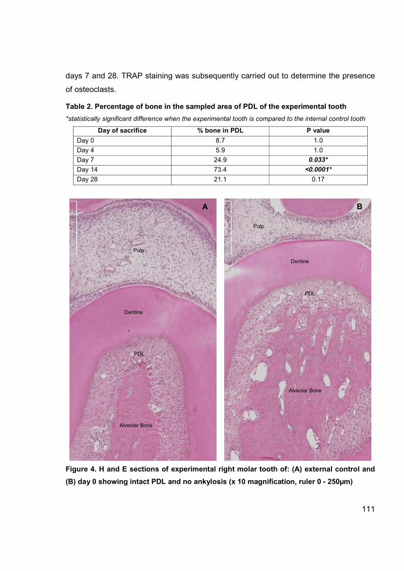
111
days 7 and 28. TRAP staining was subsequently carried out to determine the presence
of osteoclasts.
Table 2. Percentage of bone in the sampled area of PDL of the experimental tooth
*statistically significant difference when the experimental tooth is compared to the internal control tooth
Day of sacrifice % bone in PDL P value
Day 0 8.7 1.0
Day 4 5.9 1.0
Day 7 24.9 0.033*
Day 14 73.4 <0.0001*
Day 28 21.1 0.17
Figure 4. H and E sections of experimental right molar tooth of: (A) external control and
(B) day 0 showing intact PDL and no ankylosis (x 10 magnification, ruler 0 - 250µm)
Pulp
Dentine
A
PDL
Alveolar Bone
B
Dentine
Pulp
PDL
Alveolar Bone

112
Figure 5. H and E sections of experimental right molar tooth of: (A) day 4 showing intact
PDL and no ankylosis and (B) day 7 showing bony trabeculae invading the PDL (x 10
magnification, ruler 0 - 250µm)
PDL
Ankylosis
Dentine
Dentine
Pulp
Pulp
Alveolar Bone Alveolar bone
PDL
A B

113
Figure 6. H and E sections of experimental right molar tooth of: (A) day 14 showing
complete destruction of the PDL and (B) day 28 showing an intact PDL (x 10
magnification, ruler 0 - 250µm)
PDL
Ankylosis
Alveolar Bone Alveolar Bone
Dentine
Dentine
Pulp
Pulp
A B

114
Tartrate-resistant acid phosphatase (TRAP) staining
TRAP staining was conducted to determine the presence of osteoclasts. The external
control, day 4 and day 7 showed a similar expression of TRAPpositive cells. The
TRAPpositive cells were associated with bone surfaces and bone marrow spaces
within the alveolar bone and the PDL at the apex of the root with little expression in the
furcation region (Figure 7).
Figure 7. TRAP-positive cells (pink stain) in the external control (A) and the experimental
tooth at day 0 (B). (x5 magnification, ruler 0-500µm)
Pulp Dentine
A
PDL
Alveolar Bone
B
Dentine
Pulp
PDL
Alveolar Bone

115
At day 7 when the first signs of ankylosis were observed, there was an increase in
expression of TRAPpositive cells in the PDL near the ankylotic region (Figure 8). At
day 14 when significant ankylosis was present, there were few TRAPpositive cells
(Figure 8).
Figure 8. TRAP-positive cells (pink stain) in the experimental teeth at day 7 (A) and day
14 (B). (x5 magnification, ruler 0-500µm)
Pulp
Dentine
A
PDL
Alveolar Bone
B
Dentine Pulp
PDL
Alveolar Bone

116
At day 28, two rats demonstrated complete resolution of the ankylotic region, whilst the
remaining rat demonstrated partial resolution. Both of these areas were associated with
the presence of TRAPpositive cells in the furcation region of the PDL confirming that
resorption of the ankylotic bone took place (Figure 9).
Figure 9. TRAP-positive cells (pink stain) in the experimental teeth at day 28. TRAP-
positive cells localized to the area of resolving ankylosis (A) and regenerated PDL (B).
(x5 magnification, ruler 0-500µm)
Pulp
Dentine
A
PDL
Alveolar Bone
B
Dentine
Pulp
PDL
Alveolar Bone

117
Immunohistochemical detection of RANKL
RANKL was detected in bone marrow stromal cells, osteoblasts and bone lining cells,
osteoclasts, endotheliallike cells lining vessels, epithelial cells (Figure 10), odontoblasts
(Figure 11) and periodontal fibroblasts (Figure 12).
Figure 10. Expression of RANKL (pink)
in bone marrow stromal cells (A), the
basic multi-cellular unit (BMU) (B),
multi-nucleated osteoclast-like cells
(C), endothelial cells (D) and epithelial
cells (E). Counterstained with
haematoxylin and lithium carbonate.
(x30 magnification, ruler 0-100µm)
A B
C D
E

118
RANKL expression in the pulp
In the external control animals not subjected to freezing there was a punctate pattern of
positive RANKL staining which was confined to the outer layer of odontoblastlike cells
only. There was no evidence of positive staining in the inner cellrich region of the pulp.
At this stage, the pulp chamber was large with discrete definition between the outer
odontoblastlike layer and the inner cellrich zone (Figure 11).
At day 0, there was less RANKL staining in the experimental teeth when compared with
the internal control tooth but the pulp chamber was still large and the defined
odontoblast layer was visible (Figure 11).
At day 4, significant changes were evident in the pulp with the loss of the odontoblast
layer and a reduction in overall cellularity. Some RANKL positive cells were present
within the remnants of the odontoblast layer but were greatly reduced in number and
there was now evidence of RANKL positive cells within the inner region of the pulp
(Figure 11).
By day 7, the cellularity had further reduced and there were few RANKL positive cells
scattered within the remains of the pulp tissue. This effect was localised to the pulp
chamber. The radicular pulp in the tooth root appeared unaffected and resembled the
internal control (Figure 11).
At day 14, the appearance of the pulp was variable between animals. Some animals
showed calcifications within the pulp chamber and there was an increase in cellularity
and positive RANKL expression compared with day 7 (Figure 11).
At day 28, the pulp chamber more closely resembled that of the control with the re
establishment of the outer odontoblast cell layer and positive RANKL staining in this
region which was less intense than the internal control. Significant reparative dentine
had been laid down and the pulp chambers were smaller in size than the internal
controls (Figure 11).

119
Figure 11. RANKL expression in the pulp of a rat in the experimental teeth: (A) External
control, (B) day 0, (C) day 4, (D) day 7, (E) day 14, (F) day 28. RANKL positive cells stain
pink, RANKL negative stain blue (haematoxylin counter-stain). (x 20 magnification)
A B C
D F E

120
RANKL expression in the periodontal ligament
The periodontal ligament contained cells that stained positive for RANKL. The external
control animals and those sacrificed at days 0 and 4 contained a number of fibroblast
like cells showing weak cytoplasmic expression of RANKL. There was also strong
punctate expression of RANKL associated with the epithelial rests of Malassez (Figure
12, 13).
At day 7 when ankylosis was first visible, the weak cytoplasmic expression of RANKL in
the fibroblastlike PDL remains. ERM were not commonly visualised at this time point
(Figure 12).
At day 14, the ankylosis was at its maximum with almost complete obliteration of the
PDL. Some small islands remained with similar weak cytoplasmic expression of RANKL
similar to the control. No ERM were visible in the experimental teeth with ankylosis but
were still found in the internal control tooth (Figure 12).
At day 28, two of the experimental animals demonstrated intact PDL including the
presence of ERM, similar in appearance to the controls (Figure 12).

121
Figure 12. RANKL expression in the PDL of a rat in the experimental teeth: (A) External
control (see figure 13 for detail), (B) day 7, (C) day 14, (D) day 28. RANKL positive cells
stain pink, RANKL negative cells stain blue (haematoxylin counter-stain). Open arrows
indicate ERM. (x20 magnification)
Figure 13. Close up view of Figure 12 to show ERM (x30 magnification)
B A
C D
PDL
PDL
PDL
Ankylosis
Ankylosis
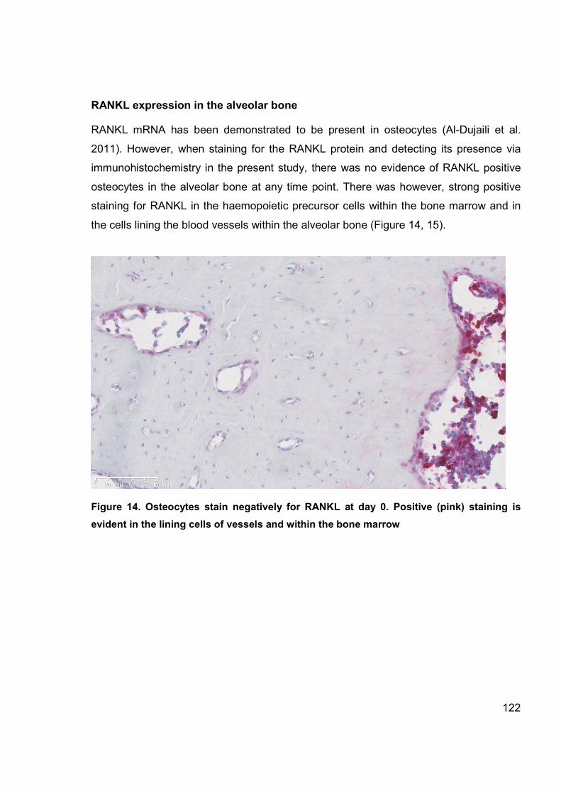
122
RANKL expression in the alveolar bone
RANKL mRNA has been demonstrated to be present in osteocytes (AlDujaili et al.
2011). However, when staining for the RANKL protein and detecting its presence via
immunohistochemistry in the present study, there was no evidence of RANKL positive
osteocytes in the alveolar bone at any time point. There was however, strong positive
staining for RANKL in the haemopoietic precursor cells within the bone marrow and in
the cells lining the blood vessels within the alveolar bone (Figure 14, 15).
Figure 14. Osteocytes stain negatively for RANKL at day 0. Positive (pink) staining is
evident in the lining cells of vessels and within the bone marrow

123
Figure 15. Alveolar bone at day 14 (maximal ankylosis). Empty lacunae are present but
osteocytes are negative for RANKL
7.5 Discussion
This investigation used an established experimental model to induce dentoalveolar
ankylosis in rats (Dreyer et al. 2000). Seven days after the application of a cold thermal
insult signs of ankylosis were present. After 14 days the PDL in the furcation region was
completely obliterated. Interestingly, after 28 days two of the three rats showed
regeneration of the PDL and were indistinguishable from the controls. This investigation,
along with previous research, found that there was no preceding resorption in the
development of dentoalveolar ankylosis in this model (Curl et al. 2014). The ankylosis in
this model appears to be of the appositional type which occurs when the PDL is
replaced by bone without prior resorption of cementum (Andreasen 1980).

124
In the control animals and at days 0 and 4 prior to development of ankylosis, there were
few TRAPpositive osteoclasts in the PDL at the furcation region. At this stage
numerous osteoclasts were present in association with vessels and the apical PDL of
the rat molar. Once ankylosis began to develop 7 days after the application of the cold
insult, some TRAPpositive osteoclasts were evident in the PDL near the region of
ankylosis. By day 14, however, there was an absence of osteoclasts in the PDL
adjacent to the ankylotic area and in the nearby blood vessels. After 28 days ankylosis
remained in one rat but the PDL showed signs of repair and there was an increase in
the number of osteoclasts in the PDL bordering the ankylotic area. In the two rats in
which the ankylosis had completely resolved, there was still evidence of an increased
number of osteoclasts in the furcation region of the PDL compared with controls. This
indicated that when the ankylosis was developing there was a lack of osteoclasts and
bone formation was favoured. However, it appeared that the reduced number of
osteoclasts was not permanent as regeneration of the PDL was associated with the
appearance of osteoclasts in the PDL. This may have resulted in resorption of the
ankylotic bone, thus allowing the PDL to regenerate. The junction between the newly
formed PDL and the cementum at the furcation region appeared uneven when
compared with the control teeth and earlier time points. This illustrated that resorption
had taken place in both the ankylotic bone and the cementum.
The resolution of the ankylotic lesion may partly be related to the experimental model.
The investigated ankylosis was the result of a thermal insult, whereas in a clinical
situation, the factors that initiate ankylosis are incompletely understood and may differ
from this model. The absence of the thermal insult at the later time points may
contribute to the regeneration of the PDL and resolution of the ankylosis.
One of the signalling mechanisms behind this may be RANKL. RANKL is known to
stimulate osteoclast differentiation and function. The roles of RANKL include the fusion
of osteoclast precursors into multinucleated cells (Lacey et al. 1998), their differentiation
into mature osteoclasts (Lacey et al. 1998), their attachment to bone surfaces (O’Brien
et al. 2000), their activation to resorb bone (Burgess et al. 1999; Lacey et al. 1998) and

125
aiding osteoclast survival by avoiding apoptosis (Lacey et al. 2000). A reduction in
RANKL expression may lead to a reduction in the number of functioning osteoclasts.
When the damaged PDL is repaired following trauma, one of the factors that might
influence whether dentoalveolar ankylosis remains or whether bone is resorbed and a
new ligament allowed to form, may be the presence of RANKL.
In this investigation, RANKL was detected in osteoblasts and bone lining cells, ERM
and fibroblasts, odontoblasts and marrow cells. Recent research has suggested that the
osteocyte is a major source of RANKL involved in bone resorption and remodelling
(Nakashima et al. 2011; Xiong and O’Brien 2012). In the current investigation, the
osteocytes in proximity to the furcation region were destroyed. This was illustrated by
the number of empty lacunae, which significantly increased at days 7 and 14 when
ankylosis was present. If osteocytes are indeed a major source of RANKL, it could be
hypothesised that osteocyte death resulted in a reduction in RANKL expression which
may have led to a reduction in osteoclast activation and therefore less bone resorption
in the response to the thermal trauma.
Evidence from bone fatigue models suggests that osteocytes produce membrane
bound RANKL in low doses under normal circumstances (Kennedy et al. 2012). What
happens after microtrauma and osteocyte death in regards to RANKL expression is not
clear. In vivo research indicates that it is the neighbouring healthy osteocytes that up
regulate RANKL production following trauma (Kennedy et al. 2012), whereas, an in vitro
experiment suggested that it is the apoptotic osteocytes that are a source of RANKL (Al
Dujaili et al. 2011). In the current investigation osteocytes were not positive for RANKL.
The area of primary examination included a region where a large number of dead
osteocytes were present. According to Kennedy et al (2012), apoptotic osteocytes do
not produce RANKL. Osteocytes located further away from the area of injury were
reported to produce RANKL. This was not observed in the current investigation. There
are two possible reasons for this. The osteocytes in the neighbouring region, although
not destroyed, may still have had key functions damaged by the hypothermic insult and
were not producing RANKL. Another explanation is that the current method of using

126
immunohistochemistry with a polyclonal antibody and light microscopy was inadequate
to detect small but possibly important levels of RANKL in the osteocytes, despite
detecting RANKLpositive cells in other tissues such as the pulp, epithelial rests of
Malassez and the bone marrow. The polyclonal antibody may not have been specific
enough. Previous studies (Kennedy et al. 2012) have detected osteocyteexpressed
RANKL in histological sections using fluorescence or have detected the presence of
RANKL mRNA (AlDujaili et al. 2011).
In health, the PDL must have the capacity to resist induction of osteogenesis through
cell signalling systems that “measure” and maintain the PDL width ensuring that the root
and alveolar bone remain separate (Melcher 1970; McCulloch 1995). Ankylosis occurs
when the PDL is repopulated with cells from a source outside the PDL such as the
alveolar bone. The presence of vital PDL cells appears crucial for repair and
regeneration of a functional PDL. In the current investigation, the ERM were associated
with strong punctate expression of RANKL. ERM were evident in the control animals
and at day 0 and 4. They were not evident at day 7 in association with the
establishment of ankylosis, even though the majority of the PDL was still intact. At day
28 when the PDL appeared intact ERM were clearly visualised again. It is possible that
the presence of the ERM is related to the presence of a normal PDL or ankylosis.
Experiments with the reimplantation of teeth after varied periods of extraoral drying
time demonstrated that the presence of a normal PDL over ankylotic repair may be
related to the presence of epithelial rests of Malassez (Löe and Waerhaug 1961). The
RANKL produced by the ERM might be one of the signalling methods involved in
maintenance of the PDL.
There are limitations to the current investigation. Occasionally osteocytes indicated
positivity for RANKL but were always in extremely close association to the PDL or
vessels. Therefore, it was hard to determine whether these cells were truly osteocytes
staining positive for RANKL or whether the plane of section made cells from the vessels
and ligament appear to be embedded in the bone. The use of a rat model has
advantages in that the PDL is similar to that of humans and there is little genetic

127
variability between animals. However, as with all animal studies, the data obtained must
be viewed with caution before applying the results to humans. The sample size in this
study was small with three rats in each group. Repeating the experiment with more rats
at each time point will increase the reliability of the findings, particularly related to the
possible healing of the ankylotic lesion. Having time points closer together, particularly
between days 14 and 28 will also allow for a more detailed analysis of the progression
of the ankylosis.
7.6 Conclusions
The development of ankylosis and the subsequent regeneration of the PDL is a
complex process controlled by many signalling factors. It is possible that RANKL plays a
role in the process, although this investigation did not indicate that osteocytederived
RANKL is a major factor. However, the following conclusions were drawn:
1) The null hypothesis that ‘an applied cold insult and subsequent ankylosis does
not affect the production of RANKL and that the amount and distribution of
RANKL will not be affected’, is rejected.
2) Changes were observed in the expression of RANKL within the pulp and PDL
subsequent to a freezing insult.
3) No changes were observed in the expression of RANKL by osteocytes in the
alveolar bone subsequent to a freezing insult.
4) The difference in the number of empty lacunae between zones indicated that the
freezing insult results in the death of osteocytes which coincides with the timing
of maximal ankylosis.
5) ERM strongly expressed RANKL and their presence or absence may play a role
in the regeneration of the PDL following dentoalveolar ankylosis.
6) A role of osteocytederived RANKL in dentoalveolar ankylosis is not observed in
this investigation but cannot be conclusively ruled out. More sensitive detection
methods may need to be employed in the future.

128
7.7 References
AlDujaili, S.A. et al., 2011. Apoptotic osteocytes regulate osteoclast precursor recruitment and differentiation in vitro. Journal of cellular biochemistry, 112(9), pp.2412–23.
Anderson, D.M. et al., 1997. A homologue of the TNF receptor and its ligand enhance Tcell growth and dendriticcell function. Nature, 390(6656), pp.175–9.
Andreasen, J.O., 1980. A timerelated study of periodontal healing and root resorption activity after replantation of mature permanent incisors in monkeys. Swedish dental journal, 4, pp.101–110.
Van Bezooijen, R.L. et al., 2004. Sclerostin is an osteocyteexpressed negative regulator of bone formation, but not a classical BMP antagonist. The Journal of Experimental Medicine, 199(6), pp.805–14.
Bucay, N. et al., 1998. OsteoprotegerinDeficient Mice Develop Early Onset Osteoporosis and Arterial Calcification. Genes & development, 12(9), pp.1260–8.
Burgess, T.L. et al., 1999. The ligand for osteoprotegerin (OPGL) directly activates mature osteoclasts. The Journal of cell biology, 145(3), pp.527–38.
Curl, L. et al., 2014. A qualitative investigation of RANKL, RANK and OPG in a rat model of transient ankylosis. Australian Orthodontic Journal, 30(2), pp.143–151.
Dreyer, C., Pierce, A. & Lindskog, S., 2000. Hypothermic insult to the periodontium: a model for the study of aseptic tooth resorption. Endodontics & Dental Traumatology, 16, pp.9–15.
Epker, B.N. & Frost, H.M., 1965a. A histological study of remodeling at the periosteal, haversian canal, cortical endosteal, and trabecular endosteal surfaces in human rib. Anatomical Record, 152, pp129135
Epker, B.N. & Frost, H.M. 1965b. Correlation of bone resorption and formation with the physical behavior of loaded bone. Journal of Dental Research, 44, pp.3341
Erausquin, J. & Devoto, F.C., 1970. Alveolodental ankylosis induced by root canal treatment in rat molars. Oral Surgery, 30(1), pp.105–116.
Gu, G. et al., 2005. Death of osteocytes turns off the inhibition of osteoclasts and triggers local bone resorption. Biochemical and biophysical research communications, 335(4), pp.1095–101.
Henriksen, K. et al., 2009. Local communication on and within bone controls bone remodeling. Bone, 44(6), pp.1026–33.

129
Ikeda, T. et al., 2001. Determination of Three Isoforms of the Receptor Activator of Nuclear FactorκΒ Ligand and Their Differential Expression in Bone and Thymus. Endocrinology, 142(4), pp.1419–1426.
Kearns, A.E., Khosla, S. & Kostenuik, P.J., 2008. Receptor activator of nuclear factor kappaβ ligand and osteoprotegerin regulation of bone remodeling in health and disease. Endocrine reviews, 29(2), pp.155–92.
Kennedy, O.D. et al., 2012. Activation of resorption in fatigueloaded bone involves both apoptosis and active proosteoclastogenic signaling by distinct osteocyte populations. Bone, 50(5), pp.1115–22.
Kurata, K. et al., 2006. Bone Marrow Cell Differentiation Induced by Mechanically Damaged Osteocytes in 3D GelEmbedded Culture. Journal of Bone and Mineral Research, 21(4), pp.616–625.
Kurol, J., 1981. lnfraocclusion of primary molars: an epidemiologic and familial study. Community Dentistry and Oral Epidemiology, 9, pp.94–102.
Lacey, D.L. et al., 1998. Osteoprotegerin ligand is a cytokine that regulates osteoclast differentiation and activation. Cell, 93(2), pp.165–76.
Lacey, D.L. et al., 2000. Osteoprotegerin ligand modulates murine osteoclast survival in vitro and in vivo. The American journal of pathology, 157(2), pp.435–48.
Lin, D. et al., 2000. Storage conditions of avulsed teeth affect the phenotype of cultured human periodontal ligament cells. Journal of periodontal research, 35(1), pp.42–50.
Line, S., Polson, A. & Zander, H., 1974. Relationship between periodontal injury, selective cell repopulation & ankylosis. Journal of periodontology, 45(10), pp.72530.
Löe, H. & Waerhaug, J., 1961. Experimental replantation of teeth in dogs and monkeys. Archives of oral biology, 3(1935), pp.176–84.
McCulloch, C., 1995. Origins and functions of cells essential for periodontal repair: the role of fibroblasts in tissue homeostasis. Oral Diseases, 1, pp.271–278.
Melcher, A., 1970. Repair of Wounds in the Periodontium of the Rat. Influence of the Periodontal Ligament on Osteogenesis. Archives of Oral Biology, 15, pp.1183–1204.
Mizuno, A et al., 1998. Severe osteoporosis in mice lacking osteoclastogenesis inhibitory factor/osteoprotegerin. Biochemical and biophysical research communications, 247(3), pp.610–5.

130
Nakashima, T. et al., 2011. Evidence for osteocyte regulation of bone homeostasis through RANKL expression. Nature medicine, 17(10), pp.1231–4.
Noble, B.S. et al., 2003. Mechanical loading: biphasic osteocyte survival and targeting of osteoclasts for bone destruction in rat cortical bone. American journal of physiology. Cell physiology, 284(4), pp.C934–43.
O’Brien, E. a, Williams, J.H. & Marshall, M.J., 2000. Osteoprotegerin ligand regulates osteoclast adherence to the bone surface in mouse calvaria. Biochemical and biophysical research communications, 274(2), pp.281–90.
Poole, K.E.S. et al., 2005. Sclerostin is a delayed secreted product of osteocytes that inhibits bone formation. FASEB journal, 19(13), pp.1842–4.
Simonet, W. et al., 1997. Osteoprotegerin: a novel secreted protein involved in the regulation of bone density. Cell, 89(2), pp.309–19.
Tan, C., 2011. A qualitative investigation of RANKL, RANK and OPG in a rat model of transient ankylosis. Honors Project, The University of Adelaide, South Australia
Tatsumi, S. et al., 2007. Targeted ablation of osteocytes induces osteoporosis with defective mechanotransduction. Cell metabolism, 5(6), pp.464–75.
Theoleyre, S. et al., 2004. The molecular triad OPG/RANK/RANKL: involvement in the orchestration of pathophysiological bone remodeling. Cytokine & growth factor reviews, 15(6), pp.457–75.
Verborgt, O. et al., 2002. Spatial Distribution of Bax and Bcl2 in Osteocytes After Bone Fatigue: Complementary Roles in Bone Remodeling Regulation? Journal of Bone and Mineral Research, 17(5), pp.60–7.
Wijenayaka, A.R. et al., 2011. Sclerostin stimulates osteocyte support of osteoclast activity by a RANKLdependent pathway. PloS one, 6(10), p.e25900.
Xiong, J. et al., 2011. Matrixembedded cells control osteoclast formation. Nature medicine, 17(10), pp.1235–41.
Xiong, J. & O’Brien, C., 2012. Osteocyte RANKL: new insights into the control of bone remodeling. Journal of bone and mineral research, 27(3), pp.499–505.
Yasuda, et al., 1998. Osteoclast differentiation factor is a ligand for osteoprotegerin/ osteoclastogenesisinhibitory factor and is identical to TRANCE/RANKL. Proceedings of the National Academy of Sciences of the USA, 95(7), pp.3597–602.
Zhao, S. et al., 2002. MLOY4 OsteocyteLike Cells Support Osteoclast Formation and Activation. Journal of Bone and Mineral Research, 17(11), pp.2068–2079.

131
8 ARTICLE 2
Expression of sclerostin in a rat model of dentoalveolar ankylosis
following hypothermal insult
Written in the style of European Journal of Orthodontics

132
8.1 Abstract
Background: Dentoalveolar ankylosis may occur spontaneously or as a sequela to
dental trauma and results in an area of bone fusing to the tooth. The understanding of
the biological processes behind the formation and repair of this lesion is incomplete.
Sclerostin is a protein produced by osteocytes that is known to inhibit bone formation.
The present study investigates the expression of sclerostin in a rat model of
dentoalveolar ankylosis.
Methods: Dentoalveolar ankylosis was induced in fifteen eight week old Sprague
Dawley rats (5 groups of 3 rats each) by the application of dry ice to the upper right first
molar tooth. An additional 3 rats served as untreated controls and the experimental rats
were sacrificed at days 0, 4, 7, 14 and 28. Immunohistochemical detection of sclerostin
was performed and the number of sclerostinpositive and negative cells as well as the
number of empty lacunae representing dead osteocytes were calculated and compared
between groups.
Results: When ankylosis was present, there was a statistically significant difference in
sclerostin expression between the areas of bone closest to, and farthest away, from the
furcation area. There was also a statistically significant increase in the number of empty
lacunae due to osteocyte death that coincides with the incidence of maximal ankylosis.
There was a nonstatistically significant trend towards reduced sclerostin expression at
days 7 and 14 followed by a slight increase in expression at day 28. The slight increase
in sclerostin expression at day 28 may indicate the establishment of a healing response.
Conclusion: The possible repair of the periodontal ligament (PDL) following trauma is
complex and the present study may contribute evidence to the possibility that the
osteocyte and sclerostin might have a role in influencing the regeneration of the PDL.
Keywords: sclerostin, ankylosis, dentoalveolar, osteocyte, immunohistochemistry

133
8.2 Introduction
Osteocytes sense loading stimuli and regulate remodelling and bone turnover
processes but little is known about how cells embedded in a mineralised matrix carry
out this regulatory role. It has been suggested that mature osteocytes send an inhibitory
signal to bone lining osteoblasts to maintain these cells in a quiescent state against their
natural tendency to reactivate the remodelling process (Poole et al. 2005). If the emitted
signal was sensitive to mechanical loading, the correct balance could be obtained
between bone formation and its inhibition. This signal has been identified as sclerostin
which is the secreted protein product of the SOST gene. Sclerostin is a negative
regulator of osteoblasts which, therefore, inhibits bone formation and appears to be
exclusively osteocyte derived (van Bezooijen et al. 2004). It exerts its effects by
interfering with Wnt signaling, a process which plays an essential role in the regulation
of bone formation (van Bezooijen et al. 2004; Li et al. 2005; Semënov et al. 2005).
A lack of sclerostin in humans results in the rare conditions of sclerosteosis and van
Buchem disease (VBD), first described in the 1950’s (van Buchem et al. 1955; Truswell
1958). The skeletal manifestations of both diseases are the result of endosteal
hyperostosis and are characterised by progressive generalised osteosclerosis. The
manifestations are most pronounced in the mandible and skull. Those patients with
sclerosteosis have a more severe phenotype compared with patients with van Buchem
disease (Moester et al. 2010). Bone biopsies of those affected show increased bone
formation including a predominance of active osteoblasts (Moester et al. 2010).
The effects of sclerostin have been shown experimentally by using SOST knockout
mice which show increased radiodensity, bone mineral density, cortical and trabecular
volume, bone formation and bone strength (Poole et al. 2005). The main mechanism of
action of sclerostin is via its inhibition of the Wnt signalling pathway. Activation of the
Wnt signalling pathway results in gene transcription that increases osteoblastic cell
differentiation and may also inhibit bone resorption by blocking the receptor activator
RANKL/RANK (Jackson et al. 2005). Sclerostin acts to block this pathway by binding to

134
the cell surface receptors LRP5 and LRP6 which subsequently prevents gene
transcription (Li et al. 2005). Sclerostin may also promote apoptosis of osteoblasts,
further inhibiting bone growth (Sutherland et al. 2004).
Dentoalveolar Ankylosis
Dentoalveolar ankylosis is defined as the fusing of cementum with alveolar bone (Kurol,
1981). Ankylosis is commonly found in two clinical situations: those of infraocclusion
which mainly affects deciduous or primary teeth, and in the permanent dentition where it
is associated with impaction or external root replacement resorption following trauma
and is frequently observed after reimplantation of avulsed permanent teeth. Ankylosed
teeth often present at a level below the occlusal plane, a condition referred to as
submergence or infraocclusion. This results from the inability of the ankylosed tooth to
follow the normal vertical development of the neighbouring teeth that are continuing to
develop and erupt.
Following trauma dentoalveolar ankylosis may occur as part of the repair process. A
healthy periodontal ligament has the capacity to resist induction to osteogenesis
through cell signalling systems that accurately ‘measure’ and maintain the PDL width,
ensuring separation of the root and alveolar bone (Melcher 1970; McCulloch 1995).
Traumatic injury disrupts this homeostasis. Investigations indicate it is the source of the
cells that repopulate the PDL space following trauma or injury that determine whether or
not ankylosis occurs (Erausquin and Devoto 1970; Lin et al. 2000; Melcher 1970; Line
et al. 1974). When the space is repopulated by cells from a source outside the true PDL
tissues (alveolar bone, bone marrow or soft connective tissue) osteoclasis and
osteogenesis occur, resulting in healing via a dentoalveolar ankylosis.
Infraocclusion following dentoalveolar ankylosis results in a number of significant
sequelae including overeruption of the opposing tooth. Adjacent teeth can tip towards
the ankylosed tooth which may lead to a loss of arch length, space loss, a shift of the
dental midline and difficulty with future extraction of the infraoccluded tooth (Messer
and Cline 1980; Andlaw 1974; Ponduri et al. 2009). Vertical alveolar growth may also be

135
hindered (Kjaer et al. 2008). When the ankylosed primary mandibular second molar has
no successor, early orthodontic and prosthodontic consultation is recommended. The
patient’s age, occlusal status and the development and condition of the infraoccluded
tooth will influence treatment requirements. Consideration may be given to the
maintenance of the ankylosed tooth, prosthetic replacement including implants and
orthodontic space closure (Kurol and Thilander 1984; Ekim and HatibovicKofman
2001).
Sclerostin in Dentistry
Sclerostin has attracted significant attention in the medical field, mainly in its role as a
potential therapy for osteoporosis. Antibodies to sclerostin have already been shown to
increase the bone formation rate and bone density in rats (Li et al 2009) and, in clinical
trials involving healthy postmenopausal women, increased bone formation markers and
bone mineral density were observed (Padhi et al. 2011).
Sclerostin appears to affect the alveolar bone and cementum. Sclerostindeficient mice
demonstrated increased mandibular basal bone dimensions and mildly increased
cementum thickness (Kuchler et al. 2014). Patients with van Buchem disease have
shown little or no sclerostin expression by cementocytes (van Bezooijen et al. 2007). It
has been suggested that the resultant hypercementosis produces a tighter junction
between the tooth and alveolar bone and may be the reason behind the reported
difficulty in extracting teeth (van Bezooijen et al. 2007). However, small sample sizes
meant the exact differences in histology could not be precisely determined and
sclerostin deficiency in cementocytes was not clearly associated with overt changes in
cementum thickness (van Bezooijen et al. 2007). Sclerostin does not appear to produce
anomalies in tooth structures such as dentine and the pulp chamber (Kuchler et al.
2014).

136
Sclerostin expression in alveolar bone appears to be influenced by orthodontic forces.
Tensile areas of bone demonstrated a downregulation of sclerostin and in areas of
compression, sclerostin was significantly higher than control sites (Sanmuganathan
2011; Nishiyama et al. 2015). It seems that when significant local strains are sensed by
the osteocyte population on the side of tension, sclerostin levels decreased and cells
were released from their suppressed Wnt signalling state and new bone was formed.
Conversely, on the side of compression, sclerostin levels were increased with continued
Wnt signalling suppression, leading to prevention of local bone formation
(Sanmuganathan 2011).
A clinical application of sclerostin has been demonstrated by the use of
sclerostin‐neutralising monoclonal antibody (Scl‐Ab) in rats with experimentally induced
periodontitis. The application of SclAb resulted in increased alveolar bone volume and
density measurements, making the experimental animals comparable to normal, healthy
controls (Taut et al. 2013).
To date, no investigations have examined the role of sclerostin in the development of
dentoalveolar ankylosis. The aim of the present investigation was to determine if the
production and expression of sclerostin is altered during the formation of dentoalveolar
ankylosis in a rat model. The null hypothesis states that ‘an applied cold insult and
subsequent ankylosis does not affect the expression of sclerostin within the alveolar
bone of exposed teeth. The amount and distribution of sclerostin will not be affected’.

137
8.3 Methods and materials
The experimental procedures were reviewed and approved by the University of
Adelaide Animal Ethics Committee for a previous study (M0832007).
Experimental procedure:
The experimental sample consisted of 18 eight week old, male, SpragueDawley rats.
Fifteen rats were subjected to a hypothermal insult and 3 rats served as an untreated
control. The rats were anaesthetised before the procedure and sacrificed using an
intramuscular injection of Hypnorm® (fentanyl citrate 0.315mg/ml and fluanisone
10mg/ml, JanssenCilag Ltd., High Wycombe, Buckinghamshire, UK) and Hypnovel®
(midazolam hydrochloride 5mg/ml, Roche Products Pty Ltd., Dee Why, Australia). The
anaesthetic agents were diluted in a 1:1 ratio with sterile water, combined and
administered at a dosage of 2.7ml/kg of body weight. The animals were restrained in a
head holding device and underwent a single 10 minute application of dry ice
(compressed carbon dioxide gas at 81°C, BOC Gases, Adelaide, Australia) to the
occlusal surface of the right maxillary first molar crown in order to produce aseptic
necrosis within the PDL and induce ankylosis (protocol developed by Dreyer et al.
2000). The upper left first molar was untreated and provided an internal control.
The rats were sacrificed on days 0, 4, 7, 14 and 28 respectively. The 3 animals in the
control group did not undergo hypothermal insult and were also sacrificed on day 0.
Following administration of anaesthetic, the rats were sacrificed via intracardiac
perfusion of 4% paraformaldehyde fixative. On completion of perfusion the maxillae
were dissected out and immersed in the same fixative for 24 hours and then stored in
phosphate buffered saline (PBS) at pH 7.3 for another 24 hours. Specimens were
decalcified using 4% ethylenediaminetetraacetic acid (EDTA) solution and were
dehydrated using a graded alcohol series and embedded in paraffin wax. The paraffin
blocks were mounted onto a Leitz 1512 Microtome (Leica, Nussloch, Germany) and
5µm sections were cut coronally through the root furcation. Sections were mounted onto
aminopropyltriethoxysilane (APTS) coated glass slides.

138
Randomly selected sections were stained with haematoxylin and eosin (H and E) to
check for orientation, identification of ankylotic areas and suitability for
immunohistological staining. The preceding experimental protocol and H and E staining
was carried out as part of a prior study (Tan et al 2011).
Immunohistochemical detection of sclerostin:
Fiftyfour sections were randomly selected from within the region of the furcation as
previous research has shown this area to have the most ankylosis (Dreyer et al 2000).
Antibody optimisation was performed prior to immunohistological staining to determine
the optimal antigen unmasking method and antibody dilution required to achieve
minimal background staining and optimal antibody specific staining. Positive control
tissue and negative controls were assessed to determine antibody specificity. Omission
of the primary antibody provided negative controls. The positive control was the alveolar
bone as osteocytes are known to express sclerostin. All staining was performed on the
same day.
The sections underwent paraffin removal and were immersed in trisEDTA buffer
(10mM, pH 9.0) in a 65°C water bath for 10 minutes before cooling for 20 minutes.
Following washing with 1x PBS three times for 5 minutes the sections were immersed in
0.3% v/v H2O2 in PBS/0.1% sodium azide for 20 minutes to block endogenous
peroxidase. The sections were again washed in 1x PBS and the primary sclerostin
antibody was applied (goat antimouse polyclonal SOST/Sclerostin [Cat No. AF1589,
RandD Systems, distributed by BioScientific Pty. Ltd., NSW, Australia]) 4µg/ml
concentration in 1x PBS/1% BSA. Sections were incubated at room temperature
overnight in a wet chamber. The sections were washed again in 1x PBS and the
secondary antibody was applied (swine antigoat Ig’s HRP [Ref no. ACI0404, Invitrogen,
distributed by Life Technologies Australia Pty Ltd, Victoria Australia] 11.7µg/ml in 1x
PBS/1% BSA) for 30 minutes. Sections were washed in PBS and the tertiary antibody
was applied (rabbit antiswine Ig’s [Code No. P 0164, Dako Australia Pty. Ltd., NSW,
Australia] 13µg/ml concentration in 1x PBS/1% BSA) for 30 minutes. Sections were

139
washed in 1x PBS before application of AEC (3Amino9ethylcarbazole) substrate
chromogen (Cat no. SK4200, Dako Australia Pty. Ltd., NSW, Australia) for 10 minutes
at room temperature with the sections covered and placed in the dark. Sections were
then rinsed with MilliQ water, counterstained with haematoxylin (10 seconds) and
lithium carbonate (30 seconds) before final washing with tap water and mounting using
Aquamount.
Histomorphometry:
The immunostained sections were digitised via a Nanozoomer Slide Scanner 2.0 series
(Hamamatsu Photonics K.K. 3256 Sunayamacho, Hamamatsu City, Shizuoka) and
viewed on a personal computer (Lenovo X220) using the Nanozoomer Digital Pathology
(NDP) software. Areas were then defined to enable analysis of the sections.
The periodontal ligament was divided into 5 regions of 0.01mm2 at the furcation region
and the percentage of bone within the PDL was determined to quantitatively assess the
degree of ankylosis viewed at a magnification factor of 20 on the NDP software (Figure
1).
Alveolar bone was delineated into 4 zones with zone 1 closest to the root furcation and
zone 4 closest to the root apex (Figure 1). To ensure consistency of the sampling area
across numerous sections, the boxes in zone 1 were placed 200 µm from the midpoint
of the furcation area of the tooth. Each zone contained 2 boxes of 0.04mm2. Counting of
sclerostin positive and negative cells as well as empty lacunae was performed at a
magnification factor of 20 using Image J software 1.47v (Wayne Rasband, National
Institutes of Health, USA).

140
Figure 1. Sampling regions in the rat molar tooth
Statistical analysis:
The data were analysed using SAS 9.3 (SAS Institute Inc., Cary, NC, USA). The
distribution of sclerostin in the alveolar bone at varying depths from the furcation, the
distribution over time periods and between internal controls and experimental teeth was
analysed using a linear mixed effects model. To account for dependency of
measurements from the same rat a random effect was also included. The significance
level was set at p<0.05.
To assess the reliability of the cell counting, 10% of the sections were randomly
selected and recounted and Intraclass Correlation Coefficients (ICC) were calculated
using SAS 9.3 and Stata/SE 13.1 (StataCorp LP).
200µm

141
8.4 Results
Error study
To assess the reliability of the examination of the histological slides 10% of the sections
were recounted (86 of a total 860 sections). Intraclass Correlation Coefficients (ICC)
were calculated using Stata and are listed, along with 95% confidence intervals, in
Table 1. The ICC describes how strongly units in the same group resemble each other.
It is an assessment of consistency or reproducibility of quantitative measurements made
by the same observer at a different time (or different observers measuring the same
quantity). The ICCs were also calculated in SAS using linear mixedeffects model
results:
ICC= Area Covariance Parameter Estimate / (Area Covariance Parameter Estimate +
Residual)
Within this linear mixedeffects model, the outcome was either positive, negative or
empty and fixed effect (the predictor) was ‘repeat’ (i.e. original measurement or
repeated measurement). ICCs found using SAS were exactly the same as ICCs found
with Stata.
In these models, all of the ICCs were 0.91 and above – an ICC of 1 denotes perfect
agreement. This is evidence that the reliability of the same examiner reexamining the
rat slides is very high.
Table 1. Intraclass Correlation Coefficients
Type of cell ICC 95% CI
Positive 0.909 0.865, 0.939
Negative 0.934 0.901, 0.956
Empty lacunae 0.955 0.933, 0.970

142
Histology
Haematoxylin and eosin stained sections were examined to assess the pulp,
periodontal ligament and the alveolar bone. No differences were noted between the
experimental tooth at day 0 and the internal control tooth when compared with those of
the external control (Figure 2).
Pulp: At days 4 and 7 the experimental tooth showed a distinct reduction in cellularity in
the central stromal region and some reduction in thickness and continuity of the
odontoblast layer (Figure 3). By day 14, a layer of tertiary dentine was visible and was
thicker at day 28. The pulp of the internal control teeth showed no disturbance to its
integrity throughout the experimental period (Figure 4).
PDL: At days 0 and 4, the PDL remained intact and comparable to both the external and
internal controls. The first signs of ankylosis were evident at day 7 in all animals (Figure
3). The PDL width decreased as a result of bony trabeculae invasion in the furcation
region. At day 14, there was widespread loss of the PDL in all animals with dense
ankylotic bone taking its place (Figure 4). By day 28, only one animal showed bone
within the PDL and the other animals revealed an intact PDL almost indistinguishable
from the internal control (Figure 4). There was no change in the PDL of the internal
control teeth throughout the experimental period. The difference in the percentage of
bone within the PDL between the experimental tooth and the internal control was
statistically significant at days 7 and 28 (Table 2).
Alveolar bone: The amount of alveolar bone in the furcation region increased at days 7
and 14 as the PDL was replaced with ankylotic bone (Figure 4). There was a statistically
significant increase in the number of empty lacunae in the furcation region as the
ankylotic lesion developed (Table 3, 4); however, by day 28, there was almost complete
filling of the lacunae by osteocytes (Figure 4).

143
Table 2. Percentage of bone in the sampled area of PDL of the experimental tooth
*statistically significant difference when the experimental tooth is compared to the internal control tooth
Day of sacrifice % bone in PDL P value
Day 0 8.7 1.0
Day 4 5.9 1.0
Day 7 24.9 0.033*
Day 14 73.4 <0.0001*
Day 28 21.1 0.17
Figure 2. H and E sections of experimental right molar tooth of: (A) external control and
(B) day 0 showing intact PDL and no ankylosis (x 10 magnification, ruler 0 - 250µm)
Pulp
Dentine
A
PDL
Alveolar Bone
B
Dentine
Pulp
PDL
Alveolar Bone

144
Figure 3. H and E sections of experimental right molar tooth of: (A) day 4 showing intact
PDL and no ankylosis and (B) day 7 showing bony trabeculae invading the PDL (x 10
magnification, ruler 0 - 250µm)
PDL
Ankylosis
Dentine
Dentine
Pulp
Pulp
Alveolar Bone Alveolar bone
PDL
A B
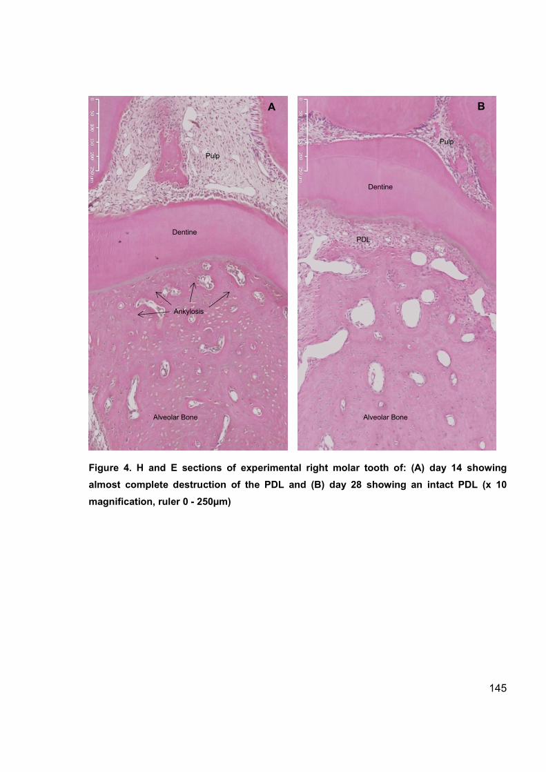
145
Figure 4. H and E sections of experimental right molar tooth of: (A) day 14 showing
almost complete destruction of the PDL and (B) day 28 showing an intact PDL (x 10
magnification, ruler 0 - 250µm)
PDL
Ankylosis
Alveolar Bone Alveolar Bone
Dentine
Dentine
Pulp
Pulp
A B

146
Immunohistochemical detection of sclerostin
Negative controls:
Positive and negative controls were performed to assess antibody specificity. Negative
controls were sections on which the antibody had been omitted. The positive control
was the alveolar bone as osteocytes are known to express sclerostin (van Bezooijen et
al. 2004).
Figure 5. Negative control. Omission of sclerostin antibody. There is an absence of
positive sclerostin staining (red) within cells. (x10 magnification, ruler 0-250µm).
Pulp
Dentine
PDL
Alveolar Bone
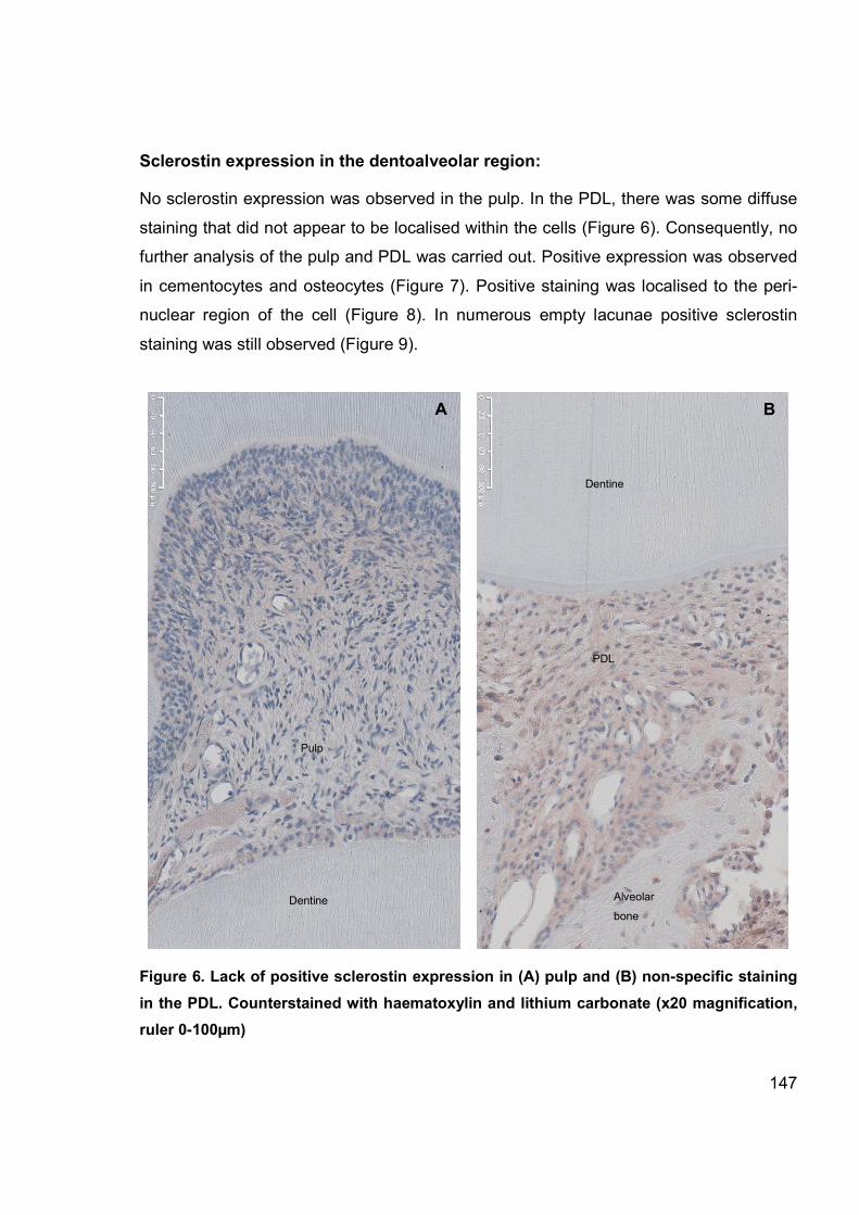
147
Sclerostin expression in the dentoalveolar region:
No sclerostin expression was observed in the pulp. In the PDL, there was some diffuse
staining that did not appear to be localised within the cells (Figure 6). Consequently, no
further analysis of the pulp and PDL was carried out. Positive expression was observed
in cementocytes and osteocytes (Figure 7). Positive staining was localised to the peri
nuclear region of the cell (Figure 8). In numerous empty lacunae positive sclerostin
staining was still observed (Figure 9).
Figure 6. Lack of positive sclerostin expression in (A) pulp and (B) non-specific staining
in the PDL. Counterstained with haematoxylin and lithium carbonate (x20 magnification,
ruler 0-100µm)
Pulp
Dentine
Dentine
PDL
Alveolar
bone
A B

148
Figure 7. Positive sclerostin expression by: (A) cementocytes and (B) osteocytes.
Positive cells stain red, counterstained with haematoxylin and lithium carbonate.
Examples of positive staining are indicated by small arrows, negative staining by large
arrows (x20 magnification, ruler 0-100µm)
Dentine
Cellular cementum
PDL
Alveolar
Bone
Alveolar
Bone

149
Figure 8. Osteocytes showing positive staining for sclerostin in the perinuclear region
(examples indicated by arrows) (x40 magnification, ruler 0-50µm)
Figure 9. Empty lacunae showing absence (large arrows) and presence of positive
sclerostin staining (small arrows) (x40 magnification, ruler 0-50µm)
Sclerost
in
Sclerost
in

150
Sclerostin expression in the alveolar bone
Empty lacunae
There was a clear reduction in cellularity at days 7 and 14 when the dentoalveolar
ankylosis was maximal (Figure 10). There was an increase in the number of empty
lacunae possibly indicating dead osteocytes.
Statistical analysis:
A statistically significant difference in the number of empty lacunae between
experimental and internal control teeth (Table 3) was observed at days 7 and 14 which
indicated that the cold insult and resulting ankylosis caused osteocyte death. There was
also a statistically significant difference (p<0.05) in the number of empty lacunae
between experimental and internal control teeth in zone 1 (Table 4) which was the area
closest to the furcation. This indicated that the osteocytes closest to the insult were
more affected than cells distant to the cold insult (Figure 10).
Table 3. Empty lacunae counts: Interaction of group and day of sacrifice
Day of Sacrifice Number of empty cells/ mm2
Adjusted P value Experimental side Internal control side
External control 0.0 0.0 0.1000
0 0.0 0.0 0.1000
4 2.0 0.2 1.000
7 17.0 0.2 0.0115*
14 16.7 1.1 0.0304*
28 1.9 0.3 1.0000
Table 4. Empty lacunae counts: Interaction of group and zone
Zone Number of empty cells/ mm2
Adjusted P value Experimental side Internal control side
1 14.0 0.4 <0.0001*
2 8.0 0.1 0.393
3 2.5 0.3 1.0000
4 0.5 0.4 1.0000

151
Figure 10. Empty lacunae and lack of osteocytes near the furcation area in an animal
sacrificed at day 14 showing significant ankylosis (x 10 magnification, ruler 0 - 250µm)
Left maxillary molar (internal control) is on the left, Right maxillary molar (Experimental
freezing) is on the right.
Sclerostin positive osteocytes
The expression of sclerostin was examined at varying distances from the furcation
region. Figure 11 shows the expression of sclerostin in zone 1 (closest to the furcation)
at various time points. The most distinctive observation was the reduction in osteocyte
numbers in association with the development of the ankylotic lesion (Figure 11D, E).
Zone of
empty
lacunae
Dentine Dentine
PDL
PDL
Alveolar
bone
Alveolar bone
Ankylosis

152
Figure 11. Expression of sclerostin in osteocytes in alveolar bone (zone 1) in the
experimental teeth: (A) External control, (B) day 0, (C) day 4, (D) day 7, (E) day 14, (F) day
28. Red indicates sclerostin positive osteocytes, blue indicates sclerostin negative
osteocytes (haematoxylin and lithium carbonate counterstain). (x20 magnification, ruler
0-100µm)
A C B
F E D

153
Statistical analysis:
The first model (Table 5, 6, 7) looks at the association between sclerostin positive cells
(outcome variable) and internal control and experimental sides of the rat, the day of
sacrifice and zone. A linear mixedeffects model was used with random effects included
in the model to account for clustering. Least square means were calculated for each
variable (Table 5). The zone had a statistically significant association with positive cell
count (global P value<0.0001) (Table 6)
Table 5. Least squares means for outcome: sclerostin positive cells versus time, zone
and side (C= internal control tooth, E= experimental tooth)
Predictor Zone Side Time Estimate Standard Error
Time External control 27.1304 4.7089
Time Day 0 36.1858 4.7089
Time Day 4 39.8146 4.7089
Time Day 7 27.6622 4.7089
Time Day 14 35.2308 4.7089
Time Day 28 31.0412 4.7089
Side C 34.6221 2.4720
Side E 31.0662 2.4720
Zone 1 27.6761 2.2264
Zone 2 32.3781 2.2264
Zone 3 35.0013 2.2264
Zone 4 36.3212 2.2264
Table 6. Global P values for outcome: sclerostin positive cells vs time, zone and side
Predictor Global P value
Time 0.3301
Zone <0.0001*
Side 0.2685
Controlling for the variables, day of sacrifice and side of the rat (experimental vs internal
control) there was a statistically significant difference between the least squares means
for sclerostin positive cells in zone 1 (closest to furcation) and zone 3 (adjusted P value
= 0.0005) and zone 1 and 4 (adjusted P value = <0.0001) after adjusting for multiple
comparisons using the Sidak adjustment (Table 7).

154
Table 7. Differences of Least Squares Means for outcome: positive cells versus zone
Zone Zone Cell count
estimate
Adjusted P value Adjusted lower
95% CI
Adjusted upper
95% CI
1 2 4.7020 0.0626 9.5514 0.1474
1 3 7.3252 0.0005* 12.1746 2.4758
1 4 8.6451 <0.0001* 13.4945 3.7957
2 3 2.6232 0.6318 7.4726 2.2262
2 4 3.9431 0.1781 8.7925 0.9063
3 4 1.3199 0.9784 6.1693 3.5295
The second model assessed the association between sclerostin positive cells (outcome
variable) and side of the rat, day of sacrifice, zone and interaction of side and zone.
Least square means were calculated for each variable (Table 8). There was a
significant interaction between zone and side when regressed against positive cell
count, adjusting for day of sacrifice (interaction P value=0.0033) (Table 9).
Table 8. Least Squares Means for outcome: sclerostin positive cells versus time and
zone*side (C=internal control tooth, E=experimental tooth)
Predictor Zone Side Time Estimate Standard Error
Time External control 27.1304 4.7089
Time Day 0 36.1858 4.7089
Time Day 4 39.8146 4.7089
Time Day 7 27.6622 4.7089
Time Day 14 35.2308 4.7089
Time Day 28 31.0412 4.7089
Zone*Side 1 C 32.5044 2.9266
Zone*Side 1 E 22.8477 2.9266
Zone*Side 2 C 35.2858 2.9266
Zone*Side 2 E 29.4704 2.9266
Zone*Side 3 C 36.0074 2.9266
Zone*Side 3 E 33.9952 2.9266
Zone*Side 4 C 34.6907 2.9266
Zone*Side 4 E 37.9517 2.9266

155
Table 9. Global P values for outcome: positive cells versus time and zone*side
Predictor Global P value
Time 0.3302
Zone <0.0001*
Side 0.2685
Zone*Side 0.0033*
When controlling for the day of sacrifice, for the experimental tooth of the rats, zone 1
had a mean positive cell count significantly less than zone 3 (adjusted P value=0.0005)
as did zones 1 compared with zone 4 (adjusted P value=<0.0001) and zone 2
compared with zone 4 (adjusted P value=0.0277). These results were statistically
significant, even when adjusted for multiple comparisons (Table 10). There was no
difference in sclerostin positive cell counts for the internal control tooth.
Table 10. Differences of Least Squares Means for outcome: positive cells versus time
and zone*side (C=internal control tooth, E=experimental tooth)
Predictor Zone Side Zone Side Estimate Raw P
value
Adjusted
P value
Adjusted
Lower
95% CL
Adjusted
Upper
95% CL
Zone*Side 1 C 1 E 9.6567 0.0118 0.2827 2.3206 21.6341
Zone*Side 1 C 2 C 2.7813 0.2776 0.9999 10.8088 5.2461
Zone*Side 1 C 3 C 3.5030 0.1717 0.9949 11.5305 4.5245
Zone*Side 1 C 4 C 2.1863 0.3933 1.0000 10.2138 5.8412
Zone*Side 1 E 2 E 6.6228 0.0100 0.2450 14.6502 1.4047
Zone*Side 1 E 3 E 11.1475 <.0001 0.0005* 19.1750 3.1200
Zone*Side 1 E 4 E 15.1040 <.0001 <0.0001* 23.1315 7.0765
Zone*Side 2 C 2 E 5.8153 0.1284 0.9787 6.1620 17.7926
Zone*Side 2 C 3 C 0.7217 0.7780 1.0000 8.7491 7.3058
Zone*Side 2 C 4 C 0.5950 0.8162 1.0000 7.4325 8.6225
Zone*Side 2 E 3 E 4.5247 0.0777 0.8962 12.5522 3.5027
Zone*Side 2 E 4 E 8.4812 0.0010 0.0277* 16.5087 0.4537
Zone*Side 3 C 3 E 2.0122 0.5984 1.0000 9.9651 13.9895
Zone*Side 3 C 4 C 1.3167 0.6071 1.0000 6.7108 9.3442
Zone*Side 3 E 4 E 3.9565 0.1228 0.9745 11.9839 4.0710
Zone*Side 4 C 4 E 3.2609 0.3934 1.0000 15.2383 8.7164

156
The third model assessed the interaction of side*time, controlling for zone. The
interaction term was not significant (P value=0.2136) (Table 11).
Table 11. Global P values for outcome: sclerostin positive cells versus zone and
time*side
Predictor Global P value
Zone <0.0001*
Time 0.3301
Side 0.2482
Side*Time 0.2136
Table 12 Positive cell counts: interaction of zone and day of sacrifice, adjusting for group
(experimental/control)
Day of
Sacrifice
Number of positive
cells/mm2
Estimate
(Adjusted
95% CI)
Adjusted
P value
Estimate
(Adjusted
95% CI)
Adjusted
P value
Estimate
(Adjusted
95% CI)
Adjusted
P value
Zone
1
Zone
2
Zone
3
Zone
4 (ref)
(Z1 vs Z4) (Z1 vs Z4) (Z2 vs Z4) (Z2 vs Z4) (Z3 vs Z4) (Z3 vs Z4)
External
control 26.3 25.4 27.3 29.5
3.2
(19.6,
13.2)
0.1000
4.1
(20.5,
12.3)
1.000
2.2
(18.6,
14.2)
1.0000
0 42.3 34.8 32.7 34.9 7.4
(9.0,23.8) 1.0000
0.1
(6.5,16.3) 1.0000
2.2
( 18.6,
14.3)
1.0000
4 33.0 38.7 43.7 43.8 10.8
(27.2,5.6) 0.9758
5.1
(21.5,
11.3)
1.0000
0.1
(16.5,
16.3)
1.0000
7 17.6 23.8 34.4 34.8
17.2
(33.6,
0.7)
0.0260*
11.0
(27.4,
5.4)
0.9615
0.4
(16.8,
16.0)
1.0000
14 25.5 37.6 34.9 42.9
17.4
(33.8,
1.0)
0.0203*
5.3
(21.7,
11.1)
1.0000
8.1
(24.5,
8.3)
1.0000
28 21.3 34.0 37.0 32.0
10.7
(27.1,
5.8)
0.9833
2.0
(14.4,
18.4)
1.0000
5.0
(11.4,
21.4)
1.0000

157
When the interaction between the day of sacrifice and zone, adjusting for the
experimental or internal control side) is examined, there was a significant difference in
the number of positive sclerostin cells between zones 1 and 4 at day 7 (adjusted P
value=0.0260) and day 14 (adjusted P value=0.0203) (Table 12). There was no
significant difference at other time points.
8.4.1 Summary of results
In the experimental tooth, sclerostin showed a trend of decreased expression in the
alveolar bone closest to the furcation area with zones 1 and 2 showing less sclerostin
expression compared with zones 3 and 4. This correlates with the data which revealed
more empty lacunae closer to the tooth where the cold insult was applied. When the day
of sacrifice was not considered, this difference between zones was not statistically
significant. However, when examining the periods displaying maximal ankylosis, there
were statistically significant differences between zones 1 and 4 in the experimental
tooth on days 7 and 14. In analysing sclerostin expression related to the day of
sacrifice, there was a slight decrease up to day 14 followed by a slight increase at day
28. When compared with the internal control tooth, this was not statistically significant.
8.5 Discussion
In the present investigation, the sclerostin protein was detected using
immunohistochemistry in osteocytes of the alveolar bone and in cementocytes in
cellular cementum. This is in accordance with the literature in which the presence of
sclerostin has only been reported in cells embedded within a mineral matrix such as
osteocytes (Winkler et al. 2003), cementocytes (Jäger et al. 2010; van Bezooijen et al.
2004) and mineralised hypertrophic chondrocytes in the human growth plate (van
Bezooijen et al. 2009). Osteoclasts, as well as osteoblasts and lining cells were
negative for sclerostin, consistent with previous research (Poole et al. 2005). SOST
mRNA has been demonstrated to be expressed in other tissues particularly during
embryogenesis. In humans, these tissues include the heart, aorta, liver and kidney (van
Bezooijen 2008).

158
Sclerostin has been suggested to have a role in tooth development. Clinical
manifestations of patients with van Buchem Disease (VBD) and sclerosteosis may
include dental anomalies such as malocclusion, hypodontia and delayed tooth eruption
although the teeth are usually structurally normal (Stephen et al. 2001). A mouse model
has been used to evaluate the possible role of sclerostin in tooth development (Naka
and Yokose 2011). Sclerostin was detected in secretory odontoblasts in fetal mouse
tooth development and adult mouse morphogenesis. Immunohistochemistry confirmed
that the expression of sclerostin was mainly present in the secretory odontoblasts
responsible for predentine secretion during the developmental stage. This provided
support for the theory that sclerostin regulates odontoblastic function during matrix
secretion in unerupted regions of the tooth and that sclerostin may play a physiological
role in tooth development (Naka and Yokose 2011). In the current study, using a model
of mature rat molars, no sclerostin was detected in the odontoblasts.
In the current study not all osteocytes were positive for sclerostin, even in control
animals. This may indicate that sclerostin expression is celldifferentiation dependent. In
vitro experiments using human and mouse cells have detected sclerostin only in late
differentiated cells (van Bezooijen et al. 2004). In human bone, sclerostin secretion by
osteocytes is a delayed event in which the cells only secrete sclerostin after the onset of
mineralisation to inhibit cortical bone formation and osteon infilling by cells of the
osteoblast lineage (Poole et al. 2005). The more sclerostinnegative osteocytes were
found in an individual osteon, the more likely it was that the osteon was in the process
of bone formation. Most new osteocytes were negative for sclerostin staining for at least
16 days. Sclerostin expression in cementocytes was also restricted to those of cellular
cementum in mice over 4 weeks old, suggesting a role for sclerostin in cementum
homeostasis and possibly regenerative processes but not in early cementogenesis
(Lehnen et al. 2012).

159
Histological examination of the alveolar bone showed that sclerostin was localised to
the perinuclear area of the osteocytes and was also present in empty lacunae following
osteocyte death after the freezing insult. The presence of sclerostin in the lacunae
supports the existing evidence that sclerostin is a secreted protein and its transport may
occur extracellularly from lacunae to lacunae (van Bezooijen et al. 2004).
The role of sclerostin is to inhibit bone formation and has been reported elsewhere
(Winkler et al. 2003; van Bezooijen et al. 2004; Poole et al. 2005). One of the main
mechanisms by which sclerostin controls bone formation is via its inhibition of Wnt
signalling in which it binds with cell surface receptors LRP5 and LRP6. The result is
prevention of transcription of genes required for osteoblast formation (Li et al. 2005). In
addition to this, sclerostin has been shown to cause osteoblast apoptosis (Sutherland et
al. 2004).
Experimental teeth that developed dentoalveolar ankylosis in the present study showed
a significantly higher proportion of empty lacunae in the area of alveolar bone closest to
the ankylotic lesion. These empty lacunae were considered to be associated with
osteocyte death. If the thermal insult caused osteocyte cell death, it may also be
possible that other key functions of the osteocyte are affected in the surviving osteocyte
population. A trend towards a decreased expression of sclerostin near the ankylotic
region when compared with bone more distant from the site of the injury, was observed.
In the normal situation, it appears that the Wnt signalling pathway is usually held in the
“off” position in bone due to the high levels of sclerostin produced by osteocytes. The
reduction of sclerostin following a cold thermal insult and dentoalveolar ankylosis may
result in hyperactivation of the Wnt signalling pathway and increased bone formation. It
is this loss of SOST/sclerostin that is responsible for the abnormal bone growth that is
seen in sclerosteosis patients (Moester et al. 2010; van Bezooijen 2008).

160
Of the animals sacrificed at day 28, one rat showed persistence of the ankylotic region,
although it was reduced in size compared with animals at day 14. The other animals at
day 28 showed no evidence of ankylosis and the histology resembled the internal
control tooth. There are possibly two explanations. The seemingly normal animals may
not have received the same degree of exposure to the cold insult and never developed
ankylosis, but given that every other animal at days 7 and 14 demonstrated significant
ankyloses, this possibility is unlikely. The other explanation could be that there is a
degree of healing response that has occurred in the two animals. The resolution of the
ankylotic lesion may partly be related to the experimental model, in which a thermal
insult induced ankylosis. In a clinical situation, the factors that initiate ankylosis are
incompletely understood and may differ from this model. The absence of the thermal
insult at the later time points may influence the regeneration of the PDL and resolution
of the ankylosis.
Dentoalveolar ankylosis has been shown to occur as a repair process following injury to
teeth and is commonly seen following trauma (Andreasen 1975; Hecova et al. 2010). In
health, the periodontal ligament must have the capacity to resist induction to
osteogenesis through cell signalling systems such as the release of cytokines and
growth factors that accurately ‘measure’ and maintain the PDL width, ensuring
separation of the root and alveolar bone (Melcher 1970; McCulloch 1995). Traumatic
injury disrupts this homeostasis. A number of studies have demonstrated that the
source of the cells which repopulate the PDL space following trauma or injury that
determine whether ankylosis occurs (Erausquin and Devoto 1970; Lin et al. 2000;
Melcher 1970; Line et al. 1974). When the space is repopulated by cells from a source
outside the true PDL tissues (such as alveolar bone, bone marrow or soft connective
tissue) osteoclasis and osteogenesis occur, resulting in healing via a dentoalveolar
ankylosis. Having vital PDL cells present after trauma to the tissue appears crucial for
repair and regeneration to a functionally normal PDL. The reduction in sclerostin
expression in this model may demonstrate that, in addition to the central role of the cells
of the periodontal ligament, the osteocyte may play a role in the tissue which
repopulates the defect. With a reduction in sclerostin and activation of the Wnt signalling

161
pathway, osteoblast production increases and osteoclastogenesis is inhibited via
RANK/RANKL. In this situation, osteogenesis predominates over osteoclastogenesis
and the bony ankylotic lesion is not removed and the PDL is not given the opportunity to
reestablish itself. In those animals that showed regeneration of the PDL, it is possible
that a recovery in the production of sclerostin allowed the balance between
osteogenesis and osteoclastogenesis to be reestablished to normal levels. In this
experiment there was a decrease in sclerostin expression up to day 14 followed by a
slight increase at day 28. This was not statistically significant when compared with the
internal control tooth, however.
The sample size in this study was small with three rats in each group. Repeating the
experiment with more rats at each time point may reduce the uncertainty related to the
possible healing of the ankylotic lesion. Having time points closer together, particularly
between days 14 and 28 will also allow for a more detailed analysis of how the
ankylosis progresses.
8.6 Conclusions
The possible repair of the PDL following trauma is complex and the present research
may contribute evidence that the osteocyte, as well as being the orchestrator of bone,
may extend its control to influencing the regeneration of the PDL. The following
conclusions were drawn from this rat model following hypothermal insult:
1) Changes were observed in the expression of sclerostin within the alveolar bone
subsequent to a freezing insult therefore, the null hypothesis is rejected.
2) The difference in the number of empty lacunae between zones indicated that the
freezing insult results in the death of osteocytes which coincided with the timing of
maximal ankylosis.
3) When ankylosis was present, there was a statistically significant difference in
sclerostin expression between the areas of bone closest to, and farthest away, from the

162
furcation area. This suggested that the death of the osteocytes resulted in a reduction in
sclerostin expression.
4) There was a nonstatistically significant trend towards reduced sclerostin expression
over time as sclerostin expression decreased at days 7 and 14 followed by a slight
increase in expression at day 28 following hypothermal insult. The slight increase in
sclerostin expression at day 28 may indicate the establishment of a healing response.
8.7 References
Andlaw, R., 1974. Submerged Deciduous Molars. A Review, with Special Reference to the Rationale of Treatment. Journal of the International Association of Dentistry for Children, 5, pp.59–66.
Andreasen, J.O., 1975. Periodontal healing after replantation of traumatically avulsed human teeth. Assessment by mobility testing and radiography. Acta Odontologica Scandinavica, 33, pp.325–335.
Van Bezooijen, R.L. et al., 2004. Sclerostin is an osteocyteexpressed negative regulator of bone formation, but not a classical BMP antagonist. The Journal of Experimental Medicine, 199(6), pp.805–14.
Van Bezooijen, R.L. et al., 2007. Sclerostin expression is absent in cementocytes in teeth and in osteocytes in bone biopsies of patients with Van Buchem disease. Bone, 40, pp.s216–2s17.
Van Bezooijen, R.L. et al., 2009. Sclerostin in mineralized matrices and van Buchem disease. Journal of dental research, 88(6), pp.569–74.
Van Bezooijen, R.L., 2008. SOST/Sclerostin: An OsteocyteDerived Inhibitor of Bone Formation that Antagonizes Canonical Wnt Signaling. In Principles of Bone Biology. pp. 139–152.
Van Buchem, F., Hadders, H. & Ubbens, R., 1955. An uncommon familial systemic disease of the skeleton: Hyperostosis corticalis generalisata familiaris. Acta Radiologica, 44, pp.109–120.
Dreyer, C., Pierce, A. & Lindskog, S., 2000. Hypothermic insult to the periodontium: a model for the study of aseptic tooth resorption. Endodontics & Dental Traumatology, 16, pp.9–15.

163
Ekim, S. & HatibovicKofman, S., 2001. A treatment decisionmaking model for infraoccluded primary molars. International Journal of Paediatric Dentistry, 11, pp.340–346.
Erausquin, J. & Devoto, F.C., 1970. Alveolodental ankylosis induced by root canal treatment in rat molars. Oral Surgery, 30(1), pp.105–116.
Hecova, H. et al., 2010. A retrospective study of 889 injured permanent teeth. Dental traumatology, 26(6), pp.466–75.
Jackson, A. et al., 2005. Gene array analysis of Wntregulated genes in C3H10T1/2 cells. Bone, 36(4), pp.585–98.
Jäger, A. et al., 2010. Localization of SOST/sclerostin in cementocytes in vivo and in mineralizing periodontal ligament cells in vitro. Journal of Periodontal Research, 45(2), pp.246–54.
Kjaer, I., FinkJensen, M. & Andreasen, J.O., 2008. Classification and sequelae of arrested eruption of primary molars. International journal of paediatric dentistry, 18(1), pp.11–7.
Kuchler, U. et al., 2014. Dental and periodontal phenotype in sclerostin knockout mice. International Journal of Oral Science, 6(2), pp.70–6.
Kurol, J., 1981. lnfraocclusion of primary molars: an epidemiologic and familial study. Community Dentistry and Oral Epidemiology, 9, pp.94–102.
Kurol, J. & Thilander, B., 1984. Infraocclusion of Primary Molars with Aplasia of the Permanent Successor. A Longitudinal Study. The Angle Orthodontist, 54(4), pp.283–294.
Lehnen, S.D.M. et al., 2012. Immunohistochemical evidence for sclerostin during cementogenesis in mice. Annals of Anatomy, 194(5), pp.415–21.
Li, X. et al., 2005. Sclerostin binds to LRP5/6 and antagonizes canonical Wnt signaling. The Journal of biological chemistry, 280(20), pp.19883–7.
Li, X. et al., 2009. Sclerostin Antibody Treatment Increases Bone Formation, Bone Mass, and Bone Strength in a Rat Model of Postmenopausal Osteoporosis. Journal of bone and mineral research, 24(4), pp.578–588.
Lin, D.G. et al., 2000. Storage conditions of avulsed teeth affect the phenotype of cultured human periodontal ligament cells. Journal of periodontal research, 35(1), pp.42–50.
164
Line, S.E., Polson, A.M. & Zander, H.A., 1974. Relationship between periodontal injury, selective cell repopulation and ankylosis. Journal of periodontology, 45(10), pp.725–730.
McCulloch, C., 1995. Origins and functions of cells essential for periodontal repair: the role of fibroblasts in tissue homeostasis. Oral Diseases, 1, pp.271–278.
Melcher, A., 1970. Repair of Wounds in the Periodontium of the Rat. Influence of the Periodontal Ligament on Osteogenesis. Archives of Oral Biology, 15, pp.1183–1204.
Messer, L.B. & Cline, J.T., 1980. Ankylosed primary molars : Results and treatment recommendations from an eightyear longitudinal study. Pediatric Dentistry, 2(1), pp.37–47.
Moester, M.J.C. et al., 2010. Sclerostin: current knowledge and future perspectives. Calcified tissue international, 87(2), pp.99–107.
Naka, T. & Yokose, S., 2011. Spatiotemporal expression of sclerostin in odontoblasts during embryonic mouse tooth morphogenesis. Journal of Endodontics, 37(3), pp.340–5.
Nishiyama, Y. et al., 2015. Changes in the spatial distribution of sclerostin in the osteocytic lacunocanalicular system in alveolar bone due to orthodontic forces, as detected on multimodal confocal fluorescence imaging analyses. Archives of Oral Biology, 60(1), pp.45–54.
Padhi, D. et al., 2011. Singledose, placebocontrolled, randomized study of AMG 785, a sclerostin monoclonal antibody. Journal of bone and mineral research, 26(1), pp.19–26.
Ponduri, S., Birnie, D.J. & Sandy, J.R., 2009. Infraocclusion of secondary deciduous molars an unusual outcome. Journal of Orthodontics, 36, pp.186–189.
Poole, K.E.S. et al., 2005. Sclerostin is a delayed secreted product of osteocytes that inhibits bone formation. FASEB journal, 19(13), pp.1842–4.
Sanmuganathan, D., 2011. The role of the osteocyte in orthodontic tooth movement in the rat dento-alveolar complex. Doctor of Clinical Dentistry thesis. The University of Adelaide, South Australia
Semënov, M., Tamai, K. & He, X., 2005. SOST is a ligand for LRP5/LRP6 and a Wnt signaling inhibitor. The Journal of biological chemistry, 280(29), pp.26770–5.
Stephen, L.X.G. et al., 2001. Dental and oral manifestations of Sclerestosis. International Dental Journal, 51, pp.287–290.

165
Sutherland, M.K. et al., 2004. Sclerostin promotes the apoptosis of human osteoblastic cells: a novel regulation of bone formation. Bone, 35(4), pp.828–35.
Tan, C., 2011. A qualitative investigation of RANKL, RANK and OPG in a rat model of transient ankylosis. Honors Project, The University of Adelaide, South Australia
Taut, A.D. et al., 2013. Sclerostin antibody stimulates bone regeneration after experimental periodontitis. Journal of Bone and Mineral Research, 28(11), pp.2347–56.
Truswell, A.S., 1958. Osteopetrosis with syndactyly. A morphological variant of AlbersSchonberg’s disease. The Journal of Bone and Joint Surgery, 40B(2), pp.209–218.
Winkler, D.G. et al., 2003. Osteocyte control of bone formation via sclerostin, a novel BMP antagonist. The EMBO journal, 22(23), pp.6267–76.

166
9 CONCLUDING REMARKS
9.1 Conclusions
The development of ankylosis and the subsequent regeneration of the PDL following
trauma is a complex issue controlled by a multitude of signalling factors. The following
conclusions were drawn from the present study of a hypothermal insult in a rat model.
1) A 10 minute application of dry ice to the upper right first molar tooth of a rat
induced dentoalveolar ankylosis that led to destruction of the PDL in the furcation
region 14 days after application.
2) After 28 days there was variation in the appearance of the PDL between animals
which may have indicated a possible healing response and biological variation.
3) The difference in the number of empty lacunae between zones indicated that the
hypothermal insult may result in the death of osteocytes which coincides with the
timing of maximal ankylosis.
4) Changes were observed in the expression of RANKL within the pulp and PDL
subsequent to a hypothermal insult
5) ERM strongly expressed RANKL and their presence or absence may play a role
in the regeneration of the PDL following dentoalveolar ankylosis.
6) No changes were observed in the expression of RANKL by osteocytes in the
alveolar bone.
7) Changes were observed in the expression of sclerostin within the alveolar bone
subsequent to a hypothermal insult. When ankylosis was present, there was a
statistically significant difference in sclerostin expression between the areas of
bone closest to, and farthest away, from the furcation area.
8) There was a nonstatistically significant trend towards reduced sclerostin
expression over time as sclerostin expression reduced at days 7 and 14 followed
by a slight increase in expression at day 28. The slight increase in sclerostin
expression at day 28 may indicate the establishment of a healing response.

167
9.2 Limitations of the study
The rats that were used had been prepared for a previous study. This limited the power
of the study as the number of animals was predetermined. Repeating the experiment
with more rats at each time point may reduce the uncertainty related to the possible
healing of the ankylotic lesion. The previous preparation of the material also meant that
the time periods between the hypothermal insult and sacrifice of the animal was fixed.
Having time points closer together, particularly between days 14 and 28 will also allow
for a more detailed analysis of the progression of ankylosis.
The antibodies used were polyclonal due to the lack of availablitiy of monoclonal
antibodies. This may have affected the specificity of the results.
As with all animal studies, the data obtained must be viewed with caution before
applying the results to humans.

168
9.3 Suggestions for future work:
The most interesting finding in the current study was the apparent healing response 28
days after the hypothermal insult. It was difficult to draw firm conclusions due to the
limitations outlined above. If the study were to be repeated, the sample size should be
increased with more time points. This would be particularly useful between days 14 and
28 during which time the ankylosis appeared to resolve.
Although this investigation did not detect the presence of osteocytederived RANKL, it
did detect significant expression of RANKL in the epithelial rests of Malassez. These
epithelial rests have the potential to play a central role in dentoalveolar ankylosis and
are worthy of further investigation.
Research in the medical field has shown sclerostin to be a highly significant molecule in
bone metabolism that is likely to play a role in dentoalveolar ankylosis.
Immunohistochemical analysis is limited to the detection of the presence or absence of
sclerostin. Further research into the role of sclerostin might engage more sensitive
methods to detect the presence of sclerostin mRNA and generating dentoalveolar
ankylosis in sclerostin knockout mice.

169
10 APPENDICES
10.1 Optimisation of immunohistochemical detection for RANKL and
sclerostin
Optimisation was performed prior to immunohistological staining to determine optimal
antibody dilution and the antigen retrieval method required to achieve minimal
background staining and optimal antibody specific staining. Positive and negative
controls were performed to assess antibody specificity.
10.2 Materials Utilised
TrisEDTA Buffer (10mM Tris Base, 1mM EDTA solution, pH 9.0)
Tris base 1.21g
EDTA 0.37g
MilliQ water 1L
5 x Phosphate Buffered Saline (pH 7.2 – 7.4)
Sodium dihydrogen orthophosphate 3.93g
Disodium hydrogen orthophosphate 15.2g
Sodium chloride 45.0g
MilliQ water 1L
5 x TrisPhosphate Buffered Saline (pH 7.27.4)
Tris base 30.275g
Sodium dihydrogen orthophosphate 3.93g
Disodium hydrogen orthophosphate 15.2g
Sodium chloride 45.0g
MilliQ water 1L

170
Endogenous Peroxidise Block
NaN3 (sodium azide) Labchem, Auburn, Australia 0.1%
H2O2 1% (added before use)
In PBS
1% BSA
1gm Albumin from Bovine Serum (electrophoresis grade), (Sigma Aldrich Pty Ltd,
Sydney, Australia)
100mL PBSx1
TrisHCl ( 0.2 M pH 8.2)
Tris base 2.42g
MilliQ water 100ml
Ajust pH to 8.2 using HCl
ImmPRESSAP AntiRabbit Ig (alkaline phosphatase) Polymer Detection Kit
Vector Laboratories, USA
Readytouse 2.5% normal horse serum blocking solution
Ready to use antirabbit Ig
Perform staining procedure at room temperature (20 25 °C)
Store at 4 °C

171
VECTOR Red Alkaline Phosphatase (AP) Substrate Kit
Vector Laboratories, USA
Store at 28 ºC
Preparation of working solution:
To 5 ml of 100 mM TrisHCl, pH 8.2 buffer
Add 2 drops (80 µl) of Reagent 1
Add 2 drops (80 µl) of Reagent 2
Add 2 drops (80 µl) of Reagent 3
Mix well before use
AEC (3Amino9ethylcarbazole) substrate chromogen
Dako Australia Pty. Ltd., NSW, Australia
Store at 4°C
Swine antigoat Ig’s HRP
Invitrogen, distributed by Life Technologies Australia Pty Ltd, Victoria Australia
Store in freezer (5 to 30°C)
Rabbit antiswine Ig’s
Dako Australia Pty. Ltd., NSW, Australia
Store at 28°C
Isotype control universal rabbit Ig
N1699
Dako Australia Pty. Ltd, NSW, Australia

172
AntisRANKL antibody
Company Abcam, United Kingdom.
Catalog number AB62516
Applications ELISA, ICC/IF, IHCP, WB
Recommended concentration of 5 µg/ml (human liver)
Description Rabbit polyclonal to sRANKL
Immunogen Synthetic peptide (Human) of 14 amino acids from near the
centre of sRANKL
Species reactivity Human, Mouse, Rat
Clonality Polyclonal
Isotype IgG
Purity Immunogen affinity purified
Form Liquid
Storage buffer: Preservative: 0.02% Sodium Azide
Constituents: PBS
Concentration 1mg/ml
Storage Store at 4°C. Aliquot and store at 20C longterm.

173
Goat antimouse polyclonal SOST/Sclerostin
Company RandD Systems, distributed by BioScientific Pty. Ltd., NSW,
Australia
Catalog Number AF1589
Applications WB, IHCF
Recommended concentration 515 µg/ml (frozen mouse
embryo)
Description Goat polyclonal to mouse SOST/Sclerostin
Immunogen Mouse myeloma cell line NS0derived
recombinant mouse SOST/Sclerostin
Gln24Tyr211
Accession # NP_077769
Species reactivity Mouse
Clonality Polyclonal
Isotype IgG
Purity Antigen Affinitypurified
Form Lyophilized from a 0.2 μm filtered solution in PBS with
Trehalose
Concentration Reconstitute at 0.2 mg/mL in sterile PBS.
Storage Use a manual defrost freezer, avoid repeated freezethaw
cycles.
12 months from date of receipt, 20 to 70 °C as supplied
1 month 2 to 8 °C under sterile conditions after reconstitution
6 months 20 to 70 °C under sterile conditions after
reconstitution.

174
10.3 Tartrate-resistant acid phosphatase (TRAP) staining protocol
Method
Tatsuo Suda Method
Solutions (to make 100ml)
Napthol ASMX phosphate 10mg
N.NDimethylformamide 1ml
0.1M Acetate Buffer (pH 5.0) 100ml
Fast Red LB violet salt F3381 60mg
Sodium Tartrate 1.15g
Make just enough volume for all slides you have
Staining solution
Glass Beaker 1:
Dissolve Napthol ASMX phosphate in N.NDimethylformamide
Once dissolved: Add half 0.1M Acetate Buffer slowly, swirling as adding
Glass Beaker 2:
Dissolve Fast Red LB violet salt F3381 in 0.1M Acetate Buffer (added a little bit
at a time)
Combine contents of beakers 1 and 2 using 2550 ml pipette
Add Sodium Tartrate and stir until dissolved
Check pH of solution – should be ~ pH 5.0
Filter and use immediately

175
Staining procedure
Dewax slides and rehydrate
Rinse slides 3 times in MilliQ
Place slides in staining chamber with filtered staining solution
Place in 37°C incubator for 30 minutes in the dark
Rinse slides in MilliQ
Counterstain with haematoxylin and lithium carbonate
Mount slides in aqueous mount only
Result
Acid phosphatase activity = red
TRAP cells are counted as positive multinucleated cells if more than 3 nuclei
10.4 Immunohistological staining protocol
10.4.1 RANKL
10.4.1.1 Paraffin removal protocol
1. Histolene 1 for 10 minutes
2. Histolene 2 for 10 minutes
3. 100% ethanol for 5 minutes
4. 95% ethanol for 5 minutes
5. Wash twice with MilliQ water and once with TrisPBS (5 minutes each time)

176
10.4.1.2 Antigen unmasking protocol
1. TrisEDTA, pH 9.0, heated to 65°C (water bath)
2. Slides immersed in trisEDTA for 10 minutes
3. Slides immersed in trisEDTA left to cool on the bench for 20 minutes
4. Wash 3 times in 1x TrisPBS for 5 minutes
10.4.1.3 Immunohistological detection method for RANKL
Protocol based on alkaline phosphatase (AP) reaction using the ImPRESS AP reagent
kit (Cat No. MP5401, Vector Laboratories, USA).
Controls
Negative control: Omission of antibody
Positive control: Human liver tissue. This was performed using the initial
immunohistochemical detection method (Avidin Biotin Complex) which was
subsequently discarded in favour of the AP method.
Procedure Day 1:
1. PAP (hydrophobic) pen around each section
2. Apply diluted horse blocking serum (yellow bottle in the kit) to all sections
3. Incubate for 30 minutes then remove excess. No wash
4. Application of primary antibody:
a. Prepare rabbit polyclonal to sRANKL antibody (ab62516) (Abcam, United
Kingdom) 1:500 dilution of 1.00mg/ml in 1x TrisPBS.
b. Apply to all sides except negative controls (omission of antibody)
c. Place in wet chamber overnight at room temperature

177
Procedure Day 2:
1. Wash in 1x TrisPBS 3 times for 5 minutes
2. Application of secondary antibody (blue bottle in the kit). Pipette onto slides and
leave for 30 minutes at room temperature
3. Wash in 1x TrisPBS 3 times for 5 minutes
4. Develop colour using Vector® Red (Vector Red Substrate Kit, SK5100, Vector
Laboratories, USA) for 10 minutes at room temperature, covered, in a dark
cupboard
5. Wash in MilliQ water 2 times for 5 minutes
6. Counterstain:
a. Haematoxylin 10 seconds
b. Wash in tap water until water runs clear
c. Lithium carbonate 30 seconds
d. Wash in tap water
7. Mount cover slips to slides using 1 drop of Aquamount whilst slides are damp
10.4.2 Sclerostin
10.4.2.1 Paraffin removal protocol
1. Histolene 1 for 10 minutes
2. Histolene 2 for 10 minutes
3. 100% ethanol for 5 minutes
4. 95% ethanol for 5 minutes
5. Wash twice with MilliQ water for 5 minutes each time
10.4.2.2 Antigen removal protocol
1. TrisEDTA, pH 9.0, heated to 65°C (water bath)
2. Slides immersed in trisEDTA for 10 minutes
3. Slides immersed in trisEDTA left to cool on the bench for 20 minutes
4. Wash 3 times in 1x PBS for 5 minutes

178
10.4.2.3 Immunohistological staining method for sclerostin
Double enhancement method
Controls
Negative control: Omission of antibody and normal goat IgG (Cat No. AB108C, RandD
Systems, distributed by BioScientific Pty. Ltd., NSW, Australia) 1:250 dilution in
PBS/BSA
Positive control: Alveolar bone
Procedure Day 1:
1. PAP (hydrophobic) pen around each section
2. Endogenous peroxidase (EP) block (0.3% v/v H2O2 in PBS/0.1% sodium azide)
for 20 minutes at room temperature
3. Wash in 1x PBS 3 times for 5 minutes
4. Application of primary antibody:
a. Prepare goat antimouse polyclonal SOST/Sclerostin (Cat No. AF1589,
RandD Systems, distributed by BioScientific Pty. Ltd., NSW, Australia)
1:50 dilution in 1x PBS/1% BSA
b. Apply to all sides except negative Place in wet chamber overnight at room
temperature
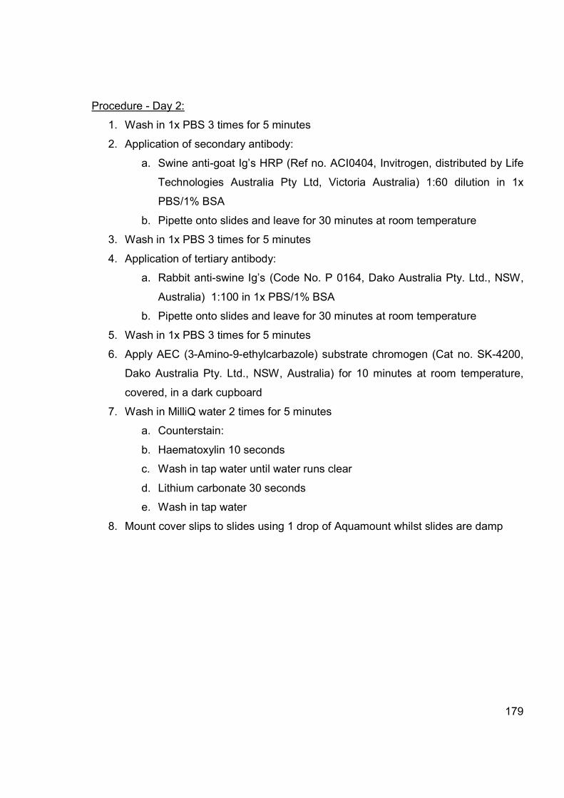
179
Procedure Day 2:
1. Wash in 1x PBS 3 times for 5 minutes
2. Application of secondary antibody:
a. Swine antigoat Ig’s HRP (Ref no. ACI0404, Invitrogen, distributed by Life
Technologies Australia Pty Ltd, Victoria Australia) 1:60 dilution in 1x
PBS/1% BSA
b. Pipette onto slides and leave for 30 minutes at room temperature
3. Wash in 1x PBS 3 times for 5 minutes
4. Application of tertiary antibody:
a. Rabbit antiswine Ig’s (Code No. P 0164, Dako Australia Pty. Ltd., NSW,
Australia) 1:100 in 1x PBS/1% BSA
b. Pipette onto slides and leave for 30 minutes at room temperature
5. Wash in 1x PBS 3 times for 5 minutes
6. Apply AEC (3Amino9ethylcarbazole) substrate chromogen (Cat no. SK4200,
Dako Australia Pty. Ltd., NSW, Australia) for 10 minutes at room temperature,
covered, in a dark cupboard
7. Wash in MilliQ water 2 times for 5 minutes
a. Counterstain:
b. Haematoxylin 10 seconds
c. Wash in tap water until water runs clear
d. Lithium carbonate 30 seconds
e. Wash in tap water
8. Mount cover slips to slides using 1 drop of Aquamount whilst slides are damp
Related Documents