An expansible aortic ring for a physiological approach to conservative aortic valve surgery Emmanuel Lansac, MD, PhD, a,b Isabelle Di Centa, MD, c Franc ¸ois Raoux, MD, d Neil Bulman-Fleming, e Adrian Ranga, e Aicha Abed, MSc, a,f Maguette Ba, MD, a Anthony Paolitto, e Didier Letourneur, PhD, a,f and Anne Meddahi-Pelle ´, MD, PhD a,g Objective: Dystrophic aortic insufficiency is characterized by dilation of the aortic annular base and sinotubular junction diameters preventing coaptation of thin and pliable cusps amenable to valve repair. An expansible aortic ring was designed to reduce dilated aortic root diameters to increase valvular coaptation height while maintaining root dynamics. The properties of the device were tested in vitro and in vivo in an ovine model. Methods: Expansible rings were composed of an elastomer core covered by polyester fabric. After in vitro anal- ysis of their mechanical properties, the rings were implanted in 6 sheep at both the level of the annular base and sinotubular junction (double subvalvular and supravalvular external aortic annuloplasty). Root dynamics were assessed by using intracardiac ultrasonography before surgical intervention and at 6 months. Histologic, scanning electron microscopic, and mechanical studies were then performed on explanted samples. Results: The expansible ring produced a significant reduction of the aortic annular base and sinotubular junction diameters. Coaptation height was increased from 2.5 0.7 mm to 6.2 1.1 mm (P<.001). Mechanical testing on 6-month explanted samples revealed no significant differences in elastic modulus. Dynamics of the root were well preserved. Histomorphologic studies showed incorporation of the material without degradation. Conclusions: Expansible aortic ring implantation produces a significant annuloplasty that increases coaptation height while preserving the dynamics of the aortic root. The effectiveness of the device in treating aortic insuf- ficiency is currently being evaluated in the prospective Conservative Aortic Valve surgery for aortic Insufficiency and Aneurysm of the Aortic Root trial comparing conservative aortic valve surgery versus mechanical valve re- placement. Dystrophic aortic insufficiency (AI) is characterized by dilation of both the aortic annular base and sinotubular junction (STJ) diameters preventing coaptation of otherwise thin and pliable cusps that are structurally close to normal. 1,2 Until recently, valve replacement was the only surgical option. Because of im- proved understanding of aortic valve dynamics, conservative aortic valve surgery was developed based on reduction of the dilated aortic root diameters (with or without cusp lesion) while preserving root dynamics with vortices (neosinuses of Val- salva) and expansibility (interleaflet triangles). 3-5 Two types of aortic valve-sparing operations were origi- nally performed for the treatment of root aneurysms: remodel- ing of the aortic root and reimplantation of the aortic valve. 6-8 The remodeling technique used a scalloped graft to provide a physiologic reconstruction of the root, but it does not address annular base dilation. 6-8 The reimplantation procedure as an inclusion technique reduces both diameters to the detriment of root dynamics. 6 We suggested combining the advantages of both approaches by placing an external subvalvular ring an- nuloplasty associated with the remodeling procedure (aortic root aneurysm; Figure 1, A) or with an external supravalvular ring annuloplasty to stabilize the STJ (isolated AI; Figure 1, B). 2,7,9 To standardize this physiologically based approach to conservative aortic valve surgery, we designed a calibrated expansible aortic ring that should reduce dilated diameters in diastole to increase valvular coaptation height while maintain- ing root systolic expansibility. The characteristics of this new device were established in vitro on the bench top. Biocompatibility of the ring and the effect of its implantation on root dynamics were studied in vivo in the ovine model. MATERIALS AND METHODS Ring Fabrication Design prerequisites for the ring were (1) target in vivo device expansion of between 5% and 15% over the cardiac cycle, (2) minimized radial thick- ness of the ring while preserving the desired dynamic behavior, (3) ring du- rability, and (4) stable and predictable bioreaction with surrounding tissues. From the INSERM, U 698, Cardiovascular Bioengineering, a Bichat University Hospi- tal, Paris, France; Cardiovascular Surgery, b Foch Hospital, Suresnes, France; Vas- cular Surgery, c Ambroise Pare ´ University Hospital, Boulogne–Billancourt, France; Cardiology, d Marie Lannelongue Hospital, Le Plessis Robinson, France; CORO- NEO, Inc, e Montre ´al, Que ´bec, Canada; Institut Galile ´e, f University Paris 13, Ville- taneuse, France; and the University of Orle ´ans, Orle ´ans, g France. E. Lansac is the recipient of a ‘‘poste d’accueil INSERM.’’ A. Abed was supported by the Agence of Biomedecine. The study was supported by CORONEO, Inc; IN- SERM; University Paris 13; and Assistance Publique des Ho ˆpitaux de Paris. Disclosures: Drs Lansac and Di Centa have Consultant Agreements with the company CORONEO, Inc. Received for publication Feb 23, 2009; revisions received April 13, 2009; accepted for publication May 17, 2009; available ahead of print July 13, 2009. Address for reprints: Anne Meddahi-Pelle ´, MD, PhD, INSERM, U 698, Hemostasis, Bio-engineering and Cardiovascular Remodelling, Cardiovascular Bioengineering Team, Bichat University Hospital, 75018 Paris, France (E-mail: anne.pelle@ inserm.fr). J Thorac Cardiovasc Surg 2009;138:718-24 0022-5223/$36.00 Copyright Ó 2009 by The American Association for Thoracic Surgery doi:10.1016/j.jtcvs.2009.05.024 718 The Journal of Thoracic and Cardiovascular Surgery c September 2009 ET/BS EVOLVING TECHNOLOGY/BASIC SCIENCE

Welcome message from author
This document is posted to help you gain knowledge. Please leave a comment to let me know what you think about it! Share it to your friends and learn new things together.
Transcript
ET
/BS
An expansible aortic ring for a physiological approach to conservativeaortic valve surgery
Emmanuel Lansac, MD, PhD,a,b Isabelle Di Centa, MD,c Francois Raoux, MD,d Neil Bulman-Fleming,e
Adrian Ranga,e Aicha Abed, MSc,a,f Maguette Ba, MD,a Anthony Paolitto,e Didier Letourneur, PhD,a,f and
Anne Meddahi-Pelle, MD, PhDa,g
Objective: Dystrophic aortic insufficiency is characterized by dilation of the aortic annular base and sinotubular
junction diameters preventing coaptation of thin and pliable cusps amenable to valve repair. An expansible aortic
ring was designed to reduce dilated aortic root diameters to increase valvular coaptation height while maintaining
root dynamics. The properties of the device were tested in vitro and in vivo in an ovine model.
Methods: Expansible rings were composed of an elastomer core covered by polyester fabric. After in vitro anal-
ysis of their mechanical properties, the rings were implanted in 6 sheep at both the level of the annular base and
sinotubular junction (double subvalvular and supravalvular external aortic annuloplasty). Root dynamics were
assessed by using intracardiac ultrasonography before surgical intervention and at 6 months. Histologic, scanning
electron microscopic, and mechanical studies were then performed on explanted samples.
Results: The expansible ring produced a significant reduction of the aortic annular base and sinotubular junction
diameters. Coaptation height was increased from 2.5� 0.7 mm to 6.2� 1.1 mm (P<.001). Mechanical testing on
6-month explanted samples revealed no significant differences in elastic modulus. Dynamics of the root were well
preserved. Histomorphologic studies showed incorporation of the material without degradation.
Conclusions: Expansible aortic ring implantation produces a significant annuloplasty that increases coaptation
height while preserving the dynamics of the aortic root. The effectiveness of the device in treating aortic insuf-
ficiency is currently being evaluated in the prospective Conservative Aortic Valve surgery for aortic Insufficiency
and Aneurysm of the Aortic Root trial comparing conservative aortic valve surgery versus mechanical valve re-
placement.
EVOLVING TECHNOLOGY/BASIC SCIENCE
Dystrophic aortic insufficiency (AI) is characterized by dilation
of both the aortic annular base and sinotubular junction (STJ)
diameters preventing coaptation of otherwise thin and pliable
cusps that are structurally close to normal.1,2 Until recently,
valve replacement was the only surgical option. Because of im-
proved understanding of aortic valve dynamics, conservative
aortic valve surgery was developed based on reduction of the
dilated aortic root diameters (with or without cusp lesion) while
preserving root dynamics with vortices (neosinuses of Val-
salva) and expansibility (interleaflet triangles).3-5
From the INSERM, U 698, Cardiovascular Bioengineering,a Bichat University Hospi-
tal, Paris, France; Cardiovascular Surgery,b Foch Hospital, Suresnes, France; Vas-
cular Surgery,c Ambroise Pare University Hospital, Boulogne–Billancourt, France;
Cardiology,d Marie Lannelongue Hospital, Le Plessis Robinson, France; CORO-
NEO, Inc,e Montreal, Quebec, Canada; Institut Galilee,f University Paris 13, Ville-
taneuse, France; and the University of Orleans, Orleans,g France.
E. Lansac is the recipient of a ‘‘poste d’accueil INSERM.’’ A. Abed was supported by
the Agence of Biomedecine. The study was supported by CORONEO, Inc; IN-
SERM; University Paris 13; and Assistance Publique des Hopitaux de Paris.
Disclosures: Drs Lansac and Di Centa have Consultant Agreements with the company
CORONEO, Inc.
Received for publication Feb 23, 2009; revisions received April 13, 2009; accepted for
publication May 17, 2009; available ahead of print July 13, 2009.
Address for reprints: Anne Meddahi-Pelle, MD, PhD, INSERM, U 698, Hemostasis,
Bio-engineering and Cardiovascular Remodelling, Cardiovascular Bioengineering
Team, Bichat University Hospital, 75018 Paris, France (E-mail: anne.pelle@
inserm.fr).
J Thorac Cardiovasc Surg 2009;138:718-24
0022-5223/$36.00
Copyright � 2009 by The American Association for Thoracic Surgery
doi:10.1016/j.jtcvs.2009.05.024
718 The Journal of Thoracic and Cardiovascular Su
Two types of aortic valve-sparing operations were origi-
nally performed for the treatment of root aneurysms: remodel-
ing of the aortic root and reimplantation of the aortic valve.6-8
The remodeling technique used a scalloped graft to provide
a physiologic reconstruction of the root, but it does not address
annular base dilation.6-8 The reimplantation procedure as an
inclusion technique reduces both diameters to the detriment
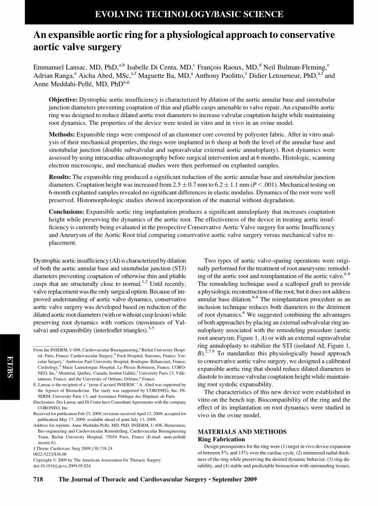
of root dynamics.6 We suggested combining the advantages
of both approaches by placing an external subvalvular ring an-
nuloplasty associated with the remodeling procedure (aortic
root aneurysm; Figure 1, A) or with an external supravalvular
ring annuloplasty to stabilize the STJ (isolated AI; Figure 1,
B).2,7,9 To standardize this physiologically based approach
to conservative aortic valve surgery, we designed a calibrated
expansible aortic ring that should reduce dilated diameters in
diastole to increase valvular coaptation height while maintain-
ing root systolic expansibility.
The characteristics of this new device were established in
vitro on the bench top. Biocompatibility of the ring and the
effect of its implantation on root dynamics were studied in
vivo in the ovine model.
MATERIALS AND METHODSRing Fabrication
Design prerequisites for the ring were (1) target in vivo device expansion
of between 5% and 15% over the cardiac cycle, (2) minimized radial thick-
ness of the ring while preserving the desired dynamic behavior, (3) ring du-
rability, and (4) stable and predictable bioreaction with surrounding tissues.
rgery c September 2009
Lansac et al Evolving Technology/Basic Science
Abbreviations and AcronymsAI ¼ aortic insufficiency
SEM ¼ scanning electron microscopy
STJ ¼ sinotubular junction
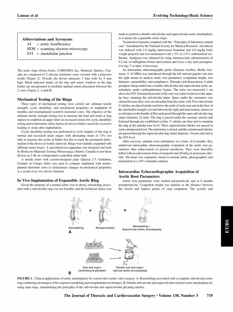
The aortic rings (Extra-Aortic; CORONEO, Inc, Montreal, Quebec, Can-
ada) are composed of 2 silicone elastomer cores covered with a polyester
textile (Figure 2). Overall, the device measures 3 mm wide by 5 mm
high. Black indicator marks on the ring and suture windows in the ring
holder are incorporated to facilitate optimal suture placement between the
2 cores (Figure 2, A and B).
Mechanical Testing of the RingsThree types of mechanical testing were carried out: ultimate tensile
strength, cyclic durability, and mechanical properties of implanted (6
months) and nonimplanted (control) elastomer cores. The objective of the
ultimate tensile strength testing was to measure the load and strain at ring
rupture to establish an upper limit on increased strains for cyclic durability
testing and to determine safety factors for device failure caused by excessive
loading or strain after implantation.
Cyclic durability testing was performed to cycle samples of the ring at
normal and increased strain ranges with alternating strain of 15% not
only to measure the cycles at failure but also to track the permanent defor-
mation of the device at weekly intervals. Rings were radially expanded with
different strain biases. A specialized test apparatus was designed and built
by Bodycote Materials Testing (Mississauga, Ontario, Canada) to test these
devices at 5 Hz in a temperature-controlled saline bath.
A tensile tester with custom-designed grips (Quasar 2.5; Galdabini,
Cardano al Campo, Italy) was used to compare implanted with nonim-
planted elastomer cores to characterize changes in mechanical properties
as a result of in vivo device function.
In Vivo Implantation of Expansible Aortic RingGiven the anatomy of a normal aortic root in sheep, remodeling associ-
ated with a subvalvular ring was not feasible, and the technical choice was
The Journal of Thoracic and
made to perform a double subvalvular and supravalvular aortic annuloplasty
to evaluate the expansible aortic rings.
Treatment of animals complied with the ‘‘Principles of laboratory animal
care’’ formulated by the National Society for Medical Research. Anesthesia
was induced with 1.0 mg/kg intravenous ketamine and 4.0 mg/kg body
weight propofol and was maintained with 1.5% to 2.5% endotracheal iso-
flurane. Analgesia was obtained by using intramuscular administration of
0.2 mL of nalbuphine before intervention and twice a day until postopera-
tive day 7 or later, if necessary.
An intracardiac ultrasonographic probe (Siemens AcuNav, Berlin, Ger-
many; 5–10 MHz) was introduced through the left internal jugular vein into
the right atrium to analyze aortic root parameters (coaptation height, root
diameter, expansibility, and compliance). Through a left thoracotomy, 6 adult
prealpine sheep underwent a double subvalvular and supravalvular aortic an-
nuloplasty under cardiopulmonary bypass. The aorta was transected 1 cm
above the STJ. External dissection of the root was achieved down to the annu-
lar base, attaining the subvalvular plane. Space under the coronaries was
cleared because they were not detached from the aortic wall. Five subvalvular
U stitches are placed inside out below the nadir of each cusp and at the base of
the interleaflet triangles (except between the right and noncoronary sinuses to
avoid injury to the bundle of His) and passed through the open subvalvular ring
(inner diameter, 22 mm). The ring is passed under the coronary arteries and
fastened through pre-established eyelets. U stitches are then tied to maintain
the ring at the annular base level. Three supravalvular stitches are passed at
each commissural level. The aortotomy is closed, and the commissural stitches
are passed through the supravalvular ring (inner diameter, 18 mm) and tied at
the STJ level.
After recovery, animals were ambulatory on a farm. At 6 months, they
underwent intracardiac ultrasonographic evaluation of the aortic root pa-
rameters after achievement of general anesthesia. They were thereafter
killed with an intravenous bolus of propofol and 20 mEq of potassium chlo-
ride. The heart was explanted, rinsed in normal saline, photographed, and
transferred to a 10% formalin solution.
Intracardiac Echocardiographic Acquisition ofAortic Root Parameters
Aortic root parameters were studied preoperatively and at 6 months
postoperatively. Coaptation height was defined as the distance between
the lowest and highest points of cusp coaptation. The systolic and
ET
/BS
FIGURE 1. Clinical applications of aortic annuloplasty for conservative aortic valve surgery. A, Remodeling associated with a complete subvalvular aortic
ring combining advantages of the original remodeling and reimplantation techniques. B, Double subvalvular and supravalvular external aortic annuloplasty by
using open rings, standardizing the principles of the subvalvular and supravalvular plicating stitches.
Cardiovascular Surgery c Volume 138, Number 3 719
Evolving Technology/Basic Science Lansac et al
ET
/BS
FIGURE 2. Expansible aortic ring before implantation. A, Cut-away schematic diagram of the device showing its key components. B, Top: Before implan-
tation, the ring is opened and mounted on a holder that is attached to the handle. The black indicator marks on the ring facilitate optimal suture placement.
Bottom: After implantation, the ends of the ring are fastened. C, Scanning electronic microscope image showing a cross-section of the ring with its rectangular
silicone core covered by a polyester sheath (insert). Scale bars ¼ 200 mm.
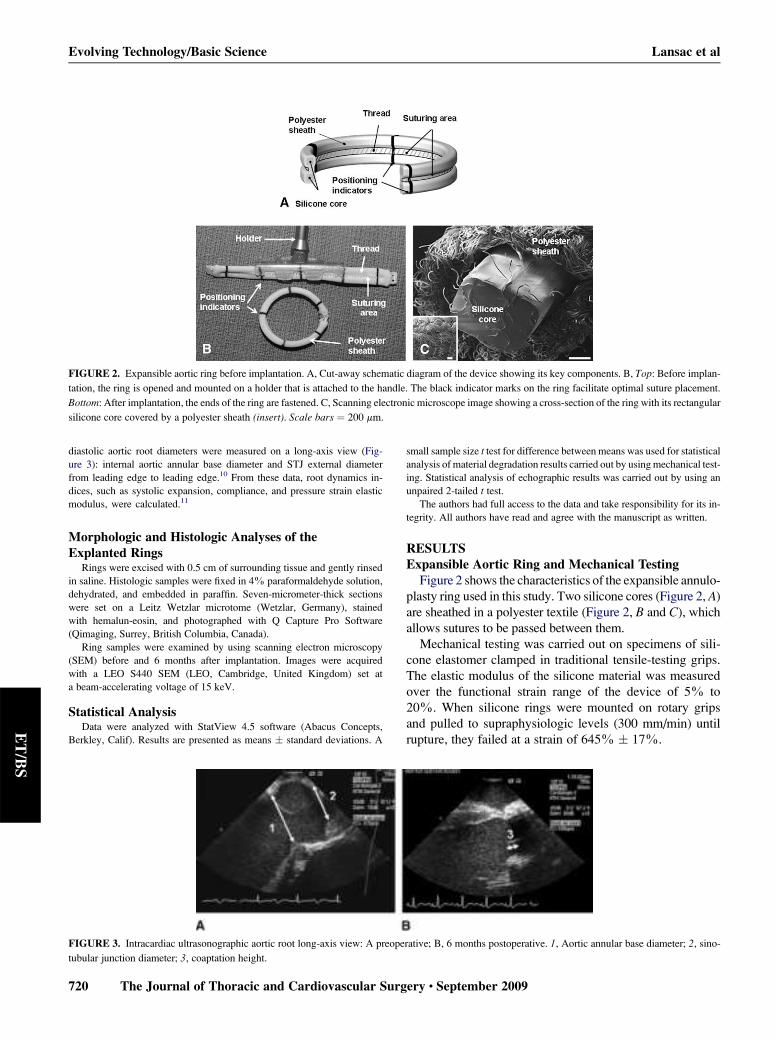
diastolic aortic root diameters were measured on a long-axis view (Fig-
ure 3): internal aortic annular base diameter and STJ external diameter
from leading edge to leading edge.10 From these data, root dynamics in-
dices, such as systolic expansion, compliance, and pressure strain elastic
modulus, were calculated.11
Morphologic and Histologic Analyses of theExplanted Rings
Rings were excised with 0.5 cm of surrounding tissue and gently rinsed
in saline. Histologic samples were fixed in 4% paraformaldehyde solution,
dehydrated, and embedded in paraffin. Seven-micrometer-thick sections
were set on a Leitz Wetzlar microtome (Wetzlar, Germany), stained
with hemalun-eosin, and photographed with Q Capture Pro Software
(Qimaging, Surrey, British Columbia, Canada).
Ring samples were examined by using scanning electron microscopy
(SEM) before and 6 months after implantation. Images were acquired
with a LEO S440 SEM (LEO, Cambridge, United Kingdom) set at
a beam-accelerating voltage of 15 keV.
Statistical AnalysisData were analyzed with StatView 4.5 software (Abacus Concepts,
Berkley, Calif). Results are presented as means � standard deviations. A
small sample size t test for difference between means was used for statistical
analysis of material degradation results carried out by using mechanical test-
ing. Statistical analysis of echographic results was carried out by using an
unpaired 2-tailed t test.
The authors had full access to the data and take responsibility for its in-
tegrity. All authors have read and agree with the manuscript as written.
RESULTSExpansible Aortic Ring and Mechanical Testing
Figure 2 shows the characteristics of the expansible annulo-
plasty ring used in this study. Two silicone cores (Figure 2, A)
are sheathed in a polyester textile (Figure 2, B and C), which
allows sutures to be passed between them.
Mechanical testing was carried out on specimens of sili-
cone elastomer clamped in traditional tensile-testing grips.
The elastic modulus of the silicone material was measured
over the functional strain range of the device of 5% to
20%. When silicone rings were mounted on rotary grips
and pulled to supraphysiologic levels (300 mm/min) until
rupture, they failed at a strain of 645% � 17%.
FIGURE 3. Intracardiac ultrasonographic aortic root long-axis view: A preoperative; B, 6 months postoperative. 1, Aortic annular base diameter; 2, sino-
tubular junction diameter; 3, coaptation height.
720 The Journal of Thoracic and Cardiovascular Surgery c September 2009
Lansac et al Evolving Technology/Basic Science
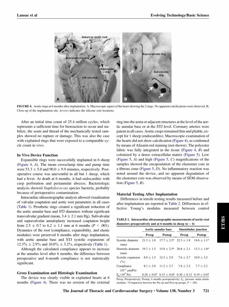
FIGURE 4. Aortic rings at 6 months after implantation. A, Macroscopic aspect of the heart showing the 2 rings. No apparent calcifications were observed. B,
Close-up of the implantation site. Arrows indicates the silicone core locations.
ET
/BS
After an initial time count of 25.4 million cycles, which
represents a sufficient time for bioreaction to occur and sta-
bilize, the seam and thread of the mechanically tested sam-
ples showed no rupture or damage. This was also the case
with explanted rings that were exposed to a comparable cy-
cle count in vivo.
In Vivo Device FunctionExpansible rings were successfully implanted in 6 sheep
(Figure 4, A). The mean crossclamp time and pump time
were 53.3 � 5.0 and 90.0 � 9.9 minutes, respectively. Post-
operative course was uneventful in all but 1 sheep, which
had a fever. At death at 6 months, it had endocarditis with
cusp perforation and periannular abscess. Bacteriologic
analysis showed Staphylococcus species bacteria, probably
because of perioperative contamination.
Intracardiac ultrasonographic analysis allowed visualization
of valvular coaptation and aortic root parameters in all cases
(Table 1). Prosthetic rings created a significant reduction of
the aortic annular base and STJ diameters without significant
transvalvular gradient (mean, 3.4� 2.1 mm Hg). Subvalvular
and supravalvular annuloplasty increased coaptation height
from 2.5 � 0.7 to 6.2 � 1.1 mm at 6 months (P < .001).
Dynamics of the root (compliance, expansibility, and elastic
modulus) were preserved 6 months after rings implantation,
with aortic annular base and STJ systolic expansions of
12.3% � 2.9% and 10.0% � 3.2%, respectively (Table 1).
Although the calculated compliance appears to increase
at the annulus level after 6 months, the difference between
preoperative and 6-month compliance is not statistically
significant.
Gross Examination and Histologic ExaminationThe device was clearly visible in explanted hearts at 6
months (Figure 4). There was no erosion of the external
The Journal of Thoracic and
ring into the aorta or adjacent structures at the level of the aor-
tic annular base or at the STJ level. Coronary arteries were
patent in all cases. Aortic cusps remained thin and pliable, ex-
cept for 1 sheep (endocarditis). Macroscopic examination of
the hearts did not show calcification (Figure 4), as confirmed
by means of Alizarin red staining (not shown). The polyester
fabric was fully integrated in the tissue (Figure 4, B) and
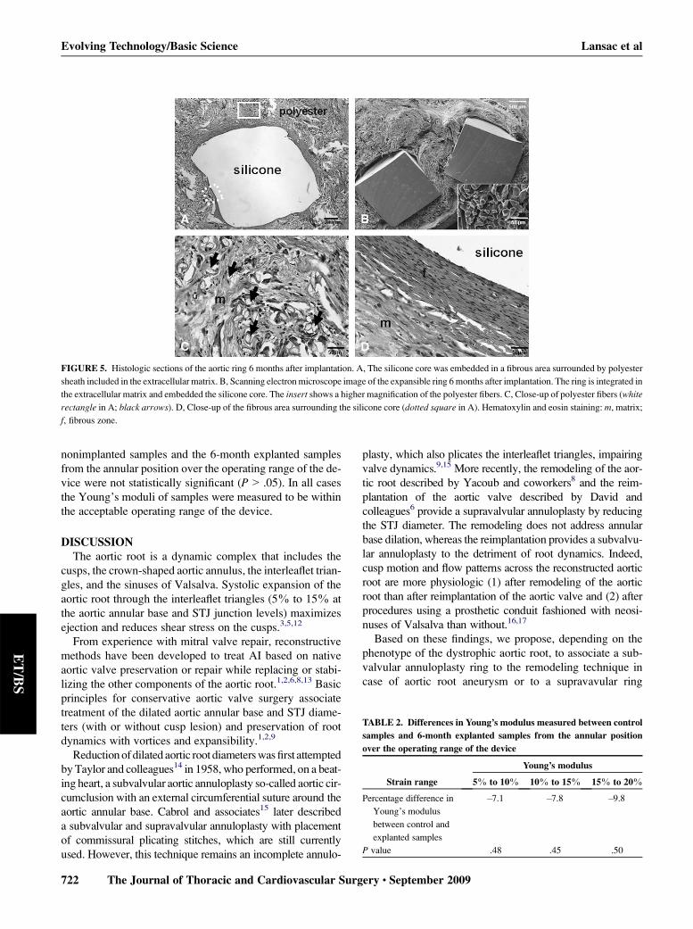
colonized by a dense extracellular matrix (Figure 5). Low
(Figure 5, A) and high (Figure 5, C) magnifications of the
samples showed the encapsulation of the elastomer core in
a fibrous zone (Figure 5, D). No inflammatory reaction was
noted around the device, and no apparent degradation of
the elastomer core was observed by means of SEM observa-
tion (Figure 5, B).
Material Testing After ImplantationDifferences in tensile testing results measured before and
after implantation are reported in Table 2. Differences in ef-
fective Young’s modulus measured between control
TABLE 1. Intracardiac ultrasonographic measurements of aortic root
diameters preoperatively and at 6 months in sheep (n ¼ 6)
Aortic annular base Sinotubular junction
Preop Postop Preop Postop
Systolic diameter
(mm)
21.3 � 1.8 17.7 � 2.5* 22.3 � 1.8 14.6 � 1.1*
Diastolic diameter
(mm)
19.7 � 1.5 15.8 � 2.5* 20.8 � 2.1 13.3 � 1.0*
Systolic expansion
(%)
8.0 � 1.5 12.3 � 2.9 7.6 � 2.7 10.0 � 3.2
Compliance
(10�1 mm/Pa)
8.1 � 2.8 11.2 � 2.7 7.8 � 3.2 7.7 � 2.2
Ep (105 Pa) 0.26 � 0.07 0.15 � 0.05 0.30 � 0.12 0.19 � 0.07
Preop, Preoperatively; Postop, 6 months postoperatively; Ep, pressure strain elastic
modulus. *Comparison between the Pre-op and Post-op groups, P< .001.
Cardiovascular Surgery c Volume 138, Number 3 721
Evolving Technology/Basic Science Lansac et al
ET
/BS
FIGURE 5. Histologic sections of the aortic ring 6 months after implantation. A, The silicone core was embedded in a fibrous area surrounded by polyester
sheath included in the extracellular matrix. B, Scanning electron microscope image of the expansible ring 6 months after implantation. The ring is integrated in
the extracellular matrix and embedded the silicone core. The insert shows a higher magnification of the polyester fibers. C, Close-up of polyester fibers (white
rectangle in A; black arrows). D, Close-up of the fibrous area surrounding the silicone core (dotted square in A). Hematoxylin and eosin staining: m, matrix;
f, fibrous zone.
nonimplanted samples and the 6-month explanted samples
from the annular position over the operating range of the de-
vice were not statistically significant (P> .05). In all cases
the Young’s moduli of samples were measured to be within
the acceptable operating range of the device.
DISCUSSIONThe aortic root is a dynamic complex that includes the
cusps, the crown-shaped aortic annulus, the interleaflet trian-
gles, and the sinuses of Valsalva. Systolic expansion of the
aortic root through the interleaflet triangles (5% to 15% at
the aortic annular base and STJ junction levels) maximizes
ejection and reduces shear stress on the cusps.3,5,12
From experience with mitral valve repair, reconstructive
methods have been developed to treat AI based on native
aortic valve preservation or repair while replacing or stabi-
lizing the other components of the aortic root.1,2,6,8,13 Basic
principles for conservative aortic valve surgery associate
treatment of the dilated aortic annular base and STJ diame-
ters (with or without cusp lesion) and preservation of root
dynamics with vortices and expansibility.1,2,9
Reduction of dilated aortic root diameters was first attempted
by Taylor and colleagues14 in 1958, who performed, on a beat-
ing heart, a subvalvular aortic annuloplasty so-called aortic cir-
cumclusion with an external circumferential suture around the
aortic annular base. Cabrol and associates15 later described
a subvalvular and supravalvular annuloplasty with placement
of commissural plicating stitches, which are still currently
used. However, this technique remains an incomplete annulo-
722 The Journal of Thoracic and Cardiovascular Su
plasty, which also plicates the interleaflet triangles, impairing
valve dynamics.9,15 More recently, the remodeling of the aor-
tic root described by Yacoub and coworkers8 and the reim-
plantation of the aortic valve described by David and
colleagues6 provide a supravalvular annuloplasty by reducing
the STJ diameter. The remodeling does not address annular
base dilation, whereas the reimplantation provides a subvalvu-
lar annuloplasty to the detriment of root dynamics. Indeed,
cusp motion and flow patterns across the reconstructed aortic
root are more physiologic (1) after remodeling of the aortic
root than after reimplantation of the aortic valve and (2) after
procedures using a prosthetic conduit fashioned with neosi-
nuses of Valsalva than without.16,17
Based on these findings, we propose, depending on the
phenotype of the dystrophic aortic root, to associate a sub-
valvular annuloplasty ring to the remodeling technique in
case of aortic root aneurysm or to a supravavular ring
TABLE 2. Differences in Young’s modulus measured between control
samples and 6-month explanted samples from the annular position
over the operating range of the device
Young’s modulus
Strain range 5% to 10% 10% to 15% 15% to 20%
Percentage difference in
Young’s modulus
between control and
explanted samples
�7.1 �7.8 �9.8
P value .48 .45 .50
rgery c September 2009
Lansac et al Evolving Technology/Basic Science
ET
/BS
(double subvalvular and supravalvular snnuloplasty) in case
of isolated AI (sinus of Valsalva<40 mm; Figure 1),2,7,9 the
subvalvular annuloplasty is associated with a second supra-
valvular ring, thus achieving a double-ring annuloplasty.
The clinical effectiveness of this physiologic approach to
conservative aortic valve surgery was confirmed in a prelim-
inary trial (96 patients) using rings obtained from a slice
of a Dacron tube graft.2,7,9 Choice of the external ring was
standardized and based on the sole measurement of internal
aortic annular base diameter with Hegar dilators. In case of
AI of grade 2 or greater, aortic rings were undersized.
To address the need for a dedicated aortic annuloplasty
device, we designed a new expansible aortic ring that pro-
vides a complete and calibrated annuloplasty while preserv-
ing root expansibility.
Previous aortic annuloplasty techniques were described.
Internal annuloplasty was proposed by using either a circular
suture,18 glutaraldehyde-tanned pericardial strips, or a sub-
valvular Gore-Tex band (W. L. Gore & Associates, Inc,
Newark, Del).19 Hahm and coworkers20 described an inter-
nal prosthetic ring at the STJ associated with a strip along the
fibrous annulus. Other experimental devices, such as 3-di-
mensional internal rings21,22 or external adjustable nylon
bands,23 were developed in animal models but never im-
planted in human subjects.
The advantages of a compliant ring as opposed to
a noncompliant ring are (1) an increase in valve triangulation
that leads to reduced leaflet stresses and superior flow
characteristics and (2) superior mobility and reduced stress
on surrounding structures (interleaflet triangles, mitral valve
annulus, and left ventricular outflow tract tissue) during the
cardiac cycle, leading to a closer approximation of physiologic
aortic root dynamics. The ring is placed externally, thus pre-
venting complications linked to endovascular prosthesis,
such as hemolytic or thromboembolic events. Moreover, the
external placement of the ring avoids placing tension on the
device’s fixation stitches by the expanding aorta.
The primary elastic element of the device is its silicone
elastomer. A design target for in vivo device expansion
was set between 5% and 15% over the cardiac cycle.
The overall device compliance is dictated by elastomer
core material and cross-section, as well as the mechanical
properties of the polyester fabric, the seam, and friction
between device components. The elastomer material is an
implantable-grade 2-part silicone.24 It is durable and has
a high tear strength and elongation to failure. Elastomer
component dimensions were designed for optimal ring
dynamics in a low-profile configuration while meeting
durability requirements because as the device cross-sec-
tional area decreases, stresses would be seen to increase
over the same strain range. The polyester was selected
from validated implantable textiles because of its suitable
mechanical properties and ability to sustain an appropriate
degree of tissue ingrowth.25
The Journal of Thoracic and
The expansible aortic ring was implanted in an orthotopic
position, allowing precise analysis of its effect on valvular
coaptation and root dynamics. After 6 months of implanta-
tion in an ovine model, aortic root diameters were signifi-
cantly reduced, ensuring a 130% increase of valvular
coaptation height. Systolic expansion at the annular base
and STJ diameters were preserved within the design target
of 5% to 15%.
Histology results obtained at 6 months corresponded to
typical remodeling processes showing polyester tissue inte-
gration and a fibrotic ring encapsulation. No adverse inflam-
matory reaction was observed. Based on macroscopic
observations of silicone cores, SEM examination, and func-
tional and mechanical studies, no evidence of damage to the
silicone component was found.
This investigation represents a feasibility and safety study
of the device, with the main limitation being device implan-
tation in a healthy animal model. However, no experimental
models of chronic AI or aortic root aneurysm are currently
available.
Effectiveness of this expansible aortic ring will be as-
sessed in the ongoing prospective multicenter Conserva-
tive Aortic Valve surgery for aortic Insufficiency and
Aneurysm of the Aortic Root (CAVIAAR) trial, which
compares conservative aortic valve surgery with mechan-
ical replacement.9 Patients with aortic root aneurysms
will undergo the remodeling of the aortic root associated
with an expansible subvalvular annuloplasty. Double
subvalvular and supravalvular expansible annuloplasty
will be performed for patients with isolated dystrophic
AI. Expanding indications of aortic valve sparing and
repair emphasize the need for dedicated devices that stan-
dardize the technique and expand use of the procedure.
Devices that preserve the natural dynamics of the root
might improve the long-term durability of conservative
aortic valve surgery.
We thank the Ecole de Chirurgie de l’Assistance Publique Hopi-
taux de Paris (Paris, France) for the use of its surgical structures and
animal facilities; G. Moulin, N. Leveque, and V. Ganachaud for
their effort and support, which were essential to the completion
of these experiments; and P. Portes (LIMHP, CNRS, Villetaneuse)
for his help in SEM experiments.
References1. ElKhoury G, Glineur D, Rubay J, Verhelst R, d’Acoz Y, Poncelet A, et al. Func-
tional classification of aortic root/valve abnormalities and their correlation with
etiologies and surgical procedures. Curr Opin Cardiol. 2005;20:115-21.
2. Lansac E, Di Centa I, Raoux F, Attar NA, Acar C, Joudinaud T, et al. A lesional
classification to standardize surgical management of aortic insufficiency towards
valve repair. Eur J Cardiothorac Surg. 2008;33:872-8.
3. Dagum P, Green GR, Nistal FJ, Daughters GT, Timek TA, Foppiano LE, et al.
Deformational dynamics of the aortic root: modes and physiologic determinants.
Circulation. 1999;100(suppl):II54-62.
4. Lansac E, Lim HS, Shomura Y, Lim KH, Rice NT, Goetz W, et al. A four-dimen-
sional study of the aortic root dynamics. Eur J Cardiothorac Surg. 2002;22:497-503.
5. Thubrikar M, Bosher LP, Nolan SP. The mechanism of opening of the aortic
valve. J Thorac Cardiovasc Surg. 1979;77:863-70.
Cardiovascular Surgery c Volume 138, Number 3 723
Evolving Technology/Basic Science Lansac et al
ET
/BS
6. David TE, Feindel CM. An aortic valve-sparing operation for patients with aortic
incompetence and aneurysm of the ascending aorta. J Thorac Cardiovasc Surg.
1992;103:617-22.
7. Lansac E, Di Centa I, Bonnet N, Leprince P, Rama A, Acar C, et al. Aortic pros-
thetic ring annuloplasty: a useful adjunct to a standardized aortic valve-sparing
procedure? Eur J Cardiothorac Surg. 2006;29:537-44.
8. Yacoub M, Fagan A, Stassano P, Radley-smith R. Result of valve conserving op-
erations for aortic regurgitation. Circulation. 1983;68(suppl III):321.
9. Lansac E, Di Centa I. From dynamic anatomy to aortic annuloplasty: the tale of the
ring. In Sakalihasan N, Kuivaniemi H, Michel JB, eds. Aortic aneurysms: new in-
sights into an old problem. Liege, Belgium: Liege University Press; 2008;199-233.
10. Roman MJ, Devereux RB, Niles NW, Hochreiter C, Kligfield P, Sato N, et al.
Aortic root dilatation as a cause of isolated, severe aortic regurgitation. Preva-
lence, clinical and echocardiographic patterns, and relation to left ventricular hy-
pertrophy and function. Ann Intern Med. 1987;106:800-7.
11. Long A, Rouet L, Vitry F, Albertini JN, Marcus C, Clement C. Compliance of ab-
dominal aortic aneurysms before and after stenting with tissue Doppler imaging:
evolution during follow-up and correlation with aneurysm diameter. Ann Vasc
Surg. 2009;23:49-59.
12. Sutton JP 3rd, Ho SY, Anderson RH. The forgotten interleaflet triangles: a review
of the surgical anatomy of the aortic valve. Ann Thorac Surg. 1995;59:419-27.
13. Carpentier A. Cardiac valve surgery—‘‘the French correction.’’ J Thorac Cardi-
ovasc Surg. 1983;86:323-37.
14. Taylor WJ, Thrower WB, Black H, Harken DE. The surgical correction of aortic
insufficiency by circumclusion. J Thorac Surg. 1958;35:192-205.
15. Cabrol C, Cabrol A, Guiraudon G, Bertrand M. [Treatment of aortic insufficiency
by means of aortic annuloplasty]. Arch Mal Coeur Vaiss. 1966;59:1305-12.
724 The Journal of Thoracic and Cardiovascular Su
16. Fries R, Graeter T, Aicher D, Reul H, Schmitz C, Bohm M, et al. In vitro compar-
ison of aortic valve movement after valve-preserving aortic replacement. J Thorac
Cardiovasc Surg. 2006;132:32-7.
17. De Paulis R, De Matteis GM, Nardi P, Scaffa R, Buratta MM, Chiariello L. Open-
ing and closing characteristics of the aortic valve after valve-sparing procedures
using a new aortic root conduit. Ann Thorac Surg. 2001;72:487-94.
18. Haydar HS, He GW, Hovaguimian H, McIrvin DM, King DH, Starr A. Valve re-
pair for aortic insufficiency: surgical classification and techniques. Eur J Cardio-
thorac Surg. 1997;11:258-65.
19. Izumoto H, Kawazoe K, Kawase T, Kim H. Subvalvular circular annuloplasty as
a component of aortic valve repair. J Heart Valve Dis. 2002;11:383-5.
20. Hahm SY, Choo SJ, Lee JW, Seo JB, Lim TH, Song JK, et al. Novel technique of
aortic valvuloplasty. Eur J Cardiothorac Surg. 2006;29:530-6.
21. Duran CM, Gallo R, Prabhakar G, Kumar N, Hainau B. New prosthetic ring for
aortic valve annuloplasty. Cardiovasc Surg. 1993;1:166-71.
22. Reimold SC, Aranki SF, Caguioa ES, Solomon SD, Birjiniuk V, Cohn LH, et al.
An external aortic root device for decreasing aortic regurgitation: in vitro and in
vivo animal studies. J Card Surg. 1994;9:304-13.
23. Gogbashian A, Ghanta RK, Umakanthan R, Rangaraj AT, Laurence RG, Fox JA,
et al. Correction of aortic insufficiency with an external adjustable prosthetic aor-
tic ring. Ann Thorac Surg. 2007;84:1001-5.
24. Chauvel-Lebret DJ, Pellen-Mussi P, Auroy P, Bonnaure-Mallet M. Evaluation
of the in vitro biocompatibility of various elastomers. Biomaterials. 1999;20:
291-9.
25. Abed A, Deval B, Assoul N, Bataille I, Portes P, Louedec L, et al. A biocompat-
ible polysaccharide hydrogel-embedded polypropylene mesh for enhanced tissue
integration in rats. Tissue Eng. 2008;14:519-27.
rgery c September 2009
Related Documents